The fi rst report of myeloma with IgD κ and free κ in Indonesia
5
Vol. 20, No. 3, August 2011 Myeloma with IgD κ and free κ 217 The first report of myeloma with IgD κ and free κ in Indonesia Riadi Wirawan, 1 Lidya Utami, 2 Abdul Muthalib 3 1 Department of Clinical Pathology, Faculty of Medicine, Universitas Indonesia, Dr Cipto Mangunkusumo National Hospital, Jakarta, Indonesia 2 Sentra Medika Cibinong Hospital, Bogor, Indonesia 3 Department of Internal Medicine, Faculty of Medicine, Universitas Indonesia, Dr Cipto Mangunkusumo National Hospital, Jakarta Abstrak Myeloma IgD κ merupakan kasus keganasan sel plasma yang jarang dijumpai, dan belum pernah dilaporkan di Indonesia. Pada keadaan normal, kadar IgD dalam darah sangat rendah, sehingga peningkatan kadar IgD dalam darah dapat terlewatkan pada pemeriksaan elektroforesis protein serum. Pada makalah ini dilaporkan kasus seorang wanita 59 tahun dengan nyeri tulang hebat. Pemeriksaan radiologi menunjukkan adanya fraktur kompresi torakal dan penyempitan foramen torakal. Diagnosis mieloma pada pasien ini ditegakkan berdasarkan kriteria WHO, stadium berdasarkan kriteria Durie Salmon, dan prognosis buruk berdasarkan International Prognostic Index dari International Myeloma Working Group. Pada elektroforesis protein serum dijumpai spike monoklonal yang kecil dan pada imunofiksasi didapatkan IgD κ and free light chain κ. (Med J Indones 2011; 20:217-21) Abstract IgD κ myeloma is a rare plasma cell neoplasm case and has never been reported before in Indonesia. In normal condition, IgD level in blood is very low, therefore increase of IgD level in myeloma could be missed by serum protein electrophoresis. A case of a 59 years old female with severe bone pain is reported. In radiology evaluation, there were thoracal compression fracture and thoracal foramen narrowing. For this patient, the myeloma diagnosis was based on WHO criteria, the stage IIIb was based on Durie and Salmon criteria, and bad prognosis with prognostic index stage III diagnosis was based on International Prognostic Index from International Myeloma Working Group, respectively. In serum protein electrophoresis we found a very small monoclonal spike and in immunofixation there were monoclonal IgD κ and free light chain κ. (Med J Indones 2011; 20:217-21) Key words: immunofixation, myeloma IgD κ, protein electrophoresis Correspondence email to: [email protected] Myeloma is a B cells malignancy which is characterized by monoclonal expansion and accumulation of abnormal plasma cells in the bone marrow, usually accompanied by monoclonal immunoglobulin secretion. The other typical findings in myeloma are bone lesion, anemia, hypercalcemia, and renal insufficiency. According to 2008 WHO classification of tumours, myeloma, which is also known as plasma cell myeloma, belongs to the group of plasma cell neoplasm, together with monoclonal gammopathy of undetermined significance (MGUS), plasmacytoma, immunoglobulin deposition disease, and osteosclerotic myeloma. 1-4 Myeloma can evolve de novo or as a progression from MGUS and smoldering multiple myeloma. Proliferation of myeloma cells will lead to bone marrow infiltration and interaction with bone marrow microenvironment, which in turn induce the secretion of cytokines, such as interleukin 6 (IL-6), IL-1β, IL- 11, tumor necrosis factor (TNF), transforming growth factor beta (TGF-β), dan receptor activator of nuclear factor kappa B ligand (RANKL). Those cytokines have important roles in proliferation and survival of myeloma cells, osteoclast activation, and osteoblast activity defect. Myeloma cells infiltration result in bone marrow failure, which shown as anemia, neutropenia, or thrombocytopenia, and bone destruction. Paraprotein produced by myeloma cells will lead to the increase of erythrocyte sedimentation rate, rouleaux formation in blood smear, increase of blood viscocity, coagulation defect, and neurological disorder. The paraprotein is detected in serum protein electrophoresis as spike in β or γ globulin area, in immunofixation as sharp band in conjuction with monoclonal immunoglobulin chain, and in urine as Bence Jones protein. Bone lesion will cause hypercalcemia, hypercalciuria, and the increase of serum ureum and creatinin due to renal function disorder. 1,2 Myeloma represents 10% of blood cancer cases. It occurs in 5 every 100.000 people in the general population, with a higher prevalence rate in males compare to females, and median age of patients is 60 years old. Myeloma is more prevalent in black people. Types of myeloma includes IgG myeloma, typically 50%, IgA myeloma, 25%, light chain myeloma 24%, IgD myeloma 1% of all cases, while IgE myeloma cases are rarely found. In 1% of the cases neither light
Transcript of The fi rst report of myeloma with IgD κ and free κ in Indonesia
Vol. 20, No. 3, August 2011 Myeloma with IgD κ and free κ 217
The fi rst report of myeloma with IgD κ and free κ in Indonesia
Riadi Wirawan,1 Lidya Utami,2 Abdul Muthalib 3
1 Department of Clinical Pathology, Faculty of Medicine, Universitas Indonesia, Dr Cipto Mangunkusumo National Hospital, Jakarta, Indonesia
2 Sentra Medika Cibinong Hospital, Bogor, Indonesia3 Department of Internal Medicine, Faculty of Medicine, Universitas Indonesia, Dr Cipto Mangunkusumo National Hospital,
Jakarta
Abstrak Myeloma IgD κ merupakan kasus keganasan sel plasma yang jarang dijumpai, dan belum pernah dilaporkan di Indonesia. Pada keadaan normal, kadar IgD dalam darah sangat rendah, sehingga peningkatan kadar IgD dalam darah dapat terlewatkan pada pemeriksaan elektroforesis protein serum. Pada makalah ini dilaporkan kasus seorang wanita 59 tahun dengan nyeri tulang hebat. Pemeriksaan radiologi menunjukkan adanya fraktur kompresi torakal dan penyempitan foramen torakal. Diagnosis mieloma pada pasien ini ditegakkan berdasarkan kriteria WHO, stadium berdasarkan kriteria Durie Salmon, dan prognosis buruk berdasarkan International Prognostic Index dari International Myeloma Working Group. Pada elektroforesis protein serum dijumpai spike monoklonal yang kecil dan pada imunofi ksasi didapatkan IgD κ and free light chain κ. (Med J Indones 2011; 20:217-21)
AbstractIgD κ myeloma is a rare plasma cell neoplasm case and has never been reported before in Indonesia. In normal condition, IgD level in blood is very low, therefore increase of IgD level in myeloma could be missed by serum protein electrophoresis. A case of a 59 years old female with severe bone pain is reported. In radiology evaluation, there were thoracal compression fracture and thoracal foramen narrowing. For this patient, the myeloma diagnosis was based on WHO criteria, the stage IIIb was based on Durie and Salmon criteria, and bad prognosis with prognostic index stage III diagnosis was based on International Prognostic Index from International Myeloma Working Group, respectively. In serum protein electrophoresis we found a very small monoclonal spike and in immunofi xation there were monoclonal IgD κ and free light chain κ. (Med J Indones 2011; 20:217-21)
Key words: immunofi xation, myeloma IgD κ, protein electrophoresis
Correspondence email to: [email protected]
Myeloma is a B cells malignancy which is characterized by monoclonal expansion and accumulation of abnormal plasma cells in the bone marrow, usually accompanied by monoclonal immunoglobulin secretion. The other typical fi ndings in myeloma are bone lesion, anemia, hypercalcemia, and renal insuffi ciency. According to 2008 WHO classifi cation of tumours, myeloma, which is also known as plasma cell myeloma, belongs to the group of plasma cell neoplasm, together with monoclonal gammopathy of undetermined signifi cance (MGUS), plasmacytoma, immunoglobulin deposition disease, and osteosclerotic myeloma.1-4
Myeloma can evolve de novo or as a progression from MGUS and smoldering multiple myeloma. Proliferation of myeloma cells will lead to bone marrow infi ltration and interaction with bone marrow microenvironment, which in turn induce the secretion of cytokines, such as interleukin 6 (IL-6), IL-1β, IL-11, tumor necrosis factor (TNF), transforming growth factor beta (TGF-β), dan receptor activator of nuclear factor kappa B ligand (RANKL). Those cytokines have important roles in proliferation and survival of myeloma cells, osteoclast activation, and osteoblast
activity defect. Myeloma cells infi ltration result in bone marrow failure, which shown as anemia, neutropenia, or thrombocytopenia, and bone destruction. Paraprotein produced by myeloma cells will lead to the increase of erythrocyte sedimentation rate, rouleaux formation in blood smear, increase of blood viscocity, coagulation defect, and neurological disorder. The paraprotein is detected in serum protein electrophoresis as spike in β or γ globulin area, in immunofi xation as sharp band in conjuction with monoclonal immunoglobulin chain, and in urine as Bence Jones protein. Bone lesion will cause hypercalcemia, hypercalciuria, and the increase of serum ureum and creatinin due to renal function disorder. 1,2
Myeloma represents 10% of blood cancer cases. It occurs in 5 every 100.000 people in the general population, with a higher prevalence rate in males compare to females, and median age of patients is 60 years old. Myeloma is more prevalent in black people.
Types of myeloma includes IgG myeloma, typically 50%, IgA myeloma, 25%, light chain myeloma 24%, IgD myeloma 1% of all cases, while IgE myeloma cases are rarely found. In 1% of the cases neither light
Wirawan et al.218 Med J Indones
or heavy chains are produced, called non secretory myeloma. 4,5 A case of IgD myeloma has never been reported before in Indonesia.
Case report
A 59 years old female patient had been treated with complains of bone pains since 1 month before she was warded. Sharp pain is felt in the vertebrae, right chest and left leg, greater in intensity as time goes on. In the physical evaluation, it was discovered that the back and exteremitas are diffi cult to be moved.
Thoracal MRI evaluation found compression fractures at thoracal (Th) 5 dan Th 10, and neural foramen narrowing at Th 2-3 right, Th 3-4 right, Th 6-7 left, Th 10-11 right dan left, caused by metastasis. Lumbal MRI showed a diffuse change in the entire lumbal area, bone metastasis in the lumbosacral area was suspected.
Complete peripheral blood evaluation showed the following result : ESR: 145 mm (n : <15), Hb : 7.2 g/dL (n : 12.0 -14.0), Ht : 23.3% (n : 37.0 – 43.0), erythrocytes :
2.59 million/uL (n : 4.00 – 5.00), leukocytes : 8,230 /uL (n : 5,000 – 10,000), thrombocytes : 70,000/uL (n : 150,000 -400,000), and differential blood count : 2/1/2/60/31/4, with nucleated erythrocytes 2/100 leucocytes. Blood smear showed normocytic normochromic erythrocytes with rouleaux formation and nucleated red blood cell 2/100 leucocytes. Leucocyte count is normal with basophilia and normal morphology. Thrombocyte count is less than normal with normal morphology.
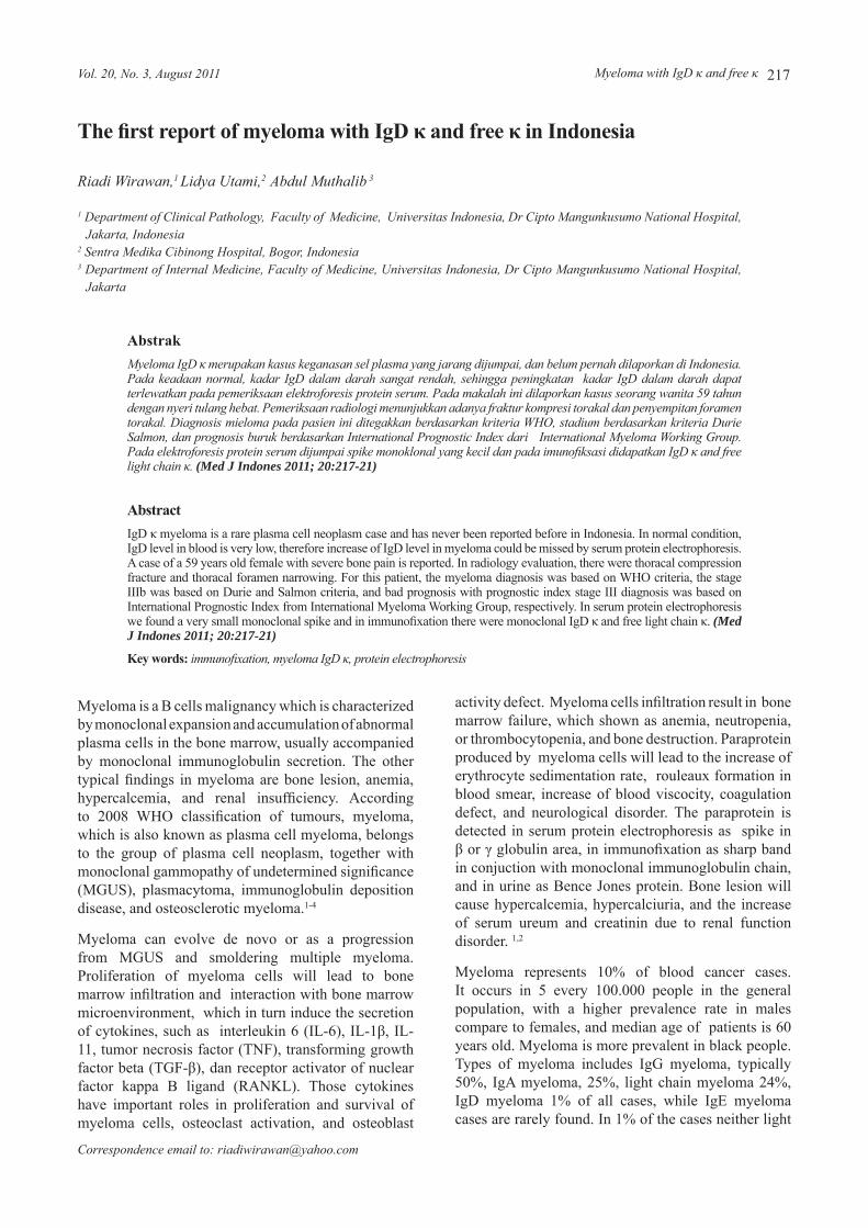
Bone marrow evaluation showed hypercellular marrow with depression of thrombopoiesis, granulopoiesis and erythropoiesis. Plasmocyte count was found 63% of the nucleated cells in the bone marrow, with abnormal morphology: uneven side of cytoplasma, halo-less, unequal sizes, as well as double nuclei, as shown in Figure 1.
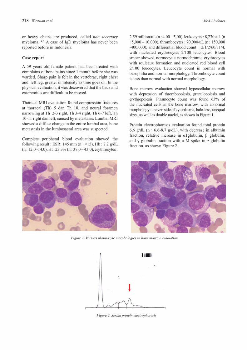
Protein electrophoresis evaluation found total protein 6,6 g/dL (n : 6,6-8,7 g/dL), with decrease in albumin fraction, relative increase in α1globulin, β globulin, and γ globulin fraction with a M spike in γ globulin fraction, as shown Figure 2.
Figure 1. Various plasmocyte morphologies in bone marrow evaluation
Figure 2. Serum protein electrophoresis
Vol. 20, No. 3, August 2011 Myeloma with IgD κ and free κ 219
Quantative immunoglobulin evaluation found a decrease in IgG level: 699 mg/dL (n : 800-1813), IgA : 74 mg/dL (n : 104-522), and IgM : 1 mg/dL (n : 52-217). Furthermore, it also found an increase in serum creatinine: 3,06 mg/dL (n : < 1,40), as well as serum calcium: 12,5 mg/dL (n : 8,1 -10,4), and β2 microglobulin : 17.341 mg/L (n :0.670-1.310).
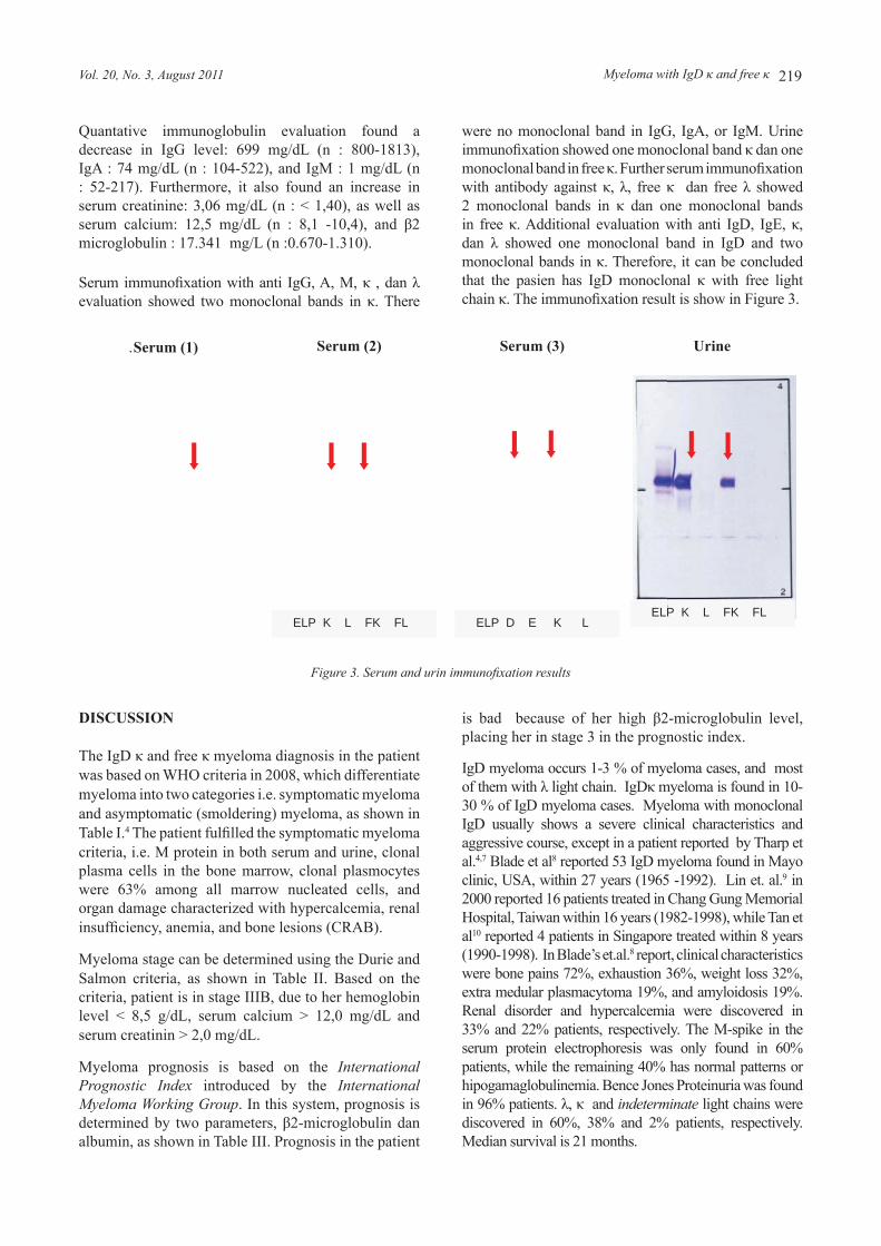
Serum immunofi xation with anti IgG, A, M, κ , dan λ evaluation showed two monoclonal bands in κ. There
were no monoclonal band in IgG, IgA, or IgM. Urine immunofi xation showed one monoclonal band κ dan one monoclonal band in free κ. Further serum immunofi xation with antibody against κ, λ, free κ dan free λ showed 2 monoclonal bands in κ dan one monoclonal bands in free κ. Additional evaluation with anti IgD, IgE, κ, dan λ showed one monoclonal band in IgD and two monoclonal bands in κ. Therefore, it can be concluded that the pasien has IgD monoclonal κ with free light chain κ. The immunofi xation result is show in Figure 3.
Figure 3. Serum and urin immunofi xation results
.Seerum (1) S
ELP
Serum (2)
K L FK FLL ELP
Serum (3)
P D E K ELP
L
Urine
P K L FK FFL
DISCUSSION
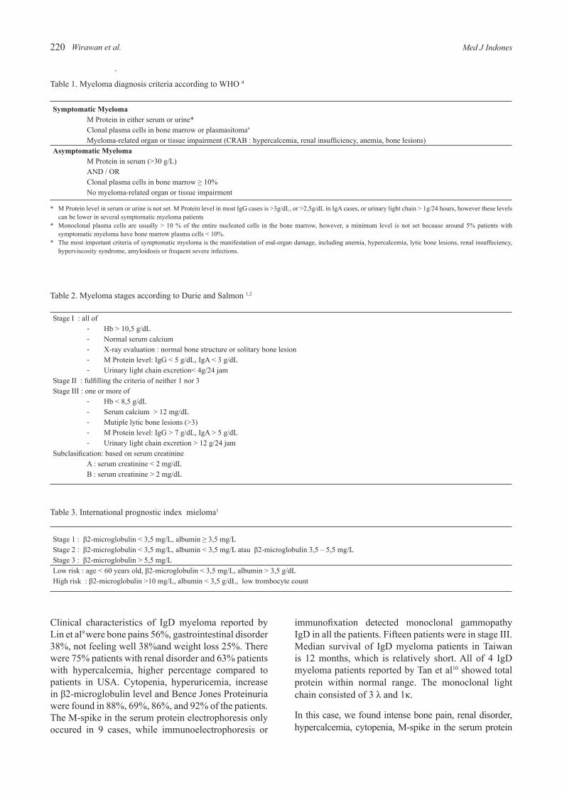
The IgD κ and free κ myeloma diagnosis in the patient was based on WHO criteria in 2008, which differentiate myeloma into two categories i.e. symptomatic myeloma and asymptomatic (smoldering) myeloma, as shown in Table I.4 The patient fulfi lled the symptomatic myeloma criteria, i.e. M protein in both serum and urine, clonal plasma cells in the bone marrow, clonal plasmocytes were 63% among all marrow nucleated cells, and organ damage characterized with hypercalcemia, renal insuffi ciency, anemia, and bone lesions (CRAB).
Myeloma stage can be determined using the Durie and Salmon criteria, as shown in Table II. Based on the criteria, patient is in stage IIIB, due to her hemoglobin level < 8,5 g/dL, serum calcium > 12,0 mg/dL and serum creatinin > 2,0 mg/dL.
Myeloma prognosis is based on the International Prognostic Index introduced by the International Myeloma Working Group. In this system, prognosis is determined by two parameters, β2-microglobulin dan albumin, as shown in Table III. Prognosis in the patient
is bad because of her high β2-microglobulin level, placing her in stage 3 in the prognostic index.
IgD myeloma occurs 1-3 % of myeloma cases, and most of them with λ light chain. IgDκ myeloma is found in 10-30 % of IgD myeloma cases. Myeloma with monoclonal IgD usually shows a severe clinical characteristics and aggressive course, except in a patient reported by Tharp et al.4,7 Blade et al8 reported 53 IgD myeloma found in Mayo clinic, USA, within 27 years (1965 -1992). Lin et. al.9 in 2000 reported 16 patients treated in Chang Gung Memorial Hospital, Taiwan within 16 years (1982-1998), while Tan et al10 reported 4 patients in Singapore treated within 8 years (1990-1998). In Blade’s et.al.8 report, clinical characteristics were bone pains 72%, exhaustion 36%, weight loss 32%, extra medular plasmacytoma 19%, and amyloidosis 19%. Renal disorder and hypercalcemia were discovered in 33% and 22% patients, respectively. The M-spike in the serum protein electrophoresis was only found in 60% patients, while the remaining 40% has normal patterns or hipogamaglobulinemia. Bence Jones Proteinuria was found in 96% patients. λ, κ and indeterminate light chains were discovered in 60%, 38% and 2% patients, respectively. Median survival is 21 months.
Wirawan et al.220 Med J Indones
Symptomatic Myeloma M Protein in either serum or urine* Clonal plasma cells in bone marrow or plasmasitoma#
Myeloma-related organ or tissue impairment (CRAB : hypercalcemia, renal insuffi ciency, anemia, bone lesions)Asymptomatic Myeloma
M Protein in serum (>30 g/L)AND / ORClonal plasma cells in bone marrow ≥ 10%No myeloma-related organ or tissue impairment
* M Protein level in serum or urine is not set. M Protein level in most IgG cases is >3g/dL, or >2,5g/dL in IgA cases, or urinary light chain > 1g/24 hours, however these levels can be lower in several symptomatic myeloma patients
* Monoclonal plasma cells are usually > 10 % of the entire nucleated cells in the bone marrow, however, a minimum level is not set because around 5% patients with symptomatic myeloma have bone marrow plasma cells < 10%.
* The most important criteria of symptomatic myeloma is the manifestation of end-organ damage, including anemia, hypercalcemia, lytic bone lesions, renal insuffeciency, hyperviscosity syndrome, amyloidosis or frequent severe infections.
Table 1. Myeloma diagnosis criteria according to WHO 4
Table 2. Myeloma stages according to Durie and Salmon 1,2
Stage I : all ofHb > 10,5 g/dL- Normal serum calcium- X-ray evaluation : normal bone structure or solitary bone lesion- M Protein level: IgG < 5 g/dL, IgA < 3 g/dL- Urinary light chain excretion< 4g/24 jam-
Stage II : fulfi lling the criteria of neither 1 nor 3Stage III : one or more of
Hb < 8,5 g/dL- Serum calcium > 12 mg/dL- Mutiple lytic bone lesions (>3)- M Protein level: IgG > 7 g/dL, IgA > 5 g/dL- Urinary light chain excretion > 12 g/24 jam-
Subclasifi cation: based on serum creatinineA : serum creatinine < 2 mg/dLB : serum creatinine > 2 mg/dL
Table 3. International prognostic index mieloma1
Stage 1 : β2-microglobulin < 3,5 mg/L, albumin ≥ 3,5 mg/LStage 2 : β2-microglobulin < 3,5 mg/L, albumin < 3,5 mg/L atau β2-microglobulin 3,5 – 5,5 mg/LStage 3 : β2-microglobulin > 5,5 mg/LLow risk : age < 60 years old, β2-microglobulin < 3,5 mg/L, albumin > 3,5 g/dLHigh risk : β2-microglobulin >10 mg/L, albumin < 3,5 g/dL, low trombocyte count
Clinical characteristics of IgD myeloma reported by Lin et al9 were bone pains 56%, gastrointestinal disorder 38%, not feeling well 38%and weight loss 25%. There were 75% patients with renal disorder and 63% patients with hypercalcemia, higher percentage compared to patients in USA. Cytopenia, hyperuricemia, increase in β2-microglobulin level and Bence Jones Proteinuria were found in 88%, 69%, 86%, and 92% of the patients. The M-spike in the serum protein electrophoresis only occured in 9 cases, while immunoelectrophoresis or
immunofi xation detected monoclonal gammopathy IgD in all the patients. Fifteen patients were in stage III. Median survival of IgD myeloma patients in Taiwan is 12 months, which is relatively short. All of 4 IgD myeloma patients reported by Tan et al10 showed total protein within normal range. The monoclonal light chain consisted of 3 λ and 1κ.
In this case, we found intense bone pain, renal disorder, hypercalcemia, cytopenia, M-spike in the serum protein

Vol. 20, No. 3, August 2011 Myeloma with IgD κ and free κ 221
electrophoresis, elevated β2-microglobulin and urinary light chain i.e. Bence Jones proteinuria, as reported by Lin et. al.9 In addition, extra medular plasmacytoma in the lumbosacral bone was found, as reported by Blade et. al.8
In several types of myeloma, such as IgD myeloma, IgE myeloma, light chain disease, and non secretory myeloma, serum protein electrophoresis often seems normal and does not show any sign of M-spike. At the moment, a novel free light chain and or κ /λ ratio evaluation with nephelometry method is being developed. This evaluation can be utilized to help serum protein electrophoresis interpretation by identifying samples which requires immunofi xation evaluation to detect small monoclonal band, which would be unclear or undetected otherwise. 11
IgD and IgE levels in the blood are so low that IgD and IgE myeloma diagnosis are often hard to justify. If the quantitative immunoglobulin evaluation of myeloma suspected patients shows a decrease of IgG, IgA, or IgM levels then a possibility of IgD or IgE myeloma should be considered. If the immunofi xation evaluation with antibody against IgG, IgA, IgM, κ , and λ does not show any monoclonal band on G, A or M but shows one or more bands on κ or λ then additional immunofi xation evaluation with anti κ , λ, free κ , free λ, and even anti IgD and anti IgE if required, needs to be done. If the protein electrophoresis does not show monoclonal characteristic, then the possibility of the myeloma suspected patient to be suffering from mieloma IgD, IgE, or non secretory myeloma should be considered.7 Free light chain level and or ratio evaluation needs to be carried out to prevent myeloma cases with no monoclonal characteristics in the serum protein electrophoresis from being undiagnosed.11
In conclusion, a 59 years old female patient was diagnosed with IgD κ myeloma according the WHO criteria, stage IIIB according to Durie dan Salmon criteria, and bad prognosis with prognostic index stage III according to the International Prognostic Index from International Myeloma Working Group. The protein electrophoresis evaluation showed a small M-spike and the immunofi xation evaluation showed monoclonal bands in the igD κ and free light chain κ. If the immunofi xation evaluation of a myeloma suspected
patient does not show M protein in IgG, IgA, IgM, but shows monoclonal band in the κ or λ in serum, additional immunofi xation evaluation with anti IgD, IgE, and light chain κ or λ needs to be performed.
REFERENCES
Terpos E, Rahemtulla A. Myeloma. In: Hoffbrand AV, 1. Catovsky D, Tuddenham EGD, editors. Postgraduate haematology. 5th ed. Massachusetts: Blackwell publishing Ltd; 2005. p. 681-702. Dispenzieri A, Lacy MQ, Greipp PR. Multiple myeloma. 2. In: Greer JP, Foerster J, Rodgers GM, Paraskevas F, Glader B, Arber DA, et al. Wintrobe’s clinical hematology 12th ed. Vol 2. Philadelphia : Lippincott Williams & Wilkins; 2009. p.2372-438.Munshi NC, Longo DL, Anderson KC. Plasma cell 3. disorders. In : Fauci AS, Kasper DL, Longo DL, Braunwald E, Hauser SL, Jameson JL, et al, Harrison’s principles of internal medicine 17th ed vol 1. New York : McGraw Hills; 2008. p.700-7.McKenna RW, Kyle RA, Kuehl WM, Grogan TM, Harris 4. NL, Coupland RW. Plasma cell neoplasms. In: Swerdlow SH, Campo E, Harris NL, Jaffe ES, Pileri SA, Stein H, et al, editors. WHO classifi cation of tumours of haematopoietic and lymphoid tissues 4th ed. Lyon: International Agency for Research on Cancer. 2008: 200-13.Datiles TB, Humphrey RL. Hypergammaglobulinemia. In: 5. Sheehan C, editors. Clinical immunology, principles and laboratory diagnosis, 2nd ed. Philadelphia: Lippincot; 1997. 317-29.Kyle RA, Rajkumar SV. Criteria for diagnosis, staging, risk 6. stratifi cation and response assessment of multiple myeloma. Leukemia. 2009; 23 : 3–9.Tharp AM, Woodruff RD, Shihabi ZK. Case report: IgD-7. Kappa Myeloma: an unusual case. Ann Clin Lab Sci. 2003; 33 (1) : 97-100.Blade J, Lust JA, Kyle RA. Immunoglobulin D multiple 8. myeloma presenting features, response to therapy, an survival in a series of 53 cases. J Clin Oncol. 1994;12:2398-404.Lin TL, Shih LY, Dunn P, Wang PN, Wu JH, Kao MC. 9. Immunoglobulin D multiple myeloma. Chang Gung Med J. 2000; 23(8):451-7.Tan Lk, Suri R, Lim HL, Ong SK. Immunoglobulin D 10. multiple myeloma in our hospital-a rare occurrence. Sing Med J. 2000; 41(10): 500-503.Hill Pg. Forsyth JM, Rai B, Mayne S. Serum free light 11. chains : an alternative to the urine Bence Jones proteins screening test for monoclonal gammopathies. Clin Chem. 2006; 59(2): 1743-8.