
Modern Ultrasonic Transducers - The Ultran Group - Ultrasound
The role of ultrasound duplex in endovenous procedures
Neophytos A. Zambas MD , PhDVascular Surgeon
Polyclinic Ygia, Limassol, Cyprus
ΚΕΑΕΧΚΥΠΡΙΑΚΗ ΕΤΑΙΡΕΙΑ ΑΓΓΕΙΑΚΗΣΚΑΙΕΝΔΑΓΓΕΙΑΚΗΣ ΧΕΙΡΟΥΡΓΙΚΗΣ
Clinical examination & duplex ultrasound = best method The most effective and accurate tool Degree of leg swelling, skin changes, venous ulceration (CEAP
Classification) DU of superficial, deep and perforator veins in the leg Proximal vein pathologies:
CT venography / MR venography for above inguinal ligament anatomic definition
Intravascular u/s for iliac vein stenosis – iliac vein stent procedures
Accredited vascular lab Appropriate equipment and personnel Validated standardized diagnostic protocols
Pre op clinical assessment
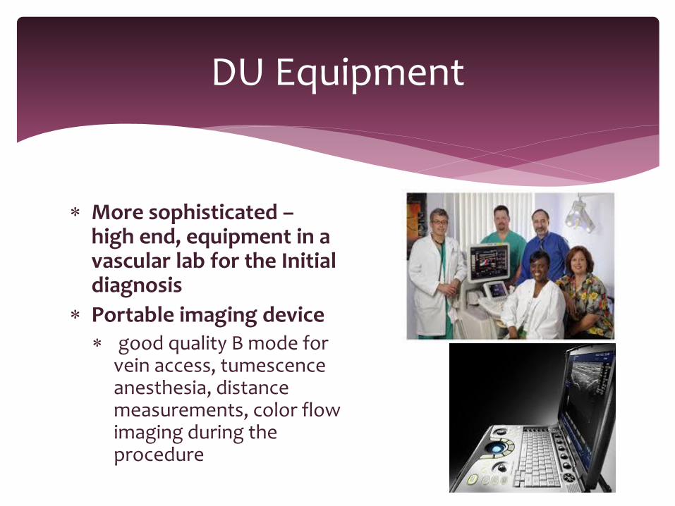
More sophisticated –high end, equipment in a vascular lab for the Initial diagnosis
Portable imaging device good quality B mode for
vein access, tumescence anesthesia, distance measurements, color flow imaging during the procedure
DU Equipment
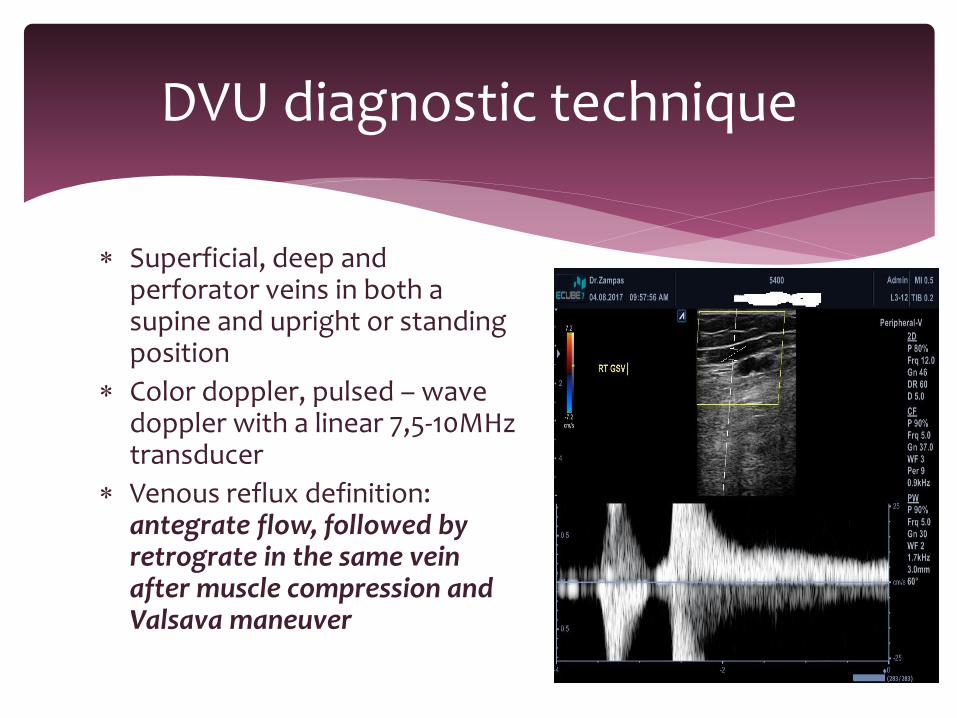
Superficial, deep and perforator veins in both a supine and upright or standing position
Color doppler, pulsed – wave doppler with a linear 7,5-10MHz transducer
Venous reflux definition: antegrate flow, followed by retrograte in the same vein after muscle compression and Valsava maneuver
DVU diagnostic technique
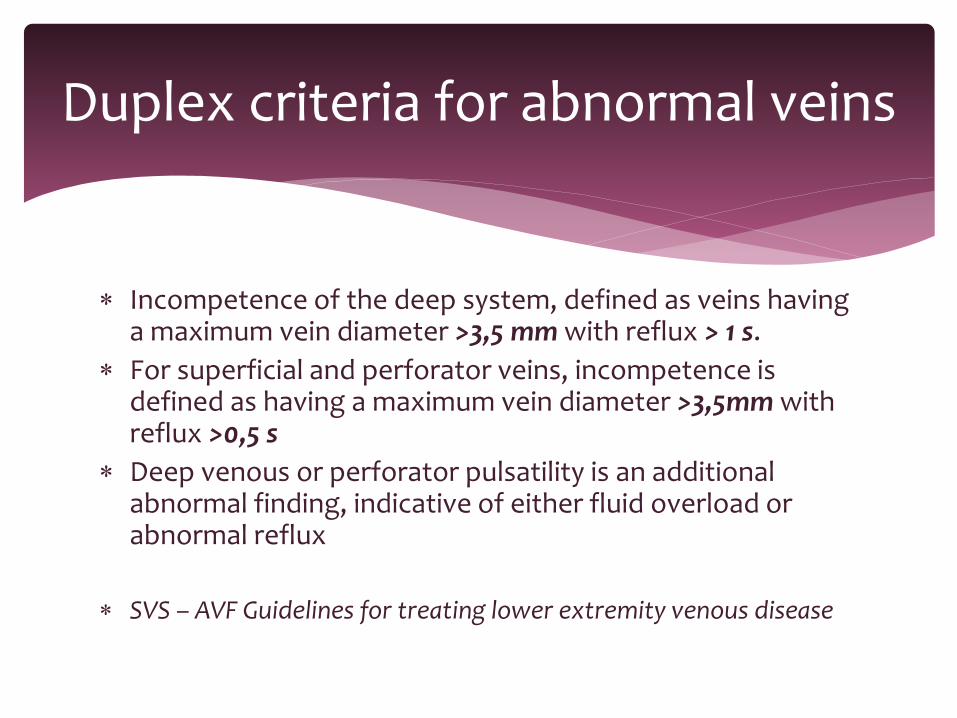
Incompetence of the deep system, defined as veins having a maximum vein diameter >3,5 mm with reflux > 1 s.
For superficial and perforator veins, incompetence is defined as having a maximum vein diameter >3,5mm with reflux >0,5 s
Deep venous or perforator pulsatility is an additional abnormal finding, indicative of either fluid overload or abnormal reflux
SVS – AVF Guidelines for treating lower extremity venous disease
Duplex criteria for abnormal veins
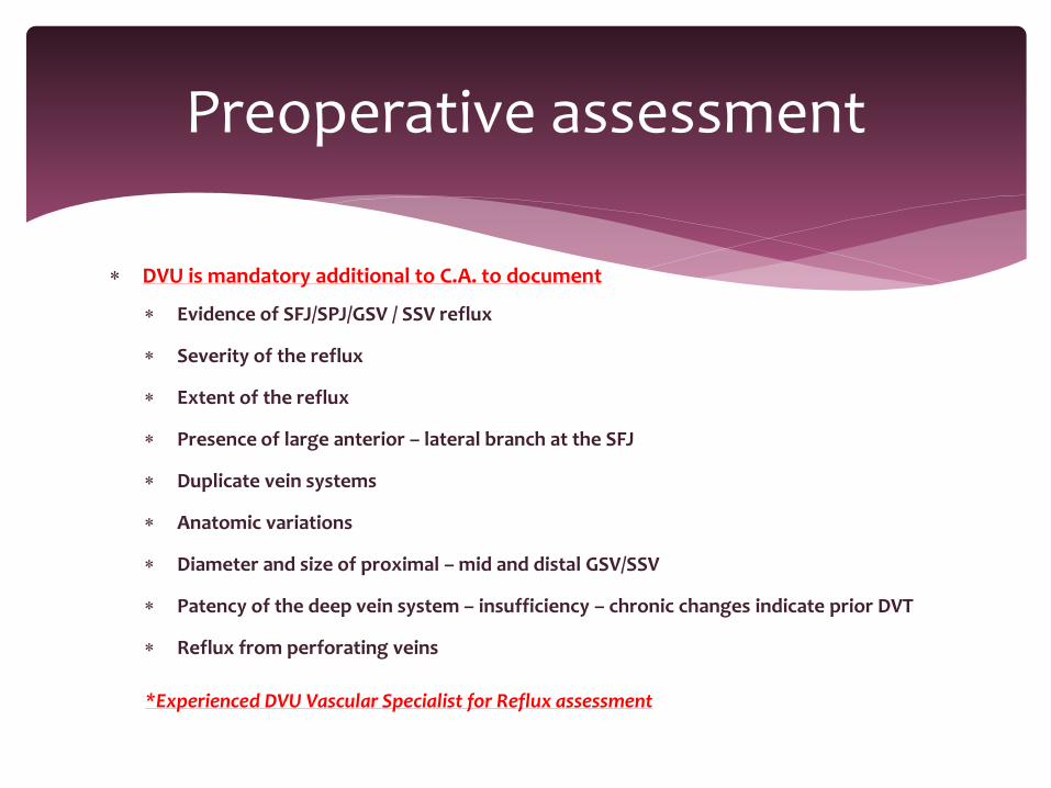
DVU is mandatory additional to C.A. to document
Evidence of SFJ/SPJ/GSV / SSV reflux
Severity of the reflux
Extent of the reflux
Presence of large anterior – lateral branch at the SFJ
Duplicate vein systems
Anatomic variations
Diameter and size of proximal – mid and distal GSV/SSV
Patency of the deep vein system – insufficiency – chronic changes indicate prior DVT
Reflux from perforating veins
*Experienced DVU Vascular Specialist for Reflux assessment
Preoperative assessment
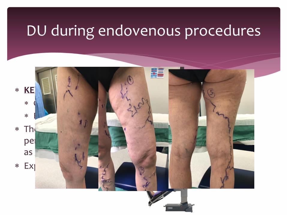
KEY:
Correct ultrasound equipment
Examination table
The treating physician is recommended to perform his or her own ultrasound as well as marking the veins to be treated .
Explain to the patient
DU during endovenous procedures
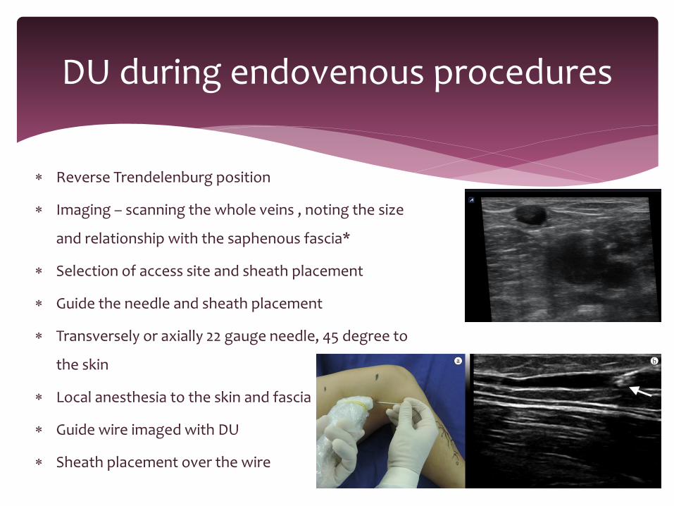
Reverse Trendelenburg position
Imaging – scanning the whole veins , noting the size
and relationship with the saphenous fascia*
Selection of access site and sheath placement
Guide the needle and sheath placement
Transversely or axially 22 gauge needle, 45 degree to
the skin
Local anesthesia to the skin and fascia
Guide wire imaged with DU
Sheath placement over the wire
DU during endovenous procedures
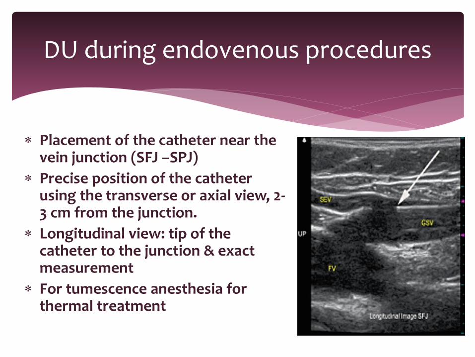
Placement of the catheter near the vein junction (SFJ –SPJ)
Precise position of the catheter using the transverse or axial view, 2-3 cm from the junction.
Longitudinal view: tip of the catheter to the junction & exact measurement
For tumescence anesthesia for thermal treatment
DU during endovenous procedures
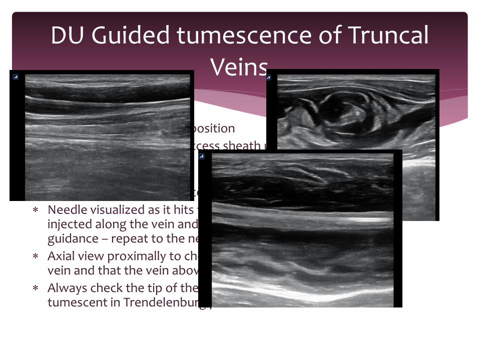
Patient in Trendelenburg position
Begins at the site of the access sheath under continuous U/S guidance – needle
Transducer in a transverse position – 45 degree angle, adjacent to the middle of the transducer and just below its inferior edge
Needle visualized as it hits the catheter – tumescent solution is injected along the vein and the catheter using continuous U/S guidance – repeat to the next segment of vein
Axial view proximally to check for adequate volume around the vein and that the vein above the catheter has been collapsed
Always check the tip of the catheter before completing the tumescent in Trendelenburg position
DU Guided tumescence of Truncal Veins
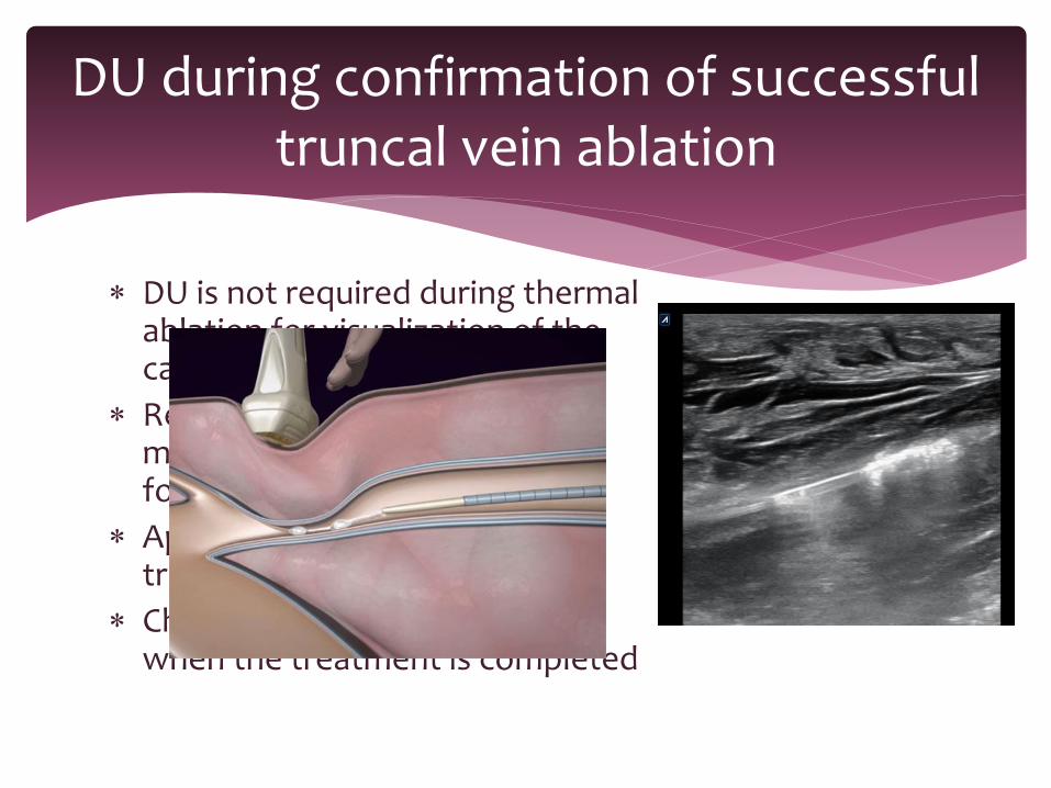
DU is not required during thermal ablation for visualization of the catheter
Recommended for non thermal methods (observed the wire, foam or glue during treatment)
Apply local pressure during treatment with the probe
Check the proximal deep veins when the treatment is completed
DU during confirmation of successful truncal vein ablation
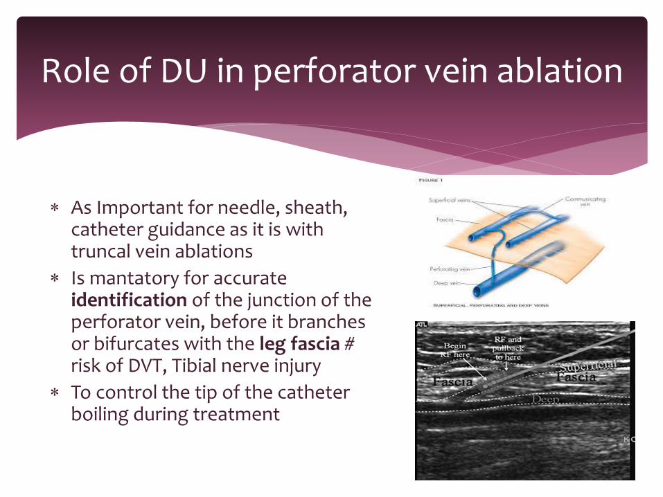
As Important for needle, sheath, catheter guidance as it is with truncal vein ablations
Is mantatory for accurate identification of the junction of the perforator vein, before it branches or bifurcates with the leg fascia # risk of DVT, Tibial nerve injury
To control the tip of the catheter boiling during treatment
Role of DU in perforator vein ablation
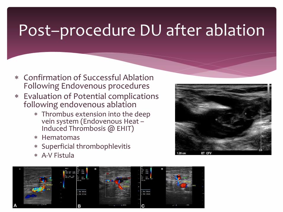
Confirmation of Successful Ablation Following Endovenous procedures
Evaluation of Potential complications following endovenous ablation
Thrombus extension into the deep vein system (Endovenous Heat –Induced Thrombosis @ EHIT)
Hematomas Superficial thrombophlevitis A-V Fistula
Post–procedure DU after ablation
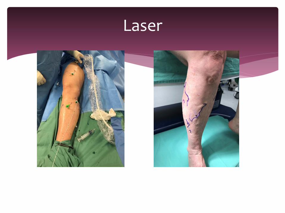
Laser
What is the role?
CRITICAL - MANTATORY
Why?
All steps of endovenous procedures Pre- op evaluation
During all endovenous procedures
DUS must be part of the training for vascular surgeons and be learned by any clinician who performs endovenous procedures
Is required in some patients to confirm the successful ablation following procedures and to evaluate patients for complication
Conclusions


![Ultrasound Imaging Physics(Basic Principles)[1]](https://static.fdocument.org/doc/165x107/5526da784a795911118b458d/ultrasound-imaging-physicsbasic-principles1.jpg)