The neurotoxin β-N-methylamino-L-alanine (BMAA)
69
The neurotoxin β-N-methylamino-L-alanine (BMAA) Sources, bioaccumulation and extraction procedures Sandra Ferreira Lage
-
Upload
hoangtuyen -
Category
Documents
-
view
224 -
download
2
Transcript of The neurotoxin β-N-methylamino-L-alanine (BMAA)
The neurotoxin
β-N-methylamino-L-alanine (BMAA)
Sources, bioaccumulation and extraction procedures
Sandra Ferreira Lage

©Sandra Ferreira Lage, Stockholm University 2016
Cover image: Cyanobacteria, diatoms and dinoflagellates microscopic
pictures taken by Sandra Ferreira Lage
ISBN 978-91-7649-455-4
Printed in Sweden by Holmbergs, Malmö 2016
Distributor: Department of Ecology, Environment and Plant Sciences,
Stockholm University

“Sinto mais longe o passado,
sinto a saudade mais perto.”
Fernando Pessoa, 1914.

Abstract
β-methylamino-L-alanine (BMAA) is a neurotoxin linked to neurodegeneration,
which is manifested in the devastating human diseases amyotrophic lateral sclerosis,
Alzheimer’s and Parkinson’s disease. This neurotoxin is known to be produced by
almost all tested species within the cyanobacterial phylum including free living as
well as the symbiotic strains. The global distribution of the BMAA producers ranges
from a terrestrial ecosystem on the Island of Guam in the Pacific Ocean to an aquatic
ecosystem in Northern Europe, the Baltic Sea, where annually massive surface
blooms occur. BMAA had been shown to accumulate in the Baltic Sea food web,
with highest levels in the bottom dwelling fish-species as well as in mollusks.
One of the aims of this thesis was to test the bottom-dwelling bioaccumulation hy-
pothesis by using a larger number of samples allowing a statistical evaluation.
Hence, a large set of fish individuals from the lake Finjasjön, were caught and the
BMAA concentrations in different tissues were related to the season of catching, fish
gender, total weight and species. The results reveal that fish total weight and fish
species were positively correlated with BMAA concentration in the fish brain.
Therefore, significantly higher concentrations of BMAA in the brain were detected
in plankti-benthivorous fish species and heavier (potentially older) individuals.
Another goal was to investigate the potential production of BMAA by other phyto-
plankton organisms. Therefore, diatom cultures were investigated and confirmed to
produce BMAA, even in higher concentrations than cyanobacteria. All diatom cul-
tures studied during this thesis work were show to contain BMAA, as well as one
dinoflagellate species. This might imply that the environmental spread of BMAA in
aquatic ecosystems is even higher than previously thought.
Earlier reports on the concentration of BMAA in different organisms have shown
highly variable results and the methods used for quantification have been intensively
discussed in the scientific community. In the most recent studies, liquid chromatog-
raphy-tandem mass spectrometry (LC-MS/MS) has become the instrument of
choice, due to its high sensitivity and selectivity. Even so, different studies show
quite variable concentrations of BMAA. In this thesis, three of the most common
BMAA extraction protocols were evaluated in order to find out if the extraction
could be one of the sources of variability. It was found that the method involving
precipitation of proteins using trichloroacetic acid gave the best performance, com-
plying with all in-house validation criteria. However, extractions of diatom and
cyanobacteria cultures with this validated method and quantified using LC-MS/MS
still resulted in variable BMAA concentrations, which suggest that also biological
reasons contribute to the discrepancies.
The current knowledge on the environmental factors that can induce or reduce
BMAA production is still limited. In cyanobacteria, production of BMAA was earli-
er shown to be negative correlated with nitrogen availability – both in laboratory
cultures as well as in natural populations. Based on this observation, it was suggest-
ed that in unicellular non-diazotrophic cyanobacteria, BMAA might take part in
nitrogen metabolism. In order to find out if BMAA has a similar role in diatoms,
BMAA was added to two diatom species in culture, in concentrations corresponding
to those earlier found in the diatoms. The results suggest that BMAA might induce a
nitrogen starvation signal in diatoms, as was earlier observed in cyanobacteria. Thus,
also in diatoms, BMAA might be involved in the nitrogen balance in the cell.
Sammanfattning
β-metylamino-L-alanin (BMAA) är ett neurotoxin som orsakar neurodegeneration
och är kopplat till förödande neurologiska sjukdomar som amyotrofisk
lateralskleros, Alzheimers och Parkinsons sjukdomar. BMAA produceras av nästan
alla analyserade cyanobakteriearter – från de som lever fritt till de som lever i
symbiotiska relationer. Dessa BMAA-producenter förekommer globalt och har
hittats i åtskilda olika ekosystem världen över – från det terrestra ekosystemet på ön
Guam i Stilla havet till det bräckta akvatiska ekosystemet i Östersjön där massiva
cyanobakterieblomningar årligen förekommer. Innan detta avhandlingsarbete
startade hade det visats att BMAA kan ackumuleras i Östersjön näringsväv med de
högsta nivåerna i de bottenlevande fiskarterna samt i vattenfiltrerande mollusker,
som tex musslor och ostron.
Ett av syftena med denna avhandling var att pröva hypotesen om ackumulering av
BMAA - särskilt i bottenlevande fiskarter - med ett tillräckligt antal prover för att
kunna utföra en statistisk analys. Ett stort antal fiskindivider fångades från en relativt
liten sjö, Finjasjön och innehållet av BMAA i olika vävnader relaterades till
fångstsäsong, kön, totalvikt och art. Resultaten visade en positiv korrelation mellan
faktorernas födomönster (planktonätande och bottenlevande) samt totalvikt med
höga koncentrationer av BMAA i fiskarnas hjärna.
Ett annat mål med avhandlingen var att undersöka om andra grupper av
växtplankton än cyanobakterier har förmåga att producera BMAA. Genom att
undersöka ett antal kulturer med kiselalger samt även dinoflagellater kunde vi påvisa
att båda dessa grupper har förmåga att producera BMAA samt att de producerar
högre nivåer av BMAA än cyanobakterier. Dessa resultat indikerar att effekten av
BMAA i vårt akvatiska ekosystem kan vara ännu högre än man tidigare trott.
Tidigare studier har visat stora variationer i koncentrationerna av BMAA och det har
lett till intensiva diskussioner angående de metoder som används för att bestämma
halterna. Flertalet nyare publikationer använder vätskekromatografi- tandem
masspektrometri (LC-MS/MS), baserat på hög känslighet och selektivitet. Trots
detta kvarstår problematiken med stora skillnader i koncentrationen av BMAA
mellan olika studier. I denna avhandling har jag studerat de tre vanligaste
extraktionsmetoderna av BMAA och utvärderat om extraktionen kan vara en av
anledningarna till den stora variationen.
Resultaten visar att den metod där triklorättiksyra används för att fälla ut proteiner i
provet gav det bästa resultatet och uppfyller samtliga kriterier för metodvalidering.
Den nuvarande kunskapen om vilka miljöfaktorer som kan inducera eller minska
ackumuleringen av BMAA i producenterna är fortfarande begränsad. Produktionen
av BMAA hos cyanobakterier har visat sig vara negativt korrelerad med tillgången
av kväve - både i laboratoriekulturer och i naturliga populationer. Baserat på denna
observation har det föreslagits att BMAA i encelliga icke- kvävefixerande
cyanobakterier kan vara involverad i kväveomsättningen. För att undersöka om
BMAA skulle kunna ha en liknande roll i kiselalger, har jag tillsatt BMAA till
kulturer med två arter av kiselalger i koncentrationer som motsvarar de som jag
funnit i kiselalgerna. Resultaten visar att BMAA tas upp av kiselalgerna och att de
reagerar på liknande sätt som vid kvävebrist. Följdaktligen så är BMAA med stor
sannolikhet även i kiselalger inblandad i den cellulära kvävebalansen.
List of Papers
The following papers, referred to by their Roman numerals, are the basis of this
thesis:
I. Lage, S., Annadotter, H., Rasmussen, U., Rydberg, S., (2015). Biotransfer
of β-N-methylamino-L-alanine (BMAA) in a eutrophicated freshwater lake.
Marine Drugs; 13(3):1185-201.
II. Jiang, L., Eriksson, J., Lage, S., Jonasson, S., Shams, S., Mehine, M., Ilag,
L., Rasmussen, U., (2014). Diatoms: a novel source for the neurotoxin
BMAA in aquatic environments. PLoS One 9: e84578.
III. Lage, S., Burian, A., Rasmussen, U., Costa, P.R., Annadotter, H., Godhe,
A., Rydberg, S., (2015). BMAA extraction of cyanobacteria samples: which
method to choose? Environmental Science and Pollution Research;
23(1):338-50.
IV. Lage, S., Ström, L., Godhe, A., Rydberg, S., (2016). The effect of exoge-
nous β-N-methylamino-L-alanine on the diatoms Phaeodactylum tricornu-
tum and Thalassiosira weissflogii. (manuscript).
My contribution to the papers:
Paper I: Performed the experiments, analyzed the data and was the main writer of
the paper.
Paper II: Participated in sample preparation, method development and manuscript
writing.
Paper III: Participated in the experimental design. Performed the experiment, de-
veloped methods, analyzed the data and was the main writer of the paper.
Paper IV: Participated in the experimental design. Participated in and supervised a
student in the experimental execution. Analyzed the data and was the main writer of
the paper.

Published papers I and II are open access. Published paper III is reprinted with per-
mission of Springer International Publishing AG with restriction that the paper must
not be reproduced or distributed separately from the thesis itself.
Additional paper completed during the PhD studies:
Lage, S., Costa, P.R., Moita, T., Eriksson, J., Rasmussen, U., Rydberg, S.J., (2014).
BMAA in shellfish from two Portuguese transitional water bodies suggests the ma-
rine dinoflagellate Gymnodinium catenatum as a potential BMAA source. Aquatic
Toxicology 152, 131-138.
Contents
Introduction 13
BMAA: a neurotoxic non-protein amino acid 13
BMAA and the paralytic disease among Guam indigenes 14
Cyanobacteria – BMAA producers 20
BMAA detection in cyanobacteria: the controversies 20
Diatoms 24
Marine toxins: producers, functions and effects 25
Nitrogen cycle: diatoms and cyanobacteria - the similarities 28
Aims 30
Comments on methods 31
Finjasjön field samples 31
Phytoplankton cultures and field samples 32
BMAA exposure experiment 33
BMAA extraction protocols and validation 33
UPLC-ESI-MS/MS 34
Statistical analysis 36
Results and discussion 37
BMAA bioaccumulation in aquatic ecosystems 37
The novel sources of BMAA – diatoms 40
BMAA extraction controversy – method validation 42
Physiological role of BMAA in diatoms – first steps towards understanding 46
Conclusions 50
Future research suggestions 53
Financial support 54
Acknowledgements 55
References 57
Abbreviations
AEG N-(2-aminoethyl) glycine
ALS amyotrophic lateral sclerosis
ALS/PDC amyotrophic lateral sclerosis/parkinsonism-dementia complex
AMPA α-amino-3-hydroxy-5-methyl-4-isoxazolepropionicacid
AQC 6-aminoquinolyl-N-hydroxysuccinimidyl carbamate
BAMA β-amino-N-methyl-alanine
BMAA β-N-methylamino-L-alanine
CCAP Culture Collection of Algae and Protozoa
CE capillary electrophoresis
CID collision induced dissociation
CNS central nervous system
D3 deuterated
DAB 2,4-diaminobutyric acid
DABA 2,3-diaminobutyric acid
DNS dansyl chloride derivatization
DW dry weight
ELISA enzyme – linked immunosorbent
ESI electrospray ionization
FMOC 9-fluorenylmethyl chloroformate
GC-MS gas chromatography – mass spectrometry
GOGAT glutamate synthase
GS glutamine synthetase
GUMACC Gothenburg University's Marine Algal Culture Collection
HILIC hydrophilic interaction liquid chromatography
HPLC-FLD high performance liquid chromatography – fluorescence detection
LC liquid chromatography
LC-MS liquid chromatography – mass spectrometry
LC-MS/MS liquid chromatography –tandem mass spectrometry
LOD limit of detection
LOQ limit of quantification
MeOH methanol
mGluRs metabotropic glutamate receptors
MS mass spectrometry
MS/MS tandem mass spectrometry
m/z mass-to-charge ratio
NMDA N-methyl-D-aspartate
nr number
PCC The Pasteur Culture Collection of Cyanobacteria
pKa acid dissociation constant at logarithmic scale
Q1 first quadrupole
Q2 second quadrupole/collision cell
Q3 third quadrupole
ROS reactive oxygen species
SCCAP The Scandinavian Culture Collection of Algae and Protozoa
S/N signal-to-noise ratio
sp. species (singular)
SPE solid phase extraction
spp. species (plural)
SRM selective reaction monitoring
TCA trichloroacetic acid
UPLC ultra-performance liquid chromatography
Xc- cystine/glutamate antiporter system
WW wet weight
13
Introduction
BMAA: a neurotoxic non-protein amino acid
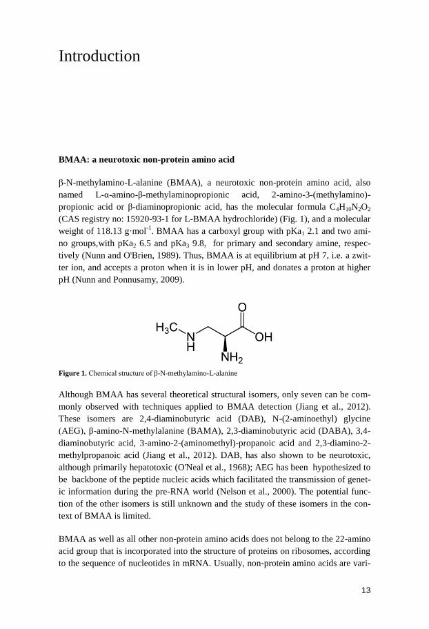
β-N-methylamino-L-alanine (BMAA), a neurotoxic non-protein amino acid, also
named L-α-amino-β-methylaminopropionic acid, 2-amino-3-(methylamino)-
propionic acid or β-diaminopropionic acid, has the molecular formula C4H10N2O2
(CAS registry no: 15920-93-1 for L-BMAA hydrochloride) (Fig. 1), and a molecular
weight of 118.13 g·mol-1
. BMAA has a carboxyl group with pKa1 2.1 and two ami-
no groups,with pKa2 6.5 and pKa3 9.8, for primary and secondary amine, respec-
tively (Nunn and O'Brien, 1989). Thus, BMAA is at equilibrium at pH 7, i.e. a zwit-
ter ion, and accepts a proton when it is in lower pH, and donates a proton at higher
pH (Nunn and Ponnusamy, 2009).
Figure 1. Chemical structure of β-N-methylamino-L-alanine
Although BMAA has several theoretical structural isomers, only seven can be com-
monly observed with techniques applied to BMAA detection (Jiang et al., 2012).
These isomers are 2,4-diaminobutyric acid (DAB), N-(2-aminoethyl) glycine
(AEG), β-amino-N-methylalanine (BAMA), 2,3-diaminobutyric acid (DABA), 3,4-
diaminobutyric acid, 3-amino-2-(aminomethyl)-propanoic acid and 2,3-diamino-2-
methylpropanoic acid (Jiang et al., 2012). DAB, has also shown to be neurotoxic,
although primarily hepatotoxic (O'Neal et al., 1968); AEG has been hypothesized to
be backbone of the peptide nucleic acids which facilitated the transmission of genet-
ic information during the pre-RNA world (Nelson et al., 2000). The potential func-
tion of the other isomers is still unknown and the study of these isomers in the con-
text of BMAA is limited.
BMAA as well as all other non-protein amino acids does not belong to the 22-amino
acid group that is incorporated into the structure of proteins on ribosomes, according
to the sequence of nucleotides in mRNA. Usually, non-protein amino acids are vari-
14
ations of protein amino acids. These changes occur through the alteration of the
amino to carboxyl relative position and/or the extent of the alkyl chain. Substitutions
along the alkyl chain, on the amino group, on any other additional functional group
and/or chiral carbons in the R-chain can also generate non-protein amino acids
(Pizzarello, 2015). BMAA itself is a variation of alanine (Brenner et al., 2003).
Many amino acids have the ability to form two different enantiomers around the
central carbon, which leads to the two different isomers, the L and the D-form. This
common feature is also applied to BMAA; however, in general, it is only the L form
that is incorporated into proteins (Bell, 2009; Dunlop et al., 2013; Glover et al.,
2014).
Fungi, bacteria, and plants can produce non-protein amino acids. These compounds
are known to be metabolites or intermediates in various metabolisms or part of the
biological structures. When consumed by other organisms several of these substanc-
es are toxic through the inhibition or disruption of existing proteins (Bell, 2003;
Harada, 2004; Vranova et al., 2011). Misincorporation of any of the 22 standard
amino acids at error rates as low as 1/10 000 may lead to neurodegeneration in la-
boratory animals (Lee et al., 2006). L-BMAA is also able to misincorporate into
human proteins and subsequently cause protein misfolding, aggregation and/or loss
of function (Dunlop et al., 2013; Glover et al., 2014). The misincorporation of
BMAA into proteins has earlier been proposed as a mechanism for bioaccumulation
as well as a mechanism for a slow release of BMAA within the central nervous
system (CNS) (Dunlop et al., 2013). However, this process could be reversed by the
presence of L-serine (Dunlop et al., 2013). Just as many other non-protein amino
acids, BMAA is not constantly present in a free form; it may also be associated with,
bound to, or incorporated into proteins (Murch et al., 2004a; Rodgers and Shiozawa,
2008; Bell, 2009; Cheng and Banack, 2009).
BMAA and the paralytic disease among Guam indigenes
BMAA has been linked to the fatal neurodegenerative diseases, amyotrophic lateral
sclerosis (ALS), Parkinson’s and Alzheimer’s disease (Banack and Cox, 2003a;
Murch et al., 2004b; Cox et al., 2009; Pablo et al., 2009). The neurotoxicity of
BMAA was discovered half a century ago, following studies conducted on the island
of Guam in the Western Pacific Ocean, where patients showed pathologies charac-
teristic of both ALS and Parkinson’s disease, i.e. amyotrophic lateral sclero-
sis/parkinsonism-dementia complex (ALS/PDC). The frequency of the disease was
abnormally high among the indigenous population of Guam, the so called Chamorro
(Vega and Bell, 1967; Spencer et al., 1987b; Spencer et al., 1991).
BMAA was later proposed to be the environmental agent causing the exceptional
high incidences of ALS/PDC among the Chamorro population (Vega and Bell,
1967; Spencer et al., 1987b; Spencer et al., 1991). The compound, BMAA, was later
15
found throughout the tissues of the cycad tree Cycas circinalis (now Cycas micro-
nesica. Hill), and was particularly abundant in the cycad seeds and immature pollen
(Vega and Bell, 1967; Hill, 1994; Banack and Cox, 2003b). An etiological study
discovered that the cycad seeds were commonly used as food source by the Chamor-
ro, who made flour from the seeds and prepared tortillas (Whiting, 1988). Thus, the
cycad seeds were proposed as the BMAA source (Vega and Bell, 1967; Spencer et
al., 1987b). However, later on, only low concentrations of BMAA were detected in
the cycad flour (Duncan et al., 1988; Duncan et al., 1989); at least too low to be the
likely cause of any neurological effect in the Chamorro (Garruto et al., 1988;
Duncan et al., 1990). Consequently, the hypothesis of the link between BMAA and
ALS/PDC in Guam was at this point abandoned.
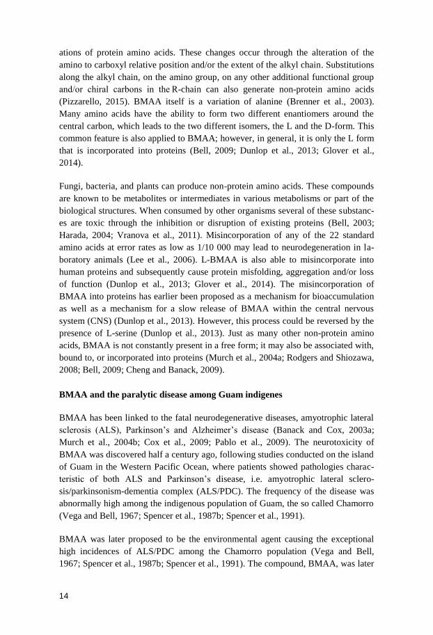
Figure 2. A photo of (a) cycad Cycas micronesica coralloid roots, (b) cyanobacteria Nostoc sp. in the
cycad coralloid roots and (c) Cycas micronesica seeds. Pictures credits: Paul Cox, Patty Stewart, and
Sandra Banack.
The interest in BMAA was brought back by the beginning of the 21st century, when
a revolutionizing study brought new facts to the story (Murch et al., 2004a). The
source of BMAA was traced to cyanobacteria of the genus Nostoc, which live sym-
biotically in the coralloid roots of cycads (Fig. 2b) and BMAA was suggested to be
then possibly transferred to the cycad seeds (Fig. 2c) (Banack and Cox, 2003b; Cox
et al., 2003; Murch et al., 2004a). The concentration of BMAA in the protein-
associated fraction was found to be much higher than the BMAA found in the free
form (Fig. 3) (Murch et al., 2004a). The protein fraction was not taken into account
in the earlier studies (Spencer et al., 1987b; Duncan et al., 1988; Whiting, 1988;
Duncan et al., 1989) due to a lack of knowledge and selectivity of the instrumenta-
tion used. Added to this, both free BMAA and protein associated BMAA were then
proven to accumulate in both cycad seeds and the wild animals (e.g. flying foxes,
pigs and deer) that forage on them (Fig. 3) (Murch et al., 2004a).
These new data could now explain how the neurotoxin BMAA had reached the
indigenous Chamorro population through biomagnification, i.e. an increasing accu-
mulation of a molecule through higher trophic levels (Fig. 3) (Banack and Cox,
2003a; Cox et al., 2003; Murch et al., 2004a). The diet of the Chamorros comprised
several wild animals in particular the flying foxes, which feed almost exclusively on
cycad seeds and evidently accumulate BMAA in high concentrations in their body
parts (Fig. 3) (Banack and Cox, 2003a; Banack et al., 2006). To further ensure
a b c
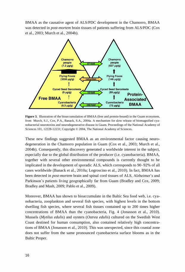
16
BMAA as the causative agent of ALS/PDC development in the Chamorro, BMAA
was detected in post-mortem brain tissues of patients suffering from ALS/PDC (Cox
et al., 2003; Murch et al., 2004b).
Figure 3. Illustration of the bioaccumulation of BMAA (free and protein-bound) in the Guam ecosystem,
from Murch, S.J., Cox, P.A., Banack, S.A., 2004a. A mechanism for slow release of biomagnified cya-
nobacterial neurotoxins and neurodegenerative disease in Guam. Proceedings of the National Academy of
Sciences 101, 12228-12231; Copyright © 2004, The National Academy of Sciences.
These new findings suggested BMAA as an environmental factor causing neuro-
degeneration in the Chamorro population in Guam (Cox et al., 2003; Murch et al.,
2004b). Consequently, this discovery generated a worldwide interest in the subject,
especially due to the global distribution of the producer (i.e. cyanobacteria). BMAA,
together with several other environmental compounds is currently thought to be
implicated in the development of sporadic ALS, which corresponds to 90–92% of all
cases worldwide (Banack et al., 2010a; Logroscino et al., 2010). In fact, BMAA has
been detected in post-mortem brain and spinal cord tissues of ALS, Alzheimer’s and
Parkinson’s patients living geographically far from Guam (Bradley and Cox, 2009;
Bradley and Mash, 2009; Pablo et al., 2009).
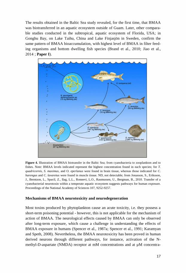
Moreover, BMAA has shown to bioaccumulate in the Baltic Sea food web, i.e. cya-
nobacteria, zooplankton and several fish species, with highest levels in the bottom
dwelling fish species, where several fish tissues contained up to 200 times higher
concentrations of BMAA than the cyanobacteria, Fig. 4 (Jonasson et al., 2010).
Mussels (Mytilus edulis) and oysters (Ostrea edulis) cultured on the Swedish West
Coast destined for human consumption, also contained relatively high concentra-
tions of BMAA (Jonasson et al., 2010). This was unexpected, since this coastal zone
does not suffer from the same pronounced cyanobacteria surface blooms as in the
Baltic Proper.
17
The results obtained in the Baltic Sea study revealed, for the first time, that BMAA
was biotransferred in an aquatic ecosystem outside of Guam. Later, other compara-
ble studies conducted in the subtropical, aquatic ecosystem of Florida, USA; in
Gonghu Bay, on Lake Taihu, China and Lake Finjasjön in Sweden, confirm the
same pattern of BMAA bioaccumulation, with highest level of BMAA in filter feed-
ing organisms and bottom dwelling fish species (Brand et al., 2010; Jiao et al.,
2014); Paper I).
Figure 4. Illustration of BMAA biotransfer in the Baltic Sea; from cyanobacteria to zooplankton and to
fishes. Note: BMAA levels indicated represent the highest concentration found in each species; for T.
quadricornis, S. maximus, and O. eperlanus were found in brain tissue, whereas those indicated for C.
harengus and C. lavaretus were found in muscle tissue. ND, not detectable; from Jonasson, S., Eriksson,
J., Berntzon, L., Spacil, Z., Ilag, L.L., Ronnevi, L.O., Rasmussen, U., Bergman, B., 2010. Transfer of a
cyanobacterial neurotoxin within a temperate aquatic ecosystem suggests pathways for human exposure.
Proceedings of the National Academy of Sciences 107, 9252-9257.
Mechanisms of BMAA neurotoxicity and neurodegeneration
Most toxins produced by phytoplankton cause an acute toxicity, i.e. they possess a
short-term poisoning potential - however, this is not applicable for the mechanism of
action of BMAA. The neurological effects caused by BMAA can only be observed
after long-term exposure, which cause a challenge in understanding the effects of
BMAA exposure in humans (Spencer et al., 1987a; Spencer et al., 1991; Karamyan
and Speth, 2008). Nevertheless, the BMAA neurotoxicity has been proved in human
derived neurons through different pathways, for instance, activation of the N-
methyl-D-aspartate (NMDA) receptor at mM concentrations and at µM concentra-
18
tions in the two metabotropic glutamate receptors 1 and 5 (mGluR1 and mGluR5
receptors, respectively), as well as in the α-amino-3-hydroxy-5-methyl-4-
isoxazolepropionicacid (AMPA)/kainate receptors (Lindstrom et al., 1990; Rao et
al., 2006; Lobner et al., 2007; Liu et al., 2009a; Liu et al., 2009b). Toxicity studies
with BMAA have shown damage both to neuron cultures and to the development of
neurodegenerative effects in animals (Karamyan and Speth, 2008; Chiu et al., 2011).
For instance, when rats were injected intraperitoneal with 6–14 μm BMAA/g body
weight, they showed weakness, convulsions and incoordination (Vega and Bell,
1967). In monkeys fed for 10 weeks with 100–350 mg/kg BMAA, corticomotoneu-
ronal dysfunction, Parkinsonian features and behavioral abnormalities were found
(Spencer et al., 1987a). In more recent studies, lower concentrations of BMAA have
been used and verified that BMAA can induce long-term cognitive deficits as well
as protein changes and fibril formation in the hippocampus of adult rodents follow-
ing neonatal exposure (Karlsson et al., 2009; Karlsson et al., 2011; Karlsson et al.,
2012).
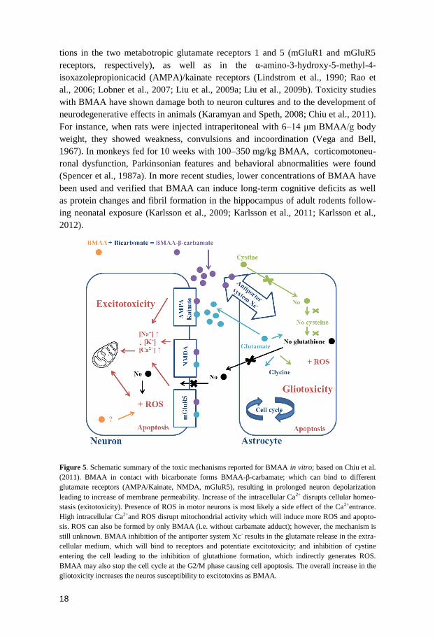
Figure 5. Schematic summary of the toxic mechanisms reported for BMAA in vitro; based on Chiu et al.
(2011). BMAA in contact with bicarbonate forms BMAA-β-carbamate; which can bind to different
glutamate receptors (AMPA/Kainate, NMDA, mGluR5), resulting in prolonged neuron depolarization
leading to increase of membrane permeability. Increase of the intracellular Ca2+ disrupts cellular homeo-
stasis (exitotoxicity). Presence of ROS in motor neurons is most likely a side effect of the Ca2+entrance.
High intracellular Ca2+and ROS disrupt mitochondrial activity which will induce more ROS and apopto-
sis. ROS can also be formed by only BMAA (i.e. without carbamate adduct); however, the mechanism is
still unknown. BMAA inhibition of the antiporter system Xc- results in the glutamate release in the extra-
cellular medium, which will bind to receptors and potentiate excitotoxicity; and inhibition of cystine
entering the cell leading to the inhibition of glutathione formation, which indirectly generates ROS.
BMAA may also stop the cell cycle at the G2/M phase causing cell apoptosis. The overall increase in the
gliotoxicity increases the neuros susceptibility to excitotoxins as BMAA.
19
BMAA is capable of forming two carbamate adducts in the presence of carbon diox-
ide/bicarbonate. The formed adducts, α-N-carboxy and β-N-carboxy, are structurally
similar to glutamate, the most important transmitter for normal brain functions in
mammals (Nunn and O'Brien, 1989; Weiss et al., 1989a). Hence, it is not surprising,
that BMAA mechanism of action involves the excess activation of glutamate recep-
tors, which subsequently leads to accumulation of intracellular Ca2+
and the genera-
tion of reactive oxygen species (ROS). This ultimately leads to destruction of the
neurons via an excitotoxic mechanism, i.e. neuronal damaged caused by high con-
centrations of extracellular glutamate (Nunn et al., 1987; Weiss et al., 1989b; Myers
and Nelson, 1990). The effects of BMAA in motor neurons have shown to be far
greater than in any other neuron cell, this through the activation of the AM-
PA/kainate receptor (Rao et al., 2006). In the AMPA/kainate receptor, BMAA in-
duces an excess activation which results in neuron degeneration; via rapid Ca2+
entry
through the AMPA/kainate channel generating mitochondrial ROS (Fig. 5) (Rao et
al., 2006).
In addition to the oxidative stress caused by the BMAA excess activation of the
glutamate receptors, BMAA also inhibit the cystine/glutamate antiporter system Xc-
in astrocytes (Liu et al., 2009a). The Xc- antiporter system is responsible for the
transport of cystine into the cell in exchange for glutamate being transported out;
thus, the presence of BMAA leads to an increase of extracellular glutamate while
glutathione is depleted inside the astrocyte. This mechanistic action will further
increase ROS causing further excitotoxicity (Fig. 5) (Liu et al., 2009a). In some of
the reported cases of sporadic forms of ALS, a decrease in the glutamate uptake
capacity in the spinal cord and motor cortex was noticed. This is likely due to an
increase in levels of extracellular glutamate which results in excitotoxicity
(Rothstein et al., 1990; Rothstein et al., 1992; Shaw, 1994; Rothstein, 1996). Also in
healthy human neurons, the exposure to BMAA causes an increased intracellular
Ca2+
influx, DNA damage and mitochondrial activity; release of lactate dehydrogen-
ase and generation of ROS, all characteristics of excitotoxicity (Chiu et al., 2012).
Although the BMAA neurotoxicity has been shown both in in vitro and in biological
models (Karamyan and Speth, 2008; Chiu et al., 2011), as well as in post-mortem
brain tissue from patients suffering of neurological diseases (Banack and Cox,
2003a; Cox et al., 2003; Murch et al., 2004b; Pablo et al., 2009), proof of the neuro-
degenerative characteristics via dietary exposure has not been presented until just
recently (Cox et al., 2016). In this study, vervet monkeys (Chlorocebus sabaeus)
were fed for 140 days with fruit supplemented with BMAA. The diet (i.e. 21 mg
kg−1
d−1
of BMAA) was projected to be equivalent to a cumulative life-time exposure
of BMAA by a Chamorro, leading to the development of neurofibrillary tangles and
β-amyloid deposits in the brains - both neurological hallmarks of Alzheimer's dis-
ease and ALS/PDC (Cox et al., 2016).
20
Cyanobacteria – BMAA producers
Cyanobacteria, previously known as blue-green algae, are included in a highly di-
verse group of ancient gram-negative photosynthetic prokaryotes, i.e. the Eubacteria
kingdom, exhibiting oxygenic photosynthesis. They are extremely important prima-
ry producers and constitute the first level of organisms in the globally distributed
aquatic food webs (Herrero and Flores, 2008). Moreover, cyanobacteria have the
ability to perform anaerobic metabolism and the capacity to use elemental sulfur for
anaerobic dark respiration (Cohen and Gurevitz, 2006). They also have an important
role in the marine nitrogen cycle, by fixing atmospheric nitrogen (N), and in the
global carbon cycle by their photosynthetic activity (Whitton and Potts, 2000). So
far, 150 genera and about 2000 species of cyanobacteria have been described (Van
Apeldoorn et al., 2007).
In terms of morphology, cyanobacteria are either single celled, colonial or filamen-
tous (Duy et al., 2000). Cyanobacteria exhibit versatile physiology, a wide ecologi-
cal tolerance and high genetic diversity, which together contributes to their competi-
tive success over a broad spectrum of environments across all global latitudes,
demonstrating the ability of the pioneering ancestors as the earliest inhabitants of
Earth (Cohen and Gurevitz, 2006). In fact, they inhabit ice fields, hot springs, de-
serts and are especially common in freshwater, brackish and marine environments
(Whitton and Potts, 2000; Cohen and Gurevitz, 2006). In view of the aquatic envi-
ronments, it is impossible to fully separate freshwater and marine cyanobacteria
species, considering that some species are capable to grown in both environments
(Burja et al., 2001).
With regard to the ability to produce BMAA, it has been recorded in a wide range of
cyanobacteria species, with verified occurrence around the world (Table 1). This
includes the cyanobacterial species that annually form the massive surface ‘blooms’
in the Baltic Sea during summer i.e. genera Nodularia and Aphanizomenon (Cox et
al., 2005; Jonasson et al., 2010).
BMAA detection in cyanobacteria: the controversies
Even though BMAA has concurrently been detected in cyanobacterial species from
different environments around the world, the levels of BMAA detected have varied
considerably (Table 1) (Cox et al., 2005; Esterhuizen and Downing, 2008; Metcalf et
al., 2008; Craighead et al., 2009; Faassen et al., 2009; Jonasson et al., 2010; Li et al.,
2010). The first report showing BMAA in cyanobacteria in 2005 was alarming,
since almost all investigated laboratory grown species, free living as well as symbi-
otic strains, contained high concentrations of BMAA (Cox et al., 2005). However,
these first results could not be reproduced by subsequent studies; BMAA could
either not be detected in cyanobacteria (Rosén and Hellenäs, 2008; Kruger et al.,
21
2010) or detected in some, but not all, (Faassen et al., 2009) or detected in all sam-
ples, but at very low concentrations (Jonasson et al., 2010) (Table 1). At first, bio-
logical reasons, such as sample origin and growth conditions of laboratory strains,
were suggested as the main cause of these discrepancies (Banack et al., 2010a;
Banack et al., 2010b). However, a more plausible reason was the diversity of the
analytical techniques applied by the different research groups and used between
studies (Faassen et al., 2012; Faassen, 2014).
Highest BMAA concentrations and percentages of detected positives samples were
found in studies using high performance liquid chromatography with fluorescence
detection (HPLC-FLD), gas chromatography with mass spectrometry detection (GC-
MS) and capillary electrophoresis (CE) for quantification (Cox et al., 2005;
Esterhuizen and Downing, 2008; Metcalf et al., 2008; Baptista et al., 2011). Where-
as studies using liquid chromatography with tandem mass spectrometry detection
(LC-MS/MS) either did not detect BMAA or reported low BMAA concentrations
(Rosén and Hellenäs, 2008; Faassen et al., 2009; Jonasson et al., 2010; Kruger et al.,
2010). This hypothesis was verified by concurrently analyzing cyanobacteria sam-
ples with both the analytical methods HPLC-FLD and LC-MS/MS (Faassen et al.,
2012). The HPLC-FLD was proven to be less selective than the LC-MS/MS, this
since the only selection criteria used are the retention time and signal fluorescence
(Faassen et al., 2012). Thus, HPLC-FLD is an uncertain method when it comes to
BMAA detection in complex biological matrices and may most certainly lead to
overestimations of the BMAA concentrations. Instead, LC-MS/MS has been shown
to give a reliable identification based on the several selection criteria, i.e. retention
time, mass-to-charge ratio (m/z) of the precursor ion, m/z of the product fragment
ions after collision-induced dissociation, and the ratio between the intensities of
respective ions transitions in MRM spectrum (Faassen et al., 2012; Jiang et al.,
2012).
In the more recent studies, BMAA analysis are most often performed with LC-
MS/MS (Faassen, 2014). However, even using this analytical method, there are
inconsistencies in the concentrations of BMAA reported between studies (Banack et
al., 2007; Jonasson et al., 2010; Spacil et al., 2010; Banack et al., 2011; Jiao et al.,
2014; Lage et al., 2014); Paper I; II and III). It is possible that these differences are
due to variation in the protocols for the extraction of BMAA, but it may also partly
be due to by biological causes (see Paper III and IV).
22
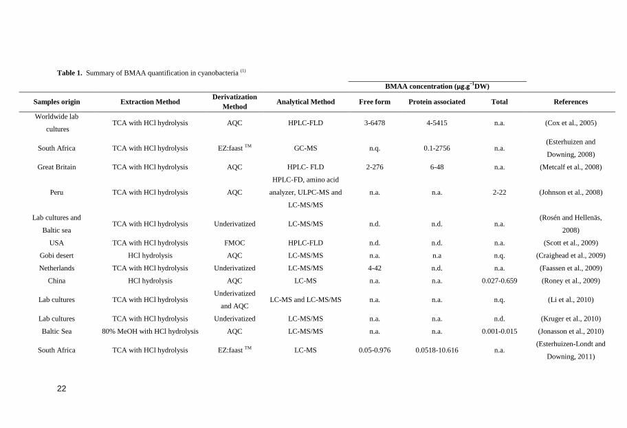
Table 1. Summary of BMAA quantification in cyanobacteria (1)
BMAA concentration (µg.g−1DW)
Samples origin Extraction Method Derivatization
Method Analytical Method Free form Protein associated Total References
Worldwide lab
cultures TCA with HCl hydrolysis AQC HPLC-FLD 3-6478 4-5415 n.a. (Cox et al., 2005)
South Africa TCA with HCl hydrolysis EZ:faast TM GC-MS n.q. 0.1-2756 n.a. (Esterhuizen and
Downing, 2008)
Great Britain TCA with HCl hydrolysis AQC HPLC- FLD 2-276 6-48 n.a. (Metcalf et al., 2008)
Peru TCA with HCl hydrolysis AQC
HPLC-FD, amino acid
analyzer, ULPC-MS and
LC-MS/MS
n.a. n.a. 2-22 (Johnson et al., 2008)
Lab cultures and
Baltic sea TCA with HCl hydrolysis Underivatized LC-MS/MS n.d. n.d. n.a.
(Rosén and Hellenäs,
2008)
USA TCA with HCl hydrolysis FMOC HPLC-FLD n.d. n.d. n.a. (Scott et al., 2009)
Gobi desert HCl hydrolysis AQC LC-MS/MS n.a. n.a n.q. (Craighead et al., 2009)
Netherlands TCA with HCl hydrolysis Underivatized LC-MS/MS 4-42 n.d. n.a. (Faassen et al., 2009)
China HCl hydrolysis AQC LC-MS n.a. n.a. 0.027-0.659 (Roney et al., 2009)
Lab cultures TCA with HCl hydrolysis Underivatized
and AQC LC-MS and LC-MS/MS n.a. n.a. n.q. (Li et al., 2010)
Lab cultures TCA with HCl hydrolysis Underivatized LC-MS/MS n.a. n.a. n.d. (Kruger et al., 2010)
Baltic Sea 80% MeOH with HCl hydrolysis AQC LC-MS/MS n.a. n.a. 0.001-0.015 (Jonasson et al., 2010)
South Africa TCA with HCl hydrolysis EZ:faast TM LC-MS 0.05-0.976 0.0518-10.616 n.a. (Esterhuizen-Londt and
Downing, 2011)
23
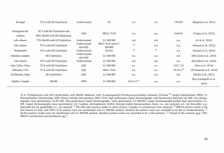
Portugal TCA with HCl hydrolysis Underivatized CE n.a. n.a. 170-810 (Baptista et al., 2011)
Portuguese lab
cultures
TCA with HCl hydrolysis and
80% MeOH with HCl Hydrolysis
AQC
HPLC- FLD
n.a.
n.a.
0.04-63
(Cianca et al., 2012)
Lab cultures 75% MeOH with HCl hydrolysis Underivatized LC-MS/MS n.d. n.d. n.a. (Li et al., 2012)
Lab cultures TCA with HCl hydrolysis Underivatized
and AQC
HPLC-FLD and LC-
MS/MS (2) n.d. n.a. (Faassen et al., 2012)
Netherlands TCA with HCl hydrolysis Underivatized ELISA (3) (3) n.a. (Faassen et al., 2013)
Spirulina samples HCl hydrolysis Underivatized
and AQC LC-MS/MS n.a. n.a. n.d. (McCarron et al., 2014)
Lab cultures TCA with HCl hydrolysis Underivatized LC-MS/MS n.d. n.d. n.a. (Reveillon et al., 2014)
Lake Taihu, China TCA with HCl hydrolysis AQC LC-MS/MS n.r. n.r. 2.01-7.23 (Jiao et al., 2014)
Nebraska, USA TCA with HCl hydrolysis AQC HPLC-FLD n.a. n.a. 1.8-25.3 (4) (Al-Sammak et al., 2014)
Al Kharrara, Qatar HCl hydrolysis AQC LC-MS/MS n.a. n.a. n.d. (Richer et al., 2015)
Québec, Canada MeOH DNS LC-MS/MS 0.01-0.3 (4) n.a. n.a. (Roy-Lachapelle et al.,
2015)
TCA: Trichloroacetic acid; HCl: Hydrochloric acid; MeOH: Methanol; AQC: 6-aminoquinolyl-N-hydroxysuccinimidyl carbamate; EZ:faast TM: propyl chloroformate; FMOC: 9-
fluorenylmethyl chloroformate; DNS: Dansyl chloride derivatization; HPLC-FLD: high performance liquid chromatography with fluorescence detection; GC-MS: Gas chroma-tography–mass spectrometry; ULPC-MS: Ultra-performance liquid chromatography- mass spectrometry; LC-MS/MS: Liquid chromatography-tandem mass spectrometry; LC-
MS: Liquid chromatography–mass spectrometry; CE: Capillary electrophoresis; ELISA: Enzyme-Linked Immunosorbent Assay; n.a.: not analyzed; n.d.: not detectable; n.q.:
detectable but not quantifiable; n.r.: not reported; (1) The table only presents studies in which at least 3 samples of cyanobacteria were analyzed; (2) BMAA positive results in the
free fraction of AQC with HPLC-FLD analysis were not corroborated with LC-MS/MS analysis, therefore the positive result was concluded to be due to misidentification; (3)
ELISA positive results were not corroborated with LC-MS/MS analysis; therefore positive result was concluded to be a false positive.; (4) Instead of the common µg.g-1 DW,
BMAA concentration was presented in µg.L-1.
24
Diatoms
We have recently shown that BMAA is not only produced by cyanobacteria (as it
has previously been thought) but also by the diverse phytoplankton groups, diatoms
and dinoflagellates (Cox et al., 2005; Lage et al., 2014); Paper II).
Diatoms are eukaryotic unicellular photoautotrophs. Their name is derived from the
Greek diatomos, meaning ‘cut in half ’, which is a reference to their distinctive two-
part cell walls made of silica, called frustule (Smetacek, 1999). Diatoms have evolu-
tionary diverged into two classes, i.e. centric and pennate, over at least 90 million
years based on the fossil record (Bowler et al., 2008). Centric and pennate diatoms
can be morphologically distinguished by their circular or elongated frustule struc-
ture, respectively. Diatoms are thought to be originated from a secondary endosym-
biotic event between a red algae and a heterotrophic eukaryotic host (Falkowski et
al., 2004). The genomes of two diatom species have been sequenced; the centric
Thalassiosira pseudonana and the pennate Phaeodactylum tricornutum (Armbrust et
al., 2004; Bowler et al., 2008). The sequences provided additional support for the
secondary endosymbiosis theory and have shown metabolic adaptations to the sur-
rounding environment; e.g. characterization of the features of central carbon me-
tabolism pathways (Bowler et al., 2010; Smith et al., 2012) Recently, knowledge
about diatom nitrogen metabolism has also increased, based on the proteomic and
metabolomic profiles in response to nitrogen starvation (Alipanah et al., 2015;
Levitan et al., 2015).
Diatoms are the most diverse group of algae and play a key role in aquatic ecosys-
tems; representing one-fifth of the photosynthesis on Earth (Nelson et al., 1995),
which generates as much organic carbon as all terrestrial rainforests combined
(Nelson et al., 1995; Field, 1998). Thus, they represent the foundation of many ma-
rine food webs and are major contributors in biogeochemical processes in aquatic
environments, especially the cycling of carbon and silicon (Mann, 1999; Sarthou et
al., 2005).
The supporting frustule allows the cell to grow large, which means a rather low
surface to volume ratio of the plasma membrane (Chisholm, 1992). This feature
makes diatoms inferior competitors in oligotrophic waters, contrary to the smaller
phytoplankton, like cyanobacteria. Even so, diatoms are more efficient in utilizing
nutrients and therefore stronger competitors in nutrient rich environments. Eutrophi-
cation, being it anthropogenic or natural, might therefore result in diatom, cyanobac-
teria and dinoflagellates blooms. It has long been known that blooms of phytoplank-
ton can be deleterious to aquatic organisms or humans (Boesch et al., 1997). Such
blooms are referred to as harmful algal blooms (Landsberg, 2002). Only a small
number of diatom species, e.g. Coscinodiscus centralis, Coscinodiscus concinnus,
Coscinodiscus wailesii, Chaetoceros convolutus, Asterionellopsis glacialis, Cera-
25
toneis closterium, Anaulus australis, Thalassiosira mala, and a few species of the
Pseudo-nitzschia genus are recognized as harmful. This through production of either
a toxin, exudates, mechanical damage due to cell morphology, and/or high biomass
accumulation (Smetacek, 1985; Fryxell and Villac, 1999; Villac et al., 2010). How-
ever, this list of species does not account for the diatom species that produce
BMAA, which so far have shown to be a common feature among all the tested dia-
tom species (Réveillon et al., 2015; Reveillon et al., 2016); Paper II and III).
Marine toxins: producers, functions and effects
Algal blooms have many detrimental consequences for the aquatic ecosystems, even
when the phytoplankton species do not produce toxic compounds. For instance,
phytoplankton blooms can increase the turbidity of aquatic ecosystems, shading
aquatic plants and thereby destroy important invertebrate and fish habitats (Paerl and
Huisman, 2008). Moreover, when blooms disrupt and decay they may deplete oxy-
gen in their surroundings, thus causing fish to die (Paerl and Huisman, 2008).
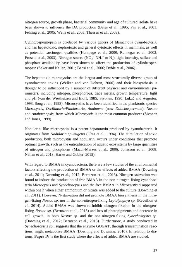
Toxin production by cyanobacteria and diatoms (Table 2) may lead to various impli-
cations in mammals depending on the target of toxicity. They can have neurotoxic
characteristics causing the inhibition of neuronal control over ion concentrations
across the cell membrane, which ultimately cause neurological insults in mammals;
or they can show hepatotoxic characteristics, causing injuries of liver and/or by
internal hemorrhages. Although their effects in humans have been well documented
(Narahashi, 1972; Berman and Murray, 1997; Sivonen and Jones, 1999; Pulido,
2008; Sivonen and Börner, 2008; Etheridge, 2010), despite much speculation and
development of several hypotheses, the ecological or physiological functions of
most toxins produced by phytoplankton have remained largely unknown. Neverthe-
less, biosynthesis of these toxins requires a high input of energy, which suggests that
their production needs to be advantageous for their producers (Sivonen and Börner,
2008). Nutrition availability, as well as light and temperature are often displayed as
a triggering factor in the production of some well-known phytoplankton toxins
(Fehling et al., 2004; Neilan et al., 2013; Van de Waal et al., 2014; Harke and
Gobler, 2015).
Anatoxins, i.e. anatoxin-a, homoanatoxin-a and anatoxin-a(s), are a group of neuro-
toxins isolated from cyanobacteria (Devlin et al., 1977; Matsunaga et al., 1989).
Anatoxin-a is potentially the most common cyanobacterial neurotoxin and is pro-
duced by Anabaena flos-aquae, Anabaena spp., A. planktonica, Aphanizomenon
spp., Cylindrospermum spp., Oscillatoria spp., Planktothrix rubescens, Phormidium
flavosum, Arthrospira fusiformis (Sivonen and Jones, 1999; Viaggiu et al., 2004;
Gugger et al., 2005). The physicochemical parameters (temperature, light intensity
and nitrogen source) and growth phase have been found positively correlated with
the toxin production (Rapala et al., 1993; Gallon et al., 1994; Gupta et al., 2002).
26
However, in contrast to the other toxins, anatoxins are produced under conditions
suboptimal for growth, e.g. during nitrogen starvation (Saker and Neilan, 2001).
Thus, most probably production of anatoxins is not a direct function of cell growth,
as suggested for the production of microcystins and nodularins (Long et al., 2001;
Neilan et al., 2013).
Saxitoxins are potent neurotoxins belonging to a group of structurally related toxins
known as the paralytic shellfish toxins. These toxins are produced by both eukaryot-
ic marine dinoflagellates, i.e. Alexandrium species, Pyrodinium bahamense,
and Gymnodinium catenatum, as well as freshwater cyanobacteria of the genera
Anabaena, Cylindrospermopsis, Aphanizomenon, Lyngbya, Raphidiopsis,
Planktothrix and Scytonema (Shumway, 1990; Neilan et al., 2013). Several studies
have correlated the production of saxitoxins with the availability of macronutrients,
temperature and light intensity. However, these studies have used different organ-
isms and methods used for toxin detection; thus, the results have so far been incon-
clusive (Sivonen and Börner, 2008; Neilan et al., 2013).
Table 2. Summary of most commonly studied cyanobacteria and diatom toxins.
Domoic acid (DA) is a neurotoxin which causes persistent neurological symptoms
(Wright et al., 1989) and triggers the so called amnesic shellfish disorder in humans
(Bates et al., 1989). DA is produced by several strains of the diatom species Pseudo-
nitzschia (Lelong et al., 2012). The macronutrient and micronutrient availability,
Chemical
group Toxins Mechanism of action Source organisms
Alkaloids
Anatoxins
(neurotoxic)
Binding irreversibly
to acetylcholine
receptors
Cyanobacteria: Anabaena flos-aquae, Anabaena sp., A.
planktonica, Aphanizomenon sp., Cylindrospermum sp.,
Oscillatoria sp., Planktothrix rubescens, Phormidium
flavosum, Arthrospira fusiformis
Saxitoxins
(neurotoxic)
Binding and blocking the
sodium channels in
neural cells
Cyanobacteria: Anabaena circinalis, Aphanizomenon
spp., Lyngbya wollei, Cylindrospermopsis raciborskii ,
Raphidiopsis brookii, Raphidiopsis sp., Planktothri sp.,
Scytonema sp. and Dinoflagellates:
Alexandrium sp., Pyrodinium bahamense,
Gymnodinium catenatum
Domoic Acid
(Neurotoxin)
Binding irreversibly
to glutamate receptors Diatoms: Pseudo-nitzschia sp.
Cylindrospermopsin
(Hepatotoxic)
Blocks protein
synthesis
Cyanobacteria: Cylindrospermopsis
raciborskii
Cyclic
Peptides
Microcystins
(Hepatotoxic) Inhibition of protein
serine/threonine phos-
phatases 1 and 2A
Cyanobacteria: Microcystis, Oscillatoria/Planktrotrix,
Anabaena, Nostoc, Anabaenopsis
Nodularins
(Hepatotoxic) Cyanobacteria: Nodularia spumigena
27
nitrogen source, growth phase, bacterial community and age of cultured isolate have
been shown to influence the DA production (Bates et al., 1995; Pan et al., 2001;
Fehling et al., 2005; Wells et al., 2005; Thessen et al., 2009).
Cylindrospermopsin is produced by various genera of filamentous cyanobacteria,
and has hepatotoxic, nephrotoxic and general cytotoxic effects in mammals, as well
as potential carcinogen qualities (Humpage et al., 2000; Runnegar et al., 2002;
Froscio et al., 2003). Nitrogen source (NO3-, NH4
+ or N2), light intensity, sulfate and
phosphate availability have been shown to affect the production of cylindrosper-
mopsin (Saker and Neilan, 2001; Bácsi et al., 2006; Dyble et al., 2006).
The hepatotoxic microcystins are the largest and most structurally diverse group of
cyanobacteria toxins (Welker and von Döhren, 2006) and their biosynthesis is
thought to be influenced by a number of different physical and environmental pa-
rameters, including nitrogen, phosphorous, trace metals, growth temperature, light
and pH (van der Westhuizen and Eloff, 1985; Sivonen, 1990; Lukač and Aegerter,
1993; Song et al., 1998). Microcystins have been identified in the planktonic species
Microcystis, Oscillatoria/Planktrotrix, Anabaena (now Dolichospermum), Nostoc
and Anabaenopsis, from which Microcystis is the most common producer (Sivonen
and Jones, 1999).
Nodularin, like microcystin, is a potent hepatotoxin produced by cyanobacteria. It
originates from Nodularia spumigena (Ohta et al., 1994). The stimulation of toxic
production, both microcystin and nodularin, occurs under conditions that promote
optimal growth, such as the eutrophication of aquatic ecosystems by large quantities
of nitrogen and phosphorus (Mazur-Marzec et al., 2006; Jonasson et al., 2008;
Neilan et al., 2013; Harke and Gobler, 2015).
With regard to BMAA in cyanobacteria, there are a few studies of the environmental
factors affecting the production of BMAA or the effects of added BMAA (Downing
et al., 2011; Downing et al., 2012; Berntzon et al., 2013). Nitrogen starvation was
found to induce the production of free BMAA in the non-nitrogen-fixing cyanobac-
teria Microcystis and Synechoscystis and the free BMAA in Microsystis disappeared
within one h when either ammonium or nitrate was added to the culture (Downing et
al., 2011). However, N-starvation did not promote BMAA biosynthesis in the nitro-
gen-fixing Nostoc sp. nor in the non-nitrogen-fixing Leptolyngbya sp. (Reveillon et
al., 2014). Added BMAA was shown to inhibit nitrogen fixation in the nitrogen-
fixing Nostoc sp. (Berntzon et al., 2013) and loss of photopigments and decrease in
cell growth, in both Nostoc sp. and the non-nitrogen-fixing Synechoscystis sp.
(Downing et al., 2012; Berntzon et al., 2013). Furthermore, a study conducted in
Synechoscystis sp., suggests that the enzyme GOGAT, through transamination reac-
tions, might metabolize BMAA (Downing and Downing, 2016). In relation to dia-
toms, Paper IV is the first study where the effects of added BMAA are studied.

28
Nitrogen cycle: diatoms and cyanobacteria - the similarities
Diatoms and cyanobacteria are phylogenetically apart; belonging to even different
domains, Eurkarya and Bacteria, respectively (Raven and Giordano, 2014). In spite
of this, it had been shown that diatom cells respond to nitrogen deficiency in a way
more similar to the responses of cyanobacteria than to those of other eukaryotes,
such as green algae and higher plants (Hockin et al. 2012). The nitrogen metabolism
of these two phytoplankton groups share several characteristics (Table 3).
Table 3. A summary of assimilation and incorporation of nitrogen in diatoms and cyanobacteria.
The nitrogen sources most commonly used by cyanobacteria are nitrate, ammoni-
um, urea, dinitrogen and some amino acids (Herrero and Flores, 2008). Nitrate
assimilation involves incorporation into the cell through an active transport system
and the intracellular two-step reduction to ammonium sequentially catalyzed by the
ferredoxin-nitrate reductase and ferredoxin-nitrite reductase (Frias et al., 1997;
Sakamoto et al., 1999; Flores et al., 2005; Flores and Herrero, 2005). However,
ammonium can also been taken up directly from the medium through permeate bio-
logical membranes or with the help of ammonium permeases (Kaneko et al., 1996;
Montesinos et al., 1998). Ammonium-repressible ureases have also been described
in cyanobacteria, which allow them to use urea as a nitrogen source (Flores et al.,
2005).
Several cyanobacteria species are able to fix atmospheric nitrogen, under aerobic
conditions. Due to the extremely oxygen sensitivity of the nitrogen fixation enzy-
matic complex (nitrogenase), cyanobacteria separate, either spatially or temporarily,
the processes of oxygenic photosynthesis and nitrogen fixation (Fay, 1992;
Haselkorn and Buikema, 1992). For instance, some filamentous cyanobacteria (e.g.
Diatoms Cyanobacteria
Nitrogen Sources nitrate, ammonium, urea and
amino acids
nitrate, ammonium, urea, amino
acids and dinitrogen
Incorporation of nitrogen GS/GOGAT-pathway GS/GOGAT-pathway
Form of nitrogen incorporated in the
GS/GOGAT-pathway Ammonium Ammonium
Enzymes NADH-nitrate reductase and
Fd-nitrite reductase Fd-nitrate reductase and
Fd-nitrite reductase
Types of GS GSII and GSIII GSI and/or GSIII, depending on
species
Types of GOGAT Fd-GOGAT , NAD(P)H-GOGAT
and NADH-GOGAT
Fd-GOGAT and NADH-
GOGAT
Molecules needed from
carbon metabolism ATP and 2-oxoglutarate ATP and 2-oxoglutarate
29
genera Anabaena and Nostoc) confine nitrogenase to heterocysts-differentiated cells
specialized in nitrogen fixation (Haselkorn and Buikema, 1992). Some unicellular as
well as a few filamentous strains express the nitrogenase activity only during the
dark periods of the light-dark growth cycles (Ohki et al., 1992; Toepel et al., 2008).
Regardless of which way ammonium enters the cyanobacterial cell, it is subsequent-
ly incorporated into carbons skeletons, mainly through the glutamine synthetase
/glutamate synthase pathway (GS/COGAT) (Flores et al., 2005). Two types of GS
(GSI and GSIII) and two types of GOGAT (ferredoxin-GOGAT and NADH-
GOGAT) have been described in cyanobacteria (Muro-Pastor et al., 2005). Carbon
skeletons required for ammonium assimilation are supplied in the form of 2-
oxoglutarate, which is synthesized by isocitrate dehydrogenase (Muro-Pastor et al.,
2001, 2005).
Diatoms, as many other phytoplankton groups including cyanobacteria, utilizes
inorganic nitrogen in the form of ammonium or nitrate (Dham et al., 2005), and
organic nitrogen like amino acids and urea (Baker et al., 2009; Solomon et al.,
2010). After entering the cell, nitrate is first reduced to nitrite by the cytosolic
NADH-dependent nitrate reductase (Allen et al., 2005). Nitrite is then transported
into the chloroplast and further reduced to ammonium by a cyanobacterium-like
ferredoxin-dependent nitrite reductase (Bowler et al., 2010). Ammonium, which can
also freely enter the cell, is assimilated by the GS/GOGAT pathway to amino acids
and other nitrogenous compounds (Zehr and Falkowski, 1988; Takabayashi et al.,
2005). Due to its secondary endosymbiosis origin, diatoms possess a plastidial glu-
tamine synthetase (GSII) (Siaut et al., 2007) plus the glutamate synthase (Fd-
GOGAT) as well as mitochondrial NAD(P)H-GOGAT and the GSIII which also
have been found in cyanobacteria (Bowler et al., 2010; Allen et al., 2011). Mito-
chondrial GSIII may catalyze the assimilation of glutamine from ammonium derived
from cytosolic catabolic reactions, e.g. deamination and hydrolysis of organic nitro-
gen (Parker and Armbrust, 2005; Hockin et al., 2012). Like in all other eukaryotic
microalgae, in diatoms, the intermediate metabolism of carbon and nitrogen metabo-
lism are closely interconnected and centered on available glutamate and 2-
oxoglutarate (Levitan et al., 2015). Also in common with cyanobacteria, diatoms
possess a complete urea cycle (Armbrust et al., 2004), which potentiates the
efficiency of nitrogen re-assimilation from catabolic processes (Allen et al., 2006).
30
Aims
Increasing evidence suggests a link between BMAA and neurodegeneration. Moreo-
ver, the occurrence and bioaccumulation of BMAA within terrestrial as well as
aquatic ecosystems around the world have been continuously reported. However, the
bioaccumulation patterns of BMAA in aquatic ecosystems have not yet been tested
with sufficient sample sizes able to statistically corroborate the bioaccumulation
hypothesis. In addition, in most studies where natural populations of phytoplankton
are collected and analyzed, the characterization of species are not performed - which
ultimately contributes to the uncertainty of the BMAA producers. Add to this, there
is a lack of validated extraction protocols for BMAA, which leads to incomparable
results between studies. The environmental conditions, which promote phytoplank-
ton production of the neurotoxin BMAA, are also unknown. In order to help solving
these issues, this thesis aims to:
Study the potential BMAA production in a freshwater system, Lake Finjasjön.
Examine the BMAA bioaccumulation pattern in a freshwater environment,
using a large number of fish samples, to allow a statistical approach.
Investigate whether, in addition to cyanobacteria, other phytoplanktonic groups
are able to produce BMAA.
Evaluate different commonly used methods for the extraction of BMAA with
criteria: linearity, precision, accuracy, matrix effect and recovery.
Establish an in-house validation of a method for the extraction of BMAA.
Take the first steps in the analysis of effects of added BMAA to the metabolism
of diatoms.
31
Comments on methods
Finjasjön field samples
In Paper I, Lake Finjasjön, located in southern Sweden (56°08' N, 13°42' E), was
used as a eutrophicated model lake in order to study the BMAA bioaccumulation
patterns in fish.
Finjasjön has been considered as a eutrophicated lake since the early 20th century
and consequently is annually affected by major blooms of toxic cyanobacteria,
mainly Aphanizomenon klebahnii and Microcystis aeruginosa (Annadotter et al.,
1999; Annadotter and Forssblad, 2011). Over the years, multiple techniques have
been unsuccessfully applied with the intention of restoring the natural state of Fin-
jasjön (Annadotter et al., 1993; Annadotter et al., 1999). In 1992, a top-down control
strategy was implemented, by reducing the populations of planktivorous and ben-
thivorous fish, in this case the cyprinids Abramis brama (bream) and Rutilus rutilus
(roach). This strategy managed to increase the population of zooplankton and conse-
quently the grazing pressure by zooplankton on phytoplankton (Annadotter et al.,
1993; Annadotter et al., 1999). Hence, after the biomanipulation of Finjasjön, the
water transparency and the native fauna and flora composition was recovered
(Annadotter et al., 1999). Finjasjön has successfully been biomanipulated. Since
1992 and up until 2007 the two cyprinid species have been intermittently removed
by trawling. From 2010 until this date the cyprinids have been removed annually, by
fyke netting during the spring spawning and in the fall by ring seining (Annadotter
and Forssblad, 2011; Annadotter and Sheet, 2014). An important fact during our
study is that the pelagic - piscivorous and plankti-benthivorous - fish species were
found to exist in equal proportion during spring 2012 (Annadotter and Sheet, 2012).
203 fish individuals were caught throughout two seasons, i.e. fall (September and
October) 2011 and spring (April) 2012, and water samples were collected from the
upper surface water in April 2012. The selection of fish species was based on the
trophic level and habitat. The number of individuals per species and the proportional
of females/males were random, depending on the catch (Table 4). In addition, the
fish species Tinca tinca (tench) n = 15, Lota lota (burbot) n = 6, Salmo trutta trutta
(trout) n = 6, Gymnocephalus cernua (ruffe) n = 15, Scardinius erythrophthalmus
(common rudd) n = 10, and Anguilla Anguilla (eel) n = 15 were caught only in
32
spring. Only data from fish species collected during both seasons were used for
statistical analysis in order to have an analogous sample size between seasons.
Weight and gender for all fish samples were determined prior to dissecting of brain,
muscle, liver, and kidney. Samples were later analysed for BMAA content.
Table 4. Number of fish individuals (females and males) collected in both seasons.
Phytoplankton cultures and field samples
In Paper II, the potential production of BMAA by diatoms was investigated. Labor-
atory diatom cultures were selected based on the species natural occurrence in the
Baltic Sea and Swedish West Coast (Table 5). Additionally, both pelagic and ben-
thic field samples were collected near Kristineberg Marine Research Station, on the
Swedish West Coast (58.2°N, 11.3°E) in summer 2010. The dominant cyanobacteri-
al and diatom morphotypes in the individual samples were determined by microsco-
py analysis using an Olympus BH-2 microscope equipped with a digital camera.
Table 5. Marine diatom strains, from Paper II.
Species Strain number Collection place
Achnanthes sp. 1CCAP 1095/1 Millport, Scotland
Navicula pelliculosa 1CCAP 1050/9 Massachusetts, USA
Proboscia inermis 1CCAP 1064/1 Brandsfield Strait, 63°15´S 58°20´W
Skeletonema marinoi 2SAAE08603 Gullmarsfjorden, Sweden
Skeletonema marinoi 2ST28 Strömstad, Sweden
Thalassiosira sp. 1CCAP 1085/15 Loch Linnhe, UK 56°28´N 50°30´W
1Culture Collection of Algae and Protozoa, Scottish Marine Institute, Dunbeg, Oban, Scotland, 2Provided
by Prof. Anna Godhe, Department of Biological and Environmental Sciences, University of Gothenburg,
Sweden.
One of the Kristineberg field samples, containing a mixture of the cyanobacteria
Leptolyngbya sp. and the diatom Naviculales, was treated with germanium dioxide
for three weeks in order to eliminate the diatoms. The diatoms absorb the germani-
Fall 2011 Spring 2012
Species Female Male Female Male
Abramis brama (bream) 0 7 14 11
Perca fluviatilis (perch) 7 2 20 0
Esox lucius (pike) 1 6 9 6
Sander lucioperca (pike-perch) 1 10 16 2
Rutilus rutilus (roach) 5 4 15 0
33
um instead of silica, which results in their frustule disintegration and ultimately the
death of the diatom, while cyanobacteria remain unaffected (Andersen, 2005). Thus,
this experiment allowed us to test if the diatoms alone contain BMAA, in the field
sample.
BMAA exposure experiment
In Paper IV, two BMAA producing diatom species, Phaeodactylum tricornutum
SCCAP K-1280 and Thalassiosira weissflogii GUMACC123 (Paper III), were
exposed to exogenous BMAA. Diatoms were shown to produce higher BMAA con-
centrations than cyanobacteria (Paper III; Reveillon et al., 2015;(Reveillon et al.,
2016). Nevertheless, in Paper IV diatoms were exposed from 2 to 20000 times
lower BMAA concentrations than cyanobacteria have been previously exposed
(Downing et al., 2012; Berntzon et al., 2013; Downing and Downing, 2016). Thus,
in Paper IV the concentrations of exogenously applied BMAA mimic the BMAA
concentrations that phytoplankton are naturally exposed to. Consequently, the ob-
tained results of the physiological effects of BMAA exposure in diatoms will not be
obscured by atypical concentrations. The effects of exposure were evaluated by the
analysis of chlorophyll a, protein and BMAA concentration in the diatoms and by
measurements of the extracellular nitrite and ammonia concentrations.
BMAA extraction protocols and validation
In Paper III, the issue of BMAA extraction protocols as a source of quantification
inconsistencies between separate studies is addressed. Therefore, the focus of Paper
III is to evaluate the efficiency of three BMAA extraction methods, commonly used
in BMAA research, in both BMAA-spiked cyanobacteria (i.e. Spirulina powder) and
in one non-spiked diatom culture. Thus, an in-house validation, taking into account
the methods linearity, precision (i.e. repeatability and intermediate precision), accu-
racy, matrix effect and recovery, was performed. Method A (used in Paper I) was
the first method used to extract BMAA from aquatic organisms (Jonasson et al.,
2010; Spacil et al., 2010). This method comprises BMAA extraction in 80% metha-
nol (MeOH) and protein hydrolysis in 6 M hydrochloric acid (HCl) for 20 h at 110
°C, followed by a solid-phase-extraction (SPE) clean-up. Relatively high BMAA
losses were registered in the recovery experiment of Paper I, where the SPE step
was suspected to be the cause. Thus, the efficiency of Method A without the SPE
step was tested in Paper III. Method B is the first method applied to extract BMAA
from diatoms and based on a 1:2, 20% MeOH:acetone protein precipitation followed
by protein hydrolysis (see Paper II). In Method B, the BMAA is fractionated into
free and protein-associated forms. This fractionation is also done in Method C; how-
ever, in Method A the BMAA results represent the total BMAA amount in the sam-
ple. Method C, used in in Paper IV, is the most commonly used protocol in BMAA
research and is based on a trichloroacetic acid (TCA) protein precipitation (Murch et
34
al., 2004a; Faassen, 2014). It also comprises a protein hydrolysis step and a clean-up
step with a centrifugal filter unit. Since the AQC derivatization is a shared step
among these three protocols, the derivatization effectiveness was also tested in Pa-
per III. Protein hydrolysis is also a joint step among all methods (i.e. method A, B
and C) that analyze protein bound BMAA (Murch et al., 2004a; Spacil et al., 2010);
Paper II). Hydrolysis with HCl is the most commonly used method of releasing
amino acids from proteins, including BMAA (Fountoulakis and Lahm, 1998;
Banack et al., 2007; Cohen, 2012). Published methods for analyzing bound BMAA
usually employ 6 M HCl hydrolysis at 110 °C in vacuo; however, the times vary
from 12 to 24 h , conditions that are sufficient to release BMAA quantitatively
(Banack et al., 2007; Cohen, 2012).
UPLC-ESI-MS/MS
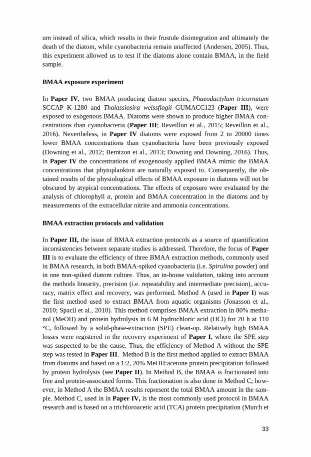
Liquid chromatography-mass spectrometry combines the versatility of LC, a tech-
nique used to separate a wide range of compounds (such as macromolecules, ionic
species, polar and high-molecular weight compounds) in a liquid mobile phase
(Dong, 2006) with the sensitivity of MS (Niessen, 2006). Thus, LC-MS is a highly
sensitive and selective method for identification of natural products in complex
mixtures (Fredenhagen et al., 2005; Furtado et al., 2007). Compounds are first sepa-
rated in the column of the LC system and later directed to the mass spectrometer by
a flow separator. They are then ionized in the flow separator and further separated in
the mass analyzer according to their (m/z) ratio (Fig. 6) (Niessen, 2006).
In this thesis, i.e. Paper I, II, III and IV, the instrument ultra-performance liquid
chromatography-electrospray tandem mass spectrometry (UPLC-ESI-MS/MS) was
employed and therefore, electrospray ionization (ESI) is used. ESI uses electrical
energy to assist the transfer of ions from solution into the gaseous phase before they
are subjected to MS analysis. This involves three consecutive steps; dispersal of a
fine spray of charge droplets, solvent evaporation and ion ejection from the highly
charged droplets tube, which is maintained at a high voltage relative to the wall of
the surrounding chamber (Ho et al., 2003). Tandem mass spectrometer consisting of
two quadrupole mass analyzers in series, i.e. Q1 and Q3, with a non-mass-resolving
quadrupole between them to act as a cell for collision-induced dissociation, i.e. Q2
(Pitt, 2009). In this analyzer, an ion (called precursor ion) from the first stage of MS
is selected and activated, to produce fragment ions, which are then analyzed in the
second stage of MS. In other words, a precursor ion is selected by mass-to-charge
ratio (m/z) on the first quadrupole (Q1), fragmented by collision-induced dissociation
in Q2, and fragment ions are detected by selected m/z in Q3 (Pitt, 2009). In the end,
the detected signal (fragment peak) is proportional to the amount of the analyte in
the sample (Fig. 6).
35
Figure 6. Scheme of the basic components of UPLC-ESI-MS/MS, adapted from Torre et al. (2015). Q1 is
labeled as 1st quadrupole mass, Q2 as collision cell and Q3 as 2nd quadrupole mass.
The proven superiority of UPLC-ESI-MS/MS over other methods has made it the
method of selection in BMAA research (Cohen, 2012; Faassen et al., 2012; Faassen,
2014). The main reason for using UPLC-ESI-MS/MS is due to the BMAA structural
isomers. These isomers have been one of the major sources of interferences on ana-
lytical methods, due to their analogous chromatographic retention properties and
identical mass. Issues that are overtaken with the UPLC-ESI-MS/MS, since precise
values of retention time, m/z of the precursor ion, pattern of the fragmentation spec-
trum after collision induced dissociation and the ratio between the intensities of
specific ions MRM transitions, needs to be reached in order to positively identify
BMAA (Faassen et al., 2012; Jiang et al., 2012). Detection of BMAA in the biolog-
ical samples was based on the quantifier fragment 459.1>119.08, also common to
the BMAA isomers, and the qualifier fragment 459.1>258.09. For the isomers N-(2-
aminoethyl) glycine (AEG) and 4-diaminobutyric acid (DAB) the qualifier frag-
ments were 459.1>214.1 and 459.1>188.1, respectively. Retention time and ratio of
fragments 119.08/258.09 was continuously controlled by comparing with the
BMAA standard in the corresponding matrix (see Paper III).
In Paper I, II, III and IV, an AQC-Tag Ultra Derivitization Kit (AQC) is used prior
to UPLC-ESI-MS/MS analysis. This method has been described as fast, reproduci-
ble and a sensitive amino acid quantitation method for biological samples, yielding a
~100% amino acid conversion (Armenta et al., 2010). AQC has been shown to
improve limits of detection (LOD) up to 3 times while reducing the analysis time 2.5
fold (Boogers et al., 2008). Derivatization is required to increase the mass of the
target amino acid, improving the Signal/Noise (S/N) ratio, doubly labelling it, which
makes the amino acid more hydrophobic, and improves the ESI response by increas-
ing the analyte surface activity. However, in BMAA research other derivatization
agents, e.g. propyl chloroformate (EZ:faast, Phenomenex, Torrance, USA); 9-
36
fluorenylmethyl chloroformate and dansyl chloride, have been used instead of AQC
(Esterhuizen and Downing, 2008; Scott et al., 2009; Esterhuizen et al., 2011;
Salomonsson et al., 2013). In addition, methods without derivatization steps, relying
exclusively on MS analysis with separation using hydrophilic interaction chroma-
tography have also been used in the detection of BMAA (Rosén and Hellenäs, 2008;
Faassen et al., 2009; Kruger et al., 2010; Faassen et al., 2012). One of the disad-
vantages of AQC is that it reacts with both primary and secondary amino groups, i.e.
reacts with all amino acids and other compounds containing amino groups (Cohen
and Michaud, 1993; Kaspar et al., 2009). Thus, in complex biological samples this
may hamper the derivatization efficiency, accurate separation and ultimately the
analysis of BMAA (Rosén and Hellenäs, 2008; Eriksson et al., 2009; Faassen et al.,
2012). Another disadvantage is that as a diamino acid, BMAA may react more slow-
ly compared to other amino acids. In addition, it might also be single or double
derivatized (Cohen, 2012). Quantification of single derivatized BMAA would be
less prone to interferences by other components present; however most published
studies quantify double-derivatized BMAA (Cohen, 2012). These disadvantages can
be overtaken if derivatization completion is assured through an adequate ratio of
total protein to derivative (i.e. the AQC reagent present in excess) (Eriksson et al.,
2009; Cohen, 2012); Paper III).
Statistical analysis
Parametric linear models were applied to the data collected in Paper I, in order to
detect any significant (p < 0.05) influence of the variables season of collection, fish
gender, total weight, and species on the response variable (i.e. BMAA concentration
in fish brain tissue) distribution. Supplementary Anova-Chi-Square tests were per-
formed to confirm linear model results. The data from BMAA concentration in fish
muscle tissue did not satisfy the parametric test assumptions, even after data trans-
formation, therefore a non-parametric Spearman’s rank correlation coefficient was
used to test whether the BMAA concentrations in fish muscle tissue were correlated
(p < 0.05) with the brain tissue. In Paper IV, analysis of variance between the con-
centrations of chlorophyll a, total protein, BMAA (both free and protein-associated),
ammonia and nitrite of a treated sample harvested at different time points and of
treated sample and controls harvested at the same time points was achieved with
one-way ANOVA. Two-way ANOVA analysis was performed in order to determine
interactions of the measured data with treatment and time. Statistical analysis of
Paper I and IV was carried out on R Statistical Software (Foundation for Statistical
Computing, Vienna, Austria). Statistics applied in Paper II, III were executed on
Microsoft Office Excel 2010 (Microsoft Corporation, WA, USA).
37
Results and discussion
BMAA bioaccumulation in aquatic ecosystems
The biomagnification of BMAA in an aquatic ecosystem was first reported in the
Baltic Sea, where the BMAA biosynthesis was suggested to be most likely done by
the cyanobacteria species Nodularia and Aphanizomenon. The BMAA produced by
the cyanobacteria was possibly transferred to marine animals that directly or indi-
rectly feed on them, i.e. zooplankton, fish, mussels and oysters, with highest BMAA
concentrations in bottom-feeding fish species as well as filter feeding mollusks
(Jonasson et al., 2010). The same trend of bioaccumulation was later found in South
Florida coastal waters in USA and in Lake Taihu in China (Brand et al., 2010;
Mondo et al., 2012; Jiao et al., 2014). At this point, the theory of BMAA bioaccu-
mulation in aquatic ecosystem had solid proofs; however, none of the studies used
enough replicates to allow a statistical approach, which was the main objective of
Paper I.
In Finjasjön, BMAA was detected in water samples (0.002 ± 0.001 to 0.006 ± 0.002
µg.g-1
DW) at comparable concentrations as in the Baltic Sea cyanobacteria (Table 1)
(Jonasson et al., 2010). However since Finjasjön suffer from both cyanobacteria and
diatom blooms year-round (Annadotter and Forssblad, 2011), the source of BMAA
is questionable (see Paper II).
In Paper I, the incidence of BMAA was higher in the plankti-benthivorous fish, i.e.
Abramis brama (bream) and Rutilus rutilus (roach), compared to the pelagic - pis-
civorous fish species, i.e. Esox lucius (pike) and Sander lucioperca (pike-perch)
(Fig.7). This is in accordance with previous studies, where BMAA accumulation
preferably was found in the benthic systems (Brand et al., 2010; Jonasson et al.,
2010; Mondo et al., 2012; Jiao et al., 2014). Thus, the results presented in Paper I
re-affirm the hypothesis of phytoplankton descending in the water column during the
bloom decline, making BMAA available for the bottom feeders, and/or that benthic
phytoplankton contain higher concentrations of BMAA than the pelagic species.
The continuous removal of A. brama and R. rutilus from Finjasjön, through the food
web manipulation scheme, resulted in a diet change, towards an increase in feeding
on benthic resources by the remaining population of this species (Persson and
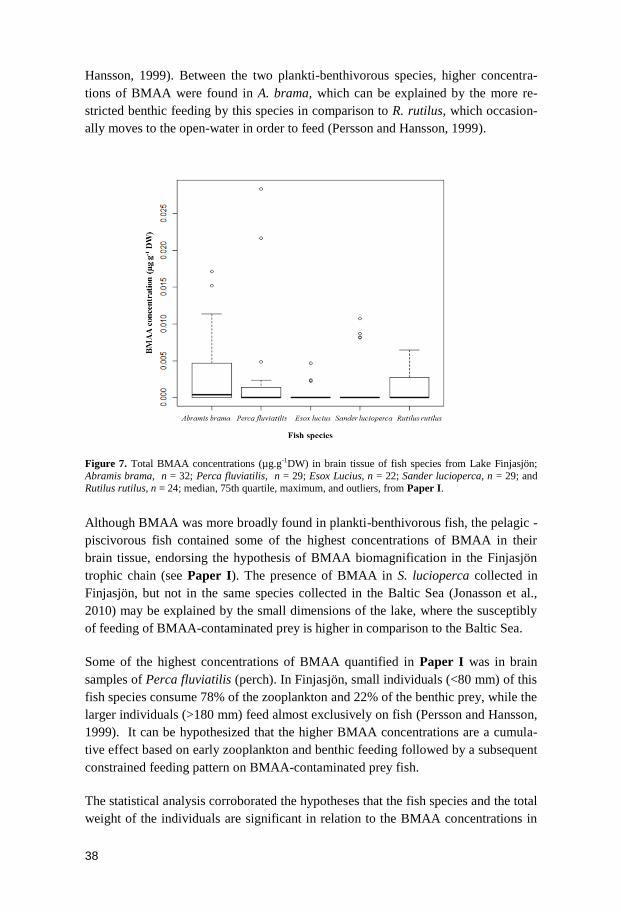
38
Hansson, 1999). Between the two plankti-benthivorous species, higher concentra-
tions of BMAA were found in A. brama, which can be explained by the more re-
stricted benthic feeding by this species in comparison to R. rutilus, which occasion-
ally moves to the open-water in order to feed (Persson and Hansson, 1999).
Figure 7. Total BMAA concentrations (µg.g-1DW) in brain tissue of fish species from Lake Finjasjön;
Abramis brama, n = 32; Perca fluviatilis, n = 29; Esox Lucius, n = 22; Sander lucioperca, n = 29; and
Rutilus rutilus, n = 24; median, 75th quartile, maximum, and outliers, from Paper I.
Although BMAA was more broadly found in plankti-benthivorous fish, the pelagic -
piscivorous fish contained some of the highest concentrations of BMAA in their
brain tissue, endorsing the hypothesis of BMAA biomagnification in the Finjasjön
trophic chain (see Paper I). The presence of BMAA in S. lucioperca collected in
Finjasjön, but not in the same species collected in the Baltic Sea (Jonasson et al.,
2010) may be explained by the small dimensions of the lake, where the susceptibly
of feeding of BMAA-contaminated prey is higher in comparison to the Baltic Sea.
Some of the highest concentrations of BMAA quantified in Paper I was in brain
samples of Perca fluviatilis (perch). In Finjasjön, small individuals (<80 mm) of this
fish species consume 78% of the zooplankton and 22% of the benthic prey, while the
larger individuals (>180 mm) feed almost exclusively on fish (Persson and Hansson,
1999). It can be hypothesized that the higher BMAA concentrations are a cumula-
tive effect based on early zooplankton and benthic feeding followed by a subsequent
constrained feeding pattern on BMAA-contaminated prey fish.
The statistical analysis corroborated the hypotheses that the fish species and the total
weight of the individuals are significant in relation to the BMAA concentrations in
39
the brain tissues. Therefore, a fish species that has a more uniform BMAA exposure
as they grow, i.e. continuous benthic feeding, will also have a higher proportion of
individuals with accumulated BMAA. Besides, in a system in which BMAA is regu-
larly available, a fish individual will most probably bioaccumulate BMAA through-
out its life, so heavier individuals, likely older, will contain higher concentrations of
BMAA (Paper I). No statistically significant relations were found between the con-
centrations of BMAA and the variables gender, and season of collection. However,
some tendencies were observed e.g. higher concentrations of BMAA were quanti-
fied in females compared to males and individuals caught during spring as compared
to the fall. Females are often larger than males (Cowx, 1983; Roff, 1983; Parker,
1992), thus, they may consume larger amounts of BMAA-contaminated prey. Most
of the individuals caught in spring were females, which may explain why higher
concentrations of BMAA were yielded during this season catchment.
As reported in previous studies (Brand et al., 2010; Jonasson et al., 2010; Mondo et
al., 2012; Jiao et al., 2014), and in Paper I, the accumulation of BMAA in the dif-
ferent fish tissues varied. In Paper I, BMAA was mostly found in the fish’s brain
tissue, sporadically detected in the muscle tissue and neither detected in liver nor in
the kidney tissue. Although not statistically tested, due to low number of positive
samples, the percentage of individuals containing BMAA in the muscle tissue were
higher in the plankti-benthivorous fish A. brama than in the other species. This
might be related to the almost exclusively benthivorous diet within this species.
Besides BMAA, mercury was also investigated and detected in the muscle tissue of
A. brama and S. lucioperca. Although the mercury concentrations were below the
EU-permitted level of 0.5mg.kg-1
WW (221/2002/EC), the synergetic neurotoxicity
of BMAA and mercury (Rush et al., 2012) may constitute a risk for human health.
There was no correlation between the concentrations of BMAA in muscle and in
brain, which may be explained by the low number of positive samples for the muscle
tissue.
With regard to the fish species exclusively caught during spring, BMAA could only
be detected in G. cernua, T. tinca and A. anguilla. In the first two species, BMAA
was quantifiable in both brain and muscle tissues, which agreed with their benthic
feeding (Bergman, 1991; Bergman and Greenberg, 1994). Furthermore, in A. anguil-
la, another bottom-dwelling fish species that instead feeds on the whole aquatic
fauna (Deelder, 1984), BMAA could only be detected in the brain tissue.
In general, BMAA concentrations detected in the fish tissues in Paper I (Fig.7) are
in agreement with the ones reported in the Baltic Sea study (Jonasson et al., 2010).
However, the concentrations of BMAA found in fish from Finjasjön are lower than
the concentrations stated in the study from Florida (58 ± 41 µg.g−1
WW in muscle,
588 ± 81 µg.g−1
WW in liver, and 1450 ± 687 µg.g-1
WW in kidney) (Mondo et al.,
40
2012); and in Lake Taihu, (concentration of BMAA in the muscle tissue was be-
tween 0.07 ± 0.021 and 35.91 ± 13.40 µg.g-1
DW) (Jiao et al., 2014). This discrepan-
cy is likely due to different methods used for extraction and quantification rather
than biological causes. Paper I and the study performed in the Baltic Sea followed
the same extraction method as well as analytical method (Table 1), whereas the
other studies did not (further discussed in Paper III).
The novel sources of BMAA – diatoms
In the study by Jonasson et al. (2010) high concentrations of BMAA were unexpect-
edly found in mussels harvested at the Swedish West Coast. This is an area where
blooms of cyanobacteria, the known BMAA producers, are less pronounced com-
pared to the typical bloom-forming areas in the Baltic Proper (Boesch et al., 2006).
At the time, it was suggested that benthic cyanobacteria might produce higher
amounts of BMAA than the bloom forming cyanobacteria, thus causing the high
BMAA concentrations detected in the mussels (Jonasson et al., 2010). However, in
Paper II, no BMAA was detected in the benthic cyanobacteria field samples from
the Swedish West Coast, containing the benthic species Calothrix sp. and Rivularia
sp. Instead, BMAA production was for the first time detected in a phytoplantonic
organism outside the cyanobacteria phylum, i.e. diatoms. The investigation of
BMAA production by this eukaryotic group was instigated since diatoms together
with dinoflagellates dominate the marine environment at the Swedish West Coast
(Granéli et al., 1989). BMAA production by the dinoflagellate Gymnodinium cate-
natum has also been confirmed (Lage et al., 2014).
In Paper II, BMAA was detected in all six diatom cultures tested, i.e. Achnanthes
sp., Navicula pelliculosa, Proboscia inermis, Thalassiosira sp. and two strains of
Skeletonema marinoi. BMAA quantification performed in the two strains of Skele-
tonema marinoi and in Thalassiosira sp. showed protein-associated BMAA concen-
trations in a range from 0.0011 ± 0.0004 and 0.00328 ± 0.0025 µg.g-1
DW. Addition-
ally, unlike cyanobacteria cultures, the cultures of diatoms were shown to produce
BMAA in successive generations.
To ensure that BMAA was not only produced by diatoms in culture, field samples
from the Swedish West Coast with mixed-species containing both cyanobacteria and
diatoms, were tested as well. Protein associated BMAA detected in the field sample
comprising the cyanobacteria Leptolyngbya sp. and diatoms belonging to the genus
Navicula was 0.0276 ± 0.011 µg.g-1
DW. However, laboratory grown cultures of
Leptolyngbya PCC73110 has been reported to contain 0.73 µg.g-1
DW BMAA (Jiang
et al., 2013), thus there was the possibility that the BMAA detected in the mixed
field sample might be due to the cyanobacterial BMAA production. A treatment
with germanium dioxide of the mixed-species sample eliminated the diatoms, while
leaving the cyanobacteria intact. This allowed us to compare the BMAA concentra-
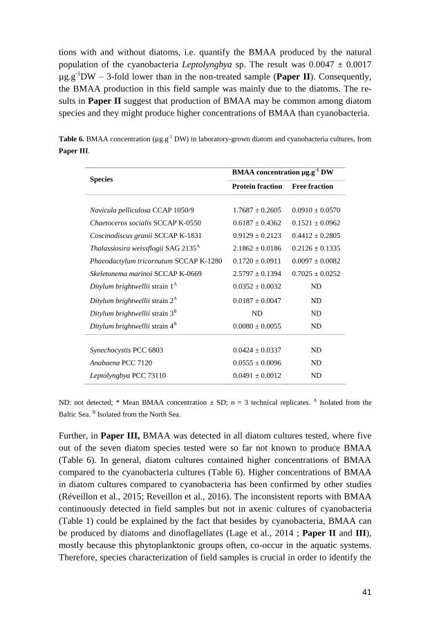
41
tions with and without diatoms, i.e. quantify the BMAA produced by the natural
population of the cyanobacteria Leptolyngbya sp. The result was 0.0047 ± 0.0017
µg.g-1
DW – 3-fold lower than in the non-treated sample (Paper II). Consequently,
the BMAA production in this field sample was mainly due to the diatoms. The re-
sults in Paper II suggest that production of BMAA may be common among diatom
species and they might produce higher concentrations of BMAA than cyanobacteria.
Table 6. BMAA concentration (µg.g-1 DW) in laboratory-grown diatom and cyanobacteria cultures, from
Paper III.
Species BMAA concentration µg.g-1 DW
Protein fraction Free fraction
Navicula pelliculosa CCAP 1050/9 1.7687 ± 0.2605 0.0910 ± 0.0570
Chaetoceros socialis SCCAP K-0550 0.6187 ± 0.4362 0.1521 ± 0.0962
Coscinodiscus granii SCCAP K-1831 0.9129 ± 0.2123 0.4412 ± 0.2805
Thalassiosira weissflogii SAG 2135A 2.1862 ± 0.0186 0.2126 ± 0.1335
Phaeodactylum tricornutum SCCAP K-1280 0.1720 ± 0.0911 0.0097 ± 0.0082
Skeletonema marinoi SCCAP K-0669 2.5797 ± 0.1394 0.7025 ± 0.0252
Ditylum brightwellii strain 1A 0.0352 ± 0.0032 ND
Ditylum brightwellii strain 2A 0.0187 ± 0.0047 ND
Ditylum brightwellii strain 3B ND ND
Ditylum brightwellii strain 4B 0.0080 ± 0.0055 ND
Synechocystis PCC 6803 0.0424 ± 0.0337 ND
Anabaena PCC 7120 0.0555 ± 0.0096 ND
Leptolyngbya PCC 73110 0.0491 ± 0.0012 ND
ND: not detected; * Mean BMAA concentration ± SD; n = 3 technical replicates. A Isolated from the
Baltic Sea. B Isolated from the North Sea.
Further, in Paper III, BMAA was detected in all diatom cultures tested, where five
out of the seven diatom species tested were so far not known to produce BMAA
(Table 6). In general, diatom cultures contained higher concentrations of BMAA
compared to the cyanobacteria cultures (Table 6). Higher concentrations of BMAA
in diatom cultures compared to cyanobacteria has been confirmed by other studies
(Réveillon et al., 2015; Reveillon et al., 2016). The inconsistent reports with BMAA
continuously detected in field samples but not in axenic cultures of cyanobacteria
(Table 1) could be explained by the fact that besides by cyanobacteria, BMAA can
be produced by diatoms and dinoflagellates (Lage et al., 2014);(Paper II and III),
mostly because this phytoplanktonic groups often, co-occur in the aquatic systems.
Therefore, species characterization of field samples is crucial in order to identify the
42
BMAA producers in a certain habitat. In addition, in Paper III, most of quantified
BMAA is present in the protein-associated form in both cyanobacteria and diatom
cultures.
BMAA extraction controversy – method validation
As previously mentioned, the varying methods is often suggested to be the main
source for inconsistencies between BMAA concentration found in different studies
(Faassen, 2014), even when the selective LC-MS/MS system has been used (Faassen
et al., 2009; Jonasson et al., 2010; Kruger et al., 2010; Faassen et al., 2012;
Salomonsson et al., 2013; Al-Sammak et al., 2014; Jiao et al., 2014; Reveillon et al.,
2014; Réveillon et al., 2015; Reveillon et al., 2016); Paper I and II). Therefore, in
Paper III, three of the most commonly applied BMAA extraction methods were
compared through an in-house validation experiment.
Method A, is based on an 80% MeOH extraction couple with a cation-exchange
solid-phase extraction (SPE) procedure (Jonasson et al., 2010; Spacil et al., 2010),
and was the method used in Paper I. In Paper I, a recovery experiment with D3-
BMAA, reveal a relatively high D3-BMAA loss in brain and muscle fish tissues
during the SPE procedure. Prior to the SPE step, D3-BMAA was added to the fish
blanks and fish samples. The recovery percentages of D3-BMAA in the blank sam-
ples was 54.9 ± 2.0% for the brain and 14.3 ± 3.5% for the muscle tissues and for
the fish samples the recoveries were 34.3 ± 6.2% for the brain and 10.7 ± 7.5% for
the muscle tissues. As a result, the SPE step was suggested as the major cause of the
low D3-BMAA recoveries. Considering this, an adaptation of Method A, which
consisted in eliminating the SPE step from the protocol, was also evaluated in Paper
III. The other methods investigated in Paper III were the 1:2, 20% MeOH: acetone
protein precipitation (Method B) used in Paper II, and the most commonly used
protocol in BMAA research, the TCA protein precipitation method (Method C)
(Murch et al., 2004a; Faassen, 2014), which were used in the study described in
Paper IV.
In the linearity test, both protein precipitation methods (B and C) display good linear
fit, i.e. r2 values of 0.99 in both protein-associated as well as the free BMAA fraction
(Paper III). Method A, with the SPE step, produced sufficient, though not ideal,
linearity, r2 of 0.97. This linearity was generated after the removal of the highest
BMAA concentration point (100 ng.mL-1
) which skewed the standard curve. How-
ever, Method A, without the SPE step, did not show any linearity, r2 of 0.74. Lack of
linearity will affect all other validation parameters, and is therefore a reason to reject
this extraction protocol. Thus, the accuracy and precision was not further investigat-
ed for this method.
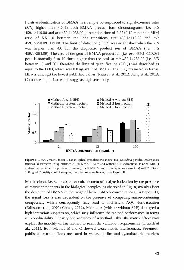
43
Positive identification of BMAA in a sample corresponded to signal-to-noise ratio
(S/N) higher than 4.0 in both BMAA product ions chromatograms, i.e. m/z
459.1>119.08 and m/z 459.1>258.09, a retention time of 2.85±0.12 min and a SRM
ratio of 5.5±1.0 between the ions transitions m/z 459.1>119.08 and m/z
459.1>258.09. 119.08. The limit of detection (LOD) was established when the S/N
was higher than 4.0 for the diagnostic product ion of BMAA (i.e. m/z
459.1>258.09). The area of the general BMAA product ion (i.e. m/z 459.1>119.08)
peak is normally 3 to 10 times higher than the peak at m/z 459.1>258.09 (i.e. S/N
between 10 and 30), therefore the limit of quantification (LOQ) was described as
equal to the LOD, which was 0.8 ng∙ mL-1
of BMAA. The LOQ presented in Paper
III was amongst the lowest published values (Faassen et al., 2012; Jiang et al., 2013;
Combes et al., 2014), which suggests high sensitivity.
Figure 8. BMAA matrix factor ± SD in spiked cyanobacteria matrix (i.e. Spirulina powder, Arthrospira
fusiformis) extracted using methods A (80% MeOH with and without SPE extraction), B (20% MeOH
and acetone protein-precipitation extraction), and C (TCA protein-precipitation extraction) with 2, 13 and
100 ng.mL–1 quality control samples; n = 3 technical replicates, from Paper III.
Matrix effect, i.e. suppression or enhancement of analyte ionization by the presence
of matrix components in the biological samples, as observed in Fig. 8, mainly affect
the detection of BMAA in the range of lower BMAA concentrations. In Paper III,
the signal loss is also dependent on the presence of competing amine-containing
compounds, which consequently may lead to inefficient AQC derivatization
(Eriksson et al., 2009; Cohen, 2012). Method A (with or without SPE) displayed a
high ionization suppression, which may influence the method performance in terms
of reproducibility, linearity and accuracy of a method - thus the matrix effect may
explain the inability of this method to reach the validation requirements (Trufelli et
al., 2011). Both Method B and C showed weak matrix interferences. Foremost-
published matrix effects measured in water, biofilm and cyanobacteria matrices
0
0.5
1
1.5
2
2 13 100
Matr
ix F
act
or
BMAA concentration (ng.mL–1)
Method A with SPE Method A without SPEMethod B protein fraction Method B free fractionMethod C protein fraction Method C free fraction
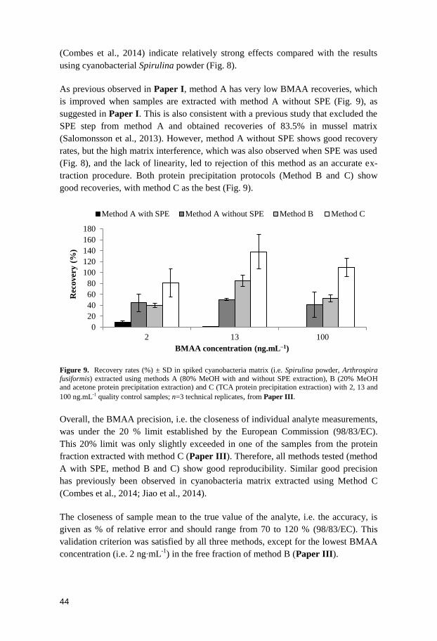
44
(Combes et al., 2014) indicate relatively strong effects compared with the results
using cyanobacterial Spirulina powder (Fig. 8).
As previous observed in Paper I, method A has very low BMAA recoveries, which
is improved when samples are extracted with method A without SPE (Fig. 9), as
suggested in Paper I. This is also consistent with a previous study that excluded the
SPE step from method A and obtained recoveries of 83.5% in mussel matrix
(Salomonsson et al., 2013). However, method A without SPE shows good recovery
rates, but the high matrix interference, which was also observed when SPE was used
(Fig. 8), and the lack of linearity, led to rejection of this method as an accurate ex-
traction procedure. Both protein precipitation protocols (Method B and C) show
good recoveries, with method C as the best (Fig. 9).
Figure 9. Recovery rates (%) ± SD in spiked cyanobacteria matrix (i.e. Spirulina powder, Arthrospira
fusiformis) extracted using methods A (80% MeOH with and without SPE extraction), B (20% MeOH and acetone protein precipitation extraction) and C (TCA protein precipitation extraction) with 2, 13 and
100 ng.mL-1 quality control samples; n=3 technical replicates, from Paper III.
Overall, the BMAA precision, i.e. the closeness of individual analyte measurements,
was under the 20 % limit established by the European Commission (98/83/EC).
This 20% limit was only slightly exceeded in one of the samples from the protein
fraction extracted with method C (Paper III). Therefore, all methods tested (method
A with SPE, method B and C) show good reproducibility. Similar good precision
has previously been observed in cyanobacteria matrix extracted using Method C
(Combes et al., 2014; Jiao et al., 2014).
The closeness of sample mean to the true value of the analyte, i.e. the accuracy, is
given as % of relative error and should range from 70 to 120 % (98/83/EC). This
validation criterion was satisfied by all three methods, except for the lowest BMAA
concentration (i.e. 2 ng∙mL-1
) in the free fraction of method B (Paper III).
0
20
40
60
80
100
120
140
160
180
2 13 100
Rec
over
y (
%)
BMAA concentration (ng.mL–1)
Method A with SPE Method A without SPE Method B Method C
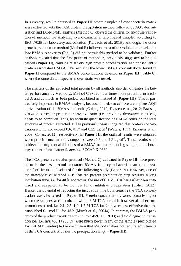
45
In summary, results obtained in Paper III where samples of cyanobacteria matrix
were extracted with the TCA protein precipitation method followed by AQC derivat-
ization and LC-MS/MS analysis (Method C) obeyed the criteria for in-house valida-
tion of methods for analyzing cyanotoxins in environmental samples according to
ISO 17025 for laboratory accreditation (Kaloudis et al., 2015). Although, the other
protein precipitation method (Method B) followed most of the validation criteria, the
low BMAA recoveries (Fig. 9) did not permit this method to be validated. Further
analysis revealed that the first pellet of method B, previously suggested to be dis-
carded (Paper II), contains relatively high protein concentration, and consequently
protein associated BMAA. This explains the lower BMAA concentrations found in
Paper II compared to the BMAA concentrations detected in Paper III (Table 6),
where the same diatom species and/or strain was tested.
The analysis of the extracted total protein by all methods also demonstrates the bet-
ter performance by Method C. Method C extract four times more protein than meth-
od A and as much as both pellets combined in method B (Paper III). This is par-
ticularly important in BMAA analysis, because in order to achieve a complete AQC
derivatization of the BMAA molecule (Cohen, 2012; Faassen et al., 2012; Faassen,
2014), a particular protein-to-derivative ratio (i.e. providing derivative in excess)
needs to be complied. Thus, an accurate quantification of BMAA relies on the total
amounts of protein extracted. It has previously been suggested that protein concen-
tration should not exceed 0.6, 0.17 and 0.25 µg∙µl-1
(Waters, 1993; Eriksson et al.,
2009; Cohen, 2012), respectively. In Paper III, the optimal results were obtained
when protein concentrations ranged between 0.3 and 2.3 µg∙µl-1
. These results were
achieved through serial dilutions of a BMAA natural containing sample, i.e. labora-
tory culture of the diatom S. marinoi SCCAP K-0669.
The TCA protein extraction protocol (Method C) validated in Paper III, have prov-
en to be the best method to extract BMAA from cyanobacteria matrix, and was
therefore the method selected for the following study (Paper IV). However, one of
the drawbacks of Method C is that the protein precipitation step requires a long
incubation time, i.e. for 48 h. Moreover, the use of 0.1 M TCA has earlier been criti-
cized and suggested to be too low for quantitative precipitation (Cohen, 2012).
Hence, the potential of reducing the incubation time by increasing the TCA concen-
tration was also tested in Paper III. Protein concentrations were, actually higher
when the samples were incubated with 0.2 M TCA for 24 h, however all other con-
centrations tested, i.e. 0.1, 0.5, 1.0, 1.5 M TCA for 24 h were less effective than the
established 0.1 mol∙L-1
for 48 h (Murch et al., 2004a). In contrast, the BMAA peak
areas of the product transition ion (i.e. m/z 459.1> 119.08) and the diagnostic transi-
tion ion (i.e. m/z 459.1>258.09) were much lower in any of the samples precipitated
for just 24 h, leading to the conclusion that Method C does not require adjustments
of the TCA concentration nor the precipitation length (Paper III).
46
Physiological role of BMAA in diatoms – first steps towards understanding
In Paper III, ten diatom strains - related in various degrees ranging from the same
species to different classes - as well as three cyanobacteria species known to produce
BMAA (Cox et al., 2005; Esterhuizen and Downing, 2008; Spacil et al., 2010;
Berntzon et al., 2013; Jiang et al., 2013) were analyzed for free and protein-
associated BMAA. Even though the in-house validated method was used, the
BMAA concentration in the protein-associated fraction showed a broad range, from
0.0080 to 2.5797 µg∙g-1
DW (Table 6). This supports the hypothesis of variability in
BMAA concentrations may be due to biological reasons such as the phytoplankton
group (i.e. cyanobacteria, diatom or dinoflagellate), strain and growing conditions
(Banack et al., 2010a; Banack et al., 2010b); Paper II).
So far, the biosynthesis, metabolism and function of BMAA in diatoms are still
unknown as well as the environmental factors that can induce or reduce its produc-
tion. As mentioned above, studies in cyanobacteria have shown a negative correla-
tion of BMAA production with nitrate and ammonium availability (Downing et al.,
2011; Scott et al., 2014). Moreover, experiments with cyanobacteria cultures sup-
plemented with BMAA had reveal signs of nitrogen starvation (Downing et al.,
2012; Berntzon et al., 2013) and proposed a mechanism of BMAA metabolism via
the GOGAT cycle (Downing and Downing, 2016). However, in the former study by
Downing and Downing (2016) cyanobacteria were exposed to 100 µM BMAA and
in the other two previous studies the BMAA concentrations added ranged from 0.05
till 42 µM (Downing et al., 2011) and from 0.01 till 50 µM (Berntzon et al., 2013).
Since, the natural BMAA concentrations in cyanobacteria range between the low
µg/g and ng/g (Table 1) some doubts may be inferred.
The variable natural production of BMAA by cyanobacteria and diatoms has also
been verified in Paper III (Table 6). Therefore, in the study described in Paper IV,
the naturally occurring concentrations of BMAA were taken into consideration when
the diatom species Phaeodactylum tricornutum and Thalassiosira weissflogii were
exposed to 0.005, 0.05 and 0.5 µM BMAA.
The exogenously applied BMAA was rapidly taken up in a concentration and time
dependent manner in the free BMAA fraction of P. tricornutum and T. weissflogii, F
(12, 40) =3.86, p<0.001 and F (12, 40) = 8.15, p<0.001, respectively (see Paper
IV). Moreover, most of the BMAA detected in the diatom cultures was in the free
form, as previously observed in cyanobacteria (Downing et al., 2012; Berntzon et
al., 2013). However, the concentrations of free BMAA in the T. weissflogii cultures
exposed to 0.5 µM BMAA were significantly lower than in the P. tricornutum cul-
tures (F (1, 13)= 17.75, p<0.01). After a 24 h hypothetical uptake, in P. tricornutum
cultures, the free BMAA concentrations were reduced during the following days,
suggesting that BMAA was metabolized (see Paper IV), as also previously ob-
served in cyanobacteria (Downing et al., 2012). In T. weissflogii, BMAA was also
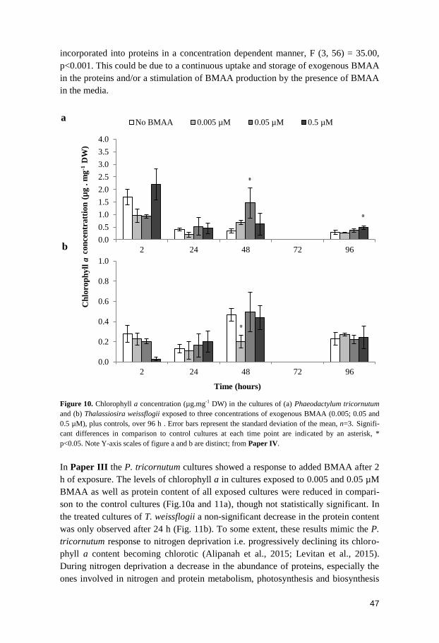
47
incorporated into proteins in a concentration dependent manner, F (3, 56) = 35.00,
p<0.001. This could be due to a continuous uptake and storage of exogenous BMAA
in the proteins and/or a stimulation of BMAA production by the presence of BMAA
in the media.
Figure 10. Chlorophyll a concentration (µg.mg-1 DW) in the cultures of (a) Phaeodactylum tricornutum
and (b) Thalassiosira weissflogii exposed to three concentrations of exogenous BMAA (0.005; 0.05 and
0.5 µM), plus controls, over 96 h . Error bars represent the standard deviation of the mean, n=3. Signifi-
cant differences in comparison to control cultures at each time point are indicated by an asterisk, *
p<0.05. Note Y-axis scales of figure a and b are distinct; from Paper IV.
In Paper III the P. tricornutum cultures showed a response to added BMAA after 2
h of exposure. The levels of chlorophyll a in cultures exposed to 0.005 and 0.05 µM
BMAA as well as protein content of all exposed cultures were reduced in compari-
son to the control cultures (Fig.10a and 11a), though not statistically significant. In
the treated cultures of T. weissflogii a non-significant decrease in the protein content
was only observed after 24 h (Fig. 11b). To some extent, these results mimic the P.
tricornutum response to nitrogen deprivation i.e. progressively declining its chloro-
phyll a content becoming chlorotic (Alipanah et al., 2015; Levitan et al., 2015).
During nitrogen deprivation a decrease in the abundance of proteins, especially the
ones involved in nitrogen and protein metabolism, photosynthesis and biosynthesis
0.0
0.5
1.0
1.5
2.0
2.5
3.0
3.5
4.0
2 24 48 72 96con
cen
tratt
ion
(µ
g .
mg
-1 D
W)
No BMAA 0.005 µM 0.05 µM 0.5 µM
0.0
0.2
0.4
0.6
0.8
1.0
2 24 48 72 96
Ch
loro
ph
yll
a !
Time (hours)
b
a
*
*
*
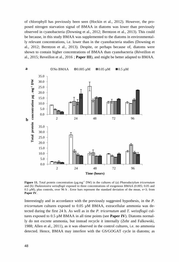
48
of chlorophyll has previously been seen (Hockin et al., 2012). However, the pro-
posed nitrogen starvation signal of BMAA in diatoms was lower than previously
observed in cyanobacteria (Downing et al., 2012; Berntzon et al., 2013). This could
be because, in this study BMAA was supplemented to the diatoms in environmental-
ly relevant concentrations, i.e. lower than in the cyanobacteria studies (Downing et
al., 2012; Berntzon et al., 2013). Despite, or perhaps because of, diatoms were
shown to contain higher concentrations of BMAA than cyanobacteria (Réveillon et
al., 2015; Reveillon et al., 2016); Paper III), and might be better adapted to BMAA.
Figure 11. Total protein concentration (µg.mg-1 DW) in the cultures of (a) Phaeodactylum tricornutum
and (b) Thalassiosira weissflogii exposed to three concentrations of exogenous BMAA (0.005; 0.05 and
0.5 µM), plus controls, over 96 h . Error bars represent the standard deviation of the mean, n=3; from Paper IV.
Interestingly and in accordance with the previously suggested hypothesis, in the P.
tricornutum cultures exposed to 0.05 µM BMAA, extracellular ammonia was de-
tected during the first 24 h. As well as in the P. tricornutum and T. weissflogii cul-
tures exposed to 0.5 µM BMAA in all time points (see Paper IV). Diatoms normal-
ly do not excrete ammonia, but instead recycle it internally (Zehr and Falkowski,
1988; Allen et al., 2011), as it was observed in the control cultures, i.e. no ammonia
detected. Hence, BMAA may interfere with the GS/GOGAT cycle in diatoms; as
0.0
5.0
10.0
15.0
20.0
25.0
30.0
35.0
2 24 48 72 96
Tota
l p
rote
in
Time (hours)
0.0
5.0
10.0
15.0
20.0
25.0
30.0
35.0
2 24 48 72 96con
cen
trati
on
µg
. m
g-1
DW
No BMAA 0.005 µM 0.05 µM 0.5 µM
b
a
49
previously been suggested for cyanobacteria (Downing and Downing, 2016).
BMAA may inhibit the synthesis and/or stimulate the degradation of glutamine,
subsequently induce glutamate release and thus signaling nitrogen depletion. As in
cyanobacteria (Berntzon et al., 2013), an imbalance of glutamate/glutamine levels
may also signal nitrogen starvation in diatoms. This since once entering the cell,
nitrate might be reduced to nitrite and successively to ammonium (Allen et al., 2005;
Bowler et al., 2010). Ammonium is assimilated to amino acids and other nitroge-
nous compounds by the joint action of GS/GOGAT (Zehr and Falkowski, 1988;
Takabayashi et al., 2005). If GS/GOGAT is somehow disrupted, ammonium will
start to intracellularly accumulate and possibly be excreted by the cell, as observed
in the cultures of both diatom species exposed to 0.5 µM BMAA, and later re-
assimilated. The re-assimilation is possible because diatoms, as cyanobacteria, pos-
sess a complete urea cycle (Armbrust, 2009). Accordingly, the concentrations of
ammonia of both species cultures at the 96 h time point were at their minimal, in
comparison with previous time points. The lack of glutamine could had led to the
up-regulation of carbamoyl phosphate synthase, and then to the uptake of the availa-
ble ammonia, which will enter the ornithine-urea cycle (Allen et al., 2011) and lead
to the recovery of the diatom cells. Accordingly, the chlorophyll concentrations at
the last experiment day were similar, though low, between treated and control cul-
tures of both species (Fig.10). Moreover, the protein content of treated and control
cultures was rather alike from 48 h to 96 h after BMAA exposure (Fig.11). There-
fore, Paper IV suggests that the physiological role of BMAA might be, together
with ammonia, to regulate/achieve a fast response of the diatoms to the rapid influx
of nitrogen in an environment with constant nitrogen fluctuations.
50
Conclusions
This thesis contributed with knowledge in the following topics:
BMAA bioaccumulation in aquatic ecosystems
Production and biotransfer of BMAA occurs not only in the brackish and marine
ecosystems, but also in the limnic ecosystem, as shown here in Lake Finjasjön.
BMAA concentrations detected in fish from Finjasjön are equivalent to those previ-
ously reported in the Baltic Sea.
There was a positive correlation between concentrations of BMAA in fish brains and
the factors species/feeding pattern and fish total weight.
Higher BMAA concentrations were detected in the brain of plankti-benthivorous
fish with higher weight.
BMAA is heterogeneously distributed between fish tissues. It was found in 29% of
the brain samples and in higher concentrations compared to in muscle tissue, where
it was found in 16% of the samples and it was not at all detected in the liver or kid-
ney.
The finding of both BMAA and mercury in muscle tissues of Abramis brama
(bream) and Sander lucioperca (pike-perch) raises some concern in view of report-
ed synergistic effects of these two toxic compounds (albeit at much higher concen-
trations than detected in this thesis).
The novel sources of BMAA
The previous suggestion that benthic cyanobacteria might contain BMAA was not
confirmed, but instead BMAA was for the first time detected in eukaryotic organ-
isms, i.e. diatoms.
BMAA was detected both in field samples containing diatoms as well as in laborato-
ry grown axenic diatom cultures.
51
Diatoms contain BMAA in higher concentrations than cyanobacteria and unlike in
cyanobacteria, the toxin is found through consecutive diatom generations – which
suggest that diatoms might be the main BMAA producers in aquatic ecosystems.
The BMAA concentrations were shown to be variable between diatom species and
strains.
Evaluation of BMAA extraction methods
The choice of extraction method was shown to be a cause of variation in measured
concentrations of BMAA.
The method starting with extraction in 80% MeOH (used in Paper I) resulted in
major losses at the SPE clean-up step, but without the SPE step, the linearity criteri-
on was not fulfilled.
The method starting with extraction in 20% MeOH followed by acetone precipita-
tion of proteins (used in Paper II) gave suitable linearity, accuracy and precision
however, the recovery did not achieve validation criteria.
The first pellet of the “20% MeOH:acetone protein precipitation protocol” was
found to contain high protein levels, and consequently protein-associated BMAA.
Thus, if applying this method, this first pellet should not be discarded.
The method involving protein precipitation using TCA (later applied in Paper III
and IV) was shown to meet in-house validation criteria.
Furthermore, the method evaluation showed that the TCA method does not require
modified TCA concentrations or incubation time adjustments, to yield optimal pro-
tein precipitation.
The concentration of total protein, i.e. the ratio of protein-to-derivative (providing
derivative in excess), was confirmed as essential to achieve a complete AQC deri-
vatization of BMAA, and therefore accurate detection and quantification.
Physiological role of BMAA in diatoms
The diatoms Phaeodactylum tricornutum and Thalassiosira weissflogii were found
to react to exogenous BMAA added at the low concentrations of 0.005, 0.05 and 0.5
M.
At 2 h, P. tricornutum had lower concentrations of chlorophyll a in the cultures
where 0.005 and 0.05 µM BMAA was added and lower concentrations of protein in
all treated cultures, in comparison to control.
52
All T. weissflogii cultures supplemented with BMAA had lower concentrations of
protein at 24 h, in comparison to control.
Both diatom species excreted ammonium (measured as ammonia) in the culture
media starting at 2 h after the addition of BMAA at 0.5 M. In both species, the
ammonia concentration was lower at the later sampling time point (96 h).
BMAA added to the cultures of P. tricornutum and T. weissflogii was taken up in a
concentration and time dependent manner in the free BMAA fractions and the
BMAA protein fraction of T. weissflogii.
The results suggest that BMAA may interfere with the diatoms GS/GOGAT cycle
and that it may be closely associated with the cellular nitrogen balance in diatoms;
thus, BMAA together with ammonium probably contributes to the fast response of
diatoms to environmental nitrogen fluctuations.
53
Future research suggestions
Based on the results in this thesis, the following investigations are suggested:
BMAA bioaccumulation in aquatic ecosystems
In order to find out which group or species of phytoplankton are the major producers
of BMAA in freshwater, brackish and marine ecosystems, phytoplankton samples
should be sampled over a year (covering all seasons) in these habitats. This sampling
should be followed by species identification and quantification of species abundance
and total BMAA. A statistical approach could then provide correlation indications
between species and/or phytoplankton group and BMAA production.
A field bioaccumulation experiment comprising important commercial fish species
and their preys and predators should be performed. So the percentage of BMAA
retained in each trophic level could be quantified. A mesocosm study should also be
carried out. Like this, both studies will complement each other and give reliable data
that could later be used in modelling BMAA accumulation in aquatic ecosystems.
Hence, the risk of consumption of BMAA-contaminated fish by human consumers
could be predicted.
The novel sources of BMAA
In view of the recent findings of BMAA producers among the diatom group, further
species of this group should be tested for BMAA production. Therefore, investigate
if as suggested for the cyanobacteria phylum, BMAA is also produced by almost all
species of the diatoms phylum (i.e. Bacillariophyta).
BMAA extraction controversy
An inter-laboratory validation of both extraction and analytical methods for BMAA
should be promoted. Thus, if the scientific community working in BMAA research
reached a consensus, all future BMAA studies could be directly compared.
Physiological role of BMAA in diatoms
Monitor the effect of exogenous BMAA in diatoms at different molecular levels by a
combined transcriptional and metabolite analysis, and thus come closer to an under-
standing of the physiological role of BMAA in diatoms.
54
Financial support
This work has been supported by funding from The Swedish Research Council For-
mas (Grant nr 2010-836).
The foundation BalticSea2020 and Stiftelsen Olle Engkvist Byggmästare funded the
Acquity UPLC coupled to Xevo-TQ-MS (Waters) via the MiMeBS program grant
awarded to Professor Birgitta Bergman.
The Department of Ecology, Environment and Plant Sciences – Stockholm Univer-
sity, Lillie and Ernst Åqvists stiftelse and Forbio – Research School in Biosystemat-
ics had provided additional financial support in the form of travel grants for courses
and international conferences attendance.
55
Acknowledgements
I would like to express my gratitude to all those who have made this thesis possible.
First and foremost, I would like to thank my supervisor Dr. Sara Rydberg for not
only giving me the opportunity to work with her, in this fascinating subject, but also
for being a great mentor. Thank you for teaching me so much and specially how to
become an independent researcher and for your confidence in me. Also thank you
for your guidance through this process; our discussions of ideas and your feedback,
it has been absolutely incomparable. Certainly I can't forget to thank you for giving
me the possibility to travel to such unforgettable places and for our constant laugh-
ing. Dr. Ulla Rasmussen, my enthusiastic co-supervisor, thanks for always being
available to help and for your support and encouragement. My gratitude goes to the
former members of the BMAA research group, Dr. Johan Eriksson and Dr. Lotta
Berntzon, for the fruitful discussions and invaluable help. Also would like to
acknowledge all former and present collaborators, for the sharing of knowledge and
for your helpful comments to my work. To all my former students, especially to
Linnea Ström, thank you for challenging me and making this journey so much more
fun!
I’m also grateful to the Department of Ecology, Environment and Plant Sciences,
Stockholm University and all its members, for the friendly and intellectually excit-
ing environment. I’m particularly thankful to the members of the Plant Physiology
section which follow and actively participated in my evolution as a PhD student.
Professor Lisbeth Jonsson and Professor Monika Winder thank you for carefully
revising my thesis and for your comments, corrections and suggestions. I would also
like to thank the opponent Professor Hanna Mazur-Marzec, committee members
Professor Ann-Sofi Rehnstam-Holm, Professor Ingvar Brandt, Professor Mats Björk
and reserve member Professor Elena Gorokhova for investing time reading and
providing your feedback on my PhD thesis. I’m honored that you have accepted to
be on my evaluation committee.
And because a PhD is not just breakthroughs but also brain breaks and break free
I’m obliged to thank the DEEP rainbow unicorn family, inhabiting the boreal forest,
seagrass meadow, ice cover, open sea and/or leaving in lab closure. Thanks to you I
was able to finish my PhD and still be a sane and happy creature. To Galochas, so
far but yet so close, thank you for your amazing friendship; it has been years now
since we started conquering the world together (Naaaants een-vwen-yaaaaaaa ma-

56
ba-gee-chi-ba-va…). To my stoical human, thank you for coping with me, especially
in the final stages of this thesis, which made my quirkiness and grumpiness reach
outrageous levels. I hope, besides many other things, to keep amusingly disagreeing
with you far into the future! Lastly, I want to thank my parents for their uncondi-
tional support and love. Aos meus pais, obrigada por sempre acreditaram em mim,
me acharem a melhor, mesmo não o sendo. Por isso, nunca deixei ou deixarei de
tentar, não ser a melhor, mas fazer o melhor de mim. Obrigada!
57
References
Al-Sammak, M.A., Hoagland, K.D., Cassada, D., Snow, D.D., 2014. Co-occurrence of the Cyanotoxins BMAA,
DABA and Anatoxin-a in Nebraska Reservoirs, Fish, and Aquatic Plants. Toxins 6, 488-508.
Alipanah, L., Rohloff, J., Winge, P., Bones, A.M., Brembu, T., 2015. Whole-cell response to nitrogen deprivation
in the diatom Phaeodactylum tricornutum. Journal of Experimental Botany 66, 6281-6296.
Allen, A.E., Dupont, C.L., Oborník, M., Horák, A., Nunes-Nesi, A., et al., 2011. Evolution and metabolic
significance of the urea cycle in photosynthetic diatoms. Nature 473, 203-207.
Allen, A.E., Ward, B.B., Song, B.K., 2005. Characterization of diatom (Bacillariophyceae) nitrate reductase genes
and their detection in marine phytoplankton communities. Journal of Phycology 41, 95-104.
Allen, A.E., Vardi, A., Bowler, C., 2006. An ecological and evolutionary context for integrated nitrogen
metabolism and related signaling pathways in marine diatoms. Current Opinion in Plant Biology 9, 264-
273.
Andersen, R.A., 2005. Algal culturing techniques. Elsevier Academic Press, London, UK.
Annadotter, H., Cronberg, G., Aagren, R., Lundstedt, B., Nilsson, P.-Ǻ., et al., 1999. Multiple techniques for lake
restoration, in: Harper, D.M., Brierley, B., Ferguson, A.J.D., Phillips, G. (Eds.), The Ecological Bases for
Lake and Reservoir Management: Proceedings of the Ecological Bases for Management of Lakes and
Reservoirs Symposium, held 19–22 March 1996, Leicester, United Kingdom. Springer Netherlands,
Dordrecht, pp. 77-85.
Annadotter, H., Cronberg, G., Jönsson, L., 1993. The toxicity of blue-green algae (cyanobacteria) in Lake
Finjasjön, Sweden in relation to Environmental Factors - A five-year study., in: Giussani, G., Callieri, C.
(Eds.), 5th International Conference on the Conservation and Management of Lakes, “Strategies for lake
ecosystems beyond 2000”. Instituto Italiano di Idrobiologia, Stresa, Italy, pp. 239-244.
Annadotter, H., Forssblad, J., 2011. Limnologisk Undersökning av Finjasjön, in: Hässleholms kommun, T.k.R.
(Ed.), Vittsjö, Sweden, pp. pp. 1-71.
Annadotter, H., Sheet, J., 2012. Limnologisk Undersökning av Finjasjön, in: Hässleholms kommun, T.k.R. (Ed.),
Vittsjö, Sweden.
Annadotter, H., Sheet, J., 2014. Limnologisk Undersökning av Finjasjön, in: Hässleholms kommun, T.k.R. (Ed.),
Vittsjö, Sweden.
Armbrust, E.V., 2009. The life of diatoms in the world's oceans. Nature 459, 185-192.
Armbrust, E.V., Berges, J.A., Bowler, C., Green, B.R., Martinez, D., et al., 2004. The genome of the diatom
Thalassiosira pseudonana: ecology, evolution, and metabolism. Science 306, 79-86.
Armenta, J.M., Cortes, D.F., Pisciotta, J.M., Shuman, J.L., Blakeslee, K., et al., 2010. Sensitive and rapid method
for amino acid quantitation in malaria biological samples using AccQ.Tag ultra-performance liquid
chromatography-electrospray ionization-MS/MS with multiple reaction monitoring. Analytical Chemistry
82, 548-558.
58
Bácsi, I., Vasas, G., Surányi, G., M-Hamvas, M., Máthé, C., et al., 2006. Alteration of cylindrospermopsin
production in sulfate- or phosphate-starved cyanobacterium Aphanizomenon ovalisporum. FEMS Micro-
biology Letters 259, 303-310.
Baker, K.M., Gobler, C.J., Collier, J.L., 2009. Urease gene sequences from algae and heterotrophic bacteria in
axenic and nonaxenic phytoplankton cultures Journal of Phycology 45, 625-634.
Banack, S.A., Caller, T.A., Stommel, E.W., 2010a. The cyanobacteria derived toxin Beta-N-methylamino-L-
alanine and amyotrophic lateral sclerosis. Toxins (Basel) 2, 2837-2850.
Banack, S.A., Cox, P.A., 2003a. Biomagnification of cycad neurotoxins in flying foxes: implications for ALS-
PDC in Guam. Neurology 61, 387-389.
Banack, S.A., Cox, P.A., 2003b. Distribution of the neurotoxic nonprotein amino acid BMAA in Cycas
micronesica. Botanical Journal of the Linnean Society 143, 165-168.
Banack, S.A., Downing, T.G., Spacil, Z., Purdie, E.L., Metcalf, J.S., et al., 2010b. Distinguishing the
cyanobacterial neurotoxin beta-N-methylamino-L-alanine (BMAA) from its structural isomer 2,4-
diaminobutyric acid (2,4-DAB). Toxicon 56, 868-879.
Banack, S.A., Johnson, H.E., Cheng, R., Cox, P.A., 2007. Production of the neurotoxin BMAA by a marine
cyanobacterium. Marine Drugs 5, 180-196.
Banack, S.A., Metcalf, J.S., Spacil, Z., Downing, T.G., Downing, S., et al., 2011. Distinguishing the
cyanobacterial neurotoxin beta-N-methylamino-L-alanine (BMAA) from other diamino acids. Toxicon
57, 730-738.
Banack, S.A., Murch, S.J., Cox, P.A., 2006. Neurotoxic flying foxes as dietary items for the Chamorro people,
Marianas Islands. Journal of Ethnopharmacology 106, 97-104.
Baptista, M.S., Cianca, R.C., Lopes, V.R., Almeida, C.M., Vasconcelos, V.M., 2011. Determination of the non-
protein amino acid beta-N-methylamino-l-alanine in estuarine cyanobacteria by capillary electrophoresis.
Toxicon 58, 410-414.
Bates, S.S., Bird, C.J., Freitas, A.S.W.d., Foxall, R., Gilgan, M., et al., 1989. Pennate Diatom Nitzschia pungens as
the Primary Source of Domoic Acid, a Toxin in Shellfish from Eastern Prince Edward Island, Canada.
Canadian Journal of Fisheries and Aquatic Sciences 46, 1203-1215.
Bates, S.S., Douglas, D.J., Doucette, G.J., Léger, C., 1995. Enhancement of domoic acid production by
reintroducing bacteria to axenic cultures of the diatom Pseudo-nitzschia multiseries. Natural Toxins 3,
428-435.
Bell, E.A., 2003. Nonprotein amino acids of plants: significance in medicine, nutrition, and agriculture. Journal of
Agricultural and Food Chemistry 51, 2854-2865.
Bell, E.A., 2009. The discovery of BMAA, and examples of biomagnification and protein incorporation involving
other non-protein amino acids. Amyotrophic Lateral Sclerosis 10 Suppl 2, 21-25.
Bergman, E., 1991. Changes in abundance of two percids, Perca fluviatilis and Gymnocephalus cernuus, along a
productivity gradient: relations to feeding strategies and competitive abilities. Canadian Journal of Fisher-
ies and Aquatic Sciences 48, 536-545.
Bergman, E., Greenberg, L.A., 1994. Competition between a planktivore, a benthivore, and a species with
ontogenetic diet shifts. Ecology 75, 1233-1245.
Berman, F.W., Murray, T.F., 1997. Domoic acid neurotoxicity in cultured cerebellar granule neurons is mediated
predominantly by NMDA receptors that are activated as a consequence of excitatory amino acid release.
Journal of Neurochemistry 69, 693-703.
Berntzon, L., Erasmie, S., Celepli, N., Eriksson, J., Rasmussen, U., et al., 2013. BMAA inhibits nitrogen fixation
in the cyanobacterium Nostoc sp. PCC 7120. Marine Drugs 11, 3091-3108.
59
Boesch, D., Hecky, R., Chair, C.O., Schindler, D., Seitzinger, S., 2006. Eutrophication of Swedish seas, Report
5509. Swedish Environmental Protection Agency, pp. 1-68.
Boesch, D.F., Anderson, D.M., Horner, R.A., Shumway, S.E., Tester, P.A., et al., 1997. Harmful Algal Blooms in
Coastal Waters: Options for Prevention, Control and Mitigation. Science for Solutions. , in: Foundation,
J.R.w.t.N.F.a.W. (Ed.), Decision Analysis Series No. 10.
Boogers, I., Plugge, W., Stokkermans, Y.Q., Duchateau, A.L., 2008. Ultra-performance liquid chromatographic
analysis of amino acids in protein hydrolysates using an automated pre-column derivatisation method.
Journal of Chromatography A 1189, 406-409.
Bowler, C., Allen, A.E., Badger, J.H., Grimwood, J., Jabbari, K., et al., 2008. The Phaeodactylum genome reveals
the evolutionary history of diatom genomes. Nature 456, 239-244.
Bowler, C., Vardi, A., Allen, A.E., 2010. Oceanographic and biogeochemical insights from diatom genomes.
Annual Review of Marine Science 2, 333-365.
Bradley, W.G., Cox, P.A., 2009. Beyond Guam: cyanobacteria, BMAA and sporadic amyotrophic lateral sclerosis.
Amyotrophic Lateral Sclerosis 10 Suppl 2, 5-6.
Bradley, W.G., Mash, D.C., 2009. Beyond Guam: the cyanobacteria/BMAA hypothesis of the cause of ALS and
other neurodegenerative diseases. Amyotrophic Lateral Sclerosis 10 Suppl 2, 7-20.
Brand, L.E., Pablo, J., Compton, A., Hammerschlag, N., Mash, D.C., 2010. Cyanobacterial Blooms and the
Occurrence of the neurotoxin beta-N-methylamino-L-alanine (BMAA) in South Florida Aquatic Food
Webs. Harmful Algae 9, 620-635.
Brenner, E.D., Stevenson, D.W., McCombie, R.W., Katari, M.S., Rudd, S.A., et al., 2003. Expressed sequence tag
analysis in Cycas, the most primitive living seed plant. Genome Biology 4, R78.
Burja, A.M., Banaigs, B., Abou-Mansour, E., Burgess, J.G., Wright, P.C., 2001. Marine cyanobacteria - a prolific
source of natural products. Tetrahedron 57, 9347-9377.
Cheng, R., Banack, S.A., 2009. Previous studies underestimate BMAA concentrations in cycad flour.
Amyotrophic Lateral Sclerosis 10 Suppl 2, 41-43.
Chisholm, S.W., 1992. Phytoplankton Size, in: Falkowski, P.G., Woodhead, A.D., Vivirito, K. (Eds.), Primary
Productivity and Biogeochemical Cycles in the Sea. Springer US, Boston, MA, pp. 213-237.
Chiu, A.S., Gehringer, M.M., Braidy, N., Guillemin, G.J., Welch, J.H., et al., 2012. Excitotoxic potential of the
cyanotoxin beta-methyl-amino-l-alanine (BMAA) in primary human neurons. Toxicon 60, 1159-1165.
Chiu, A.S., Gehringer, M.M., Welch, J.H., Neilan, B.A., 2011. Does alpha-amino-beta-methylaminopropionic acid
(BMAA) play a role in neurodegeneration? International Journal of Environmental Research and Public
Health 8, 3728-3746.
Cianca, R.C., Baptista, M.S., Silva, L.P., Lopes, V.R., Vasconcelos, V.M., 2012. Reversed-phase HPLC/FD
method for the quantitative analysis of the neurotoxin BMAA (beta-N-methylamino-L-alanine) in cyano-
bacteria. Toxicon 59, 379-384.
Cohen, S.A., 2012. Analytical techniques for the detection of alpha-amino-beta-methylaminopropionic acid.
Analyst 137, 1991-2005.
Cohen, S.A., Michaud, D.P., 1993. Synthesis of a fluorescent derivatizing reagent, 6-aminoquinolyl-N-
hydroxysuccinimidyl carbamate, and its application for the analysis of hydrolysate amino acids via high-
performance liquid chromatography. Analytical Biochemistry 211, 279-287.
Cohen, Y., Gurevitz, M., 2006. The Cyanobacteria - Ecology, Physiology and Molecular Genetics., in: Dworkin,
M., Falkow, S., Rosenberg, E. (Eds.), The Prokaryotes. A handbook on the Biology of Bacteria: Ecophys-
iology and Biochemistry. Springer, New York, pp. pp. 1074-1098.
60
Combes, A., El Abdellaoui, S., Vial, J., Lagrange, E., Pichon, V., 2014. Development of an analytical procedure
for quantifying the underivatized neurotoxin beta-N-methylamino-L-alanine in brain tissues. Analytical
and Bioanalytical Chemistry 406, 4627-4636.
Council Directive 98/83/EC on the quality of water intended for human consumption, 1998. European
Commission.
Council Directive 221/2002/EC of setting maximum levels for certain contaminants in foodstuffs, 2002. European
Commission.
Cowx, I.G., 1983. Review of the Methods for Estimating Fish Population Size from Survey Removal Data.
Aquaculture Research 14, 67-82.
Cox, P.A., Banack, S.A., Murch, S.J., 2003. Biomagnification of cyanobacterial neurotoxins and neurodegenera-
tive disease among the Chamorro people of Guam. Proceedings of the National Academy of Sciences
100, 13380-13383.
Cox, P.A., Banack, S.A., Murch, S.J., Rasmussen, U., Tien, G., et al., 2005. Diverse taxa of cyanobacteria produce
beta-N-methylamino-L-alanine, a neurotoxic amino acid. Proceedings of the National Academy of Sci-
ences 102, 5074-5078.
Cox, P.A., Davis, D.A., Mash, D.C., Metcalf, J.S., Banack, S.A., 2016. Dietary exposure to an environmental
toxin triggers neurofibrillary tangles and amyloid deposits in the brain. Proceedings of the Royal Society
of London B: Biological Sciences 283.
Cox, P.A., Richer, R., Metcalf, J.S., Banack, S.A., Codd, G.A., et al., 2009. Cyanobacteria and BMAA exposure
from desert dust: a possible link to sporadic ALS among Gulf War veterans. Amyotrophic Lateral Sclero-
sis 10 Suppl 2, 109-117.
Craighead, D., Metcalf, J.S., Banack, S.A., Amgalan, L., Reynolds, H.V., et al., 2009. Presence of the neurotoxic
amino acids beta-N-methylamino-L-alanine (BMAA) and 2,4-diamino-butyric acid (DAB) in shallow
springs from the Gobi Desert. Amyotrophic Lateral Sclerosis 10 Suppl 2, 96-100.
Deelder, C.L., 1984. Synopsis of Biological Data on the Eel Anguilla Anguilla (Linnaeus, 1758).
Devlin, J.P., Edwards, O.E., Gorham, P.R., Hunter, N.R., Pike, R.K., et al., 1977. Anatoxin-a, a toxic alkaloid
from Anabaena flos-aquae NRC-44h. Canadian Journal of Chemistry 55, 1367-1371.
Dham, V.V., Wafar, M., Heredia, A.M., 2005. Nitrogen uptake by size-fractionated phytoplankton in mangrove
waters. Aquatic Microbial Ecology 41, 281-291.
Dong, M.W., 2006. Modern HPLC for Practicing Scientists. Wiley Interscience, New Jersey.
Downing, S., Banack, S.A., Metcalf, J.S., Cox, P.A., Downing, T.G., 2011. Nitrogen starvation of cyanobacteria
results in the production of β-N-methylamino-L-alanine. Toxicon 58, 187-194.
Downing, S., Downing, T.G., 2016. The metabolism of the non-proteinogenic amino acid beta-N-methylamino-L-
alanine (BMAA) in the cyanobacterium Synechocystis PCC6803. Toxicon 115, 41-48.
Downing, S., van de Venter, M., Downing, T.G., 2012. The effect of exogenous beta-N-methylamino-L: -alanine
on the growth of Synechocystis PCC6803. Microbial Ecology 63, 149-156.
Duncan, M.W., Kopin, I.J., Crowley, J.S., Jones, S.M., Markey, S.P., 1989. Quantification of the putative
neurotoxin 2-amino-3-(methylamino)propanoic acid (BMAA) in cycadales: analysis of the seeds of some
members of the family Cycadaceae. Journal of Analytical Toxicology 13, 169-175.
Duncan, M.W., Kopin, I.J., Garruto, R.M., Lavine, L., Markey, S.P., 1988. 2-Amino-3 (methylamino)-propionic
acid in cycad-derived foods is an unlikely cause of amyotrophic lateral sclerosis/parkinsonism. The
Lancet Neurology 86, 631-632.
61
Duncan, M.W., Steele, J.C., Kopin, I.J., Markey, S.P., 1990. 2-Amino-3-(methylamino)-propanoic acid (BMAA)
in cycad flour: an unlikely cause of amyotrophic lateral sclerosis and parkinsonism-dementia of Guam.
Neurology 40, 767-772.
Dunlop, R.A., Cox, P.A., Banack, S.A., Rodgers, K.J., 2013. The Non-Protein Amino Acid BMAA Is
Misincorporated into Human Proteins in Place of l-Serine Causing Protein Misfolding and Aggregation.
PLoS ONE 8, e75376.
Duy, T.N., Lam, P.K., Shaw, G.R., Connell, D.W., 2000. Toxicology and risk assessment of freshwater
cyanobacterial (blue-green algal) toxins in water. Reviews of Environmental Contamination and Toxicol-
ogy 163, 113-185.
Dyble, J., Tester, P.A., Litaker, R.W., 2006. Effects of light intensity on cylindrospermopsin production in the
cyanobacterial HAB species Cylindrospermopsis raciborskii. African Journal of Marine Science 28, 309-
312.
Eriksson, J., Jonasson, S., Papaefthimiou, D., Rasmussen, U., Bergman, B., 2009. Improving derivatization
efficiency of BMAA utilizing AccQ-Tag in a complex cyanobacterial matrix. Amino Acids 36, 43-48.
Esterhuizen-Londt, M., Downing, T.G., 2011. Solid phase extraction of beta-N-methylamino-L-alanine (BMAA)
from South African water supplies. Water SA 37, 523-528.
Esterhuizen, M., Downing, T.G., 2008. Beta-N-methylamino-L-alanine (BMAA) in novel South African
cyanobacterial isolates. Ecotoxicology and Environmental Safety 71, 309-313.
Esterhuizen, M., Pflugmacher, S., Downing, T.G., 2011. beta-N-Methylamino-L-alanine (BMAA) uptake by the
aquatic macrophyte Ceratophyllum demersum. Ecotoxicology Environmental Safety 74, 74-77.
Etheridge, S.M., 2010. Paralytic shellfish poisoning: seafood safety and human health perspectives. Toxicon 56,
108-122.
Faassen, E., 2014. Presence of the neurotoxin BMAA in aquatic wcosystems: what do we really know? Toxins 6,
1109.
Faassen, E.J., Beekman, W., Lurling, M., 2013. Evaluation of a commercial enzyme linked immunosorbent assay
(ELISA) for the determination of the neurotoxin BMAA in surface waters. PLoS ONE 8, e65260.
Faassen, E.J., Gillissen, F., Lurling, M., 2012. A comparative study on three analytical methods for the
determination of the neurotoxin BMAA in cyanobacteria. PLoS ONE 7, e36667.
Faassen, E.J., Gillissen, F., Zweers, H.A., Lurling, M., 2009. Determination of the neurotoxins BMAA (beta-N-
methylamino-L-alanine) and DAB (alpha-,gamma-diaminobutyric acid) by LC-MSMS in Dutch urban
waters with cyanobacterial blooms. Amyotrophic Lateral Sclerosis 10 Suppl 2, 79-84.
Falkowski, P.G., Katz, M.E., Knoll, A.H., Quigg, A., Raven, J.A., et al., 2004. The evolution of modern
eukaryotic phytoplankton. Science 305, 354-360.
Fay, P., 1992. Oxygen relations of nitrogen fixation in cyanobacteria. Microbiology Reviews 56, 340-373.
Fehling, J., Davidson, K., Bates, S.S., 2005. Growth dynamics of non-toxic Pseudo-nitzschia delicatissima and
toxic P. seriata (Bacillariophyceae) under simulated spring and summer photoperiods. Harmful Algae 4,
763-769.
Fehling, J., Davidson, K., Bolch, C.J., Bates, S.S., 2004. Growth and domoic acid production by Pseudo-nitzshia
seriata (Bacillariophyceae) under phosphate and silicate limitation. Journal of Phycology 40, 674-683.
Field, C.B., 1998. Primary Production of the Biosphere: Integrating Terrestrial and Oceanic Components. Science
281.
Flores, E., Frias, J.E., Rubio, L.M., Herrero, A., 2005. Photosynthetic nitrate assimilation in cyanobacteria.
Photosynthesis Research 83, 117-133.
62
Flores, E., Herrero, A., 2005. Nitrogen assimilation and nitrogen control in cyanobacteria. Biochemical Society
Transactions 33, 164-167.
Fountoulakis, M., Lahm, H.W., 1998. Hydrolysis and amino acid composition of proteins. Journal of
Chromatography A 826, 109-134.
Fredenhagen, A., Derrien, C., Gassmann, E., 2005. An MS/MS library on an ion-trap instrument for efficient
dereplication of natural products. Different fragmentation patterns for [M + H]+ and [M + Na]+ ions.
Journal of Natural Products 68, 385-391.
Frias, J.E., Flores, E., Herrero, A., 1997. Nitrate assimilation gene cluster from the heterocyst-forming
cyanobacterium Anabaena sp. strain PCC 7120. Journal of Bacteriology 179, 477-486.
Froscio, S.M., Humpage, A.R., Burcham, P.C., Falconer, I.R., 2003. Cylindrospermopsin-induced protein
synthesis inhibition and its dissociation from acute toxicity in mouse hepatocytes. Environmental Toxi-
cology 18, 243-251.
Fryxell, G.A., Villac, M.C., 1999. Toxic and harmful marine diatoms., in: Stoermer, E.F., Smol, J.P. (Eds.), The
Diatoms: Applications for the Environmental and Earth Sciences. Cambridge University Press, Cam-
bridge pp. 419-428.
Furtado, N.A., Vessecchi, R., Tomaz, J.C., Galembeck, S.E., Bastos, J.K., et al., 2007. Fragmentation of
diketopiperazines from Aspergillus fumigatus by electrospray ionization tandem mass spectrometry (ESI-
MS/MS). Journal of Mass Spectrometry 42, 1279-1286.
Gallon, J.R., Kittakoop, P., Brown, E.G., 1994. Biosynthesis of anatoxin-a by Anabaena flos-aquae: Examination
of primary enzymic steps. Phytochemistry 35, 1195-1203.
Garruto, R.M., Yanagihara, R., Gajdusek, D.C., 1988. Cycads and amyotrophic lateral sclerosis/parkinsonism
dementia. Lancet 2, 1079.
Glover, W.B., Mash, D.C., Murch, S.J., 2014. The natural non-protein amino acid N-beta-methylamino-L-alanine
(BMAA) is incorporated into protein during synthesis. Amino Acids 46, 2553-2559.
Granéli, E., Carlsson, P., Olsson, P., Sundström, B., Granéli, W., et al., 1989. From anoxia to fish poisoning: the
last ten years of phytoplankton blooms in Swedish marine waters, in: Cosper, E.M., Bricelj, V.M., Car-
penter, E.J. (Eds.), Novel Phytoplankton Blooms: Causes and Impacts of Recurrent Brown Tides and
Other Unusual Blooms. Springer Berlin Heidelberg, Berlin, Heidelberg, pp. 407-427.
Gugger, M., Lenoir, S., Berger, C., Ledreux, A., Druart, J.C., et al., 2005. First report in a river in France of the
benthic cyanobacterium Phormidium favosum producing anatoxin-a associated with dog neurotoxicosis.
Toxicon 45, 919-928.
Gupta, N., Bhaskar, A.S.B., Rao, P.V.L., 2002. Growth characteristics and toxin production in batch cultures of
Anabaena flos-aquae: effects of culture media and duration. World Journal of Microbiology and Biotech-
nology 18, 29-35.
Harada, K., 2004. Production of secondary metabolites by freshwater cyanobacteria. Chemical and Pharmaceutical
Bulletin 52, 889-899.
Harke, M.J., Gobler, C.J., 2015. Daily transcriptome changes reveal the role of nitrogen in controlling microcystin
synthesis and nutrient transport in the toxic cyanobacterium, Microcystis aeruginosa. BMC Genomics 16,
1068.
Haselkorn, R., Buikema, W.J., 1992. Nitrogen fixation in cyanobacteria., in: Stacey, G., Burris, R.H., Evans, H.J.
(Eds.), Biological Nitrogen Fixation. Routledge, Chapman & Hall, New York, pp. 166-190.
Herrero, A., Flores, E., 2008. The Cyanobacteria : molecular biology, genomics and evolution. Caister Academic
press, Norfolk.
63
Hill, K.D., 1994. The Cycas rumphii Complex (Cycadaceae) in New Guinea and the Western Pacific. Australian
Systematic Botany 7, 543-567.
Ho, C.S., Lam, C.W.K., Chan, M.H.M., Cheung, R.C.K., Law, L.K., et al., 2003. Electrospray Ionisation Mass
Spectrometry: Principles and Clinical Applications. The Clinical Biochemist Reviews 24, 3-12.
Hockin, N.L., Mock, T., Mulholland, F., Kopriva, S., Malin, G., 2012. The response of diatom central carbon
metabolism to nitrogen starvation is different from that of green algae and higher plants. Plant Physiology
158, 299-312.
Humpage, A.R., Fenech, M., Thomas, P., Falconer, I.R., 2000. Micronucleus induction and chromosome loss in
transformed human white cells indicate clastogenic and aneugenic action of the cyanobacterial toxin,
cylindrospermopsin. Mutation Research/Genetic Toxicology and Environmental Mutagenesis 472, 155-
161.
Jiang, L., Aigret, B., De Borggraeve, W.M., Spacil, Z., Ilag, L.L., 2012. Selective LC-MS/MS method for the
identification of BMAA from its isomers in biological samples. Analytical and Bioanalytical Chemistry
403, 1719-1730.
Jiang, L., Johnston, E., Åberg, K.M., Nilsson, U., Ilag, L.L., 2013. Strategy for quantifying trace levels of BMAA
in cyanobacteria by LC/MS/MS. Analytical and Bioanalytical Chemistry 405.
Jiao, Y., Chen, Q., Chen, X., Wang, X., Liao, X., et al., 2014. Occurrence and transfer of a cyanobacterial
neurotoxin beta-methylamino-L-alanine within the aquatic food webs of Gonghu Bay (Lake Taihu,
China) to evaluate the potential human health risk. Science of the Total Environment 468-469, 457-463.
Johnson, H.E., King, S.R., Banack, S.A., Webster, C., Callanaupa, W.J., et al., 2008. Cyanobacteria (Nostoc
commune) used as a dietary item in the Peruvian highlands produce the neurotoxic amino acid BMAA.
Journal of Ethnopharmacology 118, 159-165.
Jonasson, S., Eriksson, J., Berntzon, L., Spacil, Z., Ilag, L.L., et al., 2010. Transfer of a cyanobacterial neurotoxin
within a temperate aquatic ecosystem suggests pathways for human exposure. Proceedings of the Nation-
al Academy of Sciences 107, 9252-9257.
Jonasson, S., Vintila, S., Sivonen, K., El-Shehawy, R., 2008. Expression of the nodularin synthetase genes in the
Baltic Sea bloom-former cyanobacterium Nodularia spumigena strain AV1. FEMS Microbiology Ecology
65, 31-39.
Kaloudis, T., Triantis, T.M., Hiskia, A., 2015. Basic validation protocol for the analysis of cyanotoxins in
environmental samples, in: Meriluoto, J., Spoof, L., Codd, G. (Eds.), Handbook of Cyanobacteria Moni-
toring and Cyanotoxin Analysis. John Wiley & Sons, Ltd, Athens, Greece.
Kaneko, T., Sato, S., Kotani, H., Tanaka, A., Asamizu, E., et al., 1996. Sequence analysis of the genome of the
unicellular cyanobacterium Synechocystis sp. strain PCC6803. II. Sequence determination of the entire
genome and assignment of potential protein-coding regions. DNA Research 3, 109-136.
Karamyan, V.T., Speth, R.C., 2008. Animal models of BMAA neurotoxicity: a critical review. Life Sciences 82,
233-246.
Karlsson, O., Berg, A.L., Lindstrom, A.K., Arnerup, G., Roman, E., et al., 2012. Neonatal exposure to the
cyanobacterial toxin BMAA induces changes in protein expression, and neurodegeneration in adult
hippocampus. Toxicological Sciences 130, 391-404.
Karlsson, O., Roman, E., Berg, A.L., Brittebo, E.B., 2011. Early hippocampal cell death, and late learning and
memory deficits in rats exposed to the environmental toxin BMAA (beta-N-methylamino-L-alanine)
during the neonatal period. Behavioural Brain Research 219, 310-320.
Karlsson, O., Roman, E., Brittebo, E.B., 2009. Long-term cognitive impairments in adult rats treated neonatally
with beta-N-Methylamino-L-Alanine. Toxicological Sciences 112, 185-195.
64
Kaspar, H., Dettmer, K., Gronwald, W., Oefner, P.J., 2009. Advances in amino acid analysis. Analytical and
Bioanalytical Chemistry 393, 445-452.
Kruger, T., Monch, B., Oppenhauser, S., Luckas, B., 2010. LC-MS/MS determination of the isomeric neurotoxins
BMAA (beta-N-methylamino-L-alanine) and DAB (2,4-diaminobutyric acid) in cyanobacteria and seeds
of Cycas revoluta and Lathyrus latifolius. Toxicon 55, 547-557.
Lage, S., Costa, P.R., Moita, T., Eriksson, J., Rasmussen, U., et al., 2014. BMAA in shellfish from two Portuguese
transitional water bodies suggests the marine dinoflagellate Gymnodinium catenatum as a potential
BMAA source. Aquatic Toxicology 152, 131-138.
Landsberg, J.H., 2002. The Effects of Harmful Algal Blooms on Aquatic Organisms. Reviews in Fisheries Science
10, 113-390.
Lee, J.W., Beebe, K., Nangle, L.A., Jang, J., Longo-Guess, C.M., et al., 2006. Editing-defective tRNA synthetase
causes protein misfolding and neurodegeneration. Nature 443, 50-55.
Lelong, A., Hégaret, H., Soudant, P., Bates, S.S., 2012. Pseudo-nitzschia (Bacillariophyceae) species, domoic acid
and amnesic shellfish poisoning: revisiting previous paradigms. Phycologia 51, 168-216.
Levitan, O., Dinamarca, J., Zelzion, E., Lun, D.S., Guerra, L.T., et al., 2015. Remodeling of intermediate
metabolism in the diatom Phaeodactylum tricornutum under nitrogen stress. Proceedings of the National
Academy of Sciences 112, 412-417.
Li, A., Fan, H., Ma, F., McCarron, P., Thomas, K., et al., 2012. Elucidation of matrix effects and performance of
solid-phase extraction for LC-MS/MS analysis of beta-N-methylamino-L-alanine (BMAA) and 2,4-
diaminobutyric acid (DAB) neurotoxins in cyanobacteria. Analyst 137, 1210-1219.
Li, A., Tian, Z., Li, J., Yu, R., Banack, S.A., et al., 2010. Detection of the neurotoxin BMAA within cyanobacteria
isolated from freshwater in China. Toxicon 55, 947-953.
Lindstrom, H., Luthman, J., Mouton, P., Spencer, P., Olson, L., 1990. Plant-derived neurotoxic amino acids (beta-
N-oxalylamino-L-alanine and beta-N-methylamino-L-alanine): effects on central monoamine neurons.
Journal of Neurochemistry 55, 941-949.
Liu, X., Rush, T., Zapata, J., Lobner, D., 2009a. beta-N-methylamino-l-alanine induces oxidative stress and
glutamate release through action on system Xc(-). Experimental Neurology 217, 429-433.
Liu, X.Q., Rush, T., Ciske, J., Lobner, D., 2009b. Selective death of cholinergic neurons induced by beta-
methylamino-L-alanine. NeuroReport 21, 55-58.
Lobner, D., Piana, P.M., Salous, A.K., Peoples, R.W., 2007. Beta-N-methylamino-L-alanine enhances
neurotoxicity through multiple mechanisms. Neurobiology of Disease 25, 360-366.
Logroscino, G., Traynor, B.J., Hardiman, O., Chio, A., Mitchell, D., et al., 2010. Incidence of amyotrophic lateral
sclerosis in Europe. The Journal of Neurology, Neurosurgery, and Psychiatry 81, 385-390.
Long, B.M., Jones, G.J., Orr, P.T., 2001. Cellular microcystin content in N-limited Microcystis aeruginosa can be
predicted from growth rate. Applied and Environmental Microbiology 67, 278-283.
Lukač, M., Aegerter, R., 1993. Influence of trace metals on growth and toxin production of Microcystis
aeruginosa. Toxicon 31, 293-305.
Mann, D.G., 1999. The species concept in diatoms. Phycologia 38, 437-495.
Matsunaga, S., Moore, R.E., Niemczura, W.P., Carmichael, W.W., 1989. Anatoxin-a(s), a potent anticholinester-
ase from Anabaena flos-aquae. Journal of the American Chemical Society 111, 8021-8023.
Mazur-Marzec, H., Meriluoto, J., Plinski, M., Szafranek, J., 2006. Characterization of nodularin variants in
Nodularia spumigena from the Baltic Sea using liquid chromatography/mass spectrometry/mass spec-
trometry. Rapid Communications in Mass Spectrometry 20, 2023-2032.
65
McCarron, P., Logan, A.C., Giddings, S.D., Quilliam, M.A., 2014. Analysis of beta-N-methylamino-L-alanine
(BMAA) in spirulina-containing supplements by liquid chromatography-tandem mass spectrometry.
Aquatic Biosystems 10, 5.
Metcalf, J.S., Banack, S.A., Lindsay, J., Morrison, L.F., Cox, P.A., et al., 2008. Co-occurrence of beta-N-
methylamino-L-alanine, a neurotoxic amino acid with other cyanobacterial toxins in British waterbodies,
1990-2004. Environmental Microbiology 10, 702-708.
Mondo, K., Hammerschlag, N., Basile, M., Pablo, J., Banack, S.A., et al., 2012. Cyanobacterial neurotoxin beta-
N-methylamino-L-alanine (BMAA) in shark fins. Marine Drugs 10, 509-520.
Montesinos, M.L., Muro-Pastor, A.M., Herrero, A., Flores, E., 1998. Ammonium/methylammonium permeases of
a Cyanobacterium. Identification and analysis of three nitrogen-regulated amt genes in Synechocystis sp.
PCC 6803. The Journal of Biological Chemistry 273, 31463-31470.
Murch, S.J., Cox, P.A., Banack, S.A., 2004a. A mechanism for slow release of biomagnified cyanobacterial
neurotoxins and neurodegenerative disease in Guam. Proceedings of the National Academy of Sciences
101, 12228-12231.
Murch, S.J., Cox, P.A., Banack, S.A., Steele, J.C., Sacks, O.W., 2004b. Occurrence of beta-methylamino-l-alanine
(BMAA) in ALS/PDC patients from Guam. Acta Neurologica Scandinavica 110, 267-269.
Muro-Pastor, M.I., Reyes, J.C., Florencio, F.J., 2001. Cyanobacteria perceive nitrogen status by sensing
intracellular 2-oxoglutarate levels. Journal of Biological Chemistry 276, 38320-38328.
Muro-Pastor, M.I., Reyes, J.C., Florencio, F.J., 2005. Ammonium assimilation in cyanobacteria. Photosynthesis
Research 83, 135-150.
Myers, T.G., Nelson, S.D., 1990. Neuroactive carbamate adducts of beta-N-methylamino-L-alanine and
ethylenediamine. Detection and quantitation under physiological conditions by 13C NMR. Journal of
Biological Chemistry 265, 10193-10195.
Narahashi, T., 1972. Mechanism of action of tetrodotoxin and saxitoxin on excitable membranes. Federation
proceedings 31, 1124-1132.
Neilan, B.A., Pearson, L.A., Muenchhoff, J., Moffitt, M.C., Dittmann, E., 2013. Environmental conditions that
influence toxin biosynthesis in cyanobacteria. Environmental Microbiology 15, 1239-1253.
Nelson, D.M., Tréguer, P., Brzezinski, M.A., Leynaert, A., Quéguiner, B., 1995. Production and dissolution of
biogenic silica in the ocean: Revised global estimates, comparison with regional data and relationship to
biogenic sedimentation. Global Biogeochemical Cycles 9, 359-372.
Nelson, K.E., Levy, M., Miller, S.L., 2000. Peptide nucleic acids rather than RNA may have been the first genetic
molecule. Proceedings of the National Academy of Sciences 97, 3868-3871.
Niessen, W.M.A., 2006. Liquid Chromatography-Mass Spectrometry. CRC Press, Boca Raton.
Nunn, P.B., O'Brien, P., 1989. The interaction of beta-N-methylamino-L-alanine with bicarbonate: an 1H-NMR
study. Febs Letters 251, 31-35.
Nunn, P.B., Ponnusamy, M., 2009. Beta-N-methylaminoalanine (BMAA): metabolism and metabolic effects in
model systems and in neural and other tissues of the rat in vitro. Toxicon 54, 85-94.
Nunn, P.B., Seelig, M., Zagoren, J.C., Spencer, P.S., 1987. Stereospecific acute neuronotoxicity of 'uncommon'
plant amino acids linked to human motor-system diseases. Brain Research 410, 375-379.
O'Neal, R.M., Chen, C.H., Reynolds, C.S., Meghal, S.K., Koeppe, R.E., 1968. The 'neurotoxicity' of L-2,4-
diaminobutyric acid. Biochemical Journal 106, 699-706.
Ohki, K., Zehr, J.P., Fujita, Y., 1992. Regulation of Nitrogenase Activity in Relation to the Light Dark Regime in
the Filamentous Nonheterocystous Cyanobacterium Trichodesmium Sp Nibb-1067. Journal of General
Microbiology 138, 2679-2685.
66
Ohta, T., Sueoka, E., Iida, N., Komori, A., Suganuma, M., et al., 1994. Nodularin, a potent inhibitor of protein
phosphatases 1 and 2A, is a new environmental carcinogen in male F344 rat liver. Cancer Research 54,
6402-6406.
Pablo, J., Banack, S.A., Cox, P.A., Johnson, T.E., Papapetropoulos, S., et al., 2009. Cyanobacterial neurotoxin
BMAA in ALS and Alzheimer's disease. Acta Neurologica Scandinavica 120, 216-225.
Paerl, H.W., Huisman, J., 2008. Blooms Like It Hot. Science 320, 57-58.
Pan, Y., Parsons, M.L., Busman, M., Moeller, P.D.R., Dortch, Q., et al., 2001. Pseudo-nitzschia sp. cf.
pseudodelicatissima - a confirmed producer of domoic acid from the northern Gulf of Mexico. Marine
Ecology Progress Series 220, 83-92.
Parker, G.A., 1992. The evolution of sexual size dimorphism in fish. Journal of Fish Biology 41, 1-20.
Parker, M.S., Armbrust, E.V., 2005. Synergistic effects of light, temperature, and nitrogen source on transcription
of genes for carbon and nitrogen metabolism in the centric diatom Thalassiosira pseudonana (Bacillari-
ophyceae). Journal of Phycology 41, 1142-1153.
Persson, A., Hansson, L.-A., 1999. Diet shift in fish following competitive release. Canadian Journal of Fisheries
and Aquatic Sciences 56, 70-78.
Pitt, J.J., 2009. Principles and Applications of Liquid Chromatography-Mass Spectrometry in Clinical
Biochemistry. The Clinical Biochemist Reviews 30, 19-34.
Pizzarello, S., 2015. Nonprotein Amino Acids, in: Gargaud, M., Irvine, W.M., Amils, R., Cleaves, H.J., Pinti,
D.L., Quintanilla, J.C., Rouan, D., Spohn, T., Tirard, S., Viso, M. (Eds.), Encyclopedia of Astrobiology.
Springer Berlin Heidelberg, Berlin, Heidelberg, pp. 1697-1702.
Pulido, O.M., 2008. Domoic Acid Toxicologic Pathology: A Review. Marine Drugs 6, 180-219.
Rao, S.D., Banack, S.A., Cox, P.A., Weiss, J.H., 2006. BMAA selectively injures motor neurons via
AMPA/kainate receptor activation. Experimental Neurology 201, 244-252.
Rapala, J., Sivonen, K., Luukkainen, R., Niemelä, S.I., 1993. Anatoxin-a concentration in Anabaena and
Aphanizomenon under different environmental conditions and comparison of growth by toxic and non-
toxic Anabaena-strains — a laboratory study. Journal of Applied Phycology 5, 581-591.
Raven, J.A., Giordano, M., 2014. Algae. Current Biology 24, 590-595.
Reveillon, D., Abadie, E., Sechet, V., Brient, L., Savar, V., et al., 2014. Beta-N-methylamino-L-alanine: LC-
MS/MS optimization, screening of cyanobacterial strains and occurrence in shellfish from Thau, a French
Mediterranean lagoon. Marine Drugs 12, 5441-5467.
Réveillon, D., Abadie, E., Séchet, V., Masseret, E., Hess, P., et al., 2015. β-N-methylamino-l-alanine (BMAA) and
isomers: Distribution in different food web compartments of Thau lagoon, French Mediterranean Sea.
Marine Environmental Research 110, 8-18.
Reveillon, D., Sechet, V., Hess, P., Amzil, Z., 2016. Systematic detection of BMAA (beta-N-methylamino-l-
alanine) and DAB (2,4-diaminobutyric acid) in mollusks collected in shellfish production areas along the
French coasts. Toxicon 110, 35-46.
Richer, R., Banack, S.A., Metcalf, J.S., Cox, P.A., 2015. The persistence of cyanobacterial toxins in desert soils.
Journal of Arid Environments 112, Part B, 134-139.
Rodgers, K.J., Shiozawa, N., 2008. Misincorporation of amino acid analogues into proteins by biosynthesis. Int J
Biochem Cell Biol 40, 1452-1466.
Roff, D.A., 1983. An Allocation Model of Growth and Reproduction in Fish. Canadian Journal of Fisheries and
Aquatic Sciences 40, 1395-1404.
67
Roney, B.R., Renhui, L., Banack, S.A., Murch, S., Honegger, R., et al., 2009. Consumption of fa cai Nostoc soup:
a potential for BMAA exposure from Nostoc cyanobacteria in China? Amyotrophic Lateral Sclerosis 10
Suppl 2, 44-49.
Rosén, J., Hellenäs, K.E., 2008. Determination of the neurotoxin BMAA (beta-N-methylamino-L-alanine) in
cycad seed and cyanobacteria by LC-MS/MS (liquid chromatography tandem mass spectrometry). Ana-
lyst 133, 1785-1789.
Rothstein, J.D., 1996. Excitotoxicity hypothesis. Neurology 47, S19-25; discussion S26.
Rothstein, J.D., Martin, L.J., Kuncl, R.W., 1992. Decreased glutamate transport by the brain and spinal cord in
amyotrophic lateral sclerosis. The New England Journal of Medicine 326, 1464-1468.
Rothstein, J.D., Tsai, G., Kuncl, R.W., Clawson, L., Cornblath, D.R., et al., 1990. Abnormal excitatory amino acid
metabolism in amyotrophic lateral sclerosis. Annals of Neurology 28, 18-25.
Roy-Lachapelle, A., Solliec, M., Sauvé, S., 2015. Determination of BMAA and three alkaloid cyanotoxins in lake
water using dansyl chloride derivatization and high-resolution mass spectrometry. Analytical and Bioana-
lytical Chemistry 407, 5487-5501.
Runnegar, M.T., Xie, C., Snider, B.B., Wallace, G.A., Weinreb, S.M., et al., 2002. In Vitro Hepatotoxicity of the
Cyanobacterial Alkaloid Cylindrospermopsin and Related Synthetic Analogues. Toxicological Sciences
67, 81-87.
Rush, T., Liu, X., Lobner, D., 2012. Synergistic toxicity of the environmental neurotoxins methylmercury and β-
N-methylamino-L-alanine. NeuroReport 23, 216-219
Sakamoto, T., Inoue-Sakamoto, K., Bryant, D.A., 1999. A novel nitrate/nitrite permease in the marine
Cyanobacterium Synechococcus sp. strain PCC 7002. Journal of Bacteriology 181, 7363-7372.
Saker, M.L., Neilan, B.A., 2001. Varied diazotrophies, morphologies, and toxicities of genetically similar isolates
of Cylindrospermopsis raciborskii (nostocales, cyanophyceae) from Northern Australia. Applied and
Environmental Microbiology 67, 1839-1845.
Salomonsson, M.L., Hansson, A., Bondesson, U., 2013. Development and in-house validation of a method for
quantification of BMAA in mussels using dansyl chloride derivatization and ultra-performance liquid
chromatography tandem mass spectrometry. Analytical Methods 5, 4865-4874.
Sarthou, G., Timmermans, K.R., Blain, S., Tréguer, P., 2005. Growth physiology and fate of diatoms in the ocean:
a review. Journal of Sea Research 53, 25-42.
Scott, L.L., Downing, S., Phelan, R.R., Downing, T.G., 2014. Environmental modulation of microcystin and beta-
N-methylamino-L-alanine as a function of nitrogen availability. Toxicon 87, 1-5.
Scott, P.M., Niedzwiadek, B., Rawn, D.F., Lau, B.P., 2009. Liquid chromatographic determination of the
cyanobacterial toxin beta-n-methylamino-L-alanine in algae food supplements, freshwater fish, and
bottled water. Journal of Food Protection 72, 1769-1773.
Shaw, P.J., 1994. Excitotoxicity and motor neurone disease: a review of the evidence. Journal of the Neurological
Sciences 124 Suppl, 6-13.
Shumway, S.E., 1990. A Review of the Effects of Algal Blooms on Shellfish and Aquaculture. Journal of the
World Aquaculture Society 21, 65-104.
Siaut, M., Heijde, M., Mangogna, M., Montsant, A., Coesel, S., et al., 2007. Molecular toolbox for studying
diatom biology in Phaeodactylum tricornutum. Gene 406, 23-35.
Sivonen, K., 1990. Effects of light, temperature, nitrate, orthophosphate, and bacteria on growth of and
hepatotoxin production by Oscillatoria agardhii strains. Applied and Environmental Microbiology 56,
2658-2666.
68
Sivonen, K., Börner, T., 2008. Bioactive compounds produced by cyanobacteria, in: Herrero, A., Flores, E. (Eds.),
The Cyanobacteria: molecular biology, genomics and evolution. Caister Academic press, Norfolk, pp.
159-197.
Sivonen, K., Jones, G., 1999. Cyanobacterial toxins., in: Chorus, I., Bartman, J. (Eds.), Toxic Cyanobacteria in
Water: A Guide to their Public Health Consequences, Monitoring and Management. E & Fn Spon, Lon-
don, pp. 41-111.
Smetacek, V., 1999. Revolution in the ocean. Nature 401, 647-647.
Smetacek, V.S., 1985. Role of sinking in diatom life-history cycles: ecological, evolutionary and geological
significance. Marine Biology 84, 239-251.
Smith, S.R., Abbriano, R.M., Hildebrand, M., 2012. Comparative analysis of diatom genomes reveals substantial
differences in the organization of carbon partitioning pathways. Algal Research 1, 2-16.
Solomon, C.M., Collier, J.L., Berg, G.M., Glibert, P.M., 2010. Role of urea in microbial metabolism in aquatic
systems: a biochemical and molecular review. Aquatic Microbial Ecology 59, 67-88.
Song, L., Sano, T., Li, R., Watanabe, M.M., Liu, Y., et al., 1998. Microcystin production of Microcystis viridis
(cyanobacteria) under different culture conditions. Phycological Research 46, 19.
Spacil, Z., Eriksson, J., Jonasson, S., Rasmussen, U., Ilag, L.L., et al., 2010. Analytical protocol for identification
of BMAA and DAB in biological samples. Analyst 135, 127-132.
Spencer, P.S., Hugon, J., Ludolph, A., Nunn, P.B., Ross, S.M., et al., 1987a. Discovery and partial characterization
of primate motor-system toxins. Ciba Found Symposium 126, 221-238.
Spencer, P.S., Kisby, G.E., Ludolph, A.C., 1991. Long-latency neurodegenerative disease in the western Pacific.
Geriatrics 46 Suppl 1, 37-42.
Spencer, P.S., Nunn, P.B., Hugon, J., Ludolph, A.C., Ross, S.M., et al., 1987b. Guam amyotrophic lateral
sclerosis-parkinsonism-dementia linked to a plant excitant neurotoxin. Science 237, 517-522.
Takabayashi, M., Wilkerson, F.P., Robertson, D., 2005. Response of glutamine synthetase gene transcription and
enzyme activity to external nitrogen sources in the diatom Skeletonema costatum (bacillariophyceae).
Journal of Phycology 41, 84-94.
Thessen, A.E., Bowers, H.A., Stoecker, D.K., 2009. Intra- and interspecies differences in growth and toxicity of
Pseudo-nitzschia while using different nitrogen sources. Harmful Algae 8, 792-810.
Toepel, J., Welsh, E., Summerfield, T.C., Pakrasi, H.B., Sherman, L.A., 2008. Differential transcriptional analysis
of the cyanobacterium Cyanothece sp. strain ATCC 51142 during light-dark and continuous-light growth.
Journal of Bacteriology 190, 3904-3913.
Torre, C.A.L.d.l., Blanco, J.E., Silva, J.T., Paschoalin, V.M.F., Conte Júnior, C.A., 2015. Chromatographic
detection of nitrofurans in foods of animal origin. Arquivos do Instituto Biológico 82, 1-9.
Trufelli, H., Palma, P., Famiglini, G., Cappiello, A., 2011. An overview of matrix effects in liquid chromatog-
raphy-mass spectrometry. Mass Spectrometry Reviews 30, 491-509.
Van Apeldoorn, M.E., Van Egmond, H.P., Speijers, G.J., Bakker, G.J., 2007. Toxins of cyanobacteria. Molecular
Nutrition & Food Research 51, 7-60.
Van de Waal, D.B., Smith, V.H., Declerck, S.A.J., Stam, E.C.M., Elser, J.J., 2014. Stoichiometric regulation of
phytoplankton toxins. Ecology Letters 17, 736-742.
Van der Westhuizen, A.J., Eloff, J.N., 1985. Effect of temperature and light on the toxicity and growth of the blue-
green alga Microcystis aeruginosa (UV-006). Planta 163, 55-59.
Vega, A., Bell, A., 1967. a-Amino- /5- methylaminopropionic acid, a new amino acid from seeds of Cycas
circinalis. Phytochemistry 6, 759-762.
69
Viaggiu, E., Melchiorre, S., Volpi, F., Di Corcia, A., Mancini, R., et al., 2004. Anatoxin-a toxin in the
cyanobacterium Planktothrix rubescens from a fishing pond in northern Italy. Environmental Toxicology
19, 191-197.
Villac, M.C., Doucette, G.J., Kaczmarska, I., 2010. Toxic marine diatoms.Cambridge University Press,
Cambridge.
Vranova, V., Rejsek, K., Skene, K., Formanek, P., 2011. Non-protein amino acids: plant, soil and ecosystem
interactions. Plant & Soil 342, 31-48.
Waters, 1993. Waters AccQ-Tag chemistry package instruction manual. Waters Cooperation, Milford, USA.
Weiss, J.H., Christine, C.W., Choi, D.W., 1989a. Bicarbonate dependence of glutamate receptor activation by
beta-N-methylamino-L-alanine: channel recording and study with related compounds. Neuron 3, 321-326.
Weiss, J.H., Koh, J.Y., Choi, D.W., 1989b. Neurotoxicity of beta-N-methylamino-L-alanine (BMAA) and beta-N-
oxalylamino-L-alanine (BOAA) on cultured cortical neurons. Brain Research 497, 64-71.
Welker, M., von Döhren, H., 2006. Cyanobacterial peptides - nature's own combinatorial biosynthesis. FEMS
Microbiol Rev 30, 530-563.
Wells, M.L., Trick, C.G., Cochlan, W.P., Hughes, M.P., Trainer, V.L., 2005. Domoic acid: The synergy of iron,
copper, and the toxicity of diatoms. Limnology and Oceanography 50, 1908-1917.
Whiting, M.G., 1988. Toxicity of cycads: implications for neurodegenerative diseases and cancer. Third World
Medical Research Foundation, Lyon Arboretum, University of Hawaii.
Whitton, B.A., Potts, M., 2000. The ecology of cyanobacteria : their diversity in time and space. Kluwer Academic
publisher, Dordrecht, pp. 1-10.
Wright, J.L.C., Boyd, R.K., Freitas, A.S.W.d., Falk, M., Foxall, R.A., et al., 1989. Identification of domoic acid, a
neuroexcitatory amino acid, in toxic mussels from eastern Prince Edward Island. Canadian Journal of
Chemistry 67, 481-490.
Zehr, J.P., Falkowski, P.G., 1988. Pathway of ammonium assimilation in a marine diatom determined with the
radiotracer N-13(1). Journal of Phycology 24, 588-591.