The double bond in Δ position of steroids: a matter of ... · 3 Index 1 Foreword 7 1.1 About this...
129
Università degli Studi di Milano GRADUATE SCHOOL OF VETERINARY SCIENCE FOR ANIMAL HEALTH AND FOOD SAFETY Director: Prof. Vittorio Dell’Orto Doctoral Program in Animal Nutrition and Food Safety Academic Year: 2009-2010 The double bond in Δ 1-2 position of steroids: a matter of controversy in the control of illicit treatments of farm animals Francesco Arioli Tutor: Coordinator: Prof. Giuseppe Pompa Prof. Valentino Bontempo sA sA Scuola di Dottorato in Scienze Veterinari e
Transcript of The double bond in Δ position of steroids: a matter of ... · 3 Index 1 Foreword 7 1.1 About this...
Università degli Studi di Milano
GRADUATE SCHOOL OF VETERINARY SCIENCE FOR ANIMAL HEALTH AND FOOD SAFETY
Director: Prof. Vittorio Dell’Orto
Doctoral Program in Animal Nutrition and Food Safety
Academic Year: 2009-2010
The double bond in Δ1-2 position of steroids: a matter of controversy in the control
of illicit treatments of farm animals
Francesco Arioli
Tutor: Coordinator:
Prof. Giuseppe Pompa Prof. Valentino Bontempo
sAsA
Scuola di Dottorato in Scienze Veterinarie

2
3
Index
1 Foreword 7
1.1 About this work 9
1.2 Androgen steroids 10 1.2.1 The Hypothalamus‐Hypophysary‐Gonadal Axis 10 1.2.2 Main physiologic effects of androgen steroids 11 1.2.3 Metabolism of androgen steroids 12 1.2.4 Androgen steroids and farm animals 12
1.3 Glucocorticosteroids 14 1.3.1 A short premise 14 1.3.2 Adrenal steroids 15 1.3.3 The Hypothalamus‐Hypophysary‐Adrenal Axis 16 1.3.4 Main physiologic effects of glucocorticoids 17 1.3.5 Glucocorticoid metabolism 18 1.3.6 Studies on glucocorticoid metabolism in farm animals 22 1.3.7 Veterinary uses of glucocorticoids in farm animals 23 1.3.8 Possible illicit uses of glucocorticoids 24 1.3.9 Legal aspects 25 1.3.10 Glucocorticoid residues in food of animal origin 29
1.4 References 30 2 Objectives 35
2.1 Why boldenone and prednisolone? 37 3 Evaluation of boldenone formation and related steroids transformations in veal faeces by liquid chromatography tandem mass spectrometry 39
3.1 Abstract 41
3.2 Introduction 42
3.3 Experimental 43 3.3.1 Reagents and chemicals 43 3.3.2 Animals 44 3.3.3 Faeces collection and sample preparation 44 3.3.4 LC‐MS/MS 44
4
3.3.5 Method validation 45
3.4 Results and Discussion 46
3.5 Conclusions 52
3.6 References 52 4 Evaluation of equine urine reactivity towards 17-hydroxy steroids II phase metabolites by LC-MS/MS 55
4.1 Abstract 57
4.2 Introduction 58
4.3 Experimental 59 4.3.1 Chemical and reagents 59 4.3.2 Standard solutions 59 4.3.3 Internal standards 59 4.3.4 Samples 59 4.3.5 Instrumentation and operating parameters 60 4.3.6 Method validation 60
4.4 Results and Discussion 62
4.5 Conclusions 69
4.6 References 73 5 Investigation on possible transformations of cortisol, cortisone and cortisol glucuronide in bovine faecal matter using liquid chromatography-mass spectrometry 75
5.1 Abstract 77
5.2 Key words 78
5.3 Introduction 78
5.4 Experimental 78 3.4.1 Reagents and chemicals 79 5.4.2 Animals 79 5.4.3 Faeces collection and sample preparation 81 5.4.4 LC‐MS3 81 5.4.5 Method validation 81
5
5.5 Results 83
5.6 Discussion 84
5.7 Acknowledgments 87
5.8 References 87 6. Preliminary evaluation on a possible transformation of cortisol and cortisone in bovine urine after sampling 91
6.1 Summary 92
6.2 Introduction 92
6.3 Experimental 93 6.3.1Reagents and chemicals 93 6.3.2 Sample collection and preparation 93 6.3.3 Sample extraction 94 6.3.4 LC‐MS3 analysis 94 6.3.5 Method validation 94
6.4 Results 95
6.5 Discussion 97
6.6 References 98 7. Investigation of the origin of prednisolone in cow urine 99
7.1 Abstract 101
7.2 Keywords 102
7.3 Introduction 103
7.4 Experimental 104 7.4.1 Animal treatments and sample collection 104 7.4.2 Reagents and chemicals 104 7.4.3 Sample preparation 104 7.4.4 LC–MS3 analysis 105 7.4.5 Method validation 107
6
7.5 Results 107
7.6 Discussion 113
7.7 Acknowledgments 115
7.8 References 115
8. General discussion 117
8.1 Some considerations 119
8.2 References 122
9. Summary 123
10. Acknowledgements 127

7
Foreword
CHAPTER 1

8
9
The situation of the inferior gentry, or Franklins, as they were called, who, by the law and spirit of the English constitution, were entitled to hold themselves independent of feudal tyranny, became now unusually precarious.
Sir Walter Scott, Ivanhoe
1.1 About this work
The research of drug residues in farm animal tissues or fluids is one of the means by which the National Authorities watch over the healthy, hygienic and organoleptic properties of food of animal origin. However, some substances with doubtful effects, or with a urinary threshold level due to their natural origin, like some anabolic steroid hormones, or compounds that are allowed only if a proper medical cause justifies their use, e.g. corticosteroids, complicate the tasks of the Health Authorities. These compounds have been appropriately defined “grey-zone substances” by Wim Van Thuyne in its Ph.D. thesis about doping in sport (1) and described by Scart et al:“synthetically produced hormones that are also known to be endogenous under certain conditions, dubbed ‘pseudo-endogenous’ or ‘grey zone substances’ due to their dual synthetic/endogenous nature” (2). The present work deals with the anabolic steroid boldenone, a pseudo endogenous substance with a close structural resemblance to testosterone, and with prednisolone and prednisone, which show the same resemblance respectively to cortisol and cortisone. From 2004, our research group is interested in the possibility that the presence of boldenone in farm animal biological fluids is not only due to an illicit administration but even as a “natural origin” substance. The first trial presented in this thesis regards the cattle, while the second one, carried out within a collaboration with UNIRELab, the Italian antidoping laboratory for equestrian sports, regards racing horses. Beginning from 2009, we began to study prednisolone and prednisone: the third and fourth trials are preliminary studies made with the aim to verify if the positivity to these two glucocorticosteroids could represent a matter of analogy with the observations made on boldenone.
10
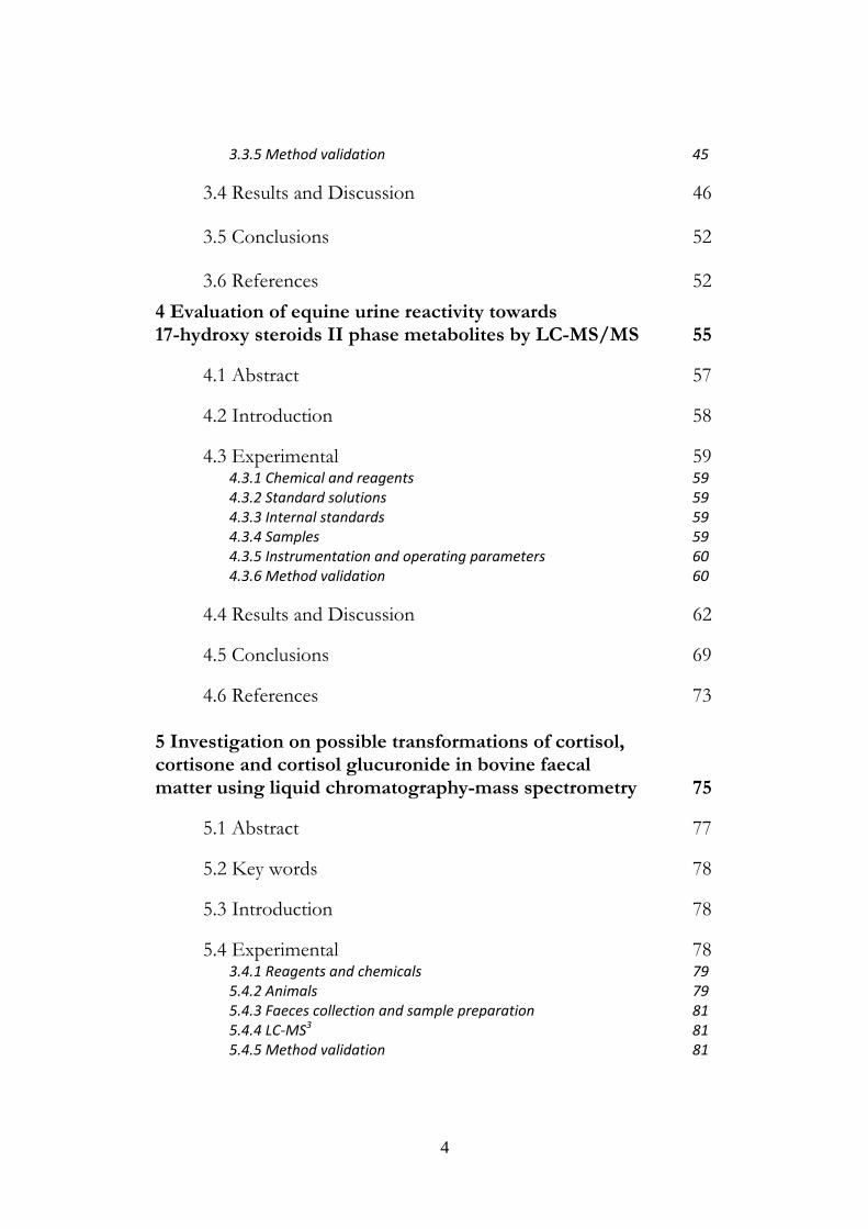
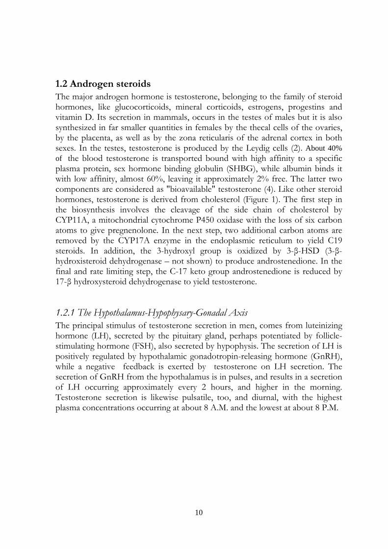
1.2 Androgen steroids The major androgen hormone is testosterone, belonging to the family of steroid hormones, like glucocorticoids, mineral corticoids, estrogens, progestins and vitamin D. Its secretion in mammals, occurs in the testes of males but it is also synthesized in far smaller quantities in females by the thecal cells of the ovaries, by the placenta, as well as by the zona reticularis of the adrenal cortex in both sexes. In the testes, testosterone is produced by the Leydig cells (2). About 40% of the blood testosterone is transported bound with high affinity to a specific plasma protein, sex hormone binding globulin (SHBG), while albumin binds it with low affinity, almost 60%, leaving it approximately 2% free. The latter two components are considered as "bioavailable" testosterone (4). Like other steroid hormones, testosterone is derived from cholesterol (Figure 1). The first step in the biosynthesis involves the cleavage of the side chain of cholesterol by CYP11A, a mitochondrial cytochrome P450 oxidase with the loss of six carbon atoms to give pregnenolone. In the next step, two additional carbon atoms are removed by the CYP17A enzyme in the endoplasmic reticulum to yield C19 steroids. In addition, the 3-hydroxyl group is oxidized by 3-β-HSD (3-β-hydroxisteroid dehydrogenase – not shown) to produce androstenedione. In the final and rate limiting step, the C-17 keto group androstenedione is reduced by 17-β hydroxysteroid dehydrogenase to yield testosterone.
1.2.1 The Hypothalamus-Hypophysary-Gonadal Axis The principal stimulus of testosterone secretion in men, comes from luteinizing hormone (LH), secreted by the pituitary gland, perhaps potentiated by follicle-stimulating hormone (FSH), also secreted by hypophysis. The secretion of LH is positively regulated by hypothalamic gonadotropin-releasing hormone (GnRH), while a negative feedback is exerted by testosterone on LH secretion. The secretion of GnRH from the hypothalamus is in pulses, and results in a secretion of LH occurring approximately every 2 hours, and higher in the morning. Testosterone secretion is likewise pulsatile, too, and diurnal, with the highest plasma concentrations occurring at about 8 A.M. and the lowest at about 8 P.M.
11
Figure 1 - Illustration of the pathway of testicular testosterone synthesis. The uptake of cholesterol and its conversion to testosterone involves numerous receptors and enzymes, including ScarB1 (Scavenger receptor class B. member 1), which is responsible for HDL uptake into Leydig cells, StAR ( Steroidogenic acute regulatory protein) that transport the cholesterol from the outer to the inner mitochondrial membrane and the steroid-converting enzymes P450scc and P450c17, which are involved in the conversion of cholesterol to testosterone. ( Figure and caption from Ref 5).
In women, LH stimulates the corpus luteum (formed from the follicle after release of the ovum) to secrete testosterone. The principal inhibitors of LH secretion in women, however, are estradiol and progesterone, not testosterone (4)
1.2.2 Main physiologic effects of androgen steroids The androgens bind to the androgen nuclear receptor NR3A, which possesses an amino-terminal domain, a DNA-binding domain, and a ligand-binding domain. Testosterone can act as an androgen either directly, or indirectly, after conversion catalyzed by a 5α-reductase, to dihydrotestosterone, which have higher affinity for the receptor. The androgen bound to the ligand-binding domain causes a conformational change in the receptor, the ligand-receptor complex can so translocate to the nucleus and via the DNA-binding domain, transcriptionally regulates the expression of androgen-responsive genes (6).
12
Androgen steroids effects can be classified as anabolic and androgenic, although many of these effects can be considered super imposable. Briefly:
Anabolic effects include growth of muscle mass and strength, increased bone density and strength, and stimulation of linear growth and bone maturation.
Androgenic effects include maturation of the sex organs, particularly the penis and the formation of the scrotum in the fetus, and at puberty the appearance of the male secondary sex characteristics.
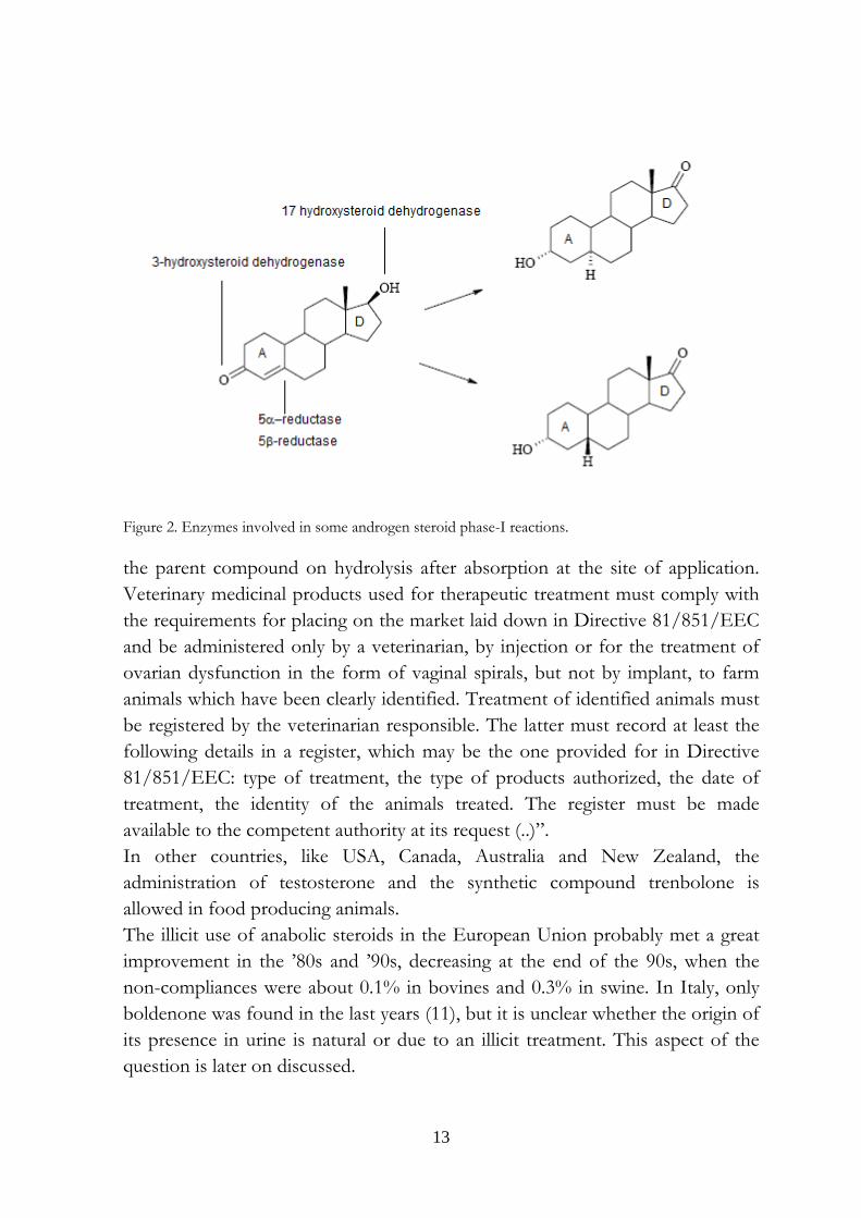
1.2.3 Metabolism of androgen steroids Androgen steroids undergo phase I and phase II reactions, that transform them into less toxic, less active, and/or more polar forms. Phase-I reactions modify the parent compound via hydrolysis, oxidation, and/or reduction (7, 8). As an example, it can be cited the non-reversible reduction of the C-4,5 double bond of 4-ene-3-one ring A which leads to the formation of two isomers, in a ratio depending on the relative catalyzing effects of 5α- and 5β-reductase enzymes (9) and the subsequent reduction of the 3-keto group, predominantly by 3α-hydroxysteroid dehydrogenase (3HSD). In D-ring, 17β-hydroxysteroid dehydrogenase (17HSD) can form 17-keto metabolites (Figure 2). Although phase-I reactions already increase the polarity and the excretion of AAS, these modifications are most often preparative stages for reactions that expose reactive sites of the analyte structure for the following phase-II, i.e. conjugation reactions. For androgen steroids, the main phase-II reactions are glucuronidation and sulfation. These conjugations occur mainly in the liver, but also in the intestinal mucosa, usually reduce the steroid activity and get him ready to the secretion with urine or bile (10).
1.2.4 Androgen steroids and farm animals In observance to the Council Directive 96/22/EC of 29 April 1996 (concerning the prohibition on the use in stockfarming of certain substances having a hormonal or thyrostatic action and of ß-agonists), the Council Directive 96/23/EC of 29 April 1996 (on measures to monitor certain substances and residues thereof in live animals and animal products) includes androgen steroids in Group A, that contains substances prohibited or firmly restricted for use in food producing animals, and specifically in category A3 - steroids. Nevertheless, the Council Directive 96/22/EC, in article 4, states:” (...) the Member States may authorize: 1. the administering to farm animals, for therapeutic purposes, of oestradiol 17 α, testosterone and progesterone and derivatives which readily yield
13
Figure 2. Enzymes involved in some androgen steroid phase-I reactions.
the parent compound on hydrolysis after absorption at the site of application. Veterinary medicinal products used for therapeutic treatment must comply with the requirements for placing on the market laid down in Directive 81/851/EEC and be administered only by a veterinarian, by injection or for the treatment of ovarian dysfunction in the form of vaginal spirals, but not by implant, to farm animals which have been clearly identified. Treatment of identified animals must be registered by the veterinarian responsible. The latter must record at least the following details in a register, which may be the one provided for in Directive 81/851/EEC: type of treatment, the type of products authorized, the date of treatment, the identity of the animals treated. The register must be made available to the competent authority at its request (..)”. In other countries, like USA, Canada, Australia and New Zealand, the administration of testosterone and the synthetic compound trenbolone is allowed in food producing animals. The illicit use of anabolic steroids in the European Union probably met a great improvement in the ’80s and ’90s, decreasing at the end of the 90s, when the non-compliances were about 0.1% in bovines and 0.3% in swine. In Italy, only boldenone was found in the last years (11), but it is unclear whether the origin of its presence in urine is natural or due to an illicit treatment. This aspect of the question is later on discussed.
14
1.3 Glucocorticosteroids
1.3.1 A short premise
In the 1930s, Edward Kendall of the Mayo Foundation in Rochester (MN, USA) isolated six hormones from the adrenal glands. He named them in the order in which they were isolated: compounds A through F. In the same years, Philip Hench, also at the Mayo Foundation, had observed that patients who had rheumatoid arthritis were sometimes relieved if they developed jaundice, during pregnancy, and immediately after unrelated surgery . He hypothesized the secretion of a natural anti-rheumatic, that he termed "Substance X". The two physicians conferred some times and agreed about the possibility that the adrenal hormones might help in treating rheumatoid arthritis and that "Substance X” could be a steroid. In January 1941, Philip Hench wrote in his notebook: "Try Compound E in rheumatoid arthritis". The anti-inflammatory effect of the compound, cortisone, was so discovered. Contemporary, in the University of Basel, Tadeus Reichstein isolated and explained the constitution of aldosterone, a hormone of the adrenal cortex, which until then had not been isolated. Reichstein also collaborated with E. Kendall and P. Hench in their work on the hormones of the adrenal cortex which culminated in the isolation of cortisone and the discovery of its therapeutic value in the treatment of rheumatoid arthritis. It was then found that many other diseases of an inflammatory nature were relieved by cortisone. Although it was found later that cortisone, like insulin, is a symptomatic drug, i.e. acts only so long as it is given to the patient, and that it does not cure the disease, the discovery of the activity of cortisone was a great step forward. It has led to our modern knowledge of the hormones of the adrenal cortex and their uses in medicine. In 1950 they were awarded of the Nobel prize “For their discoveries relating to the hormones of the adrenal cortex, their structure and biological effects” (12,13) . In 1955, a Research Group of Schering Corporation from Bloomfield (NJ, USA) described the “clinical effectiveness of two new, potent antiarthritic steroids, metacortandracin (Meticorten) and metacortandralone (Meticortelone)” The structures of these compounds were Δ1,4-pregnadiene-17α, 21-, diol-3 11, 20 trione and Δ1,4-pregnadiene-11β 17α, 21-triol-3, 20 dione. Their present names are prednisone and prednisolone, respectively. The adrenocortical activity of these two steroids was measured and resulted 3 to 4 times the activity of cortisone or cortisol (14).
15
In the same year Nobile and co-workers reported that the oxidation of the latter two corticosteroids by Corynebacterium simplex can be used to prepare their Δ1 dehydrogenated analogues (15, 16). The microbiological introduction of the double bond in position 1,2-(Δ1) of the steroid structure is still used for the production of prednisone and prednisolone, as the chemical route is inferior in both product purity and yield (17, 18).
1.3.2 Adrenal steroids
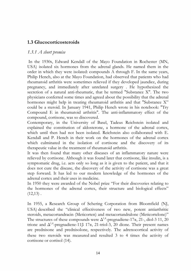
The surrenal glands, or adrenals, are endocrine glands located atop the kidneys and are constituted by an outer part - the cortex - and an inner part - the medulla. The cortex contains three zones: zona glomerulosa, fasciculata and reticularis. In these zones three main types of hormone are produced. They are, respectively: mineralocorticoids (aldosterone, deoxycorticosterone) that regulate hydro saline equilibrium, glucocorticoids (cortisol, corticosterone), active on metabolism, in stress and in the inflammatory processes, and precursors (dehydroepiandrosterone [DHEA] and androstenedione) of androgen steroids. Cholesterol is the precursor for all adrenal steroids and it is principally provided in its free form following hydrolysis, in adrenal tissue, of the circulating low-density lipoprotein (LDL) cholesterol (19). Cholesterol also can be generated de
Fig. 3. Steroidogenesis in the adrenal cortex; CYP17: steroid 17-hydoxylase; CYP11B2: aldosterone syntase; CYP11B1: steroid11-hydoxylase; Ang II: angiotensin II. The figure is a combination of two figures in Ref. 4 and 25.
16
novo within the adrenal cortex from acetyl coenzyme A (20), and there is evidence that the adrenal can use high-density lipoprotein (HDL) cholesterol (21). In the medulla, catecholamines (adrenaline, noradrenaline,, and dopamine) are produced. The biochemical pathways involved in adrenal steroidogenesis are shown in Figure 3(4, 25).
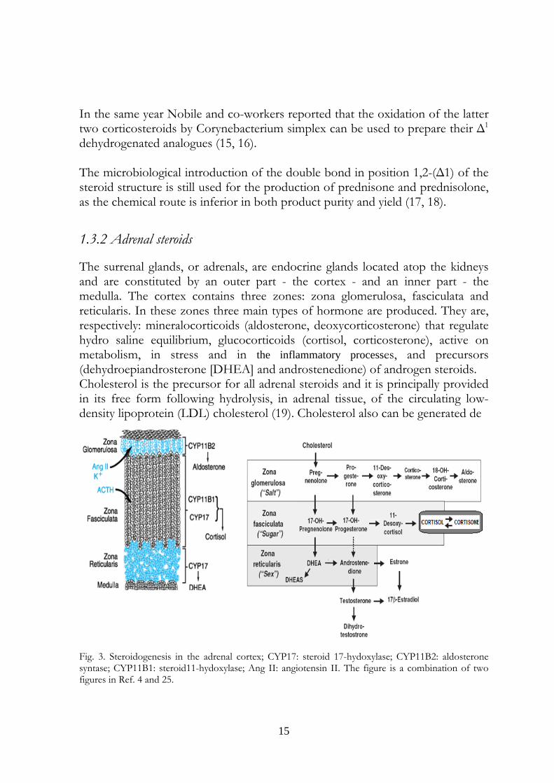
1.3.3 The Hypothalamus-Hypophysary-Adrenal Axis
The adrenal cortex is stimulated by the adrenocorticotropic hormone (ACTH, also called corticotropin), secreted from the anterior pituitary in response to
Figure 4. Hypothalamus-hypophysary-adrenal axis. (HPA)
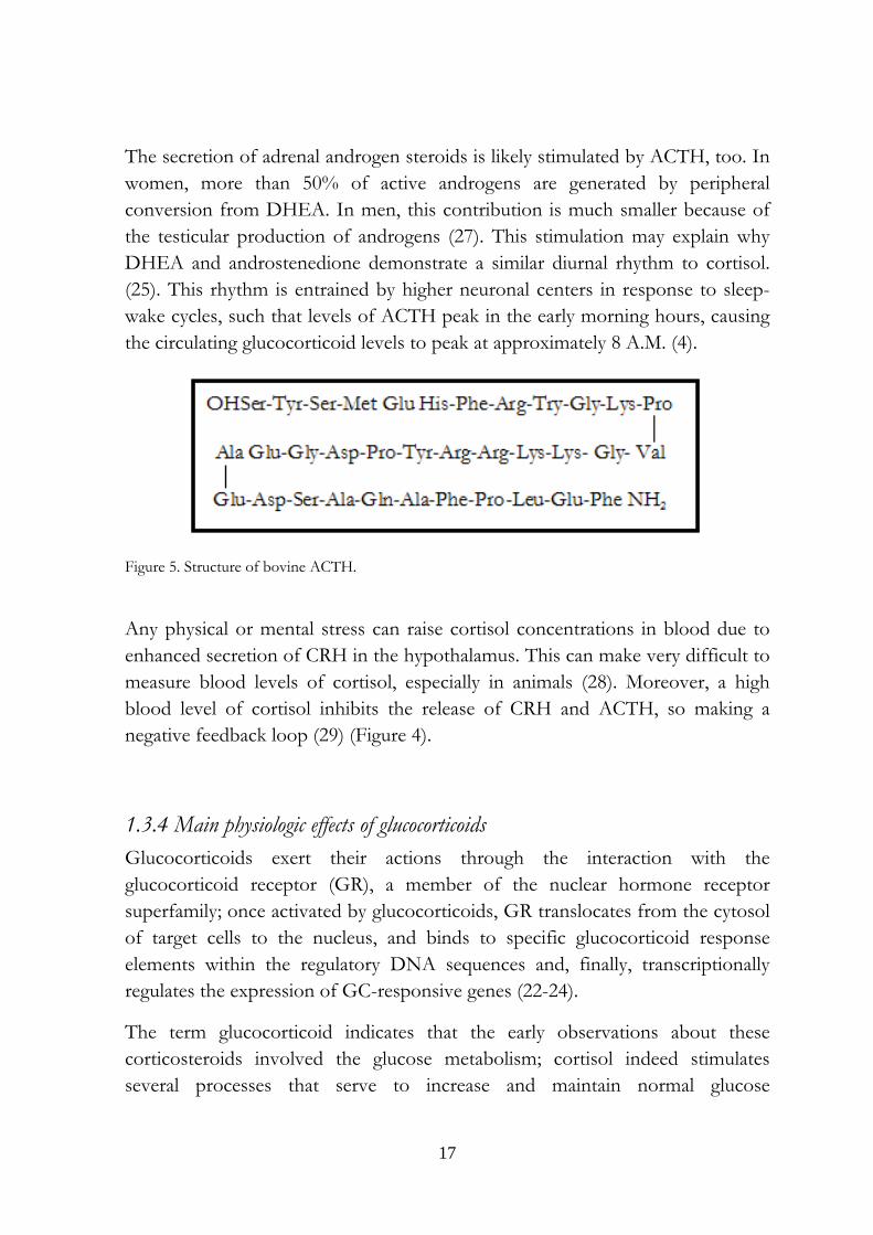
corticotropin-releasing hormone (CRH) from the hypothalamus. The cooperation, of the three glands, considered as a single system, is named hypothalamus-hypophysary-adrenal axis (HPA) (figure 4). Bovine ACTH is a 39 aminoacids peptide (26) (Figure 5) whose biological activity is closely related to the structure of NH2 terminus. More specifically, it stimulates secretion of glucocorticoids such as cortisol; aldosterone, the other major steroid hormone from the adrenal cortex is under control of the peptide Angiotensin II, potassium and, only to a lesser extent, ACTH.
STRESSOR HYPOTALAMIC CRH
HYPOPHYSARY ACTH
CCOORRTTIISSOOLL
17
The secretion of adrenal androgen steroids is likely stimulated by ACTH, too. In women, more than 50% of active androgens are generated by peripheral conversion from DHEA. In men, this contribution is much smaller because of the testicular production of androgens (27). This stimulation may explain why DHEA and androstenedione demonstrate a similar diurnal rhythm to cortisol. (25). This rhythm is entrained by higher neuronal centers in response to sleep-wake cycles, such that levels of ACTH peak in the early morning hours, causing the circulating glucocorticoid levels to peak at approximately 8 A.M. (4).
Figure 5. Structure of bovine ACTH.
Any physical or mental stress can raise cortisol concentrations in blood due to enhanced secretion of CRH in the hypothalamus. This can make very difficult to measure blood levels of cortisol, especially in animals (28). Moreover, a high blood level of cortisol inhibits the release of CRH and ACTH, so making a negative feedback loop (29) (Figure 4).
1.3.4 Main physiologic effects of glucocorticoids Glucocorticoids exert their actions through the interaction with the glucocorticoid receptor (GR), a member of the nuclear hormone receptor superfamily; once activated by glucocorticoids, GR translocates from the cytosol of target cells to the nucleus, and binds to specific glucocorticoid response elements within the regulatory DNA sequences and, finally, transcriptionally regulates the expression of GC-responsive genes (22-24).
The term glucocorticoid indicates that the early observations about these corticosteroids involved the glucose metabolism; cortisol indeed stimulates several processes that serve to increase and maintain normal glucose
18
concentrations in blood through an action on glycogen, protein and lipid metabolism (25). These effects can be so schematized:
Liver
Cortisol stimulates glycogen deposition by increasing glycogen syntase and inhibiting glycogen phosphorilase, that mobilizes it.
Glucose output increases by activating enzymes involved in gluconeogenesis, mainly glucose-6-phosphatase and phophoenolpyruvate kinase. This pathway results in the synthesis of glucose from non-hexose substrates such as amino acids and lipids.
Other tissues (Adipose tissue and muscle)
Glucose uptake and use is inhibited.
Lipolysis in adipose tissue is activated, resulting in a raise of free fatty acids and tryglicerides into the circulation; also total cholesterol increases even if HDL cholesterol falls.
Fatty acids released by lipolysis are used in tissues like muscle through their conversion into Acetyl-CoA for production of energy stored as ATP,
The released glycerol provides another substrate for gluconeogenesis.
The other major effect of glucocorticoids is constituted by the suppression of the immune system and by the anti-inflammatory actions.
Immune system
The lymphocyte counts acutely reduce in peripheral blood because of the redistribution to spleen, lymph nodes and bone marrow (30), and stimulation of apoptosis. Neutrophyl counts increase, eosinophils fall (an effect hystorically used to evaluate corticosteroid activity) (31).
19
anti-inflammatory actions
Glucocorticosteroids impair prostaglandin synthesis through the induction of lipocortins, a group of peptides that inhibit phospholipase A2 activity on arachidonic acid: the production of the mediators of inflammation prostaglandins and leucotriens, is thus inhibited. Minor anti-inflammatory effects involve inhibitory actions on macrophagae and prevention of histamine activators (25).
Finally, it can be stated the physiologic effects of these steroid hormones are a huge number: likely, no cell lacks steroid receptors. At all events the best known and studied effects of glucocorticoids are on carbohydrate metabolism, immune function and inflammation.
1.3.5 Glucocorticoid metabolism Only about 10% of circulating cortisol is free, the remaining bound to cortisol-binding globulin (CBG) also named transcortin. The excretion of free cortisol through the kidneys represents only 1% of total secretion rate.
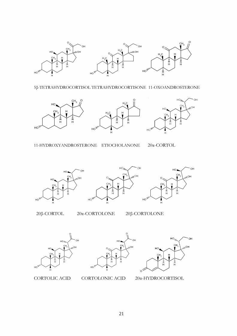
In man, the major routes of cortisol metabolism are the interconversion of cortisol (Kendall’s compound F) to cortisone (compound E) through the activity of 11β-hydroxysteroid dehydrogenases or reduction of the C4-5 double bond by either 5β-reductase or 5α-reductase to yield respectively 5β-tetrahydrocortisol (THF) or 5α-THF (allo-THF). THF, allo-THF and tetrahydrocortisone (THE) are conjugated rapidly with glucuronic acid and excreted in the urine. Downstream, cleavage of THF and THE to the C19 steroids 11-hydroxy or 11-oxo-androsterone or etiocholanolone can occur. Alternatively, reduction of the 20-oxo group by 20 α- or 20 β -hydroxysteroid dehydrogenase yields α and β cortols and cortolones, respectively, with subsequent oxidation at the C21 position to form the extremely polar metabolites, cortolic, and cortolonic acids. Hydroxylation at C6 to form 6 β-hydroxycortisol is described, as is reduction of the C20 position, which may occur without A ring reduction giving rise to 20 α-and 20 β-hydroxycortisol. Approximately 50% of secreted cortisol appears in the urine as THF, allo-THF, and THE; 25% appears as cortols/cortolones. Ten percent appears as C19 steroids, and 10% appears as cortolic/cortolonic acids. The remaining metabolites are free, unconjugated steroids (cortisol, cortisone, and 6 β- and 20 α/20 β-metabolites of F and E) (Figure 6) (25).
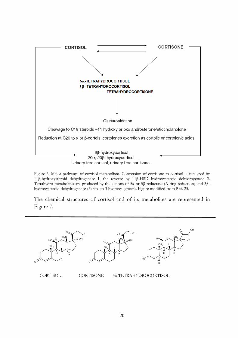
20
Figure 6. Major pathways of cortisol metabolism. Conversion of cortisone to cortisol is catalyzed by 11β-hydroxysteroid dehydrogenase 1, the reverse by 11β-HSD hydroxysteroid dehydrogenase 2. Tetrahydro metabolites are produced by the actions of 5α or 5β-reductase (A ring reduction) and 3β-hydroxysteroid dehydrogenase (3keto- to 3 hydroxy- group). Figure modified from Ref. 25. The chemical structures of cortisol and of its metabolites are represented in Figure 7.
H3COH
CH3
HO
OOH
O
H
H
H
H
CH3
CH3
O
O
O OH
OH
H
H
H
O
OHCH3HO
CH3
HO
OH
H
H
H
H
CORTISOL CORTISONE 5α-TETRAHYDROCORTISOL
21
CH3OH
CH3
HO
OOH
HO
H
H
H
H
H3COH
H3C
O
OOH
HO
H
H
H
H
O
O
HO
H3C
CH3
H
H H
H
5β-TETRAHYDROCORTISOL TETRAHYDROCORTISONE 11-OXOANDROSTERONE
HO
OHO
H H
CH3
CH3 H
H3C
H3CO
HO
H
H H
H
11-HYDROXYANDROSTERONE ETIOCHOLANONE 20α-CORTOL
CH3
CH3
HO
HO OH
HO OH
H
H
H
H
CH3
CH3
O
OHHO
OH
HOH
H
H
H
20β-CORTOL 20α-CORTOLONE 20β-CORTOLONE
CH3
CH3
O
OHHO
OHHO
HOH
H
H
H
CH3
CH3
O
OH
O
HO
OH
HOH
H
H
H
CORTOLIC ACID CORTOLONIC ACID 20α-HYDROCORTISOL
22
O
CH3
OH
H
HO
H
CH3 OH
O
OH
H
20β-HYDROCORTISOL 6β-HYDROXYCORTISOL
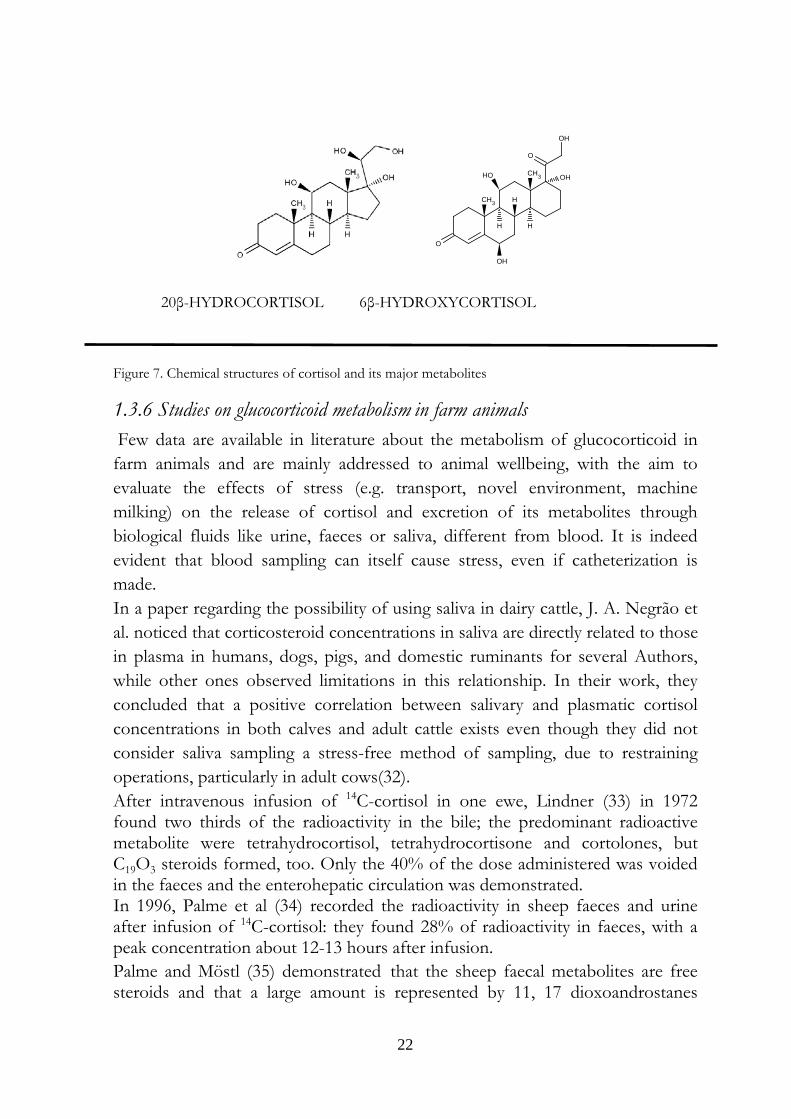
Figure 7. Chemical structures of cortisol and its major metabolites
1.3.6 Studies on glucocorticoid metabolism in farm animals Few data are available in literature about the metabolism of glucocorticoid in farm animals and are mainly addressed to animal wellbeing, with the aim to evaluate the effects of stress (e.g. transport, novel environment, machine milking) on the release of cortisol and excretion of its metabolites through biological fluids like urine, faeces or saliva, different from blood. It is indeed evident that blood sampling can itself cause stress, even if catheterization is made. In a paper regarding the possibility of using saliva in dairy cattle, J. A. Negrão et al. noticed that corticosteroid concentrations in saliva are directly related to those in plasma in humans, dogs, pigs, and domestic ruminants for several Authors, while other ones observed limitations in this relationship. In their work, they concluded that a positive correlation between salivary and plasmatic cortisol concentrations in both calves and adult cattle exists even though they did not consider saliva sampling a stress-free method of sampling, due to restraining operations, particularly in adult cows(32). After intravenous infusion of 14C-cortisol in one ewe, Lindner (33) in 1972 found two thirds of the radioactivity in the bile; the predominant radioactive metabolite were tetrahydrocortisol, tetrahydrocortisone and cortolones, but C19O3 steroids formed, too. Only the 40% of the dose administered was voided in the faeces and the enterohepatic circulation was demonstrated. In 1996, Palme et al (34) recorded the radioactivity in sheep faeces and urine after infusion of 14C-cortisol: they found 28% of radioactivity in faeces, with a peak concentration about 12-13 hours after infusion. Palme and Möstl (35) demonstrated that the sheep faecal metabolites are free steroids and that a large amount is represented by 11, 17 dioxoandrostanes
23
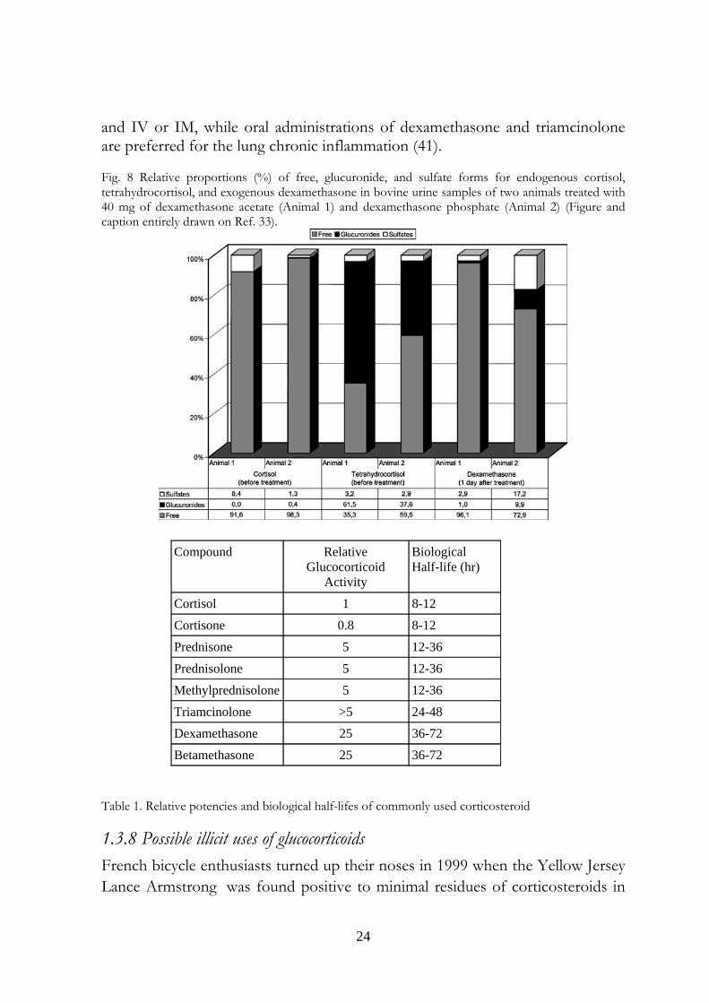
(11,17.DOAs) In this work they developed an immunoassay for detecting glucocorticoid, the 11,17.DOA test, specifically for use in ruminants but useful in a variety of species including sheep, cattle, horses, roe deer, hares, okapi, Barbary macaque, and red deer The antibody (raised in rabbit against 11-oxoetiocholanolone-3-HS:BSA) cross-reacts with 11,17-dioxoandrogens (84-100% 5α-androstanes; 5–15% 5β-androstanes). The 11,17 DOAs increase in cow faeces (36) after a two hours road transport. Similar results are described by Morrow et al (37), who conducted an experiment on Holstein dairy cows to detect corticosteroids metabolites in faecal samples. When the cows were exposed to a new housing environment, the concentrations of glucocorticoid metabolites peaked two days after the move (from 151 to 352%). In 2002, Antignac et al. (38) performed a study about corticosteroid phase II metabolites in bovine urine. The urine of two cows, “Normande” specie, weighing 400–500 kg, were analyzed for cortisol and tetrahydrocortisol (free, sulfate and glucuronide) before an intramuscular injection of 40 mg of dexamethasone acetate (Animal 1) or dexamethasone phosphate (Animal 2) and for dexamethasone (free, sulfate and glucuronide) one day after the treatment. (Figure 8). Not considering the data on dexamethasone, an exogenous, fluorinated corticosteroid, the difference between the total proportion of conjugated forms of tetrahydrocortisol (40–65%) and cortisol (2–8%) is strong. The high amount of tetrahydrocortisol glucuronide is explained by the Authors with the reduction of keto-group to hydroxil in position 3 of the A ring of tetrahydrocortisol, that probably favors the conjugation reactions.
1.3.7 Veterinary uses of glucocorticoids in farm animals The common used glucocorticoids in veterinary medicine are prednisolone, prednisone, metylprednisolone, betamethasone, dexamethasone, triamcinolone acetonide, flumethasone (39) (Table 1). In bovine, swine, ovine and caprine prednisolone, betamethasone and dexamethasone can be used, via IM or IV, in the anti-inflammatory (particularly involving locomotor and cutaneous apparatuses) and antiallergic symptomatic therapies; in pregnancy-related pathologies (gravidic toxicosis, ketosis) in case of metabolic disorders, shock and intoxications; topically prednisolone is used for conjunctivitis, blepharoconjunctivitis, keratoconjunctivitis and inflammations at the back of the eye and, by intramammary infusion, for the treatment of bovine mastitis. Betamethasone is also used for the induction of parturition in cows (40). The horse respiratory tract acute inflammation is treated with prednisolone both oral
24
and IV or IM, while oral administrations of dexamethasone and triamcinolone are preferred for the lung chronic inflammation (41). Fig. 8 Relative proportions (%) of free, glucuronide, and sulfate forms for endogenous cortisol, tetrahydrocortisol, and exogenous dexamethasone in bovine urine samples of two animals treated with 40 mg of dexamethasone acetate (Animal 1) and dexamethasone phosphate (Animal 2) (Figure and caption entirely drawn on Ref. 33).
Table 1. Relative potencies and biological half-lifes of commonly used corticosteroid
1.3.8 Possible illicit uses of glucocorticoids French bicycle enthusiasts turned up their noses in 1999 when the Yellow Jersey Lance Armstrong was found positive to minimal residues of corticosteroids in
Compound Relative Glucocorticoid
Activity
Biological Half-life (hr)
Cortisol 1 8-12
Cortisone 0.8 8-12
Prednisone 5 12-36
Prednisolone 5 12-36
Methylprednisolone 5 12-36
Triamcinolone >5 24-48
Dexamethasone 25 36-72
Betamethasone 25 36-72
25
urine. Its defense was that he used an ointment to treat a severe dermatitis, and he had a full discharge. More recently, the CONI (Italian Olympic Committee) disqualified for two months the Turin Juventus Football Club doctors for a mistake in transmitting the declaration of systemic use of corticosteroids of the football player Fabio Cannavaro, who was sting by a wasp and found positive to these substances in October 2009. Indeed, the World Anti Doping Agency (WADA) prohibits oral, intravenous, intramuscular or rectal uses of glucocorticosteroids, requires a declaration of Use for intraarticular, periarticular, peritendinous, epidural, intradermaland inhalatory administrations, and does not require any declaration when topical preparation are used (auricular, buccal, dermatological, gingival, nasal, ophthalmic and perianal disorders) (42). In zootechnics, synthetic corticosteroids may have an illicit use as growth-promoters either alone or with other active principles (i.e., steroid hormones and/or β-agonists), in order to take advantage of their synergistic effects with different illegal growth-promoting agents. In particular, dexamethasone can reverse the β2-agonist-mediated down-regulation of β2-receptors, so enhancing the repartitioning effects of β2-agonists. In addition, the glucocorticoid-mediated electrolyte imbalance results in polydipsia and polyuria, which may decrease the concentration of other illegal substances in the urine (43). Low doses of corticosteroids raise the food intake, the weight of the animal and the water retention, but reduce the food conversion index, and the proteic and lipidic anabolism. Prolonged administrations or high doses of corticosteroids can induce muscle atrophy because of the enhanced proteic catabolism that causes a decrease of animal grow rate (44). The above mentioned electrolyte imbalance, that consists into sodium retention, potassium and calcium elimination, tubular reabsorption of chlorine and sodium, results in a raise of extracellular liquid volume: a treatment in the last 30-40 days before the slaughter increases the carcass weight, but also decreases meat quality and commercial value. After the slaughter indeed, the altered ability of the meat to retain water, makes the carcass abnormally drip. This illicit treatment is generally made as a replacement of the administration of anabolic steroids, some weeks before the slaughter, to avoid their detection by health authorities and the weight loss due to interruption of anabolic steroid administration (45).
1.3.9 Legal aspects In the European Union, the use of corticosteroids in livestock is indicated for therapeutic reasons only and is regulated by the Commission Regulation (EU) No 37/2010 of 22 December 2009 on pharmacologically active substances and
26
their classification regarding maximum residue limits in foodstuffs of animal origin. MRLs (maximum residue limits) for betamethasone in cattle and pigs, dexamethasone in cattle, pigs, horses and goats; methylprednisolone and prednisolone for cattle are indicated in the Annex and reported in table 2. From 1988, the Italian National Residue Program (PNR) provides for the surveillance and the monitoring of residues of chemical substances in foods with animal origin. Until 2006 the PNR was the expression of the Legislative Decree 336 of the 4th of August 1999, that followed the indications of the Council Directive 96/22/EC of 29 April 1996 concerning the prohibition on the use in stockfarming of certain substances having a hormonal or thyrostatic action and of ß-agonists and the Council Directive 96/23/EC of 29 April 1996 on measures to monitor certain substances and residues thereof in live animals and animal products. Today, the PNR is the expression of the Legislative Decree 158 of the 16th of March 2006, that acknowledges the Directive 2003/74/EC, amending Council Directive 96/22/EC and concerning the prohibition on the use in stockfarming of certain substances having a hormonal or thyrostatic action and of β-agonists. Two categories of residues are envisaged: the “A” category includes products with an anabolic effect and substances forbidden in cattle intended for consumption and so employed fraudfully to improve the animals performances; The “B” category comprehends three families of substances. The first two ones regard veterinary drugs allowed in cattle treatment, with defined MRLs. The third family is made up of environmental contaminants as the organic chlorinated compounds, heavy metals and substances that may be absorbed by environment and enter in food chain. The analyses for the research of residues must be carried out using validated methods according to Commission Decision 2002/657/EC concerning the performance of analytical methods and the interpretation of results. Moreover the sampling procedures are reported in the Commission Decision 98/179/EC laying down detailed rules on official sampling for the monitoring of certain substances and residues thereof in live animals and animal products. Different institutions cooperate to the organization and the execution of the PNR. At a national level or, in other words, in the Ministry of Health, the General Direction of Veterinary Health and Foods coordinates all the activities concerning the fulfillment of the Project and represents the competent
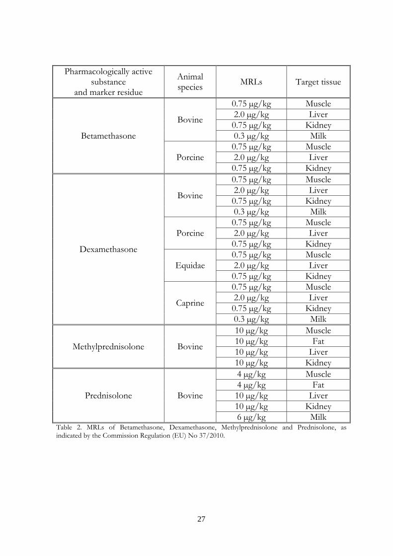
27
Pharmacologically active substance
and marker residue
Animal species MRLs Target tissue
Betamethasone
Bovine
0.75 µg/kg Muscle 2.0 µg/kg Liver 0.75 µg/kg Kidney 0.3 µg/kg Milk
Porcine 0.75 µg/kg Muscle 2.0 µg/kg Liver 0.75 µg/kg Kidney
Dexamethasone
Bovine
0.75 µg/kg Muscle 2.0 µg/kg Liver 0.75 µg/kg Kidney 0.3 µg/kg Milk
Porcine 0.75 µg/kg Muscle 2.0 µg/kg Liver 0.75 µg/kg Kidney
Equidae 0.75 µg/kg Muscle 2.0 µg/kg Liver 0.75 µg/kg Kidney
Caprine
0.75 µg/kg Muscle 2.0 µg/kg Liver 0.75 µg/kg Kidney 0.3 µg/kg Milk
Methylprednisolone Bovine
10 µg/kg Muscle 10 µg/kg Fat 10 µg/kg Liver 10 µg/kg Kidney
Prednisolone Bovine
4 µg/kg Muscle 4 µg/kg Fat 10 µg/kg Liver 10 µg/kg Kidney 6 µg/kg Milk
Table 2. MRLs of Betamethasone, Dexamethasone, Methylprednisolone and Prednisolone, as indicated by the Commission Regulation (EU) No 37/2010.
28
administrative Authority towards European Union, while the Health Superior Institute (Istituto Superiore di Sanità) coordinates the technical scientific aspects, as Reference National Laboratory for residues. On a regional scale, the Regional Residues Program (PRR) is defined according to the characteristics of the different areas, the extent of the zoo technical property, the number of slaughters, the handlings of drugs and feedstuffs; this project is sent out to territorial veterinary services that define the method of implementation of samples. The regional Councils, through the veterinary services coordinate of the activity, collect obtained data send them to the Ministry of Health every six month. The samplings are made both in breedings (primary production) and in first transformation factories, like slaughterhouses or milk collection centres. The samples are analyzed in Experimental Zooprophylactic Institutes laboratories (Istituti Zooprofilattici Sperimentali). On the basis of the analytical results, if banned substances residues are found or the content of residues of authorized substances or environmental contaminants exceed the fixed limits, administrative as well as criminal sanctions are provided, if a risk for public health is set (Article 40 of the code of criminal procedure about the adulteration of foodstuff). Finally, the Ministry of Health yearly passes all the data to the European Commission , together with the new year planning. After briefly outlining the legal aspects regarding the residues in farm animals, some considerations have to be done about the glucocorticoids. Indeed, they may be considered under two aspects: the Commission Regulation (EU) No 37/2010, in Annex sets the MRLs of dexamethasone, betamethasone, metylprednisolone and prednisolone. These substances are therefore considered legal drugs and their use is permitted in farm animals, if the suspension times are observed, so that the maximum concentrations in edible tissues are not exceeded. In the Italian PNR they are categorized in class A3, i.e. substances having an anabolic effect and unauthorized substances/steroids, unless a therapeutic use has been declared in advance by the farm veterinary surgeon. This could cause a misuse of glucocorticosteroids, illicitly used as above described, but legally declared as therapeutic agents.
29
1.3.10 Glucocorticoid residues in food of animal origin The determinations of betamethasone, dexamethasone, methylprednisolone and prednisolone MRLs are made on evaluations performed by the European Medicines Agency (EMEA) on the basis of pharmacological uses of these corticosteroids, that are indeed active principles of several human medicines. The indications include rheumatoid arthritis, severe hypersensitivity reactions, Crohn’s disease, haemolytic anaemia, leukaemias , malignant lymphoma and their administrations can be oral, intravenous or intramuscular. The preparations are generally well tolerated but adverse effects are known and include acute adrenal insufficiency and indications of glucocorticoid overactivity such as round face and wasted limbs. Growth retardation may occur in children, and resistance to infection is decreased due to suppression of the immune system. No one of the four glucocorticosteroids is considered mutagenic, carcinogenetic or genotoxic. Betamethasone and dexamethasone are contraindicated during pregnancy due to the risk to the foetus of cleft palate and intrauterine growth retardation. To confirm these contraindications the betamethasone NOEL for teratogenicity in rats is reported to be 0.4 mg/kg bw/day, while two studies on teratogenicity and foetotoxicity in rabbit with inadequate group sizes show a value of NOEL of 0.003 mg/kg bw/day and the NOEL for dexamethasone embryotoxicity in rat produces a value of 0.01 mg/kg bw/day. The normal oral dose ranges from 0.5 mg/day (maintenance dose) to 5 mg/day (attack dose) for bethametasone, from 4 to 96 mg/person for methylprednisolone, and from 5 to 150 mg/person for prednisolone. The No Observed Effect Levels (NOELs) for all this corticosteroids are calculated regarding the increase in tyrosine aminotransferase activity in rat liver at oral doses, an indicator related to reduced growth and to the activation of gluconeogenic mechanisms in which lipids and amino acids, rather than carbohydrates, are used for energy production (46): The NOEL of bethametasone is 0.004 mg/kg bw, a value that could lead to an Acceptable Daily Intake (ADI) of 0.00004 mg/kg bw/day. Betamethasone, however, is the 16β-epimer of dexamethasone, whose 16-methyl group is above the plane of the steroid moiety, i.e in α-position. Because the similar toxicological properties and glucocorticoid activities of this two corticosteroids parallel, the EMEA adopted the lower dexamethasone ADI value, 0.000015 mg/kg bw (0.0009mg/person). Methylprednisolone NOEL, is 0.016 mg/kg bw/day, and leads to an ADI of 0.00016 mg/kg bw (i.e. 0.0096mg/person). Prednisolone has an ADI of 0.0002 mg/kg bw (i.e. 0.012 mg/person) calculated by applying a safety factor of 100 to the NOEL of 20 μg/kg bw/day.
30
Based on MRLs reported in Table 2, the daily intakes would be 52% and 99% for methylprednisolone and prednisolone respectively, while the theoretical estimated consumer intake of dexamethasone and bethametasone were 0.0009125 mg/day, exceeding the ADI value of 0.0009 mg/day. The consideration of EMEA is that this would not represent a risk to human health because the substance is used only occasionally in individual animals (47-50).
1.4 References
1 )W. Van Thuyne: The grey zone in doping, Ph.D. thesis in medical sciences, Ghent University, 2006; http://www.docolab.ugent.be/PhD_WVT.pdf.
2) J. Scarth, C. Akre, L. van Ginkel, B. Le Bizec, H. De Brabander, W. Korth, J. Points, P. Teale, J. Kay: Presence and metabolism of endogenous androgenic-anabolic steroid hormones in meat-producing animals: a review, Food Addit. Contam. Part A, 26:640–671, 2009.
3) R.V. Brooks: Androgens, Clin. Endocrinol. Metab., 4: 503–520, 1975.
4) L.L. Brunton (editor-in-chief), L. Keith, K.L. Parker, N. Murri, D.K. Blumenthal (associate editors), Goodman & Gilman's the pharmacological basis of therapeutics, online 11th edition, http://www.accessmedicine.com.
5) Environmental Medicine, Institute of Public Health, University of Southern Denmark (SDU) and The National Food Institute, Technical University of Denmark (DTU): Effects of azole fungicides on the function of sex and thyroid hormones Pesticides Research financially supported by the Danish Environmental Protection Agency’s Pesticide Research Programme No. 111, 2007);http://www2.mst.dk/common/Udgivramme/Frame.asp?http://www2.mst.dk/udgiv/publications/2007/978-87-7052-538-1/html/helepubl_eng.htm
6) A.O. Brinkman, J. Trapman: Genetic analysis of androgen receptors in development and disease, Adv. Pharmacol., 47:317–341, 2000.
7) G.G. Gibson, and P. Skett: Introduction to Drug Metabolism. Gibson, G.G., Skett, P. (Eds.) Blackie Academic and Professional, Chapman & Hall, London, 1994.
31
8) S. Rendic, and F.J. Di Carlo: Human cytochrome P450 enzymes: A status report summarizing their reactions, substrates, inducers, and inhibitors. Drug Metab. Rev. 29:413–580, 1997.
9) W. Schänzer: Metabolism of anabolic androgenic steroids. Clin. Chem. 42:1001–1020, 1996.
10) T. Kuuranne: Anabolic Steroid Glucuronides. Enzyme-Assisted Synthesis and Liquid Chromatographic–Mass Spectrometric Analysis, Academic dissertation, Division of Pharmaceutical Chemistry, Viikki Drug Discovery Technology Center, Department of Pharmacy, Faculty of Science, University of Helsinki, Finland, April 2003.
11) C. Nebbia (a cura di): Residui di farmaci e contaminanti ambientali nelle produzioni animali, Edizioni EdiSES, Napoli, 2009.
12) Nobel Lectures, Physiology or Medicine 1942-1962, Elsevier Publishing Company, Amsterdam, 1964
13) http://www.mayoclinic.org/tradition-heritage/cortisone-discovery.html.
14) H.L. Herzog, A. Nobile, S. Tolksdorf, W. Charney, E.B.Hershberg, P.L. Perlman, M.M. Pechet: New antiarthritic steroids, Science, 121:176, 1955.
15) A. Nobile, W.Charney, P.L. Perlman, H.L. Herzog, C.C. Payne, M.E. Tully, M.A. Jevnik, E.B. Hershberg: Microbiological transformation of steroids I. Δ1,4-Diene-3-ketosteroids. Am. J. Chem. Soc., 77:4184, 1955.
16) T.H. Stoudt: Advances in applied microbiology, vol. 2, New York: Academic Press, p. 204, editor: Umbreit WW, 1960.
17) N.Z. Adham, A.A. El-Hady, N. Naim: Biochemical studies on the microbial Δ1-dehydrogenation of cortisol by Pseudomonas fluorescens, Process Biochemistry, 38:897–902, 2003.
18) K. Kieslich: Microbial enzymes and bioconversions: economic microbiology, vol. 5, London, Academic, pp. 369–465, editor: Rose AH., 1980.
19) J.T. Gwynne, J.F. Strauss: III. The role of lipoprotein in steroidogenesis and cholesterol metabolism in steroidogenic glands. Endocr. Rev., 3:299–329, 1982.
20) A.J. Borkowski, S. Levin, C. Delcroix, A. Mahler, V. Verhas: Blood cholesterol and hydrocortisone production in man: quantitative aspects of the
32
utilization of circulating cholesterol by the adrenals at rest and under adrenocorticotropin stimulation. J. Clin. Invest., 46:797–811, 1967.
21) S. Acton, A. Rigotti, K.T. Landschulz, S. Xu, H.H. Hobbs, M.Krieger: Identification of scavenger receptor SR-BI as a high-density lipoprotein receptor. Science, 27:518–20, 1996.
22) M.R. Yudt, J.A. Cidlowski: The glucocorticoid receptor: coding a diversity of proteins and responses through a single gene. Mol�Endocrinol.,16:1719–1726, 2002.
23) E. Sonneveld, A. Jonas, O.C. Meijer, A. Brouwer, B. van der Burg: Glucocorticoid-enhanced expression of dioxin target genes through regulation of the rat aryl hydrocarbon receptor, Toxicol. Sci., 99:455–469 2007.
24) J-M. Pascussi, S. Gerbal-Chaloin, L. Drocourt, P. Maurel, M-J. Vilarem: The expression of CYP2B6, CYP2C9 and CYP3A4 genes: a tangle of networks of nuclear and steroid receptors, Biochim. Biophys. Acta 1619:243–253, 2003.
25) W. Arlt, P.M. Stewar: Adrenal Corticosteroid Biosynthesis,Metabolism, and Action, Endocrinol Metab. Clin. N. Am., 34:293–313, 2005.
26) www.brimr.org/Roskoski/132.pdf
27) C. Longcope: Adrenal and gonadal secretion in normal females. Clin. Endocrinol. Metab., 15:213–228, 1986.
28) E. Möstl, R. Palme: Hormones as indicators of stress, Domest. Anim. Endocrinol. 23:67–74, 2002.
29) A. La Brocca: Biochimica e fisiologia della corticale surrenalica (Rassegna), Ligand Assay 11:10-18, 2006.
30) D.T. Yu, P.J. Clements, H.E. Paulus, J.B. Peter, J. Levy, E.V. Barnett: Human lymphocyte subpopulations: effect of corticosteroids, J. Clin. Invest. 53:565–571, 1974.
31) L.I. McKay, J.A. Cidlowski: Molecular control of immune/inflammatory responses: interactions between nuclear factor-kB and steroid receptor-signalling pathways, Endocr. Rev. 20:435–459, 1999.
32) J. A. Negrão, M. A. Porcionato, A. M. de Passille , and J. Rushen: Cortisol in saliva and plasma of cattle after ACTH administration and milking J. Dairy Sci. 87:1713–1718, 2004.
33
33) H.R. Lindner: Enterohepatic circulation and patterns of urinary excretion of cortisol metabolites in the ewe. J. Endocrinol.. 52:XIX-XX,1972.
34) R. Palme, P. Fischer, H. Schildorfer, M.N. Ismail: Excretion of infused 14C-steroid hormones via faeces and urine in domestic livestock,. Anim. Reprod. Sci. 43:43–63, 1996.
35) R. Palme, E. Möstl: Measurement of cortisol metabolites in faeces of sheep as a parameter of cortisol concentration in blood. Int. J. Mammal. Biol., 62 (S2):192–197, 1997.
36) R. Palme, C. Robia, W. Baumgartner, E. Möstl: Transport stress in cattle as reflected by an increase in faecal cortisol metabolites, Vet. Rec. 146:108–109, 2000.
37) C. J. Morrow, E. S. Kolver, G. A. Verkerk, L. R. Matthews: Fecal Glucocorticoid Metabolites as a Measure of Adrenal Activity in Dairy Cattle, Gen. Comp.e Endocrinol., 126:229-241, 2002.
38) J-P. Antignac, B. Le Bizec, F. Monteau, F. André: Study of natural and artificial corticosteroid phase II metabolites in bovine urine using HPLC–MS/MS, Steroids 67:873–882, 2002.
39) J.E. Riviere, M.G. Papich (editors), H.R. Adams (consulting editor): Veterinary pharmacology and therapeutics, Wiley.-Blackwell, 9th edition, 2009.
40) R. Villa (a cura di) SIVAR Prontuario terapeutico veterinario-Medicina degli animali da reddito, Edizioni veterinarie, 2008
41) S. Carli, P. Ormas, G. Re., G. Soldani: Farmacologia veterinaria, cas Editrice Idelson-Gnocchi, 2009.
42) WADA: The Prohibited List 2010, , S9. Glucocorticosteroids. 19 September 2009.
43) M. Vincenti, F. Girolami, P. Capra, M. Pazzi, M. Carletti, G. Gardini, C. Nebbia: Study of dexamethasone urinary excretion profile in cattle by LC-MS/MS: Comparison between therapeutic and growth-promoting administration, J. Agric. Food Chem., 57: 1299–1306, 2009
44) I. Istasse, V. De Haan, C. Van Enaeme , B. Buts , P. Baldwin , M. Gielen , D. Demeter , J.M. Bienfait: Effects of dexamethasone injections on performance in a pair of monozygotic cattle twins. J. Anim. Phys. Anim. Nutrit., 62:150-158 1989.
34
45) G. Ballarini:. Terapia cortisonica con Desametazone negli allevamenti di bovini da carne, Obiettivi e Documenti Veterinari, 6: 13-24, 2005.
46) K.B. Davis, P. Torrance, N.C. Parker, M.A. Suttle: Growth, body composition and hepatic tyrosine aminotransferase activity in cortisol-fed channel catfish, Ictalurus punctatus Rafines que. J. Fish Biol., 27: 177-184, 1985.
47) Committee for Veterinary Medicinal Products: Dexamethasone (extrapolation to goats), EMEA/MRL/874/03-Final, June 2004
48) Committee for Veterinary Medicinal Products: Bethametasone, EMEA/MRL/605/99-Final, June 1999,
49) Committee for Veterinary Medicinal Products: Methylprednisolone, EMEA/MRL/703/99-Final, October 1999,
50) Committee for Veterinary Medicinal Products: Prednisolone, EMEA/MRL/629/99-Final, July 1999
35
Objectives
CHAPTER 2

36
37
Never buy bread from a butcher Irish wisdom
2.1 Why Boldenone and Prednisolone?
Boldenone is a steroid occurring in the two isomeric forms 17α-boldenone and 17 β-boldenone. The latter one is commonly named boldenone. Boldenone possesses androgenic activity and differs from 17-β testosterone (testosterone) by only one double bond at the Δ1-2 position on ring A. European Directive 96/22/EC amended by Directive 2003/74/EC and 96/23/EC bans β boldenone, a class A3 substance (growth promoter steroid), in products of animal origin and in livestock. Nevertheless, there is evidence that boldenone may arise naturally in bovine urine, probably following a contamination with microorganisms both of faecal or environmental origin. In chapter 3, a contribution to the comprehension of the mechanisms leading to the presence of boldenone in faeces contaminated urine was given, while the following work regards the transformation of phase II metabolites of androgenic steroids in urine of race horses. This work, besides shows that the problem regarding boldenone presence in urine regards the controls both in farm and in sport animals. Prednisolone (Δ1,4-pregnadiene-11β,17α,21-triol-3,20-dione) is a corticosteroid with gluconeogenetic and antinflammatory activities. Its structure is very similar to the cortisol one and differs from the hormone by only one double bond at the Δ1-2 position on ring A: a close analogy with boldenone and testosterone. Prednisolone is considered a synthetic corticosteroids; in the last years, nevertheless, this certainty began to waver in the mind of some italian researchers. The exceptional percentage in prednisolone positive bovine urine sampled at the slaughterhouse, compared to the absence of this steroid in urine sampled at the farm in Lombardy, led us to cover the route already made for the boldenone problem, (i.e. the contamination of urine, chapters 5 –faecal- and 6-environmental) but particularly to study the effect of stress on a possible natural production of prednisolone by bovine organism, chapter 7).

38
39
Evaluation of boldenone formation and related steroids transformations
in veal faeces by liquid chromatography/ tandem mass spectrometry
Published in:
RAPID COMMUNICATIONS IN MASS SPECTROMETRY, 22: 217–223, 2008.
CHAPTER 3

40
41
In the midst was seen A lady of a more majestic mien,
By stature and by beauty mark’d their sovereign Queen. And as in beauty she surpass’d the choir,
So nobler than the rest was her attire; A crown of ruddy gold enclosed her brow,
Plain without pomp, and rich without a show; A branch of Agnus Castus in her hand,
She bore aloft her symbol of command. Geoffrey Chaucher, The Flower and the Leaf
3. Evaluation of boldenone formation and related steroids transformations in veal faeces by liquid chromatography/ tandem mass spectrometry Francesco Arioli1*, Matteo P. Gavinelli1, Maria L. Fracchiolla1, Alessio Casati1, Marco Fidani1, Emilia Ferrer2 and Giuseppe Pompa1 1Department of Veterinary Sciences and Technologies for Food Safety, University of Milan, Milan, Italy; 2Toxicology Laboratory, Faculty of Pharmacy, Valencia University, Burjassot, Spain *Correspondence to: F. Arioli, Department of Veterinary Sciences and Technologies for Food Safety, University of Milan, Via Celoria 10, 20133 Milan, Italy. E-mail: [email protected]
3.1 Abstract It is established that bovine urine can result positive for boldenone and androstadienedione in consequence of faecal contamination. The simple transfer of steroids to urine is one minor aspect of faecal contamination. A high de novo production of steroids in faeces after deposition and in faeces-contaminated urine is almost certainly due to microbial activity, although the precursor compounds and transformations leading to the presence of these illegal steroids are unclear. We developed a simple in vitro method – incubation of faecal matter suspended in 0.9% saline – to induce steroid transformations in faeces, and analyzed the products by liquid chromatography/tandem mass spectrometry, without the need for prior extraction. Norethandrolone was the internal standard. The linearity (R2: 0.987–0.999), sensitivity (LODs: 0.3 to 1.0 ng/mL; LOQs: 1.0 to 3.0 ng/mL), precision (intra −day CVs: 2.6–8.2; inter-day CVs: 4.5–11.5) and accuracy (percentage recovery: 89–120%) were calculated for the
42
studied steroids. Androstenedione, androstadienedione, α−and β−boldenone, testosterone and epitestosterone transformations were investigated. Mutual interconversion of steroids was observed, although 17 β−hydroxy steroids had low stability compared with 17 α−hydroxy and 17-keto steroids. The results suggest that this simple in vitro system may be an effective way of studying hormone transformations in faeces and, after analogue studies, in faeces-contaminated urine. Copyright # 2007 John Wiley & Sons, Ltd.
3.2 Introduction European Directive 96/22/EC bans β−boldenone (17β hydroxyandrosta- 1,4-dien-3-one; β−BOL), a class A3 substance (growth promoter steroid), in products of animal origin and in livestock. Urine analysis is currently used in the European Union (EU) to detect the illicit use of anabolic steroids including boldenone in cattle. Nevertheless, there is evidence that boldenone may arise naturally in urine (1). In 1996, Arts et al. (2) reported α−boldenone (17 α−hydroxyandrosta- 1,4-dien-3-one; α−BOL) up to 2.7 ng/mL, and β−BOL up to 0.1 ng/mL, in the urine of untreated calves. More recently, Nielen et al. (3) reported α−BOL, β−BOL and also androstadienedione (1,4-androstadiene-3,17-dione; ADD) in the dried faeces of untreated cattle at much higher levels than in the urine of animals administered β−BOL undecylenate by intramuscular (IM) injection. De Brabander et al. (1) reported α−BOL (1–10 ng/g) and β−BOL (0.1–2.0 ng/g) in faecal scrapings from calf skin, and Sangiorgi et al. (4) detected these substances in rectal faeces from untreated calves. We have shown that contamination of calf urine with faeces can render the urine positive for α−BOL and occasionally for β−BOL (5,6), and that considerable quantities of ADD, androstenedione (4-androstene-3,17-dione; AED) and α−BOL can be produced in faeces after emission (6). The European Union Community Reference Laboratory for Residues (RIVM) have shown that faecal contamination of urine can be a source of false positivity for unconjugated α−BOL (7). In the study, carried out on 261 veal calf urine samples suspected to contain α/β−BOL, coprostanol was used as marker of faecal contamination. It was not excluded that also unconjugated β−BOL and α−BOL-glucuronide could be endogenous or have originated from small amounts of faeces ingested by the animals. Furthermore, 28 samples containing more than 2 ng/mL of α−BOL-glucuronide were tested for the presence of 6 β−hydroxy-17 α−boldenone and 6 β−hydroxy-17 β−boldenone, marker metabolites only found after administration of β−BOL (8). The absence of this metabolite was considered a proof that α−BOL-glucuronide and
43
unconjugated β−BOL are no indicators for boldenone abuse. β−BOL-glucuronide, α−BOL-sulphate and β−BOL sulphate were never found. The authors concluded that the presence in urine of unconjugated β−BOL and α−BOL conjugates next to α−BOL are no indicators of illegal administration, but no indications were obtained about the origin of conjugated β−BOL. Investigations carried out by Le Bizec et al. (9) showed that the marker metabolite ‘‘always detectable whatever the injected boldenone form and never observable in the so-called endogenous cases’’ could be β−BOL sulphate. Some doubts however emerged about the oral administration (only 0–20% eliminated in the sulphate fraction) compared with the IM injection (up to about 70%). The mechanisms of production of α−BOL and β−BOL, but also of ADD and AED, in faeces are unclear, although it has been suggested that they arise by microbial transformation of cholesterol or phytosterols in the gut (1,7). Indeed, Egorova et al. (10) demonstrated that Mycobacterium sp. VKM Ac-1815D strain is able to cleave the side chain of sitosterols giving AED as a major product. Barthakur et al. (11) showed the ability of Mycobacterium sp. NRRL Β−3683 to transform β−sitoterols into ADD, in the presence of a 1(2)-dehydrogenase. Moreover, Grinenko et al. (12) proved that the transformation of a natural sterol mixture into AED under the action of Mycobacterium sp. S-905 (VKPM collection) is increased by aeration. This fact could imply that transformations of sterols into androgens are favoured in faeces after emission, i.e. under aerobic conditions, according to our previous work (6). In order to better understand the mechanisms that give raise to false BOL positivity of faeces-contaminated urine, it is necessary to study the biotransformations that these steroids undergo in faecal matter. We therefore further examined biotransformations of α−BOL, β−BOL, testosterone (17β-hydroxy-4-androsten-3-one; T), epitestosterone (17 α−hydroxy- 4-androsten-3-one; ET) and the precursors/metabolites ADD and AED in faeces of veal calves using a simple in vitro model, followed by analysis with liquid chromatography/ tandem mass spectrometry (LC/MS/MS).
3.3 Experimental 3.3.1 Reagents and chemicals α-BOL was obtained from Tecna (Trieste, Italy). β-BOL, ADD, AED, T and ET were purchased from Sigm α−Aldrich (St. Louis, MO, USA). Deuterated testosterone (Td3) (Sigm α−Aldrich) was tested as internal standard (see below) and norethandrolone (17α-ethyl-17β-hydroxyestr-4-en-3-one; NETA) (Alltech, Deerfield, IL, USA) was used as internal standard. All other chemicals were from
44
Fluka Chemie GmbH (Buchs, Switzerland). Standard stock solutions in ethanol (1 mg/mL) were prepared and stored at -18°C; working solutions were prepared daily by dilution of stock solutions with methanol/ water (50:50 v/v).
3.3.2 Animals Faeces from ten male Friesian veal calves, age 90–100 days, fed with milk replacer (2.8 kg/calf/day) and corn silage (1.3 kg/calf/day), were studied. The lipid content of milk replacer was 20% (35% coconut oil and 65% tallow plus lard). The animals were housed in individual stalls in a single cowshed and cared for in accordance with EU guidelines approved by the Italian Ministry of Health.
3.3.3 Faeces collection and sample preparation Rectal faeces from five veal calves were pooled. From the pooled material sample 1.3 g was suspended in 130 mL of 0.9% saline and shaken overnight at 258C. The suspension was divided into thirteen 10 mL samples: one blank (without steroid addition), six controls (held at 808C for 15 min and then spiked with a steroid) and six treatments (no heating, spiked with a steroid). A steroid (T, ET, α−BOL, β−BOL, AED or ADD) was added to each control and to each treatment tube to a final concentration of 200 ng/mL (20 mg/g faeces) and left at ambient temperature. Samples (1 mL) from each tube were collected at 0, 0.5, 1, 2, 4, 8, 24 and 48 h later. The samples were heated at 80°C for 15 min to inactivate bacteria and centrifuged (1400 g, 10 min). The supernatants were diluted 1:2 with methanol/water (50:50 v/v) and 20 mL samples analyzed by LC/MS/MS without extraction. This experimental procedure was repeated 1 week later on a pool of faeces from five different veal calves.
3.3.4 LC-MS/MS We used a slight modification of the method described in detail elsewhere.6 The LC/MS system consisted of a ThermoFinnigan Surveyor LC pump with Surveyor autosampler (San Jose, CA, USA) equipped with a LCQ DECA XP Max ion trap mass spectrometer (ThermoFinnigan). The column was a 150 mm x 2.1 mm i.d., 5 mm ODS Hypersil-Keystone (ThermoFinnigan) in an oven at 308C. The mobile phase was water with 0.1% acetic acid (A) and methanol (B), flow rate 0.3 mL/min. B was kept at 65% for 1 min, increased to 90% over 7 min, held at 90% for 1 min, and then reduced to 65% over 1 min. An isocratic period of 5 min followed with 65% B. The mass spectrometer was operated in positive atmospheric pressure chemical ionization (APCI) mode with capillary voltage 31 V, capillary temperature 2208C, discharge current 4.5 mA, vaporizer temperature 3508C and sheath and auxiliary gas (nitrogen) flow rates of 20 and 8 arbitrary units, respectively. Helium was used for collision-induced dissociation;
45
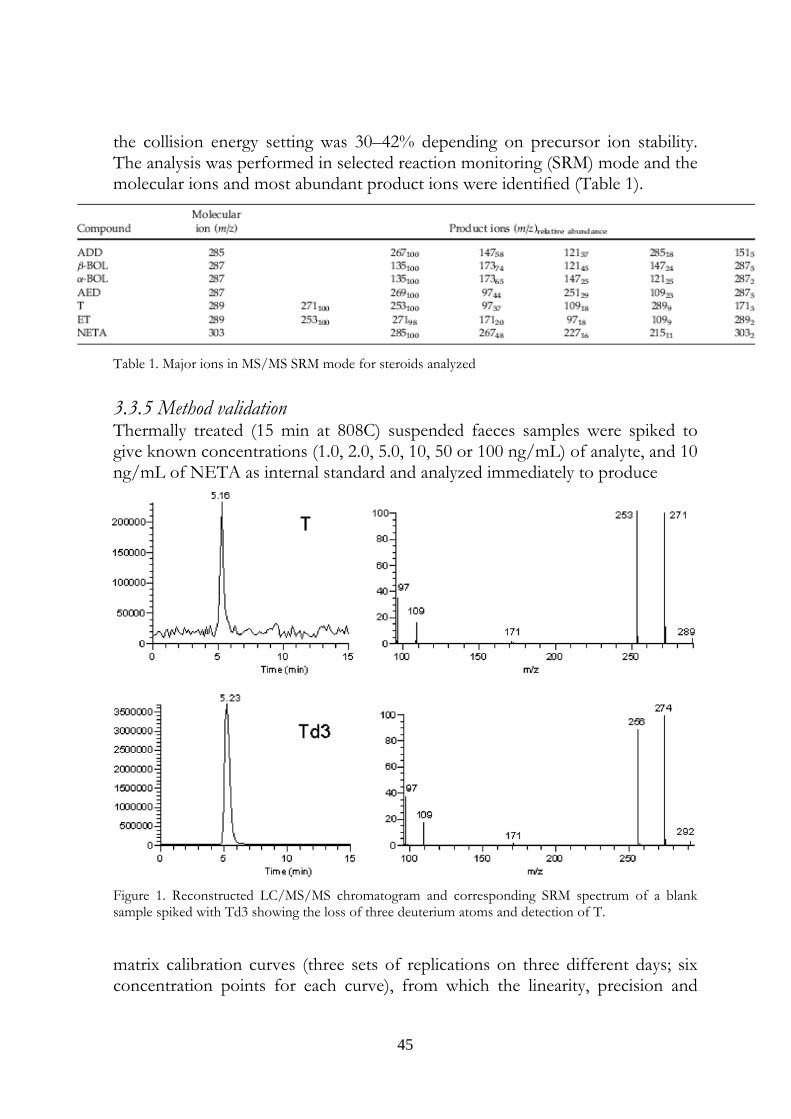
the collision energy setting was 30–42% depending on precursor ion stability. The analysis was performed in selected reaction monitoring (SRM) mode and the molecular ions and most abundant product ions were identified (Table 1).
Table 1. Major ions in MS/MS SRM mode for steroids analyzed 3.3.5 Method validation Thermally treated (15 min at 808C) suspended faeces samples were spiked to give known concentrations (1.0, 2.0, 5.0, 10, 50 or 100 ng/mL) of analyte, and 10 ng/mL of NETA as internal standard and analyzed immediately to produce
Figure 1. Reconstructed LC/MS/MS chromatogram and corresponding SRM spectrum of a blank sample spiked with Td3 showing the loss of three deuterium atoms and detection of T.
matrix calibration curves (three sets of replications on three different days; six concentration points for each curve), from which the linearity, precision and
46
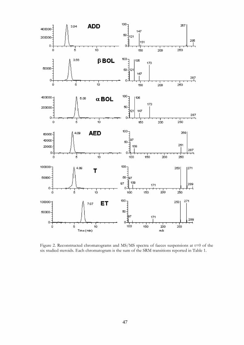
accuracy were determined. Limits of detection (LODs) and limits of quantification (LOQs) were also estimated by multiplying signal-to-noise levels by 3 and 10, respectively, in 20 unfortified samples and then converting the mean responses into ng/mL. It was found that regression coefficients (R2) were in the range 0.987–0.999 for all analytes. LODs were 0.3 ng/mL for α−BOL, β−BOL, AED and ADD, and 1.0 ng/mL for T and ET. LOQs were 1.0 ng/mL for α−BOL, β−BOL, AED and ADD and 3.0 ng/mL for T and ET. Precisions, expressed as intr α−day and inter-day coefficients of variation (CV%), were 2.6–8.2 and 4.5–11.5, respectively. Accuracy, as percentage recovery, was in the range 89–120% for all analytes. 3.4 Results and Discussion The recent study of Fidani et al. (13) showed that 17 β−hydroxy steroids could undergo transformations in poorly conserved equine urine (hence susceptible to bacterial contamination). Our previous studies (6, 14) showed similar transformations in dried calf faeces. In the present study we further investigated the biotransformations that steroids could undergo in calf faeces exposed to the environment. We analyzed faecal material after incubation in saline in order to eliminate confounding effects due to steroids present in urine. We did not perform an extraction step so that the samples could be rapidly prepared for analysis; the lack of extraction however entailed low sensibilities. The diluted suspensions analyzed had to be spiked with known and relatively high quantities of steroids in order to detect transformations. We paid particular attention to the choice of an internal standard. Preliminary experiments showed that Td3 could transform slowly into unlabelled T (Fig. 1), rendering it unsuitable as an internal standard. Further experiments showed that NETA, a 17 β−hydroxy steroid whose 17 β−hydroxy group is protected from oxidation by the presence of an ethyl group at position 17α, would be a suitable internal standard. The chromatograms and mass spectra of the test substances are shown in Fig. 2. We did not detect any of the steroids studied in the blank samples; neither did we detect any biotransformations in any of the control samples. By contrast the treatment samples showed transformations as summarized in Tables 2 and 3. The concentrations of control samples analyzed at time 0 were considered as 100% (untransformed steroids) and all data are reported as percentages of these values. The results reported in the tables are expressed as the means of two experiments. The 17 β−hydroxy steroids T and β−BOL underwent the most rapid transformations. Only 69% of T (Table 2) remained at time 0, implying
47
Figure 2. Reconstructed chromatograms and MS/MS spectra of faeces suspensions at t=0 of the six studied steroids. Each chromatogram is the sum of the SRM transitions reported in Table 1.
48
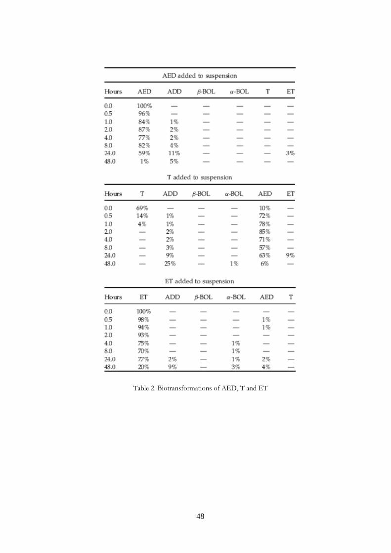
Table 2. Biotransformations of AED, T and ET
49
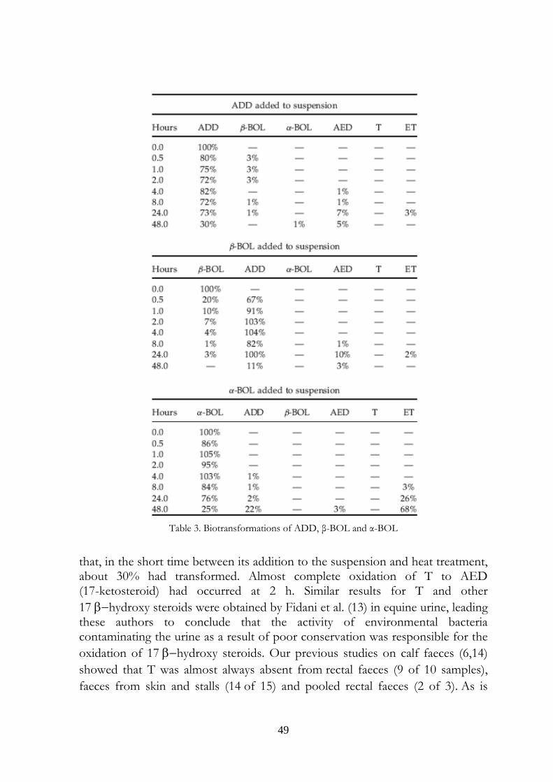
Table 3. Biotransformations of ADD, β-BOL and α-BOL
that, in the short time between its addition to the suspension and heat treatment, about 30% had transformed. Almost complete oxidation of T to AED (17-ketosteroid) had occurred at 2 h. Similar results for T and other 17 β−hydroxy steroids were obtained by Fidani et al. (13) in equine urine, leading these authors to conclude that the activity of environmental bacteria contaminating the urine as a result of poor conservation was responsible for the oxidation of 17 β−hydroxy steroids. Our previous studies on calf faeces (6,14) showed that T was almost always absent from rectal faeces (9 of 10 samples), faeces from skin and stalls (14 of 15) and pooled rectal faeces (2 of 3). As is
50
evident from Table 3, β−BOL transformed more slowly than T and had only completely disappeared from the sample after 24 h of incubation. β−BOL also oxidized to a 17-keto steroid, in this case ADD. The poor stability of β−BOL is consistent with the work of Nielen et al.,3 who detected α−BOL and ADD in bovine skin swab samples, and in dried faeces scraped from the skin. β−BOL was found only in some of the latter samples. The finding is also in agreement with the results of our earlier studies,6,14 in which β−BOL was absent or present in few samples, and was always absent from pooled bovine rectal faeces left to dry in air. The 17 α−hydroxy steroids ET and α−BOL proved to be fairly stable and had only diminished to 20–25% of their original levels after 48 h. After 48 h of α−BOL incubation, considerable quantities of ADD (22%) and ET (68%) had appeared, but only a small quantity of AED (3%), while after 48 h of ET incubation only small quantities of ADD, AED and α−BOL had accumulated. The main effect of ADD incubation under our experimental conditions was production of AED, with maximum peak (7%) after 24 h. A similar effect was observed by incubating AED, so that peak concentration (11%) of ADD was evident at 24 h. Both these steroids also gave rise to small quantities of ET, but only ADD was also able to produce a small quantity of β−BOL at 24 h. By contrast, AED seemed unable to produce T; however, it could have been produced and transformed back into AED or ADD (Fig. 3). The results of these in vitro studies provide indications as to the transformations of the studied steroids when present in faecal material, although they cannot, of course, indicate how the steroids were originally formed in the faeces. Our data further suggest that if urine is contaminated by faecal material, steroids present physiologically in the urine (T, ET and AED) may undergo transformations thereby profoundly altering the original hormone profile of the urine. In addition, we have previously shown6 that α−BOL, ET, AED and ADD neoformation may occur in dried faecal material. Urine may therefore receive doses of these steroids if contamination of urine samples from faecal crusts present on the skin or dried faeces in the stall occurs. In any event it is likely that faecal contamination also brings cholesterol and phytosterols – all potential precursors (10,12,15,16) for the neoformation of the steroids investigated in the present study. Our data are therefore consistent with the hypothesis that neoformation in urine occurs as a result of faecal contamination. An additional implication is that the types and concentrations of steroid that may be present in contaminated urine are likely to differ from those expected as a result of simple transfer from faeces to urine.
51
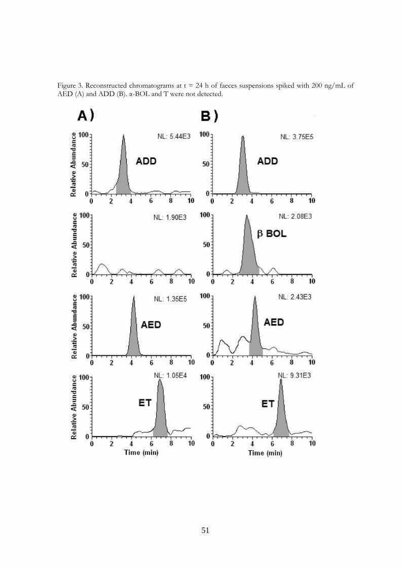
Figure 3. Reconstructed chromatograms at t = 24 h of faeces suspensions spiked with 200 ng/mL of AED (A) and ADD (B). α-BOL and T were not detected.
52
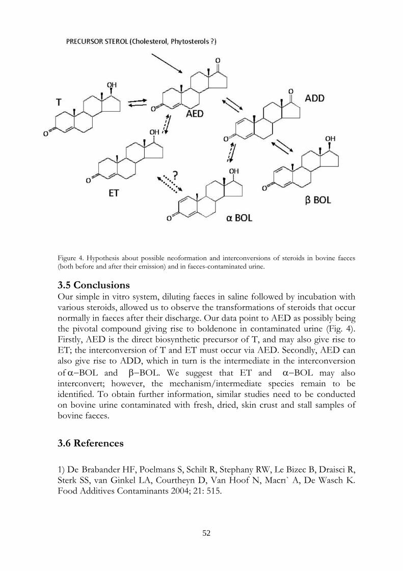
Figure 4. Hypothesis about possible neoformation and interconversions of steroids in bovine faeces (both before and after their emission) and in faeces-contaminated urine. 3.5 Conclusions Our simple in vitro system, diluting faeces in saline followed by incubation with various steroids, allowed us to observe the transformations of steroids that occur normally in faeces after their discharge. Our data point to AED as possibly being the pivotal compound giving rise to boldenone in contaminated urine (Fig. 4). Firstly, AED is the direct biosynthetic precursor of T, and may also give rise to ET; the interconversion of T and ET must occur via AED. Secondly, AED can also give rise to ADD, which in turn is the intermediate in the interconversion of α−BOL and β−BOL. We suggest that ET and α−BOL may also interconvert; however, the mechanism/intermediate species remain to be identified. To obtain further information, similar studies need to be conducted on bovine urine contaminated with fresh, dried, skin crust and stall samples of bovine faeces.
3.6 References 1) De Brabander HF, Poelmans S, Schilt R, Stephany RW, Le Bizec B, Draisci R, Sterk SS, van Ginkel LA, Courtheyn D, Van Hoof N, Macrı` A, De Wasch K. Food Additives Contaminants 2004; 21: 515.
53
2) Arts CJM, Schilt R, Schreurs M, Van Ginkel LA. Proc. Euroresidue III Conf. 1996; 808. 3) Nielen MWF, Rutgers P, van Bennekom EO, Lasaroms JJP, van Rhijn JAH. J. Chromatogr. B 2004; 801: 273.
4) Sangiorgi E, Polignano V, Gardini S. Anal. Chim. Acta 2005; 525: 239. 5) Sgoifo Rossi CA, Arioli F, Bassini A, Chiesa LM, Dell’Orto V, Montana M, Pompa G. Food Additives Contaminants 2004; 21: 756.
6) Pompa G, Arioli F, Fracchiolla ML, Sgoifo Rossi CA, Bassini A, Stella S, Biondi PA. Food Additives Contaminants 2006; 2: 126. 7) Blokland MH, van Rossum HJ, Sterk SS, van Ginkel LA, Stephany RW. Anal. Chim. Acta 2007; 586: 147. 8) Sterk SS, Blokland MH, van Ginkel LA, Schilt R, van der Vlis E, Boshuis P, van Baak MJ, Nielen MWF, van Rhijn JAH, Samson D, Keukens HJ, Stephany RW. Proc. Euroresidue V Conf. 2004; 900. 9) Le Bizec B, Courant F, Gaudin I, Bichon E, Destrez B, Schilt R, Draisci R, Monteau F, Andre´ F. Steroids 2006; 1: 1078. 10) Egorova OV, Gulevskaya SA, Puntus IF, Filonov AE, Donova MV. J. Chem. Technol. Biotechnol. 2002; 77: 141. 11) Barthakur S, Roy MK, Bera SK, Ghosh AC. J. Basic Microbiol. 1996; 36: 383. 12) Grinenko GS, Popova EV, Gabinskaya KN. Pharmaceut. Chem. J. 2002; 36: 510.
54
13) Fidani M, Casagni E, Montana M, Pasello E, Pecoraro C, Gambaro V. Rapid Commun. Mass Spectrom. 2006; 20: 2441. 14) Arioli F, Chiesa LM, Fracchiolla ML, Biondi PA, Pompa G. Vet. Res. Commun. 2005; 29 (Suppl. 2): 355. 15) Watanabe K, Aihara H, Tachi N, Nakamura R. J. Appl. Bacteriol. 1987; 62: 151. 16) Liu WH, Kuo CW, Wu KL, Lee CY, Hsu WY. J. Ind. Microbiol. 1994; 13: 167.
55
Evaluation of equine urine reactivity towards 17-hydroxy steroids
II phase metabolites by LC-MS/MS
Published in:
RAPID COMMUNICATIONS IN MASS SPECTROMETRY, 23: 65–76, 2009.
CHAPTER 4

56
57
Non hic Centauros, non Gorgonas Harpiyasque invenies: hominem pagina nostra sapit
Marcus Valerius Martialis 4. Evaluation of equine urine reactivity towards 17-hydroxy steroids II phase metabolites by LC-MS/MS
M. Fidania, M.C. Gamberinib, E. Paselloa, F. Palazzolib, P. De Iuliisa, M. Montanaa, F. Ariolic* a U.N.I.R.E. Lab. S.r.l., Via Gramsci 70, 20019 Settimo Milanese (MI), Italy; b Department of Pharmaceutical Sciences, Institute of Pharmacy, University of Modena and Reggio Emilia, Via Campi n.183, 41100 Modena, Italy; c Department of Veterinary Sciences and Technologies for Food Safety, University of Milan, Via Celoria 10, 20133 Milano, Italy * Corresponding author. Tel. +39 02 50317877; Fax: +39 02 50317890 E-mail address: [email protected]
4.1 Abstract Proper storage conditions of biological samples are fundamental to avoid microbiological contamination that can cause chemical modifications of the target analytes. A simple LC-MS/MS method through direct injection of diluted samples, without prior extraction, was carried out to evaluate the stability of phase II metabolites of boldenone and testosterone (glucuronides and sulphates) in intentionally poorly stored equine urine samples. We considered also the stability of some deuterated conjugated steroids, generally used as internal standards, like deuterated testosterone and epitestosterone glucuronides, and deuterated boldenone and testosterone sulphates. The urines were kept one day at room temperature, to mimic poor storage conditions, then spiked with the steroids written above and kept at different temperatures (-18°C, 4°C, room temperature). It has been possible to confirm the instability of glucuronide compounds when added to poorly stored equine urine samples. In particular, both 17ß and 17α-glucuronide steroids were exposed to hydrolysis phenomenon obtaining non-conjugated steroids. Only 17ß-hydroxy steroids were exposed to oxidation reactions obtaining keto derivates. Instead, 17α-hydroxy steroids presented high stability. The sulphate compounds showed a complete stability. The deuterated compounds underwent the same behaviour of the unlabelled
58
ones. The transformations were observed in urine samples kept at room temperature and 4°C temperature (at a slower rate). No modifications were observed in frozen urines. In the light of last results, the immediate freezing at -18°C of the collected samples and their instant analysis after the unfreezing is the proposed procedure for preventing and avoiding the transformations in urine, usually due to microbiological contamination.
4.2 Introduction The anabolic steroids, the most important drugs tested during racing and equestrian events, undergo phases 1 and 2 metabolism reactions. The phase 2 metabolites arise from the conjugation with glucuronic acid or sulphate, and represent the 90% of the excreted steroids(1). To detect anabolic steroids in urine, the most developed method involves the enzymatic hydrolysis of glucuronides and the chemical hydrolysis of sulphates, followed by analysis of the free steroids, by GC-MS2 with prior derivatization steps or by LC/MS (3,4). Recent developments in LC/MS methods permit the direct detection of phase II metabolites(5,6), leading to increased speed and efficiency of analysis and even the potential for track-side drug testing. The first problem to be faced in a steroid analysis is the conservation state sample, to prevent and avoid possible microbiological contaminations. Microbiological activity seems to depend on urine storage temperature, highlighting the necessity to maintain the cold chain in order to preserve urine samples unaltered, from the moment of collection to that of antidoping analysis(7). It has been verified that steroids conjugates in urine samples may undergo chemical modifications caused by bacterial contaminations (8-14). A misinterpretation of analytical results can be due to metabolic activity of bacteria (15,16). Therefore, to prevent any misinterpretation due to alteration of steroid conjugates in urine samples, it is fundamental to define suitable storage conditions prior anti-doping and clinical samples analyses. Some Authors tried to identify microorganisms potentially involved in the alterations of urinary endogeneous steroids profile (17,18). Some different studies about conjugate or non-conjugate compounds have been carried out on human urine (19-23), but few works on equine urine stability have been published yet. In a recent study, we demonstrated that 17β-hydroxy steroids can undergo transformations in poorly conserved equine urine (reactive or contaminated urine) (24). Therefore, the aim of this study was to develop a rapid test to evaluate equine urine reactivity by spiking samples with conjugate steroids and by monitoring their derivate products by LC-MS/MS analysis. This control could reveal a useful tool to evaluate the proper storage conditions of urine samples.
59
4.3 Experimental 4.3.1 Chemical and reagents Deuterated testosterone glucuronide (TGD3), boldenone glucuronide (BG), testosterone sulfate (TS), deuterated testosterone sulfate (TSD3), boldenone sulfate (BS), deuterated boldenone sulfate (BSD3), deuterated epitestosterone glucuronide (EPTG D3) and deuterated epitestosterone (EPT D3) were purchased from the National Measurement Institute (Pymble, NSW, Australia), testosterone glucuronide (TG), testosterone (T), boldione (BDI), androstenedione (AED) and deuterated testosterone (TD3) from Sigma-Aldrich (St. Louis, MO, USA), boldenone (BDN), epitestosterone (ET) and α-boldenone (αBDN from Sarstedt (Numbrecht, Germany), epitestosterone glucuronide (EPTG) from S.A.L.A.R.S (Como, Italy). Methanol, formic acid and water were purchased from J.T. Baker (Deventer, Holland). All reagents used were of HPLC grade.
4.3.2 Standard solutions Stock solutions (1 mg/mL) of all free steroids and conjugates and relative deuterated were prepared from dry chemical powder of each analyte by dissolving in methanol and stored at -18º C. The working solutions were prepared by diluting the individual stock solutions at a concentration of 1 µg/mL with methanol and stored at -18º C. All certificated standards were analysed by the LC-MS/MS developed method to evaluate the absence of interferences.
4.3.3 Internal standards The IS used were the relative deuterated substances and added at the concentration of 100 ng/mL in diluted urine samples.
4.3.4 Samples Equine urine samples were stored for 1 day at room temperature to mimic poor storage conditions. To evaluate their reactivity towards steroids, 950 µl of urine, diluted 1:5 with water, were spiked with testosterone (T) to obtain a concentration of 50 ng/mL.24 When the transformation of T into AED was observed within 2 hours, the urine was considered “reactive urine”. One hundred reactive urine samples were centrifuged at 1500g for 10 minutes, filtrated and diluted with distillate water (1:5). Every diluted urine was divided in nine portions of 450 µL, each one spiked with one of the following working solutions: TG, TGD3, BG, EPTG D3, EPTG, BS, BSD3, TS, TSD3. Nine
60
samples spiked with one of the nine different analytes were obtained. Three groups of these nine samples were prepared and stored at -18°C, 4°C as well as at room temperature. These storage temperatures were necessary to evaluate the temperature effect related to contaminated urines. The samples were analyzed in a time range variable from 10 min to 10 days after spiking.
4.3.5 Instrumentation and operating parameters An LTQ linear ion trap mass spectrometry equipped with an ESI source (Thermo-Fisher, San Josè, CA, USA) connected to a Surveyor Autosampler MS Pump (Thermo-Fisher) was used for all LC/MS analyses.Chromatography separation was performed at ambient temperature, in isocratic condition, on a reversed-phase Luna® (100 x 2 mm. 3µm; Phenomenex, San Josè, CA, USA) equipped with a C18 Guard Column® (4 mm. x 2 mm. id; Phenomenex). The mobile phase was composed of 40% formic acid 0,1% (solvent A) and 60% methanol (solvent B) at a flow rate of 0,25 mL/min. The injection volume was 5 µL. The ESI-MS/MS parameters were first optimized by direct injection of each compound individually at a concentration of 1 µg/mL at a flow rate of 20 µL/min. The mass spectrometer was operated, from 2 min to 16 min, in positive electrospray ionization MS/MS mode, with full scan product acquisition in the m/z 75-350 range for detection of T, EPT, TD3, EPTD3, AED, BDN, BDI, αBDN and in the m/z 125-500 range for detection of TG, TGD3, BG, EPTG and EPTGD3. For the positive ionization a capillary voltage of 12 V, a spray voltage of 4 kV, a capillary temperature of 275 °C were employed. The nitrogen sheath, auxiliary and sweep gas were set at 40, 20 and 5 arbitrary units respectively. The collision energy setting for the compounds ranged between 14,5% and 19,5% depending on the stability of the precursor ion. For detection of TS, BS, TSD3 and BSD3, the mass spectrometer was operated, from 16,01 min. to 26 min., in negative electrospray ionization MS/MS mode, with full scan product acquisition in the m/z 100-450 range. For the negative ionization, the tune parameters are the same of the positive ionization, while the capillary voltage was set to -8 V. The collision energy setting for the compounds ranged between 23% for boldenone sulphate and his deuterated analogue and 50% for testosterone sulphate and his deuterated analogue. Data acquisition for MS/MS was performed in 6 time segments shown in Table 1, together with precursors and product ions mass to charge ratios and relative abundances. In order to obtain data, the Xcalibur® software (version 1.4, Thermo-Fisher) was used.
4.3.6 Method validation The method was validated on blank urine samples sterilized in an autoclave for 15 min at 121°C to obtain non-microbiologically contaminated samples. After sterilization, urine was stored at-18°C. The validation was made by spiking the
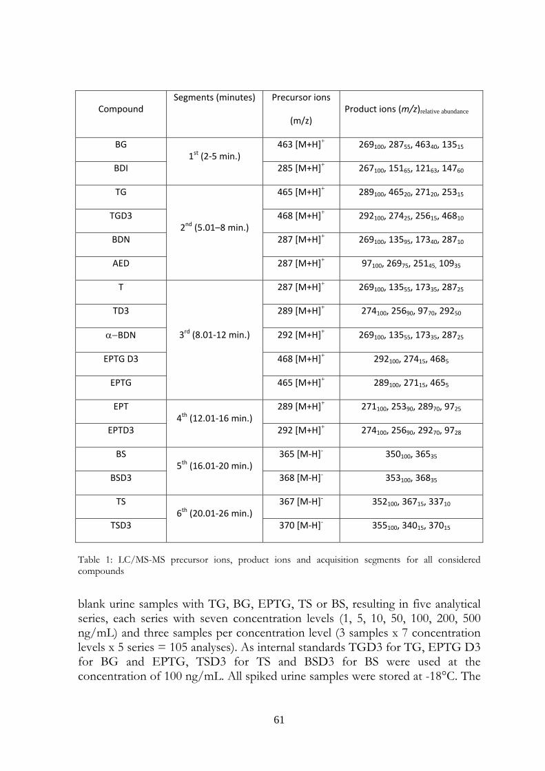
61
Compound Segments (minutes) Precursor ions
(m/z) Product ions (m/z)relative abundance
BG 1st (2‐5 min.)
463 [M+H]+ 269100, 28755, 46340, 13515
BDI 285 [M+H]+ 267100, 15165, 12163, 14760
TG
2nd (5.01–8 min.)
465 [M+H]+ 289100, 46520, 27120, 25315
TGD3 468 [M+H]+ 292100, 27425, 25615, 46810
BDN 287 [M+H]+ 269100, 13595, 17340, 28710
AED 287 [M+H]+ 97100, 26975, 25145, 10935
T
3rd (8.01‐12 min.)
287 [M+H]+ 269100, 13555, 17335, 28725
TD3 289 [M+H]+ 274100, 25690, 9770, 29250
α−BDN 292 [M+H]+ 269100, 13555, 17335, 28725
EPTG D3 468 [M+H]+ 292100, 27415, 4685
EPTG 465 [M+H]+ 289100, 27115, 4655
EPT 4th (12.01‐16 min.)
289 [M+H]+ 271100, 25390, 28970, 9725
EPTD3 292 [M+H]+ 274100, 25690, 29270, 9728
BS 5th (16.01‐20 min.)
365 [M‐H]- 350100, 36535
BSD3 368 [M‐H]- 353100, 36835
TS 6th (20.01‐26 min.)
367 [M‐H]- 352100, 36715, 33710
TSD3 370 [M‐H]- 355100, 34015, 37015
Table 1: LC/MS-MS precursor ions, product ions and acquisition segments for all considered compounds blank urine samples with TG, BG, EPTG, TS or BS, resulting in five analytical series, each series with seven concentration levels (1, 5, 10, 50, 100, 200, 500 ng/mL) and three samples per concentration level (3 samples x 7 concentration levels x 5 series = 105 analyses). As internal standards TGD3 for TG, EPTG D3 for BG and EPTG, TSD3 for TS and BSD3 for BS were used at the concentration of 100 ng/mL. All spiked urine samples were stored at -18°C. The
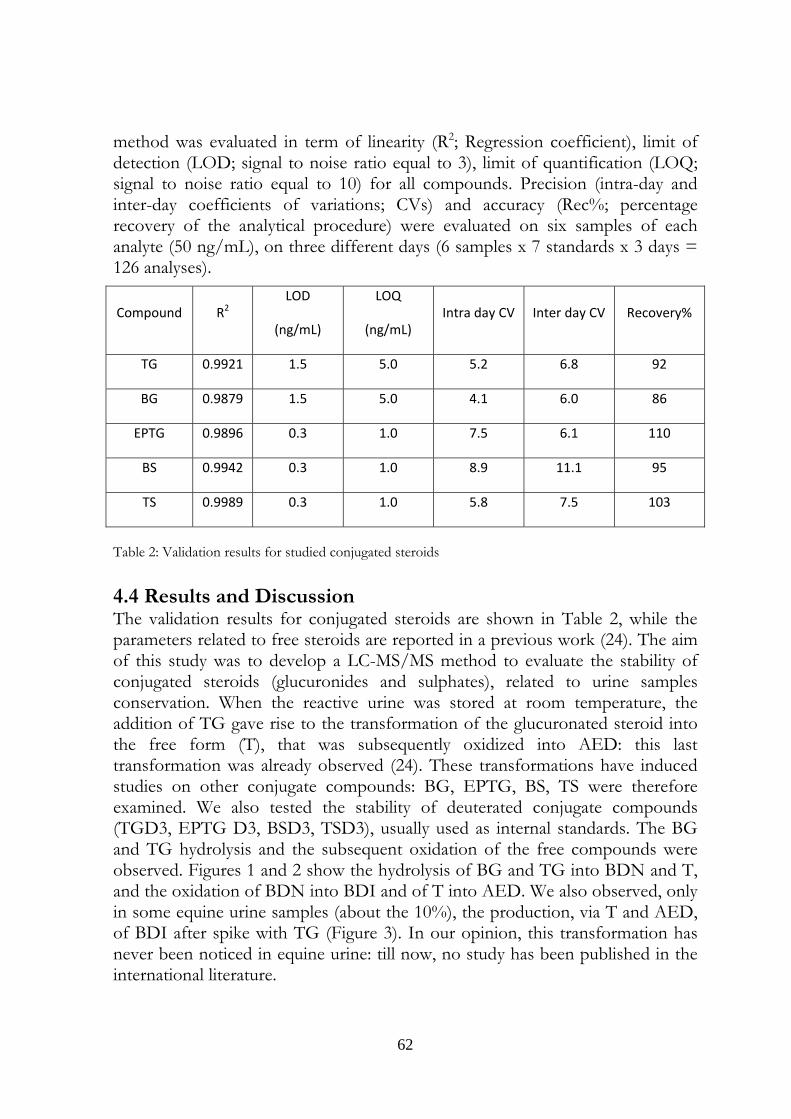
62
method was evaluated in term of linearity (R2; Regression coefficient), limit of detection (LOD; signal to noise ratio equal to 3), limit of quantification (LOQ; signal to noise ratio equal to 10) for all compounds. Precision (intra-day and inter-day coefficients of variations; CVs) and accuracy (Rec%; percentage recovery of the analytical procedure) were evaluated on six samples of each analyte (50 ng/mL), on three different days (6 samples x 7 standards x 3 days = 126 analyses).
Compound R2 LOD
(ng/mL)
LOQ
(ng/mL) Intra day CV Inter day CV Recovery%
TG 0.9921 1.5 5.0 5.2 6.8 92
BG 0.9879 1.5 5.0 4.1 6.0 86
EPTG 0.9896 0.3 1.0 7.5 6.1 110
BS 0.9942 0.3 1.0 8.9 11.1 95
TS 0.9989 0.3 1.0 5.8 7.5 103
Table 2: Validation results for studied conjugated steroids 4.4 Results and Discussion The validation results for conjugated steroids are shown in Table 2, while the parameters related to free steroids are reported in a previous work (24). The aim of this study was to develop a LC-MS/MS method to evaluate the stability of conjugated steroids (glucuronides and sulphates), related to urine samples conservation. When the reactive urine was stored at room temperature, the addition of TG gave rise to the transformation of the glucuronated steroid into the free form (T), that was subsequently oxidized into AED: this last transformation was already observed (24). These transformations have induced studies on other conjugate compounds: BG, EPTG, BS, TS were therefore examined. We also tested the stability of deuterated conjugate compounds (TGD3, EPTG D3, BSD3, TSD3), usually used as internal standards. The BG and TG hydrolysis and the subsequent oxidation of the free compounds were observed. Figures 1 and 2 show the hydrolysis of BG and TG into BDN and T, and the oxidation of BDN into BDI and of T into AED. We also observed, only in some equine urine samples (about the 10%), the production, via T and AED, of BDI after spike with TG (Figure 3). In our opinion, this transformation has never been noticed in equine urine: till now, no study has been published in the international literature.
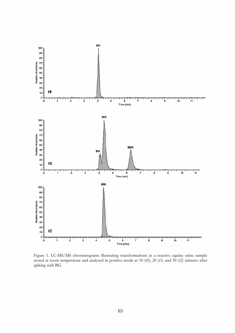
63
Figure 1. LC-MS/MS chromatograms illustrating transformations in a reactive equine urine sample stored at room temperature and analysed in positive mode at 10 (t0), 20 (t1) and 30 (t2) minutes after spiking with BG.
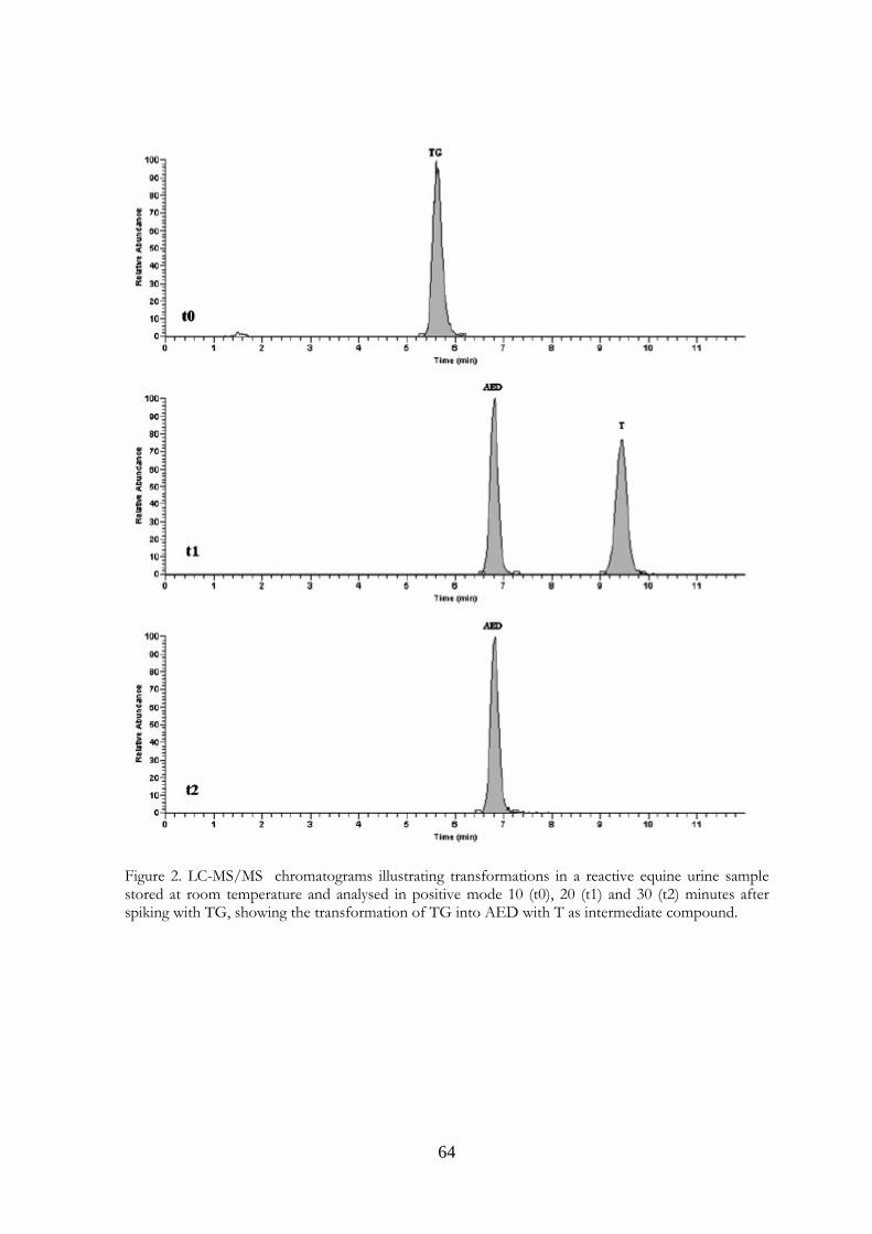
64
Figure 2. LC-MS/MS chromatograms illustrating transformations in a reactive equine urine sample stored at room temperature and analysed in positive mode 10 (t0), 20 (t1) and 30 (t2) minutes after spiking with TG, showing the transformation of TG into AED with T as intermediate compound.
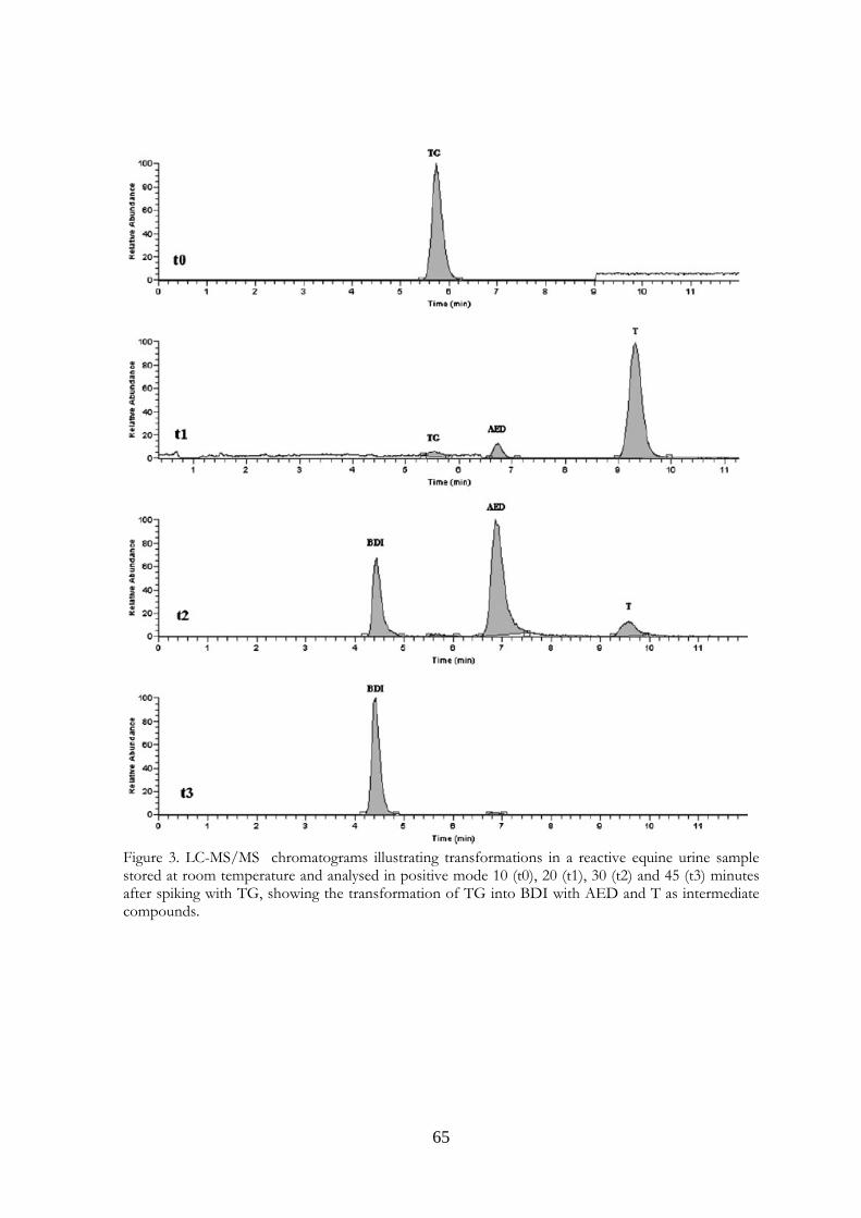
65
Figure 3. LC-MS/MS chromatograms illustrating transformations in a reactive equine urine sample stored at room temperature and analysed in positive mode 10 (t0), 20 (t1), 30 (t2) and 45 (t3) minutes after spiking with TG, showing the transformation of TG into BDI with AED and T as intermediate compounds.
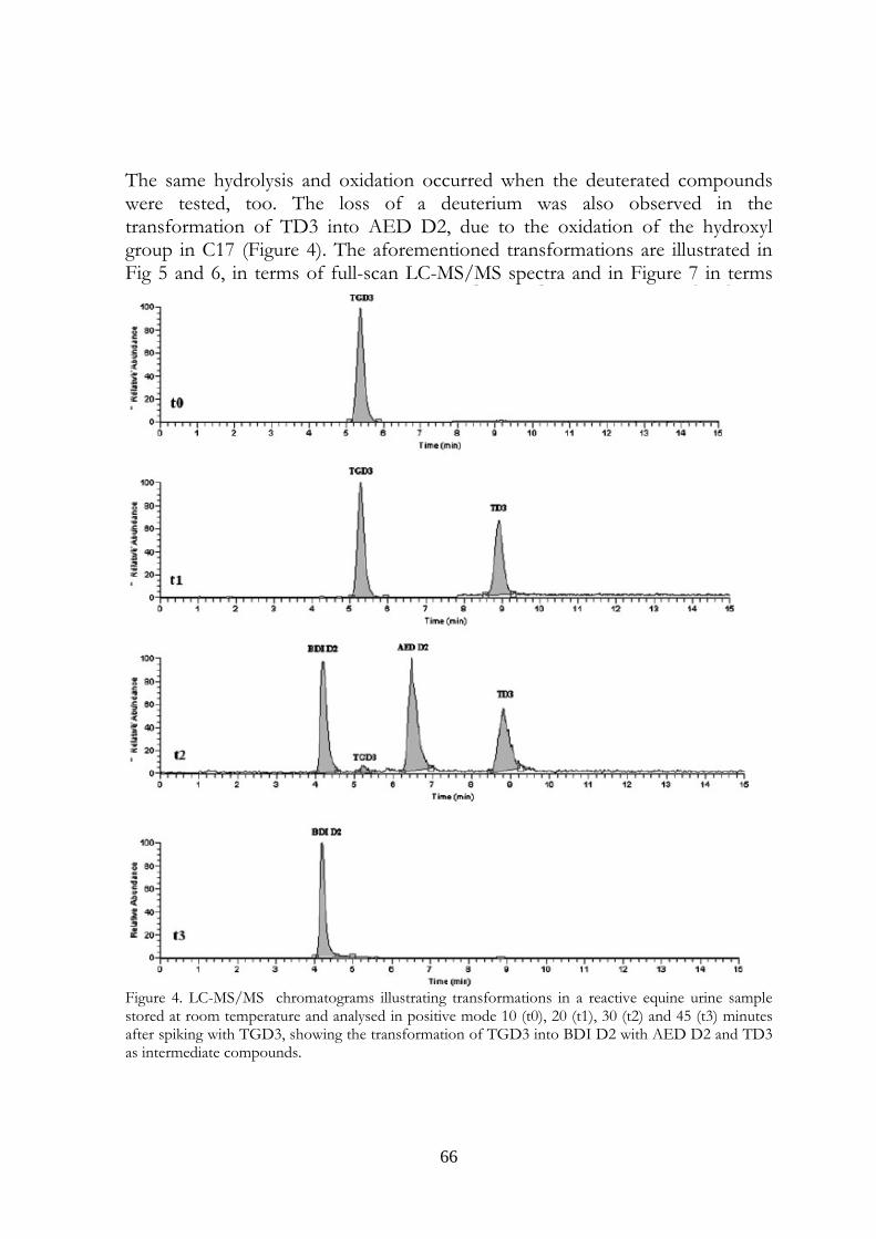
66
The same hydrolysis and oxidation occurred when the deuterated compounds were tested, too. The loss of a deuterium was also observed in the transformation of TD3 into AED D2, due to the oxidation of the hydroxyl group in C17 (Figure 4). The aforementioned transformations are illustrated in Fig 5 and 6, in terms of full-scan LC-MS/MS spectra and in Figure 7 in terms
Figure 4. LC-MS/MS chromatograms illustrating transformations in a reactive equine urine sample stored at room temperature and analysed in positive mode 10 (t0), 20 (t1), 30 (t2) and 45 (t3) minutes after spiking with TGD3, showing the transformation of TGD3 into BDI D2 with AED D2 and TD3 as intermediate compounds.
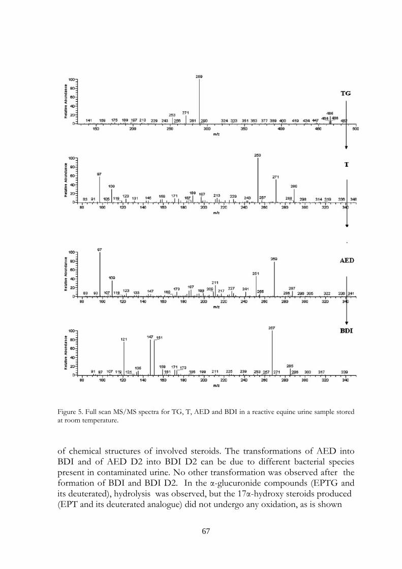
67
Figure 5. Full scan MS/MS spectra for TG, T, AED and BDI in a reactive equine urine sample stored at room temperature. of chemical structures of involved steroids. The transformations of AED into BDI and of AED D2 into BDI D2 can be due to different bacterial species present in contaminated urine. No other transformation was observed after the formation of BDI and BDI D2. In the α-glucuronide compounds (EPTG and its deuterated), hydrolysis was observed, but the 17α-hydroxy steroids produced (EPT and its deuterated analogue) did not undergo any oxidation, as is shown
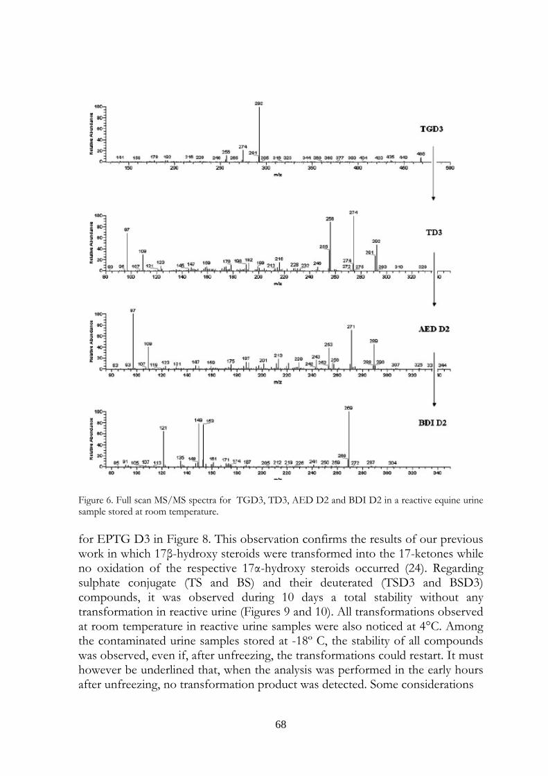
68
Figure 6. Full scan MS/MS spectra for TGD3, TD3, AED D2 and BDI D2 in a reactive equine urine sample stored at room temperature. for EPTG D3 in Figure 8. This observation confirms the results of our previous work in which 17β-hydroxy steroids were transformed into the 17-ketones while no oxidation of the respective 17α-hydroxy steroids occurred (24). Regarding sulphate conjugate (TS and BS) and their deuterated (TSD3 and BSD3) compounds, it was observed during 10 days a total stability without any transformation in reactive urine (Figures 9 and 10). All transformations observed at room temperature in reactive urine samples were also noticed at 4°C. Among the contaminated urine samples stored at -18º C, the stability of all compounds was observed, even if, after unfreezing, the transformations could restart. It must however be underlined that, when the analysis was performed in the early hours after unfreezing, no transformation product was detected. Some considerations
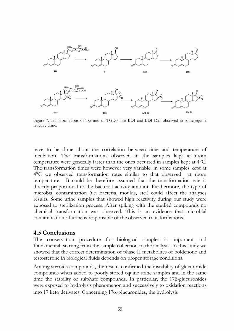
69
Figure 7. Transformations of TG and of TGD3 into BDI and BDI D2 observed in some equine reactive urine. have to be done about the correlation between time and temperature of incubation. The transformations observed in the samples kept at room temperature were generally faster than the ones occurred in samples kept at 4°C. The transformation times were however very variable: in some samples kept at 4°C we observed transformation rates similar to that observed at room temperature. It could be therefore assumed that the transformation rate is directly proportional to the bacterial activity amount. Furthermore, the type of microbial contamination (i.e. bacteria, moulds, etc.) could affect the analyses results. Some urine samples that showed high reactivity during our study were exposed to sterilization process. After spiking with the studied compounds no chemical transformation was observed. This is an evidence that microbial contamination of urine is responsible of the observed transformations.
4.5 Conclusions The conservation procedure for biological samples is important and fundamental, starting from the sample collection to the analysis. In this study we showed that the correct determination of phase II metabolites of boldenone and testosterone in biological fluids depends on proper storage conditions. Among steroids compounds, the results confirmed the instability of glucuronide compounds when added to poorly stored equine urine samples and in the same time the stability of sulphate compounds. In particular, the 17ß-glucuronides were exposed to hydrolysis phenomenon and successively to oxidation reactions into 17 keto derivates. Concerning 17α-glucuronides, the hydrolysis
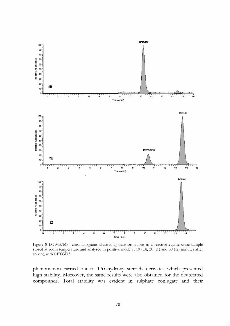
70
Figure 8 LC-MS/MS chromatograms illustrating transformations in a reactive equine urine sample stored at room temperature and analysed in positive mode at 10 (t0), 20 (t1) and 30 (t2) minutes after spiking with EPTGD3.
phenomenon carried out to 17α-hydroxy steroids derivates which presented high stability. Moreover, the same results were also obtained for the deuterated compounds. Total stability was evident in sulphate conjugate and their
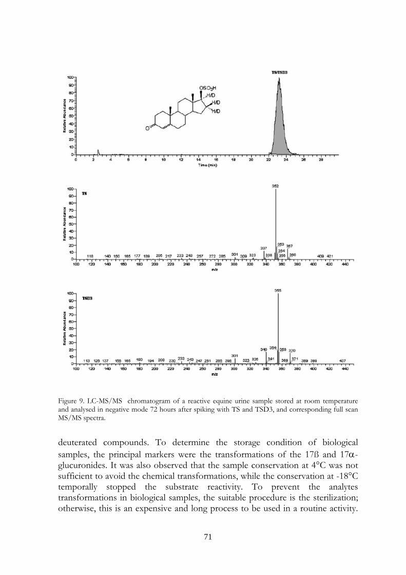
71
Figure 9. LC-MS/MS chromatogram of a reactive equine urine sample stored at room temperature and analysed in negative mode 72 hours after spiking with TS and TSD3, and corresponding full scan MS/MS spectra. deuterated compounds. To determine the storage condition of biological samples, the principal markers were the transformations of the 17ß and 17α-glucuronides. It was also observed that the sample conservation at 4°C was not sufficient to avoid the chemical transformations, while the conservation at -18°C temporally stopped the substrate reactivity. To prevent the analytes transformations in biological samples, the suitable procedure is the sterilization; otherwise, this is an expensive and long process to be used in a routine activity.
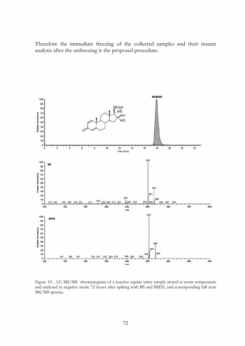
72
Therefore the immediate freezing of the collected samples and their instant analysis after the unfreezing is the proposed procedure.
Figure 10. . LC-MS/MS chromatogram of a reactive equine urine sample stored at room temperature and analysed in negative mode 72 hours after spiking with BS and BSD3, and corresponding full scan MS/MS spectra.
73
4.6 References 1) Houghton E. Proceedings of the 9thInternational Conference of Racing Analyst and Veterinarians, New Orleans, 1992; 3.
2) Roig M, Segura J,Ventura R. Anal. Chim. Acta 2007; 586: 184.
3) Yu NH, Ho ENM, Leung DKK, Wan TSM. Journal of Pharmaceutical and Biomedical Analysis 2005; 37, 1031.
4) Guan F, Uboh CE, Soma LR, Luo Y, Rudy J, Tobin T. J. Chromatogr. B 2005; 829: 56.
5) Bean KA, Henion JD. J. Chromatogr. B 1997; 690: 65.
6) Pu F, Mc Kinney AR, Stenhouse AM, Suann CJ, McLeod MD. J. Chromatogr. B 2004; 813: 241.
7) Ayotte C, Goudreault D, Charlebois A. J. Chromatogr. B 1996; 687: 3.
8) Dehennin L, Lafarge P, Dailly P, Bailloux D, Lafarge JP, J. Chromatogr. B 1996, 687: 85.
9) Borts DJ, Bowers LD. J. Mass Spectrom. 2000, 35: 50.
10) Antignac JP, Brosseaud A, Gaudin-Hirret I, Andrè F, Le Bizec B. Steroids 2005: 70: 50.
11) Buiarelli F, Cartoni GP, Coccoli F, De Rossi A, Neri B. J. Chromatogr. B 2003; 784: 1.
12) Draisci R, Palleschi L, Ferretti E, Lucentini L, delli Quadri F. J. Chromatogr. B 2003; 789: 219.
13) Kuuranne T, Kotihao T, Pedersen-Bjergaard S, Einar Rasmussen K, Leinonen A, Westwood S, Kostianen R. J. Mass Spectrom. 2003, 38: 16.
14) Van de Kerhof DH, de Boer D, Thijssen JHH, Maes RAA. J. Anal. Toxicol. 2000; 24: 102.
15) Robertson MD, Drummer OH. J. Forensic Sci. 1998; 43: 5.
16) Robertson MD, Drummer OH. J. Forensic Sci. 1995; 40: 382.
17) Saudan C, Enteza JM, Baume N, Mangin P, Saugy M. J. Chromatogr. B 2006; 844: 168.
74
18) Wilson ML, Gaido L, Clin. Infect. Dis. 2004; 38: 1150.
19) Jimenez C, De La Torre R, Segura J, Ventura R. Rap. Comm. Mass Spectrometry 2006; 20: 858.
20) De la Torre R, De la Torre X, Alia C, Segura J, Baro T, Torres-Rodriguez JM. Anal. Biochem. 2001; 289: 116.
21) Yu NH, Ho ENM, Leung DKK, Wan TSM. J. Pharm. Biomed. Anal. 2005; 37: 1031.
22) Bilton RF. Lancet 1995; 345: 1186.
23) Kickman AT, Fallon JK, Cowan DA, Walker C, Easmon S, Mackintosh D. Clinical Chemistry 2002; 48: 1799.
24) Fidani M, Casagni E, Montana M, Pasello E, Pecoraro C, Gambaro V, Rap. Comm. Mass Spectrometry 2006; 20: 2441.
75
Investigation on possible transformations of cortisol, cortisone
and cortisol glucuronide in bovine faecal matter
using liquid chromatography-mass spectrometry.
Published in:
STEROIDS, 75:350–354, 2010.
CHAPTER 5

76
77
..si qui forte mearum ineptiarum lectores eritis manusque vestras non horrebitis admovere nobis..
Gaius Valerius Catullus
5. Investigation on possible transformations of cortisol, cortisone and cortisol glucuronide in bovine faecal matter using liquid chromatography-mass spectrometry.
Francesco Ariolia,c, Marco Fidanib, Alessio Casatia, Maria L. Fracchiollab, Giuseppe Pompaa. aDepartment of Veterinary Sciences and Technologies for Food Safety, University of Milan, Milan, Italy; bU.N.I.R.E. Lab. S.r.l., Via Gramsci 70, 20019 Settimo Milanese (MI), Italy cCorrespondence: Francesco Arioli, Department of Veterinary Sciences and Technologies for Food Safety, University of Milan, Via Celoria, 10, 20133 Milan, Italy; E-mail [email protected]; tel. +390250317877; fax +390250317890 Work originated from the Department of Veterinary Sciences and Technologies for Food Safety, University of Milan, Via Celoria, 10, 20133 Milan, Italy
5.1 Abstract Given the close resemblance of the ring A structure of prednisolone and prednisone on the one hand, and of androstadienedione on the other, the transformation of cortisol and cortisone into prednisolone and prednisone in cattle faeces was evaluated. A simple method that does not involve extraction but only the 1:100 dilution of cattle faeces, spiking with 400 ng/mL cortisol, cortisone or cortisol glucuronide and incubation of the suspension, was used. The analyses were performed by HPLC-MS3 to detect the supposed Δ1 dehydrogenation of the glucocorticoids. The decision limits (CCα) and detection capabilities (CCβ) were 2.0 and 3.0 ng/mL for cortisol, cortisone and prednisolone, 3.0 and 4.0 ng/mL for cortisol glucuronide and 7.0 and 10.0 ng/mL for prednisone, respectively. Intrα−day and inter-day coefficients of variation (CV%), were 5.6–6.2 and 5.2–6.6 for cortisol glucuronide, cortisol, cortisone and prednisolone, and 16.0 and 16.2 for prednisone, respectively. The
78
recoveries were in the range 110–143% for all analytes. Regression coefficients (R2) were in the range 0.996–0.999 for all analytes. The results show the hydrolysis of the conjugated form and the dehydrogenation in ring A in diluted faeces. It is therefore predicted that urine contaminated with faeces may be positive for prednisone and prednisolone in the same way as they are positive for boldenone, i.e. as a result of microbiological dehydrogenase activity on cortisol and cortisone.
5.2 Key words Cortisol, cortisone, prednisolone, prednisone, bovine faeces, HPLC-MS3.
5.3 Introduction Prednisolone is a glucocorticoid and its structure differs from cortisol by the double bond in position 1 of ring A of the steroidal nucleus of the molecule. The same difference exists between prednisone and cortisone (Table 1). In 1955, Bunim et al. reported that prednisone and prednisolone possess increased anti-inflammatory properties (about four-fold higher (1)) in comparison with cortisone and cortisol (2). In the same year Nobile and co-workers discovered that the oxidation of the latter two corticosteroids by Corynebacterium simplex can be used to prepare their Δ1 dehydrogenated analogues (3, 4). The microbiological introduction of the double bond in position 1,2-(Δ1) of the steroid structure is still used for the production of prednisone and prednisolone, as the chemical route is inferior in both product purity and yield (5,6). In the EU, the use of corticosteroids in livestock is indicated for therapeutic reasons only and is regulated by the Council Regulation (EEC) N° 2377/90 (7) and the subsequent updates: maximum residue limits (MRLs) for betamethasone in cattle and pigs (from 0.75 µg/Kg in muscle to 2.0 µg/Kg in liver) (8), dexamethasone in cattle, pigs, horses (from 0.75 µg/Kg in milk to 2.0 µg/Kg in liver) (9) and goats (from 0.75 µg/Kg in cow’s and goat’s milk to 2.0 µg/Kg in liver) (10); methylprednisolone (not for use in milk producing animals; 10 µg/Kg in all tissues) (11) and prednisolone (from 4 μg/kg in muscle and fat; to 10 μg/kg in liver and kidney) (12) for cattle are indicated in Annex I. No MRLs are indicated for prednisone, even though the European Agency for the Evaluation of Medicinal Products (EMEA) has reported the interconversion of prednisolone and prednisone in cattle (13). The Italian National Residue Program (PNR) follows the indications of the European Directives 96/22/EC (14) and 96/23/EC (15): corticosteroids, like anabolic steroids, are therefore categorized in class A3 (substances having an
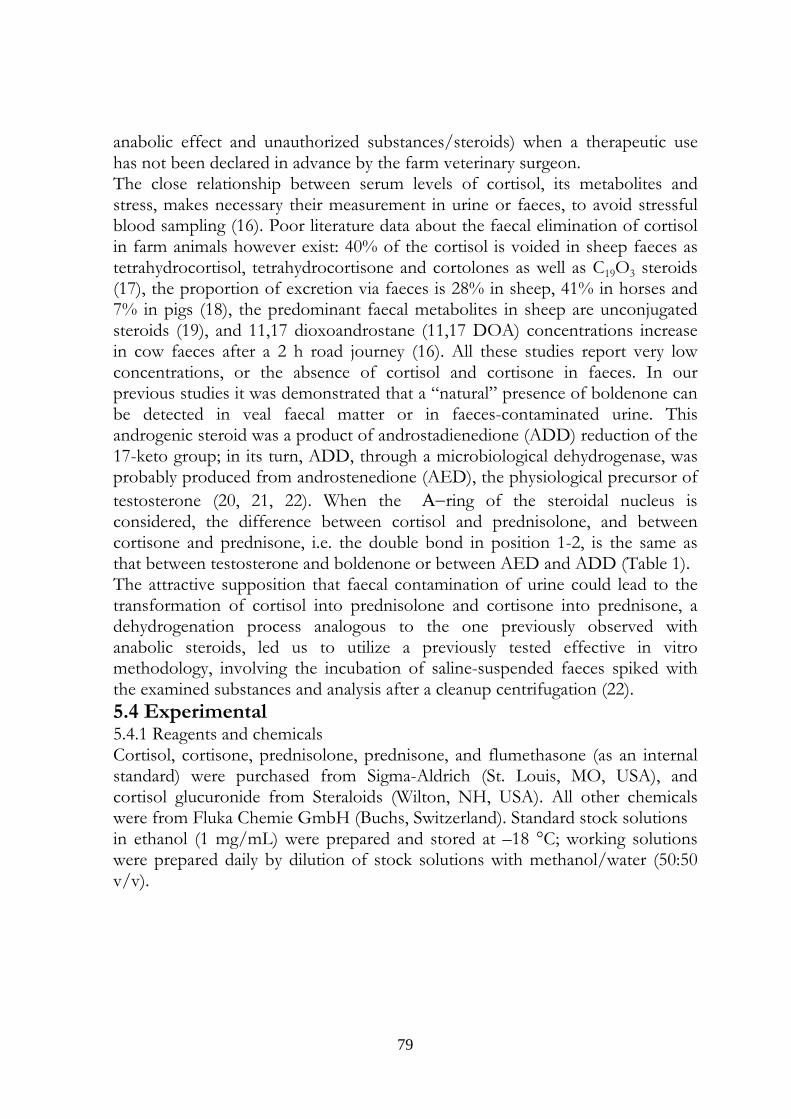
79
anabolic effect and unauthorized substances/steroids) when a therapeutic use has not been declared in advance by the farm veterinary surgeon. The close relationship between serum levels of cortisol, its metabolites and stress, makes necessary their measurement in urine or faeces, to avoid stressful blood sampling (16). Poor literature data about the faecal elimination of cortisol in farm animals however exist: 40% of the cortisol is voided in sheep faeces as tetrahydrocortisol, tetrahydrocortisone and cortolones as well as C19O3 steroids (17), the proportion of excretion via faeces is 28% in sheep, 41% in horses and 7% in pigs (18), the predominant faecal metabolites in sheep are unconjugated steroids (19), and 11,17 dioxoandrostane (11,17 DOA) concentrations increase in cow faeces after a 2 h road journey (16). All these studies report very low concentrations, or the absence of cortisol and cortisone in faeces. In our previous studies it was demonstrated that a “natural” presence of boldenone can be detected in veal faecal matter or in faeces-contaminated urine. This androgenic steroid was a product of androstadienedione (ADD) reduction of the 17-keto group; in its turn, ADD, through a microbiological dehydrogenase, was probably produced from androstenedione (AED), the physiological precursor of testosterone (20, 21, 22). When the Α−ring of the steroidal nucleus is considered, the difference between cortisol and prednisolone, and between cortisone and prednisone, i.e. the double bond in position 1-2, is the same as that between testosterone and boldenone or between AED and ADD (Table 1). The attractive supposition that faecal contamination of urine could lead to the transformation of cortisol into prednisolone and cortisone into prednisone, a dehydrogenation process analogous to the one previously observed with anabolic steroids, led us to utilize a previously tested effective in vitro methodology, involving the incubation of saline-suspended faeces spiked with the examined substances and analysis after a cleanup centrifugation (22). 5.4 Experimental 5.4.1 Reagents and chemicals Cortisol, cortisone, prednisolone, prednisone, and flumethasone (as an internal standard) were purchased from Sigma-Aldrich (St. Louis, MO, USA), and cortisol glucuronide from Steraloids (Wilton, NH, USA). All other chemicals were from Fluka Chemie GmbH (Buchs, Switzerland). Standard stock solutions in ethanol (1 mg/mL) were prepared and stored at –18 °C; working solutions were prepared daily by dilution of stock solutions with methanol/water (50:50 v/v).
80
Table 1. Structures of the studied free glucocorticoids and testosterone, androstenedione (AED), boldenone and androstadienedione (ADD).
1-2 SINGLE BOND
Cortisol
Cortisone
Testosterone
AED
1-2 DOUBLE BOND
Prednisolone
Prednisone
Boldenone
ADD
81
5.4.2 Animals Faeces from 24 male Friesian veal calves, age 120–140 days, were studied. The animals were housed on farms in North Italy in individual stalls and were cared for in accordance with EU guidelines approved by the Italian Ministry of Health. 5.4.3 Faeces collection and sample preparation Eight pools of rectal faeces, each one from three veal calves, were prepared. From each pooled material sample, 1.2 g was suspended in 120 mL of 0.9% saline and shaken overnight at 25 °C. The suspension was divided into four 30 mL samples: one blank (without the addition of glucocorticoid) and three treatments (cortisol, cortisone or cortisol glucuronide) to a final concentration of 400 ng/mL (40 µg/g faeces) and allowed to stand at ambient temperature. Internal standard flumethasone was added to the blank and the treatments. Samples (1 mL) from each tube were collected 0, 1, 2, 4, 8 and 24 h later after shaking. The samples were heated at 80 °C for 15 min to inactivate bacteria and centrifuged (3000 g, 15 min). Twenty µL of the supernatant were analysed by LC-MS3 without extraction.
5.4.4 LC-MS3 Mass spectra were recorded using a LCQ DECA XP Max ion trap mass spectrometer equipped with a Surveyor LC pump and a Surveyor autosampler (ThermoFinnigan, San Jose, CA, USA). The column was a 100 mm x 2.1 mm i.d., 3 µm Allure Biphenyl (Restek Corporation, Bellefonte, PA, USA) in an oven set at 30 °C. The mobile phase consisted of a mixture of 40% water with 0.1% formic acid and 60% methanol (isocratic elution) at a flow rate of 0.2 mL/min. The mass spectrometer was operated in negative electrospray ionization (ESI - ) mode with the following conditions: sheath and auxiliary gas (nitrogen) flow rates of 40 and 18 arbitrary units, respectively; spray voltage 5.50 kV; ion transfer capillary temperature 245 °C, capillary voltage –23 V; tube lens offset –77V. Helium was used for collision-induced dissociation. The analysis was performed in consecutive reaction monitoring (CRM) mode, and for the free forms, the formation of formiate adduct molecular ions [M+HCOO]- was observed, and the transitions to product ions were monitored, as reported in Table 2. The data acquisition software was Xcalibur® (ThermoFinnigan, San Jose, CA, USA). 5.4.5 Method validation Thermally treated (15 min at 80 °C) suspended faeces samples were spiked to give known concentrations (1.0, 2.0, 10, 50 or 100, 200 and 500 ng/mL) of analyte and of internal standard (flumethasone, 10 ng/mL), were analysed
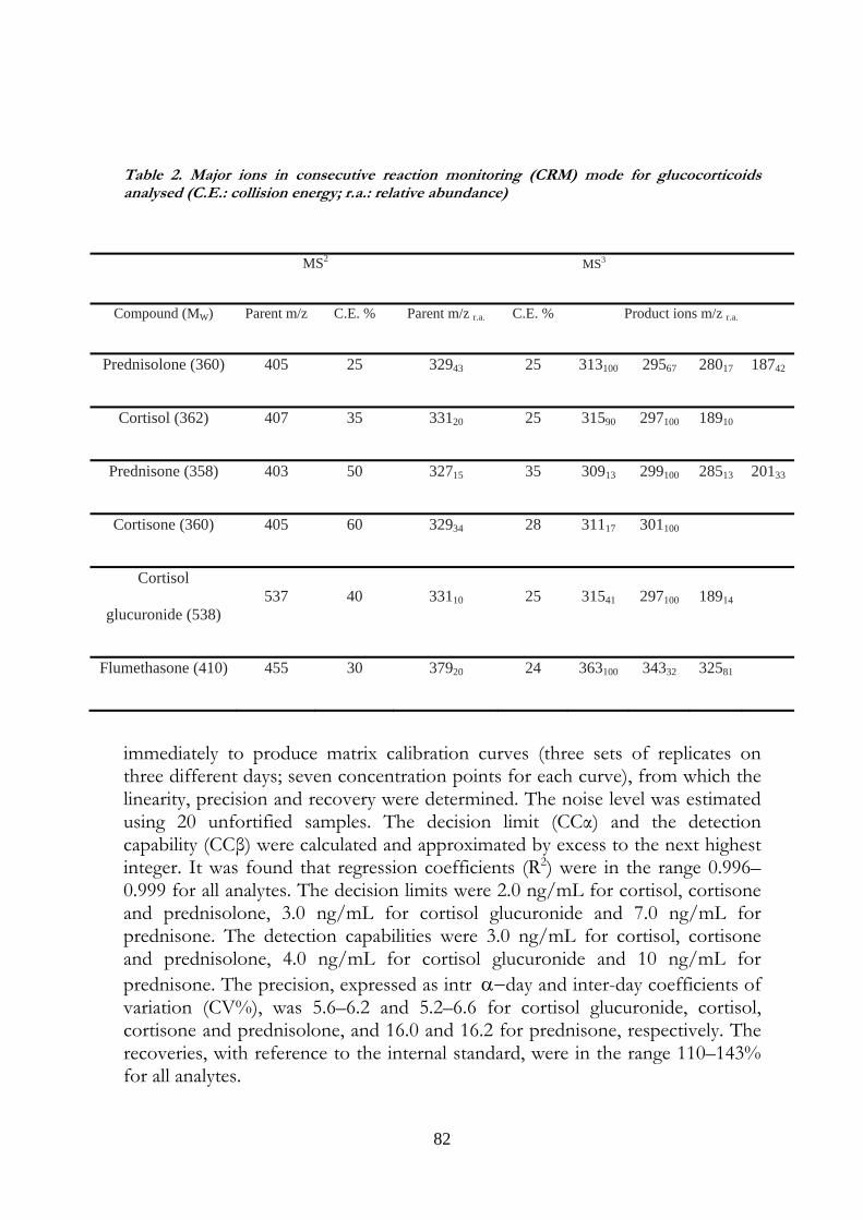
82
Table 2. Major ions in consecutive reaction monitoring (CRM) mode for glucocorticoids analysed (C.E.: collision energy; r.a.: relative abundance)
immediately to produce matrix calibration curves (three sets of replicates on three different days; seven concentration points for each curve), from which the linearity, precision and recovery were determined. The noise level was estimated using 20 unfortified samples. The decision limit (CCα) and the detection capability (CCβ) were calculated and approximated by excess to the next highest integer. It was found that regression coefficients (R2) were in the range 0.996–0.999 for all analytes. The decision limits were 2.0 ng/mL for cortisol, cortisone and prednisolone, 3.0 ng/mL for cortisol glucuronide and 7.0 ng/mL for prednisone. The detection capabilities were 3.0 ng/mL for cortisol, cortisone and prednisolone, 4.0 ng/mL for cortisol glucuronide and 10 ng/mL for prednisone. The precision, expressed as intr α−day and inter-day coefficients of variation (CV%), was 5.6–6.2 and 5.2–6.6 for cortisol glucuronide, cortisol, cortisone and prednisolone, and 16.0 and 16.2 for prednisone, respectively. The recoveries, with reference to the internal standard, were in the range 110–143% for all analytes.
MS2 MS3
Compound (MW) Parent m/z C.E. % Parent m/z r.a. C.E. % Product ions m/z r.a.
Prednisolone (360) 405 25 32943 25 313100 29567 28017 18742
Cortisol (362) 407 35 33120 25 31590 297100 18910
Prednisone (358) 403 50 32715 35 30913 299100 28513 20133
Cortisone (360) 405 60 32934 28 31117 301100
Cortisol
glucuronide (538) 537 40 33110 25 31541 297100 18914
Flumethasone (410) 455 30 37920 24 363100 34332 32581
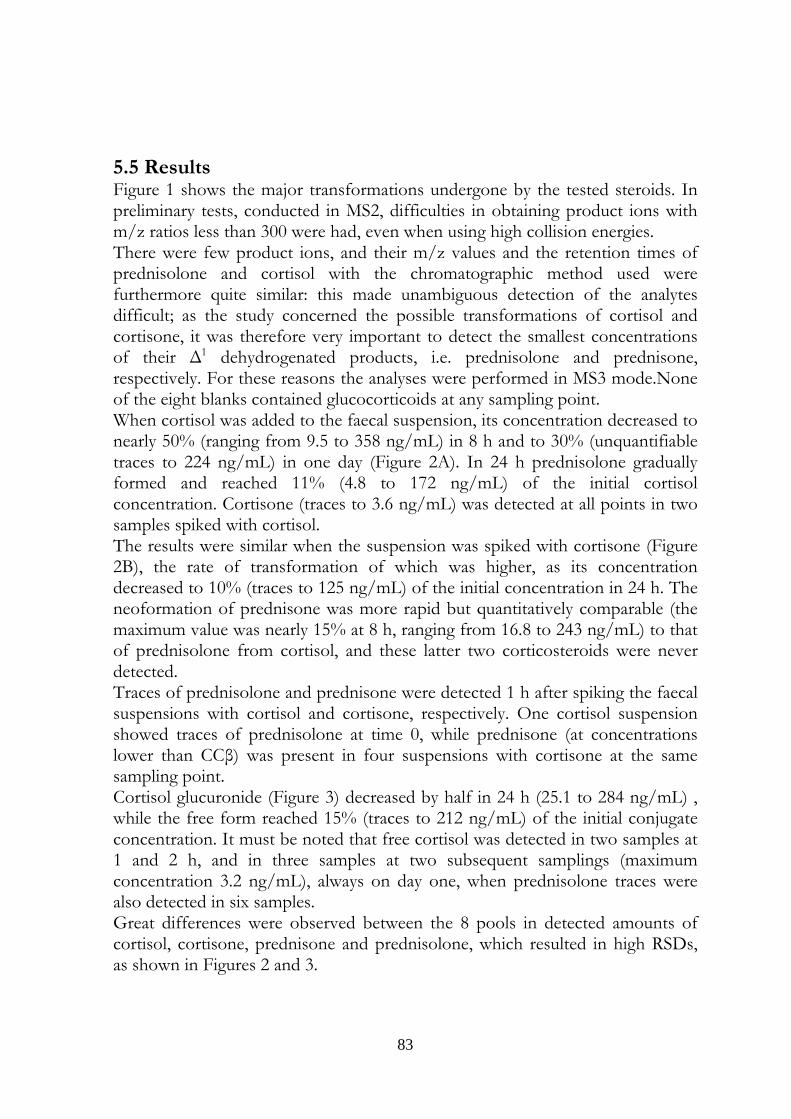
83
5.5 Results Figure 1 shows the major transformations undergone by the tested steroids. In preliminary tests, conducted in MS2, difficulties in obtaining product ions with m/z ratios less than 300 were had, even when using high collision energies. There were few product ions, and their m/z values and the retention times of prednisolone and cortisol with the chromatographic method used were furthermore quite similar: this made unambiguous detection of the analytes difficult; as the study concerned the possible transformations of cortisol and cortisone, it was therefore very important to detect the smallest concentrations of their Δ1 dehydrogenated products, i.e. prednisolone and prednisone, respectively. For these reasons the analyses were performed in MS3 mode.None of the eight blanks contained glucocorticoids at any sampling point. When cortisol was added to the faecal suspension, its concentration decreased to nearly 50% (ranging from 9.5 to 358 ng/mL) in 8 h and to 30% (unquantifiable traces to 224 ng/mL) in one day (Figure 2A). In 24 h prednisolone gradually formed and reached 11% (4.8 to 172 ng/mL) of the initial cortisol concentration. Cortisone (traces to 3.6 ng/mL) was detected at all points in two samples spiked with cortisol. The results were similar when the suspension was spiked with cortisone (Figure 2B), the rate of transformation of which was higher, as its concentration decreased to 10% (traces to 125 ng/mL) of the initial concentration in 24 h. The neoformation of prednisone was more rapid but quantitatively comparable (the maximum value was nearly 15% at 8 h, ranging from 16.8 to 243 ng/mL) to that of prednisolone from cortisol, and these latter two corticosteroids were never detected. Traces of prednisolone and prednisone were detected 1 h after spiking the faecal suspensions with cortisol and cortisone, respectively. One cortisol suspension showed traces of prednisolone at time 0, while prednisone (at concentrations lower than CCβ) was present in four suspensions with cortisone at the same sampling point. Cortisol glucuronide (Figure 3) decreased by half in 24 h (25.1 to 284 ng/mL) , while the free form reached 15% (traces to 212 ng/mL) of the initial conjugate concentration. It must be noted that free cortisol was detected in two samples at 1 and 2 h, and in three samples at two subsequent samplings (maximum concentration 3.2 ng/mL), always on day one, when prednisolone traces were also detected in six samples. Great differences were observed between the 8 pools in detected amounts of cortisol, cortisone, prednisone and prednisolone, which resulted in high RSDs, as shown in Figures 2 and 3.
84
5.6 Discussion Glucocorticoids were never detected in the blank, non-spiked samples, probably because of the 1:100 dilution of faeces in saline; nevertheless, in spiked samples, traces of glucocorticoids other than the standards used were found at time 0. The supplier indicated that the purity of the glucocorticoids in the free form was greater than or equal to 98%, without specifying what the impurity could be. An analysis of a standard cortisol solution at the concentration of 100 ng/mL was performed, to verify whether the other studied corticosteroids were present as impurities. Non-quantifiable traces of prednisolone were detected. If the decision limits (from 2 to 7 ng/mL), the variabilities (CV values nearly 6%) and the concentrations (400 ng/mL) of the corticosteroids studied here are considered, the unexpected detection of traces at time 0 is understandable. Accordingly, prednisone could constitute a greater impurity in cortisone due to its high CCα, while the presence of cortisone at very low levels in two cortisol-spiked samples was considered to be due to an impurity in the standard used. The experimental model used to test the transformation of cortisol and cortisone into their Δ1 dehydrogenated derivatives is the first step in attempts to evaluate whether prednisolone and prednisone may be considered, like boldenone and ADD, as “synthetically produced hormones that are also known to be endogenous under certain conditions, dubbed ‘pseudo-endogenous’ or ‘grey zone substances’ due to their dual synthetic/endogenous nature” (23). The faecal suspensions were therefore utilized as if they were microbiological cultures with Δ1 dehydrogenase activity, as proved for a wide variety of microorganisms (5). As expected, the hydrolysis of cortisol glucuronide was observed, which was consistent with previous results obtained in bovine faeces (unpublished work) and in poorly conserved equine urine (24), in which the glucuronide forms of anabolic steroids underwent hydrolysis into their free forms. The transformations of cortisol to prednisolone and of cortisone to prednisone are similar to what observed in bovine faeces with respect to the transformation of AED to ADD (22) i.e. the introduction of a double bond in position 1,2-(Δ1) of the steroid structure. The hydrolysis of cortisol glucuronide is furthermore consistent with the data concerning the faecal elimination of unconjugated corticosteroids (17, 25); the prednisolone traces found in six samples after one day do not indicate with certainty the dehydrogenation of the free form, even if this possibility cannot be ruled out.
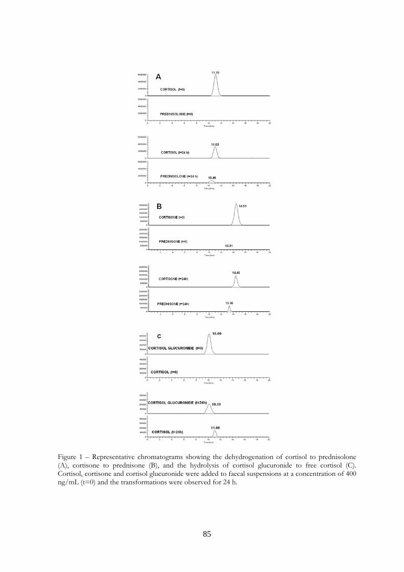
85
Figure 1 – Representative chromatograms showing the dehydrogenation of cortisol to prednisolone (A), cortisone to prednisone (B), and the hydrolysis of cortisol glucuronide to free cortisol (C). Cortisol, cortisone and cortisol glucuronide were added to faecal suspensions at a concentration of 400 ng/mL (t=0) and the transformations were observed for 24 h.
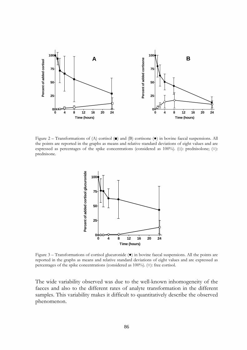
86
0 4 8 12 16 20 240
25
50
75
100
Time (hours)
Perc
ent o
f add
ed c
ortis
ol
0 4 8 12 16 20 240
25
50
75
100
Time (hours)
Perc
ent o
f add
ed c
ortis
oneA B
Figure 2 – Transformations of (A) cortisol (■) and (B) cortisone (●) in bovine faecal suspensions. All the points are reported in the graphs as means and relative standard deviations of eight values and are expressed as percentages of the spike concentrations (considered as 100%). (□): prednisolone; (○): prednisone.
0 4 8 12 16 20 240
25
50
75
100
Time (hours)
Perc
ent o
f add
ed c
ortis
ol g
lucu
roni
de
Figure 3 – Transformations of cortisol glucuronide (●) in bovine faecal suspensions. All the points are reported in the graphs as means and relative standard deviations of eight values and are expressed as percentages of the spike concentrations (considered as 100%). (○): free cortisol.
The wide variability observed was due to the well-known inhomogeneity of the faeces and also to the different rates of analyte transformation in the different samples. This variability makes it difficult to quantitatively describe the observed phenomenon.
87
Finally, even though cortisol and cortisone are very frequently absent and only sometimes present at very low levels in faeces, the increase in concentration of these glucocorticoids in stressful situations, i.e. after transport or at the slaughterhouse, should be more accurately quantified. The use of high concentrations of these substances to evaluate the supposed Δ1 dehydrogenation was therefore decided. This work is the preliminary phase of a huge study on cortisol and cortisone transformations that will be performed in vitro on faeces and urine, and in vivo on stressed bovines.
5.7 Acknowledgments This work was supported by a grant from the Regione Lombardia for the Research Project: “Presenza e persistenza di farmaci e ormoni nelle deiezioni animali e nel liquame per il potenziamento dell’attività di farmacosorveglianza”. 5.8 References 1) Brunton LL (editor-in-chief), Keith L, Parker KL, Murri N, Blumenthal DK (associate editors), Goodman & Gilman's the pharmacological basis of therapeutics, online 11th edition, http://www.accessmedicine.com. 2) Bunim JJ, Pechet MM, Bollet AJ. 1955. Studies on metacortandralone and metacortandracin in rheumatoid arthritis; antirheumatic potency, metabolic effects, and hormonal properties. JAMA. 157:311–318. 3) Nobile A, Charney W, Perlman PL, Herzog HL, Payne CC, Tully ME, Jevnik MA, Hershberg EB. 1955. Microbiological transformation of steroids I. Δ1,4-Diene-3-ketosteroids. Am J Chem Soc. 77:4184. 4) Stoudt TH. 1960. Advances in applied microbiology, vol. 2, New York: Academic Press, p. 204, editor: Umbreit WW. 5) Adham NZ, El-Hady AA, Naim N. 2003. Biochemical studies on the microbial Δ1-dehydrogenation of cortisol by Pseudomonas fluorescens, Process Biochemistry. 38:897–902. 6) Kieslich K. 1980. Microbial enzymes and bioconversions: economic microbiology, vol. 5, London, Academic, pp. 369–465, editor: Rose AH. 7) EEC Council Regulation N° 2377/1990, Official Journal of the European Community L 224, p. 1.
88
8) EEC Council Regulation N° 2593/1999, Official Journal of the European Community L 315 p. 26. 9) EEC Council Regulation N° 508/1999 Official Journal of the European Community L 60 p.16. 10) EEC Council Regulation N° 1646/2004 Official Journal of the European Community L 296 p. 5. 11) EEC Council Regulation N° 77/2002 Official Journal of the European Community L 16 p. 9. 12) EEC Council Regulation N° 2535/2000 Official Journal of the European Community L 291 p. 9. 13) Committee for Veterinary Medicinal Products: Prednisolone, EMEA/MRL/629/99-Final, July 1999, http://www.emea.europa.eu/pdfs/vet/mrls/062999en.pdf 14) EEC Council Directive N° 22/1996 Official Journal of the European Community L 12 p. 3. 15) EEC Council Directive N° 23/1996 Official Journal of the European Community L 125 p. 10. 16) Möstl E, Palme R. 2002. Hormones as indicators of stress, Domest Anim Endocrinol. 23:67–74. 17) Lindner HR. 1972. Enterohepatic circulation and patterns of urinary excretion of cortisol metabolites in the ewe. J Endocrinol. 52:XIX-XX. 18) Palme R, Fischer P, Schildorfer H, Ismail MN. 1996. Excretion of infused C-steroid hormones via faeces and urine in domestic livestock. Anim Reprod Sci. 43:43–63. 19) Palme R, Möstl E, 1997. Measurement of cortisol metabolites in faeces of sheep as a parameter of cortisol concentration in blood. Int J Mammal Biol. 62 (S2):192–197.
89
20) Sgoifo Rossi CA, Arioli F, Bassini A, Chiesa LM, Dell’Orto V, Montana M, Pompa G. 2004. Evidence for false-positive results for Boldenone testing of veal urine due to fecal cross-contamination during sampling, Food Addit Contam. 21:756–762. 21) Pompa G, Arioli F, Fracchiolla ML, Sgoifo Rossi CA, Bassini AL, Stella S, Biondi PA. 2006. Neoformation of Boldenone and related steroids in faeces of veal calves, Food Addit Contam. 23:126–132. 22) Arioli F, Gavinelli MP, Fracchiolla ML, Casati A, Fidani M, Ferrer E, Pompa G. 2008. Evaluation of boldenone formation and related steroids transformations in veal faeces by liquid chromatography/tandem mass spectrometry. Rapid Comm Mass Spectr. 22:217–223. 23) Scarth J, Akre C, van Ginkel L, Le Bizec B, De Brabander H, Korth W, Points J, Teale P, Kay J. 2009. Presence and metabolism of endogenous androgenic-anabolic steroid hormones in meat-producing animals: a review. Food Addit Contam. Part A, 26:640–671. 24) Fidani M, Gamberini MC, Pasello E, Palazzoli F, De Iuliis P, Montana M, Arioli F. 2009. Evaluation of equine urine reactivity towards phase II metabolites of 17-hydroxy steroids by liquid chromatography/tandem mass spectrometry. Rapid Comm Mass Spectr. 23:65–76. 25) Palme R, Robia C, Baumgartner W, Möstl E. 2000. Transport stress in cattle as reflected by an increase in faecal cortisol metabolites. Vet Rec. 146:108–109.

90
91
Preliminary evaluation on a possible transformation of cortisol and cortisone in
bovine urine after sampling
CHAPTER 6
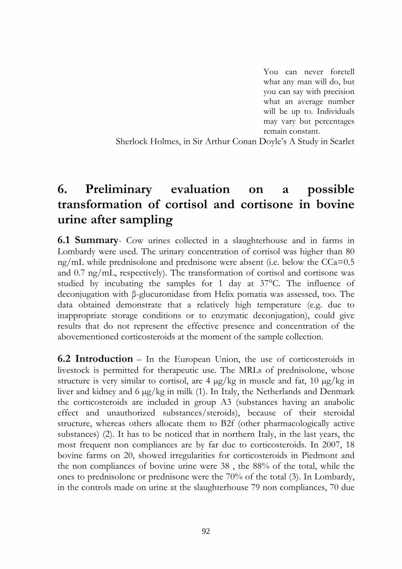
92
You can never foretell what any man will do, but you can say with precision what an average number will be up to. Individuals may vary but percentages remain constant.
Sherlock Holmes, in Sir Arthur Conan Doyle’s A Study in Scarlet
6. Preliminary evaluation on a possible transformation of cortisol and cortisone in bovine urine after sampling
6.1 Summary- Cow urines collected in a slaughterhouse and in farms in Lombardy were used. The urinary concentration of cortisol was higher than 80 ng/mL while prednisolone and prednisone were absent (i.e. below the CCa=0.5 and 0.7 ng/mL, respectively). The transformation of cortisol and cortisone was studied by incubating the samples for 1 day at 37°C. The influence of deconjugation with β-glucuronidase from Helix pomatia was assessed, too. The data obtained demonstrate that a relatively high temperature (e.g. due to inappropriate storage conditions or to enzymatic deconjugation), could give results that do not represent the effective presence and concentration of the abovementioned corticosteroids at the moment of the sample collection. 6.2 Introduction – In the European Union, the use of corticosteroids in livestock is permitted for therapeutic use. The MRLs of prednisolone, whose structure is very similar to cortisol, are 4 µg/kg in muscle and fat, 10 µg/kg in liver and kidney and 6 µg/kg in milk (1). In Italy, the Netherlands and Denmark the corticosteroids are included in group A3 (substances having an anabolic effect and unauthorized substances/steroids), because of their steroidal structure, whereas others allocate them to B2f (other pharmacologically active substances) (2). It has to be noticed that in northern Italy, in the last years, the most frequent non compliances are by far due to corticosteroids. In 2007, 18 bovine farms on 20, showed irregularities for corticosteroids in Piedmont and the non compliances of bovine urine were 38 , the 88% of the total, while the ones to prednisolone or prednisone were the 70% of the total (3). In Lombardy, in the controls made on urine at the slaughterhouse 79 non compliances, 70 due
93
to corticosteroids (89%), and more precisely 65 to prednisolone or prednisone (82%) were detected (4). The close resemblance of the A ring of prednisolone, prednisone and androstadienedione (ADD) on one side, and of cortisol, cortisone and androstenedione (AED) on the other side, is shown in Fig 1 Taking it into account, together with the observation that a previous work, concerning the transformations of anabolic steroids in equine urine, showed that from testosterone, boldenone could be produced, via its transformation into AED that dehydrogenated in position 1,2 of the ring A to give ADD (5 – chapter 4) and with the data from Piedmont ant Lombardy, the possible transformation of cortisol and/or cortisone into prednisolone/prednisone were studied. Figure 1. Chemical structure of the four corticosteroids studied in this work and of the anabolic steroids previously studied, showing their similarities in Ring A.
6.3 Experimental 6.3.1Reagents and chemicals Cortisol, cortisone, prednisolone, prednisone, flumethasone (as an internal standard) and β-glucuronidase from Helix pomatia were purchased from Sigma–Aldrich (St. Louis, MO, USA). All other chemicals were from Fluka Chemie GmbH (Buchs, Switzerland). Standard stock solutions in ethanol (1 mg/mL) were prepared and stored at −18°C; working solutions were prepared daily by dilution of stock solutions with methanol:water (50:50, v/v).
6.3.2 Sample collection and preparation In a first trial, the urines from 7 Holstein-Friesian cows, aged 27 to 80 months, were used. Five samples were from the slaughterhouse and two samples from
ANDROSTENEDIONE ANDROSTADIENEDIONE
94
two farms; all the facilities were in Lombardy. The samples, cooled at 4°C were carried to the laboratory and frozen at -18°C. In a second trial the urines from an Holstein-Friesian cow were collected at the farm and at the slaughter, with the aim to verify if β-glucuronidase from Helix pomatia, often used to catalyze the deconjugation of corticosteroids, could induce unwanted transformations, as described regarding the production of AED from testosterone (6). For the first study, 20 mL of each urine sample, underwent an incubation in a shaking water bath kept at 37±1°C. Collection of 2 mL portions were made at 0, 1, 2, 4, 8 e 24 hours. In the second trial, from each sample 3 portion of 2 mL were obtained: one portion was immediately extracted while two portions were in advance incubated at room temperature overnight. One of these last two portions contained 100 µL of β-glucuronidase. 6.3.3 Sample extraction To each 2 mL sample, 10 ng/mL of internal standard (flumethasone) and 4 mL of a mixture of tert-Buthylmethylether:Ethylacetate (80:20) were added. After a 20 min shaking in vertical rotary shaker, the samples were centrifuged for 20 min at 2000 g. The upper organic layer was collected and dried under vacuum in a centrifugal evaporator . The residue was dissolved in 200 µL of a mixture methanol:aqueous formic acid 0,1 % (50:50 v/v) and transferred in autosampler vials. 6.3.4 LC-MS3 analysis. The method of analysis has already been described in Chapter 5 (7). The analysis apparatus consisted of an AS autosampler, an LC pump Surveyor, and of an Ion trap mass spectrometer LCQ DECA XP Max (ThermoFinnigan, San Jose, CA, USA) operating in negative electrospray mode (ESI-). The chromatographic separation was achieved with an Allure Biphenyl 100 mm x 2.1 mm i.d., 3 µm (Restek Corporation, Bellefonte, PA, USA) column, kept at 30°C and an isocratic elution made with a mixture of aqueous formic acid 0.1%: methanol (40:60) having a flow of 0.2 mL/min. The acquisition of data was made in CRM (consecutive reaction monitoring) mode, the parent ion was the adduct of the corticosteroid molecule with formic acid[M+HCOO]-. In table 1 are reported the ions considered . 6.3.5 Method validation The method was validated following the Commission Decision 657/2002/CE (8) in urine with 6 concentration level, three replicates (0.5, 1.0, 2.0, 10, 50, 100
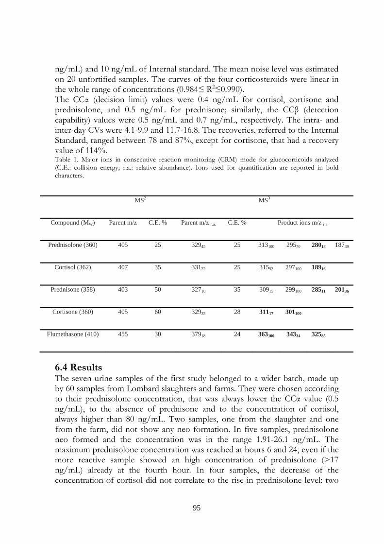
95
ng/mL) and 10 ng/mL of Internal standard. The mean noise level was estimated on 20 unfortified samples. The curves of the four corticosteroids were linear in the whole range of concentrations (0.984≤ R2≤0.990). The CCα (decision limit) values were 0.4 ng/mL for cortisol, cortisone and prednisolone, and 0.5 ng/mL for prednisone; similarly, the CCβ (detection capability) values were 0.5 ng/mL and 0.7 ng/mL, respectively. The intra- and inter-day CVs were 4.1-9.9 and 11.7-16.8. The recoveries, referred to the Internal Standard, ranged between 78 and 87%, except for cortisone, that had a recovery value of 114%. Table 1. Major ions in consecutive reaction monitoring (CRM) mode for glucocorticoids analyzed (C.E.: collision energy; r.a.: relative abundance). Ions used for quantification are reported in bold characters.
6.4 Results The seven urine samples of the first study belonged to a wider batch, made up by 60 samples from Lombard slaughters and farms. They were chosen according to their prednisolone concentration, that was always lower the CCα value (0.5 ng/mL), to the absence of prednisone and to the concentration of cortisol, always higher than 80 ng/mL. Two samples, one from the slaughter and one from the farm, did not show any neo formation. In five samples, prednisolone neo formed and the concentration was in the range 1.91-26.1 ng/mL. The maximum prednisolone concentration was reached at hours 6 and 24, even if the more reactive sample showed an high concentration of prednisolone (>17 ng/mL) already at the fourth hour. In four samples, the decrease of the concentration of cortisol did not correlate to the rise in prednisolone level: two
MS2 MS3
Compound (MW) Parent m/z C.E. % Parent m/z r.a. C.E. % Product ions m/z r.a.
Prednisolone (360) 405 25 32945 25 313100 29570 28018 18739
Cortisol (362) 407 35 33122 25 31592 297100 18916
Prednisone (358) 403 50 32718 35 30915 299100 28511 20136
Cortisone (360) 405 60 32935 28 31117 301100
Flumethasone (410) 455 30 37918 24 363100 34334 32585
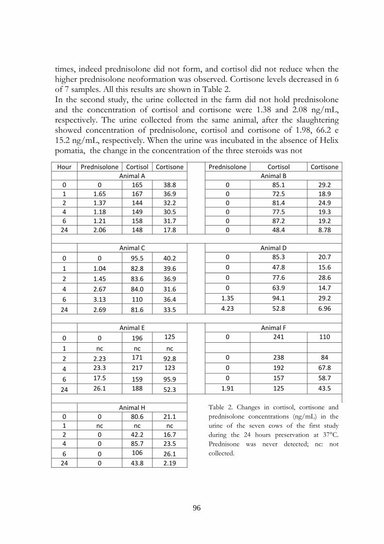
96
times, indeed prednisolone did not form, and cortisol did not reduce when the higher prednisolone neoformation was observed. Cortisone levels decreased in 6 of 7 samples. All this results are shown in Table 2. In the second study, the urine collected in the farm did not hold prednisolone and the concentration of cortisol and cortisone were 1.38 and 2.08 ng/mL, respectively. The urine collected from the same animal, after the slaughtering showed concentration of prednisolone, cortisol and cortisone of 1.98, 66.2 e 15.2 ng/mL, respectively. When the urine was incubated in the absence of Helix pomatia, the change in the concentration of the three steroids was not
Hour Prednisolone Cortisol Cortisone Prednisolone Cortisol Cortisone Animal A Animal B 0 0 165 38.8 0 85.1 29.2 1 1.65 167 36.9 0 72.5 18.9 2 1.37 144 32.2 0 81.4 24.9 4 1.18 149 30.5 0 77.5 19.3 6 1.21 158 31.7 0 87.2 19.2 24 2.06 148 17.8 0 48.4 8.78
Animal C Animal D 0 0 95.5 40.2 0 85.3 20.7
1 1.04 82.8 39.6 0 47.8 15.6
2 1.45 83.6 36.9 0 77.6 28.6
4 2.67 84.0 31.6 0 63.9 14.7
6 3.13 110 36.4 1.35 94.1 29.2
24 2.69 81.6 33.5 4.23 52.8 6.96
Animal E Animal F 0 0 196 125 0 241 110
1 nc nc nc
2 2.23 171 92.8 0 238 84
4 23.3 217 123 0 192 67.8
6 17.5 159 95.9 0 157 58.7
24 26.1 188 52.3 1.91 125 43.5
Animal H
Table 2. Changes in cortisol, cortisone and prednisolone concentrations (ng/mL) in the urine of the seven cows of the first study during the 24 hours preservation at 37°C. Prednisone was never detected; nc: not collected.
0 0 80.6 21.1 1 nc nc nc 2 0 42.2 16.7 4 0 85.7 23.5 6 0 106 26.1 24 0 43.8 2.19
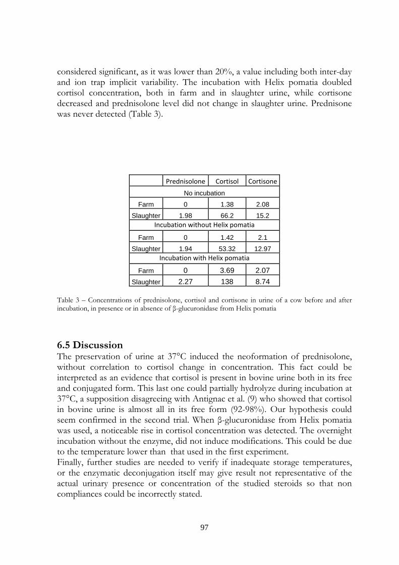
97
considered significant, as it was lower than 20%, a value including both inter-day and ion trap implicit variability. The incubation with Helix pomatia doubled cortisol concentration, both in farm and in slaughter urine, while cortisone decreased and prednisolone level did not change in slaughter urine. Prednisone was never detected (Table 3).
Prednisolone Cortisol Cortisone
No incubation
Farm 0 1.38 2.08
Slaughter 1.98 66.2 15.2 Incubation without Helix pomatia
Farm 0 1.42 2.1
Slaughter 1.94 53.32 12.97 Incubation with Helix pomatia
Farm 0 3.69 2.07 Slaughter 2.27 138 8.74
Table 3 – Concentrations of prednisolone, cortisol and cortisone in urine of a cow before and after incubation, in presence or in absence of β-glucuronidase from Helix pomatia
6.5 Discussion The preservation of urine at 37°C induced the neoformation of prednisolone, without correlation to cortisol change in concentration. This fact could be interpreted as an evidence that cortisol is present in bovine urine both in its free and conjugated form. This last one could partially hydrolyze during incubation at 37°C, a supposition disagreeing with Antignac et al. (9) who showed that cortisol in bovine urine is almost all in its free form (92-98%). Our hypothesis could seem confirmed in the second trial. When β-glucuronidase from Helix pomatia was used, a noticeable rise in cortisol concentration was detected. The overnight incubation without the enzyme, did not induce modifications. This could be due to the temperature lower than that used in the first experiment. Finally, further studies are needed to verify if inadequate storage temperatures, or the enzymatic deconjugation itself may give result not representative of the actual urinary presence or concentration of the studied steroids so that non compliances could be incorrectly stated.
98
6.6 References 1) EEC Commission Regulation N° 37/2010 Official Journal of the European Community L 15 p.1. 2) Commission staff working document on the implementation of national residue monitoring plans in the members states in 2007(Council Directive 96/23/EC); http://ec.europa.eu/food/food/chemicalsafety/residues/workdoc _2007_en.pdf. 3) Regione Piemonte. Piano regionale di controllo dei residui indesiderati nelle carni e negli animali allevati, anno 2007. 4) Regione Lombardia. Piano Nazionale Residui. Relazione attività anno 2008.
5) Fidani M., Gamberini MC., Pasello E., Palazzoli F., De Iuliis P., Montana M., Arioli F. (2009): Evaluation of equine urine reactivity towards 17-hydroxy steroids II phase metabolites by LC-MS/MS, Rapid Commun. Mass Spectrom. 23, 65–76. 6) de la Torre R., de la Torre X., Alia C., Segua J., Baro´ T., Torres-Rodriguez J.M. (2001): Changes in androgenic steroid pro. le due to urine contamination by microorganisms: a prospective study in the context of doping control Anal. Biochem. 289, 116–123. 7) Arioli F., Fidani M., Casati A., Fracchiolla ML., Pompa G. (2010): Investigation on possible transformations of cortisol, cortisone and cortisol glucuronide in bovine faecal matter using liquid chromatography-mass spectrometry, Steroids. 75. 350–354. 8) Commission Decision 657/2002 Official Journal of the European Community L 221 p.8. 9) Antignac J-P. Le Bizec B., Monteau F., André F. (2002): Study of natural and artificial corticosteroid phase II metabolites in bovine urine using HPLC-MS/MS, Steroids 67, 873–882.
99
Investigation of the origin of prednisolone in cow urine
At the printing of this thesis this work is in press and available only on line:
STEROIDS, doi:10.1016/j.steroids.2010.09.005.
CHAPTER 7

100
101
I am not young enough to know everything.
Oscar Wilde
Investigation of the origin of prednisolone in cow urine
G Pompaa, F Arioliad, A Casatia, M Fidanib, L Bertocchic, G Dusic
a Department of Veterinary Sciences and Technologies for Food Safety, University of Milan, Via Celoria 10, 20133 Milan, Italy
b U.N.I.R.E. Lab. S.r.l., Via Gramsci 70, 20019 Settimo Milanese (MI), Italy
c Istituto Zooprofilattico Sperimentale della Lombardia e dell’Emilia Romagna “B. Ubertini”, Via Bianchi 7, 25124 Brescia, Italy
d Correspondence: Francesco Arioli, Department of Veterinary Sciences and Technologies for Food Safety, University of Milan, Via Celoria, 10, 20133 Milan, Italy; E-mail [email protected]; tel. +390250317877; fax +390250317890
Work originated from the Department of Veterinary Sciences and Technologies for Food Safety, University of Milan, Via Celoria, 10, 20133 Milan, Italy
7.1 Abstract
After a two-year period of the frequent detection of prednisolone-positive bovine urine samples in the Italian region of Lombardy, studies were initiated to investigate the source. Because the majority of positive samples were detected at the slaughterhouse, researchers hypothesised that, together with increased cortisol and cortisone, a small quantity of prednisolone could be produced by the cows in stressful situations. In the present study, three dairy cows underwent intramuscular treatments with tetracosactide hexaacetate, a synthetic analogue of adrenocorticotropic hormone, to simulate stress. The animals were slaughtered at the end of the study. The results indicated that prednisolone could be detected
102
occasionally in the non-stressful state, but was consistently found in the urine of stressed cows (concentrations ranged from 1.01 to 4.08 ng/mL). To confirm the stress condition, urinary cortisol and cortisone were also detected at high concentrations in the urine, typically at concentrations of hundreds of nanograms per millilitre. The results of this preliminary study did not reveal the metabolic pathway responsible for prednisolone but suggested that this corticosteroid could be produced endogenously.
7.2 Keywords
Prednisolone, cow, urine, HPLC-MS3, ACTH
7.3 Introduction
Prednisolone (Δ1,4-pregnadiene-11β,17α,21-triol-3,20-dione) is a corticosteroid with gluconeogenetic and antinflammatory activities. Prednisone (Δ1,4-pregnadiene-17α,21- diol-3,11,20-trione), which is the inactive prodrug of prednisolone, is enzymatically activated in the liver (1). The adrenocortical activities of these two steroids are 3- to 4-times the activity of cortisone or cortisol (2). In the European Union, some corticosteroids are permitted for therapeutic use in livestock. However, the use of corticosteroids is regulated by the Commission Regulation (EEC) N° 37/2010, which sets maximum residue limits (MRLs) for betamethasone, dexamethasone, methylprednisolone and prednisolone. The use of prednisolone is permitted in cattle, and the MRLs are 4 µg/kg in muscle and fat, 10 µg/kg in liver and kidney and 6 µg/kg in milk (3). The member states are required to monitor the use of pharmacologically active substances in animals used for the production of food for human consumption. They must follow the indications of Council Directive 96/22/EC (4), which was amended by Directive 2003/74/EC (5) and 96/23/EC (6). Because of their steroidal structure, the corticosteroids are included in group A3 (substances with an anabolic effect and unauthorised substances/steroids) of National Residue Control Plan (NRCP) of some member states (Italy, the Netherlands and Denmark), whereas other states allocate them to the B2f group (other pharmacologically active substances) (7). In Italy the controls on corticosteroids use are made at the slaughterhouse and at the farm. In the first case the matrices sampled are liver, a target tissue for which MRLs are set, or urine, while at the farm the matrix sampled is urine. For this matrix, no maximum allowable concentration has been fixed. The 2008 and 2009 Italian NRCP reports (8, 9) stated that corticosteroid residues in cattle, particularly prednisolone, were an emerging problem. In 2008, the Lombard Veterinary Organisational Unit-Structure for the Control on Food of Animal Origin (Unità Organizzativa Veterinaria-Struttura di Controllo degli Alimenti di Origine Animale) found that
103
the analyses on cow urine made in farms did not give any positivity to prednisolone or prednisone. The same analyses for the urine of slaughtered cows, however, showed that positive samples accounted for 82% of all irregularities (10). Therefore, we hypothesised that the stress evoked by handling before slaughter (11) caused prednisolone or prednisone to increase to levels that
Figure 1. Chemical structures of the four steroids studied.
could be detected using current analytical methods. This stress also increased urinary levels of cortisol and its metabolites in cattle. Accordingly, in the present study we measured the urinary concentrations of cortisol, cortisone, prednisolone and prednisone (Figure 1) in dairy cows before and after intramuscular administration of tetracosactide hexaacetate (an adrenocorticotropic hormone synthetic analogue), which was used to induce stress pharmacologically. The urine of each animal was also analysed after slaughter.
104
7.4 Experimental
7.4.1 Animal treatments and sample collection
Three healthy Holstein-Friesian lactating cows, aged 44, 60 and 106 months, were housed in a Lombard farm and fed with a diet typically used in breeding dairy cows. The cows were cared for in accordance with EU guidelines approved by the Italian Ministry of Health and maintained under the control of the Istituto Zooprofilattico Sperimentale della Lombardia e dell’Emilia Romagna (IZSLER). A 2 mg intramuscular injection of tetracosactide hexaacetate (Sinacthen rp - prolonged release - Defiante Farmaceutica S.A., Punchal, Portugal), corresponding to 200 I.U. of adrenocorticotropic hormone (ACTH), was administered twice at an interval of 24 hours. Urine samples were collected (urethral catheterisation) at the farm 24 and 48 hours before the first treatment, and 2, 7 and 24 hours after the second treatment. The last collection at the farm was made 2 hours before transport to the slaughterhouse (46 hours after the second treatment), and another sample was collected directly from the urinary bladder immediately following the slaughter. After collection, the urine samples were frozen at -18°C, carried to the Toxicology Laboratory of the Department of Veterinary Sciences and Technologies for Food Safety at the University of Milan and stored at -18°C.
7.4.2 Reagents and chemicals
Cortisol, cortisone, prednisolone, prednisone and flumethasone (as an internal standard) were purchased from Sigma–Aldrich (St. Louis, MO, USA). All other chemicals were from Fluka Chemie GmbH (Buchs, Switzerland). Standard stock solutions were prepared in ethanol (1 mg/mL) and stored at −18°C. Working solutions were prepared daily by diluting the stock solutions with methanol:water (50:50 v/v).
7.4.3 Sample preparation
Two millilitres of urine was spiked with the flumethasone internal standard (10 ng/mL), and 4 mL of a mixture of tert-butyl methylether:ethyl acetate (80:20 v/v) was added in a 15-mL polypropylene tube. After shaking in a vertical rotary shaker for 20 minutes, the sample was centrifuged at 3000 x g for 15 minutes. The upper organic layer was collected with a Pasteur pipette, transferred to a 10-mL glass tube and dried under vacuum in a centrifugal evaporator. The residue was dissolved in 200 µL of a mixture of methanol-aqueous formic acid (0.1%, 50:50 v/v) and transferred to vials for LC-MS3 analysis.
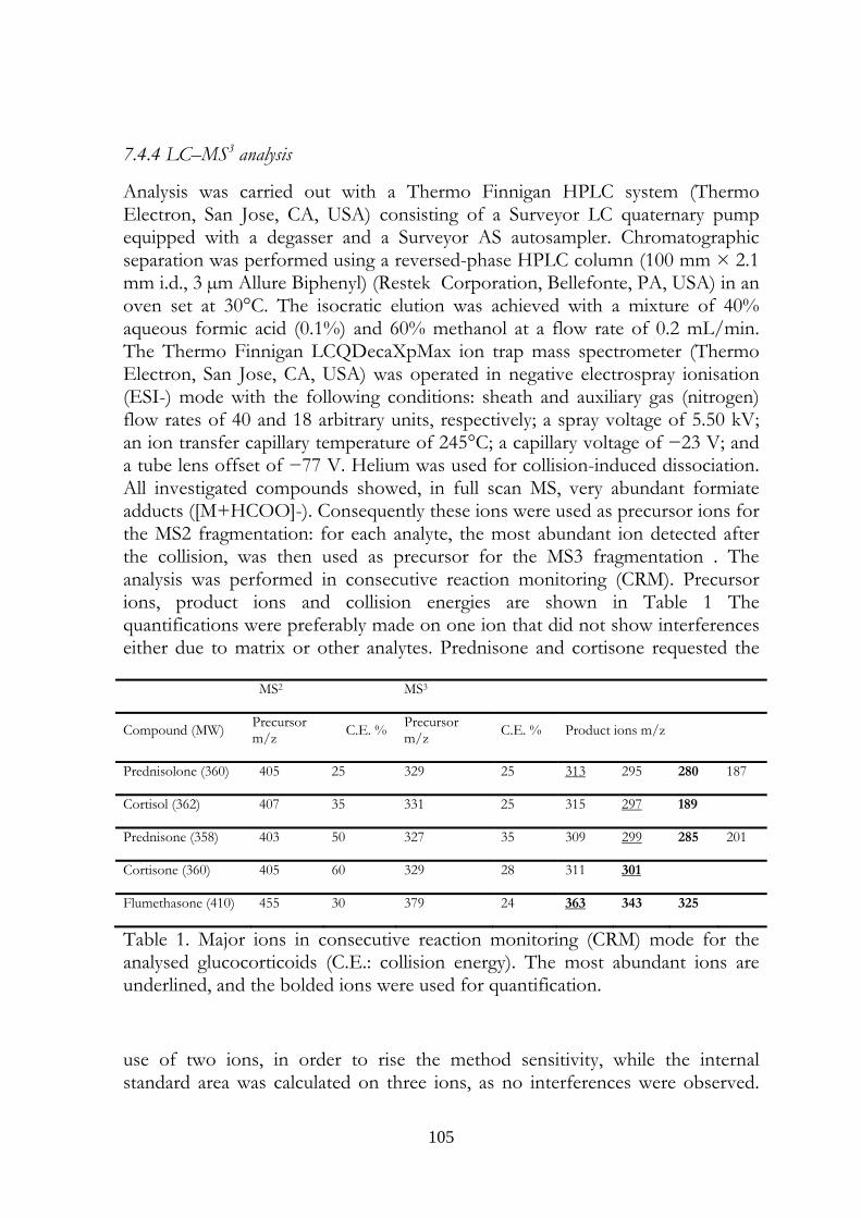
105
7.4.4 LC–MS3 analysis
Analysis was carried out with a Thermo Finnigan HPLC system (Thermo Electron, San Jose, CA, USA) consisting of a Surveyor LC quaternary pump equipped with a degasser and a Surveyor AS autosampler. Chromatographic separation was performed using a reversed-phase HPLC column (100 mm × 2.1 mm i.d., 3 µm Allure Biphenyl) (Restek Corporation, Bellefonte, PA, USA) in an oven set at 30°C. The isocratic elution was achieved with a mixture of 40% aqueous formic acid (0.1%) and 60% methanol at a flow rate of 0.2 mL/min. The Thermo Finnigan LCQDecaXpMax ion trap mass spectrometer (Thermo Electron, San Jose, CA, USA) was operated in negative electrospray ionisation (ESI-) mode with the following conditions: sheath and auxiliary gas (nitrogen) flow rates of 40 and 18 arbitrary units, respectively; a spray voltage of 5.50 kV; an ion transfer capillary temperature of 245°C; a capillary voltage of −23 V; and a tube lens offset of −77 V. Helium was used for collision-induced dissociation. All investigated compounds showed, in full scan MS, very abundant formiate adducts ([M+HCOO]-). Consequently these ions were used as precursor ions for the MS2 fragmentation: for each analyte, the most abundant ion detected after the collision, was then used as precursor for the MS3 fragmentation . The analysis was performed in consecutive reaction monitoring (CRM). Precursor ions, product ions and collision energies are shown in Table 1 The quantifications were preferably made on one ion that did not show interferences either due to matrix or other analytes. Prednisone and cortisone requested the
Table 1. Major ions in consecutive reaction monitoring (CRM) mode for the analysed glucocorticoids (C.E.: collision energy). The most abundant ions are underlined, and the bolded ions were used for quantification.
use of two ions, in order to rise the method sensitivity, while the internal standard area was calculated on three ions, as no interferences were observed.
MS2 MS3
Compound (MW) Precursor m/z C.E. % Precursor
m/z C.E. % Product ions m/z
Prednisolone (360) 405 25 329 25 313 295 280 187
Cortisol (362) 407 35 331 25 315 297 189
Prednisone (358) 403 50 327 35 309 299 285 201
Cortisone (360) 405 60 329 28 311 301
Flumethasone (410) 455 30 379 24 363 343 325
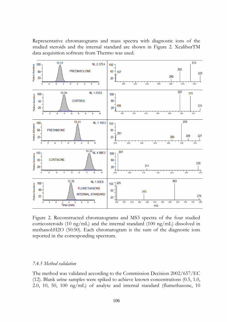
106
Representative chromatograms and mass spectra with diagnostic ions of the studied steroids and the internal standard are shown in Figure 2. XcaliburTM data acquisition software from Thermo was used.
Figure 2. Reconstructed chromatograms and MS3 spectra of the four studied corticosteroids (10 ng/mL) and the internal standard (100 ng/mL) dissolved in methanol:H2O (50:50). Each chromatogram is the sum of the diagnostic ions reported in the corresponding spectrum.
7.4.5 Method validation
The method was validated according to the Commission Decision 2002/657/EC (12). Blank urine samples were spiked to achieve known concentrations (0.5, 1.0, 2.0, 10, 50, 100 ng/mL) of analyte and internal standard (flumethasone, 10
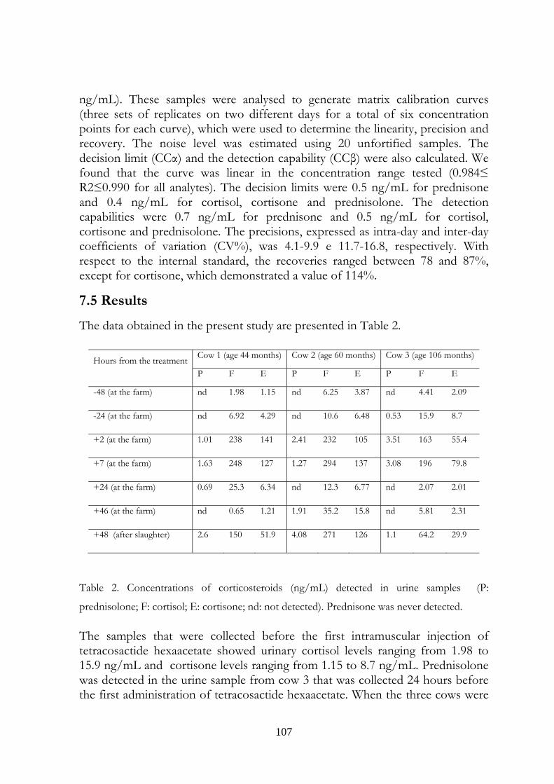
107
ng/mL). These samples were analysed to generate matrix calibration curves (three sets of replicates on two different days for a total of six concentration points for each curve), which were used to determine the linearity, precision and recovery. The noise level was estimated using 20 unfortified samples. The decision limit (CCα) and the detection capability (CCβ) were also calculated. We found that the curve was linear in the concentration range tested (0.984≤ R2≤0.990 for all analytes). The decision limits were 0.5 ng/mL for prednisone and 0.4 ng/mL for cortisol, cortisone and prednisolone. The detection capabilities were 0.7 ng/mL for prednisone and 0.5 ng/mL for cortisol, cortisone and prednisolone. The precisions, expressed as intra-day and inter-day coefficients of variation (CV%), was 4.1-9.9 e 11.7-16.8, respectively. With respect to the internal standard, the recoveries ranged between 78 and 87%, except for cortisone, which demonstrated a value of 114%.
7.5 Results
The data obtained in the present study are presented in Table 2.
Hours from the treatment Cow 1 (age 44 months) Cow 2 (age 60 months) Cow 3 (age 106 months)
P F E P F E P F E
-48 (at the farm) nd 1.98 1.15 nd 6.25 3.87 nd 4.41 2.09
-24 (at the farm) nd 6.92 4.29 nd 10.6 6.48 0.53 15.9 8.7
+2 (at the farm) 1.01 238 141 2.41 232 105 3.51 163 55.4
+7 (at the farm) 1.63 248 127 1.27 294 137 3.08 196 79.8
+24 (at the farm) 0.69 25.3 6.34 nd 12.3 6.77 nd 2.07 2.01
+46 (at the farm) nd 0.65 1.21 1.91 35.2 15.8 nd 5.81 2.31
+48 (after slaughter) 2.6 150 51.9 4.08 271 126 1.1 64.2 29.9
Table 2. Concentrations of corticosteroids (ng/mL) detected in urine samples (P:
prednisolone; F: cortisol; E: cortisone; nd: not detected). Prednisone was never detected.
The samples that were collected before the first intramuscular injection of tetracosactide hexaacetate showed urinary cortisol levels ranging from 1.98 to 15.9 ng/mL and cortisone levels ranging from 1.15 to 8.7 ng/mL. Prednisolone was detected in the urine sample from cow 3 that was collected 24 hours before the first administration of tetracosactide hexaacetate. When the three cows were
108
pharmacologically stressed, prednisolone was consistently detected in the urine both 2 hours and 7 hours after the second intramuscular injection. Concomitantly, the urinary level of cortisol increased to values ranging from 163 to 294 ng/mL. We also observed an increase in cortisone levels, which demonstrated concentrations that were approximately one-third to one-half of the cortisol levels (55.4 to 137 ng/mL). The concentrations of cortisol and cortisone at 24 and 46 hours post-stress were similar to those before tetracosactide hexaacetate treatment. Prednisolone was detected in two of six samples, in two different cows, and at different collection times. At the slaughterhouse, however, prednisolone was detected in all urine samples. The cortisol and cortisone concentrations and ratios after slaughter were similar to the concentrations observed after tetracosactide hexaacetate treatment. Prednisone was never detected. Representative chromatograms and mass spectra of urine samples collected from cow 1 twenty-four hours before the first treatment, two hours after the second treatment and after slaughter are presented in Figures 3, 4 and 5 respectively. In Figure 6, the reconstructed chromatograms of three diagnostic ions of prednisolone, detected in urine from the same animal and corresponding to the samples presented in Figure 4 and 5, are shown. Because of the unusual results observed in the present study, we asked the Drug Residue Laboratory of the official organisation IZSLER and the official anti-doping laboratory for sport horses U.N.I.R.E. Lab. Srl, to test our urine samples for the presence of prednisolone to confirm our results. The results of the analyses, performed by the U.N.I.R.E. laboratory, only through qualitative determination, fully confirmed the ones reported in this work. The IZSLER laboratory otherwise did not confirm prednisolone presence in sample collected from cow 3, 24 hours before first tetracosactide hexaacetate administration, and from cow 1 urine collected 24 hours after second injection. The analyses were carried out in the IZSLER laboratory applying a confirmatory method fully validated according to the Commission Decision 2002/657/EC (12), with a triple quadrupole mass spectrometer and at U.N.I.R.E. Lab. Srl applying a method with a linear trap mass spectrometer. In both laboratories solid phase extraction(SPE) was used to purify the sample. To confirm that the identification of prednisolone made by three laboratories operating with low mass resolution spectrometers was correct, a further analysis was made on the urine sampled from cow two 46 hours after
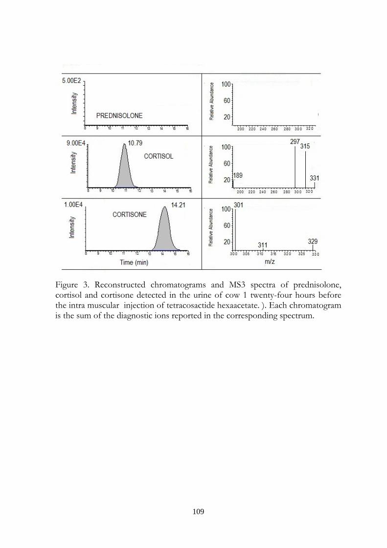
109
Figure 3. Reconstructed chromatograms and MS3 spectra of prednisolone, cortisol and cortisone detected in the urine of cow 1 twenty-four hours before the intra muscular injection of tetracosactide hexaacetate. ). Each chromatogram is the sum of the diagnostic ions reported in the corresponding spectrum.
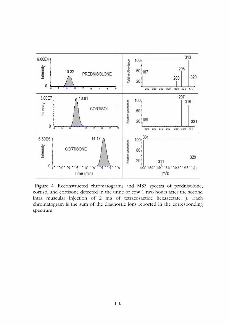
110
Figure 4. Reconstructed chromatograms and MS3 spectra of prednisolone, cortisol and cortisone detected in the urine of cow 1 two hours after the second intra muscular injection of 2 mg of tetracosactide hexaacetate. ). Each chromatogram is the sum of the diagnostic ions reported in the corresponding spectrum.
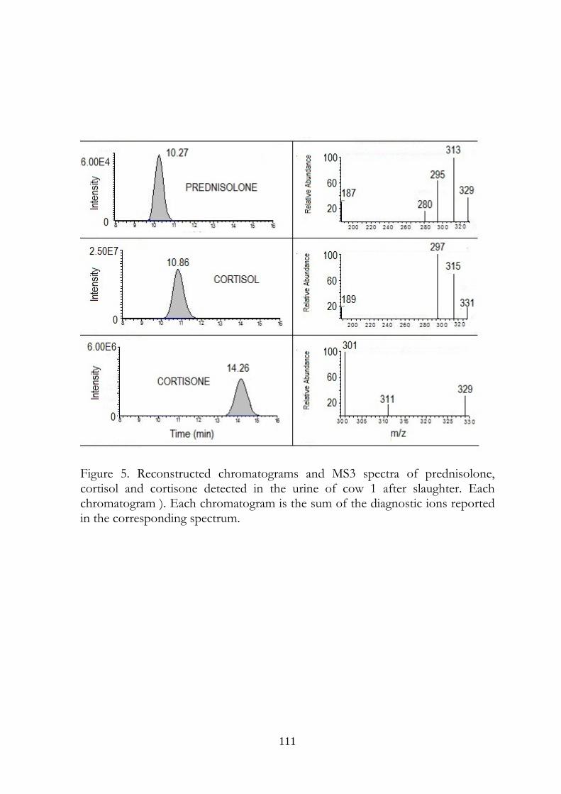
111
Figure 5. Reconstructed chromatograms and MS3 spectra of prednisolone, cortisol and cortisone detected in the urine of cow 1 after slaughter. Each chromatogram ). Each chromatogram is the sum of the diagnostic ions reported in the corresponding spectrum.
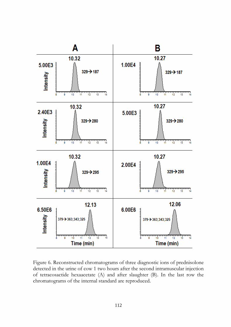
112
Figure 6. Reconstructed chromatograms of three diagnostic ions of prednisolone detected in the urine of cow 1 two hours after the second intramuscular injection of tetracosactide hexaacetate (A) and after slaughter (B). In the last row the chromatograms of the internal standard are reproduced.
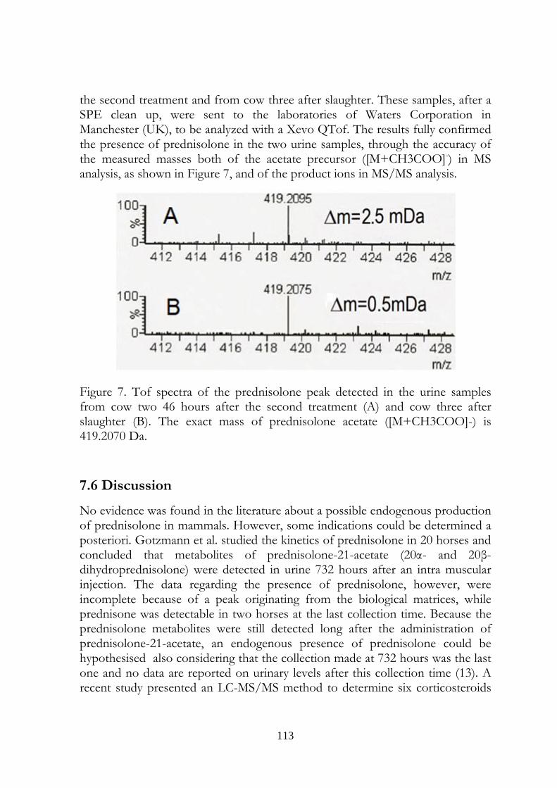
113
the second treatment and from cow three after slaughter. These samples, after a SPE clean up, were sent to the laboratories of Waters Corporation in Manchester (UK), to be analyzed with a Xevo QTof. The results fully confirmed the presence of prednisolone in the two urine samples, through the accuracy of the measured masses both of the acetate precursor ([M+CH3COO]-) in MS analysis, as shown in Figure 7, and of the product ions in MS/MS analysis.
Figure 7. Tof spectra of the prednisolone peak detected in the urine samples from cow two 46 hours after the second treatment (A) and cow three after slaughter (B). The exact mass of prednisolone acetate ([M+CH3COO]-) is 419.2070 Da.
7.6 Discussion
No evidence was found in the literature about a possible endogenous production of prednisolone in mammals. However, some indications could be determined a posteriori. Gotzmann et al. studied the kinetics of prednisolone in 20 horses and concluded that metabolites of prednisolone-21-acetate (20α- and 20β-dihydroprednisolone) were detected in urine 732 hours after an intra muscular injection. The data regarding the presence of prednisolone, however, were incomplete because of a peak originating from the biological matrices, while prednisone was detectable in two horses at the last collection time. Because the prednisolone metabolites were still detected long after the administration of prednisolone-21-acetate, an endogenous presence of prednisolone could be hypothesised also considering that the collection made at 732 hours was the last one and no data are reported on urinary levels after this collection time (13). A recent study presented an LC-MS/MS method to determine six corticosteroids
114
in bovine urine, and four samples of cow urine from the 2008 Hungarian residue control monitoring program were (presumably randomly) subjected to analysis to test the method. Prednisolone was the only corticosteroid detected in all four samples, and its concentration ranged from 0.3 to 0.9 ng/mL (14). The present study was performed to investigate data from the Official Residue Control Program of the Lombard veterinary organisational unit and to attempt to understand their observation of a high frequency of prednisolone-positive samples in slaughtered cows: in this respect, we considered appropriate to use three healthy lactating dairy cows of different ages, as in farms can be found. We did not try to make any conclusions about the physiological levels of cortisol and cortisone in bovine urine as well. Nevertheless, some considerations are proposed regarding this topic. When the cows were in supposed non-stressful conditions, the higher urinary concentrations of cortisol and cortisone were 35.2 and 15.8 ng/mL, respectively. These concentrations where however only found in the urine sample collected 46 hours after treatment from cow 2. In this sample all the laboratories detected prednisolone. This finding could represent the evidence of an uncontrolled stress condition of this cow. Regarding the samples with a doubtful presence of prednisolone (urine collected 24 hours before the treatment from cow 3 and urine collected 24 hours after the treatment from cow 1), two considerations are to be made: the concentrations of this steroid were very close to our CCβ (0.5 ng/mL) and to the CCα of the IZSLER Laboratory (0.58 ng/mL). The concentrations of cortisol were lower than in sample collected 46 hours after the treatment from cow 2, but higher than in any other sample collected in supposed non-stressful condition; cortisone showed a similar behaviour. The two doubt samples could therefore show cortisol and cortisone urinary concentrations at which low stress conditions (e.g due to the animal handling) exist, but do not induce an indisputable detection of prednisolone in urine. The values increased when a stress factor was applied to the cows (tetracosactide or transport to the slaughterhouse): cortisol concentrations ranged from 64.4 and 294 ng/mL, and cortisone concentrations ranged from 29.9 and 241 ng/mL. Despite the large amount of variability in the data, the concentrations of cortisol and cortisone in the two different conditions did not superimpose, which provided evidence that the cows were stressed. The most noteworthy result of the present study was that prednisolone was always detected in stressed cows. In comparison, prednisolone was undoubtedly detected in 1of 12 samples from supposed non-stressed cows. The possibility of a transformation of endogenous corticosteroids, such as cortisol or cortisone, in urine by microorganisms with Δ1 dehydrogenase activity has been previously studied (15). Considering the precautions that were taken for the collection (urethral catheterisation at the farm and directly from the urinary bladder at the slaughterhouse) and storage (immediate freezing at -18°C), the detection of
115
prednisolone in our samples cannot be explained by faecal (16) or massive microbial contamination. The contamination of urine, however, is an important issue because of the presence of microbial elements in the analysis environment. Prednisone was never detected, consistent with the ability of mammalian organisms to activate this compound to prednisolone in the liver via the activity of 11β-hydroxysteroid dehydrogenase (1, 17). The results observed for prednisolone in the present study suggested that it is very likely to find this corticosteroid in the urine of stressed animals. Our data, however, did not clarify whether prednisolone was a metabolite of cortisol or a by-product of its metabolic pathway. Moreover, prednisolone could also be detected under supposed non-stressful conditions. These considerations should be taken into account when setting the controls that are used to determine the illicit use of prednisolone. Further studies are needed to clarify the matter and to finally confirm the hypothesis expressed in this work.
7.7 Acknowledgments
This work was carried out with a grant from the Regione Lombardia for the Research Project: “Presenza e persistenza di farmaci e ormoni nelle deiezioni animali e nel liquame per il potenziamento dell’attività di farmacosorveglianza”. The authors wish to thank M. Silvestri (Thermo Scientific) for its technical support, and J. Cardwell and S. Scarpella (Waters Corporation) who significantly contributed to this work performing the analyses with Xevo QTof
7.8 References
1) Brunton LL (editor-in-chief), Keith L, Parker KL, Murri N, Blumenthal DK (associate editors), Goodman & Gilman's the pharmacological basis of therapeutics, online 11th edition, http://www.accessmedicine.com.
2) Herzog HL, Nobile A, Tolksdorf S, Charney W, Hershberg EB, Perlman PL, Pechet MM. 1955. New antiarthritic steroids. Science. 121:176.
3) EEC Commission Regulation N° 37/2010 Official Journal of the European Community L 15 p.1.
4) EEC Council Directive N° 22/1996 Official Journal of the European Community L 12 p. 3.
5) EEC Council Directive N°74/2003 Official Journal of the European Community L 262 p. 17
6) EEC Council Directive N° 23/1996 Official Journal of the European Community L 125 p. 10.
116
7) Commission staff working document on the implementation of national residue monitoring plans in the members states in 2007 (Council Directive 96/23/EC); http://ec.europa.eu/food/food/chemicalsafety/residues/workdoc_2007_en.pdf
8) Ministero del Lavoro, della Salute e delle Politiche Sociali; Direzione Generale della Sicurezza degli Alimenti e della Nutrizione: Piano Nazionale per la ricerca di Residui, Relazione Finale, Anno 2008; www.salute.gov.it/imgs/C_17_pubblicazioni _1146_allegato.pdf
9) Ministero della Salute; Direzione Generale della Sicurezza degli Alimenti e della Nutrizione: Relazione Finale, Piano Nazionale Residui, 2009.
10) Regione Lombardia, Unità Organizzativa Veterinaria-Struttura Controllo Alimenti di Origine Animale, Piano Nazionale Residui, Relaz. attività anno 2008; http://www.sanita.regione.lombardia.it/shared/ccurl/638/325/Relazione%20attivit%C3%A0%20PNR%202008.pdf
11) Palme R, Robia C, Baumgartner W, Möstl E. 2000. Transport stress in cattle as reflected by an increase in faecal cortisol metabolites. Vet Rec. 146:108–109.
12) EEC Commission Decison N° 657/2002 Official Journal of the European Community L 221 p. 8.
13) Gotzmann A, Bredehöft M, Thevis M and Schänzer W. 2003. Detection of prednisolone, prednisone and 20-dihydroprednisolone in human and equine urine by means of LC/MS/MS. Recent advances in doping analysis. Sport und Buch Strauß, Köln.11:33-41.
14) Tölgyesi Á, Sharma VK, Kovacsics L, Fekete J. 2010. Quantification of corticosteroids in bovine urine using selective solid phase extraction and reversed-phase liquid chromatography/tandem mass spectrometry. J. Chromatogr. B, 878:1471–1479,.
15) Bredehöft M, Baginski R, Parr MK, Thevis M, Schänzer W. 2010. Investigations of the microbial transformation of cortisol to prednisolone in urine samples. J.Steroid Biochem. Mol. Biol. doi:10.1016/j.jsbmb.2010.04.021.
16) Arioli F, Fidani M, Casati A, Fracchiolla ML, Pompa G. 2010. Investigation on possible transformations of cortisol, cortisone and cortisol glucuronide in bovine faecal matter using liquid chromatography-mass spectrometry. Steroids. 75:350–354.
17) Arlt W, Stewar PM. 2005. Adrenal Corticosteroid Biosynthesis,Metabolism and Action, Endocrinol Metab. Clin. N. Am. 34:293–313.

117
General discussion
CHAPTER 8
118
Stock your mind, stock your mind. You might be poor, your shoes might be broken, but your mind is a palace.
Frank McCourt, Angela's Ashes
8.1 Some considerations
When, from the first noughties onwards, we dealt with boldenone, and more recently we began to study the prednisolone topic, because of the significantly difference in the frequency of positivities to these drugs among farm animals’ urines, some considerations were made:
1- Does a benefit come to the farmer from the use of this substances?
2- Are the concentrations in biological samples consistent with the eventual benefits?
3- Have sampling, transport to the laboratory and storage been properly made?
4- Are the analytical methods and instrumentations changed, so improving the sensibility of analyses (a quite rhetorical question)?
The point is that if any doubt comes from these answers, studies should have to be carried out regarding the origin of the presence of the studied substances.
Boldenone and prednisolone are quite an example of this. In fact:
1- No data were found in literature about the efficacy of boldenone as growth promoter, except a degree thesis stating that it improves the colour of meat, but reduces zootechnic performances (1). On the other hand, prednisolone has a gluconeogenetic activity equal to four or five times that of cortisol but one fifth than that of dexamethasone or betamethasone and its half life is shorter than the latter two steroids (Table 1).
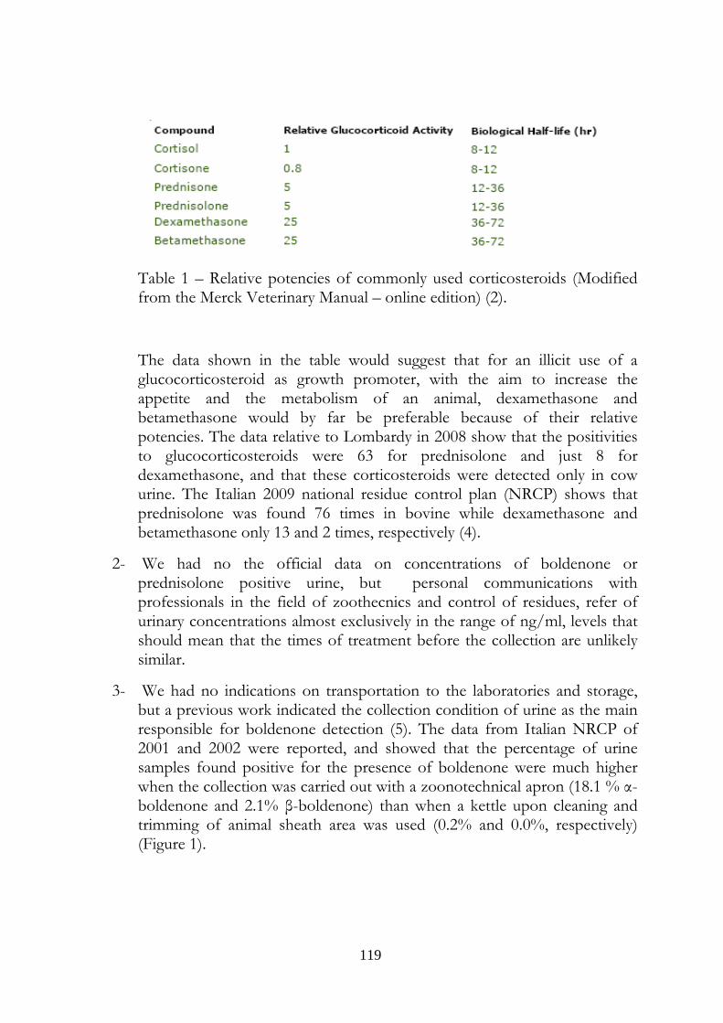
119
Table 1 – Relative potencies of commonly used corticosteroids (Modified from the Merck Veterinary Manual – online edition) (2).
The data shown in the table would suggest that for an illicit use of a glucocorticosteroid as growth promoter, with the aim to increase the appetite and the metabolism of an animal, dexamethasone and betamethasone would by far be preferable because of their relative potencies. The data relative to Lombardy in 2008 show that the positivities to glucocorticosteroids were 63 for prednisolone and just 8 for dexamethasone, and that these corticosteroids were detected only in cow urine. The Italian 2009 national residue control plan (NRCP) shows that prednisolone was found 76 times in bovine while dexamethasone and betamethasone only 13 and 2 times, respectively (4).
2- We had no the official data on concentrations of boldenone or prednisolone positive urine, but personal communications with professionals in the field of zoothecnics and control of residues, refer of urinary concentrations almost exclusively in the range of ng/ml, levels that should mean that the times of treatment before the collection are unlikely similar.
3- We had no indications on transportation to the laboratories and storage, but a previous work indicated the collection condition of urine as the main responsible for boldenone detection (5). The data from Italian NRCP of 2001 and 2002 were reported, and showed that the percentage of urine samples found positive for the presence of boldenone were much higher when the collection was carried out with a zoonotechnical apron (18.1 % α-boldenone and 2.1% β-boldenone) than when a kettle upon cleaning and trimming of animal sheath area was used (0.2% and 0.0%, respectively) (Figure 1).
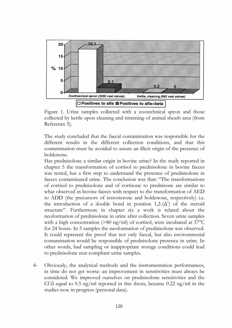
120
Figure 1. Urine samples collected with a zootechnical apron and those collected by kettle upon cleaning and trimming of animal sheath area (from Reference 5). The study concluded that the faecal contamination was responsible for the different results in the different collection conditions, and that this contamination must be avoided to assure an illicit origin of the presence of boldenone. Has prednisolone a similar origin in bovine urine? In the study reported in chapter 5 the transformation of cortisol to prednisolone in bovine faeces was tested, has a first step to understand the presence of prednisolone in faeces contaminated urine. The conclusion was that: “The transformations of cortisol to prednisolone and of cortisone to prednisone are similar to what observed in bovine faeces with respect to the transformation of AED to ADD (the precursors of testosterone and boldenone, respectively) i.e. the introduction of a double bond in position 1,2-(Δ1) of the steroid structure”. Furthermore in chapter six a work is related about the neoformation of prednisolone in urine after collection. Seven urine samples with a high concentration (>80 ng/ml) of cortisol, were incubated at 37°C for 24 hours. In 5 samples the neoformation of prednisolone was observed. It could represent the proof that not only faecal, but also environmental contamination would be responsible of prednisolone presence in urine. In other words, bad sampling or inappropriate storage conditions could lead to prednisolone non compliant urine samples.
4- Obviously, the analytical methods and the instrumentation performances, in time do not get worse: an improvement in sensitivities must always be considered. We improved ourselves on prednisolone sensitivities and the CCß equal to 0.5 ng/ml reported in this thesis, became 0.22 ng/ml in the studies now in progress (personal data).
121
A corticosteroid-peculiar element must be furthermore considered: the stress conditions an animal undergoes when transported to the slaughterhouse. Someone (6), using a vivid metaphor, defined cortisol (and noradreline) “the messenger of the wartime commander” to underline its (their) higher production in stress conditions, so that cortisol is also named the stress hormone. In the last study performed and reported in Chapter 7, a “collateral” production of prednisolone in stressful situations was hypothesized and its presence at detectable concentrations in cow urine was demonstrated, both when animals were submitted to pharmacological or slaughter stress. At last, the analogy between prednisolone and boldenone is not restricted to the similarity to cortisol and testosterone, respectively. As stated in the foreword, the production of cortisol takes place in the zona fasciculata of the adrenal cortex, while DHEA and androstenedione, precursors of testosterone, are produced (in small amounts in men, but more than 50% in women) also in the zona reticularis of adrenal cortex. The secretion of glucocorticosteroids is regulated by ACTH, that seems to have a role in androstenedione secretion, too. If a by-production of prednisolone(Δ1-2dehydrogenated cortisol) occurs during stress, a similar by production of the boldenone precursor androstadienedione, (Δ1-2dehydrogenated androstenedione), could be supposed. This hypothesis could be a fascinating topic to be dealt with in future studies on the origin of presence of boldenone and prednisolone in bovine urine.
8.2 References
1) Marcazzani M., Effects of an Hormonal Treatment With Boldenone about Productive Performances and Meat Quality of Veal Calves, Degree Thesis, Università degli Studi di Padova, 2003-04.
2)http://www.merckvetmanual.com/mvm/index.jsp?cfile=htm/bc/191604.htm
3) Regione Lombardia, Unità Organizzativa Veterinaria-Struttura Controllo degli Alimenti di Origine Animale, Piano Nazionale Residui, Relazione attività anno 2008; http://www.sanita.regione.lombardia.it/shared/ccurl/638/325/Relazione%20attivit%C3%A0%20PNR%202008.pdf
4) Ministero della Salute, Direzione Generale della Sicurezza degli Alimenti e della Nutrizione, relazione Finale, Piano Nazionale Residui 2009.
122
5) Sgoifo Rossi C.A., Arioli F., Bassini A., Chiesa L.M., Dell’Orto V., Montana M., Pompa G. 2004. Evidence for false-positive results for boldenone testing of veal urine due to faecal crosscontamination during sampling. Food Additives and Contaminants 21:756–762.
6) http://www.alessandragraziottin.it/pdf/articoli.php?ART_TYPE=AQUOT& EW_FATHER=7049
123
Summary
CHAPTER 9

124
125
This is the end, my only friend, the end Of our elaborate plans, the end
Jim Morrison, The end
Summary Two tight correlated topics were dealt in this thesis: - Bovine urine can result positive for boldenone, an androgenic steroid with the structure of testosterone with a double bond in Δ1-2 position of ring A, and androstadienedione, its 17-oxidized form, in consequence of faecal contamination, mainly through a high neo production in faeces-contaminated urine. A simple in vitro method was developed – incubation of faecal matter suspended in 0.9% saline – to induce steroid transformations in faeces, and analyzed the products by liquid chromatography/tandem mass spectrometry, without the need for prior extraction. Androstenedione, androstadienedione, α− and β−boldenone, testosterone and epitestosterone transformations were investigated. Mutual interconversions of steroids were observed, although 17 β−hydroxy steroids had lower stability if compared with 17 α−hydroxy and 17-keto steroids. This simple in vitro system proved to be an effective way of studying hormone transformations in faeces and in faeces-contaminated urine (Chapter 3). An analogous method, i.e. the direct injection, for LC/MS-MS analysis, of diluted equine urine, without prior extraction, was carried out to evaluate the stability of phase II metabolites of boldenone, testosterone and epitestosterone (glucuronides and sulphates) in intentionally poorly stored urine. Both 17β and 17 α-glucuronide steroids hydrolyzed to their correspondent hydroxysteroids, but only the 17 β epimers oxidized to the keto forms. The sulphate compounds showed a complete stability. The transformations were temperature dependent: faster at room temperature than at 4°C, while did not occur in frozen urines. This indicated that proper storage conditions of biological samples are fundamental to avoid microbiological contamination that can cause chemical modifications of androgen steroids (Chapter 4). - After a two-year period of the frequent detection of prednisolone-positive bovine urine samples in the Italian region of Lombardy, studies were initiated to investigate the source. The problem of modifications in biological fluids was therefore evaluated also regarding a possible transformation of cortisol and cortisone into prednisolone and prednisone in cattle faeces. The transformation of the II phase metabolite cortisol glucuronide was studied, too. The method consisted once more of incubation of bovine faecal matter suspended in 0.9% saline spiked with cortisol, cortisone and cortisol glucuronide (400 ng/ml). The deconjugation of the conjugated form the and the dehydrogenation in ring A
126
occurred. It was so concluded that also urine contaminated with faeces may be positive for prednisone and prednisolone in the same way as they are positive for boldenone, i.e. as a result of microbiological dehydrogenase activity on cortisol and cortisone (Chapter 5). The successive step was to assess the transformation of cortisol and cortisone in urine collected from the bladder at the slaughterhouse (i.e. without faecal contamination) and incubated at 37°C for 24 hours. The influence of deconjugation with β-glucuronidase from Helix pomatia was assessed, too. Only urine where prednisolone and prednisone were absent and cortisol was higher than 80 ng/mL were used. In this study a liquid-liquid extraction with a mixture of tert-Buthylmethylether:Ethylacetate (80:20) was performed, to concentrate the sample, so raising the sensitivity of the method. The analyses were carried out by LC MS3. It was demonstrated that a relatively high temperature (e.g. due to inappropriate storage conditions or to enzymatic deconjugation), could give results that do not represent the effective presence and concentration of the abovementioned corticosteroids at the moment of the sample collection (Chapter 6). However, because of the stressful conditions of the slaughterhouse, the by-production of prednisolone was supposed, together with the increase of cortisol and cortisone. The urines of three dairy cows were therefore collected and analyzed before and after a pharmacological induced stress (intra muscular administration of tetracosactide hexaacetate, an adrenocorticotropic hormone analogue), and before and after slaughter. Prednisolone was occasionally detected in the non-stressful state, but was consistently found in the urine of stressed cows (from 1.01 to 4.08 ng/mL), together with the raise in concentration of urinary cortisol and cortisone, so indicating a possible endogenous production of prednisolone in stressful situations (Chapter 7).

127
Acknowledgements
CHAPTER 10

128
129
I wish to thank all the people who collaborated to or enabled the execution of the studies reported in this thesis. In order to forget no one, I’ll say that their names are all in these pages, as authors or in the thanks at the end of the papers. My thanks are likewise for the Professors of the Doctoral Program in Animal Nutrition and Food Safety and for Cristiana Rossetti, the most helpful and kind (and accidentally the only one) secretary of the Graduate School of Veterinary Sciences for Animal Health and Food Safety. I should moreover like to thank: Sir Walter Scott, the Irish Wisdom, Geoffrey Chaucher, Marcus Valerius Martialis, Gaius Valerius Catullus, Sir Arthur Conan Doyle, Oscar Wilde, Frank McCourt and Jim Morrison, who kept me company in this last year. MARINA, CHIARA, MARIATERESA AND CHERUBINA: THANK YOU! (anyone of you knows why, I suppose; anyway a reason may be thought up).


![HYPERGEOMETRIC FUNCTIONS IarXiv:1309.4568v1 [math.CA] 18 Sep 2013 HYPERGEOMETRIC FUNCTIONS I IAN G. MACDONALD Contents Foreword 1 1. 2 2. Particular cases 4 3. Integral formulae 7](https://static.fdocument.org/doc/165x107/5e2f371461b5076fc4686c08/hypergeometric-functions-i-arxiv13094568v1-mathca-18-sep-2013-hypergeometric.jpg)