The β-casein–resveratrol complex: Physicochemical ...The buffer and β-casein backgrounds were...
12
J. Serb. Chem. Soc. 81 (7) 739–750 (2016) UDC 547.963.2:544.2.004.12+577.122– JSCS–4882 188:641.3:577.164.1:541.147 Original scientific paper 739 The β-casein–resveratrol complex: Physicochemical characteristics and implications for enhanced nutrition HAO CHENG 1 , YANFANG LI 1 , XUNDI YIN 2 , MURIEL SUBIRADE 3 and LI LIANG 1 * 1 State Key Lab of Food Science and Technology, School of Food Science and Technology, Jiangnan University, Wuxi, Jiangsu, China, 2 College of Textiles and Clothing, Jiangnan University, Wuxi, Jiangsu, China and 3 Chaire de recherche du Canada sur les protéines, les bio-systèmes et les aliments fonctionnels, Institut de recherche sur les nutraceutiques et les aliments fonctionnels (INAF/STELA), Université Laval, Québec, QC, Canada (Received 29 October 2015, revised 28 January, accepted 26 February 2016) Abstract: Food proteins have been widely used as carrier materials for the encapsulation and protection of bioactive molecules. Clarification of the mechanism of protein–bioactive molecule interaction is important for the deve- lopment of protein-based carrier systems. The interaction of β-casein with res- veratrol, a natural polyphenol, was studied using ultraviolet–visible absorption and fluorescence spectroscopy. It was found that the interaction shifted the protein fluorophores to a more hydrophilic environment and the polyphenol to a more hydrophobic environment. The formation of the complex with β-casein did not affect trans–cis isomerization of resveratrol or the total antioxidant activity of the protein–polyphenol system, as analyzed respectively using spec- trophotometry and the 2,2'-azinobis(3-ethylbenzothiazoline-6-sulfonic acid) assay. The protective effect of resveratrol against photodecomposition of folic acid was not affected by binding to β-casein. The data obtained should provide insight into protein–polyphenol interaction mechanisms and aid the develop- ment of β-casein-based carrier systems for the delivery of bioactive molecules. Keywords: β-casein; resveratrol; folic acid; complex; photosensitivity. INTRODUCTION Proteins have been used widely as carrier materials for the preparation of nano/micro-particles and gels because of their nutritional value and their ability to form gels and emulsions and to interact with polysaccharides. 1,2 Entrapped bioactive nutrients generally interact with carrier proteins, except that they may remain dissolved in the inner liquid phase of protein-stabilized emulsions. In order to develop effective delivery systems for bioactive nutrients, the mech- anisms of interaction between the carrier and the nutrient must be understood. * Corresponding author. E-mail: [email protected] doi: 10.2298/JSC151029025C _________________________________________________________________________________________________________________________ (CC) 2016 SCS. All rights reserved. Available on line at www.shd.org.rs/JSCS/
Transcript of The β-casein–resveratrol complex: Physicochemical ...The buffer and β-casein backgrounds were...
J. Serb. Chem. Soc. 81 (7) 739–750 (2016) UDC 547.963.2:544.2.004.12+577.122– JSCS–4882 188:641.3:577.164.1:541.147 Original scientific paper
739
The β-casein–resveratrol complex: Physicochemical characteristics and implications for enhanced nutrition
HAO CHENG1, YANFANG LI1, XUNDI YIN2, MURIEL SUBIRADE3 and LI LIANG1* 1State Key Lab of Food Science and Technology, School of Food Science and Technology, Jiangnan University, Wuxi, Jiangsu, China, 2College of Textiles and Clothing, Jiangnan
University, Wuxi, Jiangsu, China and 3Chaire de recherche du Canada sur les protéines, les bio-systèmes et les aliments fonctionnels, Institut de recherche sur les nutraceutiques et les
aliments fonctionnels (INAF/STELA), Université Laval, Québec, QC, Canada
(Received 29 October 2015, revised 28 January, accepted 26 February 2016)
Abstract: Food proteins have been widely used as carrier materials for the encapsulation and protection of bioactive molecules. Clarification of the mechanism of protein–bioactive molecule interaction is important for the deve-lopment of protein-based carrier systems. The interaction of β-casein with res-veratrol, a natural polyphenol, was studied using ultraviolet–visible absorption and fluorescence spectroscopy. It was found that the interaction shifted the protein fluorophores to a more hydrophilic environment and the polyphenol to a more hydrophobic environment. The formation of the complex with β-casein did not affect trans–cis isomerization of resveratrol or the total antioxidant activity of the protein–polyphenol system, as analyzed respectively using spec-trophotometry and the 2,2'-azinobis(3-ethylbenzothiazoline-6-sulfonic acid) assay. The protective effect of resveratrol against photodecomposition of folic acid was not affected by binding to β-casein. The data obtained should provide insight into protein–polyphenol interaction mechanisms and aid the develop-ment of β-casein-based carrier systems for the delivery of bioactive molecules.
Keywords: β-casein; resveratrol; folic acid; complex; photosensitivity.
INTRODUCTION Proteins have been used widely as carrier materials for the preparation of
nano/micro-particles and gels because of their nutritional value and their ability to form gels and emulsions and to interact with polysaccharides.1,2 Entrapped bioactive nutrients generally interact with carrier proteins, except that they may remain dissolved in the inner liquid phase of protein-stabilized emulsions. In order to develop effective delivery systems for bioactive nutrients, the mech-anisms of interaction between the carrier and the nutrient must be understood.
* Corresponding author. E-mail: [email protected] doi: 10.2298/JSC151029025C
_________________________________________________________________________________________________________________________
(CC) 2016 SCS. All rights reserved.
Available on line at www.shd.org.rs/JSCS/
740 CHENG et al.
However, most studies have hitherto focused on globular proteins, such as bovine serum albumin and β-lactoglobulin, which contain native sites for binding bio-active molecules.3,4 Collagen, soy proteins and caseins can also form complexes with bioactive molecules,5–7 although their specific native binding sites for lig-ands remain uncertain.
Bovine milk caseins are classified as intrinsically unstructured proteins con-sisting of four phosphoprotein fractions, namely αS1-casein, αS2-casein, β-cas-ein, and κ-casein. Of these fractions, β-casein (≈24 kDa) is the most hydro-phobic. The research group of Tajmir-Riahi has reported interactions of caseins with vitamins and polyphenols via hydrophilic and hydrophobic contacts.8–10 The formation of complexes with β-casein could delay the photodecomposition of folic acid.11 In addition, vitamin D3 appears to bind to β-casein with dis-sociation constants ranging from 0.06 to 0.26 μM and with 1.16–2.05 mol of vit-amin D3 bound per mol of β-casein, depending on the solution pH and ionic strength.12 It was suggested that the formation of complexes with β-casein may affect the stability and bioavailability of bioactive molecules in processed food products.
Resveratrol (3,4′,5-trihydroxystilbene) is a polyphenol present in grapes, peanuts and mulberries, and in food products such as red wine and peanut butter. Multiple biological activities generally regarded as beneficial to health have been attributed to this polyphenol, including antioxidant, anti-inflammatory, anti-athe-rogenic and growth-inhibiting activities, anti-platelet aggregation and estrogen- -like growth-promoting effects, immunomodulation and chemoprevention.13,14 However, the bioavailability of resveratrol is low due to its poor water solubility, sensitivity to environmental factors, and inability to reach target sites in the body.13 It was reported that resveratrol could bind to β-lactoglobulin, collagen, fibrinogen and caseins to form nano-complexes.6,9,15,16
The interaction of resveratrol with β-casein was reported with a binding constant of 2.3×10⁴ M–1 and a free binding energy of –12.35 kcal* mol–1.9 In this study, the interaction of β-casein with resveratrol was further investigated using ultraviolet–visible (UV–Vis) absorption and fluorescence spectroscopy and by the 2,2'-azinobis(3-ethylbenzothiazoline-6-sulfonic acid) (ABTS) assay. The effects of β-casein and/or resveratrol on the photosensitivity of folic acid, a syn-thetic form of the B group vitamin known as folates, are also discussed. The data thus gathered provide insight into the protein–polyphenol interaction mechanism and should be useful in the development of β-casein-based carrier systems for the delivery of bioactive molecules.
* 1 kcal = 4184 J
_________________________________________________________________________________________________________________________
(CC) 2016 SCS. All rights reserved.
Available on line at www.shd.org.rs/JSCS/
THE β-CASEIN–RESVERATROL COMPLEX 741
EXPERIMENTAL Materials
β-Casein from bovine milk (purity ≥ 90 %), resveratrol (trans isomer, >99 %), folic acid (≈98 %) and ABTS (≈98 %) were purchased from Sigma–Aldrich (St-Louis, MO, USA). Potassium persulfate, absolute ethanol, sodium dihydrogen phosphate (NaH2PO4⋅2H2O) and disodium hydrogen phosphate (Na2HPO4⋅12H2O) were obtained from SinoPharm (CNCM Ltd., Shanghai, China). Sample preparation
A stock solution of β-casein was prepared by dissolving the protein in phosphate buffer (10 mM, pH 7.4) to obtain a concentration of 100 μM, absorbance calculated from 280 nm using a molar extinction coefficient of 11000 M-1 cm-1,5 and was stored at 4 °C until use. A stock solution of folic acid (200 μM) was prepared for each experiment in phosphate buffer. Stock solution of resveratrol was freshly prepared by dissolving in 75% ethanol at 2 mM and then diluting to 200 μM in phosphate buffer. Samples were prepared by blending the stock solutions and phosphate buffer as required in duplicate for the measurements. Fluorescence measurements
Steady-state fluorescence was measured using a FluoroMax-4 fluorescence spectro-photometer (Horiba Jobin Yvon Inc., Edison, NJ, USA) equipped with 10-mm quartz cuv-ettes. The spectral resolution was 2.5 nm for both excitation and emission. The intrinsic fluorescence of the protein was measured at 10 μM in the absence and presence of resveratrol at 2, 5, 10 and 20 μM. Emission spectra were obtained by scanning from 295 to 450 nm using an excitation wavelength of 275 nm. Buffer and resveratrol backgrounds were subtracted from the raw spectra. Fluorescence emission spectra of 20 μM resveratrol were recorded from 330 to 540 nm with an excitation wavelength of 320 nm in the absence and presence of β-casein at 5, 10, 20 and 50 μM. Buffer and β-casein backgrounds were subtracted from the raw spectra. Fluorescence intensities of 10 μM folic acid at the emission maximum (λmax ≈455 nm) were recorded in the absence and presence of 1 μM resveratrol and/or 1 μM β-casein using an excitation wavelength of 348 nm. Buffer, resveratrol and/or β-casein backgrounds were sub-tracted from the raw spectra. Antioxidant activity
The antioxidant activities of resveratrol and β-casein were analyzed using the ABTS radical scavenging assay reported by Re et al.17 with minor modifications. In brief, ABTS and potassium persulfate stock solutions at final concentrations of 7.0 and 2.3 mM, respectively, were prepared, mixed in equal amounts and then kept in a dark at room temperature for 12–16 h to produce ABTS radical. The ABTS radical solution was diluted with phosphate buffer (pH 7.4) to an absorbance of about 0.7 at 734 nm. After mixing 2 mL of diluted solution with 160 μL of buffer or sample for 120 seconds, the absorbance was determined using a Mapada UV- -1200 UV–Vis spectrophotometer (Shanghai Mapada Instruments, China) equipped with a 1-cm cell. The total antioxidant activity of the sample was calculated as ABTS radical scav-enging activity using the following equation:
c s
cABTS scavenging 100 A A
A= − (1)
where Ac is the absorbance of the ABTS radical plus buffer and As is the absorbance of the ABTS radical plus sample.
_________________________________________________________________________________________________________________________
(CC) 2016 SCS. All rights reserved.
Available on line at www.shd.org.rs/JSCS/
742 CHENG et al.
Absorbance measurements The absorption spectra were recorded with a path length of 1 cm on a CARY 50 UV–Vis
spectrophotometer (Varian Inc.). The absorption spectra of 10 μM resveratrol were recorded under UV irradiation over the wavelength range of 250–370 nm in the absence and presence of 10 μM β-casein. The buffer and β-casein backgrounds were subtracted from the raw spectra. The absorption spectra of 10 μM β-casein mixtures without and with resveratrol at 0, 2, 5, 10, 20 and 40 μM were recorded from 250 to 450 nm and absorbance spectra of 20 μM resveratrol mixtures without and with β-casein at 0, 5, 10, 20 and 50 μM were recorded from 250 to 540 nm. Irradiation procedure
Samples in closed 10 mm quartz cuvettes were exposed to UV light using an UVL-21 ultraviolet lamp (VWR International Inc.) with peak λ of ≈365 nm and with the fluence rate set at 1 mW cm-2. Samples were analyzed every 20 min for up to 180 min.
An inherent problem of many fluorimetric procedures is the inner filter effect, referring to the absorption of light at the excitation and/or emission wavelengths by dissolved species, including the fluorophore itself. The inner filter effect caused by absorption of both exciting and emitted radiation was corrected using the following equation:18,19
ex em(corr o
/bs
) 21 0 A AF F+= (2)
where Fcorr and Fobs are, respectively, the corrected and measured fluorescence intensity, Aex and Aem are, respectively, the measured change in absorbance at the excitation and emission wavelengths.
RESULTS AND DISCUSSION
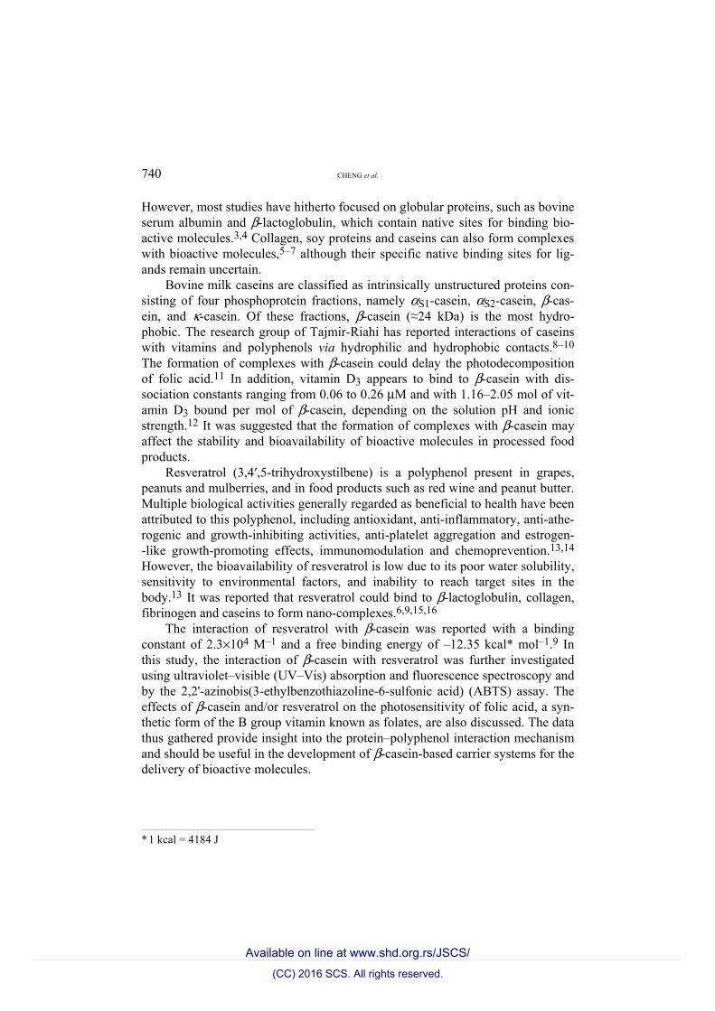
Absorbance of β-casein–resveratrol mixtures The difference spectra of the absorbance of resveratrol–β-casein mixtures
with that of β-casein at polyphenol concentrations of up to 20 μM are shown in Fig. 1A. A gradual and significant increase of the maximum concentration between 300 and 320 nm, attributed to resveratrol,15 was observed with inc-reasing polyphenol concentration. The difference spectra of the absorbance of resveratrol–β-casein mixtures with that of resveratrol at protein concentrations of up to 50 μM are presented in Fig. 1B. A gradual and significant increase in the absorbance maximum at 280 nm, attributed to β-casein, was observed.
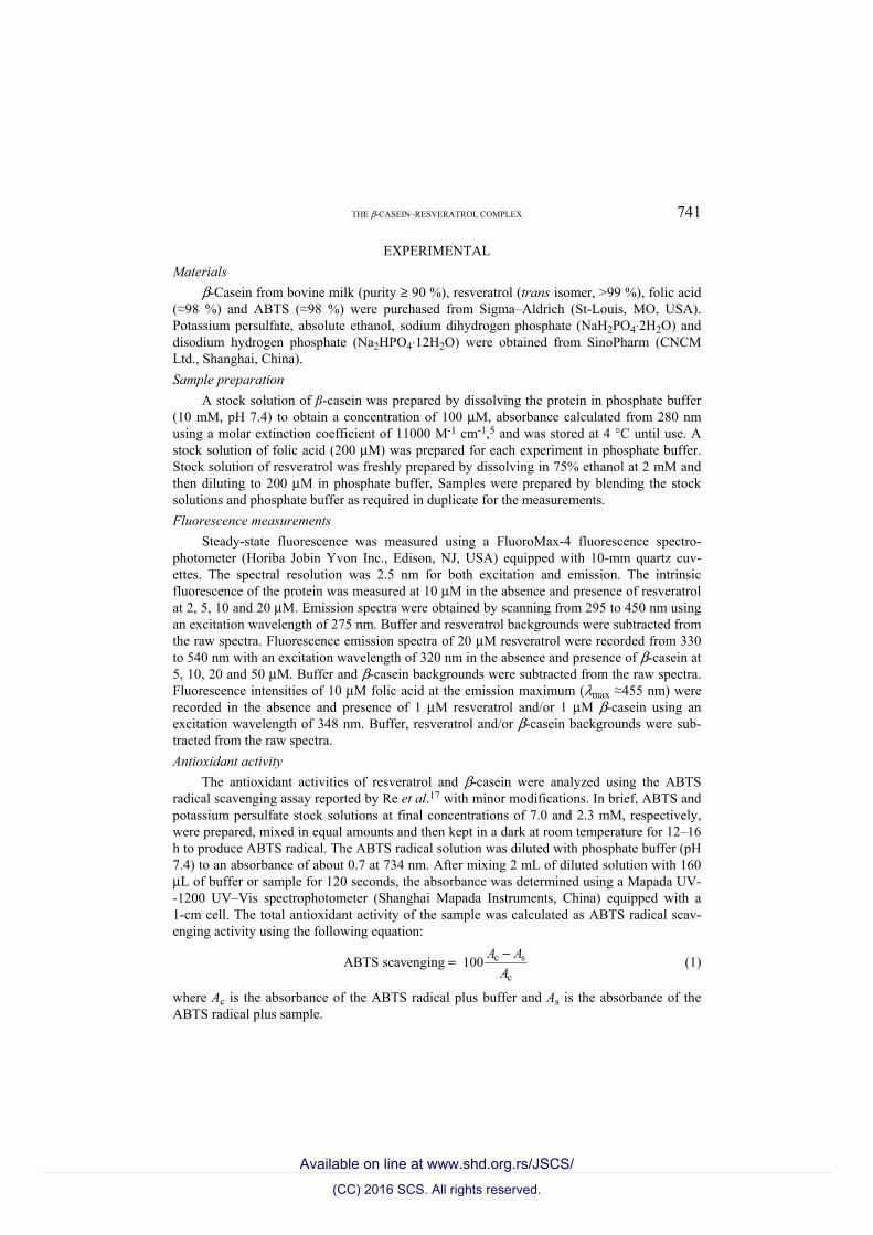
Influence of β-casein–resveratrol complex formation on the protein structure β-Casein contains one tryptophan residue and four tyrosine residues.20 The
excitation wavelength at 275 nm was used to minimize the inner-filter effect. Fluorescence spectrum of β-casein had a λmax at 335 nm with a shoulder around 300 nm in phosphate buffer at pH 7.4 (Fig. 2A). The λmax shifted gradually to longer wavelengths and reached 349 nm as the concentration of resveratrol was increased to 20 μM. A significant decrease was concomitantly observed in the fluorescence intensities attributed to the tryptophan and tyrosine residues. At 20 μM resveratrol, the intensity at λmax was about 59 % of that of pure β-casein. After correction for the inner filter effect, the resveratrol-induced β-casein fluor-
_________________________________________________________________________________________________________________________
(CC) 2016 SCS. All rights reserved.
Available on line at www.shd.org.rs/JSCS/
THE β-CASEIN–RESVERATROL COMPLEX 743
Fig. 1. A) Difference absorption spectra between resveratrol–β-casein and pure β-casein at various concentrations of resve-ratrol and B) difference absorption spectra between resveratrol–β-casein and pure resve-ratrol at various concentrations of β-casein.
Fig. 2. Fluorescence emission spectra of β-casein in the absence and presence of resveratrol at various concentrations: A) Fobs, the measured fluorescence intensity and B) Fcorr, the corrected fluorescence intensity.
_________________________________________________________________________________________________________________________
(CC) 2016 SCS. All rights reserved.
Available on line at www.shd.org.rs/JSCS/
744 CHENG et al.
escence changes were similar but less pronounced (Fig. 2B). At 20 μM resve-ratrol, the λmax was at around 338 nm and its intensity was about 88 % of that of pure protein, which remained invariable after 24 h (data not shown). These results suggest that the formation of resveratrol–β-casein complexes transferred the protein fluorophores to a more hydrophilic environment.9 In Fig. 2B, an iso-fluorescence point was seen around 364 nm, suggesting the transfer is a two-state process.
Influence of β-casein–resveratrol complex formation on the polyphenol environment
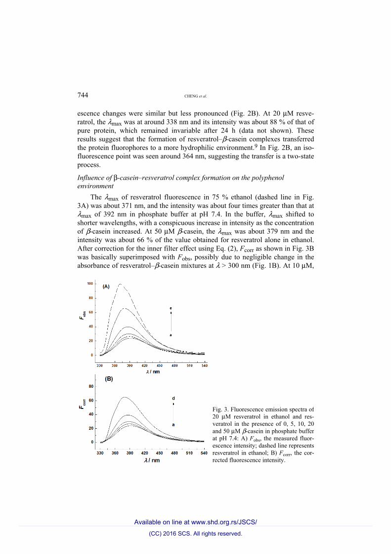
The λmax of resveratrol fluorescence in 75 % ethanol (dashed line in Fig. 3A) was about 371 nm, and the intensity was about four times greater than that at λmax of 392 nm in phosphate buffer at pH 7.4. In the buffer, λmax shifted to shorter wavelengths, with a conspicuous increase in intensity as the concentration of β-casein increased. At 50 μM β-casein, the λmax was about 379 nm and the intensity was about 66 % of the value obtained for resveratrol alone in ethanol. After correction for the inner filter effect using Eq. (2), Fcorr as shown in Fig. 3B was basically superimposed with Fobs, possibly due to negligible change in the absorbance of resveratrol–β-casein mixtures at λ > 300 nm (Fig. 1B). At 10 μM,
Fig. 3. Fluorescence emission spectra of 20 μM resveratrol in ethanol and res-veratrol in the presence of 0, 5, 10, 20 and 50 μM β-casein in phosphate buffer at pH 7.4: A) Fobs, the measured fluor-escence intensity; dashed line represents resveratrol in ethanol; B) Fcorr, the cor-rected fluorescence intensity.
_________________________________________________________________________________________________________________________
(CC) 2016 SCS. All rights reserved.
Available on line at www.shd.org.rs/JSCS/
THE β-CASEIN–RESVERATROL COMPLEX 745
the β-casein-caused change in the fluorescence of resveratrol were stable after 24 h (data not shown). The change in the resveratrol fluorescence indicates that formation of a complex with β-casein transfers the polyphenol from the aqueous solution to a more hydrophobic environment. This is consistent with the inter-action of resveratrol with β-lactoglobulin, a major protein in milk whey.15
Influence of complex formation on the antioxidant activity of the resveratrol–β-casein system
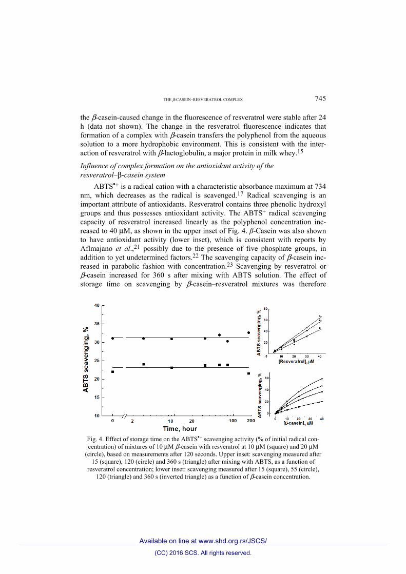
ABTS•+ is a radical cation with a characteristic absorbance maximum at 734 nm, which decreases as the radical is scavenged.17 Radical scavenging is an important attribute of antioxidants. Resveratrol contains three phenolic hydroxyl groups and thus possesses antioxidant activity. The ABTS+ radical scavenging capacity of resveratrol increased linearly as the polyphenol concentration inc-reased to 40 μM, as shown in the upper inset of Fig. 4. β-Casein was also shown to have antioxidant activity (lower inset), which is consistent with reports by Aflmajano et al.,21 possibly due to the presence of five phosphate groups, in addition to yet undetermined factors.22 The scavenging capacity of β-casein inc-reased in parabolic fashion with concentration.23 Scavenging by resveratrol or β-casein increased for 360 s after mixing with ABTS solution. The effect of storage time on scavenging by β-casein–resveratrol mixtures was therefore
Fig. 4. Effect of storage time on the ABTS•+ scavenging activity (% of initial radical con-centration) of mixtures of 10 μM β-casein with resveratrol at 10 μM (square) and 20 μM
(circle), based on measurements after 120 seconds. Upper inset: scavenging measured after 15 (square), 120 (circle) and 360 s (triangle) after mixing with ABTS, as a function of
resveratrol concentration; lower inset: scavenging measured after 15 (square), 55 (circle), 120 (triangle) and 360 s (inverted triangle) as a function of β-casein concentration.
_________________________________________________________________________________________________________________________
(CC) 2016 SCS. All rights reserved.
Available on line at www.shd.org.rs/JSCS/
746 CHENG et al.
measured after 120 s. Mixtures containing 10 and 20 μM resveratrol scavenged, respectively, about 23 and 31 % of the ABTS•+, equal to the sum of the anti-oxidant capacities of the individual components, suggesting that the formation of β-casein–resveratrol complexes did not affect the antioxidant activity of either component. Moreover, the scavenging capacity of β-casein–resveratrol com-plexes remained stable after storage for 200 h.
Masking effects of proteins on the antioxidant activity of polyphenols have been widely reported. The formation of complexes with β-lactoglobulin dec-reased the antioxidant activity of epigallocatechin 3-gallate, the most active poly-phenol present in green tea.24,25 Bovine serum albumin and caseins were shown to reduce the total antioxidant capacity of protein–flavonoid systems, β-casein and serum albumin more so than κ-casein and α-casein.26 However, the present β-casein–resveratrol system, in which the amount of bound resveratrol was about 20 % of the total, retained the total antioxidant capacity. This might be due to two possibilities: 1) the antioxidant portions of the molecules are not involved in the binding; 2) β-casein–resveratrol complexes dissociate to re-establish the equi-librium concentrations once they were diluted during the measurements of anti-oxidant activity.
Influence of β-casein–resveratrol on resveratrol and folic acid photosensitivity Resveratrol exists naturally as the trans isomer, which is believed respon-
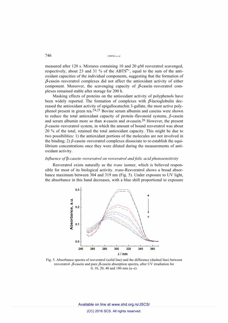
sible for most of its biological activity. trans-Resveratrol shows a broad absor-bance maximum between 304 and 319 nm (Fig. 5). Under exposure to UV light, the absorbance in this band decreases, with a blue shift proportional to exposure
Fig. 5. Absorbance spectra of resveratrol (solid line) and the difference (dashed line) between
resveratrol–β-casein and pure β-casein absorption spectra, after UV irradiation for 0, 10, 20, 40 and 180 min (a–e).
_________________________________________________________________________________________________________________________
(CC) 2016 SCS. All rights reserved.
Available on line at www.shd.org.rs/JSCS/
THE β-CASEIN–RESVERATROL COMPLEX 747
time. After 180 min, it appears as a weaker peak around 289 nm, characteristic of cis-resveratrol,27 which is found in grape wines.28,29 Trans–cis isomerization of resveratrol under daylight is slower, taking about 21–30 h,15 indicating that UV irradiation accelerates this process. Among the reported biological activities of cis-resveratrol are antioxidant activity, inhibition of collagen-induced platelet aggregation, and cancer-related kinase activities,30–32 although it is unclear whether the cis isomer is as active as the trans isomer. No significant change in the absorption spectra of resveratrol was obtained by adding 20 μM β-casein (Fig. 5), suggesting that the formation of a complex with this protein did not affect the chemical structure of the polyphenol.
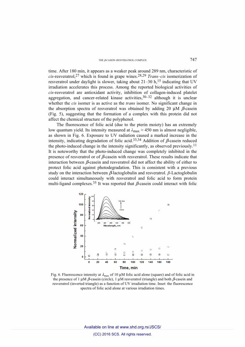
The fluorescence of folic acid (due to the pterin moiety) has an extremely low quantum yield. Its intensity measured at λmax ≈ 450 nm is almost negligible, as shown in Fig. 6. Exposure to UV radiation caused a marked increase in the intensity, indicating degradation of folic acid.33,34 Addition of β-casein reduced the photo-induced change in the intensity significantly, as observed previously.11 It is noteworthy that the photo-induced change was completely inhibited in the presence of resveratrol or of β-casein with resveratrol. These results indicate that interaction between β-casein and resveratrol did not affect the ability of either to protect folic acid against photodegradation. This is consistent with a previous study on the interaction between β-lactoglobulin and resveratrol. β-Lactoglobulin could interact simultaneously with resveratrol and folic acid to form protein multi-ligand complexes.35 It was reported that β-casein could interact with folic
Fig. 6. Fluorescence intensity at λmax of 10 μM folic acid alone (square) and of folic acid in
the presence of 1 μM β-casein (circle), 1 μM resveratrol (triangle) and both β-casein and resveratrol (inverted triangle) as a function of UV irradiation time. Inset: the fluorescence
spectra of folic acid alone at various irradiation times.
_________________________________________________________________________________________________________________________
(CC) 2016 SCS. All rights reserved.
Available on line at www.shd.org.rs/JSCS/

748 CHENG et al.
acid to form complexes with an affinity of about 7.0×10⁴ M–1.8,11 Therefore, the lack of an effect of β-casein–resveratrol complexation on the degradation of folic acid might be attributed to the binding capacity of both folic acid and resveratrol to β-casein. In addition, together with the photo-induced isomerization of the polyphenol in the absence and presence of β-casein (Fig. 5), it is suggested that the effect of resveratrol on folic acid decomposition might be attributed to its structural transition.
CONCLUSIONS
The interaction between resveratrol and β-casein shifted the protein fluoro-phore residues to a more hydrophilic environment, causing a decrease in protein fluorescence. The interaction shifted resveratrol to a more hydrophobic environ-ment, leading to an increase in the polyphnol fluorescence. The formation of a complex with β-casein did not affect the isomerization of resveratrol or the total antioxidant activity of this protein–polyphenol system. The protective effect of resveratrol against the photodecomposition of folic acid was not affected by binding to β-casein. The data obtained in this study should provide insight into protein–polyphenol interaction mechanisms and aid in the development of β-cas-ein-based carrier systems for the delivery of bioactive molecules.
Acknowledgments. This work received support from the National Natural Science Found-ation of China (NSFC Project 31201291 and 31571781). The authors thank Dr. van de Weert (Department of Pharmaceutics and Analytical Chemistry, University of Copenhagen) for help with the comments about the inner filter effect.
И З В О Д КОМПЛЕКС β-КАЗЕИНА И РЕСВЕРАТРОЛА: ФИЗИЧКОХЕМИЈСКЕ КАРАКТЕРИСТИКЕ
И ЗНАЧАЈ ЗА УНАПРЕЂЕЊЕ ИСХРАНЕ
HAO CHENG1, YANFANG LI1, XUNDI YIN2, MURIEL SUBIRADE3 и LI LIANG1
1State Key Lab of Food Science and Technology, School of Food Science and Technology, Jiangnan University, Wuxi, Jiangsu, China, 2College of Textiles & Clothing, Jiangnan University, Wuxi,
Jiangsu, China и 3Chaire de recherche du Canada sur les protéines, les bio-systèmes et les aliments fonctionnels, Institut de recherche sur les nutraceutiques et les aliments fonctionnels
(INAF/STELA), Université Laval, Québec, Canada
Протеини хране су у широкој употреби као носачи за инкапсулацију и заштиту биоактивних молекула. За развој система који се заснивају на протеинским носачима, важно је разумети механизам интеракције протеина и биоактивних молекула. У овом раду је изучавана интеракција β-казеина са ресвератролом, природним полифенолом, користећи методе UV–Vis апсорпционе и флуоресцентне спектроскопије. Показано је да интеракција помера протеинске флуорофоре према хидрофилнијој средини, а полифе-нол ка хидрофобнијој околини. Стварање комплекса са β-казеином није утицало на trans–cis изомеризацију ресвератрола или на укупну антиоксидативну активност сис-тема протеин–полифенол, што је утврђено спектрофотометријски и тестом са 2,2'-ази-нобис(3-етилбензотиазолин-6-сулфонском киселином). Заштитни ефекат ресвератрола на фотодеградацију фолне киселине није био измењен услед његовог везивања за β-казеин. Добијени резултати омогућавају сагледавање механизма интеракције про-
_________________________________________________________________________________________________________________________
(CC) 2016 SCS. All rights reserved.
Available on line at www.shd.org.rs/JSCS/

THE β-CASEIN–RESVERATROL COMPLEX 749
теин–полифенол и могу помоћи у развоју система који се заснивају на β-казеину као носачу биоактивних молекула.
(Примљено 29. октобра 2015, ревидирано 28. јануара, прихваћено 26. фебруара 2016)
REFERENCES 1. L. Y. Chen, G. E. Remondetto, M. Subirade, Trends Food Sci. Technol. 17 (2006) 272 2. O. G. Jones, D. J. McClements Adv. Colloid Interface Sci. 167 (2011) 49 3. Y. J. Hu, Y. Wang, Y. Ou-Yang, J. Zhou, Y. Liu, J. Luminesc. 130 (2010) 1394 4. F. A. Wolf, G. M. Brett, Pharmacol. Rev. 52 (2000) 207 5. P. Bourassa, C. N’soukpoé-Kossi, H. A. Tajmir-Riahi. Food Chem. 138 (2013) 444 6. J. Zhang, Q. Mi, M. Shen, Food Chem. 131 (2012a) 879 7. J. Zhang, L. Liang, Z. G. Tian, L. Y. Chen, M. Subirade, J. Phys. Chem. B 117 (2013)
14018 8. P. Bourassa, H. A. Tajmir-Riahi, J. Phys. Chem. B 116 (2012) 513 9. P. Bourassa, J. Bariyanga, H. A. Tajmir-Riahi, J. Phys. Chem. B 117 (2013) 1287
10. I. Hasni, P. Bourassa, S. Hamdani, G. Samson, R. Carpentier, H. A. Tajmir-Riahi Food Chem. 126 (2011) 630
11. J. Zhang, Y. N. Liu, X. M. Liu, Y. F. Li, X. D. Yin, M. Subirade, P. Zhou, L. Liang, Food Res. Int. 57 (2014) 162
12. S. A. Forrest, R. Y. Yada, D. R. Rousseau, J. Agr. Food Chem. 53 (2005) 8003 13. M. A. Augustin, L. Sanguansri, T. Lockett, Ann. N. Y. Acad. Sci. 1290 (2013) 107 14. C. A. De La Lastra, I. Villegas, Biochem. Soc. Trans. 35 (2007) 1156 15. L. Liang, H. A. Tajmir-Riahi, M. Subirade, Biomacromolecules 9 (2008) 50 16. J. Zhang, X. F. Dai, J. Y. Huang, Food Biophys. 7 (2012b) 35 17. R. Re, N. Pellegrini, A. Proteggente, A. Pannala, M. Yang, A. Rice-Evans, Free Radical
Biol. Med. 26 (1999) 1231 18. S. Neamtu, M. Mic, M. Bogdan, I. Turcu, J. Pharm. Biomed. Anal. 72 (2013) 134 19. M. Van de Weert, L. Stella, J. Mol. Struct. 998 (2011) 144 20. K. N. Pearce, Eur. J. Biochem. 58 (1975) 23 21. M. P. Almajano, M. E. Delgado, M. H. Gordon, Food Chem. 101 (2007) 126 22. G. Cervato, R. Cazzola, B. Cestaro, Int. J. Food Sci. Nutr. 50 (1999) 291 23. Q. Ge, X. J. Ma, Food Sci. Human Wellness 2 (2013) 68 24. A. Shpigelman, G. Israeli, Y. D. Livney, Food Hydrocolloids 24 (2010) 735 25. R. Zorilla, L. Liang, G. Remondetto, M. Subirade, Dairy Sci. Technol. 91 (2011) 629 26. M. J. T. J. Arts, G. R. M. M. Haenen, L. C. Wilms, S. A. J. N. Beetstra, C. G. M. Heijnen,
H. P. Voss, A. Bast, J. Agr. Food Chem. 50 (2002) 1184 27. L. Camont, C. H. Cottart, Y. Rhayem, V. Nivet-Antoine, R. Djelidi, F. Collin, J. L.
Beaudeux, D. Bonnefont-Rousselot, Anal. Chim. Acta 634 (2009) 121 28. D. M. Goldberg, E. Tsang, A. Karumanchiri, E. P. Diamandis, G. Soleas, E. Ng, Anal.
Chem. 68 (1996) 1688 29. J. Lopez-Hernandez, P. Paseiro-Losada, A. Sanches-Silva, M. A. Lage-Yusty, Eur. Food
Res. Technol. 225 (2007) 789 30. A. A. Bertelli, L. Giovannini, W. Bernini, M. Migliori, M. Fregoni, L. Bavaresco, A.
Bertelli, Drugs Exp. Clin. Res. 22 (1996) 61 31. G. S. Jayatilake, H. Jayasuriya, E. S. Lee, N. M. Koonchanik, R. L. Geahlen, C. L.
Ashendel, J. L. McLaughlin, C. J. Chang, J. Nat. Prod. 56 (1993) 1805 32. J. Leiro, E. Alvarez, J. A. Arranz, R. Laguna, E. Uriarte, F. Orallo, J .Leukocyte Biol. 75
(2004) 1156
_________________________________________________________________________________________________________________________
(CC) 2016 SCS. All rights reserved.
Available on line at www.shd.org.rs/JSCS/

750 CHENG et al.
33. M. K. Off, A. E. Steindal, A. C. Porojnicu, A. Juzeniene, A. Vorobey, A. Johnsson, J. Moan, J. Photochem. Photobiol., B 80 (2005) 47
34. P. Vorobey, A. E. Steindal, M. K. Off, A. Vorobey, J. Moan, Photochem. Photobiol. 82 (2006) 817
35. J. Zhang, X. M. Liu, M. Subirade, P. Zhou, L. Liang, Food Chem. 165 (2014) 256.
_________________________________________________________________________________________________________________________
(CC) 2016 SCS. All rights reserved.
Available on line at www.shd.org.rs/JSCS/