
The Borrelia afzelii outer membrane protein BAPKO 0422 ...eprints.whiterose.ac.uk/108782/1/The...
14
Biosci. Rep. (2015) / 35 / art:e00240 / doi 10.1042/BSR20150095 The Borrelia afzelii outer membrane protein BAPKO_0422 binds human factor-H and is predicted to form a membrane-spanning β -barrel Adam Dyer* 1 , Gemma Brown* 1 , Lenka Stejskal*, Peter R. Laity*† and Richard J. Bingham* 2 *Department of Biological Sciences, School of Applied Sciences, University of Huddersfield, Queensgate, Huddersfield HD1 3DH, U.K. †Present Address: Department of Materials Science and Engineering, Sir Robert Hadfield Building, Mappin Street, University of Sheffield, Sheffield S1 3JD, U.K. Synopsis The deep evolutionary history of the Spirochetes places their branch point early in the evolution of the diderms, before the divergence of the present day Proteobacteria. As a spirochete, the morphology of the Borrelia cell envelope shares characteristics of both Gram-positive and Gram-negative bacteria. A thin layer of peptidoglycan, tightly associated with the cytoplasmic membrane, is surrounded by a more labile outer membrane (OM). This OM is rich in lipoproteins but with few known integral membrane proteins. The outer membrane protein A (OmpA) domain is an eight-stranded membrane-spanning β -barrel, highly conserved among the Proteobacteria but so far unknown in the Spirochetes. In the present work, we describe the identification of four novel OmpA-like β -barrels from Borrelia afzelii, the most common cause of erythema migrans (EM) rash in Europe. Structural characterization of one these proteins (BAPKO_0422) by SAXS and CD indicate a compact globular structure rich in β -strand consistent with a monomeric β -barrel. Ab initio molecular envelopes calculated from the scattering profile are consistent with homology models and demonstrate that BAPKO_0422 adopts a peanut shape with dimensions 25 × 45 ˚ A (1 ˚ A = 0.1 nm). Deviations from the standard C-terminal signature sequence are apparent; in particular the C-terminal phenylalanine residue commonly found in Proteobacterial OM proteins is replaced by isoleucine/leucine or asparagine. BAPKO_0422 is demonstrated to bind human factor H (fH) and therefore may contribute to immune evasion by inhibition of the complement response. Encoded by chromosomal genes, these proteins are highly conserved between Borrelia subspecies and may be of diagnostic or therapeutic value. Key words: β -barrel, Borrelia, Lyme disease, outer membrane protein A (OmpA), small angle X-ray scattering (SAXS), spirochete. Cite this article as: Bioscience Reports (2015) 35, e00240, doi:10.1042/BSR20150095 INTRODUCTION Various species in the genus Borrelia are capable of zoonotic infection in humans when transmitted via the saliva of haema- tophagous ticks resulting in Lyme disease [1]). Considerable re- search has been directed at Borrelia burgdorferi sensu stricto, the most prevalent strain in North America but only a minor contributor to incidence rates in Europe and Asia. The available data on the natural hosts/vectors present in Europe show that the most common species present are Borrelia garinii and Borrelia ............................................................................................................................................................................................................................................................................................................ Abbreviations: ALBI, affinity ligand-binding immunoblot; AtVDAC1, Arabidopsis thaliana voltage-dependent anion channel 1; BamA, barrel assembly machinery; BB, Borrelia burgdorferi; BG, Borrelia garinii; BAPKO, Borrelia afzelii strain PKO; DDAO, N,N-dimethyldodecylamine-N-oxide; DipA, dicarboxylate-specific porin A; EM, erythema migrans; fH, factor H; HMM, hidden Markov model; OM, outer membrane; OmpA, outer membrane protein A; Rg , radius of gyration; TM, transmembrane. 1 These authors contributed equally to the work. 2 To whom correspondence should be addressed (email [email protected]). afzelii, the latter of which is responsible for the vast majority of observed erythema migrans (EM) rash [2,3]. Although early treatment with antibiotics can be effective, symptoms may con- tinue post-treatment probably due to a variety factors including the presence of immunogenic cell debris, survival of viable spiro- chetes [4–6] or the presence of persister phenotypes [7]. Borrelia have evolved numerous strategies to aid survival for extended periods within the mammalian host. Evasion strategies include antigenic variation [8,9], variable expression of surface antigens [10–12], invasion of immune privileged sites [13,14] and spe- cific binding of immune regulators [15]. As part of the latter, the c 2015 Authors. This is an open access article published by Portland Press Limited and distributed under the Creative Commons Attribution Licence 3.0. 1
Transcript of The Borrelia afzelii outer membrane protein BAPKO 0422 ...eprints.whiterose.ac.uk/108782/1/The...
Biosci. Rep. (2015) / 35 / art:e00240 / doi 10.1042/BSR20150095
The Borrelia afzelii outer membrane proteinBAPKO_0422 binds human factor-H and ispredicted to form a membrane-spanning β -barrelAdam Dyer*1, Gemma Brown*1, Lenka Stejskal*, Peter R. Laity*† and Richard J. Bingham*2
*Department of Biological Sciences, School of Applied Sciences, University of Huddersfield, Queensgate, Huddersfield HD1 3DH, U.K.†Present Address: Department of Materials Science and Engineering, Sir Robert Hadfield Building, Mappin Street, University of Sheffield,Sheffield S1 3JD, U.K.
SynopsisThe deep evolutionary history of the Spirochetes places their branch point early in the evolution of the diderms, beforethe divergence of the present day Proteobacteria. As a spirochete, the morphology of the Borrelia cell envelope sharescharacteristics of both Gram-positive and Gram-negative bacteria. A thin layer of peptidoglycan, tightly associatedwith the cytoplasmic membrane, is surrounded by a more labile outer membrane (OM). This OM is rich in lipoproteinsbut with few known integral membrane proteins. The outer membrane protein A (OmpA) domain is an eight-strandedmembrane-spanning β -barrel, highly conserved among the Proteobacteria but so far unknown in the Spirochetes. In thepresent work, we describe the identification of four novel OmpA-like β -barrels from Borrelia afzelii, the most commoncause of erythema migrans (EM) rash in Europe. Structural characterization of one these proteins (BAPKO_0422) bySAXS and CD indicate a compact globular structure rich in β -strand consistent with a monomeric β -barrel. Ab initiomolecular envelopes calculated from the scattering profile are consistent with homology models and demonstratethat BAPKO_0422 adopts a peanut shape with dimensions 25 × 45 A (1 A = 0.1 nm). Deviations from the standardC-terminal signature sequence are apparent; in particular the C-terminal phenylalanine residue commonly found inProteobacterial OM proteins is replaced by isoleucine/leucine or asparagine. BAPKO_0422 is demonstrated to bindhuman factor H (fH) and therefore may contribute to immune evasion by inhibition of the complement response.Encoded by chromosomal genes, these proteins are highly conserved between Borrelia subspecies and may be ofdiagnostic or therapeutic value.
Key words: β -barrel, Borrelia, Lyme disease, outer membrane protein A (OmpA), small angle X-ray scattering (SAXS),spirochete.
Cite this article as: Bioscience Reports (2015) 35, e00240, doi:10.1042/BSR20150095
INTRODUCTION
Various species in the genus Borrelia are capable of zoonoticinfection in humans when transmitted via the saliva of haema-tophagous ticks resulting in Lyme disease [1]). Considerable re-search has been directed at Borrelia burgdorferi sensu stricto,the most prevalent strain in North America but only a minorcontributor to incidence rates in Europe and Asia. The availabledata on the natural hosts/vectors present in Europe show that themost common species present are Borrelia garinii and Borrelia
. . . . . . . . . . . . . . . . . . . . . . . . . . . . . . . . . . . . . . . . . . . . . . . . . . . . . . . . . . . . . . . . . . . . . . . . . . . . . . . . . . . . . . . . . . . . . . . . . . . . . . . . . . . . . . . . . . . . . . . . . . . . . . . . . . . . . . . . . . . . . . . . . . . . . . . . . . . . . . . . . . . . . . . . . . . . . . . . . . . . . . . . . . . . . . . . . . . . . . . . . . . . . . . . . . . . . . . . . . . . . . . . . . . . . . . . . . . . . . . . . . . . . . . . . . . . . . . . . . . . . . . . . . . . . . . . . . . . . . . . . . . . . . . . . . . . . . . . . . . .
Abbreviations: ALBI, affinity ligand-binding immunoblot; AtVDAC1, Arabidopsis thaliana voltage-dependent anion channel 1; BamA, barrel assembly machinery; BB, Borrelia burgdorferi;BG, Borrelia garinii; BAPKO, Borrelia afzelii strain PKO; DDAO, N,N-dimethyldodecylamine-N-oxide; DipA, dicarboxylate-specific porin A; EM, erythema migrans; fH, factor H; HMM, hiddenMarkov model; OM, outer membrane; OmpA, outer membrane protein A; Rg, radius of gyration; TM, transmembrane.1 These authors contributed equally to the work.2 To whom correspondence should be addressed (email [email protected]).
afzelii, the latter of which is responsible for the vast majorityof observed erythema migrans (EM) rash [2,3]. Although earlytreatment with antibiotics can be effective, symptoms may con-tinue post-treatment probably due to a variety factors includingthe presence of immunogenic cell debris, survival of viable spiro-chetes [4–6] or the presence of persister phenotypes [7]. Borreliahave evolved numerous strategies to aid survival for extendedperiods within the mammalian host. Evasion strategies includeantigenic variation [8,9], variable expression of surface antigens[10–12], invasion of immune privileged sites [13,14] and spe-cific binding of immune regulators [15]. As part of the latter, the
c© 2015 Authors. This is an open access article published by Portland Press Limited and distributed under the Creative Commons Attribution Licence 3.0. 1
A. Dyer and others
ability to bind factor-H (fH) at the bacterial cell surface andso exploit the host’s own protection against the complement re-sponse is an essential virulence factor to establish infection andis thought to determine host specificity [16]. Adhesion, invasionand immune evasion is mediated primarily by a variety of surfacelipoproteins and membrane spanning β-barrels. Other notablesurface proteins include the integrin-binding BBB07 [17], theα-helical P13 channel protein [18,19] and the BesABC (Borreliaefflux system proteins A, B and C) efflux pump [20,21].
Lipoproteins are readily identified by their characteristic sig-nal sequence, consisting of a positively charged N-region, hydro-phobic H-region and a lipobox terminating with a single cysteineamino acid [22]. Genome data [23,24] has shown Borrelia has∼105 such proteins, many of which have been studied in detailand have been shown to bind to a wide range of host extracel-lular matrix proteins [25], immune regulators and cell surfacereceptors [15].
β-Barrel membrane-spanning proteins pose a significant chal-lenge to prediction methods because they are characterized byshort (<10 amino acids) membrane-spanning strands of altern-ate hydrophobic residues interspersed by highly variable loopregions [26]. The sequence conservation between orthologuescan also be very low. Consequently and probably also dueto their relative scarcity on the outer membrane (OM), veryfew β-barrel membrane-spanning proteins have been identifiedin Borrelia. Those that have been identified, BesC (BB0142)[20], dicarboxylate-specific porin A (DipA) (BB0418) [27], P66(BB0603, Oms66) [28,29] and BamA (barrel assembly ma-chinery; BB0795) [30] are all predicted to form 16–24-strandedporin-type barrels. The smaller barrel-types with 8–10 strands,commonly found in other Gram-negative bacteria have so farremained undetected in Borrelia.
Highly conserved among the Proteobacteria, the outer mem-brane protein A (OmpA)-like transmembrane (TM) domain,defined by Pfam family PF01389, consists of eight membrane-spanning antiparallel β-strands with four large extracellular loopsand three short periplasmic turns [26]. High-resolution structuraldata are available for several members of this family with arange of different functions [31–34]. This structural data con-firmed that the TM region is highly conserved and functionaldiversity is mainly due to variations in extracellular loop regions.The prototypical Escherichia coli OmpA functions as a non-specific diffusion channel and with the addition of a C-terminalpeptidoglycan-binding domain, contributes to the maintenanceof cell shape and structural integrity of the OM [35].
In addition to these physiological functions, numerous mem-bers of this family are known virulence factors including E. coliOmpA [36,37], Cronobacter sakazakii OmpA [38], Salmonellatyphimurium Rck and PagC [39,40] and Yersinia pestis Ail [31].These proteins contribute to pathogenicity by binding to a rangeof host factors mediating adhesion, invasion and resistance to thecomplement response.
To date, the OmpA-like TM domain has not been identifiedin Borrelia. Considering the similarities between the membranestructure of Borrelia and that of the Proteobacteria, we con-sidered it likely that the OmpA-like TM domain would be con-
served in both. To this aim we conducted PSI-BLAST searchesof the NCBI protein database against the available B. burgdorferiproteomes using a variety of OmpA sequences as search targets.Although larger β-barrel proteins such as BamA (BB0795) werefound, no proteins matching the criteria of an OmpA-like do-main could be identified. Consequently, we used a profile hiddenMarkov model (HMM) search strategy, which enables sequenceswith more remote homology to be detected [41]. Four homo-logous proteins were identified, all encoded by chromosomalgenes and highly conserved between different species of Borrelia.Analysis of signal sequences and bioinformatic analysis suggestthat all four proteins adopt similar topology as eight-strandedmembrane-spanning β-barrels. As the most common cause ofEM in Europe, we chose the B. afzelii homologue BAPKO_0422for further experimental work.
In the following work, we describe the production of a re-combinant version of BAPKO_0422 consisting of the predictedmembrane-spanning domain (no signal sequence) in the E. coliexpression system. Furthermore an affinity ligand-binding im-munoblot (ALBI) assay demonstrates that BAPKO_0422 bindsspecifically to human fH. Finally, data from SAXS, CD andphase partitioning demonstrate that BAPKO_0422 adopts a com-pact peanut-shaped structure rich in β-strand, consistent with amembrane-spanning β-barrel topology.
MATERIALS AND METHODS
Identification of Borrelia OmpA-like domainsProtein sequences of all available OmpA-like TM domains wereobtained from the Pfam database family PF01389 [42]. Se-quences with greater than 90 % or 40 % similarity were removedusing the Decrease Redundancy program (ExPASy) to generatetwo lists, which were then submitted to JACOP for classifica-tion [43]. Three families were identified corresponding to OmpA,OmpX and OmpW. Sequences corresponding to each family werealigned using ClustalW2 (EBI) and used to build a profile HMMusing HMMER (http://hmmer.janelia.org/). This generated sixprofile HMMs, which were then used to search the genomesof B. afzelii, B. burgdorferi and B. garinii to generate a list ofpotential OmpA-like proteins. The top scoring hits were filteredbased on the presence of a signal sequence predicted using SignalP 4.1 [44] and fold prediction using the Fold and Function As-signment System server (FFAS03) [45]. The procedure identifiedtwo proteins BB0027 and BAPKO_0422 matching all criteria. APSI-BLAST search was then conducted to identify homologousproteins in B. afzelii, B. burgdorferi and B. garinii. Topology pre-dictions were conducted using the PRED-TMBB web server [46]using the mature sequences of all homologues identified above(minus signal peptide).
Homology modellingHomology models were generated using Modeller [47] based onco-ordinates from PDB accession codes 1BXW, 1P4T, 1QJ8,
. . . . . . . . . . . . . . . . . . . . . . . . . . . . . . . . . . . . . . . . . . . . . . . . . . . . . . . . . . . . . . . . . . . . . . . . . . . . . . . . . . . . . . . . . . . . . . . . . . . . . . . . . . . . . . . . . . . . . . . . . . . . . . . . . . . . . . . . . . . . . . . . . . . . . . . . . . . . . . . . . . . . . . . . . . . . . . . . . . . . . . . . . . . . . . . . . . . . . . . . . . . . . . . . . . . . . . . . . . . . . . . . . . . . . . . . . . . . . . . . . . . . . . . . . . . . . . . . . . . . . . . . . . . . . . . . . . . . . . . . . . . . . . . . . . . . . . . . . . . . . . . . . . . . . . . . . . . . . . . . . . . . . . . . . . . . . . . . . . . . . . . . . . . . . . . . . . . . . . . . . .
2 c© 2015 Authors. This is an open access article published by Portland Press Limited and distributed under the Creative Commons Attribution Licence 3.0.
The BAPKO_0422 paralogous group of β-barrel OMPs
1QJP, 1THQ, 2ERV, 2F1T, 2K0L, 2X27, 3DZM, 3GP6 and3QRA. A structure-based multiple sequence alignment was gen-erated using the MatchMaker function in the UCSF Chimerapackage [48] with default settings. This was manually adjus-ted to minimize sequence gaps in loop regions. Sequences forthe putative Borrelia OmpA-like TM domains were then addedto this alignment and manually adjusted to match the topologyand β-strand positioning as predicted by PRED-TMBB. Multiplemodels were then generated using Modeller. Theoretical radius ofgyration (Rg) values, based on the centre of mass, were calculatedusing the UCSF Chimera package [48].
Cloning of BAPKO_042220-201
The coding region for amino acids 20–201 of BAPKO_0422(European Nucleotide Archive accession number ABH01676.1)was amplified by PCR using the forward primer 5′-GCA-TGGATCCGCAATCAAAAAGCAAAACTAT-3′ and reverseprimer 5′-ATCGAAGCTTTCATTTATTCTCCATTATATATA-3′
with the addition of BamH1 and HindIII restriction sites (under-lined). This was ligated into the pET-47b( + ) expression vector(Novagen) and confirmed by DNA sequencing. PCR primerswere synthesized by Eurofins MWG Operon.
Recombinant expression and purification ofBAPKO_042220-201
Protein expression in RosettaTM(DE3)pLysS cells (Merck Mil-lipore) was induced by the addition of 1 mM IPTG to a 3 l ofculture of cells with a D of 0.6. After 3 h, cells were harvestedby centrifugation and lysed on ice by pulsed sonication in 0.3 MNaCl, 50 mM Tris/HCl, pH 8.0. The lysate was incubated withDNase before removal of the soluble fraction by centrifugation(20 000 g for 30 min at 4 ◦C) and the pellet prepared for solu-bilization by means of several preparative washes (0.3 M NaCl,50 mM Tris/HCl, 10 mM EDTA, 1 mM DTT, 0.1 % Triton-X-100, pH 8.0). The washed inclusion body was then solubilized in8 M urea, 0.3 M NaCl, 50 mM Tris/HCl, pH 8.0, overnight andclarified by centrifugation at 16 000 g for 30 min.
Protein was loaded on to a HisTrap HP 5 ml column (GEHealthcare) in 8 M urea, 50 mM Tris/HCl, 0.3 M NaCl at pH 8.On-column refolding was performed by the use of a linear gradi-ent into 1 M urea, 50 mM Tris base, 0.3 M NaCl, 0.1 % N,N-dimethyldodecylamine-N-oxide (DDAO) at pH 8.0 over 60 mlat a flow rate of 0.5 ml/min. Refolded BAPKO_042220-201 wasthen subjected to 10 column volumes of wash buffer, 1 M urea,50 mM Tris/HCl, 1 M NaCl, 50 mM imidazole, pH 8.0, beforebeing eluted by a linear gradient of 50 mM Tris/HCl, 0.3 M NaCl,0.3 M imidazole, 0.1 % DDAO at pH 8.0.
The N-terminal 6× His-tag was removed enzymatically usingthe HRV-3 (human rhinovirus-3) protease (Sigma–Aldrich). Pur-ified BAPKO_042220-201 was incubated at room temperature for48 h in buffer 0.3 M NaCl, 0.1 % (w/v) DDAO, 50 mM Tris/HCl,pH 8.0. The reaction mixture then was passed through a HisTrapHP 5-ml column (GE Healthcare) to remove the 6× His-taggedHRV-3C and any uncleaved protein.
Protein was concentrated using a 10 NMWL Amicon Ultra-15centrifugal filter unit (Merck Millipore), centrifuged at 16 000 gfor 20 min at 10 ◦C and the protein concentration measured byspectroscopy and Bradford analysis.
Size exclusion chromatography ofBAPKO_042220-201
Following initial purification, BAPKO_042220-201 was subjectedto gel filtration and was applied to a Superdex 75 10/300 column ata flow rate of 0.5 ml/min in 0.3 M NaCl, 50 mM Tris/HCl, 0.1 %DDAO at pH 8. Peak fractions containing BAPKO_042220-201
were pooled and concentrated. BSA, ovalbumin, ribonuclease Aand vitamin B12 were prepared in the same buffer and used assize exclusion chromatography (SEC) standards.
Affinity ligand-binding immunoblot assaysALBI assays were performed to detect specific binding to humanfH. BAPKO_042220-201 (10 μg) was subjected to native-PAGEalongside human fH (HC2130, Hycult Biotech) (0.5 μg) as a pos-itive control. Negative controls consisted of BSA (10 μg, Sigma–Aldrich) and recombinant superoxide dismutase A (SodA; 10 μg)from B. burgdorferi produced from the same vector and subjec-ted to the same purification protocol. All incubations were carriedout at 4 ◦C. Proteins were immunoblotted on to PVDF membraneand blocked in 5 % milk powder made up in TBS. Followingthree 5-min washes with TBS-Tween (0.05 %), the membranewas incubated with human fH at a concentration of 73 μg/ml inTBS for 14 h with gentle agitation. This concentration is approx-imately 3-fold lower than the mean concentration of fH in humanblood (233–269 μg/ml) [49]. The membrane was further washed,as described above, before incubation with the primary antibodyfor 1 h (1:1000 of mouse monoclonal anti-Human fH, Abcam).Following washing as above, the membrane was then subjected tothe secondary antibody for 1 h (1:5000 of Goat Anti-Mouse IgGH&L, Alexa Fluor®680, Abcam). The blot was then visualized ona LI-COR Odyssey infrared imaging device by measuring emis-sion at 700 nm. A negative control blot was carried out alongsidethe experimental assay. In the latter case, human fH was replacedwith TBS for the 14-h incubation.
CD spectroscopyCD data were collected from a solution of 0.33 mg/mlBAPKO_042220-201 in 0.1 % (w/v) DDAO, 0.3 M NaCl, 30 mMTris/HCl, pH 8.0, on a Jasco J-810 Spectropolarimeter. Measure-ments were taken in duplicate over a wavelength range of 195–260 nm before background subtraction using a dialysis matchedbuffer. Data were processed using the DichroWeb server usingthe algorithm CDSSTR and reference sets 4 and 7.
Phase partitioningPhase partitioning was conducted according to published meth-ods [50]. Control proteins haemoglobin and lysozyme were
. . . . . . . . . . . . . . . . . . . . . . . . . . . . . . . . . . . . . . . . . . . . . . . . . . . . . . . . . . . . . . . . . . . . . . . . . . . . . . . . . . . . . . . . . . . . . . . . . . . . . . . . . . . . . . . . . . . . . . . . . . . . . . . . . . . . . . . . . . . . . . . . . . . . . . . . . . . . . . . . . . . . . . . . . . . . . . . . . . . . . . . . . . . . . . . . . . . . . . . . . . . . . . . . . . . . . . . . . . . . . . . . . . . . . . . . . . . . . . . . . . . . . . . . . . . . . . . . . . . . . . . . . . . . . . . . . . . . . . . . . . . . . . . . . . . . . . . . . . . . . . . . . . . . . . . . . . . . . . . . . . . . . . . . . . . . . . . . . . . . . . . . . . . . . . . . . . . . . . . . . .
c© 2015 Authors. This is an open access article published by Portland Press Limited and distributed under the Creative Commons Attribution Licence 3.0. 3
A. Dyer and others
purchased from Sigma–Aldrich. Arabidopsis thaliana voltage-dependent anion channel 1 (AtVDAC1) was refolded and purifiedaccording to published methods [51]. All temperature incubationswere for a period of 3 min. Protein solutions were made up to1 mg/ml in 10 mM Tris/HCl, pH 7.4, 150 mM NaCl, 1 % TritonX-114 at 0 ◦C. A 200 μl of sucrose cushion was prepared con-sisting of 6 % (w/v) sucrose, 10 mM Tris/HCl, pH 7.4, 150 mMNaCl and 0.06 % Triton X-114. Fifty microlitres of protein solu-tion was overlaid on the sucrose cushion and incubated at 30 ◦Cbefore centrifugation at 300 g for 3 min. The upper aqueous layer(70 μl) was removed and additional Triton X-114 was added to0.5 % concentration. After incubation at 0 ◦C and gentle mixing,this aqueous phase was overlaid on the same sucrose cushion, in-cubated at 30 ◦C and then centrifuged at 300 g for 3 min to pelletthe detergent phase. The upper aqueous phase was removed andreceived an additional 2 % Triton X-114. This was then cooled to0 ◦C, mixed, then incubated at 30 ◦C before a final centrifugationat 300 g for 3 min (pellet discarded). The supernatant (aqueousphase) was then analysed by SDS/PAGE alongside the previousdetergent phase.
SAXSSAXS data were acquired using a Bruker Nanostar from solu-tions of BAPKO_042220-201 in 0.1 % (w/v) DDAO, 0.3 M NaCl,50 mM Tris/HCl, pH 8.0. Protein samples of ∼100 μl were sealedin a 1.5 mm bore quartz glass capillary and the sample chamberwas evacuated to minimize background scattering. The sampleto detector distance was 106.85 cm. The s-axis and beam centrewere calibrated using the scattering pattern of silver-behenatesalt (d-spacing = 5.84 nm). The momentum transfer was definedas s = 4πsin(θ )/λ. Although detergent solutions exhibit scat-tering similar to that of water, proper background subtractionis essential to ensure good quality data. In addition, the pres-ence of protein can sequester detergent from the surroundingsolution and alter the monomer/micelle equilibrium. For thisreason, samples were dialysed against the blank buffer at 4 ◦Cfor 5 days prior to data collection. Each scattering data set con-sisted of 10 × 2400 s exposures. Scattering data were collectedfrom His-tagged BAPKO_042220-201 at 3.3, 4.5 and 5.5 mg/mland from native BAPKO_042220-201 at 2.5, 3.5 and 4.5 mg/ml. Foreach sample, the equivalent scattering data collected for dialysis-matched buffer (10 × 2400 s) was subtracted using Primus soft-ware. Equivalent scattering data were also acquired for lysozymeusing identical buffers (result not shown).
Rg values were evaluated using both the Guinier approximationand also in real space from the entire scattering pattern using theindirect transform program GNOM.
Ab initio molecular envelopes were calculated using the DAM-AVER program suite, part of the ATSAS package [52,53]. DAM-MIN generated 20 independent models by simulated annealing(P1 symmetry) using a bead model, which were then aligned andoutliers removed before averaging (Supplementary Figures S1and S2, Supplementary Tables S1 and S2). A filtered model wasgenerated in DAMFILT. Figures were generated in PyMOL (ThePyMOL Molecular Graphics System, Version 1.7.4 Schrodinger).
RESULTS
Identification and topology prediction of OmpA-likeTM domains in BorreliaThe sequences of known OmpA-like membrane-spanningdomains, defined by Pfam family PF01389 [42], show high con-servation in only a small number of key positions, specifically thevarious residues involved in forming the two aromatic-girdles,several glycine residues and residues involved in ion pair interac-tions. The aliphatic residues orientated towards the membrane-interior are highly variable between different proteins whereasthe loop-regions are exceptionally variable. Because of this lowsequence conservation, we employed a sensitive HMM-basedsearch strategy to find potential OmpA-like domains in the genusBorrelia. The results revealed four chromosomally encoded pro-teins, consisting of three closely related paralogous sequences,BAPKO_0422, BAPKO_0423, BAPKO_0591 and the moredistantly related BAPKO_0026. An analysis of whole genome se-quences of the 35 species in the Lyme-borreliosis group and sevenspecies in the relapsing fever group [54] revealed conserved or-thologues of these proteins in all known Borrelia species. Orderedlocus names from the three main strains known to be pathogenicto humans (B. afzelii, B. burgdorferi and B. garinii) are shownin Table 1. All the sequences identified are currently annotatedas putative uncharacterized proteins. Fold recognition using theFFAS03 server [45] suggested that BAPKO_0026 was moreclosely related to E. coli OmpW, whereas the BAPKO_0422 para-logous group (BAPKO_0422, BAPKO_0423, BAPKO_0591)bore similarities to a range of proteins including the Neisseriameningitidis protein NspA and the E. coli proteins OmpA andOmpW (Table 1). However, an analysis of sequence similarityand turn/loop lengths with comparison to the E. coli homologuesdid not reveal any consistent features, suggesting the Borrelialproteins share the same basic topology as OmpA/OmpW/NspAbut are not closely related to any of these proteins in particular.
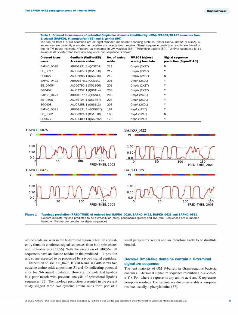
Topology predictions (PRED-TMBB) of the proteins listed inTable 1 match an eight-stranded β-barrel with long extracellularloops and short periplasmic turns (Figures 1 and 2). Both N- andC-termini are predicted to be periplasmic. A consensus topologywas generated based on the PRED-TMBB results and manualinspection of conserved of aliphatic residues (shaded grey, Fig-ures 2A and 2B). The topology prediction failed for BB0405 andBG0407 and some manual interpretation was required based onthe sequence similarity to other members of the group. Someuncertainty remains about the positioning of the first strand (in-dicated by dashed lines in Figure 2). The sequence alignmentreveals that TM regions are more conserved than loop regions,which vary in sequence and length between the four homologues.Side chains predicted to occupy the barrel interior are in generalmore conserved than neighbouring residues exposed to the hy-drophobic lipid-bilayer, as might be expected considering theconstraints imposed by packing inside the barrel.
Signal sequence prediction (Signal P) [44] suggests that nineout of the 12 homologues listed in Table 1 contain a functionalsignal sequence (Figures 2A and 2B). Multiple positively charged
. . . . . . . . . . . . . . . . . . . . . . . . . . . . . . . . . . . . . . . . . . . . . . . . . . . . . . . . . . . . . . . . . . . . . . . . . . . . . . . . . . . . . . . . . . . . . . . . . . . . . . . . . . . . . . . . . . . . . . . . . . . . . . . . . . . . . . . . . . . . . . . . . . . . . . . . . . . . . . . . . . . . . . . . . . . . . . . . . . . . . . . . . . . . . . . . . . . . . . . . . . . . . . . . . . . . . . . . . . . . . . . . . . . . . . . . . . . . . . . . . . . . . . . . . . . . . . . . . . . . . . . . . . . . . . . . . . . . . . . . . . . . . . . . . . . . . . . . . . . . . . . . . . . . . . . . . . . . . . . . . . . . . . . . . . . . . . . . . . . . . . . . . . . . . . . . . . . . . . . . . .
4 c© 2015 Authors. This is an open access article published by Portland Press Limited and distributed under the Creative Commons Attribution Licence 3.0.
The BAPKO_0422 paralogous group of β-barrel OMPs
Table 1 Ordered locus names of potential OmpA-like domains identified by HMM/FFAS03/BLAST searches fromB. afzelii (BAPKO), B. burgdorferi (BB) and B. garinii (BG)The top hit from FFAS03 searches are all eight-stranded membrane-spanning proteins (either OmpA, OmpW or NspA). Allsequences are currently annotated as putative uncharacterized proteins. Signal sequence prediction results are based onthe no TM neural network. *Present as monomer in OM vesicles [65]. †fH-binding activity [59]. ‡UniProt sequence is 11amino acids shorter than GenBank sequence, full sequence is shown.
Ordered locusname
GenBank (UniProtKB)Accession codes
No. of aminoacids
FFAS03 highestscoring template
Signal sequenceprediction (SignalP 4.1)
BAPKO_0026 ABH01291.1 (Q0SPD7) 211 OmpW (2X27) N
BB_0027 AAC66429.1 (O51058) 212 OmpW (2X27) Y
BG0027 AAU06886.1 (Q662Y5) 212 OmpW (2X27) N
BAPKO_0422 ABH01676.1 (Q0SNA2) 201 OmpA (2K0L) Y
BB_0405* AAC66795.1 (O51366) 203 OmpW (2X27) Y
BG0407† AAU07257.1 (Q661L4) 203 OmpW (2X27) Y
BAPKO_0423 ABH01677.1 (Q0SNA1) 203 OmpA (2K0L) Y
BB_0406 AAC66794.1 (O51367) 203 OmpA (2K0L) Y
BG0408 AAU07258.1 (Q661L3) 203 OmpA (2K0L) Y
BAPKO_0591 ABH01831.1 (G0IQB2‡) 181 NspA (1P4T) Y
BB_0562 AAC66924.1 (O51510) 180 NspA (1P4T) N
BG0572 AAU07409.1 (Q660W2) 179 NspA (1P4T) Y
Figure 1 Topology prediction (PRED-TMBB) of ordered loci BAPKO_0026, BAPKO_0422, BAPKO_0423 and BAPKO_0591Colours indicate regions predicted to be extracellular (blue), periplasmic (green) and TM (red). Sequences are numberedbased on the mature protein (no signal sequence).
amino acids are seen in the N-terminal region, a feature consist-ently found in confirmed signal sequences from both spirochetesand proteobacteria [55,56]. With the exception of BB0562, allsequences have an alanine residue in the predicted − 1 positionand so are expected to be processed by a type-I signal peptidase.
Inspection of BAPKO_0423, BB0406 and BG0408 shows twocysteine amino acids at positions 31 and 40, indicating potentialsites for N-terminal lipidation. However, the potential lipoboxis a poor match with previous analysis of spirochetal lipoboxsequences [22]. The topology prediction presented in the presentstudy suggest these two cysteine amino acids form part of a
small periplasmic region and are therefore likely to be disulfidebonded.
Borrelia OmpA-like domains contain a C-terminalsignature sequenceThe vast majority of OM β-barrels in Gram-negative bacteriacontain a C-terminal signature sequence resembling Z-x-Z-x-Z-x-Y-x-F>, where x represents any amino acid and Z-representsnon-polar residues. The terminal residue is invariably a non-polarresidue, usually a phenylalanine [57].
. . . . . . . . . . . . . . . . . . . . . . . . . . . . . . . . . . . . . . . . . . . . . . . . . . . . . . . . . . . . . . . . . . . . . . . . . . . . . . . . . . . . . . . . . . . . . . . . . . . . . . . . . . . . . . . . . . . . . . . . . . . . . . . . . . . . . . . . . . . . . . . . . . . . . . . . . . . . . . . . . . . . . . . . . . . . . . . . . . . . . . . . . . . . . . . . . . . . . . . . . . . . . . . . . . . . . . . . . . . . . . . . . . . . . . . . . . . . . . . . . . . . . . . . . . . . . . . . . . . . . . . . . . . . . . . . . . . . . . . . . . . . . . . . . . . . . . . . . . . . . . . . . . . . . . . . . . . . . . . . . . . . . . . . . . . . . . . . . . . . . . . . . . . . . . . . . . . . . . . . . .
c© 2015 Authors. This is an open access article published by Portland Press Limited and distributed under the Creative Commons Attribution Licence 3.0. 5
A. Dyer and others
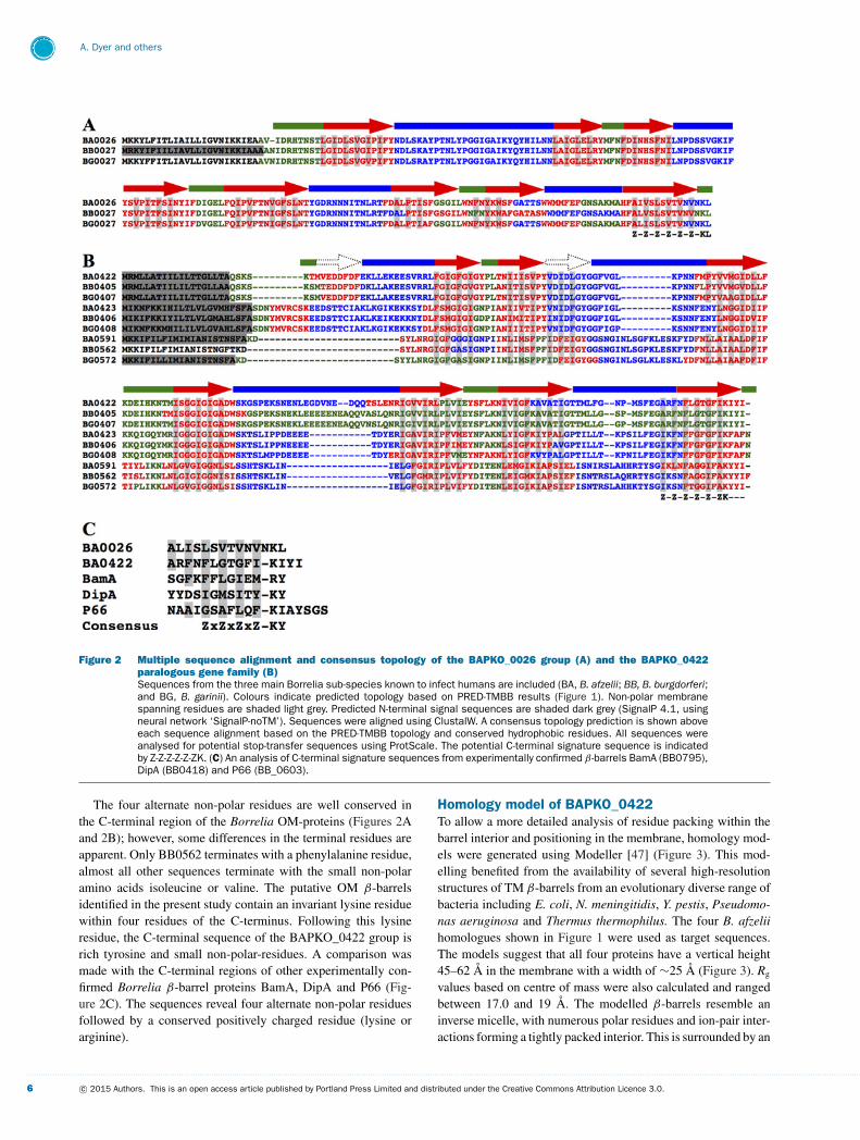
Figure 2 Multiple sequence alignment and consensus topology of the BAPKO_0026 group (A) and the BAPKO_0422paralogous gene family (B)Sequences from the three main Borrelia sub-species known to infect humans are included (BA, B. afzelii; BB, B. burgdorferi;and BG, B. garinii). Colours indicate predicted topology based on PRED-TMBB results (Figure 1). Non-polar membranespanning residues are shaded light grey. Predicted N-terminal signal sequences are shaded dark grey (SignalP 4.1, usingneural network ‘SignalP-noTM’). Sequences were aligned using ClustalW. A consensus topology prediction is shown aboveeach sequence alignment based on the PRED-TMBB topology and conserved hydrophobic residues. All sequences wereanalysed for potential stop-transfer sequences using ProtScale. The potential C-terminal signature sequence is indicatedby Z-Z-Z-Z-Z-ZK. (C) An analysis of C-terminal signature sequences from experimentally confirmed β -barrels BamA (BB0795),DipA (BB0418) and P66 (BB_0603).
The four alternate non-polar residues are well conserved inthe C-terminal region of the Borrelia OM-proteins (Figures 2Aand 2B); however, some differences in the terminal residues areapparent. Only BB0562 terminates with a phenylalanine residue,almost all other sequences terminate with the small non-polaramino acids isoleucine or valine. The putative OM β-barrelsidentified in the present study contain an invariant lysine residuewithin four residues of the C-terminus. Following this lysineresidue, the C-terminal sequence of the BAPKO_0422 group isrich tyrosine and small non-polar-residues. A comparison wasmade with the C-terminal regions of other experimentally con-firmed Borrelia β-barrel proteins BamA, DipA and P66 (Fig-ure 2C). The sequences reveal four alternate non-polar residuesfollowed by a conserved positively charged residue (lysine orarginine).
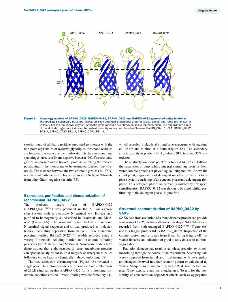
Homology model of BAPKO_0422To allow a more detailed analysis of residue packing within thebarrel interior and positioning in the membrane, homology mod-els were generated using Modeller [47] (Figure 3). This mod-elling benefited from the availability of several high-resolutionstructures of TM β-barrels from an evolutionary diverse range ofbacteria including E. coli, N. meningitidis, Y. pestis, Pseudomo-nas aeruginosa and Thermus thermophilus. The four B. afzeliihomologues shown in Figure 1 were used as target sequences.The models suggest that all four proteins have a vertical height45–62 A in the membrane with a width of ∼25 A (Figure 3). Rg
values based on centre of mass were also calculated and rangedbetween 17.0 and 19 A. The modelled β-barrels resemble aninverse micelle, with numerous polar residues and ion-pair inter-actions forming a tightly packed interior. This is surrounded by an
. . . . . . . . . . . . . . . . . . . . . . . . . . . . . . . . . . . . . . . . . . . . . . . . . . . . . . . . . . . . . . . . . . . . . . . . . . . . . . . . . . . . . . . . . . . . . . . . . . . . . . . . . . . . . . . . . . . . . . . . . . . . . . . . . . . . . . . . . . . . . . . . . . . . . . . . . . . . . . . . . . . . . . . . . . . . . . . . . . . . . . . . . . . . . . . . . . . . . . . . . . . . . . . . . . . . . . . . . . . . . . . . . . . . . . . . . . . . . . . . . . . . . . . . . . . . . . . . . . . . . . . . . . . . . . . . . . . . . . . . . . . . . . . . . . . . . . . . . . . . . . . . . . . . . . . . . . . . . . . . . . . . . . . . . . . . . . . . . . . . . . . . . . . . . . . . . . . . . . . . . .
6 c© 2015 Authors. This is an open access article published by Portland Press Limited and distributed under the Creative Commons Attribution Licence 3.0.
The BAPKO_0422 paralogous group of β-barrel OMPs
Figure 3 Homology models of BAPKO_0026, BAPKO_0422, BAPKO_0423 and BAPKO_0591 generated using ModellerThe predicted secondary structure shows an eight-stranded antiparallel β -barrel (blue). Loops and turns are shown inyellow, α-helices are shown in green. Aromatic-girdle residues are shown as sticks representation. The approximate limitsof the aliphatic region are indicated by dashed lines. Rg values calculated in Chimera: BAPKO_0026 18.9 A, BAPKO_042216.8 A, BAPKO_0423 18.1 A, BAPKO_0591 18.4 A.
exterior band of aliphatic residues predicted to interact with thenon-polar acyl chains of Borrelia glycolipids. Aromatic residuesare frequently observed at the lipid-water interface in membranespanning β-barrels of Gram-negative bacteria [26]. Two aromaticgirdles are present in the Borrelia proteins, allowing the verticalpositioning in the membrane to be estimated (dashed line, Fig-ure 3). The distance between the two aromatic girdles (24–27 A)is consistent with the hydrophobic distance (∼26 A) of β-barrelsfrom other Gram-negative bacteria [58].
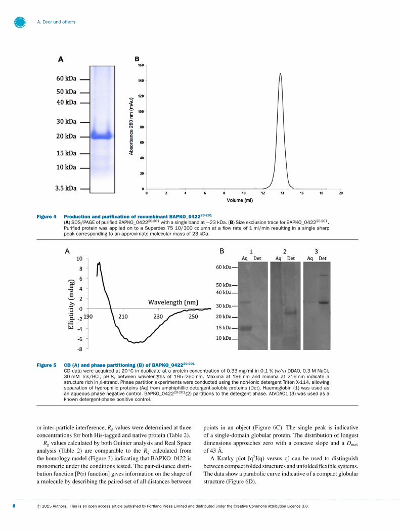
Expression, purification and characterization ofrecombinant BAPKO_0422The predicted mature form of BAPKO_0422(BAPKO_042220-201) was produced in the E. coli expres-sion system with a cleavable N-terminal 6× His-tag andpurified to homogeneity as described in ‘Materials and Meth-ods’ (Figure 4A). The resultant protein lacked a functionalN-terminal signal sequence and so was produced as inclusionbodies, facilitating separation from native E. coli membraneproteins. Purified BAPKO_042220-201 readily refolded using avariety of methods including dilution and on-column refoldingprotocols (see Materials and Methods). Numerous studies havedemonstrated that eight-stranded β-barrel membrane proteinscan spontaneously refold in lipid bilayers or detergent micellesfollowing either heat- or chemically-induced unfolding [58].
The size exclusion chromatogram (Figure 4B) revealed asingle peak. The elution volume corresponds to a molecular massof 23 kDa indicating that BAPKO_0422 forms a monomer un-der the conditions tested. Protein folding was confirmed by CD,
which revealed a classic β-strand-type spectrum with maximaat 196 nm and minima at 216 nm (Figure 5A). The secondarystructure analysis predicts 40 % β-sheet, 20 % turn and 35 % un-ordered.
The relatively low cloud point of Triton X-114 (∼23 ◦C) allowsthe separation of amphiphilic integral membrane proteins fromwater-soluble proteins at physiological temperatures. Above thecloud point, aggregation of detergent micelles results in a two-phase system consisting of an aqueous phase and a detergent-richphase. This detergent phase can be readily isolated by low speedcentrifugation. BAPKO_0422 was shown to be amphiphilic, par-titioning to the detergent phase (Figure 5B).
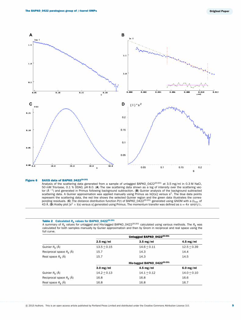
Structural characterization of BAPKO_0422 bySAXSSAXS data from a solution of a monodisperse protein can providea measure of the Rg and overall molecular shape. SAXS data wererecorded from both untagged BAPKO_042220-201 (Figure 6A)and His-tagged protein (6His-BAPKO_0422). Inspection of theGuinier region and residuals from linear fitting (Figure 6B) re-vealed linearity an indication of good quality data with minimalaggregation.
Radiation damage may result in sample aggregation or proteinmisfolding through the course of an experiment. Scattering datawere compared from initial and final images with no signific-ant changes observed in either scattering form or calculated Rg
values. Samples were analysed by SDS/PAGE both before andafter X-ray exposure and were unchanged. To test for the pos-sibility of concentration dependent effects such as aggregation
. . . . . . . . . . . . . . . . . . . . . . . . . . . . . . . . . . . . . . . . . . . . . . . . . . . . . . . . . . . . . . . . . . . . . . . . . . . . . . . . . . . . . . . . . . . . . . . . . . . . . . . . . . . . . . . . . . . . . . . . . . . . . . . . . . . . . . . . . . . . . . . . . . . . . . . . . . . . . . . . . . . . . . . . . . . . . . . . . . . . . . . . . . . . . . . . . . . . . . . . . . . . . . . . . . . . . . . . . . . . . . . . . . . . . . . . . . . . . . . . . . . . . . . . . . . . . . . . . . . . . . . . . . . . . . . . . . . . . . . . . . . . . . . . . . . . . . . . . . . . . . . . . . . . . . . . . . . . . . . . . . . . . . . . . . . . . . . . . . . . . . . . . . . . . . . . . . . . . . . . . .
c© 2015 Authors. This is an open access article published by Portland Press Limited and distributed under the Creative Commons Attribution Licence 3.0. 7
A. Dyer and others
Figure 4 Production and purification of recombinant BAPKO_042220-201
(A) SDS/PAGE of purified BAPKO_042220-201 with a single band at ∼23 kDa. (B) Size exclusion trace for BAPKO_042220-201.Purified protein was applied on to a Superdex 75 10/300 column at a flow rate of 1 ml/min resulting in a single sharppeak corresponding to an approximate molecular mass of 23 kDa.
Figure 5 CD (A) and phase partitioning (B) of BAPKO_042220-201
CD data were acquired at 20 ◦C in duplicate at a protein concentration of 0.33 mg/ml in 0.1 % (w/v) DDAO, 0.3 M NaCl,30 mM Tris/HCl, pH 8, between wavelengths of 195–260 nm. Maxima at 196 nm and minima at 216 nm indicate astructure rich in β -strand. Phase partition experiments were conducted using the non-ionic detergent Triton X-114, allowingseparation of hydrophilic proteins (Aq) from amphiphillic detergent-soluble proteins (Det). Haemoglobin (1) was used asan aqueous phase negative control. BAPKO_042220-201(2) partitions to the detergent phase. AtVDAC1 (3) was used as aknown detergent-phase positive control.
or inter-particle interference, Rg values were determined at threeconcentrations for both His-tagged and native protein (Table 2).
Rg values calculated by both Guinier analysis and Real Spaceanalysis (Table 2) are comparable to the Rg calculated fromthe homology model (Figure 3) indicating that BAPKO_0422 ismonomeric under the conditions tested. The pair-distance distri-bution function [P(r) function] gives information on the shape ofa molecule by describing the paired-set of all distances between
points in an object (Figure 6C). The single peak is indicativeof a single-domain globular protein. The distribution of longestdimensions approaches zero with a concave slope and a Dmax
of 43 A.A Kratky plot [q2I(q) versus q] can be used to distinguish
between compact folded structures and unfolded flexible systems.The data show a parabolic curve indicative of a compact globularstructure (Figure 6D).
. . . . . . . . . . . . . . . . . . . . . . . . . . . . . . . . . . . . . . . . . . . . . . . . . . . . . . . . . . . . . . . . . . . . . . . . . . . . . . . . . . . . . . . . . . . . . . . . . . . . . . . . . . . . . . . . . . . . . . . . . . . . . . . . . . . . . . . . . . . . . . . . . . . . . . . . . . . . . . . . . . . . . . . . . . . . . . . . . . . . . . . . . . . . . . . . . . . . . . . . . . . . . . . . . . . . . . . . . . . . . . . . . . . . . . . . . . . . . . . . . . . . . . . . . . . . . . . . . . . . . . . . . . . . . . . . . . . . . . . . . . . . . . . . . . . . . . . . . . . . . . . . . . . . . . . . . . . . . . . . . . . . . . . . . . . . . . . . . . . . . . . . . . . . . . . . . . . . . . . . . .
8 c© 2015 Authors. This is an open access article published by Portland Press Limited and distributed under the Creative Commons Attribution Licence 3.0.
The BAPKO_0422 paralogous group of β-barrel OMPs
Figure 6 SAXS data of BAPKO_042220-201
Analysis of the scattering data generated from a sample of untagged BAPKO_042220-201 at 3.5 mg/ml in 0.3 M NaCl,50 mM Tris-base, 0.1 % DDAO, pH 8.0. (A) The raw scattering data shown as a log of intensity over the scattering vec-tor (A− 1) and generated in Primus following background subtraction. (B) Guinier analysis of the background subtractedscattering data. A Guinier approximation was applied manually using Primus as In[I(s)] versus s2. The blue data pointsrepresent the scattering data, the red line shows the selected Guinier region and the green data illustrates the corres-ponding residuals. (C) The distance distribution function P(r) of BAPKO_042220-201 generated using GNOM with a Dmax of43 A. (D) Kratky plot [s2 × I(s) versus s] generated using Primus. The momentum transfer was defined as s = 4π sin(θ )/λ.
Table 2 Calculated Rg values for BAPKO_042220-201
A summary of Rg values for untagged and His-tagged BAPKO_042220-201 calculated using various methods. The Rg wascalculated for both samples manually by Gunier approximation and then by Gnom in reciprocal and real space using thefull curve.
Untagged BAPKO_042220-201
2.5 mg/ml 3.5 mg/ml 4.5 mg/ml
Guinier Rg (A) 13.5 +− 0.15 14.6 +− 0.11 12.5 +− 0.39
Reciprocal space Rg (A) 15.7 14.3 14.4
Real space Rg (A) 15.7 14.3 14.5
His-tagged BAPKO_042220-201
3.0 mg/ml 4.5 mg/ml 6.0 mg/ml
Guinier Rg (A) 14.2 +− 0.13 14.1 +− 0.12 14.0 +− 0.10
Reciprocal space Rg (A) 16.8 16.8 16.6
Real space Rg (A) 16.8 16.8 16.7
. . . . . . . . . . . . . . . . . . . . . . . . . . . . . . . . . . . . . . . . . . . . . . . . . . . . . . . . . . . . . . . . . . . . . . . . . . . . . . . . . . . . . . . . . . . . . . . . . . . . . . . . . . . . . . . . . . . . . . . . . . . . . . . . . . . . . . . . . . . . . . . . . . . . . . . . . . . . . . . . . . . . . . . . . . . . . . . . . . . . . . . . . . . . . . . . . . . . . . . . . . . . . . . . . . . . . . . . . . . . . . . . . . . . . . . . . . . . . . . . . . . . . . . . . . . . . . . . . . . . . . . . . . . . . . . . . . . . . . . . . . . . . . . . . . . . . . . . . . . . . . . . . . . . . . . . . . . . . . . . . . . . . . . . . . . . . . . . . . . . . . . . . . . . . . . . . . . . . . . . . .
c© 2015 Authors. This is an open access article published by Portland Press Limited and distributed under the Creative Commons Attribution Licence 3.0. 9
A. Dyer and others
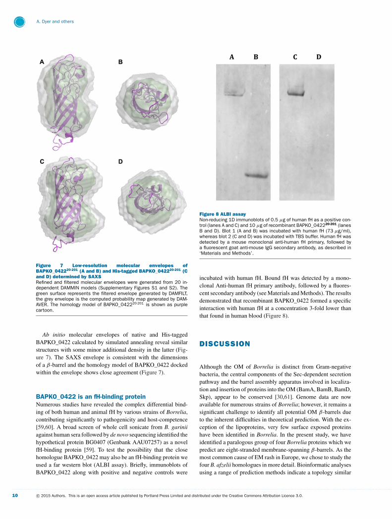
Figure 7 Low-resolution molecular envelopes ofBAPKO_042220-201 (A and B) and His-tagged BAPKO_042220-201 (Cand D) determined by SAXSRefined and filtered molecular envelopes were generated from 20 in-dependent DAMMIN models (Supplementary Figures S1 and S2). Thegreen surface represents the filtered envelope generated by DAMFILT,the grey envelope is the computed probability map generated by DAM-AVER. The homology model of BAPKO_042220-201 is shown as purplecartoon.
Ab initio molecular envelopes of native and His-taggedBAPKO_0422 calculated by simulated annealing reveal similarstructures with some minor additional density in the latter (Fig-ure 7). The SAXS envelope is consistent with the dimensionsof a β-barrel and the homology model of BAPKO_0422 dockedwithin the envelope shows close agreement (Figure 7).
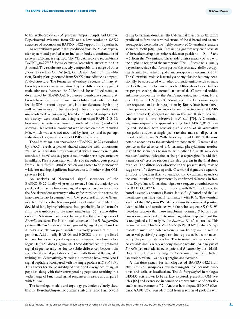
BAPKO_0422 is an fH-binding proteinNumerous studies have revealed the complex differential bind-ing of both human and animal fH by various strains of Borrelia,contributing significantly to pathogenicity and host-competence[59,60]. A broad screen of whole cell sonicate from B. gariniiagainst human sera followed by de novo sequencing identified thehypothetical protein BG0407 (Genbank AAU07257) as a novelfH-binding protein [59]. To test the possibility that the closehomologue BAPKO_0422 may also be an fH-binding protein weused a far western blot (ALBI assay). Briefly, immunoblots ofBAPKO_0422 along with positive and negative controls were
Figure 8 ALBI assayNon-reducing 1D immunoblots of 0.5 μg of human fH as a positive con-trol (lanes A and C) and 10 μg of recombinant BAPKO_042220-201 (lanesB and D). Blot 1 (A and B) was incubated with human fH (73 μg/ml),whereas blot 2 (C and D) was incubated with TBS buffer. Human fH wasdetected by a mouse monoclonal anti-human fH primary, followed bya fluorescent goat anti-mouse IgG secondary antibody, as described in‘Materials and Methods’.
incubated with human fH. Bound fH was detected by a mono-clonal Anti-human fH primary antibody, followed by a fluores-cent secondary antibody (see Materials and Methods). The resultsdemonstrated that recombinant BAPKO_0422 formed a specificinteraction with human fH at a concentration 3-fold lower thanthat found in human blood (Figure 8).
DISCUSSION
Although the OM of Borrelia is distinct from Gram-negativebacteria, the central components of the Sec-dependent secretionpathway and the barrel assembly apparatus involved in localiza-tion and insertion of proteins into the OM (BamA, BamB, BamD,Skp), appear to be conserved [30,61]. Genome data are nowavailable for numerous strains of Borrelia; however, it remains asignificant challenge to identify all potential OM β-barrels dueto the inherent difficulties in theoretical prediction. With the ex-ception of the lipoproteins, very few surface exposed proteinshave been identified in Borrelia. In the present study, we haveidentified a paralogous group of four Borrelia proteins which wepredict are eight-stranded membrane-spanning β-barrels. As themost common cause of EM rash in Europe, we chose to study thefour B. afzelii homologues in more detail. Bioinformatic analysesusing a range of prediction methods indicate a topology similar
. . . . . . . . . . . . . . . . . . . . . . . . . . . . . . . . . . . . . . . . . . . . . . . . . . . . . . . . . . . . . . . . . . . . . . . . . . . . . . . . . . . . . . . . . . . . . . . . . . . . . . . . . . . . . . . . . . . . . . . . . . . . . . . . . . . . . . . . . . . . . . . . . . . . . . . . . . . . . . . . . . . . . . . . . . . . . . . . . . . . . . . . . . . . . . . . . . . . . . . . . . . . . . . . . . . . . . . . . . . . . . . . . . . . . . . . . . . . . . . . . . . . . . . . . . . . . . . . . . . . . . . . . . . . . . . . . . . . . . . . . . . . . . . . . . . . . . . . . . . . . . . . . . . . . . . . . . . . . . . . . . . . . . . . . . . . . . . . . . . . . . . . . . . . . . . . . . . . . . . . . .
10 c© 2015 Authors. This is an open access article published by Portland Press Limited and distributed under the Creative Commons Attribution Licence 3.0.
The BAPKO_0422 paralogous group of β-barrel OMPs
to the well-studied E. coli proteins OmpA, OmpX and OmpW.Experimental evidence from CD and a low-resolution SAXSstructure of recombinant BAPKO_0422 support this hypothesis.
As recombinant protein was produced from the E. coli expres-sion system and purified from inclusion bodies, confirmation ofprotein refolding is required. The CD data indicate recombinantBAPKO_042220-201 forms extensive secondary structure rich inβ-strand. The results are directly comparable to a range of otherβ-barrels such as OmpW [62], OmpA and OprF [63]. In addi-tion, Kratky plots generated from SAXS data indicate a compact,folded structure. The formation of tertiary structure of many β-barrels proteins can be monitored by the difference in apparentmolecular mass between the folded and the unfolded states, asdetermined by SDS/PAGE. Numerous membrane-spanning β-barrels have been shown to maintain a folded state when solubil-ized in SDS at room temperature, but once denatured by boilingwill remain in an unfolded state [64]. Therefore, gel-shift assaysare conducted by comparing boiled and unboiled samples. Gel-shift assays were conducted using recombinant BAPKO_0422;however, the protein remained unmodified by heat (results notshown). This result is consistent with studies on the 24-strandedP66, which was also not modified by heat [28] and is perhapsindicative of a general feature of OMPs in Borrelia.
The ab initio molecular envelope of BAPKO_0422 determinedby SAXS reveals a peanut shaped structure with dimensions25 × 45 A. This structure is consistent with a monomeric eight-stranded β-barrel and suggests a multimeric porin-type structureis unlikely. This is consistent with data on the orthologous proteinfrom B. burgdorferi BB0405, which was shown to be monomericwhile not making significant interactions with other major OM-proteins [65].
An analysis of N-terminal signal sequences of theBAPKO_0422 family of proteins revealed that the majority arepredicted to have a functional signal sequence and so may enterthe Sec-dependent secretory pathway for translocation across theinner membrane. In common with OM-proteins from other Gram-negative bacteria the Borrelia proteins identified in Table 1 aredevoid of long hydrophobic stretches, precluding lateral transferfrom the translocase to the inner membrane [66]. Some differ-ences in N-terminal sequence between the three sub-species ofBorrelia are seen. The N-terminal sequence of the B. burgdorferiprotein BB0562 may not be recognized by signal peptidase I asit lacks a small non-polar residue normally present at the − 1position. Additionally BA0026 and BG0027 are not predictedto have functional signal sequences, whereas the close ortho-logue BB0027 does (Figure 2). These differences in predictedsignal sequence may be due to subtle differences between thespirochetal signal peptides compared with those of the signal Ptraining set. Alternatively, Borrelia is known to have three type-Isignal peptidases compared with the single protein in E. coli [67].This allows for the possibility of concerted divergence of signalpeptides along with their corresponding peptidase resulting in awider range of functional signal sequences in Borrelia comparedwith E. coli.
The homology models and topology predictions clearly showthat the Borrelia OmpA-like domains listed in Table 1 are devoid
of any C-terminal domains. The C-terminal residues are thereforepredicted to form the terminal strand of the β-barrel and as suchare expected to contain the highly conserved C-terminal signaturesequence motif [68]. This 10-residue signature sequence consistsof three alternating non-polar residues at positions − 9, − 7 and− 5 from the C-terminus. These side chains make contact withthe aliphatic region of the membrane. The − 3 residue is usuallya tyrosine residue that forms part of the aromatic girdle occupy-ing the interface between polar and non-polar environments [57].The C-terminal residue is usually a phenylalanine but may occa-sionally be substituted with other aromatic amino acids or morerarely other non-polar amino acids. Although not essential forproper processing, the aromatic nature of the C-terminal residueenhances processing by the BamA apparatus, facilitating barrelassembly in the OM [57,69]. Variations in the C-terminal signa-ture sequence and their recognition by BamA have been shownto be species specific, in particular, many Proteobacterial OMPshave a positively charged residue in the penultimate position,whereas this is never observed in E. coli [70]. A C-terminalsignature sequence is apparent among the BAPKO_0422 fam-ily and BA0026, both consisting of a series of six alternativenon-polar residues, a single lysine residue and a small polar ter-minal motif (Figure 2). With the exception of BB0562, the mostnotable exception to the standard proteobacterial C-terminal se-quence is the absence of a C-terminal phenylalanine residue.Instead the sequences terminate with either the small non-polarresidues leucine, isoleucine or the polar asparagine. In addition,a number of tyrosine residues are also present in the final threeresidues. The differences observed in the present study may besuggestive of a Borrelia-specific C-terminal signature sequence.In order to confirm this, we analysed the C-terminal strands ofthe small number of experimentally confirmed β-barrels in Bor-relia. DipA has a C-terminal signature sequence reminiscent ofthe BAKPO_0422 family, terminating with K-Y. In addition, thebarrel assembly apparatus BamA, is itself a β-barrel and the lastmembrane-spanning strand terminates with R-Y. The terminalstrand of the OM porin P66 also contains the conserved positivelysine residue and terminates with the polar sequence S-G-S. Wetherefore propose that these membrane-spanning β-barrels con-tain a Borrelia-specific C-terminal signature sequence and thisis recognized efficiently by the Borrelia BamA apparatus. Thissequence resembles Z-x-Z-x-Z-x-Z-[KR]-[ILNY], where Z rep-resents a small non-polar residue, x can be any amino acid. Aconserved positively charged residue is present, but is not neces-sarily the penultimate residue. The terminal residue appears tobe variable and is rarely a phenylalanine residue. An analysis ofBorrelia proteins identified as potential β-barrels by the TMBB-DataBase [71] reveals a range of C-terminal residues includingisoleucine, valine, lysine, asparagine and tyrosine.
A literature search for homologues of BAPKO_0422 fromother Borrelia subspecies revealed insights into possible func-tions and cellular localization. The B. burgdorferi homologueBB0405 was shown to be surface exposed, present in OM ves-icles [65] and expressed in conditions representative of both tickand host environments [72]. Another homologue, BB0407 (Gen-bank AAU07257) was identified from a screen of proteins with
. . . . . . . . . . . . . . . . . . . . . . . . . . . . . . . . . . . . . . . . . . . . . . . . . . . . . . . . . . . . . . . . . . . . . . . . . . . . . . . . . . . . . . . . . . . . . . . . . . . . . . . . . . . . . . . . . . . . . . . . . . . . . . . . . . . . . . . . . . . . . . . . . . . . . . . . . . . . . . . . . . . . . . . . . . . . . . . . . . . . . . . . . . . . . . . . . . . . . . . . . . . . . . . . . . . . . . . . . . . . . . . . . . . . . . . . . . . . . . . . . . . . . . . . . . . . . . . . . . . . . . . . . . . . . . . . . . . . . . . . . . . . . . . . . . . . . . . . . . . . . . . . . . . . . . . . . . . . . . . . . . . . . . . . . . . . . . . . . . . . . . . . . . . . . . . . . . . . . . . . . .
c© 2015 Authors. This is an open access article published by Portland Press Limited and distributed under the Creative Commons Attribution Licence 3.0. 11
A. Dyer and others
fH-binding activity suggesting a possible role in virulence [59].This fH-binding activity led us to investigate the fH binding ofBAPKO_0422, which we confirmed using an ALBI assay. Theseresults and the high sequence similarity suggest that the ortho-logous group BAPKO_0422, BG0407 and BB0405 are expressedin the mammalian host, exposed at the cell surface and bind tohuman fH.
In summary, the data presented suggest that BAPKO_0422forms a membrane-spanning β-barrel. The topology predictionmatches the OmpA-membrane spanning domain defined by Pfamfamily PF01389, consisting of eight membrane-spanning β-strands linked by short periplasmic turns and longer extracellularloops. For the first time, this extends the species distribution of thePF01389 domain to include members of the Spirochete phylum.The orthologous group consisting of BAPKO_0422, BB0405 andBG0407 are proposed to play a role in virulence by binding tohost fH, therefore abrogating the host complement response andreducing antigenicity of surface exposed loops. Further work isrequired to establish the host-specific fH-binding activity of theremaining homologues and to determine any other physiologicalfunctions of these proteins in Borrelia. Surface exposed epitopesmay enhance recombinant immunoblots currently used in dia-gnostic tests for Lyme disease.
AUTHOR CONTRIBUTION
Adam Dyer, Gemma Brown, Lenka Stejskal and Peter Laity designedand conducted experiments and performed data analysis. RichardBingham designed the study and supervised the research. All au-thors participated in writing the paper.
ACKNOWLEDGEMENTS
We are grateful to the following: Ibad Kureshi and members of theHPC-RC team at the University of Huddersfield for computationalsupport, Andrew Leech for CD data collection, Gabriele Margos,Stephanie Vollmer, Ruth Mitchell and Freddie Seelig for tick col-lection and DNA extraction and to George Psakis and AlexandreBoulbrima for provision of AtVDAC1.
FUNDING
This work was supported by the Biochemical Society (to L.S.); andthe University of Huddersfield, Department of Biological Sciences.
REFERENCES
1 Steere, A.C., Coburn, J. and Glickstein, L. (2004) The emergenceof Lyme disease. J. Clin. Invest. 113, 1093–1101CrossRef PubMed
2 Margos, G., Vollmer, S.A., Ogden, N.H. and Fish, D. (2011)Population genetics, taxonomy, phylogeny and evolution of Borreliaburgdorferi sensu lato. Infect. Genet. Evol. 11, 1545–1563CrossRef PubMed
3 Ornstein, K., Berglund, J., Nilsson, I., Norrby, R. and Bergstrom, S.(2001) Characterization of Lyme borreliosis isolates from patientswith erythema migrans and neuroborreliosis in southern Sweden.J. Clin. Microbiol. 39, 1294–1298 CrossRef PubMed
4 Oksi, J., Marjamaki, M., Nikoskelainen, J. and Viljanen, M.K.(1999) Borrelia burgdorferi detected by culture and PCR in clinicalrelapse of disseminated Lyme borreliosis. Ann. Med. 31, 225–232CrossRef PubMed
5 Schmidli, J., Hunziker, T., Moesli, P. and Schaad, U.B. (1988)Cultivation of Borrelia burgdorferi from joint fluid three monthsafter treatment of facial palsy due to Lyme borreliosis. J. Infect.Dis. 158, 905–906 CrossRef PubMed
6 Preac-Mursic, V., Weber, K., Pfister, H.W., Wilske, B., Gross, B.,Baumann, A. and Prokop, J. (1989) Survival of Borrelia burgdorferiin antibiotically treated patients with Lyme borreliosis. Infection17, 355–359 CrossRef PubMed
7 Feng, J., Wang, T., Shi, W., Zhang, S., Sullivan, D., Auwaerter, P.G.and Zhang, Y. (2014) Identification of novel activity against Borreliaburgdorferi persisters using an FDA approved drug library. Emerg.Microbes Infect. 3, e49 CrossRef PubMed
8 Coutte, L., Botkin, D.J., Gao, L. and Norris, S.J. (2009) Detailedanalysis of sequence changes occurring during vlsE antigenicvariation in the mouse model of Borrelia burgdorferi infection.PLoS Pathog. 5, e1000293 CrossRef PubMed
9 Rogovskyy, A.S. and Bankhead, T. (2013) Variable VlsE is criticalfor host reinfection by the Lyme disease spirochete. PLoS One 8,e61226 CrossRef PubMed
10 Tilly, K., Bestor, A. and Rosa, P.A. (2013) Lipoprotein succession inBorrelia burgdorferi: similar but distinct roles for OspC and VlsE atdifferent stages of mammalian infection. Mol. Microbiol. 89,216–227 CrossRef PubMed
11 Zhang, J.R., Hardham, J.M., Barbour, A.G. and Norris, S.J. (1997)Antigenic variation in Lyme disease borreliae by promiscuousrecombination of VMP-like sequence cassettes. Cell 89, 275–285CrossRef PubMed
12 Crother, T.R., Champion, C.I., Whitelegge, J.P., Aguilera, R., Wu,X.Y., Blanco, D.R., Miller, J.N. and Lovett, M.A. (2004) Temporalanalysis of the antigenic composition of Borrelia burgdorferi duringinfection in rabbit skin. Infect. Immun. 72, 5063–5072
13 Dietrich, T., Geissdorfer, W., Schlotzer-Schrehardt, U., Holbach, L.,Schoerner, C. and Seitz, B. (2008) Borrelia-associated crystallinekeratopathy with intracorneal detection of Borrelia garinii byelectron microscopy and polymerase chain reaction. Cornea 27,498–500 CrossRef PubMed
14 Livengood, J.A. and Gilmore, R.D. (2006) Invasion of humanneuronal and glial cells by an infectious strain of Borreliaburgdorferi. Microbes Infect. 8, 2832–2840 CrossRef PubMed
15 Pulzova, L. and Bhide, M. (2014) Outer surface proteins ofBorrelia: peerless immune evasion tools. Curr. Protein Pept. Sci.15, 75–88 CrossRef PubMed
16 Kurtenbach, K., de Michelis, S., Etti, S., Schafer, S.M., Sewell,H.S., Brade, V. and Kraiczy, P. (2002) Host association of Borreliaburgdorferi sensu lato–the key role of host complement. TrendsMicrobiol. 10, 74–79 CrossRef PubMed
17 Behera, A.K., Durand, E., Cugini, C., Antonara, S., Bourassa, L.,Hildebrand, E., Hu, L.T. and Coburn, J. (2008) Borrelia burgdorferiBBB07 interaction with integrin alpha3beta1 stimulates productionof pro-inflammatory mediators in primary human chondrocytes.Cell Microbiol. 10, 320–331 PubMed
18 Ostberg, Y., Pinne, M., Benz, R., Rosa, P. and Bergstrom, S. (2002)Elimination of channel-forming activity by insertional inactivation ofthe p13 gene in Borrelia burgdorferi. J. Bacteriol. 184, 6811–6819CrossRef PubMed
19 Barcena-Uribarri, I., Thein, M., Barbot, M., Sans-Serramitjana, E.,Bonde, M., Mentele, R., Lottspeich, F., Bergstrom, S. and Benz, R.(2014) Study of the protein complex, pore diameter, andpore-forming activity of the Borrelia burgdorferi P13 porin. J. Biol.Chem. 289, 18614–18624 CrossRef PubMed
. . . . . . . . . . . . . . . . . . . . . . . . . . . . . . . . . . . . . . . . . . . . . . . . . . . . . . . . . . . . . . . . . . . . . . . . . . . . . . . . . . . . . . . . . . . . . . . . . . . . . . . . . . . . . . . . . . . . . . . . . . . . . . . . . . . . . . . . . . . . . . . . . . . . . . . . . . . . . . . . . . . . . . . . . . . . . . . . . . . . . . . . . . . . . . . . . . . . . . . . . . . . . . . . . . . . . . . . . . . . . . . . . . . . . . . . . . . . . . . . . . . . . . . . . . . . . . . . . . . . . . . . . . . . . . . . . . . . . . . . . . . . . . . . . . . . . . . . . . . . . . . . . . . . . . . . . . . . . . . . . . . . . . . . . . . . . . . . . . . . . . . . . . . . . . . . . . . . . . . . . .
12 c© 2015 Authors. This is an open access article published by Portland Press Limited and distributed under the Creative Commons Attribution Licence 3.0.
The BAPKO_0422 paralogous group of β-barrel OMPs
20 Bunikis, I., Denker, K., Ostberg, Y., Andersen, C., Benz, R. andBergstrom, S. (2008) An RND-type efflux system in Borreliaburgdorferi is involved in virulence and resistance to antimicrobialcompounds. PLoS Pathog 4, e1000009 CrossRef PubMed
21 Greene, N.P., Hinchliffe, P., Crow, A., Ababou, A., Hughes, C. andKoronakis, V. (2013) Structure of an atypical periplasmic adaptorfrom a multidrug efflux pump of the spirochete Borrelia burgdorferi.FEBS Lett. 587, 2984–2988 CrossRef PubMed
22 Setubal, J.C., Reis, M., Matsunaga, J. and Haake, D.A. (2006)Lipoprotein computational prediction in spirochaetal genomes.Microbiology 152, 113–121 CrossRef PubMed
23 Fraser, C.M., Casjens, S., Huang, W.M., Sutton, G.G., Clayton, R.,Lathigra, R., White, O., Ketchum, K.A., Dodson, R. and Hickey, E.K.(1997) Genomic sequence of a Lyme disease spirochaete, Borreliaburgdorferi. Nature 390, 580–586 CrossRef PubMed
24 Casjens, S.R., Mongodin, E.F., Qiu, W.G., Dunn, J.J., Luft, B.J.,Fraser-Liggett, C.M. and Schutzer, S.E. (2011) Whole-genomesequences of two Borrelia afzelii and two Borrelia garinii Lymedisease agent isolates. J. Bacteriol. 193, 6995–6996CrossRef PubMed
25 Cabello, F.C., Godfrey, H.P. and Newman, S.A. (2007) Hidden inplain sight: Borrelia burgdorferi and the extracellular matrix. TrendsMicrobiol. 15, 350–354 CrossRef PubMed
26 Schulz, G.E. (2000) Beta-barrel membrane proteins. Curr. Opin.Struct. Biol. 10, 443–447 CrossRef PubMed
27 Thein, M., Bonde, M., Bunikis, I., Denker, K., Sickmann, A.,Bergstrom, S. and Benz, R. (2012) DipA, a pore-forming protein inthe outer membrane of Lyme disease spirochetes exhibitsspecificity for the permeation of dicarboxylates. PLoS One 7,e36523 CrossRef PubMed
28 Kenedy, M.R., Luthra, A., Anand, A., Dunn, J.P., Radolf, J.D. andAkins, D.R. (2013) Structural modeling and physicochemicalcharacterization provide evidence that P66 forms a beta-barrel inthe Borrelia burgdorferi outer membrane. J. Bacteriol. 196,859–872 CrossRef PubMed
29 Skare, J.T., Mirzabekov, T.A., Shang, E.S., Blanco, D.R.,Erdjument-Bromage, H., Bunikis, J., Bergstrom, S., Tempst, P.,Kagan, B.L., Miller, J.N. and Lovett, M.A. (1997) The Oms66 (p66)protein is a Borrelia burgdorferi porin. Infect. Immun. 65,3654–3661 PubMed
30 Lenhart, T.R. and Akins, D.R. (2010) Borrelia burgdorferi locusBB0795 encodes a BamA orthologue required for growth andefficient localization of outer membrane proteins. Mol. Microbiol.75, 692–709 CrossRef PubMed
31 Yamashita, S., Lukacik, P., Barnard, T.J., Noinaj, N., Felek, S.,Tsang, T.M., Krukonis, E.S., Hinnebusch, B.J. and Buchanan, S.K.(2011) Structural insights into Ail-mediated adhesion in Yersiniapestis. Structure 19, 1672–1682 CrossRef PubMed
32 Hong, H.D., Patel, D.R., Tamm, L.K. and van den Berg, B. (2006)The outer membrane protein OmpW forms an eight-strandedbeta-barrel with a hydrophobic channel. J. Biol. Chem. 281,7568–7577 CrossRef PubMed
33 Vogt, J. and Schulz, G.E. (1999) The structure of the outermembrane protein OmpX from Escherichia coli reveals possiblemechanisms of virulence. Structure 7, 1301–1309CrossRef PubMed
34 Pautsch, A. and Schulz, G.E. (1998) Structure of the outermembrane protein A transmembrane domain. Nat. Struct. Biol. 5,1013–1017 CrossRef PubMed
35 Koebnik, R., Locher, K.P. and van Gelder, P. (2000) Structure andfunction of bacterial outer membrane proteins: barrels in anutshell. Mol. Microbiol. 37, 239–253 CrossRef PubMed
36 Mittal, R., Krishnan, S., Gonzalez-Gomez, I. and Prasadarao, N.V.(2011) Deciphering the roles of outer membrane protein Aextracellular loops in the pathogenesis of Escherichia coli K1meningitis. J. Biol. Chem. 286, 2183–2193CrossRef PubMed
37 Prasadarao, N.V. (2002) Identification of Escherichia coli outermembrane protein A receptor on human brain microvascularendothelial cells. Infect. Immun. 70, 4556–4563
38 Nair, M.K., Venkitanarayanan, K., Silbart, L.K. and Kim, K.S.(2009) Outer membrane protein A (OmpA) of Cronobactersakazakii binds fibronectin and contributes to invasion of humanbrain microvascular endothelial cells. Foodborne Pathog. Dis. 6,495–501 CrossRef PubMed
39 Heffernan, E.J., Harwood, J., Fierer, J. and Guiney, D. (1992) TheSalmonella typhimurium virulence plasmid complement resistancegene rck is homologous to a family of virulence-related outermembrane protein genes, including pagC and ail. J. Bacteriol. 174,84–91 PubMed
40 Crago, A.M. and Koronakis, V. (1999) Binding of extracellularmatrix laminin to Escherichia coli expressing the Salmonella outermembrane proteins Rck and PagC. FEMS Microbiol. Lett. 176,495–501 CrossRef PubMed
41 Eddy, S.R. (1998) Profile hidden Markov models. Bioinformatics14, 755–763 CrossRef PubMed
42 Finn, R.D., Bateman, A., Clements, J., Coggill, P., Eberhardt, R.Y.,Eddy, S.R., Heger, A., Hetherington, K., Holm, L., Mistry, J. et al.(2014) Pfam: the protein families database. Nucleic. Acids Res.42, D222–230 CrossRef PubMed
43 Sperisen, P. and Pagni, M. (2005) JACOP: a simple and robustmethod for the automated classification of protein sequences withmodular architecture. BMC Bioinformatics 6, 216CrossRef PubMed
44 Petersen, T.N., Brunak, S., von Heijne, G. and Nielsen, H. (2011)SignalP 4.0: discriminating signal peptides from transmembraneregions. Nat. Methods 8, 785–786 CrossRef PubMed
45 Jaroszewski, L., Li, Z., Cai, X.H., Weber, C. and Godzik, A. (2011)FFAS server: novel features and applications. Nucleic. Acids. Res.39, W38–44 CrossRef PubMed
46 Bagos, P.G., Liakopoulos, T.D., Spyropoulos, I.C. and Hamodrakas,S.J. (2004) A Hidden Markov Model method, capable of predictingand discriminating beta-barrel outer membrane proteins. BMCBioinformatics 5, 29 CrossRef PubMed
47 Eswar, N., Webb, B., Marti-Renom, M.A., Madhusudhan, M.S.,Eramian, D., Shen, M.Y., Pieper, U. and Sali, A. (2007)Comparative protein structure modeling using MODELLER. Curr.Protoc. Protein Sci. Chapter 2, Unit 2 9
48 Pettersen, E.F., Goddard, T.D., Huang, C.C., Couch, G.S.,Greenblatt, D.M., Meng, E.C. and Ferrin, T.E. (2004) UCSFChimera–a visualization system for exploratory research andanalysis. J. Comput. Chem. 25, 1605–1612CrossRef PubMed
49 Kopp, A., Hebecker, M., Svobodova, E. and Jozsi, M. (2012) Factorh: a complement regulator in health and disease, and a mediatorof cellular interactions. Biomolecules 2, 46–75CrossRef PubMed
50 Bordier, C. (1981) Phase separation of integral membrane proteinsin Triton X-114 solution. J. Biol. Chem. 256, 1604–1607PubMed
51 Mertins, B., Psakis, G., Grosse, W., Back, K.C., Salisowski, A.,Reiss, P., Koert, U. and Essen, L.O. (2012) Flexibility of theN-terminal mVDAC1 segment controls the channel’s gatingbehavior. PLoS One 7, e47938 CrossRef PubMed
52 Volkov, V.V. and Svergun, D.I. (2003) Uniqueness of ab initio shapedetermination in small-angle scattering. J. Appl. Cryst. 36,860–864 CrossRef
53 Svergun, D.I. (1999) Restoring low resolution structure of biologicalmacromolecules from solution scattering using simulatedannealing. Biophys. J. 76, 2879–2886 CrossRef PubMed
54 Di, L., Pagan, P.E., Packer, D., Martin, C.L., Akther, S., Ramrattan,G., Mongodin, E.F, Fraser, C.M., Schutzer, S.E., Luft, B.J. et al.(2014) BorreliaBase: a phylogeny-centered browser of Borreliagenomes. BMC Bioinformatics 15, 233 CrossRef PubMed
. . . . . . . . . . . . . . . . . . . . . . . . . . . . . . . . . . . . . . . . . . . . . . . . . . . . . . . . . . . . . . . . . . . . . . . . . . . . . . . . . . . . . . . . . . . . . . . . . . . . . . . . . . . . . . . . . . . . . . . . . . . . . . . . . . . . . . . . . . . . . . . . . . . . . . . . . . . . . . . . . . . . . . . . . . . . . . . . . . . . . . . . . . . . . . . . . . . . . . . . . . . . . . . . . . . . . . . . . . . . . . . . . . . . . . . . . . . . . . . . . . . . . . . . . . . . . . . . . . . . . . . . . . . . . . . . . . . . . . . . . . . . . . . . . . . . . . . . . . . . . . . . . . . . . . . . . . . . . . . . . . . . . . . . . . . . . . . . . . . . . . . . . . . . . . . . . . . . . . . . . .
c© 2015 Authors. This is an open access article published by Portland Press Limited and distributed under the Creative Commons Attribution Licence 3.0. 13
A. Dyer and others
55 Cullen, P.A., Haake, D.A. and Adler, B. (2004) Outer membraneproteins of pathogenic spirochetes. FEMS Microbiol Rev 28,291–318 CrossRef PubMed
56 Pugsley, A.P. (1993) The complete general secretory pathway ingram-negative bacteria. Microbiol. Rev. 57, 50–108 PubMed
57 Struyve, M., Moons, M. and Tommassen, J. (1991)Carboxy-terminal phenylalanine is essential for the correctassembly of a bacterial outer membrane protein. J. Mol. Biol. 218,141–148 CrossRef PubMed
58 Tamm, L.K., Hong, H. and Liang, B. (2004) Folding and assemblyof beta-barrel membrane proteins. Biochim. Biophys. Acta 1666,250–263 CrossRef PubMed
59 Bhide, M.R., Escudero, R., Camafeita, E., Gil, H., Jado, I. andAnda, P. (2009) Complement factor H binding by different Lymedisease and relapsing fever Borrelia in animals and human. BMCRes. Notes 2, 134 CrossRef PubMed
60 Kraiczy, P. and Stevenson, B. (2013) Complementregulator-acquiring surface proteins of Borrelia burgdorferi:structure, function and regulation of gene expression. Ticks TickBorne Dis. 4, 26–34 CrossRef PubMed
61 Guina, T., Helfet-Hilliker, D., Ramamurthy, V. and Oliver, D. (1998)Sequence and phylogenetic analysis of the Borrelia burgdorferisecA gene. Biochim. Biophys. Acta 1371, 24–30CrossRef PubMed
62 Albrecht, R., Zeth, K., Soding, J., Lupas, A. and Linke, D. (2006)Expression, crystallization and preliminary X-ray crystallographicstudies of the outer membrane protein OmpW from Escherichiacoli. Acta. Crystallogr. Sect. F Struct. Biol. Cryst. Commun. 62,415–418 CrossRef PubMed
63 Sugawara, E., Steiert, M., Rouhani, S. and Nikaido, H. (1996)Secondary structure of the outer membrane proteins OmpA ofEscherichia coli and OprF of Pseudomonas aeruginosa. J. Bacteriol.178, 6067–6069 PubMed
64 Fairman, J.W., Noinaj, N. and Buchanan, S.K. (2011) The structuralbiology of beta-barrel membrane proteins: a summary of recentreports. Curr. Opin. Struct. Biol. 21, 523–531CrossRef PubMed
65 Yang, X., Promnares, K., Qin, J., He, M., Shroder, D.Y., Kariu, T.,Wang, Y. and Pal, U. (2011) Characterization of multiproteincomplexes of the Borrelia burgdorferi outer membrane vesicles. J.Proteome Res. 10, 4556–4566 CrossRef PubMed
66 Xie, K., Hessa, T., Seppala, S., Rapp, M., von Heijne, G. andDalbey, R.E. (2007) Features of transmembrane segments thatpromote the lateral release from the translocase into the lipidphase. Biochemistry 46, 15153–15161 CrossRef PubMed
67 Paetzel, M., Dalbey, R.E. and Strynadka, N.C. (2000) The structureand mechanism of bacterial type I signal peptidases. A novelantibiotic target. Pharmacol. Ther. 87, 27–49
68 Tommassen, J. (2010) Assembly of outer-membrane proteins inbacteria and mitochondria. Microbiology 156, 2587–2596CrossRef PubMed
69 Gessmann, D., Chung, Y.H., Danoff, E.J., Plummer, A.M., Sandlin,C.W., Zaccai, N.R. and Fleming, K.G. (2014) Outer membranebeta-barrel protein folding is physically controlled by periplasmiclipid head groups and BamA. Proc. Natl. Acad. Sci. U.S.A. 111,5878–5883 CrossRef PubMed
70 Robert, V., Volokhina, E.B., Senf, F., Bos, M.P., Van Gelder, P. andTommassen, J. (2006) Assembly factor Omp85 recognizes itsouter membrane protein substrates by a species-specificC-terminal motif. PLoS Biol 4, e377 CrossRef PubMed
71 Freeman, Jr, T.C. and Wimley, W.C. (2012) TMBB-DB: atransmembrane beta-barrel proteome database. Bioinformatics28, 2425–2430 CrossRef PubMed
72 Brooks, C.S., Vuppala, S.R., Jett, A.M. and Akins, D.R. (2006)Identification of Borrelia burgdorferi outer surface proteins. Infect.Immun. 74, 296–304 CrossRef PubMed
Received 27 April 2015/23 June 2015; accepted 6 July 2015
Accepted Manuscript online 9 July 2015, doi 10.1042/BSR20150095
. . . . . . . . . . . . . . . . . . . . . . . . . . . . . . . . . . . . . . . . . . . . . . . . . . . . . . . . . . . . . . . . . . . . . . . . . . . . . . . . . . . . . . . . . . . . . . . . . . . . . . . . . . . . . . . . . . . . . . . . . . . . . . . . . . . . . . . . . . . . . . . . . . . . . . . . . . . . . . . . . . . . . . . . . . . . . . . . . . . . . . . . . . . . . . . . . . . . . . . . . . . . . . . . . . . . . . . . . . . . . . . . . . . . . . . . . . . . . . . . . . . . . . . . . . . . . . . . . . . . . . . . . . . . . . . . . . . . . . . . . . . . . . . . . . . . . . . . . . . . . . . . . . . . . . . . . . . . . . . . . . . . . . . . . . . . . . . . . . . . . . . . . . . . . . . . . . . . . . . . . .
14 c© 2015 Authors. This is an open access article published by Portland Press Limited and distributed under the Creative Commons Attribution Licence 3.0.

![The Borrelia afzelii outer membrane protein BAPKO 0422 ...eprints.hud.ac.uk/25212/1/BinghamBorr.pdf · bers of this family are known virulence factors including E. coli OmpA [36,37],](https://static.fdocument.org/doc/165x107/5e80ec0e2d6afc71fc518da2/the-borrelia-afzelii-outer-membrane-protein-bapko-0422-bers-of-this-family-are.jpg)


![μ approach to robust stability domains in the space of ...eprints.whiterose.ac.uk/114455/1/μ approach to robust stability domains.pdfsuch as Middlebrook criterion [7], [8]. A key](https://static.fdocument.org/doc/165x107/5e95b6d8269fea172b4b7bcf/-approach-to-robust-stability-domains-in-the-space-of-approach-to-robust.jpg)


![BLAST - Cosmic and Galactic star formation from a stratospheric …moriond.in2p3.fr/J16/transparencies/3_tuesday/1_morning/... · 2016. 3. 22. · Projected 2015 depth [μK-arcmin]](https://static.fdocument.org/doc/165x107/5feee15e422d97274c4ae70f/blast-cosmic-and-galactic-star-formation-from-a-stratospheric-2016-3-22-projected.jpg)