The active site of O-GlcNAc transferase imposes ...davapc1.bioch.dundee.ac.uk/pdf/ogtpep.pdf ·...
9
744 VOLUME 22 NUMBER 9 SEPTEMBER 2015 NATURE STRUCTURAL & MOLECULAR BIOLOGY ARTICLES O-linked β-N-acetylglucosamine (O-GlcNAc) is an abundant and essential post-translational modification of intracellular proteins in metazoans and plants 1,2 . Hundreds of O-GlcNAc–modified nuclear and cytoplasmic proteins (O-GlcNAc proteins) have been identified. They fall into a variety of functional classes including transcription factors, ribosomal proteins, translational factors, signaling proteins, cytoskeletal proteins and components of the nuclear pore complex, as reviewed previously 3 . Like protein phosphorylation, O-GlcNAcylation is a rapidly reversible post-translational modification 4 . In recent years O-GlcNAc has been implicated in a multitude of cellular functions such as regulation of gene expression 5–7 , circadian rhythms 8,9 , vesicle traf- ficking and protein localization 10,11 , and signaling 12–14 . Remarkably, unlike phosphorylation, which is orchestrated by over 600 serine/ threonine kinases and phosphatases in metazoans 15 , the O-GlcNAc modification is regulated by only two opposing enzymes. The enzyme catalyzing addition of O-GlcNAc onto proteins is OGT 16,17 , and the enzyme responsible for catalyzing cleavage of O-GlcNAc from modified proteins is a glycoside hydrolase known as O-GlcNAcase (OGA) 18,19 . OGT has, in addition to its catalytic domain, an N-terminal domain comprising 13.5 tetratricopeptide repeats (TPRs) arranged in a super- helical spiral abutting the active site 20–23 (Fig. 1a). This TPR domain is believed to mediate protein-protein interactions and is required for recognition of certain protein substrates 24,25 . However, recent reports of crystal structures of human OGT (hOGT) in complex with sub- strate peptides have shown that the substrates bind the active site in an extended but ordered fashion, thus suggesting that the enzyme may recognize features proximal to the O-GlcNAc site 23,26,27 . A number of proteomics studies have been conducted to identify OGT substrates 28–33 . These studies exploit various new methods of enriching and labeling O-GlcNAc–modified peptides derived from proteins in cell lysates and tissues for site mapping through MS. Upon compiling of the identified O-GlcNAc sites, certain sequence motifs have been observed flanking the O-GlcNAc modification 32,34–36 . It is not clear whether such motifs are restricted to the limited subset of proteins in the cell recognized by the OGT TPR repeats or whether they are imposed by restrictions on peptide binding in the OGT active site. Here, we aim to dissect the specificity of the hOGT active site by using a substrate library of synthetic peptides. This approach defines the preferred hOGT peptide sequon as [TS][PT][VT]S/T[RLV][ASY] (with the O-GlcNAc–modified amino acid in bold), and several hits from this screen are previously validated O-GlcNAc sites on proteins in vivo and in vitro. We report crystal structures of hOGT with four acceptor peptides, revealing conserved peptide conformations in the (−3 to +2) subsites, despite differences in amino acid sequence and a paucity of enzyme-substrate interactions of a sequence-specific nature. RESULTS O-GlcNAc transferase possesses acceptor-peptide specificity To gain insight into possible sequence specificity, we measured hOGT activity against a library of 720 biotinylated 13–amino acid peptides derived from the human proteome 37 (Supplementary Table 1). We developed a high throughput–compatible OGT scintillation prox- imity assay, using UDP-[ 3 H]GlcNAc, and included an α-crystallin– derived peptide (NH 2 -AIPVSREEK-(biotin)-COOH, CRYA1) as a reference standard. We captured the reaction product, radiolabeled O-GlcNAcylated peptide, on streptavidin-coated FlashPlates, thus allowing for direct quantification on a scintillation counter without a need for removing excess substrate (averages of two independent 1 Medical Research Council Protein Phosphorylation and Ubiquitylation Unit, College of Life Sciences, University of Dundee, Dundee, UK. 2 Division of Molecular Microbiology, College of Life Sciences, University of Dundee, Dundee, UK. 3 These authors contributed equally to this work. Correspondence should be addressed to D.M.F.v.A. ([email protected]). Received 3 March; accepted 6 July; published online 3 August 2015; doi:10.1038/nsmb.3063 The active site of O-GlcNAc transferase imposes constraints on substrate sequence Shalini Pathak 1,3 , Jana Alonso 1,3 , Marianne Schimpl 1,3 , Karim Rafie 1 , David E Blair 1 , Vladimir S Borodkin 1 , Alexander W Schüttelkopf 1 , Osama Albarbarawi 1 & Daan M F van Aalten 1,2 O-GlcNAc transferase (OGT) glycosylates a diverse range of intracellular proteins with O-linked N-acetylglucosamine (O-GlcNAc), an essential and dynamic post-translational modification in metazoans. Although this enzyme modifies hundreds of proteins with O-GlcNAc, it is not understood how OGT achieves substrate specificity. In this study, we describe the application of a high-throughput OGT assay to a library of peptides. We mapped sites of O-GlcNAc modification by electron transfer dissociation MS and found that they correlate with previously detected O-GlcNAc sites. Crystal structures of four acceptor peptides in complex with Homo sapiens OGT suggest that a combination of size and conformational restriction defines sequence specificity in the −3 to +2 subsites. This work reveals that although the N-terminal TPR repeats of OGT may have roles in substrate recognition, the sequence restriction imposed by the peptide-binding site makes a substantial contribution to O-GlcNAc site specificity. npg © 2015 Nature America, Inc. All rights reserved.
Transcript of The active site of O-GlcNAc transferase imposes ...davapc1.bioch.dundee.ac.uk/pdf/ogtpep.pdf ·...
744 VOLUME 22 NUMBER 9 SEPTEMBER 2015 nature structural & molecular biology
a r t i c l e s
O-linked β-N-acetylglucosamine (O-GlcNAc) is an abundant and essential post-translational modification of intracellular proteins in metazoans and plants1,2. Hundreds of O-GlcNAc–modified nuclear and cytoplasmic proteins (O-GlcNAc proteins) have been identified. They fall into a variety of functional classes including transcription factors, ribosomal proteins, translational factors, signaling proteins, cytoskeletal proteins and components of the nuclear pore complex, as reviewed previously3. Like protein phosphorylation, O-GlcNAcylation is a rapidly reversible post-translational modification4. In recent years O-GlcNAc has been implicated in a multitude of cellular functions such as regulation of gene expression5–7, circadian rhythms8,9, vesicle traf-ficking and protein localization10,11, and signaling12–14. Remarkably, unlike phosphorylation, which is orchestrated by over 600 serine/threonine kinases and phosphatases in metazoans15, the O-GlcNAc modification is regulated by only two opposing enzymes. The enzyme catalyzing addition of O-GlcNAc onto proteins is OGT16,17, and the enzyme responsible for catalyzing cleavage of O-GlcNAc from modified proteins is a glycoside hydrolase known as O-GlcNAcase (OGA)18,19. OGT has, in addition to its catalytic domain, an N-terminal domain comprising 13.5 tetratricopeptide repeats (TPRs) arranged in a super-helical spiral abutting the active site20–23 (Fig. 1a). This TPR domain is believed to mediate protein-protein interactions and is required for recognition of certain protein substrates24,25. However, recent reports of crystal structures of human OGT (hOGT) in complex with sub-strate peptides have shown that the substrates bind the active site in an extended but ordered fashion, thus suggesting that the enzyme may recognize features proximal to the O-GlcNAc site23,26,27.
A number of proteomics studies have been conducted to identify OGT substrates28–33. These studies exploit various new methods of
enriching and labeling O-GlcNAc–modified peptides derived from proteins in cell lysates and tissues for site mapping through MS. Upon compiling of the identified O-GlcNAc sites, certain sequence motifs have been observed flanking the O-GlcNAc modification32,34–36. It is not clear whether such motifs are restricted to the limited subset of proteins in the cell recognized by the OGT TPR repeats or whether they are imposed by restrictions on peptide binding in the OGT active site. Here, we aim to dissect the specificity of the hOGT active site by using a substrate library of synthetic peptides. This approach defines the preferred hOGT peptide sequon as [TS][PT][VT]S/T[RLV][ASY] (with the O-GlcNAc–modified amino acid in bold), and several hits from this screen are previously validated O-GlcNAc sites on proteins in vivo and in vitro. We report crystal structures of hOGT with four acceptor peptides, revealing conserved peptide conformations in the (−3 to +2) subsites, despite differences in amino acid sequence and a paucity of enzyme-substrate interactions of a sequence-specific nature.
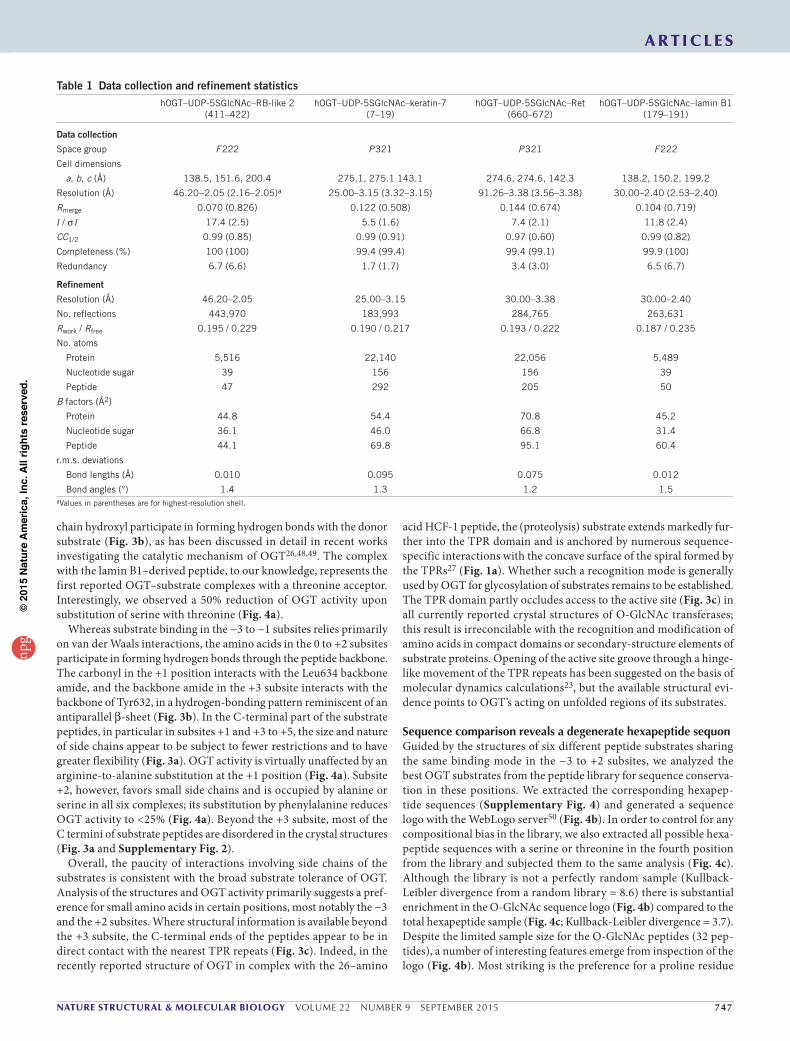
RESULTSO-GlcNActransferasepossessesacceptor-peptidespecificityTo gain insight into possible sequence specificity, we measured hOGT activity against a library of 720 biotinylated 13–amino acid peptides derived from the human proteome37 (Supplementary Table 1). We developed a high throughput–compatible OGT scintillation prox-imity assay, using UDP-[3H]GlcNAc, and included an α-crystallin–derived peptide (NH2-AIPVSREEK-(biotin)-COOH, CRYA1) as a reference standard. We captured the reaction product, radiolabeled O-GlcNAcylated peptide, on streptavidin-coated FlashPlates, thus allowing for direct quantification on a scintillation counter without a need for removing excess substrate (averages of two independent
1Medical Research Council Protein Phosphorylation and Ubiquitylation Unit, College of Life Sciences, University of Dundee, Dundee, UK. 2Division of Molecular Microbiology, College of Life Sciences, University of Dundee, Dundee, UK. 3These authors contributed equally to this work. Correspondence should be addressed to D.M.F.v.A. ([email protected]).
Received 3 March; accepted 6 July; published online 3 August 2015; doi:10.1038/nsmb.3063
The active site of O-GlcNAc transferase imposes constraints on substrate sequenceShalini Pathak1,3, Jana Alonso1,3, Marianne Schimpl1,3, Karim Rafie1, David E Blair1, Vladimir S Borodkin1, Alexander W Schüttelkopf1, Osama Albarbarawi1 & Daan M F van Aalten1,2
O-GlcNActransferase(OGT)glycosylatesadiverserangeofintracellularproteinswithO-linkedN-acetylglucosamine(O-GlcNAc),anessentialanddynamicpost-translationalmodificationinmetazoans.AlthoughthisenzymemodifieshundredsofproteinswithO-GlcNAc,itisnotunderstoodhowOGTachievessubstratespecificity.Inthisstudy,wedescribetheapplicationofahigh-throughputOGTassaytoalibraryofpeptides.WemappedsitesofO-GlcNAcmodificationbyelectrontransferdissociationMSandfoundthattheycorrelatewithpreviouslydetectedO-GlcNAcsites.CrystalstructuresoffouracceptorpeptidesincomplexwithHomo sapiensOGTsuggestthatacombinationofsizeandconformationalrestrictiondefinessequencespecificityinthe−3to+2subsites.ThisworkrevealsthatalthoughtheN-terminalTPRrepeatsofOGTmayhaverolesinsubstraterecognition,thesequencerestrictionimposedbythepeptide-bindingsitemakesasubstantialcontributiontoO-GlcNAcsitespecificity.
npg
© 2
015
Nat
ure
Am
eric
a, In
c. A
ll rig
hts
rese
rved
.
nature structural & molecular biology VOLUME 22 NUMBER 9 SEPTEMBER 2015 745
screens of the peptide library in Fig. 1b). The CRYA1 reference peptide is known to be a relatively poor OGT substrate38. Although all peptides contained at least one potential serine or threonine O-GlcNAc site (2.8 on average), only 70 out of 720 peptides produced a radiometric signal greater than that obtained for the CRYA1 reference peptide. This implied that OGT has a substantial level of substrate specificity dictated by the peptide-binding site alone. However, almost all of the 70 top hits contained more than one serine or threonine, thus preventing direct interrogation of sequence conservation around the O-GlcNAc–acceptor site in the absence of site-mapping data.
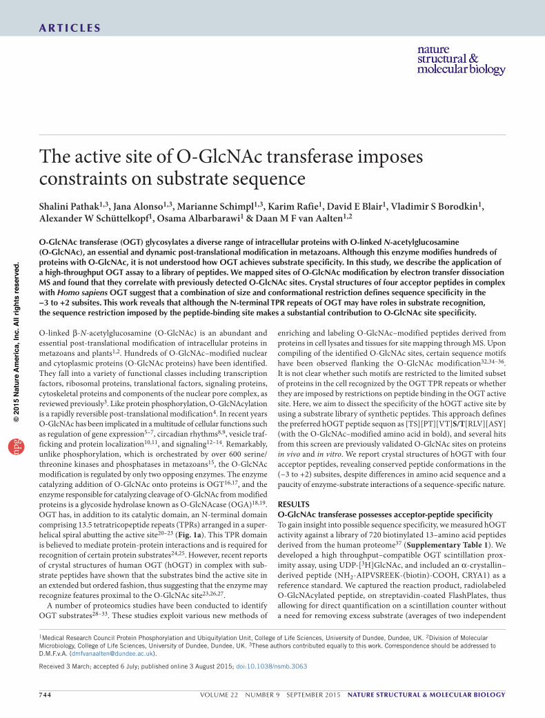
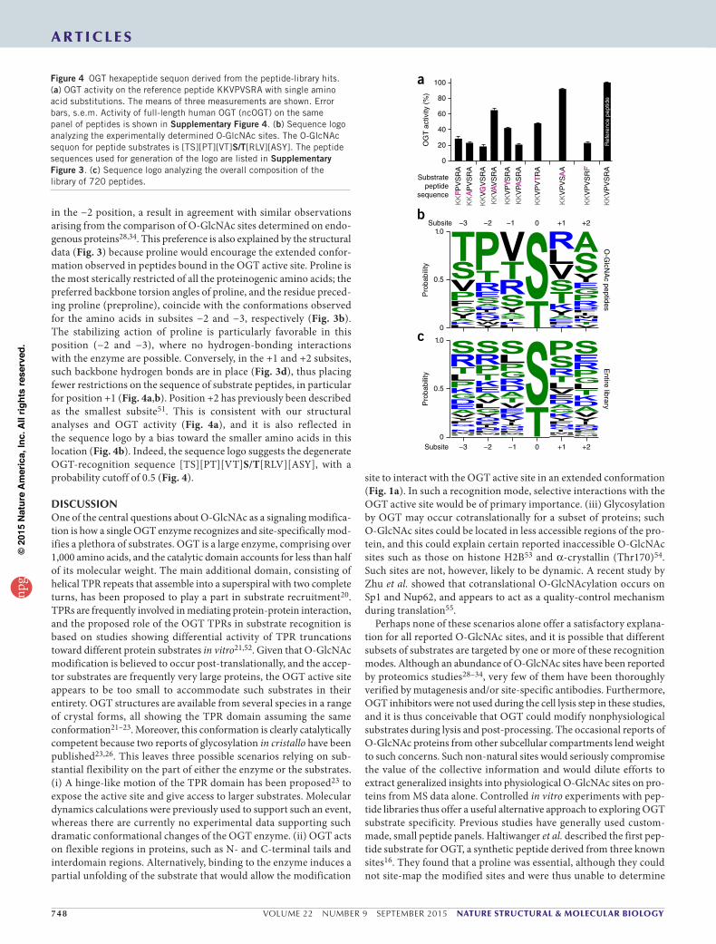
PeptideO-GlcNAcylationislimitedtospecificpositionsTo allow for MS mapping of the specific sites of O-GlcNAc modifica-tion on the best substrates from the peptide-library screen, we resyn-thesized these peptides without the biotin tag (because fragmentation products of biotin would otherwise impede the interpretation of MS data). We enzymatically O-GlcNAcylated these peptides in vitro and analyzed them by electron transfer dissociation (ETD) MS. ETD-MS/MS of the peptides generated fragmentation patterns cover-ing the majority of c- and z-type ions, thus allowing precise mapping of the O-GlcNAc sites. For instance, the ETD-MS/MS spectrum of the in vitro O-GlcNAcylated synthetic peptide KENSPAVTPVSTA,
the top hit from the screen matched a peptide from the protein retinoblastoma-like protein 2 (RBL2; Fig. 2), and showed a strong c10
1+ peak corresponding to a serine plus 203 Da representing the sugar moiety. We specified an expectation value below 0.1 for each peptide fragmentation to ensure reliable designation of the O-GlcNAc sites (Supplementary Fig. 1; fragmentation spectra for all peptides in Supplementary Data Set 1).
OGTpeptidesubstratesarepredictiveforO-GlcNAcylatedproteinsFor those peptides shown to be good OGT substrates, we proceeded to investigate whether the corresponding proteins have previously been reported to be O-GlcNAc modified. The transcription factor FOXO1 has been reported to be O-GlcNAcylated at Ser319 (ref. 39), in agreement with our ETD-MS/MS data from the peptide alone (Fig. 2). Similarly, a tryptic peptide from insulin receptor substrate-1, spanning residues 981–998, has been observed to bear an O-GlcNAc modification on either Ser984 or Ser985 (refs. 40,41). Using ETD-MS/MS, we observed that both Ser984 and Ser985 were O-GlcNAc modified (Fig. 2 and Supplementary Fig. 1). Interestingly, we also
identified Ser400 as being O-GlcNAc modi-fied on a peptide derived from Tau; the same amino acid was recently described as an O-GlcNAc site42. Five of the O-GlcNAc peptides (RBL2, α-crystallin B chain, GSK3β, lamin A and Hsp27) identified from our screen (Fig. 2 and Supplementary Fig. 1) are derived from proteins that have been reported to be O-GlcNAc proteins with either unknown or nonmatching O-GlcNAc sites11,43–46.
SubstratepeptidesbindOGTwithacommonconformationSeveral studies have reported crystal struc-tures of hOGT in complex with substrate peptides and the donor analog UDP-5S-GlcNAc47; notably a 14–amino acid peptide containing the O-GlcNAc site on Ser347
OGT catalytic core
OGT TPR repeats
Substrate
UDP-GlcNAc
XXXS/TXX
OGT activity0% 100%
a bFigure 1 OGT shows substrate selectivity at the peptide level. (a) Schematic representation of OGT substrate recognition, highlighting three possible binding modes: (i) recognition of globular parts of the substrate protein by as-yet-unidentified regions on OGT, (ii) recognition of unfolded regions by the TPR domain or (iii) recognition of a target peptide sequence by the OGT active site. (b) OGT activity on a library of 720 synthetic peptides. The average of two independent measurements is depicted in a heat map, scaled from no activity over background (0%, blue) to the highest activity (100%, red), observed for peptide KENSPCVTPVSTA. The layout of the heat map corresponds to peptide 1 from the Jerini peptide library (Online Methods and Supplementary Table 1) in the upper-left corner and peptide 720 in the lower-right corner.
200 300 400 500 600 700 800 900 1,000 1,100 1,200 1,300 1,400 1,500m/z
05
10
20
30
40
50
60
70
80
90
100
Rel
ativ
e ab
unda
nce
(%)
1,503.76
1,486.65
1,330.741,284.751,040.52
751.941,432.74
743.40572.37
1,031.56941.52 1,230.73643.46
1,360.741,117.48 701.54389.27 878.40843.63274.46
1,375.66
N SP A T P V sV T A
TV AP SN E
S N E
c+1
y+1
z+1
E> <N N> <S S> <P P> A> V> T> P> V> <s s> <T T> <A A – (COOH)
a+1 101.11 230.15 344.20 431.23 528.28 599.32 698.38 799.43 896.48 995.55 1,285.66 1,386.71 1,457.75
c+1 146.13 275.17 389.21 476.25 573.30 644.34 743.41 844.45 941.51 1,040.57 1,330.69 1,431.73 1,502.77
y+1 1,375.66 1,246.62 1,132.57 1,045.54 948.49 877.45 778.39 677.34 580.28 481.21 191.10 90.06 1,503.75
z+1 1,360.65 1,231.61 1,117.56 1,030.53 933.48 862.44 763.37 662.33 565.27 466.20 176.10 75.05 1,488.74
[NH3]+
Figure 2 OGT modifies specific sites on peptide substrates. ETD-MS/MS spectrum of O-GlcNAcylated RBL2 peptide. The spectrum shows the c+1 fragment ions in orange, which unequivocally assign the O-GlcNAc site to Ser11, as well as the z+1 ions in green and the y+1 ions in blue.
a r t i c l e snp
g©
2015
Nat
ure
Am
eric
a, In
c. A
ll rig
hts
rese
rved
.
746 VOLUME 22 NUMBER 9 SEPTEMBER 2015 nature structural & molecular biology
a r t i c l e s
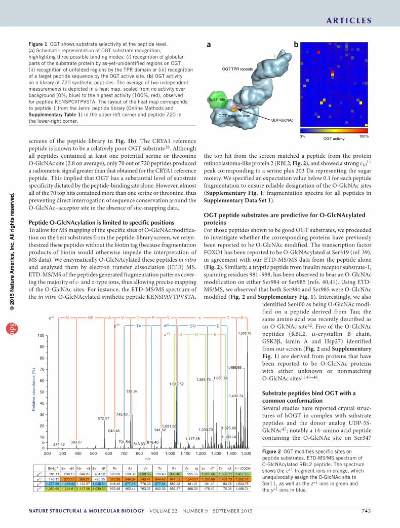
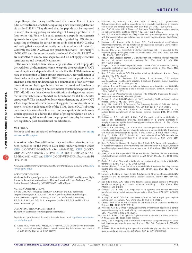
of casein kinase 2α (CK2)23,48, a 13–amino acid peptide based on Ser395 from the TAK1-binding protein TAB1 (ref. 26) and, recently, a 26–amino acid peptide derived from the host cell factor 1 (HCF-1 (ref. 27), whose fate is believed to be proteolytic cleavage by OGT rather than glycosylation). These studies have provided the first insights into how hOGT substrates interact with the active site. It was noted that the peptides bind the active site of OGT in the same orientation and with similar, extended conformations. To explore possible links between peptide sequence and binding modes, we determined the crystal structures of four peptide hits from the screen in complex with hOGT and UDP-5S-GlcNAc. We obtained complexes with the peptides derived from retinoblastoma-like protein 2 (RBL2411–422, KENPAVTPVSTA), proto-oncogene tyrosine protein kinase receptor Ret (Ret660–672, AQAFPVSYSSSGA), keratin-7 (KER77–19, SPVFTSRSAAFSC) and lamin B1 (LAMIN179–191, KLSPSPSSRVTVS) by soaking hOGT crystals and carried out refinement against synchrotron diffraction data (Fig. 3, Supplementary Fig. 2 and Table 1). Despite these peptides’ containing multiple serines and threonines, in all cases the position of target serine or threonine in the enzyme active site was in agreement with the site-mapping results obtained through MS. As observed in the previously published hOGT–peptide complexes, the additional complexes reported here showed peptides binding the active site in the same orientation and in an extended conformation. A comparison of all six complexes highlighted several interesting common features (Fig. 3). Strikingly, all structures revealed a conserved backbone conformation of the peptides in the −3 to +2 subsites (Fig. 3a), with backbone torsion angles characteristic of the extended conformation observed in β-strands (−160° < phi < −50° and 100° < psi < 180°) (Fig. 3b). Beyond this −3 to +2 region, the peptide conformations diverge (Fig. 3a).
OGT is known to use an ordered bi-bi catalytic mechanism, in which the donor substrate binds the enzyme first and contributes to creating the binding site for the incoming acceptor peptide. UDP-GlcNAc interacts primarily with the C-terminal lobe of the catalytic
domain (C-cat), leaving part of the sugar nucleotide molecular surface exposed to the solvent (Fig. 3c). This area is subsequently covered by the peptide substrate binding in subsites −3 to 0. The side chain in the −3 subsite occupies a shallow pocket flanked by hydrophobic amino acids from the C-cat domain and the uracil moiety of UDP-GlcNAc (Fig. 3d). Notably, the six OGT–peptide complexes contain isosteric amino acids, either a valine or a threonine, in this position, which fill the shallow pocket and form van der Waals interactions with the uracil and surrounding enzyme side chains. Substantially larger side chains would be difficult to accommodate in this position without some change in the peptide backbone conformation. We investigated the importance of these interactions and size restrictions by deter-mining the effect of single amino acid substitutions on OGT activity (Fig. 4a and Supplementary Fig. 3). Both the change to a very small (alanine) or a bulky (phenylalanine) amino acid in −3 resulted in <30% residual activity, thus corroborating the structural insights. The −2 subsite was occupied by a proline in three out of the six struc-tures. The conformational rigidity of proline appeared to be favorable in stabilizing the extended conformation of the peptide. Nevertheless, larger side chains, such as the phenylalanine and arginine, were also accommodated, but they projected away from the enzyme and toward the solvent (Fig. 3a). Substrate peptides with very small side chains in the −2 subsite were disfavored by OGT; Alanine reduced the activity to 65%, but the effect was particularly pronounced for glycine (<20%), which offers the least conformational rigidity (Fig. 4a). The −1 subsite appeared to tolerate large side chains, such as that of the tyro-sine observed in Ret and TAB1, without undue distortion (the rotamer adopted by the tyrosine side chain is observed in 13% of tyrosines in high-resolution crystal structures of proteins), although OGT activity was higher on peptides with valine in the −1 position (100%) than those with tyrosine (42%) or alanine (21%) (Fig. 4a).
The acceptor serine or threonine in the 0 subsite necessarily repre-sented the position in the peptide with the least degree of positional and conformational flexibility. Both the backbone amide and the side
+1 +1
+2 +2
–2–3
–1–1
–3–2
0 0
–3–3
–3
–3
–3
–3
-2
–2
–2
–2–2
–2
–1
–1
–1
–1
–1–1
+1
+1
+1
+1
+1
+1
+2
+2
+2
+2
0
0
0
0
00
Phi (degrees)
Psi
(de
gree
s)
–180 –135 –90 –45
180
135
90
+1
+2
0TPRs
N-cat
C-cat
ID
–1
–2
–3–4
RBL2RetKer7TAB1CK2Lamin
a
b c d
Figure 3 Substrate peptides bind the active site of OGT with similar conformations in the −3 to +2 subsites. (a) Stereo view of the active site region of OGT (gray surface), in complex with donor substrate analog UDP-5S-GlcNAc (black sticks) and different substrate peptides derived from the following proteins: RBL2, keratin-7 (Ker7), Ret, lamin B1, CK2 (PDB 4GYY) and TAB1 (PDB 4AY6). Peptides are colored by position in the active site to illustrate the conformational similarities within three amino acids before (−3 to −1 subsites) and two amino acids beyond (+1 to +2 subsites) the acceptor serine (the 0 subsite). Amino acids beyond the −3 to +2 subsites are shown as white sticks. For clarity, the TPR domain has been omitted. Individual structures including unbiased Fo − Fc electron density are shown in Supplementary Figure 2. (b) Subsection of a Ramachandran plot showing backbone torsion angles of the six different substrate peptides. (c) Domain structure of the crystallized fragment of OGT (amino acids 312–1031) in surface representation, with UDP-5S-GlcNAc shown as black sticks and substrate peptides in cartoon representation. The N-terminal TPR domain is formed by tetratricopeptide repeats, N-cat and C-cat, the N- and C-terminal lobes, respectively, of the catalytic domain; ID, intervening domain. (d) Hydrogen-bonding interactions tethering the RBL2 peptide in the active site of OGT, depicted as dashed lines. Colors as in a.
npg
© 2
015
Nat
ure
Am
eric
a, In
c. A
ll rig
hts
rese
rved
.
nature structural & molecular biology VOLUME 22 NUMBER 9 SEPTEMBER 2015 747
a r t i c l e s
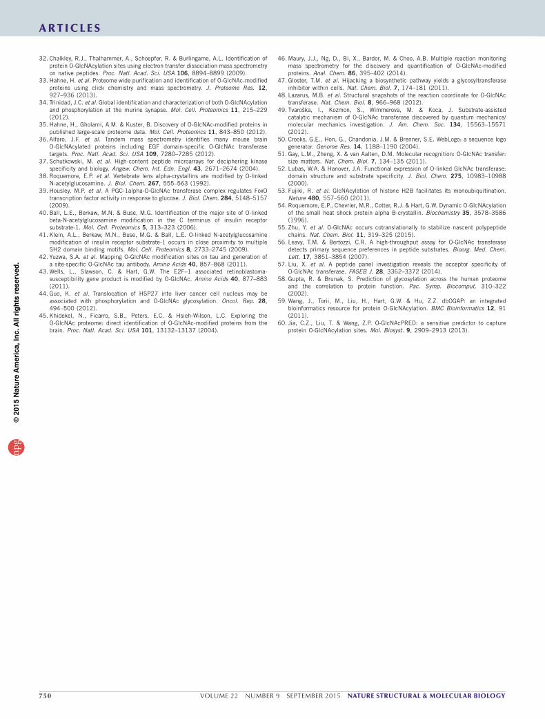
chain hydroxyl participate in forming hydrogen bonds with the donor substrate (Fig. 3b), as has been discussed in detail in recent works investigating the catalytic mechanism of OGT26,48,49. The complex with the lamin B1–derived peptide, to our knowledge, represents the first reported OGT–substrate complexes with a threonine acceptor. Interestingly, we observed a 50% reduction of OGT activity upon substitution of serine with threonine (Fig. 4a).
Whereas substrate binding in the −3 to −1 subsites relies primarily on van der Waals interactions, the amino acids in the 0 to +2 subsites participate in forming hydrogen bonds through the peptide backbone. The carbonyl in the +1 position interacts with the Leu634 backbone amide, and the backbone amide in the +3 subsite interacts with the backbone of Tyr632, in a hydrogen-bonding pattern reminiscent of an antiparallel β-sheet (Fig. 3b). In the C-terminal part of the substrate peptides, in particular in subsites +1 and +3 to +5, the size and nature of side chains appear to be subject to fewer restrictions and to have greater flexibility (Fig. 3a). OGT activity is virtually unaffected by an arginine-to-alanine substitution at the +1 position (Fig. 4a). Subsite +2, however, favors small side chains and is occupied by alanine or serine in all six complexes; its substitution by phenylalanine reduces OGT activity to <25% (Fig. 4a). Beyond the +3 subsite, most of the C termini of substrate peptides are disordered in the crystal structures (Fig. 3a and Supplementary Fig. 2).
Overall, the paucity of interactions involving side chains of the substrates is consistent with the broad substrate tolerance of OGT. Analysis of the structures and OGT activity primarily suggests a pref-erence for small amino acids in certain positions, most notably the −3 and the +2 subsites. Where structural information is available beyond the +3 subsite, the C-terminal ends of the peptides appear to be in direct contact with the nearest TPR repeats (Fig. 3c). Indeed, in the recently reported structure of OGT in complex with the 26–amino
acid HCF-1 peptide, the (proteolysis) substrate extends markedly fur-ther into the TPR domain and is anchored by numerous sequence-specific interactions with the concave surface of the spiral formed by the TPRs27 (Fig. 1a). Whether such a recognition mode is generally used by OGT for glycosylation of substrates remains to be established. The TPR domain partly occludes access to the active site (Fig. 3c) in all currently reported crystal structures of O-GlcNAc transferases; this result is irreconcilable with the recognition and modification of amino acids in compact domains or secondary-structure elements of substrate proteins. Opening of the active site groove through a hinge-like movement of the TPR repeats has been suggested on the basis of molecular dynamics calculations23, but the available structural evi-dence points to OGT’s acting on unfolded regions of its substrates.
SequencecomparisonrevealsadegeneratehexapeptidesequonGuided by the structures of six different peptide substrates sharing the same binding mode in the −3 to +2 subsites, we analyzed the best OGT substrates from the peptide library for sequence conserva-tion in these positions. We extracted the corresponding hexapep-tide sequences (Supplementary Fig. 4) and generated a sequence logo with the WebLogo server50 (Fig. 4b). In order to control for any compositional bias in the library, we also extracted all possible hexa-peptide sequences with a serine or threonine in the fourth position from the library and subjected them to the same analysis (Fig. 4c). Although the library is not a perfectly random sample (Kullback-Leibler divergence from a random library = 8.6) there is substantial enrichment in the O-GlcNAc sequence logo (Fig. 4b) compared to the total hexapeptide sample (Fig. 4c; Kullback-Leibler divergence = 3.7). Despite the limited sample size for the O-GlcNAc peptides (32 pep-tides), a number of interesting features emerge from inspection of the logo (Fig. 4b). Most striking is the preference for a proline residue
Table 1 Data collection and refinement statisticshOGT–UDP-5SGlcNAc–RB-like 2
(411–422)hOGT–UDP-5SGlcNAc–keratin-7
(7–19)hOGT–UDP-5SGlcNAc–Ret
(660–672)hOGT–UDP-5SGlcNAc–lamin B1
(179–191)
Data collection
Space group F 222 P 321 P 321 F 222
Cell dimensions
a, b, c (Å) 138.5, 151.6, 200.4 275.1, 275.1 143.1 274.6, 274.6, 142.3 138.2, 150.2, 199.2
Resolution (Å) 46.20−2.05 (2.16–2.05)a 25.00–3.15 (3.32–3.15) 91.26–3.38 (3.56–3.38) 30.00–2.40 (2.53–2.40)
Rmerge 0.070 (0.826) 0.122 (0.508) 0.144 (0.674) 0.104 (0.719)
I / σ I 17.4 (2.5) 5.5 (1.6) 7.4 (2.1) 11.8 (2.4)
CC1/2 0.99 (0.85) 0.99 (0.91) 0.97 (0.60) 0.99 (0.82)
Completeness (%) 100 (100) 99.4 (99.4) 99.4 (99.1) 99.9 (100)
Redundancy 6.7 (6.6) 1.7 (1.7) 3.4 (3.0) 6.5 (6.7)
Refinement
Resolution (Å) 46.20–2.05 25.00–3.15 30.00–3.38 30.00–2.40
No. reflections 443,970 183,993 284,765 263,631
Rwork / Rfree 0.195 / 0.229 0.190 / 0.217 0.193 / 0.222 0.187 / 0.235
No. atoms
Protein 5,516 22,140 22,056 5,489
Nucleotide sugar 39 156 156 39
Peptide 47 292 205 50
B factors (Å2)
Protein 44.8 54.4 70.8 45.2
Nucleotide sugar 36.1 46.0 66.8 31.4
Peptide 44.1 69.8 95.1 60.4
r.m.s. deviations
Bond lengths (Å) 0.010 0.095 0.075 0.012
Bond angles (°) 1.4 1.3 1.2 1.5aValues in parentheses are for highest-resolution shell.
npg
© 2
015
Nat
ure
Am
eric
a, In
c. A
ll rig
hts
rese
rved
.
748 VOLUME 22 NUMBER 9 SEPTEMBER 2015 nature structural & molecular biology
a r t i c l e s
in the −2 position, a result in agreement with similar observations arising from the comparison of O-GlcNAc sites determined on endo-genous proteins28,34. This preference is also explained by the structural data (Fig. 3) because proline would encourage the extended confor-mation observed in peptides bound in the OGT active site. Proline is the most sterically restricted of all the proteinogenic amino acids; the preferred backbone torsion angles of proline, and the residue preced-ing proline (preproline), coincide with the conformations observed for the amino acids in subsites −2 and −3, respectively (Fig. 3b). The stabilizing action of proline is particularly favorable in this position (−2 and −3), where no hydrogen-bonding interactions with the enzyme are possible. Conversely, in the +1 and +2 subsites, such backbone hydrogen bonds are in place (Fig. 3d), thus placing fewer restrictions on the sequence of substrate peptides, in particular for position +1 (Fig. 4a,b). Position +2 has previously been described as the smallest subsite51. This is consistent with our structural analyses and OGT activity (Fig. 4a), and it is also reflected in the sequence logo by a bias toward the smaller amino acids in this location (Fig. 4b). Indeed, the sequence logo suggests the degenerate OGT-recognition sequence [TS][PT][VT]S/T[RLV][ASY], with a probability cutoff of 0.5 (Fig. 4).
DISCUSSIONOne of the central questions about O-GlcNAc as a signaling modifica-tion is how a single OGT enzyme recognizes and site-specifically mod-ifies a plethora of substrates. OGT is a large enzyme, comprising over 1,000 amino acids, and the catalytic domain accounts for less than half of its molecular weight. The main additional domain, consisting of helical TPR repeats that assemble into a superspiral with two complete turns, has been proposed to play a part in substrate recruitment20. TPRs are frequently involved in mediating protein-protein interaction, and the proposed role of the OGT TPRs in substrate recognition is based on studies showing differential activity of TPR truncations toward different protein substrates in vitro21,52. Given that O-GlcNAc modification is believed to occur post-translationally, and the accep-tor substrates are frequently very large proteins, the OGT active site appears to be too small to accommodate such substrates in their entirety. OGT structures are available from several species in a range of crystal forms, all showing the TPR domain assuming the same conformation21–23. Moreover, this conformation is clearly catalytically competent because two reports of glycosylation in cristallo have been published23,26. This leaves three possible scenarios relying on sub-stantial flexibility on the part of either the enzyme or the substrates. (i) A hinge-like motion of the TPR domain has been proposed23 to expose the active site and give access to larger substrates. Molecular dynamics calculations were previously used to support such an event, whereas there are currently no experimental data supporting such dramatic conformational changes of the OGT enzyme. (ii) OGT acts on flexible regions in proteins, such as N- and C-terminal tails and interdomain regions. Alternatively, binding to the enzyme induces a partial unfolding of the substrate that would allow the modification
site to interact with the OGT active site in an extended conformation (Fig. 1a). In such a recognition mode, selective interactions with the OGT active site would be of primary importance. (iii) Glycosylation by OGT may occur cotranslationally for a subset of proteins; such O-GlcNAc sites could be located in less accessible regions of the pro-tein, and this could explain certain reported inaccessible O-GlcNAc sites such as those on histone H2B53 and α-crystallin (Thr170)54. Such sites are not, however, likely to be dynamic. A recent study by Zhu et al. showed that cotranslational O-GlcNAcylation occurs on Sp1 and Nup62, and appears to act as a quality-control mechanism during translation55.
Perhaps none of these scenarios alone offer a satisfactory explana-tion for all reported O-GlcNAc sites, and it is possible that different subsets of substrates are targeted by one or more of these recognition modes. Although an abundance of O-GlcNAc sites have been reported by proteomics studies28–34, very few of them have been thoroughly verified by mutagenesis and/or site-specific antibodies. Furthermore, OGT inhibitors were not used during the cell lysis step in these studies, and it is thus conceivable that OGT could modify nonphysiological substrates during lysis and post-processing. The occasional reports of O-GlcNAc proteins from other subcellular compartments lend weight to such concerns. Such non-natural sites would seriously compromise the value of the collective information and would dilute efforts to extract generalized insights into physiological O-GlcNAc sites on pro-teins from MS data alone. Controlled in vitro experiments with pep-tide libraries thus offer a useful alternative approach to exploring OGT substrate specificity. Previous studies have generally used custom-made, small peptide panels. Haltiwanger et al. described the first pep-tide substrate for OGT, a synthetic peptide derived from three known sites16. They found that a proline was essential, although they could not site-map the modified sites and were thus unable to determine
KK
VP
YS
RA
KK
VP
VS
RA
KK
FP
VS
RA
KK
AP
VS
RA
KK
VG
VS
RA
KK
VA
VS
RA
KK
VPA
SR
A
KK
VP
VT
RA
KK
VP
VS
AA
KK
VP
VS
RF
Substratepeptide
sequence
0
20
40
60
80
100
OG
T a
ctiv
ity (
%)
Ref
eren
ce p
eptid
e
+2+10−1−2−3Subsite
Pro
babi
lity
1.0
0
0.5
Entire library
Pro
babi
lity
1.0
0
0.5
O-G
lcNA
c peptides
Subsite +2+10−1−2−3
a
b
c
Figure 4 OGT hexapeptide sequon derived from the peptide-library hits. (a) OGT activity on the reference peptide KKVPVSRA with single amino acid substitutions. The means of three measurements are shown. Error bars, s.e.m. Activity of full-length human OGT (ncOGT) on the same panel of peptides is shown in Supplementary Figure 4. (b) Sequence logo analyzing the experimentally determined O-GlcNAc sites. The O-GlcNAc sequon for peptide substrates is [TS][PT][VT]S/T[RLV][ASY]. The peptide sequences used for generation of the logo are listed in Supplementary Figure 3. (c) Sequence logo analyzing the overall composition of the library of 720 peptides.
npg
© 2
015
Nat
ure
Am
eric
a, In
c. A
ll rig
hts
rese
rved
.
nature structural & molecular biology VOLUME 22 NUMBER 9 SEPTEMBER 2015 749
a r t i c l e s
the proline position. Leavy and Bertozzi used a small library of pep-tides derived from α-crystallin, exploiting a new assay using detection via azido-ELISA56. They showed that the peptide can be improved in many places, suggesting an advantage of having a proline in +2 (but not in −2). Finally, Liu et al. generated a peptide-mutagenesis approach to explore motifs previously reported by MS studies, suggesting preference for certain amino acids close to the O-GlcNAc and noting that sites predominantly occur in random-coil regions57. Currently available O-GlcNAc site–prediction servers—YinOYang58, dbOGAP59 and the more recently developed O-GlcNAcPRED60—are restricted to amino acid sequence and do not apply structural restraints around the modification sites.
The work described here uses a large and diverse set of peptides derived from the human proteome to explore the specificity of OGT toward peptides, independently of the role that the TPR domain may have in recognition of large protein substrates. Cocrystallization of identified acceptor peptides with OGT showed that the peptide is teth-ered into a common binding mode by a combination of van der Waals interactions and hydrogen bonds that restrict torsional freedom in the −3 to +2 subsites only. These structural constraints together with ETD-MS/MS data then allowed identification of a degenerate sequon that is remarkably similar to what has been found for O-GlcNAc sites on proteins34. This is a notable advance in understanding of how OGT selects its protein substrates because it suggests that constraints in the active site alone, independently of the TPRs, dictate OGT substrate preference to a considerable extent. Future experiments could use a similar approach to address the effect of phosphorylation on OGT substrate recognition, to address the proposed interplay between the two regulatory post-translational modifications.
METhODSMethods and any associated references are available in the online version of the paper.
Accession codes. X-ray diffraction data and refined structures have been deposited in the Protein Data Bank under accession codes 4XI9 (hOGT–UDP-5SGlcNAc–Ret (660–672)), 4XIF (hOGT– UDP-5SGlcNAc–keratin-7 (7–19)), 5C1D (hOGT–UDP-5SGlcNAc–RB-like 2 (411–422)) and 5BNW (hOGT–UDP-5SGlcNAc–lamin B1 (179–191)).
Note: Any Supplementary Information and Source Data files are available in the online version of the paper.
AcKnOWlEDgMEntSWe thank the European Synchrotron Radiation Facility (ESRF) and Diamond Light Source for beam time and assistance. This work was funded by a Wellcome Trust Senior Research Fellowship (WT087590MA) to D.M.F.v.A.
AUtHOR cOntRIBUtIOnSS.P. and D.M.F.v.A. conceived the study; S.P., D.E.B. and K.R. performed the peptide assays; M.S., K.R. and D.M.F.v.A. performed structural biology; V.S.B. performed peptide synthesis; J.A. and O.A. performed MS studies; S.P., M.S., A.W.S. and D.M.F.v.A. interpreted the data; S.P., M.S. and D.M.F.v.A. wrote the manuscript.
cOMPEtIng FInAncIAl IntEREStSThe authors declare no competing financial interests.
Reprints and permissions information is available online at http://www.nature.com/reprints/index.html.
1. Lubas, W.A., Frank, D.W., Krause, M. & Hanover, J.A. O-Linked GlcNAc transferase is a conserved nucleocytoplasmic protein containing tetratricopeptide repeats. J. Biol. Chem. 272, 9316–9324 (1997).
2. O’Donnell, N., Zachara, N.E., Hart, G.W. & Marth, J.D. Ogt-dependent X-chromosome-linked protein glycosylation is a requisite modification in somatic cell function and embryo viability. Mol. Cell. Biol. 24, 1680–1690 (2004).
3. Hart, G.W., Housley, M.P. & Slawson, C. Cycling of O-linked β-N-acetylglucosamine on nucleocytoplasmic proteins. Nature 446, 1017–1022 (2007).
4. Hart, G.W. et al. O-GlcNAcylation of key nuclear and cytoskeletal proteins: reciprocity with O-phosphorylation and putative roles in protein multimerization. Glycobiology 6, 711–716 (1996).
5. Hanover, J.A., Krause, M.W. & Love, D.C. Post-translational modifications: bittersweet memories: linking metabolism to epigenetics through O-GlcNAcylation. Nat. Rev. Mol. Cell Biol. 13, 312–321 (2012).
6. Sinclair, D.A. et al. Drosophila O-GlcNAc transferase (OGT) is encoded by the Polycomb group (PcG) gene, super sex combs (sxc). Proc. Natl. Acad. Sci. USA 106, 13427–13432 (2009).
7. Daou, S. et al. Crosstalk between O-GlcNAcylation and proteolytic cleavage regulates the host cell factor-1 maturation pathway. Proc. Natl. Acad. Sci. USA 108, 2747–2752 (2010).
8. Durgan, D.J. et al. O-GlcNAcylation, novel post-translational modification linking myocardial metabolism and cardiomyocyte circadian clock. J. Biol. Chem. 286, 44606–44619 (2011).
9. Kim, E.Y. et al. A role for O-GlcNAcylation in setting circadian clock speed. Genes Dev. 26, 490–502 (2012).
10. Geng, F., Zhu, W., Anderson, R.A., Leber, B. & Andrews, D.W. Multiple post-translational modifications regulate E-cadherin transport during apoptosis. J. Cell Sci. 125, 2615–2625 (2012).
11. Lefebvre, T. et al. Evidence of a balance between phosphorylation and O-GlcNAc glycosylation of Tau proteins–a role in nuclear localization. Biochim. Biophys. Acta 1619, 167–176 (2003).
12. Yang, X. et al. Phosphoinositide signalling links O-GlcNAc transferase to insulin resistance. Nature 451, 964–969 (2008).
13. Pathak, S. et al. O-GlcNAcylation of TAB1 modulates TAK1-mediated cytokine release. EMBO J. 31, 1394–1404 (2012).
14. Rotty, J.D., Hart, G.W. & Coulombe, P.A. Stressing the role of O-GlcNAc: linking cell survival to keratin modification. Nat. Cell Biol. 12, 847–849 (2010).
15. Manning, G., Whyte, D.B., Martinez, R., Hunter, T. & Sudarsanam, S. The protein kinase complement of the human genome. Science 298, 1912–1934 (2002).
16. Haltiwanger, R.S., Holt, G.D. & Hart, G.W. Enzymatic addition of O-GlcNAc to nuclear and cytoplasmic proteins: identification of a uridine diphospho-N-acetylglucosamine:peptide beta-N-acetylglucosaminyltransferase. J. Biol. Chem. 265, 2563–2568 (1990).
17. Kreppel, L.K., Blomberg, M.A. & Hart, G.W. Dynamic glycosylation of nuclear and cytosolic proteins: cloning and characterization of a unique O-GlcNAc transferase with multiple tetratricopeptide repeats. J. Biol. Chem. 272, 9308–9315 (1997).
18. Dong, D.L. & Hart, G.W. Purification and characterization of an O-GlcNAc selective N-acetyl-beta-D-glucosaminidase from rat spleen cytosol. J. Biol. Chem. 269, 19321–19330 (1994).
19. Gao, Y., Wells, L., Comer, F.I., Parker, G.J. & Hart, G.W. Dynamic O-glycosylation of nuclear and cytosolic proteins: cloning and characterization of a neutral, cytosolic beta-N-acetylglucosaminidase from human brain. J. Biol. Chem. 276, 9838–9845 (2001).
20. Jínek, M. et al. The superhelical TPR-repeat domain of O-linked GlcNAc transferase exhibits structural similarities to importin α. Nat. Struct. Mol. Biol. 11, 1001–1007 (2004).
21. Clarke, A.J. et al. Structural insights into mechanism and specificity of O-GlcNAc transferase. EMBO J. 27, 2780–2788 (2008).
22. Martinez-Fleites, C. et al. Structure of an O-GlcNAc transferase homolog provides insight into intracellular glycosylation. Nat. Struct. Mol. Biol. 15, 764–765 (2008).
23. Lazarus, M.B., Nam, Y., Jiang, J., Sliz, P. & Walker, S. Structure of human O-GlcNAc transferase and its complex with a peptide substrate. Nature 469, 564–567 (2011).
24. Iyer, S.P. & Hart, G.W. Roles of the tetratricopeptide repeat domain in O-GlcNAc transferase targeting and protein substrate specificity. J. Biol. Chem. 278, 24608–24616 (2003).
25. Kreppel, L.K. & Hart, G.W. Regulation of a cytosolic and nuclear O-GlcNAc transferase: role of the tetratricopeptide repeats. J. Biol. Chem. 274, 32015–32022 (1999).
26. Schimpl, M. et al. O-GlcNAc transferase invokes nucleotide sugar pyrophosphate participation in catalysis. Nat. Chem. Biol. 8, 969–974 (2012).
27. Lazarus, M.B. et al. HCF-1 is cleaved in the active site of O-GlcNAc transferase. Science 342, 1235–1239 (2013).
28. Vosseller, K. et al. O-linked N-acetylglucosamine proteomics of postsynaptic density preparations using lectin weak affinity chromatography and mass spectrometry. Mol. Cell. Proteomics 5, 923–934 (2006).
29. Cole, R.N. & Hart, G.W. Cytosolic O-glycosylation is abundant in nerve terminals. J. Neurochem. 79, 1080–1089 (2001).
30. Wells, L. et al. Mapping sites of O-GlcNAc modification using affinity tags for serine and threonine post-translational modifications. Mol. Cell. Proteomics 1, 791–804 (2002).
31. Khidekel, N. et al. Probing the dynamics of O-GlcNAc glycosylation in the brain using quantitative proteomics. Nat. Chem. Biol. 3, 339–348 (2007).
npg
© 2
015
Nat
ure
Am
eric
a, In
c. A
ll rig
hts
rese
rved
.
750 VOLUME 22 NUMBER 9 SEPTEMBER 2015 nature structural & molecular biology
32. Chalkley, R.J., Thalhammer, A., Schoepfer, R. & Burlingame, A.L. Identification of protein O-GlcNAcylation sites using electron transfer dissociation mass spectrometry on native peptides. Proc. Natl. Acad. Sci. USA 106, 8894–8899 (2009).
33. Hahne, H. et al. Proteome wide purification and identification of O-GlcNAc-modified proteins using click chemistry and mass spectrometry. J. Proteome Res. 12, 927–936 (2013).
34. Trinidad, J.C. et al. Global identification and characterization of both O-GlcNAcylation and phosphorylation at the murine synapse. Mol. Cell. Proteomics 11, 215–229 (2012).
35. Hahne, H., Gholami, A.M. & Kuster, B. Discovery of O-GlcNAc-modified proteins in published large-scale proteome data. Mol. Cell. Proteomics 11, 843–850 (2012).
36. Alfaro, J.F. et al. Tandem mass spectrometry identifies many mouse brain O-GlcNAcylated proteins including EGF domain-specific O-GlcNAc transferase targets. Proc. Natl. Acad. Sci. USA 109, 7280–7285 (2012).
37. Schutkowski, M. et al. High-content peptide microarrays for deciphering kinase specificity and biology. Angew. Chem. Int. Edn. Engl. 43, 2671–2674 (2004).
38. Roquemore, E.P. et al. Vertebrate lens alpha-crystallins are modified by O-linked N-acetylglucosamine. J. Biol. Chem. 267, 555–563 (1992).
39. Housley, M.P. et al. A PGC-1alpha-O-GlcNAc transferase complex regulates FoxO transcription factor activity in response to glucose. J. Biol. Chem. 284, 5148–5157 (2009).
40. Ball, L.E., Berkaw, M.N. & Buse, M.G. Identification of the major site of O-linked beta-N-acetylglucosamine modification in the C terminus of insulin receptor substrate-1. Mol. Cell. Proteomics 5, 313–323 (2006).
41. Klein, A.L., Berkaw, M.N., Buse, M.G. & Ball, L.E. O-linked N-acetylglucosamine modification of insulin receptor substrate-1 occurs in close proximity to multiple SH2 domain binding motifs. Mol. Cell. Proteomics 8, 2733–2745 (2009).
42. Yuzwa, S.A. et al. Mapping O-GlcNAc modification sites on tau and generation of a site-specific O-GlcNAc tau antibody. Amino Acids 40, 857–868 (2011).
43. Wells, L., Slawson, C. & Hart, G.W. The E2F–1 associated retinoblastoma-susceptibility gene product is modified by O-GlcNAc. Amino Acids 40, 877–883 (2011).
44. Guo, K. et al. Translocation of HSP27 into liver cancer cell nucleus may be associated with phosphorylation and O-GlcNAc glycosylation. Oncol. Rep. 28, 494–500 (2012).
45. Khidekel, N., Ficarro, S.B., Peters, E.C. & Hsieh-Wilson, L.C. Exploring the O-GlcNAc proteome: direct identification of O-GlcNAc-modified proteins from the brain. Proc. Natl. Acad. Sci. USA 101, 13132–13137 (2004).
46. Maury, J.J., Ng, D., Bi, X., Bardor, M. & Choo, A.B. Multiple reaction monitoring mass spectrometry for the discovery and quantification of O-GlcNAc-modified proteins. Anal. Chem. 86, 395–402 (2014).
47. Gloster, T.M. et al. Hijacking a biosynthetic pathway yields a glycosyltransferase inhibitor within cells. Nat. Chem. Biol. 7, 174–181 (2011).
48. Lazarus, M.B. et al. Structural snapshots of the reaction coordinate for O-GlcNAc transferase. Nat. Chem. Biol. 8, 966–968 (2012).
49. Tvaroška, I., Kozmon, S., Wimmerova, M. & Koca, J. Substrate-assisted catalytic mechanism of O-GlcNAc transferase discovered by quantum mechanics/molecular mechanics investigation. J. Am. Chem. Soc. 134, 15563–15571 (2012).
50. Crooks, G.E., Hon, G., Chandonia, J.M. & Brenner, S.E. WebLogo: a sequence logo generator. Genome Res. 14, 1188–1190 (2004).
51. Gay, L.M., Zheng, X. & van Aalten, D.M. Molecular recognition: O-GlcNAc transfer: size matters. Nat. Chem. Biol. 7, 134–135 (2011).
52. Lubas, W.A. & Hanover, J.A. Functional expression of O-linked GlcNAc transferase: domain structure and substrate specificity. J. Biol. Chem. 275, 10983–10988 (2000).
53. Fujiki, R. et al. GlcNAcylation of histone H2B facilitates its monoubiquitination. Nature 480, 557–560 (2011).
54. Roquemore, E.P., Chevrier, M.R., Cotter, R.J. & Hart, G.W. Dynamic O-GlcNAcylation of the small heat shock protein alpha B-crystallin. Biochemistry 35, 3578–3586 (1996).
55. Zhu, Y. et al. O-GlcNAc occurs cotranslationally to stabilize nascent polypeptide chains. Nat. Chem. Biol. 11, 319–325 (2015).
56. Leavy, T.M. & Bertozzi, C.R. A high-throughput assay for O-GlcNAc transferase detects primary sequence preferences in peptide substrates. Bioorg. Med. Chem. Lett. 17, 3851–3854 (2007).
57. Liu, X. et al. A peptide panel investigation reveals the acceptor specificity of O-GlcNAc transferase. FASEB J. 28, 3362–3372 (2014).
58. Gupta, R. & Brunak, S. Prediction of glycosylation across the human proteome and the correlation to protein function. Pac. Symp. Biocomput. 310–322 (2002).
59. Wang, J., Torii, M., Liu, H., Hart, G.W. & Hu, Z.Z. dbOGAP: an integrated bioinformatics resource for protein O-GlcNAcylation. BMC Bioinformatics 12, 91 (2011).
60. Jia, C.Z., Liu, T. & Wang, Z.P. O-GlcNAcPRED: a sensitive predictor to capture protein O-GlcNAcylation sites. Mol. Biosyst. 9, 2909–2913 (2013).
a r t i c l e snp
g©
2015
Nat
ure
Am
eric
a, In
c. A
ll rig
hts
rese
rved
.
nature structural & molecular biologydoi:10.1038/nsmb.3063
ONLINEMEThODSPeptide-library screening. A peptide library comprising 720 biotinylated pro-tein kinase substrate peptides, consisting of two 384-well polypropylene plates, each containing 360 peptides (0.25 nmol each in rows A1 to O24), was obtained from JPT Peptide Technologies37. Peptides were solubilized in 62.5 µl of 50 mM Tris-HCl, pH 7.5, to give a final concentration of 4 µM, with a PlateMate Plus liquid handling robot (Thermo Scientific Matrix). A solution of either control (no protein) or hOGT313–1046 protein (300 nM, Glycobiochem UK) and UDP-GlcNAc substrate stock (1 µM), consisting of 910 nM cold UDP-GlcNAc (Sigma) and 90 nM UDP-[3H]GlcNAc radioactive tracer (0.4 Ci/ mmol) (American Radiolabeled Chemicals) was made in buffer consisting of 50 mM Tris, pH 7.5, 2 mM DTT and 0.1 mg/ml bovine serum albumin (BSA). The peptide screen was carried out in duplicate and consisted of one quality-control plate con-taining high controls, mutant α-A crystallin (CRYA1M) (NH2-AIPVSRAEK (biotin)-COOH) in columns 1–8 and 17–24, and native α-A crystallin (CRYA1) (NH2-AIPVSREEK(biotin)-COOH) in columns 9–16. The screening plates consisted of library peptides in rows A1–O24, with low controls in P1–P24. 10 µl of 4 µM peptide was added to all 384-well polypropylene U-bottom plates with a PlateMate Plus liquid-handling robot as outlined above. The reaction was then initiated by the addition of 10 µl of control (no OGT/UDP-GlcNAc) to both the QC plate (columns 9–16) and the assay plates P13–P24 to provide low controls. All other positions received OGT/UDP-GlcNAc. Plates were incubated at room temperature on a microtiter plate shaker (Heidolph) for 4 h. The reaction was stopped by the addition of 40 µl of 100 µM phosphoric acid, pH 4, and 750 µM MgCl2 with a Wellmate liquid handling robot (Thermo Scientific Matrix). The stopped reactions were incubated for an additional 10 min at room temperature. 56 µl from the QC and screening plates was transferred to 384-well Streptavidin FlashPlates (PerkinElmer) with a PlateMate plus liquid-handling robot. Plates were then sealed and incubated overnight at room temperature before determination of scintillation counts with a Top Count plate reader (PerkinElmer).
The quality of the resultant screening data was assessed by coefficient of variance (CV), robust Z prime and SSMD calculations carried out on the QC plate and the high and low controls included on each of the screening repli-cates. Scintillation counts generated from the screen were converted into relative activity with the CRYA1 peptide as standard.
Peptide synthesis. Peptides were synthesized on a microwave-assisted CEM Liberty instrument with standard Fmoc protocol on Rink amide low-load MBHA resin (0.24 mmol/g) (Novabiochem). Coupling steps were performed with 2.5 eq of amino acid with respect to the resin loading capacity, PyBOP (2.5 eq) and DIPEA (5 eq), at 70 °C for 5 min, except for serine and histidine, which were coupled at 50 °C for 10 min. Deprotection was carried out with 20% piperid-ine in DMF at 70 °C for 0.5 min and then for 3 min with the fresh portion of the deprotection mixture. Peptides were manually cleaved from the resin with TFA/H2O/TIPS/DODT 1:0.05:0.02:0.05 (2 mL) for 2 h at RT. For methionine-containing peptides, the cleavage cocktail was supplemented with NH4I (0.05 g) and Me2S (0.1 mL). The peptides were precipitated with cold (0 °C) diethyl ether (40 mL) and collected by centrifugation at 4,000g, at 4 °C for 15 min. The pellet was resuspended in cold ether and centrifuged again; this step was repeated twice. The peptides were dried with argon flow. The crude peptides were purified by HPLC on Gilson instrument with a Waters Xbridge 19 × 100 mm peptide separa-tion technology column (flow rate 25 mL/min), with a linear gradient 5–95% of MeCN in 0.1% TFA in water over 10 min. The appropriate fractions were pooled and concentrated in vacuo. Finally, the residue was redissolved in 20% AcOH (3 mL) and freeze dried. The identity of the synthetic peptides was confirmed by LC-MS on a Bruker microTOF instrument.
hOGT activity measurement. hOGT activity was determined in reactions containing 50 nM His6-hOGT312–1031 or ncOGT in 50 mM Tris-HCl, pH 7.5, 0.1 mg/mL BSA, 10 µM sodium dithionate and 0.5 mM of peptide in a total volume of 100 µL. Reaction mixtures were preincubated for 15 min and initiated by addition of UDP-GlcNAc to a final concentration of 50 µM. The reaction was stopped after 30 min (short construct) or 4 h (ncOGT) at 21 °C by addition of 200 µL of 75 µM pyrocatechol violet in 25 mM HEPES, pH 7.4, 10 mM NaCl, 50% (v/v) MeOH and 15 µM fluorophore, a UDP-sensitive xanthene-based Zn(ii) complex described previously61,62. UDP formation was detected fluorimetrically
on a Gemini EM fluorescence Microplate reader (Molecular Devices) at excita-tion and emission wavelengths of 485 nm and 530 nm, respectively. Turnover did not exceed 10% for either substrate. Data are presented as averages of three measurements, with error bars showing the s.e.m.
In vitro O-GlcNAcylation of peptides for mass spectrometry. The resynthesized peptides were O-GlcNAcylated in a final volume of 20 µl with the following final concentrations of each component: peptide (2 µM), OGT enzyme (2 µM), UDP-GlcNAc (4 mM) and reaction buffer (25 mM ammonium bicarbonate (Sigma-Aldrich LC-MS grade) and 1 mM DTT). The reaction mixture was incubated for 3 h at 30 °C. For LC-MS analysis, excess UDP-GlcNAc and OGT were removed with NanoSep centrifugal columns (molecular-weight cutoff 10 kDa, PALL Life Sciences). The resulting filtrate was dried in a SpeedVac (Thermo Fisher Scientific) and stored at −20 °C until further analysis.
O-GlcNAcylation analysis by LC-MS/MS. Identification of the O-GlcNAc sites was performed by ESI-IT-ETD (ElectroSpray IonTrap Electron Transfer Dissociation) MS coupled to a nano-LC system (Ultimate 3000 RSLC, Dionex). Dried peptides were resuspended in 100 µL of 0.5% HCOOH, and 10 µL was injected for MS analysis. In vitro O-GlcNAcylated peptides were concentrated on a trap column (2 cm × 100 µm, Dionex) at 10 µL/min and separated on a 15 cm × 75 µm Pepmap C18 reversed-phase column (Thermo Fisher Scientific). Peptides were eluted by a linear 60-min gradient of 95% A/5% B to 90% B (A, H2O and 0.1% HCOOH; B, 80% acetonitrile (ACN) and 0.08% HCOOH) at 300 nl/min into a LTQ Velos ETD (Thermo Fisher Scientific). MS spectra were acquired in positive mode: first MS full scans were acquired, and this was followed by MS/MS in ETD mode. Up to five of the most intense precursors were selected for ETD fragmentation with an activation time of 300 ms and nondynamic exclusion.
Proteome Discoverer v1.4 (Thermo Scientific) was used to process raw LC-MS/MS data, applying the Mascot (version 2.3.02, Matrix Science) search-engine algorithm against an in house–established peptide database with the following Mascot parameters: 2+, 3+, 4+ and 5+ ions; precursor mass tolerance, 100 p.p.m.; fragment tolerance, 0.6 Da; and no missed cleavages. The variable modifica-tions included were oxidation (M) (15.99 Da), dioxidation (M) (31.98 Da) and HexNAc (ST) (+203.0794 Da). All MS/MS data and database results were manu-ally inspected in detail to check the accurate assignment of fragment ions with the above software. Peptides with an expectation value (exp value) smaller than 0.1 are considered to be precise O-GlcNAc site assignments.
Crystallography. Human OGT (amino acids 312–1031) was recombinantly expressed as a cleavable GST fusion in Escherichia coli BL21(DE3)pLysS and purified and cocrystallized with the substrate analog UDP-5S-GlcNAc47 as described previously26 for the complexes with peptides derived from keratin-7 (SPVFTSRSAAFSC) and Ret (AQAFPVSYSSSGA). Crystals were soaked with an excess of synthetic peptide for 20–60 min and cryoprotected in a saturated lithium sulfate solution before being flash frozen in liquid nitrogen. For the complex with the RBL2 peptide (KENPAVTPVSTA), the reservoir solution was supplemented with 0.5 M (NH4)2SO4, and hOGT was cocrystallized with 2.5 mM UDP-5S-GlcNAc and 2.5 mM peptide and crystal seeds. Seeds were generated from hOGT crystals grown in 1.3 M dl-malic acid, and 0.1 M Bis-Tris pro-pane, pH 6.4. Crystals were cryoprotected by a 2-s immersion in 2.5 M sodium malonate, pH 7, before being flash frozen in liquid nitrogen. For the complex with the lamin B1 peptide (KLSPSPSSRVTVS), hOGT was crystallized in 1.3 M dl-malic acid, pH 6.4, 0.1 M Bis-Tris propane, pH 6.4 and crystal seeds. Seeds were generated from hOGT crystals grown in 1.3 M dl-malic acid, and 0.1 M Bis-Tris propane, pH 6.4. Crystals were soaked for 60 min with excess peptide and 2.5 mM UDP-5S-GlcNAc. Crystals were cryoprotected by a 2-s immersion in 2.5 M dl-malic acid, pH 7, before being flash frozen in liquid nitrogen. Data were collected at the European Synchrotron Radiation Facility (ESRF) beamlines ID23-2 and ID30A-3 and on Diamond Light Source beamline I04-1, and were processed with MOSFLM63, XDS64 and DENZO65. 2% (Ret, keratin-7) or 5% (RBL2, lamin) of all reflections were set aside as an Rfree test set. Structures were solved with MOLREP66 with one molecule of PDB 3PE4 (ref. 23) as a search model. Crystals belonging to F222 (RBL2, lamin B) have one molecule per asym-metric unit; whereas crystals belonging to space group P321 (keratin-7 and Ret) have four molecules per asymmetric unit, so noncrystallographic symmetry (NCS) restraints were imposed during refinement with REFMAC5 (ref. 66),
npg
© 2
015
Nat
ure
Am
eric
a, In
c. A
ll rig
hts
rese
rved
.
nature structural & molecular biology doi:10.1038/nsmb.3063
and four-fold NCS averaging was used to improve the maps for model building in COOT67. Ligand topology for UDP-5S-GlcNAc was created with PRODRG68, and donor and acceptor substrates were manually placed. Table 1 gives a sum-mary of data collection and refinement statistics, and Supplementary Figure 2 shows the unbiased difference electron density before modeling of the ligands.
61. Ojida, A., Takashima, I., Kohira, T., Nonaka, H. & Hamachi, I. Turn-on fluorescence sensing of nucleoside polyphosphates using a xanthene-based Zn(II) complex chemosensor. J. Am. Chem. Soc. 130, 12095–12101 (2008).
62. Lee, H.S. & Thorson, J.S. Development of a universal glycosyltransferase assay amenable to high-throughput formats. Anal. Biochem. 418, 85–88 (2011).
63. Battye, T.G., Kontogiannis, L., Johnson, O., Powell, H.R. & Leslie, A.G. iMOSFLM: a new graphical interface for diffraction-image processing with MOSFLM. Acta Crystallogr. D Biol. Crystallogr. 67, 271–281 (2011).
64. Kabsch, W. Xds. Acta Crystallogr. D Biol. Crystallogr. 66, 125–132 (2010).65. Otwinowski, Z. & Minor, W. Processing of X-ray diffraction data collected in
oscillation mode. Methods Enzymol. 276, 307–326 (1997).66. Murshudov, G.N., Vagin, A.A. & Dodson, E.J. Refinement of macromolecular
structures by the maximum-likelihood method. Acta Crystallogr. D Biol. Crystallogr. 53, 240–255 (1997).
67. Emsley, P. & Cowtan, K. Coot: model-building tools for molecular graphics. Acta Crystallogr. D Biol. Crystallogr. 60, 2126–2132 (2004).
68. Schüttelkopf, A.W. & van Aalten, D.M.F. PRODRG: a tool for high-throughput crystallography of protein-ligand complexes. Acta Crystallogr. D Biol. Crystallogr. 60, 1355–1363 (2004).
npg
© 2
015
Nat
ure
Am
eric
a, In
c. A
ll rig
hts
rese
rved
.


![Medical Ozone Reduces the Risk of γ-Glutamyl Transferase ... · Previously, ozone’s protective effects against liver damage such as MTX-induced hepatotoxicity in rats [9], CCl](https://static.fdocument.org/doc/165x107/606bd1351d0ec53c2b5c31f0/medical-ozone-reduces-the-risk-of-glutamyl-transferase-previously-ozoneas.jpg)


![Physiology & Behaviorproducts, which play important roles in sexual arousal and reproduction in frogs [31]. At a practical level, this remarkable change in sexual moti-vation imposes](https://static.fdocument.org/doc/165x107/60ce49563d6bb7562a0ba333/physiology-products-which-play-important-roles-in-sexual-arousal-and-reproduction.jpg)