The active site architecture of Pisum sativum β-carbonic...
12
The EMBO Journal Vol.19 No.7 pp.1407–1418, 2000 The active site architecture of Pisum sativum β-carbonic anhydrase is a mirror image of that of α-carbonic anhydrases Matthew S.Kimber 1,3 and Emil F.Pai 1,2,3,4 1 Department of Molecular and Medical Genetics and 2 Departments of Biochemistry and of Medical Biophysics, University of Toronto and 3 Protein Engineering Network of Centers of Excellence, 1 King’s College Circle, Toronto, Canada M5S 1A8 4 Corresponding author e-mail: [email protected] We have determined the structure of the β-carbonic anhydrase from the dicotyledonous plant Pisum sativum at 1.93 Å resolution, using a combination of multiple anomalous scattering off the active site zinc ion and non-crystallographic symmetry averaging. The mol- ecule assembles as an octamer with a novel dimer of dimers of dimers arrangement. Two distinct patterns of conservation of active site residues are observed, implying two potentially mechanistically distinct classes of β-carbonic anhydrases. The active site is located at the interface between two monomers, with Cys160, His220 and Cys223 binding the catalytic zinc ion and residues Asp162 (oriented by Arg164), Gly224, Gln151, Val184, Phe179 and Tyr205 interacting with the sub- strate analogue, acetic acid. The substrate binding groups have a one to one correspondence with the functional groups in the α-carbonic anhydrase active site, with the corresponding residues being closely superimposable by a mirror plane. Therefore, despite differing folds, α- and β-carbonic anhydrase have converged upon a very similar active site design and are likely to share a common mechanism. Keywords: β-carbonic anhydrase/convergent evolution/ X-ray crystallography/zinc enzyme Introduction β-carbonic anhydrase (β-CA; EC 4.2.1.1) is an enzyme that catalyses the reversible hydration of carbon dioxide. This enzyme has been found in species from all three domains of life, with representatives in several eubacterial species including Escherichia coli (Guilloton et al., 1992), in the thermophilic archaeote Methanobacterium thermo- autotrophicum (Smith and Ferry, 1999) and in a variety of higher plants and algae. Homologous sequences of uncertain function are found in the fungus Saccharomyces cerevisiae and in the nematode Caenorhabditis elegans. In addition to β-CA, there exist two other enzymes that share the same catalytic function. γ-carbonic anhydrase (γ-CA) was first identified in 1994 in the archaeote Methanosarcina thermophila, and homologous sequences have also been found in plants and eubacteria (Alber and Ferry, 1994). Thus far, little is known about the details of γ-CA biochemistry although the enzyme’s structure has been solved, revealing a highly unusual fold, a trimer of © European Molecular Biology Organization 1407 left-handed β-helices (Kisker et al., 1996). α-carbonic anhydrase (α-CA) was the first CA discovered, and is often simply referred to as the CA. It is found across all domains of life, but is best studied in animals where it occurs in a number of isozymic forms (numbered I–VII) differing widely in their kinetic properties and tissue distribution. Among the best characterized isozymes is α-CAII, which is found in many cell types, including erythrocytes, where it facilitates rapid exchange of CO 2 in the respiratory cycle. α-CAs also play a role in other physiological processes such as tissue mineralization and intra-ocular pressure regulation. Much of the clinical interest in this enzyme stems from the latter role where it is the target of the sulfonamidyl glaucoma drugs (Liljas et al., 1994; Lindskog, 1997). The reaction catalysed by α-CA proceeds via a two- stage ping pong mechanism: EZn 2 OH – CO 2 H 2 O ≥ EZn 2 OH 2 HCO 3 – (1) EZn 2 H 2 O B ≥ EZn 2 OH – BH (2) where E is the enzyme and B is a buffer molecule. The value of k cat /K m may be obtained from equilibrium isotope exchange kinetics, and reaches a value of 1.8 10 9 M – 1 s –1 for the α-CAII reaction between CO 2 and hydroxide, essentially at the limit of diffusion control. The rate of the overall reaction may be measured by monitoring the rate of evolution of protons in a saturated solution of CO 2 . This parameter reflects the rate limiting proton transfer step, as shown by a strong hydrogen isotope effect on k cat and the dependence of the reaction rate on the amount and nature of the buffer present (Ren and Lindskog, 1992). k cat for the fast α-CAII isozyme is 1 10 6 s –1 . The affinity of the enzyme for the substrate CO 2 is very low, with spectroscopic studies placing K m at ~10 mM. Crystallo- graphic studies of α-CA reveal a 30 kDa monomeric protein folded in an extended, predominantly antiparallel β-sheet that wraps around to form a 15 Å deep conical depression housing the active site (Liljas et al., 1972). β-CA has been most intensively studied in higher plants because of the critical supporting role it plays in the physiology of photosynthesis. The primary role of this enzyme is to minimize resistance to the diffusion of CO 2 from the stomatal air spaces, where CO 2 is initially absorbed, to the chloroplast stroma where carbon is fixed by the enzyme ribulose bisphosphate carboxylase/ oxygenase (RuBisCO). In C 4 plants the protein is expressed predominantly in the cytoplasm of mesophyll cells, where by converting CO 2 to HCO 3 – it provides substrate for phosphoenolpyruvate carboxylase and is thus an integral component of the CO 2 concentrating mechanism. In the more common C 3 plants, β-CA is a major component of leaf protein (0.5–2% of the total) and is localized primarily in the stroma of the chloroplast, although significant
-
Upload
nguyenthuan -
Category
Documents
-
view
213 -
download
0
Transcript of The active site architecture of Pisum sativum β-carbonic...
The EMBO Journal Vol.19 No.7 pp.1407–1418, 2000
The active site architecture of Pisum sativumβ-carbonic anhydrase is a mirror image of that ofα-carbonic anhydrases
Matthew S.Kimber1,3 and Emil F.Pai1,2,3,4
1Department of Molecular and Medical Genetics and 2Departments ofBiochemistry and of Medical Biophysics, University of Toronto and3Protein Engineering Network of Centers of Excellence,1 King’s College Circle, Toronto, Canada M5S 1A84Corresponding authore-mail: [email protected]
We have determined the structure of the β-carbonicanhydrase from the dicotyledonous plantPisum sativumat 1.93 Å resolution, using a combination of multipleanomalous scattering off the active site zinc ion andnon-crystallographic symmetry averaging. The mol-ecule assembles as an octamer with a novel dimer ofdimers of dimers arrangement. Two distinct patternsof conservation of active site residues are observed,implying two potentiallymechanistically distinct classesof β-carbonic anhydrases. The active site is located atthe interface between two monomers, with Cys160,His220 and Cys223 binding the catalytic zinc ion andresidues Asp162 (oriented by Arg164), Gly224, Gln151,Val184, Phe179 and Tyr205 interacting with the sub-strate analogue, acetic acid. The substrate bindinggroups have a one to one correspondence with thefunctional groups in the α-carbonic anhydrase activesite, with the corresponding residues being closelysuperimposable by a mirror plane. Therefore, despitediffering folds, α- and β-carbonic anhydrase haveconverged upon a very similar active site design andare likely to share a common mechanism.Keywords: β-carbonic anhydrase/convergent evolution/X-ray crystallography/zinc enzyme
Introduction
β-carbonic anhydrase (β-CA; EC 4.2.1.1) is an enzymethat catalyses the reversible hydration of carbon dioxide.This enzyme has been found in species from all threedomains of life, with representatives in several eubacterialspecies including Escherichia coli (Guilloton et al., 1992),in the thermophilic archaeote Methanobacterium thermo-autotrophicum (Smith and Ferry, 1999) and in a varietyof higher plants and algae. Homologous sequences ofuncertain function are found in the fungus Saccharomycescerevisiae and in the nematode Caenorhabditis elegans.In addition to β-CA, there exist two other enzymes thatshare the same catalytic function. γ-carbonic anhydrase(γ-CA) was first identified in 1994 in the archaeoteMethanosarcina thermophila, and homologous sequenceshave also been found in plants and eubacteria (Alber andFerry, 1994). Thus far, little is known about the details ofγ-CA biochemistry although the enzyme’s structure hasbeen solved, revealing a highly unusual fold, a trimer of
© European Molecular Biology Organization 1407
left-handed β-helices (Kisker et al., 1996). α-carbonicanhydrase (α-CA) was the first CA discovered, and isoften simply referred to as the CA. It is found across alldomains of life, but is best studied in animals where itoccurs in a number of isozymic forms (numbered I–VII)differing widely in their kinetic properties and tissuedistribution. Among the best characterized isozymes isα-CAII, which is found in many cell types, includingerythrocytes, where it facilitates rapid exchange of CO2in the respiratory cycle. α-CAs also play a role in otherphysiological processes such as tissue mineralization andintra-ocular pressure regulation. Much of the clinicalinterest in this enzyme stems from the latter role where itis the target of the sulfonamidyl glaucoma drugs (Liljaset al., 1994; Lindskog, 1997).
The reaction catalysed by α-CA proceeds via a two-stage ping pong mechanism:
EZn2�OH– � CO2 � H2O ≥ EZn2�OH2 � HCO3– (1)
EZn2�H2O � B ≥ EZn2�OH– � BH� (2)
where E is the enzyme and B is a buffer molecule. Thevalue of kcat/Km may be obtained from equilibrium isotopeexchange kinetics, and reaches a value of 1.8 � 109 M–
1s–1 for the α-CAII reaction between CO2 and hydroxide,essentially at the limit of diffusion control. The rate ofthe overall reaction may be measured by monitoring therate of evolution of protons in a saturated solution of CO2.This parameter reflects the rate limiting proton transferstep, as shown by a strong hydrogen isotope effect on kcatand the dependence of the reaction rate on the amountand nature of the buffer present (Ren and Lindskog, 1992).kcat for the fast α-CAII isozyme is 1 � 106 s–1. The affinityof the enzyme for the substrate CO2 is very low, withspectroscopic studies placing Km at ~10 mM. Crystallo-graphic studies of α-CA reveal a 30 kDa monomericprotein folded in an extended, predominantly antiparallelβ-sheet that wraps around to form a 15 Å deep conicaldepression housing the active site (Liljas et al., 1972).
β-CA has been most intensively studied in higher plantsbecause of the critical supporting role it plays in thephysiology of photosynthesis. The primary role of thisenzyme is to minimize resistance to the diffusion of CO2from the stomatal air spaces, where CO2 is initiallyabsorbed, to the chloroplast stroma where carbon isfixed by the enzyme ribulose bisphosphate carboxylase/oxygenase (RuBisCO). In C4 plants the protein is expressedpredominantly in the cytoplasm of mesophyll cells, whereby converting CO2 to HCO3
– it provides substrate forphosphoenolpyruvate carboxylase and is thus an integralcomponent of the CO2 concentrating mechanism. In themore common C3 plants, β-CA is a major component ofleaf protein (0.5–2% of the total) and is localized primarilyin the stroma of the chloroplast, although significant
M.S.Kimber and E.F.Pai
activity is also found in the cytoplasm of photosyntheticcells (Badger and Price, 1994). In the stroma, where thealkaline pH stabilizes bicarbonate relative to CO2, thepresence of β-CA in association with the Calvin cycleenzyme complex, which includes RuBisCO, promotesefficient photosynthesis by providing a CO2 source inclose proximity to the CO2 sink (Jebanathirajah andColeman, 1998). Inhibition of β-CA production by anti-sense expression can to some degree be compensated forby the plant by increasing stomatal conductance, at the cost,however, of excessive water loss (Majeau et al., 1994).
Historically, the study of the biochemistry of β-CA hasdeveloped as a sideline to studies of the α-CA enzymes,and indeed, it was not until the first plant sequences weredetermined that scientists realized that the two enzymeswere non-homologous (Fawcett et al., 1990). Con-sequently, understanding of the β-CA enzymes hasadvanced quickly by drawing on the experimental tech-niques and functional models developed in the study ofα-CA, with experimental results generally assessed againstthe α-CA benchmarks. Kinetic characterization of theβ-CA enzyme has shown that the reaction has much incommon with that of α-CA. kcat/Km for the pea enzymeis 1.8 � 108 M–1s–1, while kcat is 4 � 105 s–1 (Johanssonand Forsman, 1993); therefore, this enzyme is almost asfast and as efficient as α-CAII, the fastest of the α-CAisozymes. The reaction is activated by increasing pH,but the behaviour is more complicated than in α-CA,preventing the assignment of a single pKa value (Johanssonand Forsman, 1993). The observed strong hydrogen isotopeeffect again indicates the presence of a rate limiting protontransfer step, presumably corresponding to step 2 of thereaction outlined above for α-CA (Johansson and Forsman,1994; Rowlett et al., 1994). The mature β-CA monomerhas a mol. wt of ~25 kDa (chloroplastic β-CAs aregenerally nuclear encoded and so are initially expressedwith a two-component signal presequence of ~100 aminoacids to direct them to the chloroplast’s stroma; Johanssonand Forsman, 1992). The reported oligomeric nature of theenzyme encompasses a large variety of states; publishedestimates of molecular weights indicate octamers (Kandelet al., 1978; Rumeau et al., 1996), hexamers, tetramers(Hiltonen et al., 1998; Smith and Ferry, 1999) and dimers.The molecule has been shown to bind one zinc moleculeper subunit, and a combination of extended X-ray absorp-tion fine structure (EXAFS) and mutagenesis studies inthe spinach enzyme and mutagenesis studies in the peaenzyme have led to the proposal that the metal ligandsare Cys160, His220 and Cys223; the same studies alsoconcluded that zinc appears essential for catalysis (Provartet al., 1993; Bracey et al., 1994; Rowlett et al., 1994).
In summary, biochemical characterization of the β-CAenzyme has revealed a number of close parallels withα-CA, but understanding of the details of the mechanismhas been hampered by the lack of structural context inwhich to interpret these results. Here, we report thestructure of the β-CA from the common pea, Pisumsativum, at 1.93 Å in complex with the inhibitor acetic acid.
Results and discussion
The structure was determined by multiple anomalousdispersion (MAD) off the active site zinc ions combined
1408
with 8-fold non-crystallographic symmetry (NCS) aver-aging and phase extension. The asymmetric unit containstwo hemi-octamers (the first containing molecules A–D,the second molecules E–H), with each hemi-octamer beingone half of an octamer located on a crystallographic 2-foldaxis. Residues N-terminal to amino acid 120, althoughpartially stabilized for some monomers by crystal contacts,do not appear integral to the enzyme’s structure, insteadforming a flexible linker between the molecule and thesite of post-stromal-import proteolytic processing.
One peculiarity of these crystals is that layers ofmolecules with high mean temperature (B)-factors (58.7,65.6, 52.7 and 55.9 for an average of 58 Å2) alternatewith layers of molecules with low mean B-factors (39.5,30.6, 35.6 and 31.7 for an average of 34 Å2) in the latticec direction. Not all of the independent molecules inthe asymmetric unit are therefore equally well defined,with the four molecules with low B-factors showingthe expected level of detail, and the other four havingweaker density and resembling molecules solved at lowerresolution.
Structure of β-CA
Comparison of the β-CA structure with the database ofexisting structures using the program DALI revealedthat while there are elements of the fold that resemblepreviously known structures, the overall fold is novel. Aswas anticipated from the absence of sequence conservation,the β-CA fold resembles neither that of α-CA nor that ofγ-CA. It therefore confirms that there are indeed (at least)three protein folds that have independently evolved theCA function.
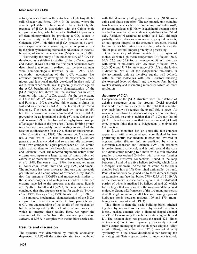
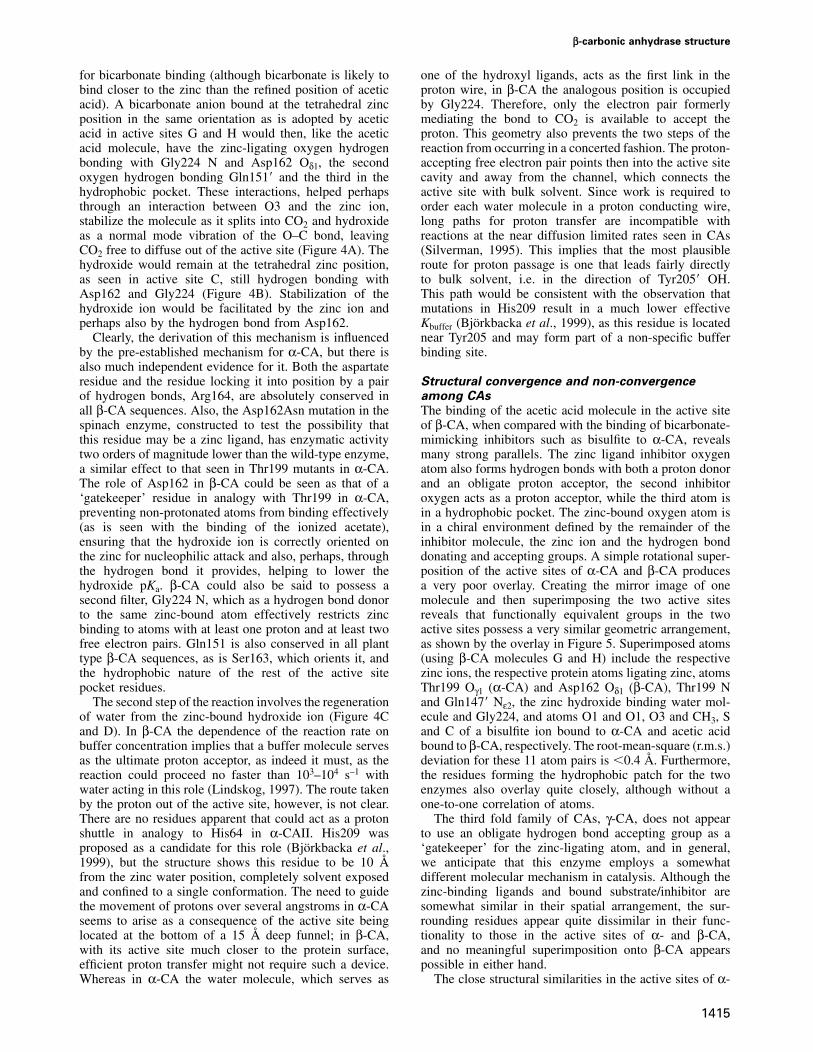
The β-CA monomer has an unusually non-compactappearance, with a wedge-shaped core flanked by twoprotruding motifs that mediate interactions involved inoligomerization (Figure 1A). As predicted by circulardichroism (Johansson and Forsman, 1993), the structureis predominantly α-helical, and is built around the coreof a dinucleotide-binding fold motif with a four-strandedparallel β-sheet ordered 2–1–3–4 with α-helices formingright-handed crossover connections. Found in the loopbetween β3 and β4 are five helices (α5–α9), which forma compact subdomain. At the end of strand β4 the chaindoubles back into a fifth C-terminal antiparallel β-strand.Pairs of monomers are joined up to form dimers throughan extensive interface that buries 27% (3255 of 12 119 Å2)of the monomer’s surface area (Figure 1B), a substantialportion of which is mediated by helices α1 and α2, whichform a finger that wraps most of the way around the secondmolecule. Strands β2 from each of the two monomers crossat a 60° angle in an antiparallel fashion forming a pair ofhydrogen bonds between residues 179 and 179� (num-bering as in Provart et al., 1993).
This dimer is then the basic building block stitchedtogether by interactions mediated by strand β5 into aloosely packed octamer with a diamond-shaped channelof ~35 � 15 Å running through the centre (Figure 1C andD). The octamer does not possess the usual 422 (dimerof tetramers) point group symmetry previously inferredfrom electron micrographs of the chickpea enzyme (Alievet al., 1986), but rather has 222 (dimer of dimers)symmetry with the above described dimer forming therepeating unit. This oligomerization arrangement is novel,
β-carbonic anhydrase structure
Fig. 1. Fold and oligomeric organization of β-CA. (A) Ribbon of the β-CA monomer, with secondary structure nomenclature as indicated. Thecolour is graded from blue to red, N-terminus to C-terminus. (B) Ribbon diagram of the β-CA dimer. One monomer is coloured as in (A), the otheris in white. Extensive contacts are mediated by helices α1 and α2 as they wrap around the secondary monomer. (C) Ribbon diagram of the β-CAoctamer. The octamer is assembled as a dimer of dimers of dimers. Arrows indicate the approximate position of the symmetry elements: redarrows, crystallographic 2-fold symmetry elements; black arrows, NCS elements that apply to the whole octamer; and cyan arrows, local non-crystallographic 2-fold axes that apply only to one dimer. Interactions between dimers are primarily mediated through strand β5. (D) Molecularsurface of the β-CA octamer. Each monomer is coloured differently to highlight the complex interweaving of molecules that occurs in octamerassembly, with each monomer contacting five other monomers. (E) View of half an octamer (the other half omitted for clarity) down thecrystallographic 2-fold axis. At the interface formed here, the dimer–dimer interaction buries a substantial portion of the molecular surface, althoughmost of the interactions are mediated by water molecules. (F) View of half an octamer as seen down a non-crystallographic 2-fold axis. Here, farless surface area is buried and almost all of the interactions are mediated by strand β5. The configuration of the monomers as seen here may bemapped onto that in Figure 1E by a 54° twist around a vector approximately colinear with the β5 strand.
with no precedent in the Research Collaboratory forStructural Bioinformatics (RBSC) protein structure data-base. The assembly of a complex with 222 point groupsymmetry from an object that possesses intrinsic 2-foldsymmetry necessitates that one molecular surface mediatestwo distinct types of interactions, much as is observed inthe assembly of hexameric and pentameric units in a viral
1409
capsid from a single structural protein. This phenomenonis seen here, where different interactions are observed atthe interfaces mediated by the crystallographic and thenon-crystallographic 2-fold axes (Figure 1C–F). Almostall protein–protein interactions responsible for formingthe octamer from dimers are mediated through the inter-action of strand β5, which pairs up in an antiparallel
M.S.Kimber and E.F.Pai
fashion with its equivalent in the second dimer in aninteraction strongly reminiscent of a strand exchange event.There is, however, no evidence that compact versions ofthe enzyme, where strand β5 folds back to form a sixthstrand, exist in any natural β-CA. The dimer–dimerpacking along the NCS axis is mediated almost entirelyby strand β5, with only 342 of the total 2504 Å2 buriedat this interface (total surface area for a pair of dimers is33 263 Å2) being mediated by other interactions. Althoughthe interface generated by the crystallographic 2-fold axisburies a substantial proportion of the monomer’s surface(compare Figure 1E and F), only 1472 Å2 is renderedsolvent inaccessible (this is beyond the β5–β5� interaction,which buries a further 2011 Å2), as almost all interactionsbetween the molecules are mediated through ordered watermolecules. One type of interface can be generated fromthe other by fixing one dimer and rotating the second 54°around an axis roughly colinear with the β-strands. Thismovement is accommodated by a slight twisting of theβ5–β5� strand at residues Leu319 and Leu323, where theβ5–β5� pair separates from β4.
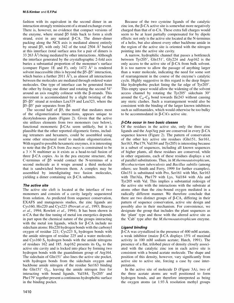
The second half of β5, the motif that mediates mostof the oligomerization interactions, appears unique todicotyledonous plants (Figure 2). Given that the activesite utilizes elements from two monomers (see below),functional monomeric β-CAs seem unlikely, but it isplausible that the other reported oligomeric forms, includ-ing tetramers and hexamers, could be assembled usingsome other structural motif to mediate oligomerization.With regard to possible hexameric enzymes, it is interestingto note that the β-CA from Zea mays is constrained to bea 3 � N multimer as it exists as a head-to-tail fusion ofthree β-CA copies. As in the pea enzyme structure, theC-terminus of β5 would contact the N-terminus of asecond molecule as it wraps around a third moleculethat is sandwiched between them. A complex may beassembled by interdigitating two fusion molecules,yielding a dimer containing six β-CA subunits.
The active site
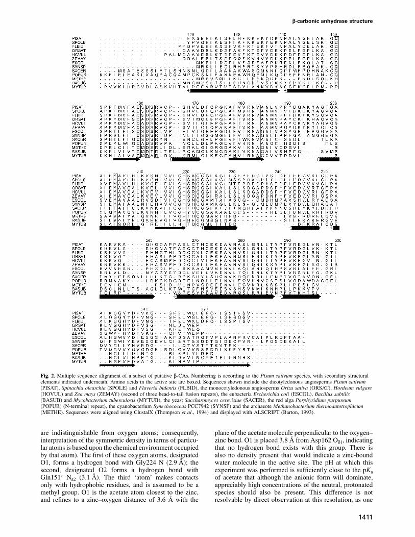
The active site cleft is located at the interface of twomonomers and consists of a cavity largely sequesteredfrom solution. As predicted from sequence conservation,EXAFS and mutagenesis studies, the zinc ligands areCys160, His220 and Cys223 (Provart et al., 1993; Braceyet al., 1994; Rowlett et al., 1994). It has been shown inα-CA that the fine tuning of metal ion energetics dependsin part upon the chemical nature of the groups interactingwith the metal ion ligands, which for β-CA include nosidechain atoms: His220 hydrogen bonds with the carbonyloxygen of residue 221; Cys223 Sγ hydrogen bonds withthe amide nitrogen of residue 225 and a water molecule;and Cys160 Sγ hydrogen bonds with the amide nitrogensof residues 162 and 185. Asp162 presents its Oδ1 to theactive site cavity and is locked into place by forming twohydrogen bonds with the guanidinium group of Arg164.The sidechain of Gln151� also lines the active site pocket,with hydrogen bonds from the sidechain oxygen andbackbone amide nitrogen of the residue Ser163 bindingthe Gln151� Oε1, leaving the amide nitrogen free forinteracting with bound ligands. Val184, Tyr205� andPhe179� together present a continuous hydrophobic surfacein the binding pocket.
1410
Because of the two cysteine ligands of the catalyticzinc ion, the β-CA active site is somewhat more negativelycharged than that of α-CA. These extra full charges wouldseem to be at least partially compensated for by dipoleeffects: not only is the active site located at the N-terminusof a helix, but also almost every other backbone amide inthe region of the active site is oriented with the nitrogenpointing into the active site cavity.
A narrow, hydrophilic channel that passes a bottleneckbetween Tyr205�, Gln151�, Gly224 and Asp162 is theonly access to the active site of β-CA from bulk solvent.It is too narrow to allow the passage of anything largerthan a water molecule, indicating the need for some sortof rearrangement in the course of the enzyme’s catalyticcycle. Highly suggestive in this regard is the deep finger-like hydrophobic pocket lining the far edge of Tyr205�.This empty space would allow the widening of the solventaccess channel by rotating the Tyr205� sidechain 30°around the Cα–Cβ bond towards Ile243 without causingany steric clashes. Such a rearrangement would also beconsistent with the binding of the larger known inhibitorssuch as sulfonamides, which would otherwise be too bulkyto be accommodated in β-CA’s active site.
β-CAs occur in two basic classes
Of the residues in the active site, only the three zincligands and the Asp/Arg pair are conserved in every β-CAsequence known (Figure 2). The pattern of conservationof the other key active site residues (Gln151, Ser161,Ser163, Phe179, Val184 and Tyr205) is interesting becausein a subset of sequences, including all known sequencesof higher plants, all these residues are conserved, whilein other organisms, each of these residues displays a setof parallel substitutions. Thus, in M.thermoautotrophicum,Mycobacterium tuberculosis and Bacillus subtilis (amongothers; see Smith and Ferry, 1999 for further examples),Gln151 is substituted with Pro, Ser161 with Met, Ser163with Thr/Ala, Phe179 with Lys, Val184 with Ala andTyr205 with Val. This implies a substantial redesign ofthe active site with the interactions with the substrate atatoms other than the zinc-bound oxygen mediated in aradically different manner. We therefore conclude thatthere are two distinct groups of β-CA, differing in theirpattern of sequence conservation, active site design andpossibly also in their mechanism. For convenience, wedesignate the group that includes the plant sequences asthe ‘plant’ type and those with the altered active site asthe ‘Cab’ type after the M.thermoautotrophicum enzyme.
Ligand binding
β-CA was crystallized in the presence of 400 mM acetate,a weak inhibitor (maize β-CA displays 15% of maximalactivity in 100 mM sodium acetate; Hatch, 1991). Thepresence of a flat, trilobed piece of density closely associ-ated with the catalytic zinc ion in each active site isconsistent with a bound acetate molecule. The shape andposition of this density, however, vary significantly fromactive site to active site, forcing a case by case inter-pretation.
In the active site of molecule D (Figure 3A), two ofthe three acetate atoms are well positioned to formhydrogen bonds, and are therefore interpreted as beingthe oxygen atoms (at 1.93 Å resolution methyl groups
β-carbonic anhydrase structure
Fig. 2. Multiple sequence alignment of a subset of putative β-CAs. Numbering is according to the Pisum sativum species, with secondary structuralelements indicated underneath. Amino acids in the active site are boxed. Sequences shown include the dicotyledonous angiosperms Pisum sativum(PISAT), Spinachia olearchia (SPOLE) and Flaveria bidentis (FLBID), the monocotyledonous angiosperms Oriza sativa (ORSAT), Hordeum vulgare(HOVUL) and Zea mays (ZEMAY) (second of three head-to-tail fusion repeats), the eubacteria Escherichia coli (ESCOL), Bacillus subtilis(BASUB) and Mycobacterium tuberculosis (MYTUB), the yeast Saccharomyces cerevisiae (SACER), the red alga Porphyridium purpureum(POPUR) (N-terminal repeat), the cyanobacterium Synechococcus PCC7942 (SYNSP) and the archaeote Methanobacterium thermoautotrophicum(METHE). Sequences were aligned using ClustalX (Thompson et al., 1994) and displayed with ALSCRIPT (Barton, 1993).
are indistinguishable from oxygen atoms; consequently,interpretation of the symmetric density in terms of particu-lar atoms is based upon the chemical environment occupiedby that atom). The first of these oxygen atoms, designatedO1, forms a hydrogen bond with Gly224 N (2.9 Å); thesecond, designated O2 forms a hydrogen bond withGln151� Nε2 (3.1 Å). The third ‘atom’ makes contactsonly with hydrophobic residues, and is assumed to be amethyl group. O1 is the acetate atom closest to the zinc,and refines to a zinc–oxygen distance of 3.6 Å with the
1411
plane of the acetate molecule perpendicular to the oxygen–zinc bond. O1 is placed 3.8 Å from Asp162 Oδ1, indicatingthat no hydrogen bond exists with this group. There isalso no density present that would indicate a zinc-boundwater molecule in the active site. The pH at which thisexperiment was performed is sufficiently close to the pKaof acetate that although the anionic form will dominate,appreciably high concentrations of the neutral, protonatedspecies should also be present. This difference is notresolvable by direct observation at this resolution, as one
M.S.Kimber and E.F.Pai
1412

β-carbonic anhydrase structure
Fig. 4. Proposed mechanism for β-CA. (A) Bicarbonate bound in the active site, in a position similar to that seen for the acetic acid molecule,forming hydrogen bonds with Gly224, Asp162 and Glu151. (B) This molecule decomposes into CO2, which continues to interact with Gln151� andhydroxide. (C) CO2 diffuses out of the active site leaving a hydroxide ion bound to the zinc. This ion then accepts a proton from a buffer moleculein bulk solvent, leaving water bound at the active site zinc (D).
cannot distinguish charged from uncharged states or thepresence from the absence of protons. Because O1 issignificantly displaced away from the hydrogen bondacceptor Asp162, and no compensating interaction isformed, we interpret the ligand in this active site to bethe unprotonated acetate ion.
Similar density is observed in the active site of moleculeC, where this density coexists with a strong (2.0σ) densitypeak closely associated with the zinc ion. The acetatedensity lends itself to the same interpretation as above,with essentially the same binding geometry, while theextra peak, occupying the fourth tetrahedral zinc positionand well resolved from the acetate density, is interpretedas a water molecule/hydroxide ion. Refined as a watermolecule, it occupies a position where it can form hydrogenbonds with both Gly224 N (3.3 Å) and Asp162 Oδ1 (2.9 Å).
In the active sites of molecules G (Figure 3B) and H,the inhibitor’s O1 is 2.5 Å from the zinc ion, completingthe canonical tetrahedral geometry with respect to theother zinc ligands. This oxygen atom forms hydrogenbonds with both Asp162 Oδ1 (3.0 Å) and Gly119 N (3.1 Å)with good tetrahedral geometry. O2 hydrogen bonds withGln151� Nε2 (2.9 Å) and its second free electron pair
Fig. 3. Ligand binding in various of the active sites of β-CA. (A) Stereo picture of the active site of molecule D showing σA-weighted electrondensity for the acetate ion. The map is contoured at 1.5σ (blue) and 8σ (orange). (B) Stereo picture of the active site of molecule G showingσA-weighted electron density for the acetic acid molecule. The map is contoured at 1.5σ (blue) and 8σ (orange). (C) Binding mode of acetic acid inthe β-CA active site, with zinc–ligand interactions shown as solid lines and hydrogen bonds as dashed lines. The binding site is composed of twomolecules, with one molecule shown in green and the other one in brown.
1413
points at the edge of the ring of Phe179� (3.4 Å). Themethyl group makes van der Waals interactions with thesidechains of Phe179�, Val184 and Tyr205�. Since in orderto form the hydrogen bond with both Asp162 Oδ1 andGly119 N, O1 would need to be protonated and sp3
hybridized, we infer that this molecule is acetic acid.It should be noted that the zinc–oxygen distance
observed in molecules G and H is significantly longerthan that generally observed for zinc–oxygen bonds (1.95–2.1 Å). Although this may be real, perhaps a consequenceof the neutral nature of the ligating species, it is morelikely that the density in these active sites is a superpositionof two different conformations, a major one with aceticacid bound closely to the zinc, and a minor one withacetate bound, similar to molecule D. The inhibitor wouldthen refine to a position that is the weighted averageof these two, resulting in the zinc–O1 distance beingoverestimated. In general, the ligand position may bebiased away from the zinc, and distances observed forthese two active sites should be interpreted with caution.
The higher temperature factors associated with the otherfour molecules in the asymmetric unit cause the densityto be less defined. Generally, the inhibitor molecules in
M.S.Kimber and E.F.Pai
Fig. 5. Active site overlay of α-CA and β-CA. Superimposition of the mirror image of the active site of α-CA with bisulfite bound (RCSB id code5cac) (Håkansson et al., 1992) in plum onto the active site of β-CA with acetic acid bound in beige. The atoms used in the overlay include therespective zinc divalent cations; the zinc ligands His96 Nε2, His94 Nε2 and His119 Nδ2 (α-CA) versus His120 Nε2, Cys223 Sγ and Cys160 Sγ(β-CA); bisulfite atoms S, O2 and O3 and the acetic acid atoms C, O1 and CH3; among inhibitor/substrate interacting moieties, Thr199 Oγ1 onAsp162 Oδ2 and Thr199 N on Gln147� Nε2, water on Gly224 N. This superposition also maps the residues of the respective hydrophobic patches,Phe179�, Val184 and Tyr205� versus Val121, Val143, Leu198 and Trp209, approximately onto one another, although for these atoms there are not thesame clear one-to-one-correspondences.
these active sites refine to positions equivalent to those inmolecules G and H.
The observation of distinct binding modes in differentactive sites in the asymmetric unit implies the existenceof some mechanism by which residues in a given activesite can sense their crystallographic environment (asotherwise there would be no correlation between succes-sive unit cells and one would simply observe the averagedensity). Superposition of all four well defined bindingsites, however, reveals no significant differences consist-ently associated with a particular binding mode. Havinga zinc ion with only three strong ligands, as is observedhere in monomer D, would be anticipated to be relativelyunstable and has never been reported for any of the α-CAor γ-CA complex structures. Possibly the ionic acetate isable to compete with acetic acid for binding in part dueto stabilizing electrostatic interactions with the zinc ion,but it seems likely that it is only the high concentrationof the acetate ion at this pH that allows this binding modeto be observed.
Proposed model for β-CA catalysis
Before discussing potential mechanisms for β-CA, it isuseful to review what several decades of detailed studieshave taught us about the same reaction as it occurs inthe analogous enzyme α-CA. Here, the most importantfunctional group is the zinc ion, which is coordinated bythree histidyl ligands with the fourth position availablefor the binding of water. The zinc-bound water alsohydrogen bonds with a second water molecule and Thr199Oγ1. The latter group acts as a hydrogen bond donor withrespect to Glu106, and therefore allows only atoms capableof acting as hydrogen bond donors to bind to the zinc ionwith tetrahedral geometry. Thr199 acts as a filter and isoften called the ‘gatekeeper’ residue. Together with
1414
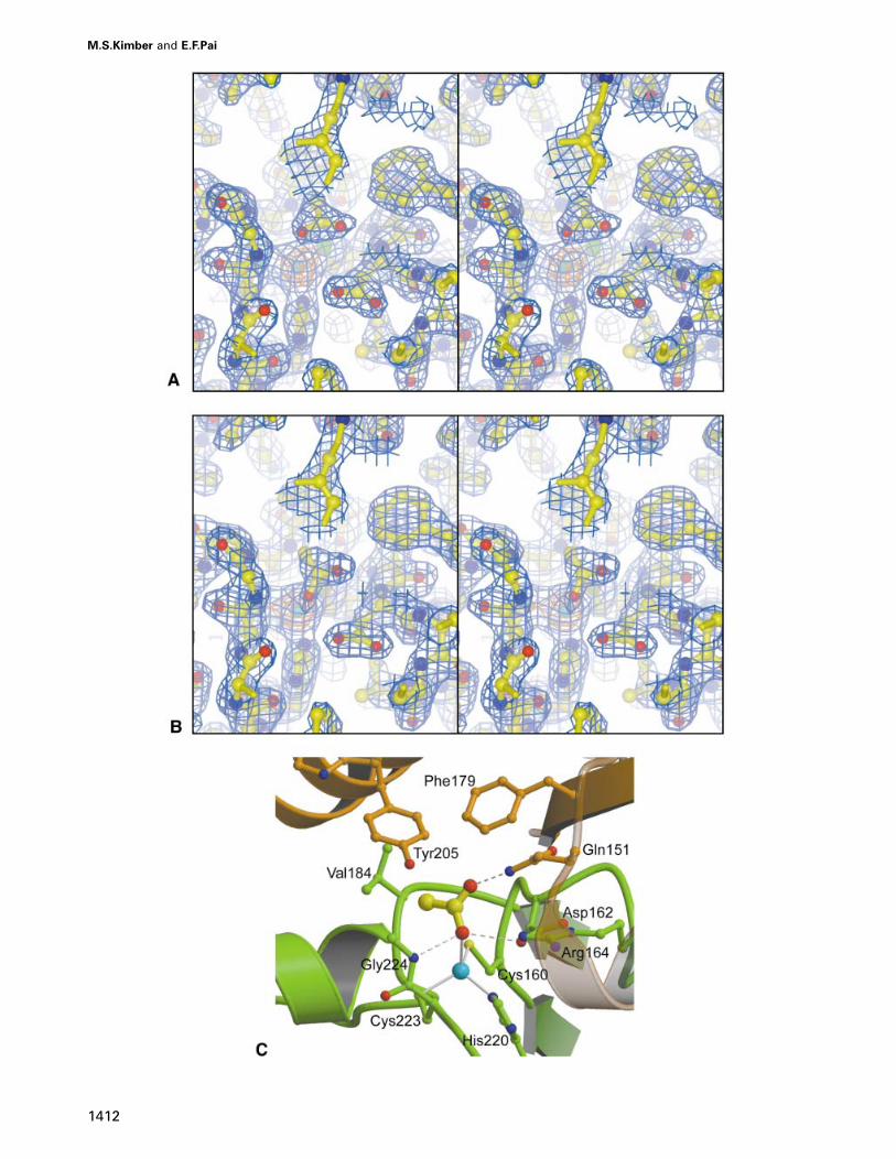
Glu106, it is absolutely conserved and enzymes mutatedin these positions show 100-fold reduced activity. Thezinc ion acts as a Lewis acid, which, with some help fromthe hydrogen bond provided by Thr199, lowers the pKaof water from 15.8 to ~7, allowing the generation of astable OH– ion at physiological pH. The water molecule,which forms the third ligand for the zinc-bound water, isthe first member of a proton wire conducting a proton toHis64, which in turn acts as a temporary way station forthe proton prior to its transfer to a buffer molecule.This residue adopts alternative conformations with theimidazole group close to the zinc for accepting the protonand then more solvent exposed to allow transfer to abuffer species in bulk solution; for this reason, it is knownas the proton shuttle. His64 is found only in a subset ofα-CAs, including α-CAII, and is critical for fast turnover.The OH– ion is the nucleophile that attacks the electrophilicCO2 carbon. CO2 binding is very weak and the modeof interaction is not well understood. The extensivehydrophobic patch consisting of Val121, Val143, Leu198and Trp209 has been proposed to play a role, and thebackbone amide nitrogen of Thr199, which forms ahydrogen bond to bicarbonate analogues, is believed toalso hydrogen bond with CO2 helping to electrophilicallyactivate the CO2 molecule. The product, bicarbonate, isbound through the same interactions: the zinc-boundoxygen is protonated and hydrogen bonds with Thr199Oγ1 and a water molecule; the second oxygen hydrogenbonds with Thr199 N; and the third oxygen is buried in thehydrophobic pocket (Liljas et al., 1994; Lindskog, 1997).
Since acetic acid and bicarbonate are isoelectronicexcept at the methyl group, which, in any case, is buriedin a hydrophobic pocket, the interactions made by the twospecies in the active site are highly similar and so aceticacid binding (Figure 3C) would seem a reasonable model
β-carbonic anhydrase structure
for bicarbonate binding (although bicarbonate is likely tobind closer to the zinc than the refined position of aceticacid). A bicarbonate anion bound at the tetrahedral zincposition in the same orientation as is adopted by aceticacid in active sites G and H would then, like the aceticacid molecule, have the zinc-ligating oxygen hydrogenbonding with Gly224 N and Asp162 Oδ1, the secondoxygen hydrogen bonding Gln151� and the third in thehydrophobic pocket. These interactions, helped perhapsthrough an interaction between O3 and the zinc ion,stabilize the molecule as it splits into CO2 and hydroxideas a normal mode vibration of the O–C bond, leavingCO2 free to diffuse out of the active site (Figure 4A). Thehydroxide would remain at the tetrahedral zinc position,as seen in active site C, still hydrogen bonding withAsp162 and Gly224 (Figure 4B). Stabilization of thehydroxide ion would be facilitated by the zinc ion andperhaps also by the hydrogen bond from Asp162.
Clearly, the derivation of this mechanism is influencedby the pre-established mechanism for α-CA, but there isalso much independent evidence for it. Both the aspartateresidue and the residue locking it into position by a pairof hydrogen bonds, Arg164, are absolutely conserved inall β-CA sequences. Also, the Asp162Asn mutation in thespinach enzyme, constructed to test the possibility thatthis residue may be a zinc ligand, has enzymatic activitytwo orders of magnitude lower than the wild-type enzyme,a similar effect to that seen in Thr199 mutants in α-CA.The role of Asp162 in β-CA could be seen as that of a‘gatekeeper’ residue in analogy with Thr199 in α-CA,preventing non-protonated atoms from binding effectively(as is seen with the binding of the ionized acetate),ensuring that the hydroxide ion is correctly oriented onthe zinc for nucleophilic attack and also, perhaps, throughthe hydrogen bond it provides, helping to lower thehydroxide pKa. β-CA could also be said to possess asecond filter, Gly224 N, which as a hydrogen bond donorto the same zinc-bound atom effectively restricts zincbinding to atoms with at least one proton and at least twofree electron pairs. Gln151 is also conserved in all planttype β-CA sequences, as is Ser163, which orients it, andthe hydrophobic nature of the rest of the active sitepocket residues.
The second step of the reaction involves the regenerationof water from the zinc-bound hydroxide ion (Figure 4Cand D). In β-CA the dependence of the reaction rate onbuffer concentration implies that a buffer molecule servesas the ultimate proton acceptor, as indeed it must, as thereaction could proceed no faster than 103–104 s–1 withwater acting in this role (Lindskog, 1997). The route takenby the proton out of the active site, however, is not clear.There are no residues apparent that could act as a protonshuttle in analogy to His64 in α-CAII. His209 wasproposed as a candidate for this role (Bjorkbacka et al.,1999), but the structure shows this residue to be 10 Åfrom the zinc water position, completely solvent exposedand confined to a single conformation. The need to guidethe movement of protons over several angstroms in α-CAseems to arise as a consequence of the active site beinglocated at the bottom of a 15 Å deep funnel; in β-CA,with its active site much closer to the protein surface,efficient proton transfer might not require such a device.Whereas in α-CA the water molecule, which serves as
1415
one of the hydroxyl ligands, acts as the first link in theproton wire, in β-CA the analogous position is occupiedby Gly224. Therefore, only the electron pair formerlymediating the bond to CO2 is available to accept theproton. This geometry also prevents the two steps of thereaction from occurring in a concerted fashion. The proton-accepting free electron pair points then into the active sitecavity and away from the channel, which connects theactive site with bulk solvent. Since work is required toorder each water molecule in a proton conducting wire,long paths for proton transfer are incompatible withreactions at the near diffusion limited rates seen in CAs(Silverman, 1995). This implies that the most plausibleroute for proton passage is one that leads fairly directlyto bulk solvent, i.e. in the direction of Tyr205� OH.This path would be consistent with the observation thatmutations in His209 result in a much lower effectiveKbuffer (Bjorkbacka et al., 1999), as this residue is locatednear Tyr205 and may form part of a non-specific bufferbinding site.
Structural convergence and non-convergence
among CAs
The binding of the acetic acid molecule in the active siteof β-CA, when compared with the binding of bicarbonate-mimicking inhibitors such as bisulfite to α-CA, revealsmany strong parallels. The zinc ligand inhibitor oxygenatom also forms hydrogen bonds with both a proton donorand an obligate proton acceptor, the second inhibitoroxygen acts as a proton acceptor, while the third atom isin a hydrophobic pocket. The zinc-bound oxygen atom isin a chiral environment defined by the remainder of theinhibitor molecule, the zinc ion and the hydrogen bonddonating and accepting groups. A simple rotational super-position of the active sites of α-CA and β-CA producesa very poor overlay. Creating the mirror image of onemolecule and then superimposing the two active sitesreveals that functionally equivalent groups in the twoactive sites possess a very similar geometric arrangement,as shown by the overlay in Figure 5. Superimposed atoms(using β-CA molecules G and H) include the respectivezinc ions, the respective protein atoms ligating zinc, atomsThr199 Oγ1 (α-CA) and Asp162 Oδ1 (β-CA), Thr199 Nand Gln147� Nε2, the zinc hydroxide binding water mol-ecule and Gly224, and atoms O1 and O1, O3 and CH3, Sand C of a bisulfite ion bound to α-CA and acetic acidbound to β-CA, respectively. The root-mean-square (r.m.s.)deviation for these 11 atom pairs is �0.4 Å. Furthermore,the residues forming the hydrophobic patch for the twoenzymes also overlay quite closely, although without aone-to-one correlation of atoms.
The third fold family of CAs, γ-CA, does not appearto use an obligate hydrogen bond accepting group as a‘gatekeeper’ for the zinc-ligating atom, and in general,we anticipate that this enzyme employs a somewhatdifferent molecular mechanism in catalysis. Although thezinc-binding ligands and bound substrate/inhibitor aresomewhat similar in their spatial arrangement, the sur-rounding residues appear quite dissimilar in their func-tionality to those in the active sites of α- and β-CA,and no meaningful superimposition onto β-CA appearspossible in either hand.
The close structural similarities in the active sites of α-
M.S.Kimber and E.F.Pai
and β-CAs lead us to conclude that these two enzymeshave convergently evolved to a remarkable degree.Coupled with the close matches in the biochemical dataavailable for the two enzymes, we further conclude thatthe enzymes are also likely to utilize a very similarmechanism. They should then serve as excellent modelsfor one another and detailed comparisons of the results ofexperiments performed in parallel on the two should helpadvance understanding of both. Having such a pair of‘twins’ is extremely useful, as experiments difficult tocarry out in one system may prove more tractable in theother. For example, it has been suggested that a hydrogenbond between CO2 and Thr199 N in α-CA is needed forelectrophilic activation of CO2 (Håkansson et al., 1992),but as the interaction is mediated by a mainchain atomnot only are sequence conservation arguments inapplic-able, but also the issue is unresolvable by mutagenesisstudies. Not only does the finding of a similar motif inthe (conserved) Gln151 sidechain amide of β-CA helplend credence to the idea that such a group has somefunctional role, but also the fact that the analogoussubstrate–enzyme interaction is mediated by a sidechainmeans that the exact functional role should now beamenable to experimental inquiry.
Similar motifs found in a pair of convergently evolvedenzymes highlight them as being of potential functionalimportance in much the same way as sequence conserva-tion within an enzyme family does. Here, one feature bothconserved in and converged upon by α-CA and plant typeβ-CA (and also to a lesser degree in γ-CA) is the extensiveactive site hydrophobic patch. In α-CA, these residueshave been proposed to perform many functions, includingforming a CO2 binding site or in some way helpingturnover (by excluding solvent, stabilizing the transitionstate, etc.). However, given that α-CA binds CO2 with103- to 104-fold lower affinity than RuBisCO, althoughthe hydrophobic residues may bind CO2 they are not likelyto be optimized for this function. While it is true thatmutagenesis of these amino acids to charged or largeramino acids is very disruptive to α-CAII turnover, somemutations to hydrophilic residues of similar volume areonly moderately disruptive. For example, the Val121Ser,Val143Asn and Leu198Ser mutants show approximately3-, 3- and 5-fold reduction in CO2 hydration activity,respectively (Fierke et al., 1991; Nair et al., 1991; Krebset al., 1993). Since some α-CA isozymes are 104-fold lessactive than α-CAII, disruptions of this magnitude shouldbe accommodated in the α-CA active site, yet even in theleast active isozymes the hydrophobic nature of theseresidues is absolutely conserved. This implies a verystrong selective pressure against hydrophilic residues inthese positions, with selective pressure not being primarilyfor maximal turnover. We propose that substrate discrim-ination is what is exerting this pressure.
Most enzymes act on substrates with a large number ofatoms, most of which are not affected during the catalyticcycle and so afford multiple potential strong and unalteringinteractions, which allows for clear energetic discrimin-ation between substrates and non-substrates. Not only isCA’s substrate small, but also the charge and hydrogenbonding behaviour of almost every atom changes duringthe catalytic cycle. This means that there are very fewfunctionalities, and even fewer consistent ones, with which
1416
the enzyme can make the strong interactions that wouldallow energetic discrimination between the substrate mol-ecules and other, potentially inhibitory molecules. Thefact that α-CA and β-CA (and to a lesser degree γ-CA)display appreciable inhibition constants for almost anysmall anion, including many present at appreciable levelsin normal cellular environments (Johansson and Forsman,1993; Alber and Ferry, 1994; Liljas et al., 1994), atteststo the fact that CAs have not fully solved this problem,and as such the overall productivity of the enzyme in vivois likely to be less constrained by the enzyme’s maximalpotential turnover (as measured in in vitro assays) thanby competitive inhibition by the anions present in thecellular environment. By minimizing the number of poten-tial hydrogen bond acceptors and donors around the zincion, the presence of the hydrophobic residues helps ensurethat the binding energy of such molecules is as unfavour-able as possible. Weak substrate binding is one necessaryconsequence of this, but since OH– is intrinsically highlynucleophilic and CO2 is intrinsically highly electrophilic,the bicarbonate–CO2 equilibration step remains faster thanproton transfer. Other structural features shared by CAswould also seem to play a role in maximizing discrimin-ation. The gatekeeper residue, Thr199 in α-CA and Asp162in β-CA, is set up as an obligatory hydrogen bond acceptornear the zinc ion, forcing unprotonated inhibitors to bindto the zinc ion with suboptimal geometry and thereforelower energy. Also, in all three CAs the active site isconfined and sequestered from bulk solvent. Since maximalturnover is limited by how fast protons can be passedto buffer molecules in bulk solvent, this might seemcounterproductive, but forcing molecules to approach theactive site through a restricted diameter passage couldhelp, by steric means, to filter out larger molecular anionsthat might otherwise also be inhibitory.
Materials and methods
Crystallization, data collection and processingProtein was purified and crystals grown as previously described(M.S.Kimber, J.R.Coleman and E.F.Pai, submitted). Briefly, the proteinwas crystallized by vapour diffusion against 16% polyethylene glycol(PEG) 4000 MW (Fluka), 400 mM ammonium acetate, 50 mM dithio-threitol and 100 mM sodium citrate pH 5.0 at 4°C. All data werecollected with crystals flash-frozen at 100 K in artificial mother liquorwith 25% PEG 4000 and 30% ethylene glycol as cryoprotectant. Crystalswere of the orthorhombic space group C222 with cell parametersa � 136.9, b � 143.3, c �202.1 Å and α � β � γ � 90°. The crystalswere generally highly anisotropic in their scattering. A three wavelengthMAD data set was collected at the National Synchrotron Light Source,beamline X8-C, using an MAR345 area detector. The 1.93 Å highresolution data set was collected at the Advanced Photon Source beamline14C (BioCARS) with a Quantum Q4 CCD detector. All data werereduced using DENZO and scaled using SCALEPACK (Otwinowskiand Minor, 1997) (Table I).
Phase determinationThe structure was determined by MAD using the signal from the intrinsiczinc atoms only. The program SOLVE (Terwilliger and Berendzen, 1999)was used to find a solution to the heavy atom substructure using thethree wavelength MAD dataset at 3.8 Å. Eight zinc ions were foundwith high occupancies (0.85–1.05), yielding a figure of merit (FOM) of0.58. While elements of secondary structure were visible in the MADmap, connectivity was relatively poor and the map was unsuitable forchain tracing. The disposition of the zinc sites allowed initial determin-ation of the location and orientation of two of the NCS 2-fold axes.Phase improvement and extension to 2.7 Å was then performed in theCCP4 program DM (Cowtan and Main, 1996) using techniques including
β-carbonic anhydrase structure
Table I. Data collection statistics
λ1 (peak) λ2 (edge) λ3 (remote) Highresolution
Wavelength (Å) 1.281323 1.282716 1.00000 1.0000Resolution (Å) 30–2.7 30–2.8 30–2.7 40–1.93No. of observations 253 785 144 276 166 306 779 081No. unique 49 623 43 192 48 177 138 484Rsym
a 0.066 0.048 0.053 0.067(last shell) 0.309 0.179 0.212 0.277I/σ 12.2 15.6 13.8 13.0(last shell) 4.4 5.1 3.8 3.7Completeness 0.908 0.876 0.877 0.932(last shell) 0.839 0.722 0.679 0.785
aRsym � ΣhklΣi|Ii – �I�|/ΣhklΣi�I� where Ii is the ith measurement ofthe reflection intensity I and �I� is the weighted mean of allmeasurements of I.
Table II. Refinement statistics
Resolution range (Å) 40–1.93Rcryst (%)a 22.7Rfree (%)b 25.2R.m.s. deviations
bond lengths (Å) 0.012bond angles (°) 1.5torsion angles (°) 22improper torsion angles (°) 1.0
aRcryst � Σhkl|Fobs – Fcalc|/Fobs where Fobs and Fcalc are the observedand the calculated structure factors, respectively, and the summation isover the 99% of reflections used for model refinement.bRfree as for Rcryst except summed only over the 1% of reflections notused for model refinement.
solvent flattening and 3-fold NCS averaging with a sphere being usedas the NCS mask. The improvement in the map was sufficient to allowmuch of the secondary structure to be recognized in a skeletonizationand the accurate determination of the remaining NCS elements togetherwith the definition of a more accurate mask. Eight-fold symmetryaveraging, coupled with solvent flattening and histogram matching inDM produced a high quality map with an overall FOM of 0.78. Theprotein was then fully traced into this map using the molecular graphicsprogram O (Jones et al., 1991).
RefinementSimulated annealing torsion angle refinement against amplitude max-imum likelihood targets was performed in CNS version 0.5 (Brungeret al., 1998). Individual B-factor refinement was also used in thesame program. Rounds of refinement were interspersed with rounds ofrebuilding in O. The model was initially refined using strict NCS withthe dimer as asymmetric unit, which was later released to eight moleculeswith strong harmonic restraints. No NCS restraints were used in thefinal rounds of refinement. The current model contains residues 120–329, 117–329, 116–329, 120–329, 118–329, 116–329, 110–329 and119–329 for molecules A–H respectively, with no chain breaks. Theasymmetric unit also contains eight Zn2� ions, eight Cl– ions, four Cu2�
ions (at half occupancy, a tentative interpretation of a strong densitypeak between Cys166 and Cys166� at the monomer–monomer interface),eight acetate molecules and 894 water molecules (Table II). Figureswere prepared using SPOCK and MOLSCRIPT (Kraulis, 1991) andrendered using Raster3D (Merritt and Murphy, 1994).
Acknowledgements
The authors wish to thank Dr John R.Coleman for bringing this problemto our attention, for his generous gift of the pCA plasmid-containingcells and for helpful discussions. We are grateful to the staff at BNLX8-C and APS BioCARS for assistance with data collection. Thisresearch was supported by the National Science and Engineering Councilof Canada through an Industrial Research Chair to E.F.P. M.S.K. is therecipient of a Medical Research Council of Canada post-graduate student
1417
scholarship and a University of Toronto Open Scholarship. A joint grantfrom the Medical Research and National Sciences and EngineeringCouncils of Canada enabled use of beamline X8-C at the NationalSynchrotron Light Source. Use of the Advanced Photon Source wassupported by the US Department of Energy, Basic Energy Sciences,Office of Science, under contract No. W-31-109-Eng-38. Atomic coordin-ates have been deposited at the RCSB structure database, id 1EKJ.
References
Alber,B.E. and Ferry,J.G. (1994) A carbonic anhydrase from the archaeonMethanosarcina thermophila. Proc. Natl Acad. Sci. USA, 91, 6909–6913.
Aliev,D.A., Guliev,N.M., Mamedov,A.M. and Tsuprun,V.L. (1986)Physico-chemical properties and quaternary structure of carbonicanhydrase from Cicer arietinum leaves. Biokhimiya, 51, 1785–1794.
Badger,M.R. and Price,G.D. (1994) The role of carbonic anhydrase inphotosynthesis. Annu. Rev. Plant Physiol. Plant Mol. Biol., 45,369–392.
Barton,G.J. (1993) ALSCRIPT: a tool to format multiple sequencealignments. Protein Eng., 6, 37–40.
Bjorkbacka,H., Johansson,I.M. and Forsman,C. (1999) Possible roles forHis 208 in the active-site region of chloroplast carbonic anhydrasefrom Pisum sativum. Arch. Biochem. Biophys., 361, 17–24.
Bracey,M.H., Christiansen,J., Tovar,P., Cramer,S.P. and Bartlett,S.G.(1994) Spinach carbonic anhydrase: investigation of the zinc-bindingligands by site-directed mutagenesis, elemental analysis and EXAFS.Biochemistry, 33, 13126–13131.
Brunger,A.T. et al. (1998) Crystallography & NMR system: a newsoftware suite for macromolecular structure determination. ActaCrystallogr. D, 54, 905–921.
Cowtan,K.D. and Main,P. (1996) Phase combination and cross-validationin iterated density modification calculations. Acta Crystallogr. D, 52,43–48.
Fawcett,T.W., Browse,J.A., Volokita,M. and Bartlett,S.G. (1990) Spinachcarbonic anhydrase primary structure deduced from the sequence ofa cDNA clone. J. Biol. Chem., 265, 5414–5417.
Fierke,C.A., Calderone,T.L. and Krebs,J.F. (1991) Functionalconsequences of engineering the hydrophobic pocket of carbonicanhydrase II. Biochemistry, 30, 11054–11063.
Guilloton,M.B., Korte,J.J., Lamblin,A.F., Fuchs,J.A. and Anderson,P.M.(1992) Carbonic anhydrase in Escherichia coli. A product of the cynoperon. J. Biol. Chem., 267, 3731–3734.
Håkansson,K., Carlsson,M., Svensson,L.A. and Liljas,A. (1992) Structureof native and apo carbonic anhydrase II and structure of some of itsanion–ligand complexes. J. Mol. Biol., 227, 1192–1204.
Hatch,M.D. (1991) Carbonic anhydrase assay: strong inhibition of theleaf enzyme by CO2 in certain buffers. Anal. Biochem., 192, 85–89.
Hiltonen,T., Bjorkbacka,H., Forsman,C., Clarke,A.K. and Samuelsson,G.(1998) Intracellular β-carbonic anhydrase of the unicellular greenalga Coccomyxa. Cloning of the cDNA and characterization of thefunctional enzyme overexpressed in Escherichia coli. Plant Physiol.,117, 1341–1349.
Jebanathirajah,J.A. and Coleman,J.R. (1998) Association of carbonicanhydrase with a Calvin cycle enzyme complex in Nicotiana tabacum.Planta, 204, 177–182.
Johansson,I.M. and Forsman,C. (1992) Processing of the chloroplasttransit peptide of pea carbonic anhydrase in chloroplasts and inEscherichia coli. Identification of two cleavage sites. FEBS Lett., 314,232–236.
Johansson,I.M. and Forsman,C. (1993) Kinetic studies of pea carbonicanhydrase. Eur. J. Biochem., 218, 439–446.
Johansson,I.M. and Forsman,C. (1994) Solvent hydrogen isotope effectsand anion inhibition of CO2 hydration catalysed by carbonic anhydrasefrom Pisum sativum. Eur. J. Biochem., 224, 901–907.
Jones,T.A., Zou,J.Y., Cowan,S.W. and Kjeldgaard,M. (1991) Improvedmethods for binding protein models in electron density maps and thelocation of errors in these models. Acta Crystallogr. A, 47, 110–119.
Kandel,M., Gornall,A.G., Cybulsky,D.L. and Kandel,S.I. (1978)Carbonic anhydrase from spinach leaves. Isolation and some chemicalproperties. J. Biol. Chem., 253, 679–685.
Kisker,C., Schindelin,H., Alber,B.E., Ferry,J.G. and Rees,D.C. (1996) Aleft-hand β-helix revealed by the crystal structure of a carbonicanhydrase from the archaeon Methanosarcina thermophila. EMBO J.,15, 2323–2330.
Kraulis,P.J. (1991) MOLSCRIPT: a program to produce both detailed
M.S.Kimber and E.F.Pai
and schematic plots of protein structures. J. Appl. Crystallogr., 24,946–950.
Krebs,J.F., Rana,F., Dluhy,R.A. and Fierke,C.A. (1993) Kinetic andspectroscopic studies of hydrophilic amino acid substitutions in thehydrophobic pocket of human carbonic anhydrase II. Biochemistry,32, 4496–4505.
Liljas,A. et al. (1972) Crystal structure of human carbonic anhydrase C.Nature New Biol., 235, 131–137.
Liljas,A., Håkansson,K., Jonsson,B.H. and Xue,Y. (1994) Inhibition andcatalysis of carbonic anhydrase. Recent crystallographic analyses. Eur.J. Biochem., 219, 1–10.
Lindskog,S. (1997) Structure and mechanism of carbonic anhydrase.Pharmacol. Ther., 74, 1–20.
Majeau,N., Arnoldo,M.A. and Coleman,J.R. (1994) Modification ofcarbonic anhydrase activity by antisense and over-expressionconstructs in transgenic tobacco. Plant Mol. Biol., 25, 377–385.
Merritt,E.A. and Murphy,M.E.P. (1994) Raster3D version 2.0: a programfor photorealistic molecular graphics. Acta Crystallogr. D, 50, 869–873.
Nair,S.K., Calderone,T.L., Christianson,D.W. and Fierke,C.A. (1991)Altering the mouth of a hydrophobic pocket. Structure and kineticsof human carbonic anhydrase II mutants at residue Val-121. J. Biol.Chem., 266, 17320–17325.
Otwinowski,Z. and Minor,W. (1997) Processing of X-ray diffractiondata collected in oscillation mode. Methods Enzymol., 276, 307–326.
Provart,N.J., Majeau,N. and Coleman,J.R. (1993) Characterization ofpea chloroplastic carbonic anhydrase. Expression in Escherichia coliand site-directed mutagenesis. Plant Mol. Biol., 22, 937–943.
Ren,X. and Lindskog,S. (1992) Buffer dependence of CO2 hydrationcatalyzed by human carbonic anhydrase I. Biochim. Biophys. Acta,1120, 81–86.
Rowlett,R.S., Chance,M.R., Wirt,M.D., Sidelinger,D.E., Royal,J.R.,Woodroffe,M., Wang,Y.F., Saha,R.P. and Lam,M.G. (1994) Kineticand structural characterization of spinach carbonic anhydrase.Biochemistry, 33, 13967–13976.
Rumeau,D., Cuine,S., Fina,L., Gault,N., Nicole,M. and Peltier,G. (1996)Subcellular distribution of carbonic anhydrase in Solanum tuberosumL. leaves: characterization of two compartment-specific isoforms.Planta, 199, 79–88.
Silverman,D.N. (1995) Proton transfer in carbonic anhydrase measuredby equilibrium isotope exchange. Methods Enzymol., 249, 479–503.
Smith,K.S. and Ferry,J.G. (1999) A plant-type (β-class) carbonicanhydrase in the thermophilic methanoarchaeon Methanobacteriumthermoautotrophicum. J. Bacteriol., 181, 6247–6253.
Terwilliger,T.C. and Berendzen,J. (1999) Automated MAD and MIRstructure solution. Acta Crystallogr. D, 55, 849–861.
Thompson,J.D., Higgins,D.G. and Gibson,T.J. (1994) CLUSTAL W:improving the sensitivity of progressive multiple sequence alignmentthrough sequence weighting, position-specific gap penalties and weightmatrix choice. Nucleic Acids Res., 22, 4673–4680.
Received December 22, 1999; revised February 7, 2000;accepted February 9, 2000
1418


![Calcareous sponge genomes reveal complex -carbonic … · 2017. 8. 29. · or characterize CA-proteins from the calcareous sponge S. ciliatum have not been successful [22]. Only recently,](https://static.fdocument.org/doc/165x107/60d35117c3bc180d086fdbcc/calcareous-sponge-genomes-reveal-complex-carbonic-2017-8-29-or-characterize.jpg)


![The Effects of Pharmacological Carbonic Anhydrase ...S-nitrosylation targets upon infection with the oomycete Phytophthora infestans [14]. Additionally, it is worth noting that the](https://static.fdocument.org/doc/165x107/60f89da2a24b6b558f15cb7b/the-effects-of-pharmacological-carbonic-anhydrase-s-nitrosylation-targets-upon.jpg)