TGF-β1 polymorphisms and familial aggregation of liver ... · PDF fileTGF-β1...
14
©FUNPEC-RP www.funpecrp.com.br Genetics and Molecular Research 14 (3): 8147-8160 (2015) TGF-β1 polymorphisms and familial aggregation of liver cancer in Guangxi, China P.Q. Wan, J.Z. Wu, L.Y. Huang, J.L. Wu, Y.H. Wei and Q.Y. Ning Department of Infectious Disease, The First Affiliated Hospital of Guangxi Medical University, Nanning, Guangxi Zhuang Autonomous Region, China Corresponding author: J.Z. Wu E-mail: [email protected] Genet. Mol. Res. 14 (3): 8147-8160 (2015) Received July 16, 2014 Accepted January 13, 2015 Published July 27, 2015 DOI http://dx.doi.org/10.4238/2015.July.27.3 ABSTRACT. The goal of present study was to investigate the relationship between polymorphisms of TGF-β1 and familial aggregation of liver cancer in Guangxi Zhuang, Han, and Yao populations. We conducted a population-based case-control family study of liver cancer in Guanxi, China. A total of 214 individuals from 37 case families were surveyed for polymorphisms in TGF-β1. We genotyped six functional TGF-β1 polymorphisms: rs1800469, rs2241715, rs2241716, rs11466345, rs8105161, and rs747857. Levels of TGF-β1, hepatitis B surface antigen, and anti-hepatitis C virus in all serum samples were detected using the enzyme-linked immunoassay method, and presence of hepatitis B virus (HBV) DNA was determined using polymerase chain reaction amplification. A standardized questionnaire was used to collect information from subjects, including alcohol consumption, smoking, eating, and water drinking habits. The results were compared with those from 214 control individuals. The results showed that the TGF-β1 genotypes rs1800469, rs2241715, rs2241715, and rs8105161 were more frequent in patients than in controls. The risk factors for familial aggregation of liver cancer in Guangxi were determined, from high to low, to be: drinking sugared beverages > alcohol consumption > HBV
Transcript of TGF-β1 polymorphisms and familial aggregation of liver ... · PDF fileTGF-β1...
©FUNPEC-RP www.funpecrp.com.brGenetics and Molecular Research 14 (3): 8147-8160 (2015)
TGF-β1 polymorphisms and familial aggregation of liver cancer in Guangxi, China
P.Q. Wan, J.Z. Wu, L.Y. Huang, J.L. Wu, Y.H. Wei and Q.Y. Ning
Department of Infectious Disease, The First Affiliated Hospital of Guangxi Medical University, Nanning, Guangxi Zhuang Autonomous Region, China
Corresponding author: J.Z. WuE-mail: [email protected]
Genet. Mol. Res. 14 (3): 8147-8160 (2015)Received July 16, 2014Accepted January 13, 2015Published July 27, 2015DOI http://dx.doi.org/10.4238/2015.July.27.3
ABSTRACT. The goal of present study was to investigate the relationship between polymorphisms of TGF-β1 and familial aggregation of liver cancer in Guangxi Zhuang, Han, and Yao populations. We conducted a population-based case-control family study of liver cancer in Guanxi, China. A total of 214 individuals from 37 case families were surveyed for polymorphisms in TGF-β1. We genotyped six functional TGF-β1 polymorphisms: rs1800469, rs2241715, rs2241716, rs11466345, rs8105161, and rs747857. Levels of TGF-β1, hepatitis B surface antigen, and anti-hepatitis C virus in all serum samples were detected using the enzyme-linked immunoassay method, and presence of hepatitis B virus (HBV) DNA was determined using polymerase chain reaction amplification. A standardized questionnaire was used to collect information from subjects, including alcohol consumption, smoking, eating, and water drinking habits. The results were compared with those from 214 control individuals. The results showed that the TGF-β1 genotypes rs1800469, rs2241715, rs2241715, and rs8105161 were more frequent in patients than in controls. The risk factors for familial aggregation of liver cancer in Guangxi were determined, from high to low, to be: drinking sugared beverages > alcohol consumption > HBV
8148P.Q. Wan et al.
©FUNPEC-RP www.funpecrp.com.brGenetics and Molecular Research 14 (3): 8147-8160 (2015)
DNA-positive > rs1800469 TT homozygous genotype > rs2241715 TT homozygous genotype. The results suggested that TGF-β1 rs1800469 TT and rs2241715 TT homozygote genotypes represent the genetic factors underlying familial clustering of liver cancer in Guangxi, and that drinking water use, alcohol consumption, and testing positive for HBV DNA are the main environmental factors contributing to familial aggregation of liver cancer in Guangxi.
Key words: Transforming growth factor-β1; Gene polymorphism; Liver cancer; Familial aggregation; Risk factors; Chinese population
INTRODUCTION
In humans, the transforming growth factor-β1 (TGF-β1) gene is located on chromo-some 19q13, and contains seven exons and six introns. The TGF-β1 polypeptide is a member of the TGF-β superfamily of cytokines. It is a secreted protein that performs many cellu-lar functions, including the control of cell growth, cell proliferation, cell differentiation, and apoptosis. TGF-β1 plays an important role in controlling the immune system, and shows dif-ferent activities on different types of cells, or on cells at different developmental stages.
Since the expression levels of TGF-β1 are assumed to be predominantly under ge-netic control, predisposition to disease might be associated with distinct allelic changes at the TGF-β1 locus (Park et al., 2006; Shah et al., 2006a,b; Prasad et al., 2007; Sripriya et al., 2007; Thys et al., 2007; Toyoda et al., 2007; Weng et al., 2007). TGF-β1 gene polymorphism is as-sociated with nasopharyngeal carcinoma (Hu et al., 2012), gastric cancer (Li et al., 2008; Lin et al., 2010; Guo et al., 2011), lung cancer (Kang et al., 2006; Helmig et al., 2009; Yuan et al., 2009; Teixeira et al., 2011; Togashi et al., 2011), head and neck cancer (Lundberg et al., 2010), liver cancer (Kim et al., 2003; Qi et al., 2009), bladder cancer (Castillejo et al., 2009), esopha-geal cancer (Wei et al., 2007a), pancreatic cancer (Wei et al., 2007a), and uterine and other cancers (Stanczuk et al., 2002). To our knowledge, few studies exist that have attempted to clarify the association between TGF-β1 single nucleotide polymorphisms (SNPs) and familial aggregation of liver cancer. The Guangxi region in China has a high incidence of liver cancer (Wu et al., 2009). The analysis of familial aggregation of liver cancer is very complicated, and includes the potential contributions of environmental factors, including water pollution, aflatoxin exposure, and hepatitis B or C virus infection. We conducted the present case-control study to investigate the association between TGF-β1 polymorphisms (rs1800469, rs2241715, rs2241716, rs11466345, rs8105161, and rs747857) and familial aggregation of liver cancer in Guangxi, China.
MATERIAL AND METHODS
Subjects
This case-control study was carried out with the approval of the Ethics Committee of the First Affiliated Hospital of Guangxi Medical University. Subject recruitment and sample collection were performed only after obtaining the written informed consent of the partici-pants. All patients resided in the Province of Guangxi, China, an area with a high incidence
8149TGF-β1 polymorphisms and familial liver cancer in China
©FUNPEC-RP www.funpecrp.com.brGenetics and Molecular Research 14 (3): 8147-8160 (2015)
of liver cancer. The patient cohort consisted of 214 individuals from 37 families. Patient re-sults were compared with 214 control individuals. Volunteer subjects, predominantly family members, or recruited by word of mouth and able to provide informed, written consent for participation were recruited as controls. The patients and controls were interviewed using a standardized questionnaire, including: drinking alcohol, eating, and smoking habits, exposure to hepatitis B virus (HBV), family history of cancer, and, the relationship of the other family members with liver cancer to the proband.
Methods
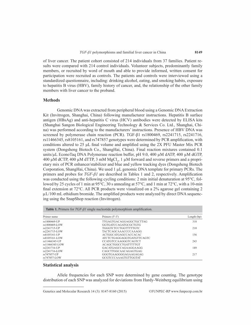
Genomic DNA was extracted from peripheral blood using a Genomic DNA Extraction Kit (Invitrogen, Shanghai, China) following manufacturer instructions. Hepatitis B surface antigen (HBsAg) and anti-hepatitis C virus (HCV) antibodies were detected by ELISA kits (Shanghai Sangon Biological Engineering Technology & Services Co. Ltd., Shanghai, Chi-na) was performed according to the manufacturers’ instructions. Presence of HBV DNA was screened by polymerase chain reaction (PCR). TGF-β1 rs1800469, rs2241715, rs2241716, rs11466345, rs8105161, and rs747857 genotypes were determined by PCR amplification, with conditions altered to 25 μL final volume and amplified using the 2X PFU Master Mix PCR system (Dongsheng Biotech Co., ShangHai, China). Final reaction mixtures contained 0.1 units/µL EconoTaq DNA Polymerase reaction buffer, pH 9.0, 400 µM dATP, 400 µM dGTP, 400 µM dCTP, 400 µM dTTP, 3 mM MgCl2, 1 µM forward and reverse primers and a propri-etary mix of PCR enhancer/stabilizer and blue and yellow tracking dyes (Dongsheng Biotech Corporation, ShangHai, China). We used 1 µL genomic DNA template for primary PCRs. The primers and probes for TGF-β1 are described in Tables 1 and 2, respectively. Amplification was conducted using the following cycling conditions: 2 min initial denaturation at 95°C, fol-lowed by 25 cycles of 1 min at 95°C, 30 s annealing at 57°C, and 1 min at 72°C, with a 10-min final extension at 72°C. All PCR products were visualized on a 2% agarose gel containing 2 mL/100 mL ethidium bromide. The amplified products were analyzed by direct DNA sequenc-ing using the SnapShop reaction (Invitrogen).
Primer name Primers (5'-3') Length (bp)
rs1800469-UP TTGAGTGACAGGAGGCTGCTTAG 318rs1800469-LOW GGTGATCCAGATGCGCTGTG rs2241715-UP TGGGTCTCCTGGTTTTTGTC 210rs2241715-LOW TACTCAGCAAACCCCAAAGG rs8105161-UP ACTGGCATGAGCCACCACAC 156rs8105161-LOW ATCTCTGAGGAGGTGATGTTCAGTC rs11466345-UP CCATGTCCAAGGGTCAGTCT 243rs11466345-LOW ACAGCTGGCCTGATTTTTGT rs2241716-UP GACATGAGCCAGAAGGAAGG 189rs2241716-LOW CAGCTTGGCAACAGAGTGAG rs747857-UP GGGTGAAGGGGAGAAGAGAG 217rs747857-LOW GCGTCCCAAAGTGTTGGTAT
Table 1. Primers for TGF-β1 single nucleotide polymorphism amplification.
Statistical analysis
Allele frequencies for each SNP were determined by gene counting. The genotype distribution of each SNP was analyzed for deviations from Hardy-Weinberg equilibrium using
8150P.Q. Wan et al.
©FUNPEC-RP www.funpecrp.com.brGenetics and Molecular Research 14 (3): 8147-8160 (2015)
c2 analyses. A c2 test and conditional logistic regression were performed to identify statisti-cal associations between genotype and disease status. The Student t-test was used to evaluate differences among serum levels of TFG-β1. The statistical analysis was carried out using the SPSS version 17.0 statistical software package (SPSS, Inc., Chicago, IL, USA).
Probe name Allele Probe sequence (5'-3')
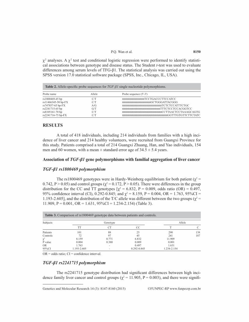
rs1800469-45 bp C/T ttttttttttttttttttttttttttttTCCTGACCCTTCCATCCrs11466345-50 bp-FX C/T ttttttttttttttttttttttttttttttttttttGCTGGGATTACGGGrs747857-65 bp-FX A/G ttttttttttttttttttttttttttttttttttttttttttttttttttTCTCTCCATTTCTGCrs2241715-65 bp G/T ttttttttttttttttttttttttttttttttttttttttttttttttTTTCTCCTCCACGGTCCrs8105161-70 bp C/T ttttttttttttttttttttttttttttttttttttttttttttttttttCCTTAACTCCTAAAGCAGTGrs2241716-75 bp-FX C/T tttttttttttttttttttttttttttttttttttttttttttttttttttttttttGGTTTGTGTTCTTCTATC
Table 2. Allele-specific probe sequences for TGF-β1 single nucleotide polymorphisms.
RESULTS
A total of 418 individuals, including 214 individuals from families with a high inci-dence of liver cancer and 214 healthy volunteers, were recruited from Guangxi Province for this study. Patients comprised a total of 214 Guangxi Zhuang, Han, and Yao individuals, 154 men and 60 women, with a mean ± standard error age of 34.5 ± 5.4 years.
Association of TGF-β1 gene polymorphisms with familial aggregation of liver cancer
TGF-β1 rs1800469 polymorphism
The rs1800469 genotypes were in Hardy-Weinberg equilibrium for both patient (χ2 = 0.742, P > 0.05) and control groups (χ2 = 0.172, P > 0.05). There were differences in the group distribution for the CC and TT genotypes [χ2 = 6.832, P = 0.009, odds ratio (OR) = 0.497, 95% confidence interval (CI), 0.292-0.845; and χ2 = 8.159, P = 0.004, OR = 1.763, 95%CI = 1.193-2.605], and the distribution of the T/C allele was different between the two groups (χ2 = 11.909, P = 0.001, OR = 1.631, 95%CI = 1.234-2.154) (Table 3).
Subjects Genotype Allele
TT CT CC T C
Patients 101 88 25 290 138Controls 72 97 45 241 187χ2 8.159 0.771 6.832 11.909 P value 0.004 0.380 0.009 0.001 OR 1.763 - 0.497 1.631 95%CI 1.193-2.605 - 0.292-0.845 1.234-2.154
OR = odds ratio; CI = confidence interval.
Table 3. Comparison of rs1800469 genotype data between patients and controls.
TGF-β1 rs2241715 polymorphism
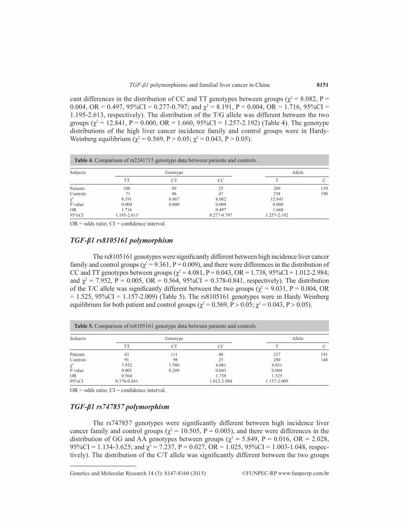
The rs2241715 genotype distribution had significant differences between high inci-dence family liver cancer and control groups (χ2 = 11.905, P = 0.003), and there were signifi-
8151TGF-β1 polymorphisms and familial liver cancer in China
©FUNPEC-RP www.funpecrp.com.brGenetics and Molecular Research 14 (3): 8147-8160 (2015)
cant differences in the distribution of CC and TT genotypes between groups (χ2 = 8.082, P = 0.004, OR = 0.497, 95%CI = 0.277-0.797; and χ2 = 8.191, P = 0.004, OR = 1.716, 95%CI = 1.195-2.613, respectively). The distribution of the T/G allele was different between the two groups (χ2 = 12.841, P = 0.000, OR = 1.660, 95%CI = 1.257-2.192) (Table 4). The genotype distributions of the high liver cancer incidence family and control groups were in Hardy-Weinberg equilibrium (χ2 = 0.569, P > 0.05; χ2 = 0.043, P > 0.05).
Subjects Genotype Allele
TT CT CC T C
Patients 100 89 25 289 139Controls 71 96 47 238 190χ2 8.191 0.467 8.082 12.841 P value 0.004 0.000 0.004 0.000 OR 1.716 0.497 1.660 95%CI 1.195-2.613 0.277-0.797 1.257-2.192
OR = odds ratio; CI = confidence interval.
Table 4. Comparison of rs2241715 genotype data between patients and controls.
TGF-β1 rs8105161 polymorphism
The rs8105161 genotypes were significantly different between high incidence liver cancer family and control groups (χ2 = 9.361, P = 0.009), and there were differences in the distribution of CC and TT genotypes between groups (χ2 = 4.081, P = 0.043, OR = 1.738, 95%CI = 1.012-2.984; and χ2 = 7.952, P = 0.005, OR = 0.564, 95%CI = 0.378-0.841, respectively). The distribution of the T/C allele was significantly different between the two groups (χ2 = 9.031, P = 0.004, OR = 1.525, 95%CI = 1.157-2.009) (Table 5). The rs8105161 genotypes were in Hardy Weinberg equilibrium for both patient and control groups (χ2 = 0.569, P > 0.05; χ2 = 0.043, P > 0.05).
Subjects Genotype Allele
TT CT CC T C
Patients 63 111 40 237 191Controls 91 98 25 280 148χ2 7.952 1.580 4.081 9.031 P value 0.005 0.209 0.043 0.004 OR 0.564 1.738 1.525 95%CI 0.378-0.841 1.012-2.984 1.157-2.009
OR = odds ratio; CI = confidence interval.
Table 5. Comparison of rs8105161 genotype data between patients and controls.
TGF-β1 rs747857 polymorphism
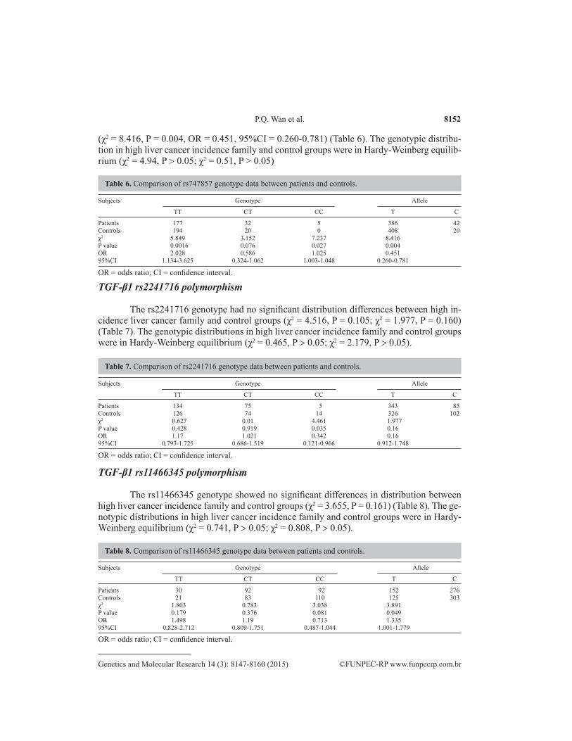
The rs747857 genotypes were significantly different between high incidence liver cancer family and control groups (χ2 = 10.505, P = 0.005), and there were differences in the distribution of GG and AA genotypes between groups (χ2 = 5.849, P = 0.016, OR = 2.028, 95%CI = 1.134-3.625; and χ2 = 7.237, P = 0.027, OR = 1.025, 95%CI = 1.003-1.048, respec-tively). The distribution of the C/T allele was significantly different between the two groups
8152P.Q. Wan et al.
©FUNPEC-RP www.funpecrp.com.brGenetics and Molecular Research 14 (3): 8147-8160 (2015)
(χ2 = 8.416, P = 0.004, OR = 0.451, 95%CI = 0.260-0.781) (Table 6). The genotypic distribu-tion in high liver cancer incidence family and control groups were in Hardy-Weinberg equilib-rium (χ2 = 4.94, P > 0.05; χ2 = 0.51, P > 0.05)
Subjects Genotype Allele
TT CT CC T C
Patients 177 32 5 386 42Controls 194 20 0 408 20χ2 5.849 3.152 7.237 8.416 P value 0.0016 0.076 0.027 0.004 OR 2.028 0.586 1.025 0.451 95%CI 1.134-3.625 0.324-1.062 1.003-1.048 0.260-0.781
OR = odds ratio; CI = confidence interval.
Table 6. Comparison of rs747857 genotype data between patients and controls.
TGF-β1 rs2241716 polymorphism
The rs2241716 genotype had no significant distribution differences between high in-cidence liver cancer family and control groups (χ2 = 4.516, P = 0.105; χ2 = 1.977, P = 0.160) (Table 7). The genotypic distributions in high liver cancer incidence family and control groups were in Hardy-Weinberg equilibrium (χ2 = 0.465, P > 0.05; χ2 = 2.179, P > 0.05).
TGF-β1 rs11466345 polymorphism
The rs11466345 genotype showed no significant differences in distribution between high liver cancer incidence family and control groups (χ2 = 3.655, P = 0.161) (Table 8). The ge-notypic distributions in high liver cancer incidence family and control groups were in Hardy-Weinberg equilibrium (χ2 = 0.741, P > 0.05; χ2 = 0.808, P > 0.05).
Subjects Genotype Allele
TT CT CC T C
Patients 134 75 5 343 85Controls 126 74 14 326 102χ2 0.627 0.01 4.461 1.977 P value 0.428 0.919 0.035 0.16 OR 1.17 1.021 0.342 0.16 95%CI 0.793-1.725 0.686-1.519 0.121-0.966 0.912-1.748
OR = odds ratio; CI = confidence interval.
Table 7. Comparison of rs2241716 genotype data between patients and controls.
Subjects Genotype Allele
TT CT CC T C
Patients 30 92 92 152 276Controls 21 83 110 125 303χ2 1.803 0.783 3.038 3.891 P value 0.179 0.376 0.081 0.049 OR 1.498 1.19 0.713 1.335 95%CI 0.828-2.712 0.809-1.751 0.487-1.044 1.001-1.779
OR = odds ratio; CI = confidence interval.
Table 8. Comparison of rs11466345 genotype data between patients and controls.
8153TGF-β1 polymorphisms and familial liver cancer in China
©FUNPEC-RP www.funpecrp.com.brGenetics and Molecular Research 14 (3): 8147-8160 (2015)
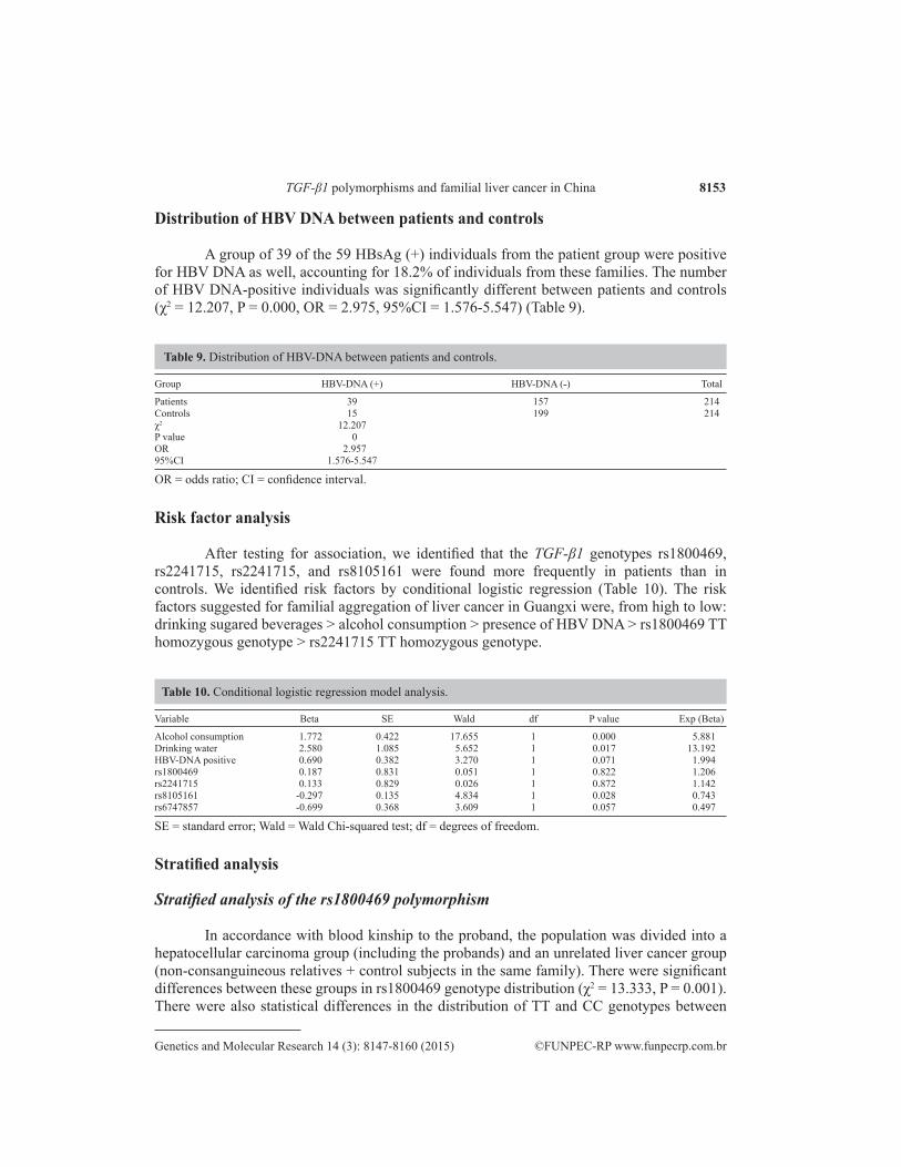
Distribution of HBV DNA between patients and controls
A group of 39 of the 59 HBsAg (+) individuals from the patient group were positive for HBV DNA as well, accounting for 18.2% of individuals from these families. The number of HBV DNA-positive individuals was significantly different between patients and controls (χ2 = 12.207, P = 0.000, OR = 2.975, 95%CI = 1.576-5.547) (Table 9).
Group HBV-DNA (+) HBV-DNA (˗) Total
Patients 39 157 214Controls 15 199 214χ2 12.207 P value 0 OR 2.957 95%CI 1.576-5.547
OR = odds ratio; CI = confidence interval.
Table 9. Distribution of HBV-DNA between patients and controls.
Risk factor analysis
After testing for association, we identified that the TGF-β1 genotypes rs1800469, rs2241715, rs2241715, and rs8105161 were found more frequently in patients than in controls. We identified risk factors by conditional logistic regression (Table 10). The risk factors suggested for familial aggregation of liver cancer in Guangxi were, from high to low: drinking sugared beverages > alcohol consumption > presence of HBV DNA > rs1800469 TT homozygous genotype > rs2241715 TT homozygous genotype.
Variable Beta SE Wald df P value Exp (Beta)
Alcohol consumption 1.772 0.422 17.655 1 0.000 5.881Drinking water 2.580 1.085 5.652 1 0.017 13.192HBV-DNA positive 0.690 0.382 3.270 1 0.071 1.994rs1800469 0.187 0.831 0.051 1 0.822 1.206rs2241715 0.133 0.829 0.026 1 0.872 1.142rs8105161 -0.297 0.135 4.834 1 0.028 0.743rs6747857 -0.699 0.368 3.609 1 0.057 0.497
SE = standard error; Wald = Wald Chi-squared test; df = degrees of freedom.
Table 10. Conditional logistic regression model analysis.
Stratified analysis
Stratified analysis of the rs1800469 polymorphism
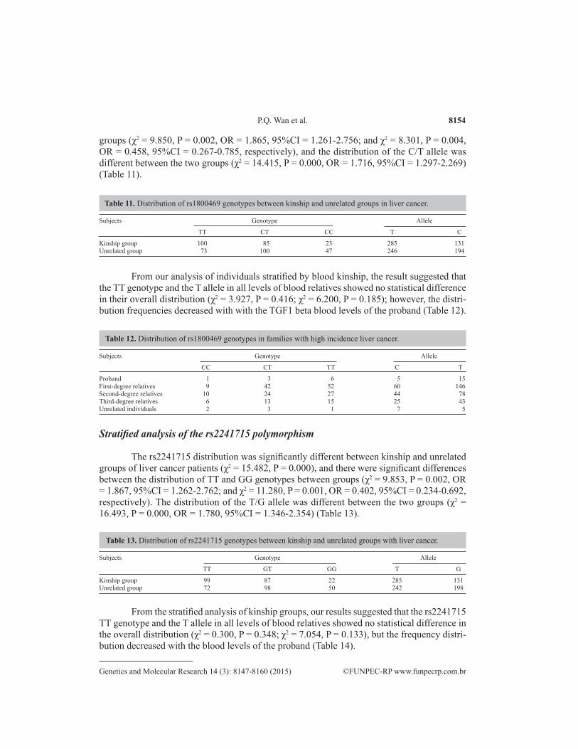
In accordance with blood kinship to the proband, the population was divided into a hepatocellular carcinoma group (including the probands) and an unrelated liver cancer group (non-consanguineous relatives + control subjects in the same family). There were significant differences between these groups in rs1800469 genotype distribution (χ2 = 13.333, P = 0.001). There were also statistical differences in the distribution of TT and CC genotypes between
8154P.Q. Wan et al.
©FUNPEC-RP www.funpecrp.com.brGenetics and Molecular Research 14 (3): 8147-8160 (2015)
groups (χ2 = 9.850, P = 0.002, OR = 1.865, 95%CI = 1.261-2.756; and χ2 = 8.301, P = 0.004, OR = 0.458, 95%CI = 0.267-0.785, respectively), and the distribution of the C/T allele was different between the two groups (χ2 = 14.415, P = 0.000, OR = 1.716, 95%CI = 1.297-2.269) (Table 11).
Subjects Genotype Allele
TT CT CC T C
Kinship group 100 85 23 285 131Unrelated group 73 100 47 246 194
Table 11. Distribution of rs1800469 genotypes between kinship and unrelated groups in liver cancer.
From our analysis of individuals stratified by blood kinship, the result suggested that the TT genotype and the T allele in all levels of blood relatives showed no statistical difference in their overall distribution (χ2 = 3.927, P = 0.416; χ2 = 6.200, P = 0.185); however, the distri-bution frequencies decreased with with the TGF1 beta blood levels of the proband (Table 12).
Subjects Genotype Allele
CC CT TT C T
Proband 1 3 6 5 15First-degree relatives 9 42 52 60 146Second-degree relatives 10 24 27 44 78Third-degree relatives 6 13 15 25 43Unrelated individuals 2 3 1 7 5
Table 12. Distribution of rs1800469 genotypes in families with high incidence liver cancer.
Stratified analysis of the rs2241715 polymorphism
The rs2241715 distribution was significantly different between kinship and unrelated groups of liver cancer patients (χ2 = 15.482, P = 0.000), and there were significant differences between the distribution of TT and GG genotypes between groups (χ2 = 9.853, P = 0.002, OR = 1.867, 95%CI = 1.262-2.762; and χ2 = 11.280, P = 0.001, OR = 0.402, 95%CI = 0.234-0.692, respectively). The distribution of the T/G allele was different between the two groups (χ2 = 16.493, P = 0.000, OR = 1.780, 95%CI = 1.346-2.354) (Table 13).
Subjects Genotype Allele
TT GT GG T G
Kinship group 99 87 22 285 131Unrelated group 72 98 50 242 198
Table 13. Distribution of rs2241715 genotypes between kinship and unrelated groups with liver cancer.
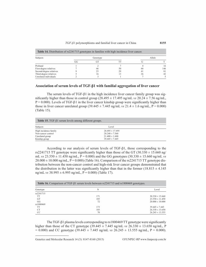
From the stratified analysis of kinship groups, our results suggested that the rs2241715 TT genotype and the T allele in all levels of blood relatives showed no statistical difference in the overall distribution (χ2 = 0.300, P = 0.348; χ2 = 7.054, P = 0.133), but the frequency distri-bution decreased with the blood levels of the proband (Table 14).
8155TGF-β1 polymorphisms and familial liver cancer in China
©FUNPEC-RP www.funpecrp.com.brGenetics and Molecular Research 14 (3): 8147-8160 (2015)
Association of serum levels of TGF-β1 with familial aggregation of liver cancer
The serum levels of TGF-β1 in the high incidence liver cancer family group was sig-nificantly higher than those in control group (28.495 ± 17.495 ng/mL vs 20.24 ± 7.56 ng/mL, P = 0.000). Levels of TGF-b1 in the liver cancer kinship group were significantly higher than those in liver cancer unrelated group (39.445 ± 7.445 ng/mL vs 21.4 ± 1.6 ng/mL, P = 0.000) (Table 15).
Subjects Genotype Allele
GG GT TT G T
Proband 1 2 7 4 16First-degree relatives 7 46 50 60 146Second-degree relatives 9 23 29 41 81Third-degree relatives 5 16 13 26 42Unrelated individuals 2 3 1 7 5
Table 14. Distribution of rs2241715 genotypes in families with high incidence liver cancer.
Subjects Level
High incidence family 28.495 ± 17.495Non-cancer control 20.240 ± 7.560Unrelated group 21.400 ± 1.600Kinship group 39.445 ± 7.445
Table 15. TGF-β1 serum levels among different groups.
According to our analysis of serum levels of TGF-β1, those corresponding to the rs2241715 TT genotype were significantly higher than those of the GT (30.330 ± 15.660 ng/mL vs 23.550 ± 11.450 ng/mL, P = 0.000) and the GG genotypes (30.330 ± 15.660 ng/mL vs 20.000 ± 10.000 ng/mL, P = 0.000) (Table 16). Comparison of the rs2241715 TT genotype dis-tribution between the non-cancer control and high-risk liver cancer groups demonstrated that the distribution in the latter was significantly higher than that in the former (18.815 ± 4.145 ng/mL vs 38.995 ± 6.995 ng/mL, P = 0.000) (Table 17).
Genotype N Level
rs2241715 TT 171 30.330 ± 15.660 GT 185 23.550 ± 11.450 GG 72 20.000 ± 10.000rs1800469 TT 173 39.445 ± 7.445 CT 185 26.330 ± 13.650 CC 70 26.245 ± 13.555
Table 16. Comparison of TGF-β1 serum levels between rs2241715 and rs1800469 genotypes.
The TGF-β1 plasma levels corresponding to rs1800469 TT genotype were significantly higher than those of the CT genotype (39.445 ± 7.445 ng/mL vs 26.330 ± 13.650 ng/mL, P = 0.000) and CC genotype (39.445 ± 7.445 ng/mL vs 26.245 ± 13.555 ng/mL, P = 0.000).
8156P.Q. Wan et al.
©FUNPEC-RP www.funpecrp.com.brGenetics and Molecular Research 14 (3): 8147-8160 (2015)
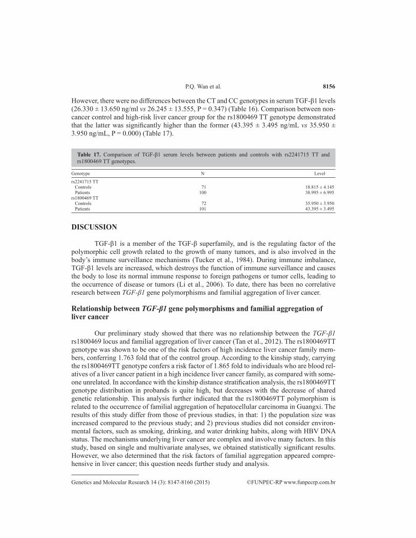
However, there were no differences between the CT and CC genotypes in serum TGF-β1 levels (26.330 ± 13.650 ng/ml vs 26.245 ± 13.555, P = 0.347) (Table 16). Comparison between non-cancer control and high-risk liver cancer group for the rs1800469 TT genotype demonstrated that the latter was significantly higher than the former (43.395 ± 3.495 ng/mL vs 35.950 ± 3.950 ng/mL, P = 0.000) (Table 17).
Genotype N Level
rs2241715 TT Controls 71 18.815 ± 4.145 Patients 100 38.995 ± 6.995rs1800469 TT Controls 72 35.950 ± 3.950 Patients 101 43.395 ± 3.495
Table 17. Comparison of TGF-β1 serum levels between patients and controls with rs2241715 TT and rs1800469 TT genotypes.
DISCUSSION
TGF-β1 is a member of the TGF-β superfamily, and is the regulating factor of the polymorphic cell growth related to the growth of many tumors, and is also involved in the body’s immune surveillance mechanisms (Tucker et al., 1984). During immune imbalance, TGF-β1 levels are increased, which destroys the function of immune surveillance and causes the body to lose its normal immune response to foreign pathogens or tumor cells, leading to the occurrence of disease or tumors (Li et al., 2006). To date, there has been no correlative research between TGF-β1 gene polymorphisms and familial aggregation of liver cancer.
Relationship between TGF-β1 gene polymorphisms and familial aggregation of liver cancer
Our preliminary study showed that there was no relationship between the TGF-β1 rs1800469 locus and familial aggregation of liver cancer (Tan et al., 2012). The rs1800469TT genotype was shown to be one of the risk factors of high incidence liver cancer family mem-bers, conferring 1.763 fold that of the control group. According to the kinship study, carrying the rs1800469TT genotype confers a risk factor of 1.865 fold to individuals who are blood rel-atives of a liver cancer patient in a high incidence liver cancer family, as compared with some-one unrelated. In accordance with the kinship distance stratification analysis, the rs1800469TT genotype distribution in probands is quite high, but decreases with the decrease of shared genetic relationship. This analysis further indicated that the rs1800469TT polymorphism is related to the occurrence of familial aggregation of hepatocellular carcinoma in Guangxi. The results of this study differ from those of previous studies, in that: 1) the population size was increased compared to the previous study; and 2) previous studies did not consider environ-mental factors, such as smoking, drinking, and water drinking habits, along with HBV DNA status. The mechanisms underlying liver cancer are complex and involve many factors. In this study, based on single and multivariate analyses, we obtained statistically significant results. However, we also determined that the risk factors of familial aggregation appeared compre-hensive in liver cancer; this question needs further study and analysis.
8157TGF-β1 polymorphisms and familial liver cancer in China
©FUNPEC-RP www.funpecrp.com.brGenetics and Molecular Research 14 (3): 8147-8160 (2015)
Chow et al. (2005) and Healy et al. (2009) presented analyses of the TGF-β1 promoter, wherein the rs1800469 promoter was found to interfere with the identification of functional transcription factors, which influenced the gene expression levels of TGF-β1. The rs1800469 locus containing a cytosine base promoted selective binding of AP1, which lowered the binding potential of other transcriptional activating factors, which led to lower plasma TGF-β1 levels (Shah et al., 2006a). The plasma levels of TGF-β1 associated with the CC genotype of rs1800469 were significantly lower than those of the TT genotype (Guo et al., 2011), and the individual plasma TGF-β1 levels of the TGF-β1 rs1800469TT genotype was 2 times that of non-TT individuals (Grainger et al., 1999). The results of this study showed that the plasma TGF-β1 levels associated with the rs1800469TT genotype were significantly higher than those of the CT and CC genotypes (P = 0.000, P = 0.000), and the rs1800469 polymorphism is the genetic factor that affects the expression of TGF-beta1. However, analysis of the different groups showed that the rs1800469TT genotype conferred higher overall serum TGF-β1 expression levels to the high liver cancer incidence family group than the overall cancer-free controls (P = 0.000, P = 0.000). Therefore, the rs1800469 gene polymorphism appears not to be the only genetic factor affecting plasma TGF-β1 expression; it may interact with other related components of familial liver cancer aggregation, or play a role with other susceptibility genes segregating in familial liver cancer.
TGF-β1 inflammation-related cytokine production, and TGF-β1 gene expression or dysfunction are associated with liver fibrosis and hepatocellular carcinoma, as is chronic HBV infection (Gewaltig et al., 2002; Kim et al., 2003; Wang, 2003). Gong et al. (2008) reported on the relationship between TGF-β1 gene rs2241715 polymorphism and hepatocellular carci-noma, wherein they found that the rs2241715TT genotype frequencies in patients with liver cirrhosis and hepatocellular carcinoma were higher than those of a control group; however, to date there is no related report on rs2241715 gene polymorphism and familial clustering of hepatocellular carcinoma. This study found that the distributions of the rs2241715TT geno-type and the T allele were significantly higher in high liver cancer incidence families than in control families without cancer (χ2 = 8.191, P = 0.004; χ2 = 12.841, P = 0.000). Examination of the association between the gene polymorphism and drinking, smoking, and drinking water habits, HBsAg levels, and presence of HBV-DNA by logistic regression analysis found that when rs2241715TT was entered into the model, the corresponding regression coefficient was positive. This finding suggested that the TGF-β1 rs2241715 gene polymorphism is a risk fac-tor for familial aggregation of liver cancer and that carrying the rs2241715TT genotype is one of the cancer predisposing risk factors for high liver cancer incidence family members, with a risk 1.716 times higher than that of the control group. Subsequent analysis based on the level of kinship suggested that carrying the rs2241715TT genotype represents the genetic risk factor predisposing blood relatives of a liver cancer patient in high incidence families to liver cancer, with the risk factor of liver cancer being 1.867 times that of an unrelated person. These results serve to further illustrate that the rs2241715 polymorphism is related to familial clustering of liver cancer in Guangxi.
The incidences reported of liver cancer are closely related to degree of blood relation-ship (Chen et al., 1998), with the highest incidence among first-degree relatives, followed by second-degree relatives, third-degree relatives, and the general population. Stratified analysis by kinship distance with the proband demonstrated that the rs2241715TT genotype and the T allele have a very high frequency distribution in the probands, which decreases in first-, sec-ond-, and third-degree relatives. Therefore, the rs2241715TT genotype and the T allele may
8158P.Q. Wan et al.
©FUNPEC-RP www.funpecrp.com.brGenetics and Molecular Research 14 (3): 8147-8160 (2015)
be one of the genetic factors underlying the occurrence of familial clustering of hepatocellular carcinoma occurrence in Guangxi, wherein the distribution and effect of this polymorphism decreases with decreasing relative level.
The quantity of serum TGF-β1 expression in individuals with the rs2241715TT gen-otype was higher than those with genotypes TG or GG (P = 0.000, P = 0.000). This sug-gested that rs2241715 gene polymorphism is related to serum TGF-β1 expression level, and is also one of the determining genetic factors of TGF-β1 expression levels. However, the grouping analysis showed that in the high liver cancer incidence family group, the genotype rs2241715TT plasma TGF-β1 expression levels were higher overall than in the control cancer free family group (P = 0.000). Therefore, the rs2241715 gene polymorphism is not the only genetic factor affecting plasma TGF-β1 expression; it may interact with other related factors contributing to liver cancer familial aggregation, or play a role in conjunction with other sus-ceptibility genes involved in familial clustering of liver cancer.
The rs2241716 and rs2241715 polymorphisms demonstrate complete linkage, but there was no difference in distribution of the rs2241716 genotype and allele between high liver cancer incidence family and non-cancer control groups (χ2 = 4.516, P = 0.105; χ2 = 1.977, P = 0.160). There was no obvious correlation between the rs2241716 polymorphism and familial clustering of liver cancer, rs2241715 played a more important role in familial clustering of HCC occurrence. There have been no reports of rs11466345 association with liver cancer and this study found that there was no obvious correlation between the rs11466345 gene polymor-phism and familial clustering of liver cancer. There have also been no reports of rs8105161 or rs747857 association with liver cancer and this study found that these two loci had no obvious correlation with familial clustering of liver cancer in Guangxi.
Relationship between HBV DNA and family aggregation of liver cancer
HBV infection is one of the risk factors for liver cancer, but its mechanism is not well understood. Studies (Sheng et al., 2009) have shown that familial clustering of liver cancer is related to positive HBsAg status. In this study, HBV-DNA positive status was found to be a risk factor of liver cancer familial aggregation (B = 0.690, Wald value 0.382). Because the HBV DNA status of patients with HBSAg is generally positive while HBV-negative patients are rarely HBSAg positive, it is more exact to state that being HBV-DNA (+) is a risk factor for liver cancer familial aggregation.
This is the first study to analyze blood TGF-β1 expression level by family relation-ship or kinship. The overall TGF-β1 levels in the high incidence liver cancer family group were higher than those in the control non-cancer family group (P = 0.000), and blood TGF-β1 levels were higher in the liver cancer-related group than in the liver cancer unrelated group (P = 0.000). Therefore, the overall levels of TGF-β1 and rs1800469TT, rs2241715TT genotype frequency distributions are in agreement. These results further suggest that the rs1800469TT and rs2241715TT genotypes are risk factors for liver cancer familial aggregation in Guangxi.
This study also took into consideration environmental factors such as gender, age, nationality, and HBSAg status. The relationship among TGF-β1 gene polymorphisms, pres-ence of HBV-DNA, and liver cancer family aggregation was considered by single analysis, and the comprehensive lifestyle factors of smoking, alcohol consumption, and drinking water habits were considered by multiple factors analysis. Our results showed that the rs1800469TT and rs2241715TT genotypes are genetic risk factors of liver cancer familial aggregation in
8159TGF-β1 polymorphisms and familial liver cancer in China
©FUNPEC-RP www.funpecrp.com.brGenetics and Molecular Research 14 (3): 8147-8160 (2015)
Guangxi, while alcohol consumption, quality of drinking water, and HBV DNA-positive sta-tus are environmental factors that contribute to the risk of family aggregation liver cancer in Guangxi. The largest effect on family aggregation of cancer in Guangxi is alcohol consump-tion, followed by the quality of drinking water. We are currently investigating what kinds of carcinogens are present in sugared beverages related to the high incidence of liver cancer. In summary, both environmental and genetic factors may impact the familial aggregation of liver cancer in Guangxi Zhuang, Han, and Yao populations.
Conflicts of interest
The authors declare no conflict of interest.
ACKNOWLEDGMENTS
Research supported by the National Natural Science Foundation of China, #30960170; the Foundation of Guangxi Provincial Education Department, #201203YB048; and the Foun-dation of Guangxi Provincial Education Department, #201202ZD021.
REFERENCES
Castillejo A, Rothman N, Murta-Nascimento C, Malats N, et al. (2009). TGFB1 and TGFBR1 polymorphic variants in relationship to bladder cancer risk and prognosis. Int. J. Cancer 124: 608-613.
Chen CH, Huang GT, Lee HS, Yang PM, et al. (1998). Clinical impact of screening first-degree relatives of patients with hepatocellular carcinoma. J. Clin. Gastroenterol. 27: 236-239.
Chow KM, Szeto CC, Poon P, Lau WY, et al. (2005). Transforming growth factor-β1 gene polymorphism in renal transplant recipients. Ren. Fail. 27: 671-675.
Gewaltig J, Mangasser-Stephan K, Gartung C, Biesterfeld S, et al. (2002). Association of polymorphisms of the transforming grow th factor-beta1 gene with the rate of progression of HCV-induced liver fibrosis. Clin. Chim. Acta 316: 83-94.
Gong GD, Yu J, Huang ZH, Du DL, et al. (2008). The individuals with rs2241715 T/T genotype of the TGF-β1 gene have an increased risk for developing HBV infection-associated diseases. Tumor 28: 502-505.
Grainger DJ, Heathcote K, Chiano M, Snieder H, et al. (1999). Genetic control of the circulating concentration of transforming growth factor type beta1. Hum. Mol. Genet. 8: 93-97.
Guo W, Dong Z, Guo Y, Chen Z, et al. (2011). Polymorphisms of transforming growth factor-b1 associated with increased risk of gastric cardia adenocarcinoma in north China. Int. J. Immunogenet. 38: 215-224.
Healy J, Dionne J, Bélanger H, Larivière M, et al. (2009). Functional impact of sequence variation in the promoter region of TGF-β1. Int. J. Cancer 125: 1483-1489.
Helmig S, Belwe A and Schneider J (2009). Association of transforming growth factor β1 gene polymorphisms and asbestos-induced fibrosis and tumors. J. Invest. Med. 57: 655-661.
Hu S, Zhou G, Zhang L, Jiang H, et al. (2012). The effects of functional polymorphisms in the TGF β1 gene on nasopharyngeal carcinoma susceptibility. Otolaryngol. Head Neck Surg. 146: 579-584.
Kang HG, Chae MH, Park JM, Kim EJ, et al. (2006). Polymorphisms in TGF-β1 gene and the risk of lung cancer. Lung Cancer 52: 1-7.
Kim YJ, Lee HS, Im JP, Min BH, et al. (2003). Association of transforming growth factor-β1 gene polymorphisms with a hepatocellular carcinoma risk in patients with chronic hepatitis B virus infection. Exp. Mol. Med. 35: 196-202.
Li MO, Wan YY, Sanjabi S, Roberston AK, et al. (2006). Transforming growth factor-beta regulation of immune responses. Annu. Rev. Immunol. 24: 99-146.
Li X, Yue ZC, Zhang YY, Bai J, et al. (2008). Elevated serum level and gene polymorphisms of TGFβ1 in gastric cancer. J. Clin. Lab. Anal. 22: 164-171.
Lin XD, Li C, Shi Y, Chen Y, et al. (2010). Correlation of polymorphism of Nme1-1465 T > C and TGFβ1-509 T > C with genetic susceptibility to gastric carcinoma. Zhonghua Bing Li Xue Za Zhi. 39: 681-685.
Lundberg M, Saarilahti K, Mӓkatie AA and Mattila PS (2010). TGF-β1 genetic polymorphism is associated with survival
8160P.Q. Wan et al.
©FUNPEC-RP www.funpecrp.com.brGenetics and Molecular Research 14 (3): 8147-8160 (2015)
in head and neck squamous cell carcinoma independent of the severity of chemoradiotherapy induced mucositis. Oral Oncol. 46: 369-372.
Park KH, Lo Han SG, Whang YM, Lee HJ, et al. (2006). Single nucleotide polymorphisms of the TGFB1 gene and lung cancer risk in a Korean population. Cancer Genet. Cytogenet. 169: 39-44.
Prasad P, Tiwari AK, Kumar KM, Ammini AC, et al. (2007). Association of TGF-beta1, TNF-alpha, CCR2 and CCR5 gene polymorphisms in type-2 diabetes and renal insufficiency among Asian Indians. BMC Med. Genet. 8: 20.
Qi P, Chen YM, Wang H, Fang M, et al. (2009). -509C>T polymorphism in the TGF-β1 gene promoter, impact on the hepatocellular carcinoma risk in Chinese patients with chronic hepatitis B virus infection. Cancer Immunol. Immunother. 58: 1433-1440.
Tan L, Wu JZ, Wu JL, Wan PQ, et al. (2012). Relationship between a TGF-β1 gene polymorphism and HBV infection and familial clustering of hepatocellular carcinoma. Shijie Huaren Xiaohua Zazhi 20: 514-518.
Shah R, Hurley CK and Posch PE (2006a). A molecular mechanism for the differential regulation of TGF-β1 expression due to the common SNP-509C-T(c.-1347C>T). Hum. Genet. 120: 461-469.
Shah R, Rahaman B, Hurley CK and Posch PE (2006b). Allelic diversity in the TGFB1 regulatory region: characterization of novel functional single nucleotide polymorphisms. Hum. Genet. 119: 61-74.
Sheng Yi, Deng W, Yu JH, Zhang CY, et al. (2009). Study on the relationship between familial clustering of hepatocellular carcinoma and polymorphism of cytochrome P450 2E1 gene in Zhuang population, Guangxi. Chin. J. Epidemiol. 30: 153-154.
Sripriya S, George R, Arvind H, Baskaran M, et al. (2007). Transforming growth factor beta-1 -509C> T polymorphism in Indian patients with primary open angle glaucoma. Mol. Diagn. Ther. 11: 151-154.
Stanczuk GA, Tswana SA, Bergstrom S and Sibanda EN (2002). Polymorphism in codons 10 and 25 of the transforming growth factor-beta 1 (TGF-1) gene in patients with invasive squamous cell carcinoma of the uterine cervix. Eur. J. Immunogenet. 29: 417-421.
Teixeira AL, Araújo A, Coelho A, Ribeiro R, et al. (2011). Influence of TGFB1+869T>C functional polymorphism in non-small cell lung cancer (NSCLC) risk. J. Cancer Res. Clin. Oncol. 137: 435-439.
Thys M, Schrauwen I, Vanderstraeten K, Janssens K, et al. (2007). The coding polymorphism T263I in TGF-beta 1 is associated with otosclerosis is in two independent populations. Hum. Mol. Genet. 16: 2021-2030.
Togashi Y, Masago K, Fujita S, Kim YH, et al. (2011). Association of the transforming growth factor β1 promoter polymorphism, C-509T, with smoking status and survival in advanced non-small cell lung cancer. Oncol. Rep. 25: 377-382.
Toyoda T, Nakamura K, Yamada K, Thanseem I, et al. (2007). SNP analyses of growth factor genes EGF, TGF beta-1, and HGF reveal haplotypic association of EGF with autism. Biochem. Biophys. Res. Commun. 360: 715-720.
Tucker RF, Shipley GD, Moses HL and Holley RW (1984). Growth inhibitor from BSC-1 cells closely related to platelet type beta transforming growth factor. Science 226: 705-707.
Wang FS (2003). Current status and prospects of studies on human genetic Alleles associated with hepatitis B virus infection. World J .Gastroenterol. 9: 641-644.
Wei YS, Xu QQ, Wang CF, Pan Y, et al. (2007a). Genetic variation in transforming growth factor-beta1 gene associated with increased risk of esophageal squamous cell carcinoma. Tissue Antigens 70: 464-469.
Wei YS, Zhu YH, Du B, Yang ZH, et al. (2007b). Association of transforming growth factor-β1 gene polymorphisms with genetic susceptibility to nasopharyngeal carcinoma. Clin. Chim. Acta 380: 165-169.
Weng H, Mertens PR, Gressner AM and Dooley S (2007). IFN-gamma abrogates profibrogenic TGF-beta signaling in liver by targeting expression of inhibitory and receptor Smads. J. Hepatol. 46: 295-303.
Wu JZ, Li GJ, Chen WQ, Zhang N, et al. (2009). Primary epidemiology investigation in the area with high mortality of hepatocellular carcinoma discovered recently in Guangxi province. Int. Med. China 4: 678-680.
Yuan X, Liao Z, Liu Z, Wang LE, et al. (2009). Single nucleotide polymorphism at rs1982073:T869C of the TGFβ1 gene is associated with the risk of radiation pneumonitis in patients with non-small-cell lung cancer treated with definitive radiotherapy. J. Clin. Oncol. 27: 3370-3378.