Testicular Caspase-3 and -Catenin Regulators Predicted via ...
23
metabolites H OH OH Article Testicular Caspase-3 and β-Catenin Regulators Predicted via Comparative Metabolomics and Docking Studies Mohammed S. Hifnawy 1 , Mahmoud A. Aboseada 2 , Hossam M. Hassan 3 , Asmaa M. AboulMagd 4 , Adel F. Tohamy 5, † , Samraa H. Abdel-Kawi 6,7 , Mostafa E. Rateb 3,8,9 , El Moataz Bellah El Naggar 10 , Miaomiao Liu 11 , Ronald J. Quinn 11 , Hani A. Alhadrami 12,13, * and Usama Ramadan Abdelmohsen 14,15, * 1 Department of Pharmacognosy, Faculty of Pharmacy, Cairo University, Cairo 11865, Egypt; [email protected] 2 Department of Pharmacognosy, Faculty of Pharmacy, Nahda University, Beni-Suef 62513, Egypt; [email protected] 3 Department of Pharmacognosy, Faculty of Pharmacy, Beni-Suef University, Beni-Suef 62513, Egypt; [email protected] (H.M.H.); [email protected] (M.E.R.) 4 Department of Pharmaceutical Chemistry, Faculty of Pharmacy, Nahda University, Beni-Suef 62513, Egypt; [email protected] 5 Department of Toxicology and Forensic Medicine, Faculty of Veterinary Medicine, Cairo University, Cairo 11865, Egypt; [email protected] 6 Department of Medical Histology and Cell Biology, Faculty of Medicine, Beni-Suef University, Beni-Suef 62513, Egypt; [email protected] 7 Department of Basic Science, Faculty of Dentistry, Nahda University, Beni-Suef 62513, Egypt 8 Marine Biodiscovery Centre, University of Aberdeen, Aberdeen AB24 3UE, UK 9 School of Computing, Engineering and Physical Sciences, University of West Scotland, Paisley PA1 2BE, UK 10 Department of Pharmacognosy, Faculty of Pharmacy, Damanhur University, Elbehira 22511, Egypt; [email protected] 11 Griffith Institute for Drug Discovery, Griffith University, Brisbane, QLD 4111, Australia; miaomiao.liu@griffith.edu.au (M.L.); r.quinn@griffith.edu.au (R.J.Q.) 12 Faculty of Applied Medical Sciences, Department of Medical Laboratory Technology, King Abdulaziz University, P. O. Box 80402, Jeddah 21589, Saudi Arabi 13 King Fahd Medical Research Centre, King Abdulaziz University, P. O. Box 80402, Jeddah 21589, Saudi Arabia 14 Department of Pharmacognosy, Faculty of Pharmacy, Minia University, Minia 61519, Egypt 15 Department of Pharmacognosy, Faculty of Pharmacy, Deraya University, Universities Zone, New Minia City 61111, Egypt * Correspondence: [email protected] (H.A.A.); [email protected] (U.R.A.) † Current affiliation: Higher Colleges of Technology, Faculty of Health Sciences (Veterinary Medicine), Sharjah Mens’s Campus, Sharjah 25026, UAE. Received: 5 December 2019; Accepted: 6 January 2020; Published: 11 January 2020 Abstract: Many routes have been explored to search for effective, safe, and affordable alternatives to hazardous female contraceptives. Herbal extracts and their secondary metabolites are some of the interesting research areas to address this growing issue. This study aims to investigate the effects of ten different plant extracts on testicular spermatogenesis. The correlation between the chemical profile of these extracts and their in vivo effect on male reproductive system was evaluated using various techniques. Approximately 10% of LD 50 of hydro-methanolic extracts were orally administrated to rats for 60 days. Semen parameters, sexual organ weights, and serum levels of male sex hormones in addition to testes histopathology, were evaluated. Moreover, metabolomic analysis using (LC-HRESIMS), multivariate analysis (PCA), immunohistochemistry (caspase-3 and β-catenin), and a docking study were performed. Results indicated that three plant extracts significantly decreased epididymal sperm density and motility. Moreover, their effects on testicular cells were Metabolites 2020, 10, 31; doi:10.3390/metabo10010031 www.mdpi.com/journal/metabolites
Transcript of Testicular Caspase-3 and -Catenin Regulators Predicted via ...
metabolites
H
OH
OH
Article
Testicular Caspase-3 and β-Catenin RegulatorsPredicted via Comparative Metabolomics andDocking Studies
Mohammed S. Hifnawy 1, Mahmoud A. Aboseada 2, Hossam M. Hassan 3 ,Asmaa M. AboulMagd 4 , Adel F. Tohamy 5,† , Samraa H. Abdel-Kawi 6,7,Mostafa E. Rateb 3,8,9 , El Moataz Bellah El Naggar 10, Miaomiao Liu 11, Ronald J. Quinn 11,Hani A. Alhadrami 12,13,* and Usama Ramadan Abdelmohsen 14,15,*
1 Department of Pharmacognosy, Faculty of Pharmacy, Cairo University, Cairo 11865, Egypt;[email protected]
2 Department of Pharmacognosy, Faculty of Pharmacy, Nahda University, Beni-Suef 62513, Egypt;[email protected]
3 Department of Pharmacognosy, Faculty of Pharmacy, Beni-Suef University, Beni-Suef 62513, Egypt;[email protected] (H.M.H.); [email protected] (M.E.R.)
4 Department of Pharmaceutical Chemistry, Faculty of Pharmacy, Nahda University, Beni-Suef 62513, Egypt;[email protected]
5 Department of Toxicology and Forensic Medicine, Faculty of Veterinary Medicine, Cairo University,Cairo 11865, Egypt; [email protected]
6 Department of Medical Histology and Cell Biology, Faculty of Medicine, Beni-Suef University,Beni-Suef 62513, Egypt; [email protected]
7 Department of Basic Science, Faculty of Dentistry, Nahda University, Beni-Suef 62513, Egypt8 Marine Biodiscovery Centre, University of Aberdeen, Aberdeen AB24 3UE, UK9 School of Computing, Engineering and Physical Sciences, University of West Scotland, Paisley PA1 2BE, UK10 Department of Pharmacognosy, Faculty of Pharmacy, Damanhur University, Elbehira 22511, Egypt;
[email protected] Griffith Institute for Drug Discovery, Griffith University, Brisbane, QLD 4111, Australia;
[email protected] (M.L.); [email protected] (R.J.Q.)12 Faculty of Applied Medical Sciences, Department of Medical Laboratory Technology, King Abdulaziz
University, P. O. Box 80402, Jeddah 21589, Saudi Arabi13 King Fahd Medical Research Centre, King Abdulaziz University, P. O. Box 80402, Jeddah 21589, Saudi Arabia14 Department of Pharmacognosy, Faculty of Pharmacy, Minia University, Minia 61519, Egypt15 Department of Pharmacognosy, Faculty of Pharmacy, Deraya University, Universities Zone,
New Minia City 61111, Egypt* Correspondence: [email protected] (H.A.A.); [email protected] (U.R.A.)† Current affiliation: Higher Colleges of Technology, Faculty of Health Sciences (Veterinary Medicine),
Sharjah Mens’s Campus, Sharjah 25026, UAE.
Received: 5 December 2019; Accepted: 6 January 2020; Published: 11 January 2020�����������������
Abstract: Many routes have been explored to search for effective, safe, and affordable alternativesto hazardous female contraceptives. Herbal extracts and their secondary metabolites are some ofthe interesting research areas to address this growing issue. This study aims to investigate theeffects of ten different plant extracts on testicular spermatogenesis. The correlation between thechemical profile of these extracts and their in vivo effect on male reproductive system was evaluatedusing various techniques. Approximately 10% of LD50 of hydro-methanolic extracts were orallyadministrated to rats for 60 days. Semen parameters, sexual organ weights, and serum levels of malesex hormones in addition to testes histopathology, were evaluated. Moreover, metabolomic analysisusing (LC-HRESIMS), multivariate analysis (PCA), immunohistochemistry (caspase-3 and β-catenin),and a docking study were performed. Results indicated that three plant extracts significantlydecreased epididymal sperm density and motility. Moreover, their effects on testicular cells were
Metabolites 2020, 10, 31; doi:10.3390/metabo10010031 www.mdpi.com/journal/metabolites
Metabolites 2020, 10, 31 2 of 23
also assured by histopathological evaluations. Metabolomic profiling of the bioactive plant extractsshowed the presence of diverse phytochemicals, mostly oleanane saponins, phenolic diterpenes, andlupane triterpenes. A docking study on caspase-3 enzyme showed that oleanane saponins possessedthe highest binding affinity. An immunohistochemistry assay on β-catenin and caspase-3 indicatedthat Albizzia lebbeck was the most active extract for decreasing immunoexpression of β-catenin, whileRosmarinus officinalis showed the highest activity for increasing immunoexpression of caspase-3.The spermatogenesis decreasing the activity of A. lebbeck, Anagallis arvensis, and R. officinalis can bemediated via up-regulation of caspase-3 and down-regulation of β-catenin existing in testis cells.
Keywords: testicular spermatogenesis; β-catenin; caspase-3; Albizzia lebbeck; Anagallis arvensis;Rosmarinus officinalis; metabolomic profiling; docking
1. Introduction
In developing countries, populations are increasing rapidly. High population numbers have thepotential to lead to numerous economic, environmental, resource, and social problems [1]. Recently,researchers have given priority to the use of medicinal plants for their contraceptive properties, with amajority of them being used as female contraceptive agents, while some affect the male reproductivesystem [2,3]. Albizzia lebbeck L. Benth (Mimosoidae), Anagallis arvensis L. (primulaceae), and Calendulaofficinalis L. (Astraceae) are annual plants containing oleanane saponins which have been linked tovarious biological activites affecting sperm [4–6]. A. lebbeck extracts have exhibited anti-inflammatory,antiallergic, and antidiabetic activities [7–9]. Additionally, the saponins found in the bark of this plantare also reported to have adverse effects on the male reproductive system [10], while its saponin-richroot and pod fractions were shown to cause spermicidal action in human semen [11]. A. arvensisextract has been used in folk remedies for bronchial asthma, fever, and rheumatism [12]. On top ofthis, anagalligenone sapogenin isolated from A. arvensis caused the instantaneous immobilization ofspermatozoa in one minute [13]. The flower extract of C. officinalis, commonly known as marigold [14],has been used to treat HIV and cancer [15,16]. Moreover, the saponin-rich fraction from marigoldflowers has exhibited potent spermicidal activity [17].
Apium graveolens L., Anethum graveolens L. (Apiaceae), and Menthae piperitae (Lamiaceae) are richin polyphenolics [18–20] and volatile constituents [21–23]. Essential oil and extracts of A. graveolens(celery) have been utilized for numerous medical problems such as hepatic disorders, peptic ulcers,and hypertension [22,24,25]. It was studied for its anti-fertility effect in males, suggesting the highcontent of phytoestrogens (flavonoids) seemed to be responsible for decreasing sex hormone levels inthe blood [26]. Anethum graveolens (dill) is on the vital list of plants for home complementary medicine,as its essential oil and extracts can help in GIT disorders, have antioxidant and anti-hyperlipidemiceffects [27,28], and its hydro-methanolic extract adversely affects male fertility [29]. The essential oiland extracts of M. pepperitae (peppermint) have been used as antimicrobial and hepatoprotective agentsand to improve menstrual disorders [30–32]. Peppermint tea has been reported to affect male sexhormones levels and its essential oil has been shown to possess potent spermicidal activity [33,34].
Rosmarinus officinalis L. (rosemary) is a common household plant containing mainly phenolicditerpenes, triterpenes, polyphenolics, and other essential oil constituents [35–37]. Extracts of rosemaryhave been used as antipyretic and anti-inflammatory agents and to relieve respiratory disorders [38,39].Administration of high doses of rosemary leaf extract to male albino rats led to adverse effects on themale reproductive system [40].
Hibiscus sabdariffa L. (rosel) is also considered as one of the polyphenol-rich plants, especially highin anthocyanins [41]. Their extracts made from the calyces possess a remarkable antihypertensiveeffect [42]. It was reported that sub-chronic administration of calyces extract to male rats or mice caused
Metabolites 2020, 10, 31 3 of 23
harmful effects on testis ultrastructure, suggesting that protocatechuic acid, the main active constituent,is responsible for this effect based on its high structural similarity with polyphenolic gossypol [43,44].
Calotropis procera Asclepiadaceae (Ait) R.Br. is a common xerophytic perennial shrub. Aerial partshave been found to contain mainly cardiac glycosides and flavonoids [45,46]. Its root extract has beenused in traditional medicine for the treatment of leprosy, ulcers, tumors, and piles [47]. Its flowerextract was reported to cause functional alteration in the genital organs of male mice [47], and thecrude extract of roots and flowers of C. procera proved to produce spermicidal action to adult male ratsemen [48]. Calotropin cardenolide may be responsible for the anti-male fertility activity of C. procera,as reported by Sarma et al. [49].
Lactuca sativa (Lettuce, Asteraceae) was reported to contain mainly sesquiterpene lactones [50].Its latex sap and hydroalcoholic extracts possess sedative, anticonvulsant, and antifungalproperties [51–53]. Furthermore, hydro-alcoholic extracts of its seeds were found to exhibitantispermatogenic effects for adult male mice [54].
Dereplication has become commonplace in natural products research, allowing for the rapididentification of known metabolites in complex mixtures [55,56]. Dereplication with LC-MS andsubsequent database searches, using MarinLit [57] and AntiBase [58], makes screening samples forknown natural products significantly easier. It aids in saving time and reducing the possibility ofredundancy of re-isolation throughout natural product discovery programs. Metabolomics can bedefined as the comprehensive analysis of molecules in a biological system under a given series ofconditions [59]. At the biochemical standard, the metabolome is most closely linked to the phenotype,providing insight into biological functions [60].
As part of our ongoing research on the aforementioned plants, this study aims at investigatingthe chemical and biological profiles of each plant. In this approach, metabolomic analysis usingliquid chromatography coupled with high-resolution electrospray ionization mass spectrometry(LC-HRESIMS) was applied to initially assess and dereplicate the secondary metabolites of thetested plants. Subsequently, we evaluate datasets for correlations between their effect on the malereproductive system and the associated chemical profile in order to identify or suggest mechanisticpathways by which plant extracts influence some morphological features of male rat testes as well assemen parameters.
2. Results
2.1. Sperm Density and Motility
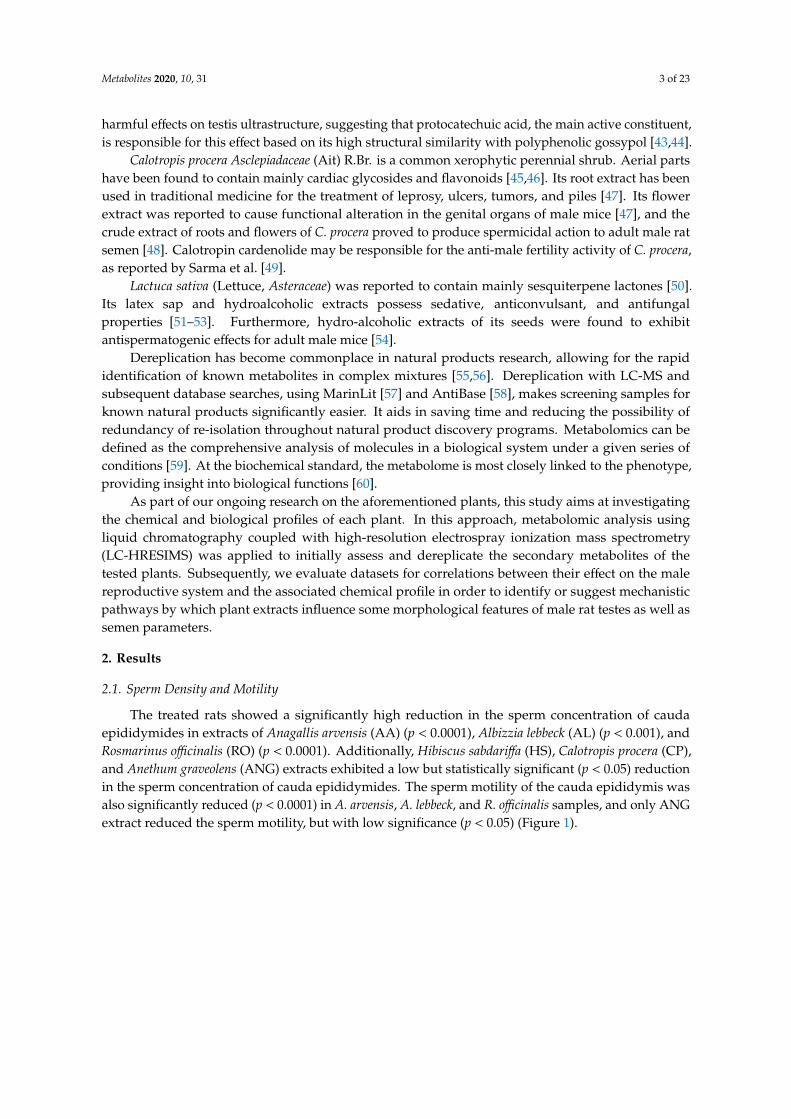
The treated rats showed a significantly high reduction in the sperm concentration of caudaepididymides in extracts of Anagallis arvensis (AA) (p < 0.0001), Albizzia lebbeck (AL) (p < 0.001), andRosmarinus officinalis (RO) (p < 0.0001). Additionally, Hibiscus sabdariffa (HS), Calotropis procera (CP),and Anethum graveolens (ANG) extracts exhibited a low but statistically significant (p < 0.05) reductionin the sperm concentration of cauda epididymides. The sperm motility of the cauda epididymis wasalso significantly reduced (p < 0.0001) in A. arvensis, A. lebbeck, and R. officinalis samples, and only ANGextract reduced the sperm motility, but with low significance (p < 0.05) (Figure 1).
Metabolites 2020, 10, 31 4 of 23
Metabolites 2019, 9, x 4 of 24
Figure 1. (A) Mean epididymal sperm number (×106 cells/mL) of rats exposed to different plant
extracts. (B) Mean percentage of epididymal sperm motility of rats exposed to different plant
extracts. AG: Apium graveolens, ANG: Anethum graveolens, AL: Albizzia lebbeck, MP: Menthae piperitae,
LS: Lactuca sativa, AA: Anagallis arvensis, CP: Calotropis procera, RO: Rosmarinus officinalis, CO:
Calendula officinalis, and HS: Hibiscus sabdariffa. Values are expressed as mean ± S.E.M. For each
group, N = 7. * Significantly different from control at p < 0.05, *** significantly different from control
at p < 0.001, and **** significantly different from control at p < 0.0001.
2.2. Hormonal Assay and Effect on Organ Weights
No significant difference was realized between the weights of treated rats and their sexual
organs (testis and seminal vesicle) compared to the control group. Additionally, there was no effect
on sex hormones levels (testosterone, FSH, and LH) of the treated animal group in comparison to the
untreated group (Supplementary Materials, Figures S1 and S2).
2.3. Histological Analysis
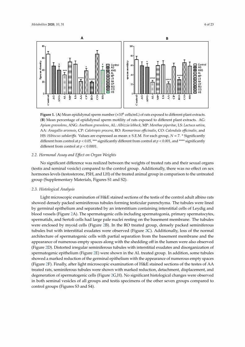
Light microscopic examination of H&E stained sections of the testis of the control adult albino
rats showed densely packed seminiferous tubules forming testicular parenchyma. The tubules were
lined by germinal epithelium and separated by an interstitium containing interstitial cells of Leydig
and blood vessels (Figure 2A). The spermatogenic cells including spermatogonia, primary
spermatocytes, spermatids, and Sertoli cells had large pale nuclei resting on the basement
membrane. The tubules were enclosed by myoid cells (Figure 2B). In the RO treated group, densely
packed seminiferous tubules but with interstitial exudates were observed (Figure 2C). Additionally,
loss of the normal architecture of spermatogenic cells with partial separation from the basement
membrane and the appearance of numerous empty spaces along with the shedding off in the lumen
were also observed (Figure 2D). Distorted irregular seminiferous tubules with interstitial exudates
and disorganization of spermatogenic epithelium (Figure 2E) were shown in the AL treated group.
In addition, some tubules showed a marked reduction of the germinal epithelium with the
appearance of numerous empty spaces (Figure 2F). Finally, after light microscopic examination of
H&E stained sections of the testes of AA treated rats, seminiferous tubules were shown with marked
reduction, detachment, displacement, and degeneration of spermatogenic cells (Figure 2G,H). No
significant histological changes were observed in both seminal vesicles of all groups and testis
specimens of the other seven groups compared to control groups (Figures S3 and S4).
A B
Figure 1. (A) Mean epididymal sperm number (×106 cells/mL) of rats exposed to different plant extracts.(B) Mean percentage of epididymal sperm motility of rats exposed to different plant extracts. AG:Apium graveolens, ANG: Anethum graveolens, AL: Albizzia lebbeck, MP: Menthae piperitae, LS: Lactuca sativa,AA: Anagallis arvensis, CP: Calotropis procera, RO: Rosmarinus officinalis, CO: Calendula officinalis, andHS: Hibiscus sabdariffa. Values are expressed as mean ± S.E.M. For each group, N = 7. * Significantlydifferent from control at p < 0.05, *** significantly different from control at p < 0.001, and **** significantlydifferent from control at p < 0.0001.
2.2. Hormonal Assay and Effect on Organ Weights
No significant difference was realized between the weights of treated rats and their sexual organs(testis and seminal vesicle) compared to the control group. Additionally, there was no effect on sexhormones levels (testosterone, FSH, and LH) of the treated animal group in comparison to the untreatedgroup (Supplementary Materials, Figures S1 and S2).
2.3. Histological Analysis
Light microscopic examination of H&E stained sections of the testis of the control adult albino ratsshowed densely packed seminiferous tubules forming testicular parenchyma. The tubules were linedby germinal epithelium and separated by an interstitium containing interstitial cells of Leydig andblood vessels (Figure 2A). The spermatogenic cells including spermatogonia, primary spermatocytes,spermatids, and Sertoli cells had large pale nuclei resting on the basement membrane. The tubuleswere enclosed by myoid cells (Figure 2B). In the RO treated group, densely packed seminiferoustubules but with interstitial exudates were observed (Figure 2C). Additionally, loss of the normalarchitecture of spermatogenic cells with partial separation from the basement membrane and theappearance of numerous empty spaces along with the shedding off in the lumen were also observed(Figure 2D). Distorted irregular seminiferous tubules with interstitial exudates and disorganization ofspermatogenic epithelium (Figure 2E) were shown in the AL treated group. In addition, some tubulesshowed a marked reduction of the germinal epithelium with the appearance of numerous empty spaces(Figure 2F). Finally, after light microscopic examination of H&E stained sections of the testes of AAtreated rats, seminiferous tubules were shown with marked reduction, detachment, displacement, anddegeneration of spermatogenic cells (Figure 2G,H). No significant histological changes were observedin both seminal vesicles of all groups and testis specimens of the other seven groups compared tocontrol groups (Figures S3 and S4).
Metabolites 2020, 10, 31 5 of 23Metabolites 2019, 9, x 5 of 24
Figure 2. A photomicrograph of a paraffin section in testis. (A) Testis of group I (control group)
showing densely packed seminiferous tubules (black arrows), separated by an interstitium
containing interstitial cells of Leydig and blood vessels (white arrows) (H&E ×200). (B) The
spermatogenic cells including spermatogonia (SG), primary spermatocytes (SC), spermatids (SD)
and spermatozoa (SZ). Sertoli cells (S) had large pale nuclei resting on the basement membrane. The
tubules are enclosed by myoid cells (arrow) (H&E ×400). (C) Testis of group H (Rosmarinus officinalis
treated group) showing densely packed seminiferous tubules but with interstitial exudates (Astrix)
(H&E ×200). (D) Loss of the normal architecture of spermatogenic cells with separation from the
basement membrane and appearance of numerous empty spaces (arrow) (H&E ×400). (E) Testis of
group C (Albizia lebbeck pods treated group) showing distorted irregular seminiferous tubules with
interstitial exudates (Astrix) and disorganization of spermatogenic epithelium (H&E ×200). (F)
Marked reduction of germinal epithelium and appearance of a lot of empty spaces (Astrix) (H&E
A
Figure 2. A photomicrograph of a paraffin section in testis. (A) Testis of group I (control group)showing densely packed seminiferous tubules (black arrows), separated by an interstitium containinginterstitial cells of Leydig and blood vessels (white arrows) (H&E ×200). (B) The spermatogenic cellsincluding spermatogonia (SG), primary spermatocytes (SC), spermatids (SD) and spermatozoa (SZ).Sertoli cells (S) had large pale nuclei resting on the basement membrane. The tubules are enclosed bymyoid cells (arrow) (H&E ×400). (C) Testis of group H (Rosmarinus officinalis treated group) showingdensely packed seminiferous tubules but with interstitial exudates (Astrix) (H&E ×200). (D) Loss ofthe normal architecture of spermatogenic cells with separation from the basement membrane andappearance of numerous empty spaces (arrow) (H&E ×400). (E) Testis of group C (Albizia lebbeck podstreated group) showing distorted irregular seminiferous tubules with interstitial exudates (Astrix) anddisorganization of spermatogenic epithelium (H&E×200). (F) Marked reduction of germinal epitheliumand appearance of a lot of empty spaces (Astrix) (H&E ×400). (G) Testis of group F (Anagallis arvensistreated group) showing seminiferous tubules with marked detachment (arrows) and disorganization ofspermatogenic epithelium (H&E ×200). (H) Marked reduction, detachment (arrows), displacement anddegeneration of spermatogenic cells (H&E ×400).
Metabolites 2020, 10, 31 6 of 23
2.4. Multivariate Analysis
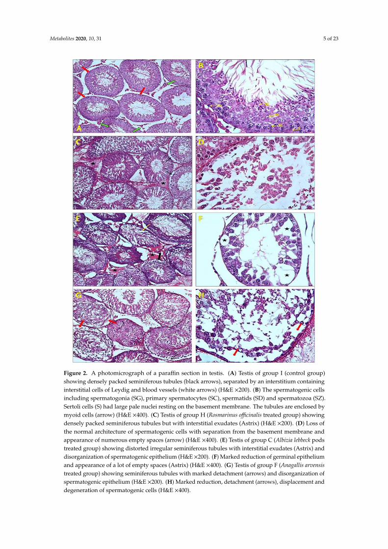
Processed data were analyzed by SIMCA-P V 13.0 (Umetrics, Umeå, Sweden) using principalcomponent analysis (PCA), the unsupervised statistical analysis method. PCA was utilized to detectdiffering features found in the outlying extracts to aid the prioritization of the extracts with interestingbioactive metabolomes. Two predominant outliers, AA and AL were observed, indicating that therewas high variance in the secondary metabolites found in these two extracts as they lay farthermostfrom the main group of aggregated samples in the score plot (Figure 3).
Metabolites 2019, 9, x 6 of 24
×400). (G) Testis of group F (Anagallis arvensis treated group) showing seminiferous tubules with
marked detachment (arrows) and disorganization of spermatogenic epithelium (H&E ×200). (H)
Marked reduction, detachment (arrows), displacement and degeneration of spermatogenic cells
(H&E ×400).
2.4. Multivariate Analysis
Processed data were analyzed by SIMCA-P V 13.0 (Umetrics, Umeå, Sweden) using principal
component analysis (PCA), the unsupervised statistical analysis method. PCA was utilized to detect
differing features found in the outlying extracts to aid the prioritization of the extracts with
interesting bioactive metabolomes. Two predominant outliers, AA and AL were observed,
indicating that there was high variance in the secondary metabolites found in these two extracts as
they lay farthermost from the main group of aggregated samples in the score plot (Figure 3).
Figure 3. Principal component score plot analysis of ten extracts clustered according to features (m/z
ratios) from mass spectral data (R2 = 0.4). 1: AG, 2: ANG, 3: MP, 4: RO, 5: AA, 6: CO, 7: AL, 8: CP, 9:
LS, and 10: HS.
2.5. Chemical Diversity of Natural Products in Plant Extracts
In this ongoing study, the mass resolution was 50,000 (at m/z 400), that is high enough to
distinguish closely related compounds. The total number of features detected in ten plant extracts by
LC-HRMS is documented in the supplementary material (Supplementary Tables S1–S10), and the
highest numbers of features were detected in the two outlaying extracts in addition to the bioactive
RO extract is documented in Table 1, Figure 4.
Figure 3. Principal component score plot analysis of ten extracts clustered according to features (m/zratios) from mass spectral data (R2 = 0.4). 1: AG, 2: ANG, 3: MP, 4: RO, 5: AA, 6: CO, 7: AL, 8: CP, 9:LS, and 10: HS.
2.5. Chemical Diversity of Natural Products in Plant Extracts
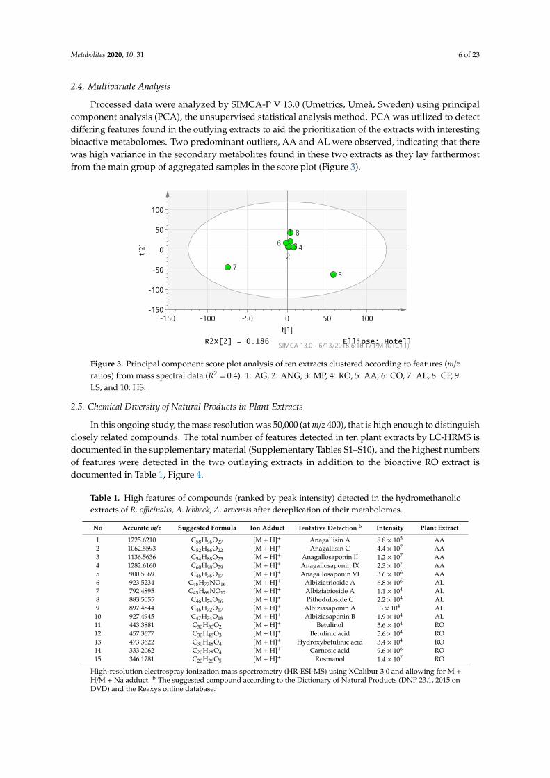
In this ongoing study, the mass resolution was 50,000 (at m/z 400), that is high enough to distinguishclosely related compounds. The total number of features detected in ten plant extracts by LC-HRMS isdocumented in the supplementary material (Supplementary Tables S1–S10), and the highest numbersof features were detected in the two outlaying extracts in addition to the bioactive RO extract isdocumented in Table 1, Figure 4.
Table 1. High features of compounds (ranked by peak intensity) detected in the hydromethanolicextracts of R. officinalis, A. lebbeck, A. arvensis after dereplication of their metabolomes.
No Accurate m/z Suggested Formula Ion Adduct Tentative Detection b Intensity Plant Extract
1 1225.6210 C58H96O27 [M + H]+ Anagallisin A 8.8 × 10⁵ AA2 1062.5593 C52H86O22 [M + H]+ Anagallisin C 4.4 × 10⁷ AA3 1136.5636 C54H88O25 [M + H]+ Anagallosaponin II 1.2 × 10⁷ AA4 1282.6160 C60H98O29 [M + H]+ Anagallosaponin IX 2.3 × 10⁷ AA5 900.5069 C46H76O17 [M + H]+ Anagallosaponin VI 3.6 × 10⁶ AA6 923.5234 C48H77NO16 [M + H]+ Albiziatrioside A 6.8 × 10⁶ AL7 792.4895 C43H69NO12 [M + H]+ Albiziabioside A 1.1 × 10⁴ AL8 883.5055 C46H74O16 [M + H]+ Pitheduloside C 2.2 × 10⁴ AL9 897.4844 C46H72O17 [M + H]+ Albiziasaponin A 3 × 10⁴ AL
10 927.4945 C47H74O18 [M + H]+ Albiziasaponin B 1.9 × 10⁴ AL11 443.3881 C30H50O2 [M + H]+ Betulinol 5.6 × 10⁴ RO12 457.3677 C30H48O3 [M + H]+ Betulinic acid 5.6 × 10⁴ RO13 473.3622 C30H48O4 [M + H]+ Hydroxybetulinic acid 3.4 × 10⁴ RO14 333.2062 C20H28O4 [M + H]+ Carnosic acid 9.6 × 10⁶ RO15 346.1781 C20H26O5 [M + H]+ Rosmanol 1.4 × 10⁷ RO
High-resolution electrospray ionization mass spectrometry (HR-ESI-MS) using XCalibur 3.0 and allowing for M +H/M + Na adduct. b The suggested compound according to the Dictionary of Natural Products (DNP 23.1, 2015 onDVD) and the Reaxys online database.
Metabolites 2020, 10, 31 7 of 23Metabolites 2019, 9, x 8 of 24
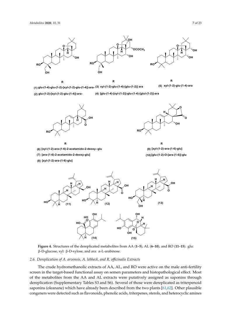
Figure 4. Structures of the dereplicated metabolites from AA (1–5), AL (6–10), and RO (11–15). glu:
β-D-glucose, xyl: β-D-xylose, and ara: α-L-arabinose.
Figure 4. Structures of the dereplicated metabolites from AA (1–5), AL (6–10), and RO (11–15). glu:β-D-glucose, xyl: β-D-xylose, and ara: α-L-arabinose.
2.6. Dereplication of A. arvensis, A. lebbeck, and R. officinalis Extracts
The crude hydromethanolic extracts of AA, AL, and RO were active on the male anti-fertilityscreen in the target-based functional assay on semen parameters and histopathological effect. Mostof the metabolites from the AA and AL extracts were putatively assigned as saponins throughdereplication (Supplementary Tables S3 and S6). Several of those were dereplicated as triterpenoidsaponins (oleanane) which have already been described from the two plants [61,62]. Other plausiblecongeners were detected such as flavonoids, phenolic acids, triterpenes, sterols, and heterocyclic amines
Metabolites 2020, 10, 31 8 of 23
(Supplementary Tables S3 and S6). RO extract contained many metabolites, including flavonoids,phenolic acids, triterpenes (lupane derivatives), and phenolic diterpenes (Supplementary Table S9). Byapplying an algorithm to the data from this extract to calculate the intensity of each m/z of parent ionpeaks, a plethora of metabolites with high intensities were detected, especially triterpenes (lupanederivatives) and phenolic diterpenes (Table 1).
2.7. Modelling Study
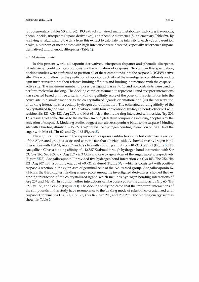
In this present work, all saponin derivatives, triterpenes (lupane) and phenolic diterpenes(abietatriene) could induce apoptosis via the activation of caspases. To confirm this speculation,docking studies were performed to position all of these compounds into the caspase-3 (1GFW) activesite. This would allow for the prediction of apoptotic activity of the investigated constituents and togain further insight into their relative binding affinities and binding interactions with the caspase-3active site. The maximum number of poses per ligand was set to 10 and no constraints were used toperform molecular docking. The docking complex assumed to represent ligand-receptor interactionswas selected based on three criteria: (i) binding affinity score of the pose, (ii) its orientation into theactive site in a similar manner as the co-crystallized ligands orientation, and (iii) the preservationof binding interactions, especially hydrogen bond formation. The estimated binding affinity of theco-crystallized ligand was −11.425 Kcal/mol, with four conventional hydrogen bonds observed withresidue His 121, Gly 122, Arg 207, and Met 61. Also, the indole ring interacted with residue Trp 206.This result gives some clue as to the mechanism of high feature compounds inducing apoptosis by theactivation of caspase-3. Modeling studies suggest that albizeasaponin A binds to the caspase-3 bindingsite with a binding affinity of −15.227 Kcal/mol via the hydrogen bonding interaction of the OHs of thesugar with Met 61, Thr 62, and Cys 163 (Figure 5).
The significant increase in the expression of caspase-3 antibodies in the testicular tissue sectionof the AL treated group is associated with the fact that albiziabioside A showed five hydrogen bondinteractions with Met 61, Arg 207, and Cys 163 with a binding affinity of−10.731 Kcal/mol (Figure 5C,D).Anagallicin C has a binding affinity of −12.587 Kcal/mol through hydrogen bond interaction with Ser63, Cys 163, Ser 205, and Arg 207 via 3 OHs and one oxygen atom of the sugar moiety, respectively(Figure 5E,F). Anagallosaponin II provided five hydrogen bond interaction via Cys 163, Phe 252, His121, Arg 207 with a binding energy of −9.921 Kcal/mol (Figure 5G), which is consistent with positivecaspase-3 reaction in the cytoplasm of germinal cells of the AA treated group. Anagallosaponin IX,which is the third-highest binding energy score among the investigated derivatives, showed the keybinding interaction of the co-crystallized ligand which includes hydrogen bonding interactions ofArg 207 and Met 61. In addition, other interactions can be observed for the amino acids Gly 60, Thr62, Cys 163, and Ser 205 (Figure 5H). The docking study indicated that the important interactions ofthe compounds in this study have resemblance to the binding mode of celastrol co-crystallized withcaspase-3 enzyme via His 121, Gly 122, Cys 163, Asn 208, and Phe 252. The binding energy score isshown in Table 2.
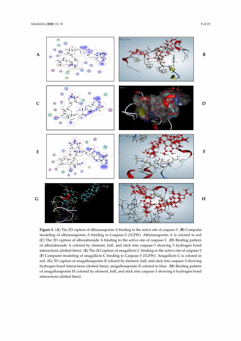
Metabolites 2020, 10, 31 9 of 23Metabolites 2019, 9, x 10 of 24
Figure 5. (A) The 2D caption of albizeasaponin A binding to the active site of caspase-3. (B)
Computer modeling of albizeasaponin A binding to Caspase-3 (1GFW). Albizeasaponin A is colored
in red. (C) The 2D caption of albiziabioside A binding to the active site of caspase-3. (D) Binding
pattern of albiziabioside A colored by element, ball, and stick into caspase-3 showing 5 hydrogen
bond interactions (dotted lines). (E) The 2D caption of anagallicin C binding to the active site of
caspase-3. (F) Computer modeling of anagallicin C binding to Caspase-3 (1GFW). Anagallicin C is
colored in red. (G) 3D caption of anagallosaponin II colored by element, ball, and stick into caspase-3
showing hydrogen bond interactions (dotted lines), anagallosaponin II colored in blue. (H) Binding
pattern of anagallosaponin IX colored by element, ball, and stick into caspase-3 showing 6 hydrogen
bond interactions (dotted lines).
A B
C D
E F
G H
Figure 5. (A) The 2D caption of albizeasaponin A binding to the active site of caspase-3. (B) Computermodeling of albizeasaponin A binding to Caspase-3 (1GFW). Albizeasaponin A is colored in red.(C) The 2D caption of albiziabioside A binding to the active site of caspase-3. (D) Binding patternof albiziabioside A colored by element, ball, and stick into caspase-3 showing 5 hydrogen bondinteractions (dotted lines). (E) The 2D caption of anagallicin C binding to the active site of caspase-3.(F) Computer modeling of anagallicin C binding to Caspase-3 (1GFW). Anagallicin C is colored inred. (G) 3D caption of anagallosaponin II colored by element, ball, and stick into caspase-3 showinghydrogen bond interactions (dotted lines), anagallosaponin II colored in blue. (H) Binding patternof anagallosaponin IX colored by element, ball, and stick into caspase-3 showing 6 hydrogen bondinteractions (dotted lines).
Metabolites 2020, 10, 31 10 of 23
Table 2. The binding energy score ranked results of target compounds-caspase-3 complexbinding conformations.
Compound Name Binding Energy Score * Average Number of Poses per Run
23-OH-betulinic acid −7.474 10Albizeasaponin A −15.227 10Albiziabioside A −10.731 10Albiziasaponin B −14.591 10Albiziatrioside A −8.712 10
Anagallicin A −11.562 10Anagallicin C −12.587 10
Anagallosaponin II −9.921 10Anagallosaponin IX −13.087 10Anagallosaponin VI −10.545 10
Betulinic acid −7.628 10Betulinol −9.518 9
Carnosic acid −9.362 8Pitheduloside C −10.143 10
Rosmanol −9.902 4
* The shown score is the mean of three consecutive runs. The docking method was validated by a successfulpose-retrieval docking experiment of the ligand (score: −11.425).
2.8. Immunohistochemical Assay
2.8.1. β-Catenin
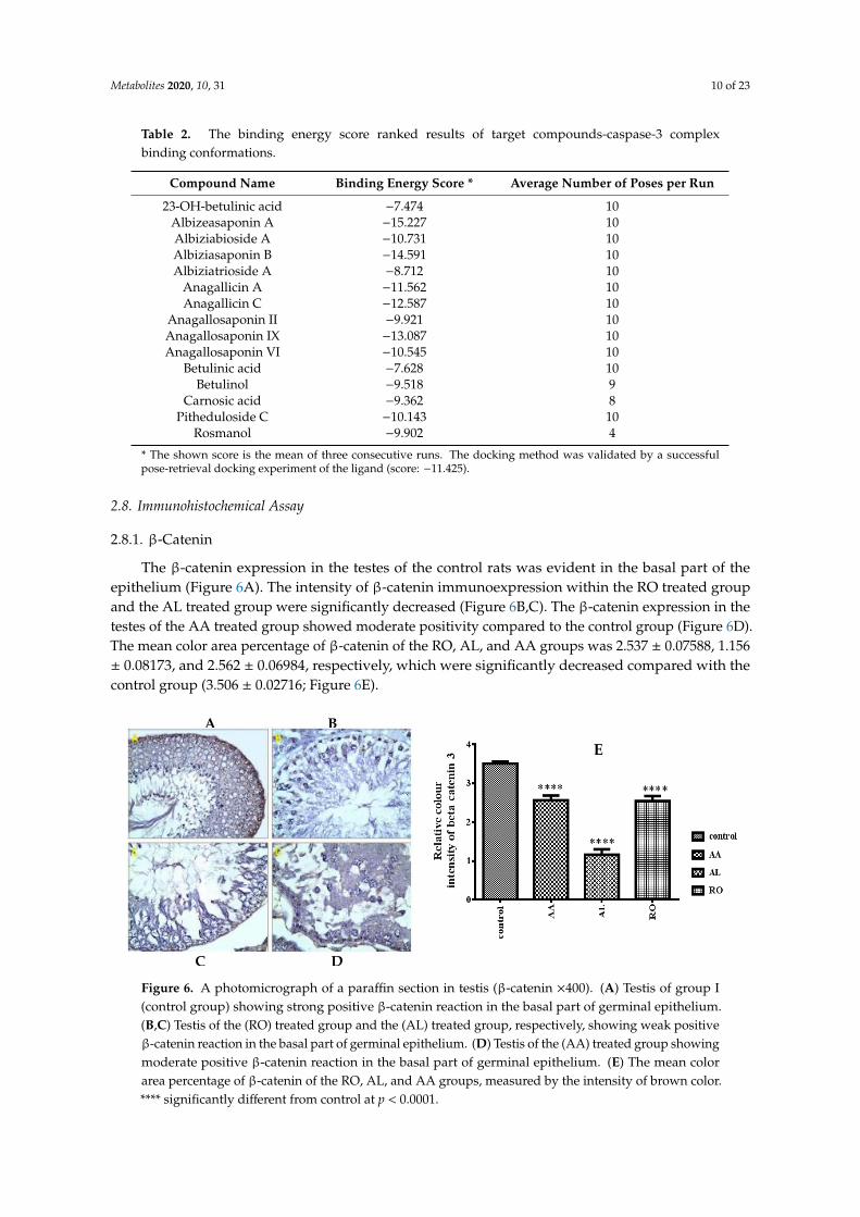
The β-catenin expression in the testes of the control rats was evident in the basal part of theepithelium (Figure 6A). The intensity of β-catenin immunoexpression within the RO treated groupand the AL treated group were significantly decreased (Figure 6B,C). The β-catenin expression in thetestes of the AA treated group showed moderate positivity compared to the control group (Figure 6D).The mean color area percentage of β-catenin of the RO, AL, and AA groups was 2.537 ± 0.07588, 1.156± 0.08173, and 2.562 ± 0.06984, respectively, which were significantly decreased compared with thecontrol group (3.506 ± 0.02716; Figure 6E).
Metabolites 2019, 9, x 11 of 24
Table 2. The binding energy score ranked results of target compounds-caspase-3 complex binding
conformations.
Compound Name Binding Energy Score * Average Number of Poses per Run
23-OH-betulinic acid −7.474 10
Albizeasaponin A −15.227 10
Albiziabioside A −10.731 10
Albiziasaponin B −14.591 10
Albiziatrioside A −8.712 10
Anagallicin A −11.562 10
Anagallicin C −12.587 10
Anagallosaponin II −9.921 10
Anagallosaponin IX −13.087 10
Anagallosaponin VI −10.545 10
Betulinic acid −7.628 10
Betulinol −9.518 9
Carnosic acid −9.362 8
Pitheduloside C −10.143 10
Rosmanol −9.902 4
* The shown score is the mean of three consecutive runs. The docking method was validated by a
successful pose-retrieval docking experiment of the ligand (score: –11.425).
2.8. Immunohistochemical Assay
2.8.1. β-catenin
The β-catenin expression in the testes of the control rats was evident in the basal part of the
epithelium (Figure 6A). The intensity of β-catenin immunoexpression within the RO treated group
and the AL treated group were significantly decreased (Figure 6B,C). The β-catenin expression in the
testes of the AA treated group showed moderate positivity compared to the control group (Figure
6D). The mean color area percentage of β-catenin of the RO, AL, and AA groups was 2.537 ± 0.07588,
1.156 ± 0.08173, and 2.562 ± 0.06984, respectively, which were significantly decreased compared with
the control group (3.506 ± 0.02716; Figure 6E).
Figure 6. A photomicrograph of a paraffin section in testis (β-catenin ×400). (A) Testis of group I
(control group) showing strong positive β-catenin reaction in the basal part of germinal epithelium.
(B,C) Testis of the (RO) treated group and the (AL) treated group, respectively, showing weak
positive β-catenin reaction in the basal part of germinal epithelium. (D) Testis of the (AA) treated
group showing moderate positive β-catenin reaction in the basal part of germinal epithelium. (E) The
mean color area percentage of β-catenin of the RO, AL, and AA groups, measured by the intensity of
brown color. **** significantly different from control at p < 0.0001.
E
A B
C D
Figure 6. A photomicrograph of a paraffin section in testis (β-catenin ×400). (A) Testis of group I(control group) showing strong positive β-catenin reaction in the basal part of germinal epithelium.(B,C) Testis of the (RO) treated group and the (AL) treated group, respectively, showing weak positiveβ-catenin reaction in the basal part of germinal epithelium. (D) Testis of the (AA) treated group showingmoderate positive β-catenin reaction in the basal part of germinal epithelium. (E) The mean colorarea percentage of β-catenin of the RO, AL, and AA groups, measured by the intensity of brown color.**** significantly different from control at p < 0.0001.
Metabolites 2020, 10, 31 11 of 23
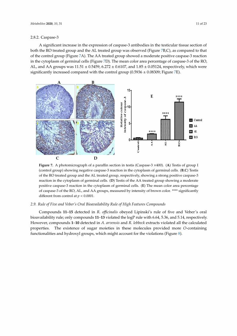
2.8.2. Caspase-3
A significant increase in the expression of caspase-3 antibodies in the testicular tissue section ofboth the RO treated group and the AL treated group was observed (Figure 7B,C), as compared to thatof the control group (Figure 7A). The AA treated group showed a moderate positive caspase-3 reactionin the cytoplasm of germinal cells (Figure 7D). The mean color area percentage of caspase-3 of the RO,AL, and AA groups was 11.51 ± 0.5459, 6.272 ± 0.6107, and 1.85 ± 0.05124, respectively, which weresignificantly increased compared with the control group (0.5936 ± 0.08309; Figure 7E).
Metabolites 2019, 9, x 12 of 24
2.82. Caspase-3
A significant increase in the expression of caspase-3 antibodies in the testicular tissue section of
both the RO treated group and the AL treated group was observed (Figure 7B,C), as compared to
that of the control group (Figure 7A). The AA treated group showed a moderate positive caspase-3
reaction in the cytoplasm of germinal cells (Figure 7D). The mean color area percentage of caspase-3
of the RO, AL, and AA groups was 11.51 ± 0.5459, 6.272 ± 0.6107, and 1.85 ± 0.05124, respectively,
which were significantly increased compared with the control group (0.5936 ± 0.08309; Figure 7E).
Figure 7. A photomicrograph of a paraffin section in testis (Caspase-3 ×400). (A) Testis of group I
(control group) showing negative caspase-3 reaction in the cytoplasm of germinal cells. (B,C) Testis
of the RO treated group and the AL treated group, respectively, showing a strong positive caspase-3
reaction in the cytoplasm of germinal cells. (D) Testis of the AA treated group showing a moderate
positive caspase-3 reaction in the cytoplasm of germinal cells. (E) The mean color area percentage of
caspase-3 of the RO, AL, and AA groups, measured by intensity of brown color. **** significantly
different from control at p < 0.0001
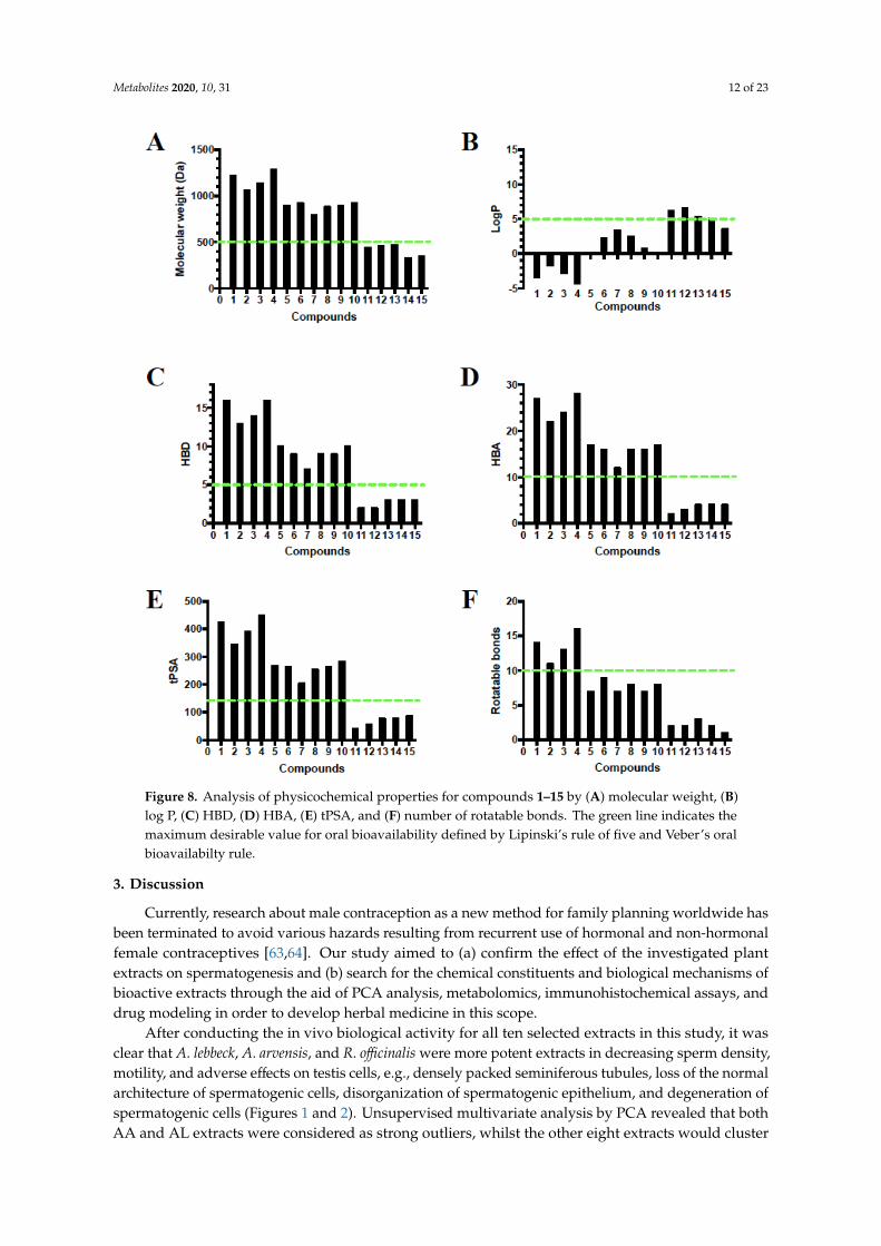
2.9. Rule of Five and Veber’s Oral Bioavailability Rule of High Features Compounds
Compounds 11–15 detected in R. officinalis obeyed Lipinski’s rule of five and Veber’s oral
bioavailability rule; only compounds 11–13 violated the logP rule with 6.64, 5.36, and 5.14,
respectively. However, compounds 1–10 detected in A. arvensis and R. lebbeck extracts violated all the
calculated properties. The existence of sugar moieties in these molecules provided more
O-containing functionalities and hydroxyl groups, which might account for the violations (Figure 8).
E
A B
C D
Figure 7. A photomicrograph of a paraffin section in testis (Caspase-3 ×400). (A) Testis of group I(control group) showing negative caspase-3 reaction in the cytoplasm of germinal cells. (B,C) Testisof the RO treated group and the AL treated group, respectively, showing a strong positive caspase-3reaction in the cytoplasm of germinal cells. (D) Testis of the AA treated group showing a moderatepositive caspase-3 reaction in the cytoplasm of germinal cells. (E) The mean color area percentageof caspase-3 of the RO, AL, and AA groups, measured by intensity of brown color. **** significantlydifferent from control at p < 0.0001.
2.9. Rule of Five and Veber’s Oral Bioavailability Rule of High Features Compounds
Compounds 11–15 detected in R. officinalis obeyed Lipinski’s rule of five and Veber’s oralbioavailability rule; only compounds 11–13 violated the logP rule with 6.64, 5.36, and 5.14, respectively.However, compounds 1–10 detected in A. arvensis and R. lebbeck extracts violated all the calculatedproperties. The existence of sugar moieties in these molecules provided more O-containingfunctionalities and hydroxyl groups, which might account for the violations (Figure 8).
Metabolites 2020, 10, 31 12 of 23Metabolites 2019, 9, x 13 of 24
Figure 8. Analysis of physicochemical properties for compounds 1–15 by (A) molecular weight, (B)
log P, (C) HBD, (D) HBA, (E) tPSA, and (F) number of rotatable bonds. The green line indicates the
maximum desirable value for oral bioavailability defined by Lipinski’s rule of five and Veber’s oral
bioavailabilty rule.
3. Discussion
Currently, research about male contraception as a new method for family planning worldwide
has been terminated to avoid various hazards resulting from recurrent use of hormonal and
non-hormonal female contraceptives [63,64]. Our study aimed to (a) confirm the effect of the
investigated plant extracts on spermatogenesis and (b) search for the chemical constituents and
biological mechanisms of bioactive extracts through the aid of PCA analysis, metabolomics,
immunohistochemical assays, and drug modeling in order to develop herbal medicine in this scope.
After conducting the in vivo biological activity for all ten selected extracts in this study, it was
clear that A. lebbeck, A. arvensis, and R. officinalis were more potent extracts in decreasing sperm
density, motility, and adverse effects on testis cells, e.g., densely packed seminiferous tubules, loss of
the normal architecture of spermatogenic cells, disorganization of spermatogenic epithelium, and
degeneration of spermatogenic cells (Figures 1 and 2). Unsupervised multivariate analysis by PCA
revealed that both AA and AL extracts were considered as strong outliers, whilst the other eight
Figure 8. Analysis of physicochemical properties for compounds 1–15 by (A) molecular weight, (B)log P, (C) HBD, (D) HBA, (E) tPSA, and (F) number of rotatable bonds. The green line indicates themaximum desirable value for oral bioavailability defined by Lipinski’s rule of five and Veber’s oralbioavailabilty rule.
3. Discussion
Currently, research about male contraception as a new method for family planning worldwide hasbeen terminated to avoid various hazards resulting from recurrent use of hormonal and non-hormonalfemale contraceptives [63,64]. Our study aimed to (a) confirm the effect of the investigated plantextracts on spermatogenesis and (b) search for the chemical constituents and biological mechanisms ofbioactive extracts through the aid of PCA analysis, metabolomics, immunohistochemical assays, anddrug modeling in order to develop herbal medicine in this scope.
After conducting the in vivo biological activity for all ten selected extracts in this study, it wasclear that A. lebbeck, A. arvensis, and R. officinalis were more potent extracts in decreasing sperm density,motility, and adverse effects on testis cells, e.g., densely packed seminiferous tubules, loss of the normalarchitecture of spermatogenic cells, disorganization of spermatogenic epithelium, and degeneration ofspermatogenic cells (Figures 1 and 2). Unsupervised multivariate analysis by PCA revealed that bothAA and AL extracts were considered as strong outliers, whilst the other eight extracts would cluster
Metabolites 2020, 10, 31 13 of 23
together, including RO, even though it was one of the three most biologically active extracts (Figure 3).Therefore, to find an explanation for this outcome, metabolomics using LC-HRMS and dereplication ofall investigated extracts were conducted to detect different metabolite classes, distinguish betweenthem, and understand the reason for the variability in the anti-male fertility activities (SupplementaryTables S1–S10).
In accordance with metabolomics and dereplication of the ten tested extracts, it was clear that inboth outliers AL and AA, oleanane saponins were detected as high features, while lupane triterpenes,phenolic diterpenes, and some polyphenolics (flavonoids and phenolic acids) were detected in highconcentrations in RO (Table 1, Figure 4). Most of these metabolites were actually reported in thesethree bioactive plants, e.g., albizeasaponins A–B were isolated from A. lebbeck bark; albiziatriozide wasisolated from A. lebbeck leaves [4,65]; anagallisins A–C, anagallosaponins II, VI, and IX were isolatedfrom A. arvensis [5,62,66]; and betulinol, betulinic acid, 23-hydroxybetulinic, rosmanol, and carnosicacid were isolated from R. officinalis [67–71].
Extensive searches about these compound classes and their effects on spermatogenesis showedthat there were numerous studies that evaluated the antispermatogenic effect of these metabolites;more specifically, the antispermatogenic effect of oleanane saponins was investigated [10,72], theeffect of lupeol and lupeol-rich plant extracts on spermatogenesis was examined [73–76], and theaction of diterpenes on testes was discussed [77,78]. There is no available data to confirm the adverseeffects of polyphenolics on the male reproductive system except gossypol [3,79–81], and all studiesreported about this action emphasized the cytoprotective and antioxidant activities for male sexualorgans and sperm after the administration of polyphenolic-rich plants [82–85]. Therefore, our studyfocused specifically on oleanane saponins, lupane triterpenes, and phenolic diterpenes, and to elucidatethe mechanism of how they affect spermatogenesis, while polyphenolics were outside the scope ofthis study.
The classes of compounds which drew our attention were reported to have apoptotic actionon different cancer cell lines via up-regulation of caspase-3 or down-regulation of β-catenin,concerning phenolic diterpenes [86–89], betulinic acid and its natural derivatives [90,91], and oleananesaponins [92–95]. In particular, sasanguasaponin (oleanane saponin) isolated from defatted seedsof Camellia oleifera was reported to induce spermatogenic cell apoptosis in vivo via a caspase-3activation reaction [72], and Tripterygium wilfordii extract, a male contraceptive diterpene-rich plant thatdemonstrated an apoptosis action on male rat germ cells and induced a modification for epigenetichistone [96]. Therefore, these metabolites could probably induce apoptosis in testes tissues. Our studywas directed towards an immunohistochemical assay for the three most bioactive extracts (RO, AL,and AA) by measuring the degree of reaction between each extract and the two proteins (β-cateninand caspase-3). Their expression plays an essential role in causing apoptosis, particularly in testes cells(β-catenin), and generally in other organs cells including testes (caspase-3; Figures 6 and 7). Clearly,spermatid-specific deletion of β-catenin resulted in disruption of adherence junctions between Sertolicells and elongating spermatids, acrosomal defects, increased germ cell apoptosis, and in consequence,reduced sperm count and impaired fertility. In addition, caspase-3 is a member of the cysteine-asparticacid protease (caspase) family, where its sequential activation is considered as one of important keys inthe cellular apoptosis process [97,98].
Morphometric study of testes sections of RO, AL, and AA after application of β-catenin andcaspase-3 immunohistochemistry indicated a significant decrease and increase of immunoexpressionof both β-catenin and caspase-3, respectively, in all three treatment groups, which confirmed theoccurrence of apoptosis in these specimens (Figures 6 and 7). Moreover, high concentration metabolites,as detected via LC-MS, were subjected to modeling studies measuring the extent of binding between thetarget compounds and caspase enzyme (Table 2, Figure 5) to prove if these compounds are responsiblefor the incidence of apoptosis in testes tissues. Results suggested that these high concentrationconstituents may be substantially contributing to a reduction in spermatogenesis, particularly oleanane
Metabolites 2020, 10, 31 14 of 23
saponins, due to their relative binding affinities, and binding interactions with the caspase-3 active sitehad the highest docking score.
4. Material and Methods
4.1. Chemicals and Reagents
HPLC grade acetonitrile was purchased from TEDIA Company Inc. 79 (Fairfield, CT, USA).Ultra-pure water was purified by an EPED super purification system (Nanjing, China). Formic acidwas obtained from Merck KGaA (Darmstadt, Germany). Methanol (MeOH) used for extraction,formaldehyde, glacial acetic acid, and ethanol used for the preparation of 10% formalin and Davidson’ssolution were purchased from El-Nasr Company for Pharmaceuticals and Chemicals, Egypt, and wasdistilled before use.
4.2. Plant Material
Leaves of Menthae piperitae (MP) and Apium graveolens (AG) and flowers of Calendula officinalis(CO) were collected in February 2016 from plants cultivated in the campus of Cairo University, Cairo,Egypt, while seeds of Lactuca sativa (LS) and Anethum graveolens (ANG), leaves of Rosmarinus officinalis(RO) and calyces of Hibiscus sabdariffa (HS) were purchased in February 2016 from Harrraz, an officialseed trader, Cairo, Egypt. In addition, aerial parts of Calotropis procera (CP) were collected in March2016 from a desert land, located in the desert Cairo-Ismaili road, Egypt. Albizia lebbeck pods (AL) werecollected in March 2016 from a field for medicinal plants, El Qanater El Khayreya, Qalyubia, Egypt,and a Anagallis arvensis (AA) whole plant was collected in January 2016 from a field found in BanhaCapital, Qalyubia, Egypt. Authentication of the plants was established by Professor Abdel-Halim A.Mohammed, Horticultural Research Institute, Department of Flora and Phytotaxonomy Researches,Dokki, Cairo, Egypt. Voucher specimens (2017-BuPD 51–60), respectively, were deposited at theDepartment of Pharmacognosy, Faculty of Pharmacy, Cairo University, Egypt.
4.3. Plant Extraction
The air-dried, powdered plant parts (1 kg) of previously mentioned plants were extracted bymaceration with 80% MeOH at room temperature, and then concentrated under reduced pressureusing a rotary evaporator (IKA, Königswinter, Germany) to a syrupy consistency. The concentratedmethanolic extract yields were 60, 105, 85, 230, 75, 100, 80, 200, 150, and 120 g of AL, CP, HS, LS, AG,AA, ANG, CO, RO, and MP, respectively. All the resulting dried extracts were kept at 4 ◦C for thebiological and metabolomic investigations.
4.4. Oral Acute Toxicity Study
This study for ten tested samples, previously mentioned, was performed to determine their safedoses [99]. For each extract, 24 adult male rats (Sprague–Dawley) of about 150 ± 20 g were starvedovernight and randomly divided into 4 groups (6 rats each). Rats were kept overnight fasting thensubjected to a daily sole dose of 1000, 2000, 4000, and 5000 mg/kg b.w of each examined extract. Theobservation of animals individually performed after 24 and 72 h with a daily observation. After thisperiod, the LD50 was calculated, and the pharmacological evaluation of the extracts was carried outat a fixed daily dose of 10% of LD50. As shown in Supplementary Table S11, all previously reportedtoxicity studies and LD50 of the investigated extracts or their alternatives had a wide therapeutic indexand safety margins, and no adverse effects or toxicities were produced except in very high doses and/orchronic administration of few number of evaluated plants. These findings were considered to be inhigh agreement with our oral acute toxicity study of chosen plant extracts, and thus suggesting thatusing of fixed daily doses of 10% of LD50 for consecutive 60 days could not produce any mortality orserious toxicity of different organs.
Metabolites 2020, 10, 31 15 of 23
4.5. Animals Grouping, Modelling and Drugs Administration
Seventy-seven adult male albino rats (150–200 g each), in agreement with guidelines of the careand use for laboratory animals of the National Institutes of Health [100], was approved by the ResearchEthics Committee for Animal Experimentation, Department of Pharmacology and Toxicology, Facultyof Pharmacy, Minia University, Egypt (project code No. 2018:022). Rats were housed and then bredunder systematized conditions in the pre-clinical animal house. Rats were kept in stainless steelcages (seven per cage), fed a suitable diet, and allowed unlimited access to drinking water. Theywere acclimatized to the environment for one week before the commencement of the experiment. Allconditions were also made to diminish animal suffering.
The 77 male rats were divided into 11 groups, each group consisting of seven animals. Eachindividual rat in each group between the ten treatment groups was orally received 10% of LD50 dailydose for 60 days of its specific extract for its definite group. This daily oral dose was 500 mg/kg, b.wfor all ten treatment groups except AL and AA groups, of which the dose was 200 mg/kg, b.w, and thecontrol group received the vehicle (normal saline, 2 mL/kg, b.w). At day 60, rats were anaesthetized bythiopental injection (45 mg/kg b.w), then the blood samples were collected using the retino-orbitalsinus, centrifuged for 15 min (3000 rpm/min) to obtain the serum that stored at −80 ◦C for estimationof LH, FSH and testosterone levels. An incision was made longitudinally in the scrotum by the waythat both testes were exposed. Cauda epididymis was chosen for the collection of semen samplesused in semen analysis. The testes, prostate and seminal vesicles were dissected and weighed. Testeswere preserved in Davidson’s solution, but seminal vesicles were preserved in 10% neutral formalinsolution until histopathological examination processing.
4.6. Hormonal Assay
Blood samples were collected in the fasting state between 07:00 and 10:00 h. Serum LH,total testosterone, and FSH were quantitatively determined by commercial electrochemiluminescenceimmunoassay methods (Elecsys 2010, Roche Diagnostics, Mannheim, Germany). For all parameters,the interassay and intraassay coefficients of variation were <10% and 8%, respectively.
4.7. Semen Analysis
This analysis in epididymis was performed by the cutting of cauda epididymis surgically by usingsurgical blades, then squeezed into a sterile clean watch glass to obtain semen content. The contentwas diluted with 0.09% NaCl isotonic solution and mixed for estimation of the sperm progressivemotility and density, as described by [101].
4.8. Histological Analysis
Sexual organs specimens were fixed in the previously mentioned solutions. They were dehydratedin an increasing gradient of ethanol, cleared in xylene, and embedded in paraffin. Serial sections of5–7 µm thickness were cut and subjected to H&E staining [102].
4.9. Metabolomics and PCA Analyses
The crude hydromethanolic extracts of ten plants, previously mentioned above, were subjected tometabolomic analysis using LC-HR-ESI-MS analytical techniques [103]. Briefly, the total extract ofeach plant (1 mg/mL in MeOH) was uploaded on an Accela HPLC (Thermo Fisher Scientific, Bremen,Germany) combined with Accela UV–vis and Exactive (Orbitrap) mass spectrometer from ThermoFisher Scientific (Bremen, Germany). The mobile phase composed of HPLC grade water (A) andacetonitrile (B) with 0.1% formic acid in each solvent. The gradient elution started at a flow rate of300 µL/min with 10% B linearly increased to 100% B within 30 min and remained isocratic for the next5 min before linearly decreasing back to 10% B for the following 1 min. Afterwards, the mobile phasewas equilibrated for 9 min before the next injection. The mass range was set from m/z (mass-to-charge
Metabolites 2020, 10, 31 16 of 23
ratio) 100–2000 for ESI-MS using in-source CID (collision-induced dissociation) mechanism andm/z 50–1000 for MS/MS using untargeted HCD (high energy collision dissociation). The raw datawere imported to MZmine 2.12, a framework for the disparate analysis of mass spectrometry data.Deconvolution of chromatogram was then performed followed by deisotoping of peaks. The retentiontime normalizer was applied for chromatographic alignment and gap-filling. For the combinationof negative and positive ionization mode data files that were generated by MZmine, Excel macroswere used. Peaks produced from the samples were then extracted. The Excel macro was used fordereplicating each m/z ion peak with constituents in the customized database (using RT and m/zthreshold of ± 5 ppm), which provided the putative identities of all metabolomes in the total extract(s)in details. The macro was then applied for detection the top 20 features (arranged ascendingly bypeak intensity) and the corresponding putative identities by producing a list for the extract. Therefore,diverse constituents were detected in selected extracts by rapprochement with some in-house andonline databases.
The resultant data matrices were introduced to the Extended Statistical tool EZinfo v2.0 software(Umetrics AB, Umeå, Sweden) for multivariate analysis. The data were scaled to unit variancefor principal component analysis (PCA) to give an overview of the repeatability of tested samples.The samples with high repeatability should cluster together in the score plot of PCA.
4.10. Modelling Study
Docking calculations were carried out using the molecular operating environment (MOE).The structure of the compounds was built by Chemdraw Ultra 8.0. Docking calculations were carriedout on a caspase-3 complex (PDB code 1GFW, http://www.rcsb.org/pdb/home/home.do). A dockingsimulation was performed on the test compounds with the following protocol. (1) Enzyme structureswere checked for missing atoms, bonds, and contacts. (2) Hydrogen atoms were added to theenzyme structure. (3) The ligand molecules were constructed using the builder module and wereenergy-minimized. (4) The active site was generated using the MOEAlpha site finder. (5) Ligands weredocked within the caspase-3 active site using MOEDock with simulated annealing utilized as the searchprotocol and the CHARMm molecular mechanics force field. (6) The lowest energy conformation ofthe docked ligand complex was selected and subjected to a further energy minimization using theCHARMm force field.
To determine the accuracy of this docking protocol, the crystallized ligand, was re-docked into thecaspase-3 active site. This procedure was repeated three times, and the best ranked solutions of theligand exhibited RMSD values of 1.89 Å from the position of the crystallized ligand for caspase-3. Ingeneral, RMSD values smaller than 2.0 Å indicate that the docking protocol is capable of accuratelypredicting the binding orientation of the crystallized ligand. Lowest energy aligned conformation(s)were identified. This protocol was thus deemed to be suitable for the docking of the test compoundsinto the active site model of the caspase-3 complex.
4.11. Immunohistochemical Assay
Testis specimens were fixed in Davidson’s solution. They were dehydrated in an increasing gradientof ethanol, cleared in xylene, and embedded in paraffin. Serial sections of 5–7 µm thickness were cutand subjected to immunohistochemical staining, as follows: (a) 1-β-catenin (it is a ready-to-use rabbitpolyclonal antibody; Santa Cruz Biotechnology, catalogue number sc-7199); and (b) 2-anti-caspase-3antibody to demonstrate apoptosis (it is a ready-to-use rabbit polyclonal antibody; NEO markers,Thermo Scientific Laboratories, USA, catalogue number RB-1197-R7) [104].
4.11.1. Immunohistochemical Procedure
Sections were incubated in H2O2 for 15 min then rinsed in PBS (Sigma-Aldrich, Saint Louis, MO,USA). They were boiled in 10 Mm citrate buffer pH 6 (Lab Vision Corporation Laboratories, catalogno AP 9003) for 10 min for antigen retrieval and left to cool in room temperature, then rinsed in PBS.
Metabolites 2020, 10, 31 17 of 23
To eliminate non-specific background, sections were incubated with two drops of Ultra V Block for5 min and then incubated for 60 min with two drops of the primary antibody (this was omitted inthe negative control). Afterwards, slides were incubated for 10 min with two drops of biotinylatedgoat anti-polyvalent secondary antibody at room temperature, then rinsed well with PBS. Followed byincubation with two drops of streptavidin-peroxidase for 10 min at room temperature then washed inPBS. One drop of DAB Plus chromogen was mixed with 2 mL of DAB Plus substrate, then applied tothe slides and incubated for 10 min at room temperature, then rinsed well with distilled water. Slideswere then counterstained with Mayer’s hematoxylin (Lab Vision Corporation Laboratories, cat noTA-060-MH), dehydrated, and mounted.
4.11.2. Morphometric Study
Data were obtained using a Leica Qwin 500 C image analyzer computer system (Kasr Alaini, CairoUniversity, Cairo, Egypt). All measurements were done using ×400 magnification and within10 nonoverlapping fields for each specimen. The following were measured: mean height ofspermatogenic epithelium, and area% for β-catenin and caspase-3 immunopositive cells.
4.12. Rule of Five and Veber’s Oral Bioavailability Rule of High Feature Compounds
The four Lipinski’s properties molecular weight (MW), log P, hydrogen bond acceptor (HBA),and hydrogen bond donor (HBD) as well as two additional descriptors topological polar surfacearea (tPSA) and numbers of rotation bonds of compounds (1–15) were calculated by Instant JChemversion 17.10.0 from ChemAxon Ltd (Budapest, Hungary) of the molecules and projected onto adrug-like cut-off threshold of Lipinski’s rules and Veber’s oral bioavailability rule (Figure 8). Lipinski’srule of five, which considers orally active compounds and defines four simple physicochemicalparameter ranges (MW ≤ 500, logP ≤ 5, HBD ≤ 5, and HBA ≤ 10) was combined with Veber’s oralbioavailability rule, which includes two additional parameter ranges (tPSA≤ 140 Å, number of rotatablebonds ≤10) [105,106].
4.13. Statistical Analysis
All data were expressed as the mean ± standard error (S.E.) of 7 rats per experimentalgroup. Statistical analysis was performed using a one-way ANOVA test followed by theStudent–Newman–Keuls multiple comparisons test by the aid of Graphpad prism5 and Graphpad instant2computer software (San Diego, CA, USA), with values of p < 0.05 considered statistically significant.
5. Conclusions
The present work reveals the significant effects of hydromethanolic extracts of A. lebbck pods andA. arvensis and R. officinalis leaves on the testicular structure. In addition, metabolomic analysis of thesethree plants displayed their ability to biosynthesize and accumulate various secondary metabolites,predominantly oleanane saponins in both A. lebbck pods and A. arvensis and lupane tritepenes andphenolic diterpenes in R. officinalis leaves, which largely suggests their contribution to the adverseaction on the male reproductive system (antispermatogenesis) through their apoptotic effects ontestes tissue mediated by influencing on β-catenin and caspase-3 proteins. These findings mighthelp broaden the application of these metabolites in future phytotherapy to decrease the incidenceof pregnancy without using female contraceptives. Future investigation of more molecular aspectsand cellular mechanisms of male contraceptive potential of these potent plants and their suggestedactive/major metabolites is therefore recommended in the near future, especially betulinic acid and itsderivatives, which were seen to only violate the logP rule, and thus may be considered as promisingdrug candidates. Therefore, more studies concerning the safety, efficacy, and reversibility of thesesuggested drug candidates are required.
Metabolites 2020, 10, 31 18 of 23
Supplementary Materials: The following are available online at http://www.mdpi.com/2218-1989/10/1/31/s1,Figure S1: A: Mean body weights of rats exposed to different plant extracts (AG, ANG, AL, MP, LS, AA, CP, RO,CO and HS). B: Mean prostate gland relative weights of rats. C: Mean seminal vesicle relative weights of rats. D:Mean testis relative weight of rats. Values are expressed as mean ± S.E.M. For each group N = 7. * Significantlydifferent from control at P < 0.05, Figure S2: Hormonal parameter of treated rats after 2 months of therapy withdifferent plant extracts (AG, ANG, AL, MP, LS, AA, CP, RO, CO and HS). A: FSH, B: testosterone and C: LH, FigureS3: A photomicrograph of a paraffin section in seminal vesicle (H&E ×100), Figure S4: A photomicrograph of aparaffin section in testis (H&E ×100) of low significant and non-significant plant extracts, Table S1: LC-HRESIMSanalysis of Apium graveolens extract, Table S2: LC-HRESIMS analysis of Anethum graveolens extract, Table S3:LC-HRESIMS analysis of Albizia lebbeck extract, Table S4: LC-HRESIMS analysis of Mentha piperita extract, Table S5:LC-HRESIMS analysis of Lactuca sativa extract, Table S6: LC-HRESIMS analysis of Anagallis arvensis extract, TableS7: LC-HRESIMS analysis of Hibiscus sabdariffa extract, Table S8: LC-HRESIMS analysis of Calendula officinalisextract, Table S9: LC-HRESIMS analysis of Rosmarinus officinalis extract, Table S10: LC-HRESIMS analysis ofCalotropis procera extract, Table S11: Toxicity and adverse effects doses of investigated plants.
Author Contributions: The original idea, planning of work, writing original paper and group administration wascarried by M.S.H., M.A.A., H.M.H. and M.E.R. Metabolomic study was carried out by U.R.A., Docking study wascarried by A.M.A., Immunohistochemistry was carried by S.H.A.-K., and Bioavailability was added by M.L. andR.J.Q. Review & Editing by A.F.T., E.M.B.E.N. and H.A.A. All authors have read and agreed to the publishedversion of the manuscript.
Funding: This project was funded by the Deanship of Scientific Research (DSR), King Abdulaziz University,Jeddah, under grant No (D1441-150-142). The authors, therefore, gratefully acknowledge DSR technical andfinancial support.
Acknowledgments: The authors gratefully acknowledge the DSR, King Abdulaziz University for their technicaland financial support. We also acknowledge M. G. Ewis, N. E. Mahmoud and M. A. Darwish for assisting in thein vivo work and A. M. Mansour for providing the initial training in semen analysis of rats.
Conflicts of Interest: The authors declare there is no conflict of interest.
Abbreviations
LD50 Lethal dose 50FSH Follicular stimulating hormonePBS Phosphate buffer salineHBA Hydrogen bond acceptortPSA Two additional descriptors topological polar surface areaLH Luteinizing hormonePCA Principle component analysisDAB 3,3′DiaminobenzidineHBD Hydrogen bond donorANOVA One way analysis of variance
References
1. Ramakrishnan, P.S. Increasing population and declining biological resources in the context of global changeand globalization. J. Biosci. 2001, 26, 465–479. [CrossRef] [PubMed]
2. Maurya, R.; Srivastava, S.; Kulshreshta, D.K.; Gupta, C.M. Traditional Remedies for Fertility Regulation.Curr. Med. Chem. 2004, 11, 1431–1450. [CrossRef] [PubMed]
3. Cruz, S.C.D.; Vaithinathan, S.; Jubendradass, R.; Mathur, P.P. Effects of plants and plant products on thetestis. Asian J. Androl. 2010, 12, 468–479. [CrossRef] [PubMed]
4. Pal, B.C.; Achari, B.; Yoshikawa, K.; Arihar, S. Saponins from Albizia lebbeck. Phytochemistry 1995, 38,1287–1291. [CrossRef]
5. Shoji, N.; Umeyama, A.; Yoshikawa, K.; Arihara, S. Structures of anagallosaponins I-V and their companionsubstances from Anagallis arvensis L. Chem. Pharm. Bull. 1994, 42, 1750–1755. [CrossRef]
6. D’Ambrosio, M.; Ciocarlan, A.; Colombo, E.; Guerriero, A.; Pizza, C.; Sangiovanni, E.; Dell’Agli, M.Structure and cytotoxic activity of sesquiterpene glycoside esters from Calendula officinalis L.: Studies on theconformation of viridiflorol. Phytochemistry 2015, 117, 1–9. [CrossRef]
7. Babu, N.P.; Pandikumar, P.; Ignacimuthu, S. Anti-inflammatory activity of Albizia lebbeck Benth., anethnomedicinal plant, in acute and chronic animal models of inflammation. J. Ethnopharmacol. 2009,125, 356–360. [CrossRef]
Metabolites 2020, 10, 31 19 of 23
8. Venkatesh, P.; Mukherjee, P.K.; Kumar, N.S.; Bandyopad, A.; Fukui, H.; Mizuguchi, H.; Islam, N. Anti-allergicactivity of standardized extract of Albizia lebbeck with reference to catechin as a phytomarker. Immunopharmacol.Immunotoxicol. 2010, 32, 272–276. [CrossRef]
9. Ahmed, D.; Kumar, V.; Verma, A.; Gupta, P.S.; Kumar, H.; Dhingra, V. Antidiabetic,renal/hepatic/pancreas/cardiac protective and antioxidant potential of methanol/dichloromethane extract ofAlbizzia lebbeck Benth. stem bark (ALEx) on streptozotocin induced diabetic rats. Complement. Altern. Med.2014, 14, 243. [CrossRef]
10. Gupta, R.S.; Chaudhary, R.; Yadav, R.K.; Verma, S.K.; Dobhal, M.P. Effect of saponins of Albizia lebbeck (L.)Benth bark on the reproductive system of male albino rats. J. Ethnopharmacol. 2005, 96, 31–36. [CrossRef]
11. Setty, B.S.; Kamboj, V.P.; Garg, H.S.; Khanna, N.M. Spermicidal potential of saponins isolated from indianmedicinal plants. Contraception 1976, 14, 571–578. [CrossRef]
12. Tiwari, S. Plants: A rich source of herbal medicine. J. Nat. Prod. 2008, 1, 27–35.13. Banerji, R.; Srivastava, A.K.; Misra, G.; Nigam, S.K.; Singh, S.; Nigam, S.C.; Saxena, R.C. Steroid and
triterpenoid saponins as spermicidal agents. Indian Drugs 1979, 17, 6–8.14. García, D.; Sánchez, E.; Crespo, M.; Carballo, C. Estudio farmacognóstico de caléndula (Calendula officinalis
L.). Rev. Cuba. Plantas Med. 1996, 1, 21–25.15. Maria, Y.; Dias, C.; Vicentini, F.T.M.C.; Nomizo, A.; Fernanda, R.; José, M.; Fonseca, V. Protective effect of
Calendula officinalis extract against UVB-induced oxidative stress in skin: Evaluation of reduced glutathionelevels and matrix metalloproteinase secretion. J. Ethnopharmacol. 2010, 127, 596–601.
16. Kalvatchev, Z.; Walder, R.; Garzaro, D. Anti-HIV activity of extracts from Calendula officinalis flowers. Biomed.Pharmacother. 1997, 51, 176–180. [CrossRef]
17. Parkhurst, R.M.; Stolzenberg, S.J. Saponin-Containing Spermatocidal Compositions. U.S. Patent 3,886,272,27 May 1975.
18. Yıldız, L.; Bas, K.S.; Tütem, E. Combined HPLC-CUPRAC (cupric ion reducing antioxidant capacity) assayof parsley, celery leaves, and nettle. Talanta 2008, 77, 304–313. [CrossRef]
19. Abdulmanea, K.; Prokudina, E.A.; Lanková, P.; Vaní, L. Immunochemical and HPLC identification ofisoflavonoids in the Apiaceae family. Biochem. Syst. Ecol. 2012, 45, 237–243. [CrossRef]
20. Figueroa, M.G.; Rocha-guzmán, N.E.; Mercado-silva, E.; Loarca-piña, G.; Reynoso-camacho, R. Effect ofchemical elicitors on peppermint (Mentha piperita) plants and their impact on the metabolite profile andantioxidant capacity of resulting infusions. Food Chem. 2014, 156, 273–278. [CrossRef]
21. Sun, Z.; Wang, H.; Wang, J.; Zhou, L.; Yang, P. Chemical composition and anti-Inflammatory, cytotoxicand antioxidant activities of essential oil from leaves of Mentha piperita grown in China. PLoS ONE 2014, 9,e114767. [CrossRef]
22. Baananou, S.; Bouftira, I.; Mahmoud, A. Antiulcerogenic and antibacterial activities of Apium graveolensessential oil and extract. Nat. Prod. Res. 2013, 27, 1075–1083. [CrossRef] [PubMed]
23. Yeom, H.; Kang, J.S.; Kim, G.; Park, I. Insecticidal and acetylcholine esterase inhibition activity of Apiaceaeplant essential oils and their constituents against adults of German cockroach (Blattella germanica). Agric.Food Chem. 2012, 60, 7194–7203. [CrossRef] [PubMed]
24. Asadi-samani, M.; Kafash-farkhad, N.; Azimi, N.; Fasihi, A.; Alinia-ahandani, E.; Rafieian-kopaei, M.Medicinal plants with hepatoprotective activity in Iranian folk medicine. Asian Pac. J. Trop. Biomed. 2015, 5,146–157. [CrossRef]
25. Jorge, V.-G.; Ángel, J.-R.L.; Adrián, T.-S.; Francisco, A.-C.; Anuar, S.-G.; Samuel, E.-S.; Ángel, S.-O.;Emmanuel, H.-N. Vasorelaxant activity of extracts obtained from Apium graveolens: Possible source forvasorelaxant molecules isolation with potential antihypertensive effect. Asian Pac. J. Trop. Biomed. 2013, 3,776–779. [CrossRef]
26. Modaresi, M.; Ghalamkari, G.; Jalalizand, A. The effect of celery (Apium graveolens) extract on the reproductivehormones in male mice. APCBEE Procedia 2012, 4, 99–104. [CrossRef]
27. Jana, S.; Shekhawat, G.S. Anethum graveolens: An Indian traditional medicinal herb and spice. Pharmacogn.Rev. 2010, 4, 179–184.
28. Orhan, I.E.; Senol, F.S. Phytochemical contents and enzyme inhibitory and antioxidant properties of Anethumgraveolens L. dill) samples cultivated under organic and conventional agricultural conditions. Food Chem.Toxicol. 2013, 59, 96–103. [CrossRef]
Metabolites 2020, 10, 31 20 of 23
29. Monsefi, M.; Zahmati, M.; Masoudi, M.; Javidnia, K. Effects of Anethum graveolens L. on fertility in male rats.Eur. J. Contracept. Reprod. Health Care 2011, 16, 488–497. [CrossRef]
30. Kizil, S.; Hasimi, N.; Tolan, V.; Kilinc, E.; Yuksel, U. Mineral content, essential oil components and biologicalactivity of two Mentha species (M. piperita L., M. spicata L.). Turk. J. Field Crops 2010, 15, 148–153.
31. Sharma, A.; Sharma, M.K.; Kumar, M. Protective effect of Mentha piperita against arsenic-induced toxicity inliver of swiss albino mice. Basic Clin. Pharmacol. Toxicol. 2007, 100, 249–257. [CrossRef]
32. Heshmati, A.; Dolatian, M.; Mojab, F. The effect of peppermint (Mentha piperita) capsules on the severity ofprimary dysmenorrhea. J. Herb. Med. 2016, 6, 137–141. [CrossRef]
33. Akdogan, M.; Ozguner, M.; Kocak, A.; Oncu, M.; Cicek, E. Effects of peppermint teas on plasma luteinizinghormone levels and testicular tissue in rats. Urology 2004, 64, 394–398. [CrossRef] [PubMed]
34. Buch, J.G.; Dikshit, R.K.; Mansuri, S.M. Effect of certain volatile oils on ejaculated human spermatozoa.Indian J. Med. Res. 1988, 87, 361–363. [PubMed]
35. Chopa, C.S.; Descamps, L.R. Composition and biological activity of essential oils against Metopolophiumdirhodum (Hemiptera: Aphididae) cereal crop pest. Pest Manag. Sci. 2012, 68, 1492–1500. [CrossRef] [PubMed]
36. Kontogianni, V.G.; Tomic, G.; Nikolic, I.; Nerantzaki, A.A.; Sayyad, N.; Stosic-grujicic, S.; Stojanovic, I.;Gerothanassis, I.P.; Tzakos, A.G. Phytochemical profile of Rosmarinus officinalis and Salvia officinalis extractsand correlation to their antioxidant and anti-proliferative activity. Food Chem. 2013, 136, 120–129. [CrossRef]
37. Linares, I.B.; Arráez-Román, D.; Herrero, M.; Ibáñez, E.; Segura-Carretero, A.; Fernández-Gutiérrez, A.Comparison of different extraction procedures for the comprehensive characterization of bioactive phenoliccompounds in Rosmarinus officinalis by reversed-phase high-performance liquid chromatography with diodearray detection coupled to electrospray time. J. Chromatogr. A 2011, 1218, 7682–7690. [CrossRef]
38. González-Trujano, M.E.; Peña, E.I.; Martínez, A.L.; Moreno, J.; Guevara-Fefer, P.; Deciga-Campos, M.;López-Muñoz, F.J. Evaluation of the antinociceptive effect of Rosmarinus officinalis L. using three differentexperimental models in rodents. J. Ethnopharmacol. 2007, 111, 476–482. [CrossRef]
39. Aqel, M.B. Relaxant effect of the volatile oil of Romarinus officinalis on tracheal smooth muscle. J.Ethmopharmacol. 1991, 33, 57–62. [CrossRef]
40. Nusier, M.K.; Bataineh, H.N.; Daradkah, H.M. Adverse effects of rosemary (Rosmarinus officinalis L.) onreproductive function in adult male rats. Exp. Biol. Med. 2007, 232, 809–813.
41. Alonso-villaverde, C.; Joven, J.; Menøndez, J.A.; Segura-carretero, A.; Fernµndez-gutiørrez, A. Shortcommunication direct characterization of aqueous extract of Hibiscus sabdariffa using HPLC with diode arraydetection coupled to ESI and ion trap MS. J. Sep. Sci. 2009, 32, 3441–3448.
42. Ali, B.H.; Al Wabel, N.; Blunden, G. Phytochemical, Pharmacological and toxicological aspects of Hibiscussabdariffa L.: A Review. Phyther. Res. 2005, 375, 369–375. [CrossRef] [PubMed]
43. Mahmoud, Y.I. Effect of extract of Hibiscus on the ultrastructure of the testis in adult mice. Acta Histochem.2012, 114, 342–348. [CrossRef] [PubMed]
44. Orisakwe, O.E.; Husaini, D.C.; Afonne, O.J. Testicular effects of sub-chronic administration of Hibiscussabdariffa calyx aqueous extract in rats. Reprod. Toxicol. 2004, 18, 295–298. [CrossRef] [PubMed]
45. Seiber, J.N.; Nelson, C.J.; Lee, S.M. Cardenolides in the latex and leaves of seven Asclepias species andCalotropis procera. Phytochemistry 1982, 21, 2343–2348. [CrossRef]
46. Nenaah, G.E. Potential of using flavonoids, latex and extracts from Calotropis procera (Ait.) as grain protectantsagainst two coleopteran pests of stored rice. Ind. Crop. Prod. 2013, 45, 327–334. [CrossRef]
47. Sharma, N.; Jacob, D. Inhibition of fertility and functional alteration in the genital organs of male swiss albinomouse after administration of Calotropis procera flower extract. Pharm. Biol. 2001, 39, 403–407. [CrossRef]
48. Qureshi, M.A.; Qureshi, N.M.; Arshad, R.; Begum, R. A study on the antisperm activity in extracts fromdifferent parts of Calotropis procera. Pak. J. Zool. 1991, 23, 161–166.
49. Sarma, K.; Roychoudhury, S.; Sankar Bora, S.; Dehury, B.; Parida, P.; Das, S.; Das, R.; Dohutia, C.; Nath, S.;Deb, B. Molecular modeling and dynamics simulation analysis of KATNAL1 for identification of novelinhibitor of sperm maturation. Comb. Chem. High Throughput Screen. 2017, 20, 82–92. [CrossRef]
50. Michalska, K.; Stojakowska, A.; Malarz, J.; Doležalová, I.; Lebeda, A.; Kisiel, W. Systematic implications ofsesquiterpene lactones in Lactuca species. Biochem. Syst. Ecol. 2009, 37, 174–179. [CrossRef]
51. Giordani, R.; Siepaio, M.; Moulin-Traffort, J.; Regli, P. Antifungal action of latex saps from Lactuca sativa L.and Asclepias curassavica L. Mycosis 1990, 33, 383–392.
Metabolites 2020, 10, 31 21 of 23
52. Harsha, S.N.; Anilakumar, K.R. Anxiolytic property of hydro-alcohol extract of Lactuca sativa and its effect onbehavioral activities of mice. J. Biomed. Res. 2013, 27, 37. [PubMed]
53. Ghorbani, A.; Rakhshandeh, H.; Reza, H. Potentiating Effects of Lactuca sativa on Pentobarbital-InducedSleep. Iran. J. Pharm. Res. 2013, 12, 401–406. [PubMed]
54. Ahangarpour, A.; Oroojan, A.A.; Radan, M. Effect of aqueous and hydro-alcoholic extracts of lettuce (Lactucasativa) seed on testosterone level and spermatogenesis in NMRI mice. Iran. J. Reprod. Med. 2014, 12, 65–72.[PubMed]
55. Bobzin, S.C.; Yang, S.; Kasten, T.P. LC-NMR: A new tool to expedite the dereplication and identification ofnatural products. J. Ind. Microbiol. Biotechnol. 2000, 25, 342–345. [CrossRef] [PubMed]
56. Tawfike, A.F.; Viegelmann, C.; Edrada-Ebel, R. Metabolomics and dereplication strategies in natural products.In Metabolomics Tools for Natural Product Discovery; Humana Press: Totowa, NJ, USA, 2013; pp. 227–244.
57. Blunt, J. MarinLit; University of Canterbury: Christchurch, New Zealand, 2012.58. Laatsch, H. Antibase Version 4.0—The Natural Compound Identifier; Wiley-VCH Verlag GmbH & Co. KGaA:
Weinheim, Germany, 2012.59. Fiehn, O. Combining genomics, metabolome analysis, and biochemical modelling to understand metabolic
networks. Int. J. Genom. 2001, 2, 155–168. [CrossRef] [PubMed]60. Van Der Werf, M.J.; Jellema, R.H.; Hankemeier, T. Microbial metabolomics: Replacing trial-and-error by
the unbiased selection and ranking of targets. J. Ind. Microbiol. Biotechnol. 2005, 32, 234–252. [CrossRef][PubMed]
61. Sirohi, S.K.; Goel, N.; Singh, N. Influence of Albizia lebbeck Saponin and Its Fractions on In vitro gas productionkinetics, rumen methanogenesis, and rumen fermentation characteristics. Vet. Sci. 2014, 2014, e498218.
62. Soberón, J.R.; Sgariglia, M.A.; Pastoriza, A.C.; Soruco, E.M.; Jäger, N.; Labadie, G.R.; Sampietro, D.A.;Vattuone, M.A. Antifungal activity and cytotoxicity of extracts and triterpenoid saponins obtained from theaerial parts of Anagallis arvensis L. J. Ethnopharmacol. 2017, 203, 233–240. [CrossRef]
63. Cleland, J.; Bernstein, S.; Ezeh, A.; Faundes, A.; Glasier, A.; Innis, J. Family planning: The unfinished agenda.Lancet 2006, 368, 1810–1827. [CrossRef]
64. Bleakley, H.; Canning, D.; Field, E.; Finkelstein, A.; Mullainathan, S.; Newhouse, J.; Olken, B.; Pop-eleches, C.;Urdinola, P.; Zaslavsky, A. Contraception as development? New evidence from family planning in Colombia.Econ. J. 2005, 120, 709–736.
65. Ueda, M.; Tokunaga, T.; Okazaki, M.; Noriko, U. A potential source of bioactive saponin from the leaves ofAlbizzia lebbeck. Nat. Prod. Res. 2003, 17, 329–335. [CrossRef] [PubMed]
66. Shoji, N.; Umeyama, A.; Yoshikawa, K.; Arihara, S. Triterpenoid glycosides from Anagallis arvensis.Phytochemistry 1994, 37, 1397–1402. [CrossRef]
67. Machado, D.G.; Cunha, M.P.; Neis, V.B.; Balen, G.O.; Colla, A.; Bettio, L.E.B.; Oliveira, Á.; Pazini, F.L.;Dalmarco, J.B.; Simionatto, E.L. Antidepressant-like effects of fractions, essential oil, carnosol and betulinicacid isolated from Rosmarinus officinalis L. Food Chem. 2013, 136, 999–1005. [CrossRef] [PubMed]
68. Razboršek, M.I.; Voncina, D.B.; Dolecek, V.; Voncina, E. Determination of major phenolic acids, phenolicditerpenes and triterpenes in Rosemary (Rosmarinus officinalis L.) by gas chromatography and massspectrometry. Acta Chim. Slov. 2007, 54, 60–67.
69. Mahmoud, A.A.; Al-Shihry, S.S.; Son, B.W. Diterpenoid quinones from Rosemary (Rosmarinus officinalis L.).Phytochemistry 2005, 66, 1685–1690. [CrossRef]
70. Wellwood, C.R.L.; Cole, R.A. Relevance of carnosic acid concentrations to the selection of rosemary, Rosmarinusofficinalis (L.), accessions for optimization of antioxidant yield. J. Agric. Food Chem. 2004, 52, 6101–6107.[CrossRef]
71. Petiwala, S.M.; Johnson, J.J. Diterpenes from rosemary (Rosmarinus officinalis): Defining their potential foranti-cancer activity. Cancer Lett. 2015, 367, 93–102. [CrossRef]
72. Li, J.; Wang, Z.; Shi, D.; Chen, Y. Adult exposure to sasanguasaponin induces spermatogenic cell apoptosisin vivo through increased oxidative stress in male mice. Toxicol. Ind. Health 2010, 26, 691–700.
73. Gupta, R.S.; Bhatnager, A.K.; Joshi, Y.C.; Sharma, R.; Sharma, A. Suppression of fertility in male albino ratsfollowing α-amyrin acetate administration. Pharm. Biol. 2004, 42, 98–104. [CrossRef]
74. Gupta, R.S.; Bhatnager, A.K.; Joshi, Y.C.; Sharma, M.C.; Khushalani, V.; Kachhawa, J.B.S. Induction ofantifertility with lupeol acetate in male albino rats. Pharmacology 2005, 75, 57–62. [CrossRef]
Metabolites 2020, 10, 31 22 of 23
75. Padashetty, S.A.; Mishra, S.H. Effect of terpenoidal fraction of Echinops echinatus roots on reproductiveparameters of male rats. J. Nat. Med. 2007, 61, 452–457. [CrossRef]
76. Gupta, R.; Kachhawa, J.B.S.; Gupta, R.S.; Sharma, A.K.; Sharma, M.C.; Dobhal, M.P. Phytochemical evaluationand antispermatogenic activity of Thevetia peruviana methanol extract in male albino rats. Hum. Fertil. 2011,14, 53–59. [CrossRef] [PubMed]
77. Wang, Z.-P.; GU, Z.-P.; You, Y.X.G.-D.; Cao, L.; Mao, B.-Y.; Qian, S.-Z. Effects of tripchlorolide on theepididymides and testes of rats. Pituitary 1999, 28, 25–79.
78. Ni, B.; Zhu, T.; Jiang, Z.; Zhang, R.; Zhang, T.; Zhang, L. In vitro and in silico approaches for analyzing thetoxicological effect of triptolide on Cx43 in Sertoli cells. Toxicol. Mech. Methods 2008, 18, 717–724. [CrossRef][PubMed]
79. Yu, Z.; Chan, H.C. Gossypol as a male antifertility agent-why studies should have been continued. Int. J.Androl. 1998, 21, 2–7. [CrossRef] [PubMed]
80. Coutinho, E.M. Gossypol: A contraceptive for men. Contraception 2002, 65, 259–263. [CrossRef]81. Wang, X.; Howell, C.P.; Chen, F. Gossypol—A polyphenolic compound from cotton plant. Adv. Food Nutr.
Res. 2009, 58, 215–263.82. Türk, G.; Atessahin, A.; Sönmez, M.; Çeribasi, A.O.; Yüce, A. Improvement of cisplatin-induced injuries to
sperm quality, the oxidant-antioxidant system, and the histologic structure of the rat testis by ellagic acid.Fertil. Steril. 2008, 89, 1474–1481. [CrossRef]
83. Silva, E.C.B.; Cajueiro, J.F.P.; Silva, S.V.; Soares, P.C.; Guerra, M.M.P. Effect of antioxidants resveratrol andquercetin on in vitro evaluation of frozen ram sperm. Theriogenology 2012, 77, 1722–1726. [CrossRef]
84. Ögretmen, F.; Inanan, B.E.; Öztürk, M. Protective effects of propolis on cryopreservation of common carp(Cyprinus carpio) sperm. Cryobiology 2014, 68, 107–112. [CrossRef]
85. Yousef, M.I.; Salama, A.F. Propolis protection from reproductive toxicity caused by aluminium chloride inmale rats. Food Chem. Toxicol. 2009, 47, 1168–1175. [CrossRef] [PubMed]
86. López-Jiménez, A.; García-Caballero, M.; Medina, M.Á.; Quesada, A.R. Anti-angiogenic properties ofcarnosol and carnosic acid, two major dietary compounds from rosemary. Eur. J. Nutr. 2013, 52, 85–95.[CrossRef] [PubMed]
87. Cheng, A.-C.; Lee, M.-F.; Tsai, M.-L.; Lai, C.-S.; Lee, J.H.; Ho, C.-T.; Pan, M.-H. Rosmanol potently inducesapoptosis through both the mitochondrial apoptotic pathway and death receptor pathway in human colonadenocarcinoma COLO 205 cells. Food Chem. Toxicol. 2011, 49, 485–493. [CrossRef] [PubMed]
88. Moran, A.E.; Carothers, A.M.; Weyant, M.J.; Redston, M.; Bertagnolli, M.M. Carnosol inhibits β-catenintyrosine phosphorylation and prevents adenoma formation in the C57bl/6j/Min/+ (Min/+) mouse. CancerRes. 2005, 1097–1105.
89. De Roche, M.; Rutherford, T.J.; Gupta, D.; Veprintsev, D.B.; Saxty, B.; Freund, S.M.; Bienz, M. An intrinsicallylabile α-helix abutting the BCL9-binding site of β-catenin is required for its inhibition by carnosic acid. Nat.Commun. 2012, 3, 610–680.
90. Fulda, S.; Debatin, K. Betulinic acid induces apoptosis through a direct effect on mitochondria inneuroectodermal tumors. Med. Pediatr. Oncol. 2000, 35, 616–618. [CrossRef]
91. Wang, D.; Liu, J.; Yin, M.; Li, X.; Lou, G.; Liu, Y.; Chen, X. Betulin induces apoptosis of HeLa cell lines in vitroand its possible mechanism. Tumor 2012, 32, 234–238.
92. Haridas, V.; Higuchi, M.; Jayatilake, G.S.; Bailey, D.; Mujoo, K.; Blake, M.E.; Arntzen, C.J.; Gutterman, J.U.Avicins: Triterpenoid saponins from Acacia victoriae (Bentham) induce apoptosis by mitochondrialperturbation. Proc. Natl. Acad. Sci. USA 2001, 98, 5821–5826. [CrossRef]
93. Han, L.-T.; Li, J.; Huang, F.; Yu, S.-G.; Fang, N.-B. Triterpenoid saponins from Anemone flaccida induceapoptosis activity in HeLa cells. J. Asian Nat. Prod. Res. 2009, 11, 122–127. [CrossRef]
94. Bian, Q.; Liu, P.; Gu, J.; Song, B. Tubeimoside-1 inhibits the growth and invasion of colorectal cancer cellsthrough the Wnt/β-catenin signaling pathway. Int. J. Clin. Exp. Pathol. 2015, 8, 12517–12524.
95. Lee, K.M.; Yun, J.H.; Lee, D.H.; Park, Y.G.; Son, K.H.; Nho, C.W.; Kim, Y.S. Chikusetsusaponin IVa methylester induces cell cycle arrest by the inhibition of nuclear translocation of β-catenin in HCT116 cells. Biochem.Biophys. Res. Commun. 2015, 459, 591–596. [CrossRef] [PubMed]
96. Xiong, J.; Wang, H.; Guo, G.; Wang, S.; He, L.; Chen, H.; Wu, J. Male germ cell apoptosis and epigenetichistone modification induced by Tripterygium wilfordii Hook F. PLoS ONE 2011, 6, e0020751. [CrossRef][PubMed]
Metabolites 2020, 10, 31 23 of 23
97. Chang, Y.-F.; Lee-Chang, J.S.; Harris, K.Y.; Sinha-Hikim, A.P.; Rao, M.K. Role of β-catenin in post-meioticmale germ cell differentiation. PLoS ONE 2011, 6, e28039. [CrossRef] [PubMed]
98. Khurshid, A.; Vishal, M.B.; Mohammad, H.B.; Ashwini, K.S.; Saif, K.; Mohammad, A.K. Identificationof potent caspase-3 inhibitors for treatment of multi-neurodegenerative diseases using pharmacophoremodeling and docking approaches. CNS Neurol. Disord. Targets 2014, 13, 1346–1353.
99. OECD. Guidance document on acute oral toxicity testing. In Environmental Health and Safety Monograph Serieson Testing and Assessment; No. 23; OECD: Paris, France, 2000.
100. Garber, J.; Barbee, R.; Bielitzki, J.; Clayton, L.; Donovan, J.; Hendriksen, C.F. Guide for the Care and Use ofLaboratory Animals, 8th ed.; National Academic Press: Washington, DC, USA, 2010; ISBN 0309186633.
101. Bearden, H.J.; Fuquay, J.W. Applied Animal Reproduction; Reston Publishing Company, Inc.: Reston, VA, USA,1980; ISBN 0835902498.
102. Bancroft, J.D.; Gamble, M. Theory and Practice of Histological Techniques, 5th ed.; Elsevier Health Sciences:London, UK, 2008; ISBN 0443102791.
103. Abdelhafez, O.H.; Fawzy, M.A.; Fahim, J.R.; Desoukey, S.Y.; Krischke, M.; Mueller, M.J.; Abdelmohsen, U.R.Hepatoprotective potential of Malvaviscus arboreus against carbon tetrachloride-induced liver injury in rats.PLoS ONE 2018, 13, e020236. [CrossRef]
104. Suvarna, K.; Layton, C.; Bancroft, J. Bancroft’s Theory and Practice of Histological Techniques, 7th ed.; ChurchillLivingstone of Elsevier: Philadelphia, PA, USA, 2013.
105. Lipinski, C.A. Lead-and drug-like compounds: The rule-of-five revolution. Drug Discov. Today Technol. 2004,1, 337–341. [CrossRef]
106. Ebejer, J.-P.; Charlton, M.H.; Finn, P.W. Are the physicochemical properties of antibacterial compounds reallydifferent from other drugs? J. Cheminform. 2016, 8, 30. [CrossRef]
© 2020 by the authors. Licensee MDPI, Basel, Switzerland. This article is an open accessarticle distributed under the terms and conditions of the Creative Commons Attribution(CC BY) license (http://creativecommons.org/licenses/by/4.0/).
![SI 2 column - University of Michigan · ∑ +w 1 δ[s q(i),s t (j)] +w 2 Ps t (j,k)L q(i,k) k=1 20 ... where P[Sq(i),conf] is the probability of the predicted secondary structure](https://static.fdocument.org/doc/165x107/5ed044334d28cd6d54471427/si-2-column-university-of-michigan-a-w-1-s-qis-t-j-w-2-ps-t-jkl.jpg)