TAVI IN BICUSPID AOV AND VALVE-IN-VALVE · 2017-05-15 · TIPS AND TRICKS –VALVE IN VALVE • In...
67
TAVI IN BICUSPID AOV AND VALVE - IN - VALVE Petros S. Dardas, MD, FESC St Lukes ’ Hospital Thessaloniki, GREECE 6o ΣΥΝΕΔΡΙΟ ΔΙΑΚΑΘΕΤΗΡΙΑΚΗΣ ΘΕΡΑΠΕΙΑΣ ΚΑΡΔΙΑΚΩΝ ΒΑΛΒΙΔΟΠΑΘΕΙΩΝ ΑΘΗΝΑ 2017
Transcript of TAVI IN BICUSPID AOV AND VALVE-IN-VALVE · 2017-05-15 · TIPS AND TRICKS –VALVE IN VALVE • In...

TAVI IN BICUSPID AOV AND VALVE-IN-VALVE
Petros S. Dardas, MD, FESCSt Lukes’ Hospital
Thessaloniki, GREECE
6o ΣΥΝΕΔΡΙΟ ΔΙΑΚΑΘΕΤΗΡΙΑΚΗΣ ΘΕΡΑΠΕΙΑΣ ΚΑΡΔΙΑΚΩΝ ΒΑΛΒΙΔΟΠΑΘΕΙΩΝΑΘΗΝΑ 2017
BICUSPID AOV

•
•
•
Annulus – larger than anticipated
Sizing is difficult – oval annulus
Calcification – can be extensive, especially under the commisure
Surgical experience

Potential Problems in Bicuspids
• Often heavily calcified- Incomplete valve expansion
- Paravalvar leak
- Annulus rupture
• Frequently associated with
ascending aortic aneurysm- Risk of rupture/dissection
• Oval shaped valve area- Risk of paravalvar leak
- Long-term durability of the TAVI valve?
For these reasons bicuspid valves had been excluded
from all randomized trials
Relative contraindication for TAVI according to guidelines
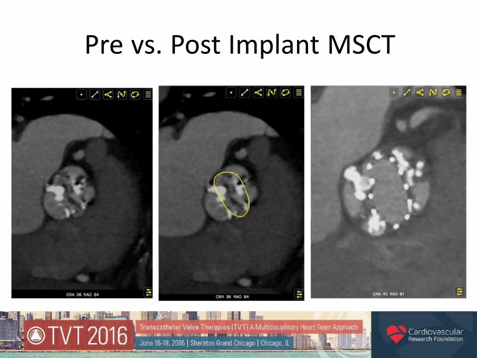
Pre vs. Post Implant MSCT

Sizing: What are the options?
Annular method
Perimeter - 73.2 mmArea - 393 mm2
Diameter 27 x 17 mm
29 mm CoreValve
Commissure-to-Commissure
Diameter 25.9 mm
29 mm CoreValve
Annulus size > balloon size + 3 mm
Select a 1-size (3-mm) larger valve
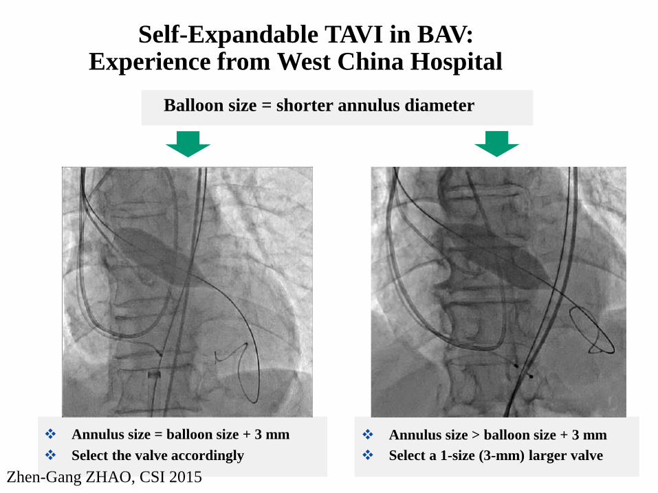
Self-Expandable TAVI in BAV:Experience from West China Hospital
Balloon size = shorter annulus diameter
Annulus size = balloon size + 3 mm
Select the valve accordingly
Zhen-Gang ZHAO, CSI 2015
Valve size in bicuspid valves
•Avoid oversizing
- Risk of rupture
• Usually it is safe to undersize
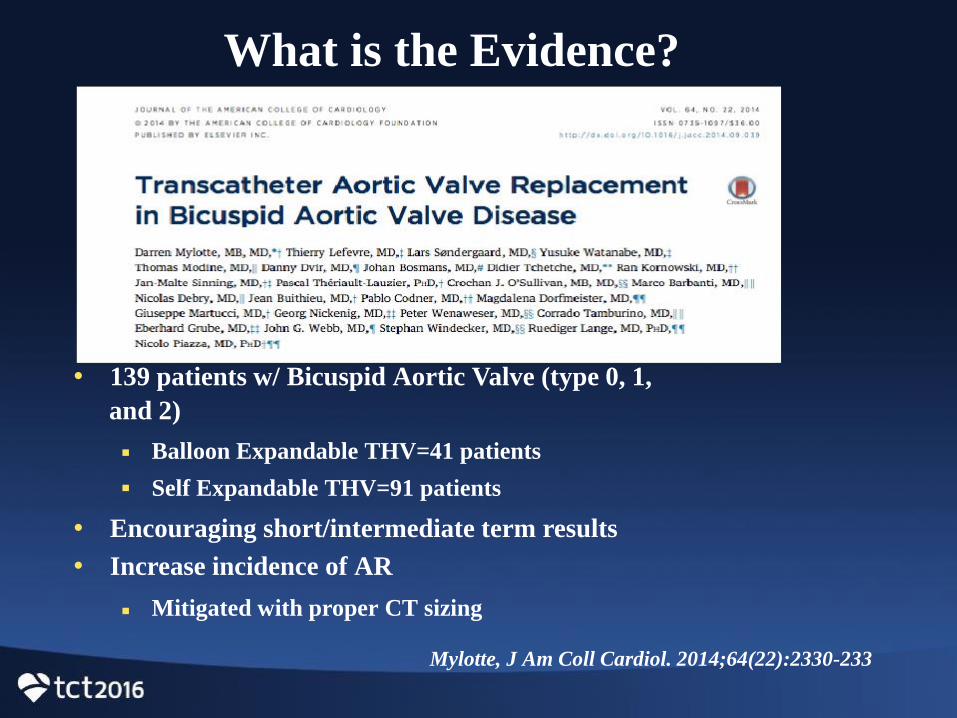
What is the Evidence?
• 139 patients w/ Bicuspid Aortic Valve (type 0, 1,
and 2)
Balloon Expandable THV=41 patients
Self Expandable THV=91 patients
• Encouraging short/intermediate term results
• Increase incidence of AR
Mitigated with proper CT sizing
Mylotte, J Am Coll Cardiol. 2014;64(22):2330-233
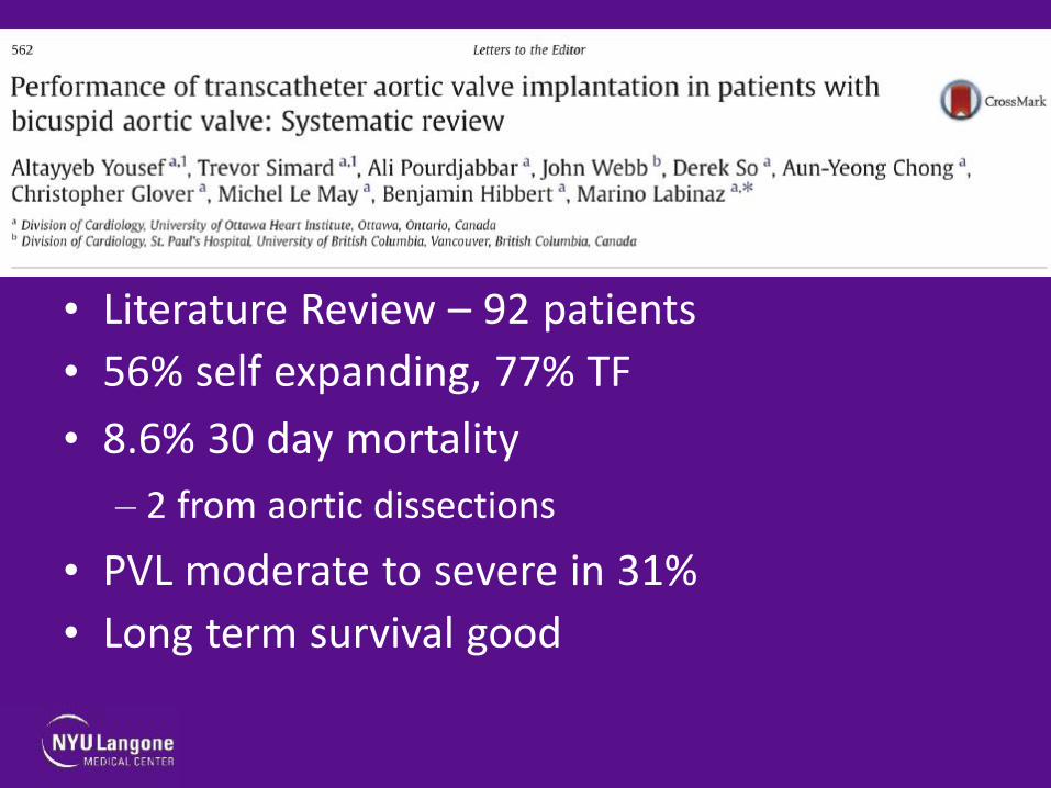
• Literature Review – 92 patients
• 56% self expanding, 77% TF
• 8.6% 30 day mortality
– 2 from aortic dissections
• PVL moderate to severe in 31%
• Long term survival good

More Evidence in Favor of Bicuspid
Perlman et al J Am Coll Cardiol Intv. 2016;9(8):817-824
• 51 Patients with Bicuspid Aortic Valve anatomy underwent
THV with SAPIEN 3 valve
• No Embolization Noted
• PVL
• None/Trivial AR noted in 63%
• Mild AR in 37%
• No moderate or severe PVL noted

Venus A Trial – 1 yr survival

Who Should We Not Treat?
• Aortopathy (>4.5 cm) if operative
• Younger, lower risk?
– Calcification pattern
– Size
• Grossly too large
• Coronary Anatomy

Case 1Cardiac Team Work
• 75 female
• Coarctation operated 1969
• CABG x 2 1999
– LIMA - LAD
– Gastroepiploic – dom Cx
• Bilateral carotid stenting
• Increasing SOB and angina

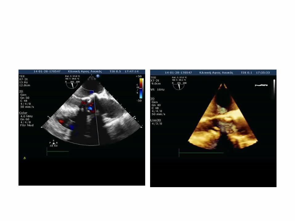
• ECHO– Severe AS – gradient 65mmHg, AVA 0.6 cm2– Bicuspid valve– EF 40%– PHT 55mmHg– Asc Ao = 4.0 cm
• LHC– Patent grafts– Severe peripheral vessel disease – severe iliac
stenoses– Severe L subclavian disease
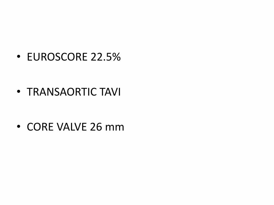
• EUROSCORE 22.5%
• TRANSAORTIC TAVI
• CORE VALVE 26 mm

Mini aortotomy Wire in LV

Deployment Deployment

Valve expanded Fully expanded – minimal AR
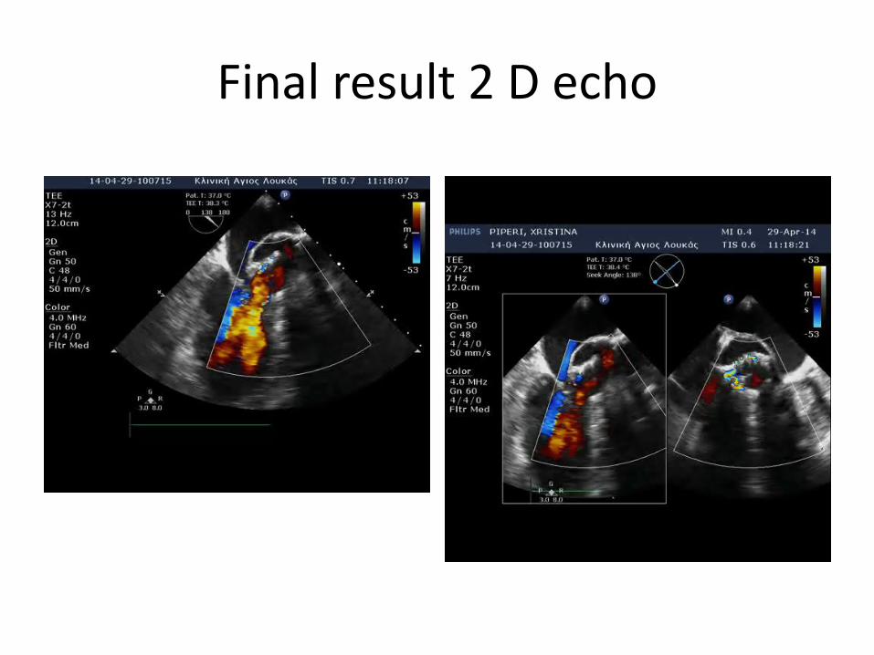
Final result 2 D echo

TIPS AND TRICKS - BICUSPID
• CT sizing either annular or supraannular• Smaller valve downsize according to supraannular
commissure to commissure size• Predilatation and balloon sizing for heavy calcification• High implantation as true stenosis is not at the annulus
but slightly higher • Both projections to assess valve• Bicuspid valve: ao fragile• Oval shape post implantation don’t overexpand
because valve may disrupt – especially in LVOT calcification
Valve in Valve

Avoids redo operation
Less trauma
Faster recovery
Easier Procedure
Less/no contrast
Near Perfect Implantzone
VIV - attractive treatment option
Every patient with degeneratedSurgical valve is NOT for VIV

NativeanatomyTHV
Main Concerns with VIV Aortic
SHV
Malposition
High Gradients
CoronaryObstruction
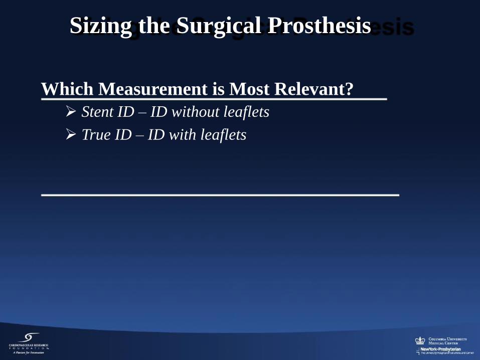
Sizing the Surgical Prosthesis
Which Measurement is Most Relevant?
Stent ID – ID without leaflets
True ID – ID with leaflets

Why use True ID - Example
• Helps choose correct size TAVI Valve to avoid excess oversizing
CE Porcine: size 27
Stent ID - 25
True ID – 23
Sapien XT - Not 29 but 26
CoreValve - Not 29 but 26

Porcine
2mm
* Courtesy V. Bapat
Bovine Pericardium
Inside Stent
1mm
Bovine Pericardium
Outside Stent
0mm
> >
Impact of Leaflets on True ID of Prosthesis
True ID = Stent ID – Leaflets
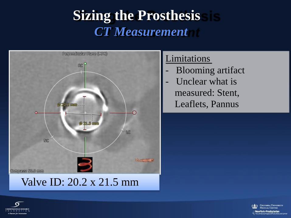
Sizing the ProsthesisCT Measurement
Limitations
- Blooming artifact
- Unclear what is
measured: Stent,
Leaflets, Pannus
Valve ID: 20.2 x 21.5 mm
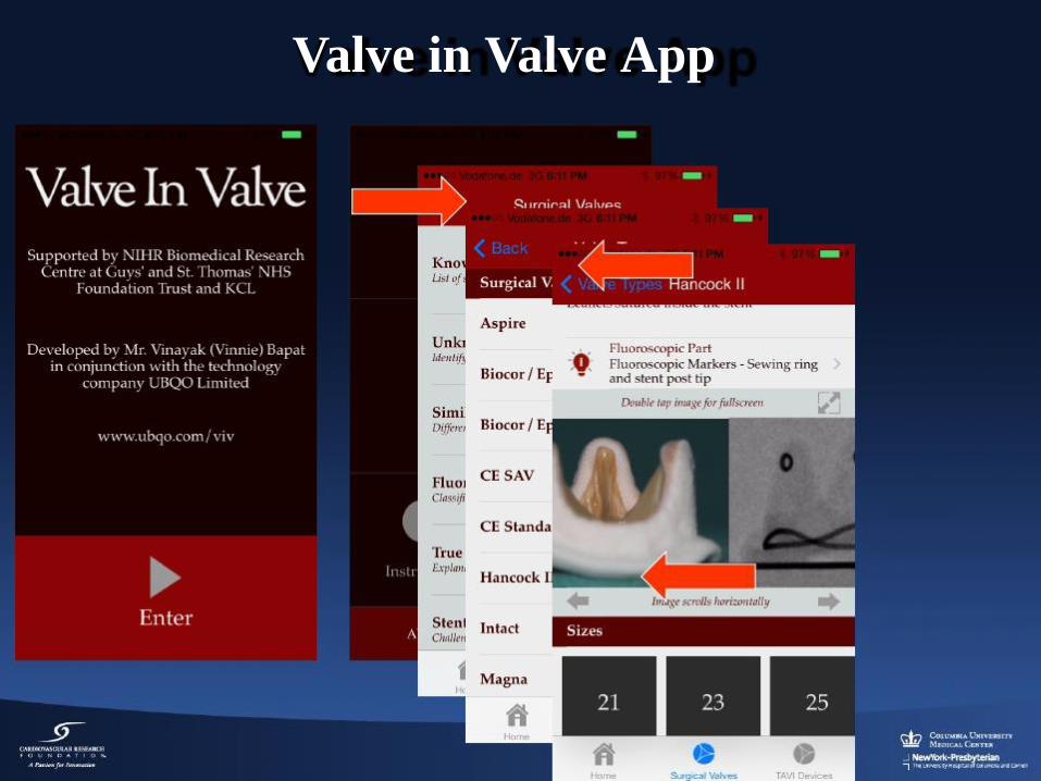
Valve in Valve App

Valve in Valve App
Review this
information just
before each case!

Correct Placement Requires
Understanding of Valve Geometry
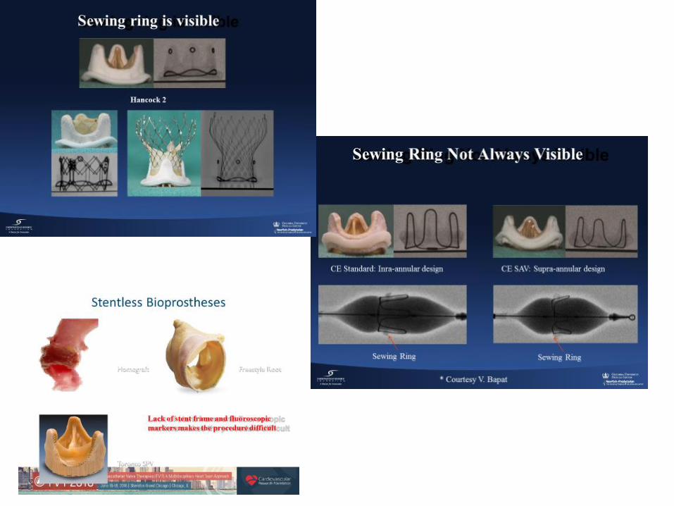
Biocor/ Epic– Thin wire at the level of sewing ring
Narrowest Portion –
Sewing Ring
Identifying Sewing Ring
Crucial
Place Sapien 15%
Below Sewing Ring
* Courtesy V. Bapat

What is the proper location?
• Valve should be positioned based on neo-annulus– Sapien – 10-15% below– CoreValve – 4-5mm below
• Malposition leads to improper seal and anchoring– Too high
• Embolization
– Too low• PVL• Poor hemodyanamics
• Repositional valve helpful
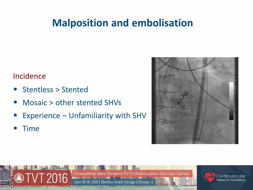
Malposition and embolisation
Incidence
•
•
•
•
Stentless > Stented
Mosaic > other stented SHVs
Experience – Unfamiliarity with SHV
Time

Evaluation of Medtronic CoreValveEvolut R Valve Size and Position on
Valve-in-Valve Hemodynamics
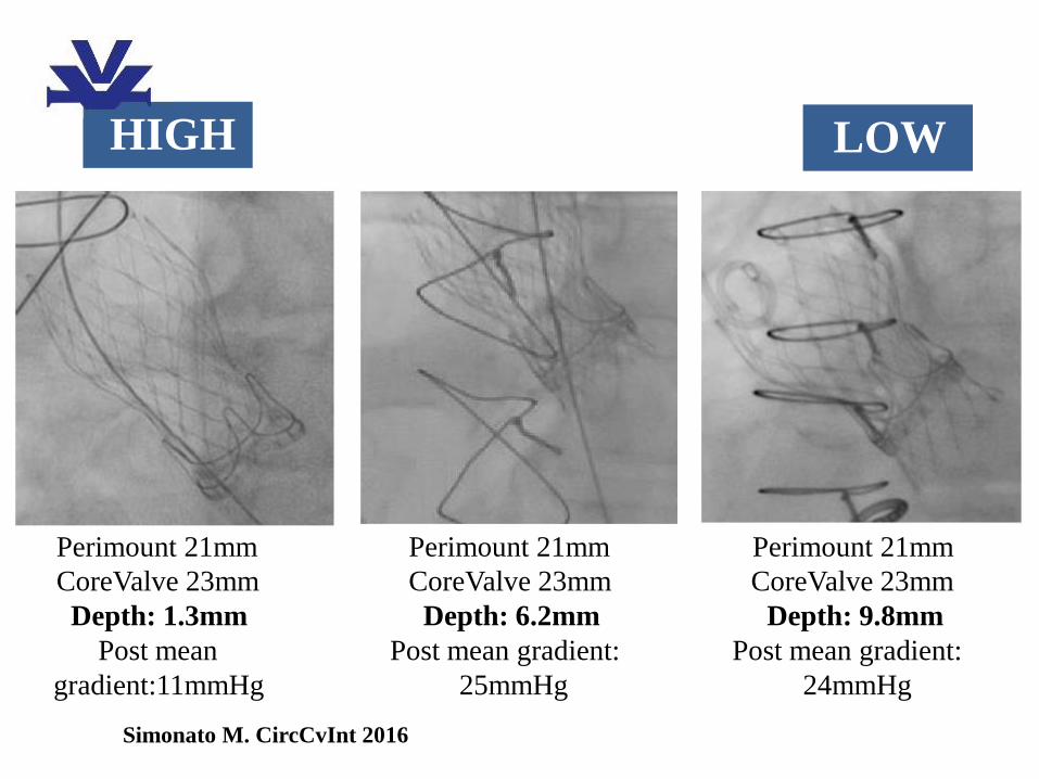
Perimount 21mm
CoreValve 23mm
Depth: 1.3mm
Post mean
gradient:11mmHg
Perimount 21mm
CoreValve 23mm
Depth: 6.2mm
Post mean gradient:
25mmHg
Perimount 21mm
CoreValve 23mm
Depth: 9.8mm
Post mean gradient:
24mmHg
HIGH LOW
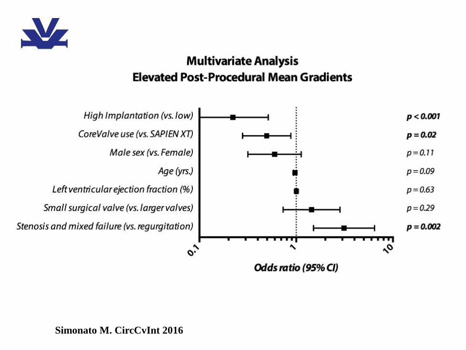
Simonato M. CircCvInt 2016

Perimount 21mm
SAPIEN 23mm
Depth: 0.41%
Post mean
gradient:17mmHg
Perimount 21mm
SAPIEN 23mm
Depth: 25.41%
Post mean gradient:
33mmHg
Pericarbon 21mm
SAPIEN 23mm
Depth: 43.93%
Post mean gradient:
50mmHg
HIGH LOW
Simonato M. CircCvInt 2016

Implantation Depth and Gradients
Simonato M. CircCvInt 2016
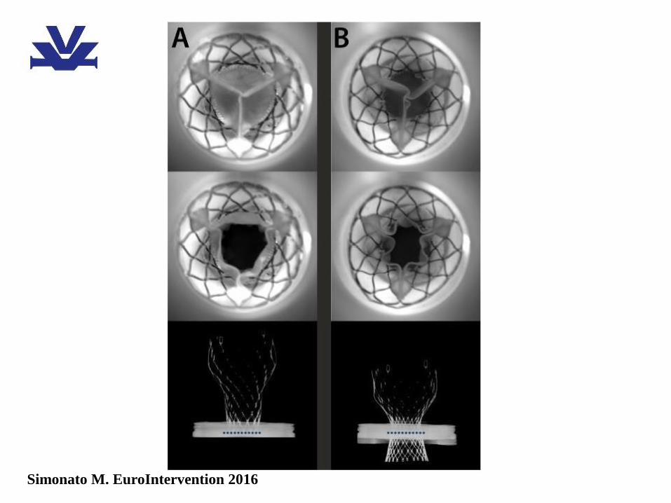
Simonato M. EuroIntervention 2016

Simonato M. EuroIntervention 2016
Simonato M. CircCvInt 2016

Coronary Obstruction
• Function of
Smaller anatomy
Narrow sinuses
Oversizing- Stent post deflection
Valves with leaflet outside the stent
HighLow


Screening for Coronary Occlusion
• Aortogram – Relationship of poststo coronary ostia
• CT Scan
– Coronary ostia
– Sinus width

St. Jude TrifectaMitroflow
Be Cautious with Certain ValveTypes
3F Valve
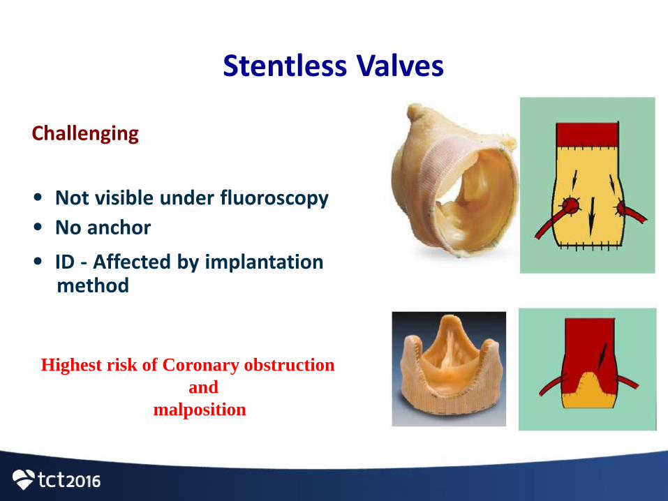
Stentless Valves
Challenging
• Not visible under fluoroscopy
• No anchor
• ID - Affected by implantationmethod
Highest risk of Coronary obstruction
and
malposition
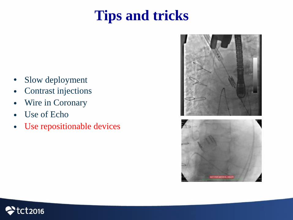
Tips and tricks
•
•
•
•
•
Slow deployment
Contrast injections
Wire in Coronary
Use of Echo
Use repositionable devices
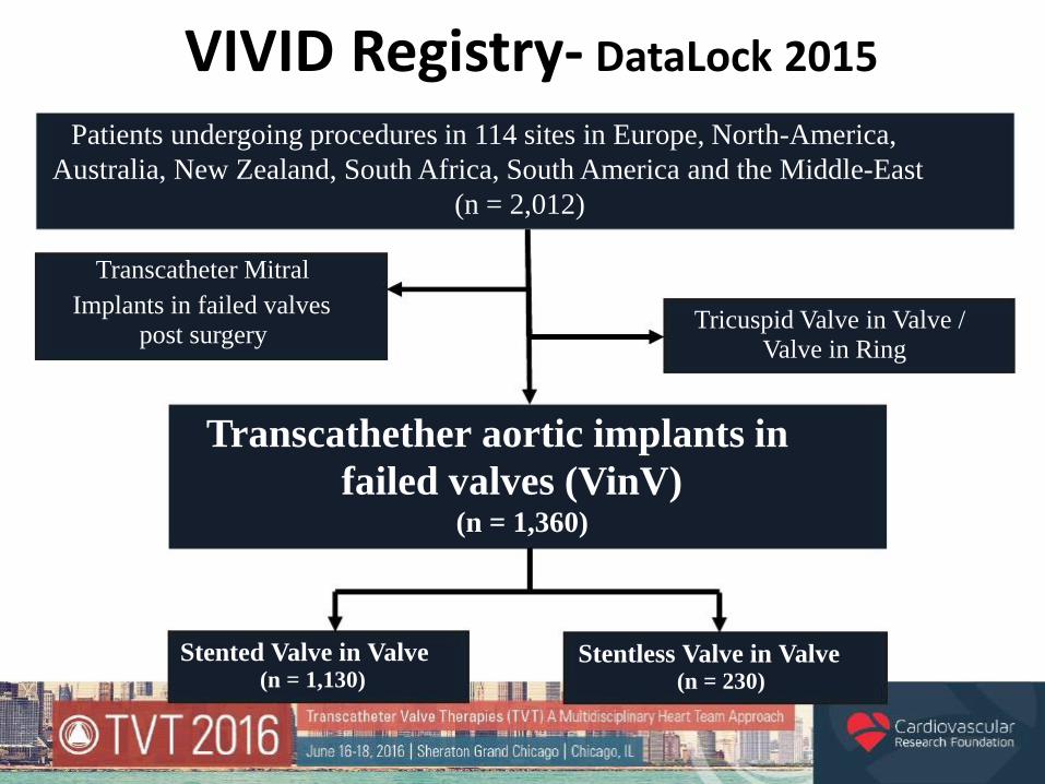
Transcathether aortic implants in
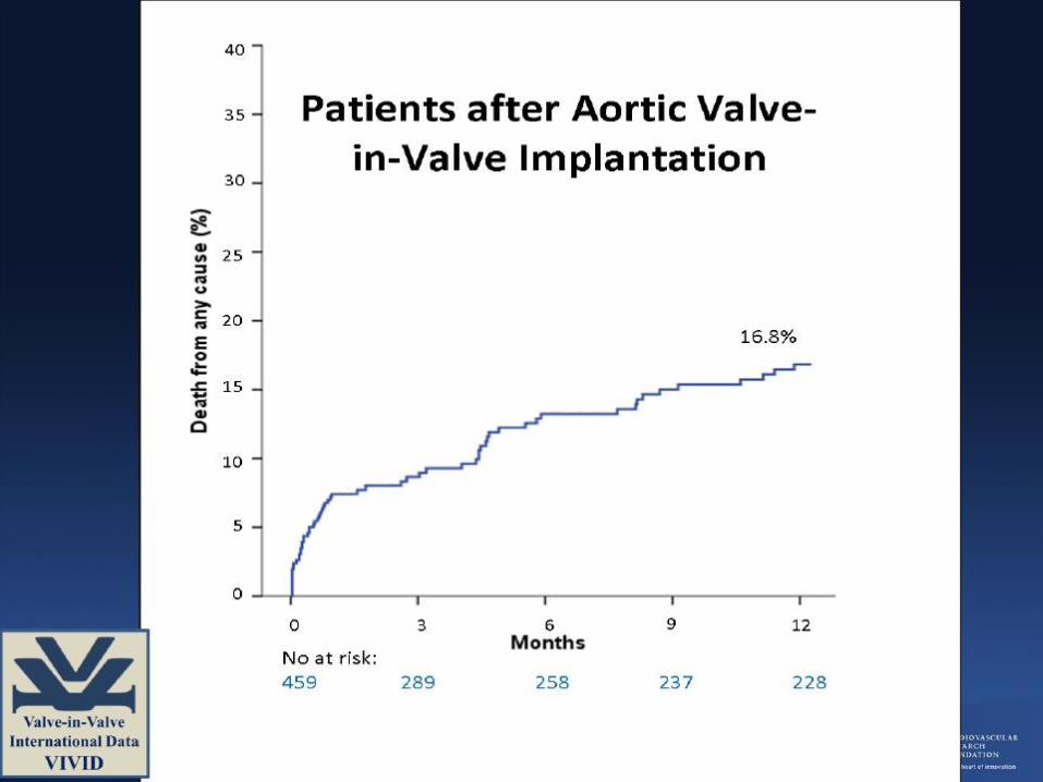
failed valves (VinV)(n = 1,360)
VIVID Registry- DataLock 2015
Patients undergoing procedures in 114 sites in Europe, North-America,
Australia, New Zealand, South Africa, South America and the Middle-East
(n = 2,012)
Transcatheter Mitral
Implants in failed valvespost surgery
Stented Valve in Valve(n = 1,130)
Stentless Valve in Valve(n = 230)
Tricuspid Valve in Valve /Valve in Ring


21
PCR 2016
Coronary Obstruction Following Transcatheter Aortic Valve Implantation for
Degenerative Bioprosthetic Surgical Valves: Insights from the Valve-in-Valve
International Data (VIVID) Registry
34 / 1,508 (2.25%)
Correlates per Multivariate analysis:
Stentless surgical valves (OR 8.76, p<0.001)
Stented with externally mounted leaflets (OR 4.95, p=0.001).
Thirty-day mortality was 52.9% in the coronary obstruction group vs. 3.9% in the
control group.
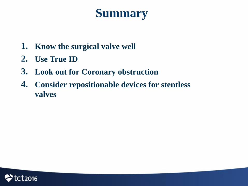
Summary
1.
2.
3.
4.
Know the surgical valve well
Use True ID
Look out for Coronary obstruction
Consider repositionable devices for stentless
valves
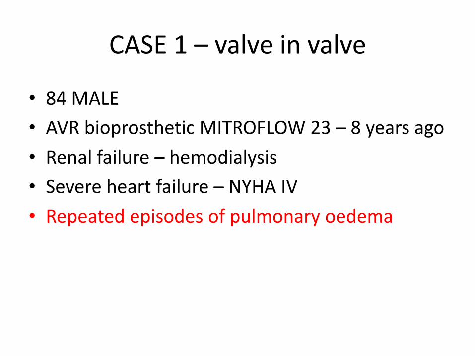
CASE 1 – valve in valve
• 84 MALE
• AVR bioprosthetic MITROFLOW 23 – 8 years ago
• Renal failure – hemodialysis
• Severe heart failure – NYHA IV
• Repeated episodes of pulmonary oedema
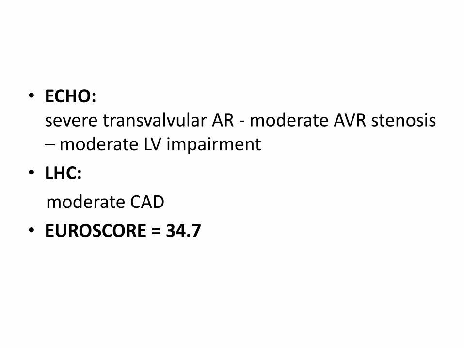
• ECHO:severe transvalvular AR - moderate AVR stenosis – moderate LV impairment
• LHC:
moderate CAD
• EUROSCORE = 34.7

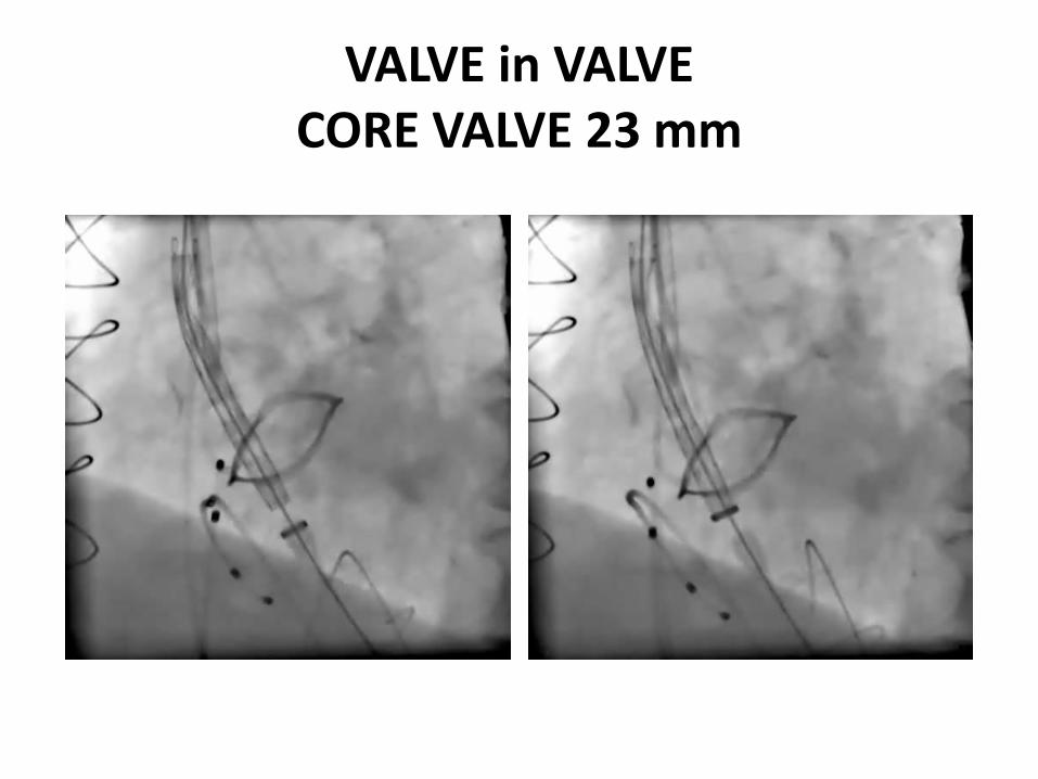
VALVE in VALVECORE VALVE 23 mm


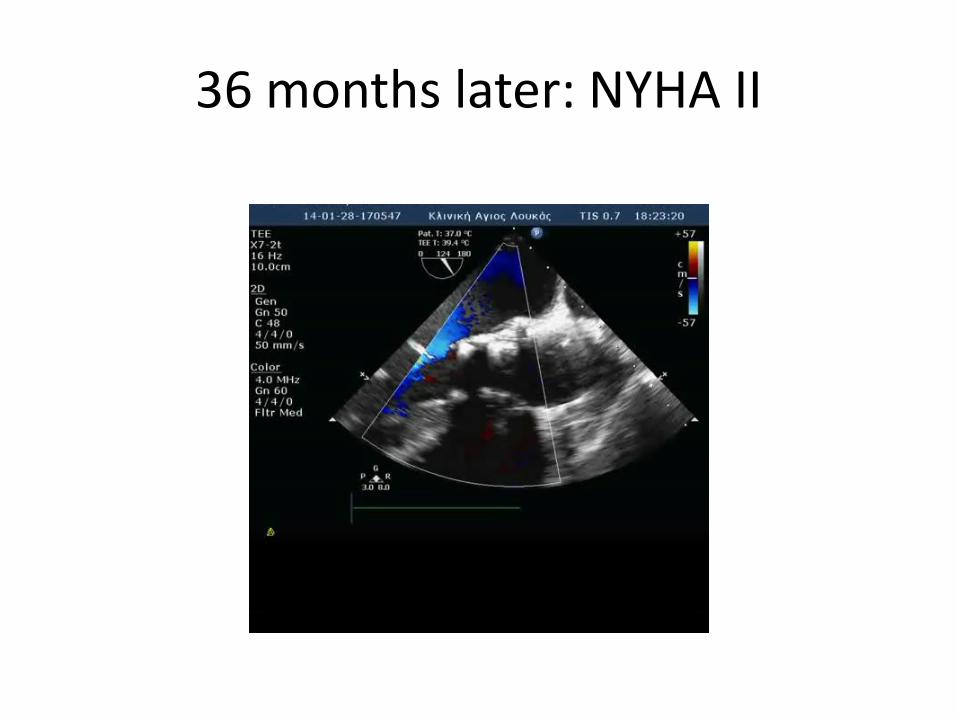
36 months later: NYHA II
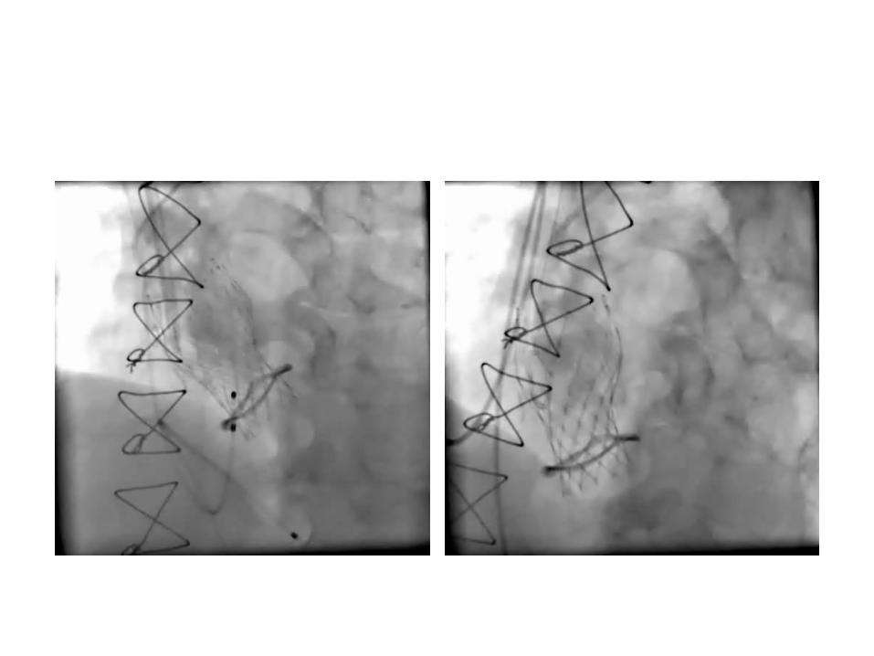
CASE 2 – valve in valve
• 84 female
• AVR St Jude 2001
• AVR severe stenosis – mean gradient 55 mmHg
• Moderate LV dysfunction
• Acute LVF
• Logistic Euroscore 48,46%

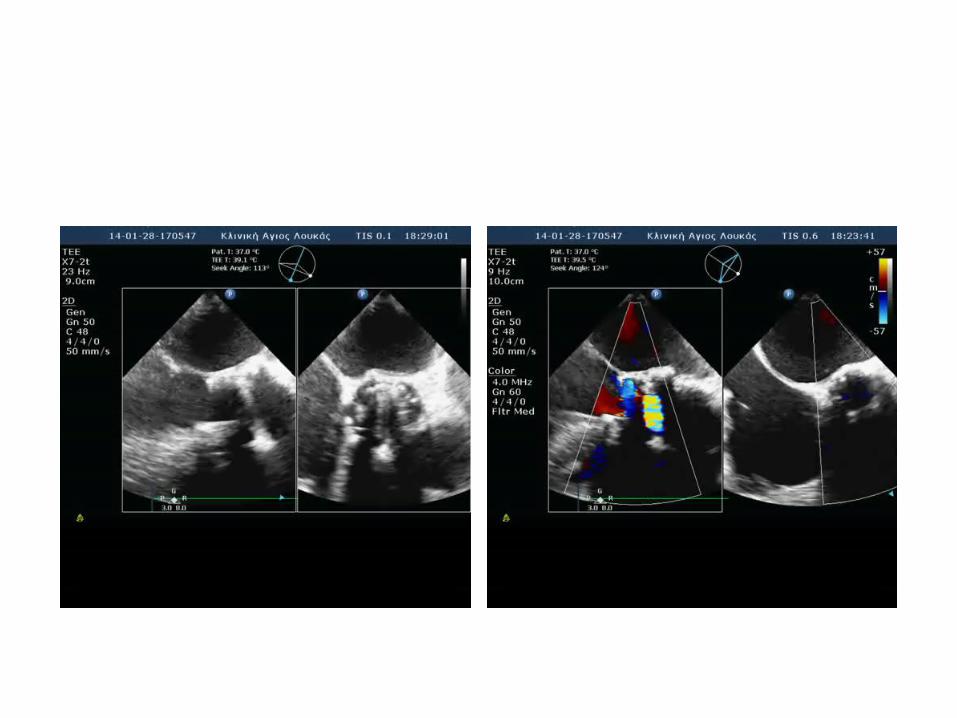
EVOLUT R 23mm
Initial too deep Recapture
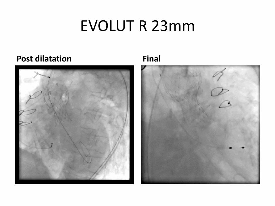
EVOLUT R 23mm
Post dilatation Final

EVOLUT R 23 mm ECHO
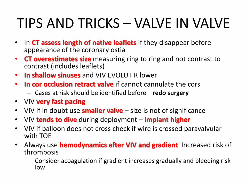
TIPS AND TRICKS – VALVE IN VALVE• In CT assess length of native leaflets if they disappear before
appearance of the coronary ostia• CT overestimates size measuring ring to ring and not contrast to
contrast (includes leaflets)• In shallow sinuses and VIV EVOLUT R lower• In cor occlusion retract valve if cannot cannulate the cors
– Cases at risk should be identified before – redo surgery
• VIV very fast pacing• VIV if in doubt use smaller valve – size is not of significance• VIV tends to dive during deployment – implant higher• VIV if balloon does not cross check if wire is crossed paravalvular
with TOE• Always use hemodynamics after VIV and gradient Increased risk of
thrombosis – Consider acoagulation if gradient increases gradually and bleeding risk
low