Tau oligomers mediate α-synuclein toxicity and can be ...
14
RESEARCH ARTICLE Open Access Tau oligomers mediate α-synuclein toxicity and can be targeted by immunotherapy Julia E. Gerson 1,2 , Kathleen M. Farmer 1 , Natalie Henson 1 , Diana L. Castillo-Carranza 1 , Mariana Carretero Murillo 1 , Urmi Sengupta 1 , Alan Barrett 2 and Rakez Kayed 1,2* Abstract Background: We have evaluated the efficacy of targeting the toxic, oligomeric form of tau protein by passive immunotherapy in a mouse model of synucleinopathy. Parkinson’s disease and Lewy body dementia are two of the most common neurodegenerative disorders and are primarily characterized by the accumulation of α-synuclein in Lewy bodies. However, evidence shows that smaller, oligomeric aggregates are likely the most toxic form of the protein. Moreover, a large body of research suggests that α-synuclein interacts with tau in disease and may act in a synergistic mechanism, implicating tau oligomers as a potential therapeutic target. Methods: We treated seven-month-old mice overexpressing mutated α-synuclein (A53T mice) with tau oligomer- specific monoclonal antibody (TOMA) and a control antibody and assessed both behavioral and pathological phenotypes. Results: We found that A53T mice treated with TOMA were protected from cognitive and motor deficits two weeks after a single injection. Levels of toxic tau oligomers were specifically decreased in the brains of TOMA-treated mice. Tau oligomer depletion also protected against dopamine and synaptic protein loss. Conclusion: These results indicate that targeting tau oligomers is beneficial for a mouse model of synucleinopathy and may be a viable therapeutic strategy for treating diseases in which tau and α-synuclein have a synergistic toxicity. Keywords: Tau, Immunotherapy, Oligomers, α-synuclein, Synucleinopathies, Neurodegeneration Background Parkinson’ s disease (PD) and Lewy body dementia (LBD), behind Alzheimer’ s disease (AD), are the most common neurodegenerative disorders with no effective therapies targeting the cause of disease. Passive im- munotherapy is a promising mode of treatment, with a recent anti-amyloid oligomer antibody showing promis- ing results [1]. After years of failure targeting amyloid-β protein, new promising approaches for AD targeting tau toxicity are coming to the forefront [2]. The pathological hallmarks of PD are cytoplasmic inclu- sions called Lewy bodies (LB), comprised primarily of α-synuclein, along with hyperphosphorylated tau and other sequestered proteins [3–8] in dopaminergic neurons. However, the importance of LB to the neurotoxicity in dis- ease has been questioned [9–13]. A number of studies have shown that oligomeric α-synuclein is the toxic species, rather than fibrils comprising LBs [14–17], and that α- synuclein oligomers may be the most effective therapeutic target [18]. In spite of the clear prevalence of α-synuclein pathology in disease, one of the greatest genetic risk factors for PD is tau, the role of which is understudied and poorly understood [19]. Phosphorylated tau aggregates have been reported in numerous synucleinopathy mouse models [20–25], suggesting a possible synergistic interaction between α-synuclein and tau in mediating neurodegenera- tion in PD, as α-synuclein may increase tau aggregation [23, 26–29] and tau may have a similar effect on α- synuclein. While neurofibrillary tangles (NFTs) characterize tauopathies and are not correlative of synucleinopathies, recent studies suggest that intermediate forms of tau—tau oligomers—that form prior to or independently of NFTs, * Correspondence: [email protected] 1 Departments of Neurology, Neuroscience and Cell Biology, University of Texas Medical Branch, Galveston, TX 77555, USA 2 Sealy Center for Vaccine Development, University of Texas Medical Branch, Galveston, TX 77555, USA © The Author(s). 2018 Open Access This article is distributed under the terms of the Creative Commons Attribution 4.0 International License (http://creativecommons.org/licenses/by/4.0/), which permits unrestricted use, distribution, and reproduction in any medium, provided you give appropriate credit to the original author(s) and the source, provide a link to the Creative Commons license, and indicate if changes were made. The Creative Commons Public Domain Dedication waiver (http://creativecommons.org/publicdomain/zero/1.0/) applies to the data made available in this article, unless otherwise stated. Gerson et al. Molecular Neurodegeneration (2018) 13:13 https://doi.org/10.1186/s13024-018-0245-9
Transcript of Tau oligomers mediate α-synuclein toxicity and can be ...
RESEARCH ARTICLE Open Access
Tau oligomers mediate α-synuclein toxicityand can be targeted by immunotherapyJulia E. Gerson1,2, Kathleen M. Farmer1, Natalie Henson1, Diana L. Castillo-Carranza1, Mariana Carretero Murillo1,Urmi Sengupta1, Alan Barrett2 and Rakez Kayed1,2*
Abstract
Background: We have evaluated the efficacy of targeting the toxic, oligomeric form of tau protein by passiveimmunotherapy in a mouse model of synucleinopathy. Parkinson’s disease and Lewy body dementia are two of themost common neurodegenerative disorders and are primarily characterized by the accumulation of α-synuclein inLewy bodies. However, evidence shows that smaller, oligomeric aggregates are likely the most toxic form of theprotein. Moreover, a large body of research suggests that α-synuclein interacts with tau in disease and may act in asynergistic mechanism, implicating tau oligomers as a potential therapeutic target.
Methods: We treated seven-month-old mice overexpressing mutated α-synuclein (A53T mice) with tau oligomer-specific monoclonal antibody (TOMA) and a control antibody and assessed both behavioral and pathologicalphenotypes.
Results: We found that A53T mice treated with TOMA were protected from cognitive and motor deficits two weeksafter a single injection. Levels of toxic tau oligomers were specifically decreased in the brains of TOMA-treated mice.Tau oligomer depletion also protected against dopamine and synaptic protein loss.
Conclusion: These results indicate that targeting tau oligomers is beneficial for a mouse model of synucleinopathyand may be a viable therapeutic strategy for treating diseases in which tau and α-synuclein have a synergistic toxicity.
Keywords: Tau, Immunotherapy, Oligomers, α-synuclein, Synucleinopathies, Neurodegeneration
BackgroundParkinson’s disease (PD) and Lewy body dementia(LBD), behind Alzheimer’s disease (AD), are the mostcommon neurodegenerative disorders with no effectivetherapies targeting the cause of disease. Passive im-munotherapy is a promising mode of treatment, with arecent anti-amyloid oligomer antibody showing promis-ing results [1]. After years of failure targeting amyloid-βprotein, new promising approaches for AD targeting tautoxicity are coming to the forefront [2].The pathological hallmarks of PD are cytoplasmic inclu-
sions called Lewy bodies (LB), comprised primarily ofα-synuclein, along with hyperphosphorylated tau and othersequestered proteins [3–8] in dopaminergic neurons.
However, the importance of LB to the neurotoxicity in dis-ease has been questioned [9–13]. A number of studies haveshown that oligomeric α-synuclein is the toxic species,rather than fibrils comprising LBs [14–17], and that α-synuclein oligomers may be the most effective therapeutictarget [18]. In spite of the clear prevalence of α-synucleinpathology in disease, one of the greatest genetic risk factorsfor PD is tau, the role of which is understudied and poorlyunderstood [19]. Phosphorylated tau aggregates have beenreported in numerous synucleinopathy mouse models[20–25], suggesting a possible synergistic interactionbetween α-synuclein and tau in mediating neurodegenera-tion in PD, as α-synuclein may increase tau aggregation[23, 26–29] and tau may have a similar effect on α-synuclein. While neurofibrillary tangles (NFTs) characterizetauopathies and are not correlative of synucleinopathies,recent studies suggest that intermediate forms of tau—tauoligomers—that form prior to or independently of NFTs,
* Correspondence: [email protected] of Neurology, Neuroscience and Cell Biology, University ofTexas Medical Branch, Galveston, TX 77555, USA2Sealy Center for Vaccine Development, University of Texas Medical Branch,Galveston, TX 77555, USA
© The Author(s). 2018 Open Access This article is distributed under the terms of the Creative Commons Attribution 4.0International License (http://creativecommons.org/licenses/by/4.0/), which permits unrestricted use, distribution, andreproduction in any medium, provided you give appropriate credit to the original author(s) and the source, provide a link tothe Creative Commons license, and indicate if changes were made. The Creative Commons Public Domain Dedication waiver(http://creativecommons.org/publicdomain/zero/1.0/) applies to the data made available in this article, unless otherwise stated.
Gerson et al. Molecular Neurodegeneration (2018) 13:13 https://doi.org/10.1186/s13024-018-0245-9

are the true toxic species in disease and the optimumtargets for anti-tau therapies [30–35].Tau and α-synuclein have been shown to interact in
disease by multiple different groups. Large genome-wideassociation studies have revealed an increased risk forPD and α-synuclein toxicity associated with changes tothe gene encoding tau [36, 37]. Additionally, the proteinshave been found to co-exist in disease and α-synucleinaggregation has been demonstrated to cause an increasein tau fibril levels [26, 38]. Increasing evidence also sug-gests that comorbidity of AD pathology in synucleinopa-thies is common [39]. Moreover, decreasing tau in asynucleinopathy mouse model has been shown to beprotective [40]. We have shown that, unlike fibrils, oligo-meric α-synuclein can seed the formation of tau oligo-mers [27]. Given the presence of tau oligomers in PDand LBD human brains [41], this highlights their import-ance in synucleinopathies. Thus, oligomeric tau mayrepresent a new therapeutic target for synucleinopathies.A53T mice overexpress a mutation in α -synuclein
that leads to PD in humans and have been shown tohave both tau and α-synuclein aggregates [42], resultingin motor and cognitive deficits [43, 44]. Therefore, wechose to specifically target oligomeric tau by passive im-munotherapy in A53T mice with tau oligomer-specificmonoclonal antibody (TOMA) to evaluate the efficacy oftau oligomers as a novel drug target for the treatment ofsynucleinopathies.
MethodsPreparation of brain homogenateFrozen frontal cortical brain tissue from PD subjects was re-ceived in blocks from Oregon Health & Science University.Tissues used for research were approved by the InstitutionalEthics Committee. Brains were homogenized in PBS witha protease inhibitor cocktail (catalog no. 11836145001;Roche Applied Science, Indianapolis, IN, USA), using a1:3 dilution of tissue: PBS (w/v). Samples were centrifugedat 10,000 rpm for 10 min at 4 °C. Supernatants were ali-quoted, snap-frozen, and stored at − 80 °C until use. Forinsoluble tau fractions, pellets were resuspended in 10%Sarkosyl in PBS, vortexed, and sonicated, then centrifugedfor 30 min at 100,000 x g at 4 °C.
Non-denaturing filter trap assayTau, amyloid-β and α-synuclein oligomers and taumonomer and fibrils were prepared as previously de-scribed [45]. Filter Trap assay was performed to detecttau oligomers with conformational antibodies in theabsence of reducing agents. Briefly, 1 μg of oligomeric tauwas applied to nitrocellulose membranes, previously satu-rated with TBS-T, through the use of a vacuum-based bio-slot apparatus. Then, membranes were blocked with 10%nonfat milk in TBS-T overnight at 4 °C. The next day,
membranes were probed with the oligomer-specific tauantibodies, T22 (1:250) and TOMA (1:250) as well as totaltau antibody, tau5 (1:10000), or α-synuclein antibody, 4D6(1:5000), or amyloid-β antibody, 6E10 (1:5000), diluted in5% nonfat milk for 1 h at RT. Membranes were thenwashed three times with TBS-T and incubated with horse-radish peroxidase-conjugated IgG anti-rabbit (1:5000) andanti-mouse (1:5000) secondary antibodies as appropriate.Membranes were washed again three times in TBS-T andECL plus (GE Healthcare) was used for signal detection.
AnimalsThis study was conducted in a facility approved by theAmerican Association for the Accreditation of Labora-tory Animal Care, and all experiments were performedin accordance with the National Institutes of HealthGuide for the Care and Use of Laboratory Animals andapproved by the Institutional Animal Care and UseCommittee of the University of Texas Medical Branch(UTMB). For immunotherapy experiments, both maleand female animals were balanced in each group todetermine whether sex differences were present. A53Tmice (The Jackson laboratory; B6;C3-Tg(Prnp-SNCA*A53T)83Vle/J) and nontransgenic littermate(WT) animals were bred at UTMB and were raised freeof enrichment following weaning to prevent any effecton behavioral test performance. Mice were housed at theUTMB animal care facility and maintained according toU.S. Department of Agriculture standards (12 h light/dark cycle with food and water available ad libitum).
Experimental design and statistical analysisTreatment groups were assigned at random using a ran-dom number generator. Two-tailed power analysis usingCohen’s Power Table when α = 0.05 was conducted todetermine the appropriate sample size for each immuno-therapy group based on the means and standard devia-tions from a pilot immunotherapy study in A53T mice.It was determined that a sample size of 16 would allowus to detect a difference in discrimination index of 0.45between groups on the novel object recognition task80% of the time. Additional animals were included inA53T groups to protect against any unforeseen attritionor sex-dependent effects. Half of the mice in each groupwere maintained after behavioral analysis to assess mor-tality using a Kaplan-Meier survival curve, analyzed bythe Log-rank (Mantel Cox) test with a Bonferroni adjust-ment for multiple comparisons (threshold for signifi-cance p < 0.025).
ImmunizationSeven-month-old homozygous A53T mice were givenintravenous injections of 120 μL per animal of either of1 mg/mL TOMA (n = 17), or anti-Rhodamine antibody
Gerson et al. Molecular Neurodegeneration (2018) 13:13 Page 2 of 14
(IgG, nonspecific; catalog #GTX29093; Genetex; n = 23)for the control treatment group (Ctrl IgG). Wildtypelittermates were given 120 μL of saline injected intraven-ously (n = 16). Injections were repeated a second timeon seven-month old homozygous A53T mice treatedwith either TOMA (n = 5) or saline (n = 4) in order toconfirm that differences were not due to toxicity of non-specific IgG antibody in Ctrl IgG mice. For intravenousinjections, mice were placed in a restrainer (BraintreeScientific), and an inch of the tail was shaved and placedin warm water to dilate veins. Mice were then injectedvia the lateral tail vein, returned to home cages, and keptunder observation.
Behavioral analysesPrior to performing behavioral tasks, mice were habitu-ated to frequent experimenter handling. All tasks werescored and analyzed by experimenters blinded to treat-ment group. To determine the effect of immunotherapyon behavioral deficits, the cognition and motor pheno-type of A53T mice was evaluated using the NOR,rotarod, footprint and nesting tests. The NOR test wasperformed 2 weeks after immunization, followed by thetwo motor tasks (Fig. 2).
Novel object recognition (NOR)The novel object recognition task (NOR) utilizes thenatural tendency of rodents to preferentially explorenovel objects and environments over those that arefamiliar. A53T mice have been shown to exhibit memorydeficits which may be detected by NOR starting at6 months of age [44, 46]. Mice were habituated to theNOR task and allowed to freely explore the white open-field arena (55 cm in diameter; 60 cm in height) for15 min on the first day. The next day, mice were placedin the arena for the training phase with two identical ob-jects, either spheres or cubes, and allowed to explore for15 min. On the third day, mice were placed again in thearena for 15 min with one familiar object previouslyexplored in the training phase and one novel objectdiffering in color and shape, but sharing a common sizeand volume. After each trial, the apparatus was thor-oughly cleaned using 70% Ethanol and allowed to dryprior to placement of a new mouse. Trials were recordedand time spent exploring each object was measuredusing ANY-Maze software. Exploration was defined byhead orientation within 2 cm of the object or physicalcontact with the object. The percentage of total timespent exploring the familiar object versus the novel objectwas measured. In order to control for any differences inexploratory behavior, the discrimination index was alsocalculated as the time spent exploring the familiar objectsubtracted from the time spent exploring the novel object,divided by the total time spent exploring both objects.
Thus, a higher discrimination index is indicative of func-tioning object discrimination and improved cognitivefunction. Object exploration data were analyzed usingGraph Pad Prism 5.04 sofware by one-way analysis ofvariance (ANOVA) and Tukey’s post-hoc test.
Rotarod testA53T mice do not demonstrate gross motor deficitsuntil around nine to 12 months of age [44, 47]. Rotarodmeasures the ability of a rodent to maintain balance ona rotating rod (diameter 3.2 cm) and may be used toevaluate both motor coordination and hyperactivity.Mice were first habituated in one session of four trials toreach a baseline level of performance. The next day,mice were tested in one session of four trials (Rotarodmeter; Stoelting). Five mice were placed onto the rod,one per testing station. The speed started at 4 rpm andaccelerated at 0.1 rpm/s. The latency to fall from therotating rod was determined. Latency significantly higherthan that of wildtype was determined to be indicative ofa hyperactivity phenotype. Data were analyzed usingGraph Pad Prism 5.04 sofware by one-way analysis ofvariance (ANOVA) and Tukey’s post-hoc test.
Footprint testA53T mice have been shown to have gait alterationssimilarly to what is seen in PD disease patients clinically[44, 48]. The footprint test was used to compare the gaitof A53T transgenic mice in each treatment group withthat of wild-type control mice. Mice were first trained towalk along a 50-cm-long, 10-cm-wide runway (with 10-cm-high walls) into an enclosed box containing fruitcereal. All mice had four training trials. To obtain foot-prints, hindfeet and forefeet of each mouse were coatedwith blue and red nontoxic paints, respectively. One dayfollowing training, mice were allowed to complete atesting trial. Any mouse that did not walk continuouslyalong path was given another trial. A fresh sheet of whitepaper was placed on the floor of the runway for eachrun. Footprints were analyzed for different gait parame-ters measured in centimeters as has been previouslydescribed [49], including stride length (average forwarddistance between each stride), hindpaw width and fore-paw width (average distance between left and right hindand front footprints, respectively), and front footprint/hind footprint overlap for uniformity of steps (a valueof zero is recorded when the center of the hind andfront footprints overlap and the distance between thetwo centers was recorded for those which did not over-lap). For each step parameter, six to eight values weremeasured from each run, excluding footprints made atthe beginning and the end of the run to avoid inaccuratemeasurements during initiation and completion ofmovement. The mean value of each set of measurements
Gerson et al. Molecular Neurodegeneration (2018) 13:13 Page 3 of 14

was compared between groups by one-way ANOVA andTukey’s post-hoc test using Graph Pad Prism 5.04software.
Nesting testThe nesting task is designed to utilize the innate abilityof both male and female mice to build nests for heat,shelter, and reproduction and has been shown to beaffected by very minor sensorimotor deficits [50]. A53Tmice begin to show minor deficits that can be detectedby nesting behavior as young as two to 3 months of age,prior to any gross locomotor phenotype [44, 51]. The nest-ing task was completed as previously described [44, 50].Two hours prior to the dark phase, each mouse was placedin an individual testing cage identical to home cage andgiven a single cotton nestlet. At 1 h and 24 h after place-ment, nesting quality was determined by two independentresearchers blinded to treatment group based on previouslydefined criteria described as follows; 1: nestlet not visiblytouched; 2: nestlet partially torn but at least 50% remaininguntouched; 3: 50-90% of nestlet shredded but scattereddistribution throughout the cage; 4: at least 90% of nestletis shredded and a compact, flat nest can be identified; 5: anearly perfect nest with over 90% shredded and walls of thenest higher than the mouse’s body. Nesting scores wereaveraged and analyzed using Graph Pad Prism 5.04software by the Kruskal-Wallis non-parametric ANOVAand Tukey’s post-hoc test.
Tissue collectionTo evaluate the pathological presence of oligomeric tauin synucleinopathy mice, brain samples were collectedfrom A53T [51] and non-transgenic control mice at dif-ferent ages. For the immunotherapy experiment, A53Tmice from each group were randomly selected followingcompletion of behavioral testing (n = 7/group), as well asa second set of treated mice (n = 4-5/treatment group)not tested behaviorally for confirmation. Mice wereeuthanized with CO2 and brains were collected foranalysis. The right hemisphere of each brain was snap-frozen and homogenized for biochemical analysis. Theleft hemisphere of each brain was embedded in optimalcutting temperature medium (OCT), rapid frozen andsectioned. All sections (7 μm) were processed simultan-eously under the same conditions.
ImmunohistochemistrySections were fixed in methanol at − 20 °C, permeabilizedwith 0.5% Triton-X, incubated with 3% hydrogen peroxideto quench endogenous peroxidases and blocked in 5%goat serum for 1 h. Sections were then incubated over-night at 4 °C with either T22 for tau oligomers (1:200) ortyrosine hydroxylase antibody (Millipore) for dopamine(1:200). The following day, sections were washed in PBS
three times for 10 min each and incubated with biotinyl-ated goat anti-rabbit IgG (1:200; Vector Laboratories) for1 h. Sections were then washed three times for 10 mineach in PBS and processed using an ABC reagent kit(Vector Laboratories) and visualized with DAB substrate(Vector Laboratories) according to the manufacturer’srecommendations. Lastly, sections were counterstainedwith hematoxylin (Vector Laboratories) for nuclearstaining and mounted. To quantify TH-positive cells, weacquired bright field images using a Nikon MultizoomAZ100 microscope equipped with a Nikon DS-2 M colorCCD camera (Nikon Instruments Inc.). Three sections permouse (n = 6-8 per treatment group) were imaged forquantification. Four visual fields from the olfactory bulb ofeach section were used to quantify TH-positive cell bodiesusing the ImageJ plugin, counting cell. Results wereanalyzed by one-way ANOVA with the Bonferroni posthoc test.
ImmunofluorescenceSections used for immunofluorescence were fixed in 4%paraformaldehyde, permeabilized with 0.5% Triton-Xand blocked in 5% goat serum for 1 h. Prior topermeabilization step, sections evaluated for ProteinaseK (PK) sensitivity were treated with 10 μg/ml PK (SigmaAldrich, cat. #P2308) for 10 min. Sections labeled withantibodies raised in mouse went through a secondblocking step with unconjugated Fab anti-mouse IgG(40 μg/ml; Jackson Immunoresearch) for 1 h to blockendogenous IgG immediately following blocking withserum. Sections were then incubated overnight at 4 °Cwith either tau oligomer-specific polyclonal antibodyT22 (1:250), α-synuclein oligomer-specific antibodySyn33 (1:100), total α-synuclein antibody 4D6 (1:400;Abcam, cat. #ab1903), Lewy body antibody LB509(1:300; Abcam, cat. #ab27766) or GFAP (1:300; Covance,cat. #SM1-224). The following day, sections were washedin PBS three times for 10 min each and incubated withgoat anti-rabbit IgG Alexa-488 and anti-mouse IgGAlexa-568 (1:500; Invitrogen) for 1 h. Sections were thenwashed in PBS three times for 10 min each and incu-bated with DAPI (Invitrogen) to label nuclei for 5 min atroom temperature, washed three times for 5 min each,and were mounted with Fluoromount G (SouthernBiotech). The sections were examined using a NikonA1R MP Laser scanning microscope. LB509 labeledsections were analyzed using Cell Counter in Image-J(National Institutes of Health) for number of Lewybody-like structures (n = 6-8/group). For all other anti-bodies, the total level of fluorescence was measured foreach randomly selected cell using Image-J, as well as thelevel of background from three different surroundingregions without fluorescence (n = 6-8/group). To correctthe level of fluorescence for background and cell size,
Gerson et al. Molecular Neurodegeneration (2018) 13:13 Page 4 of 14
the background multiplied by the area of the region wassubtracted from the total fluorescence. Corrected fluor-escence was equilibrated to nuclear fluorescencemeasured by DAPI to control for differences in celldensity. Results were analyzed by one-way ANOVA withthe Bonferroni post hoc test.
Western blot analysisPBS soluble fractions of brain extracts containing 20 μgof total protein were loaded (without boiling) on precastNuPAGE 4-12% Bis-Tris gels (Invitrogen) for SDS-PAGEanalysis. Gels were subsequently transferred onto nitro-cellulose membranes and blocked overnight at 4 °C with10% nonfat dry milk. Membranes were then probed for1 h at room temperature with T22 (1:250), Tau5(1:2000), F8H7 (1:200), Syn33 (1:500), α-synucleinphospho S129 (1:1000; Abcam cat #ab59264), tyrosinehydroxylase (1:1000), synapsin 1 (1:1000), synaptophysin(1:500), and β-actin (1:2000) diluted in 5% nonfat drymilk. T22, tyrosine hydroxylase, α-synuclein phosphoS129, Syn33 and Synapsin 1 were detected with an HRP-conjugated anti-rabbit IgG (1:3000, GE Healthcare),while Tau5, synaptophysin, F8H7, and β-actin weredetected with an HRP-conjugated anti-mouse IgG (1:3000,GE Healthcare), diluted in 5% milk. ECL (Advansta) wasused to visualize the bands, which were normalized tocorresponding β-actin levels. Densitometric analysis wasperformed using ImageJ software (National Institutes ofHealth). Analyses were completed in triplicate and analyzedby one-way ANOVA with the Bonferroni post hoc test.
ELISAFor ELISA we used Nunc immobilizer amino modules,96 well plates (Thermo Fisher Scientific). Plates werecoated with 60 ng/well of brain homogenate using 0.1 Msodium bicarbonate, pH 9.6, as coating buffer, followedby overnight incubation at 4 °C, plates were washed 3times with Tris-buffered saline (TBST) with low Tween(0.01%), then blocked for 3 h at room temperature with10% nonfat dry milk in TBST. The plates were thenwashed with TBST. Primary antibodies, T22 (1:250),Syn33 and 4D6 (1:1000) diluted in 5% nonfat dry milk inTBST were then added and allowed to react for 1 h atroom temperature. The plates were then washed 3 timeswith TBST and 100 μl of horseradish peroxidase-conjugated anti-rabbit and anti-mouse IgG (1:3000; GEHealthcare Life Sciences, Pittsburgh, PA) were added,followed by incubation for 1 h at room temperature.Finally, plates were washed three times with TBST anddeveloped with 3,3′,5,5′,-tetramethylbenzidine (TMB-1)substrate from DAKO (Carpinteria, CA). The reactionwas stopped with 100 μl 1 M HCl and absorbances wereread at 450 nm using a POLARstar OMEGA platereader (BMG Labtechnologies, Melbourne, Australia).
All measurements were performed in triplicate andanalyzed by one-way ANOVA with the Bonferroni posthoc test.
ResultsElevated levels of tau oligomers in α-synuclein mousemodels and PDWe previously characterized the presence of elevated tauoligomeric species in synucleinopathy human brains.The presence of phosphorylated and aggregated tau hasbeen identified in synucleinopathy mouse model, Prnp-SNCA*A53T, and here we show that tau oligomers arealso increased in the brains of these mice (Fig. 1a-e). Itis important to note that these animals are not designedto overexpress tau, yet, using the tau oligomer-specificantibody, T22, we found increased levels of oligomerictau in the brains of these animals. Tau oligomers wereelevated in brain regions important for cognitive func-tion (hippocampus), as well as motor areas includingpons and cerebellum. The molecular weight of theseSDS stable oligomers is similar to those found in mousemodels of tauopathy [30, 31, 52]. Additionally, tauoligomers were detected at elevated levels in the brainof PD patients by Western blot with TOMA (Fig. 1f),as previously shown. Specificity of previously charac-terized tau oligomer-specific antibodies [53–55] is fur-ther demonstrated by non-denaturing filter trap assay(Fig. 1g).
TOMA protects against behavioral deficits in A53T miceIV injection of TOMA has been shown to specificallymodulate tau oligomers and reverse the phenotypes inthree different animal models of tauopathy and Alzheimer’sdisease without any side effects [53–55]. To test whethertau oligomer immunotherapy can protect against deficits ina synucleinopathy mouse model, we injected 7-month-oldA53T mice with either TOMA or a control IgG and com-pared them to wild-type littermate controls injected withsaline. Two weeks after injection, we conducted behavioraltasks for cognition and motor behavior, as summarized(Fig. 2a). A53T mice showed memory deficits beginning at6 months of age [44, 46]. Using the NOR task, we foundthat A53T mice injected with control IgG antibody hadsignificantly lower discrimination index scores whencompared to wild-type animals (p = 0.0099), indicating acognitive function deficit, based on the inability to discrim-inate between novel and familiar objects. A53T miceinjected with TOMA showed no significant differencesfrom wildtype or control IgG (Fig. 2b). No differences weredetected between wild-type animals or either A53T treat-ment group in the rotarod task (data not shown), indicatingthat gross motor impairment was not measurable at thisage. Thus we utilized additional motor tasks that providemore sensitivity at younger ages in A53T mice. We
Gerson et al. Molecular Neurodegeneration (2018) 13:13 Page 5 of 14

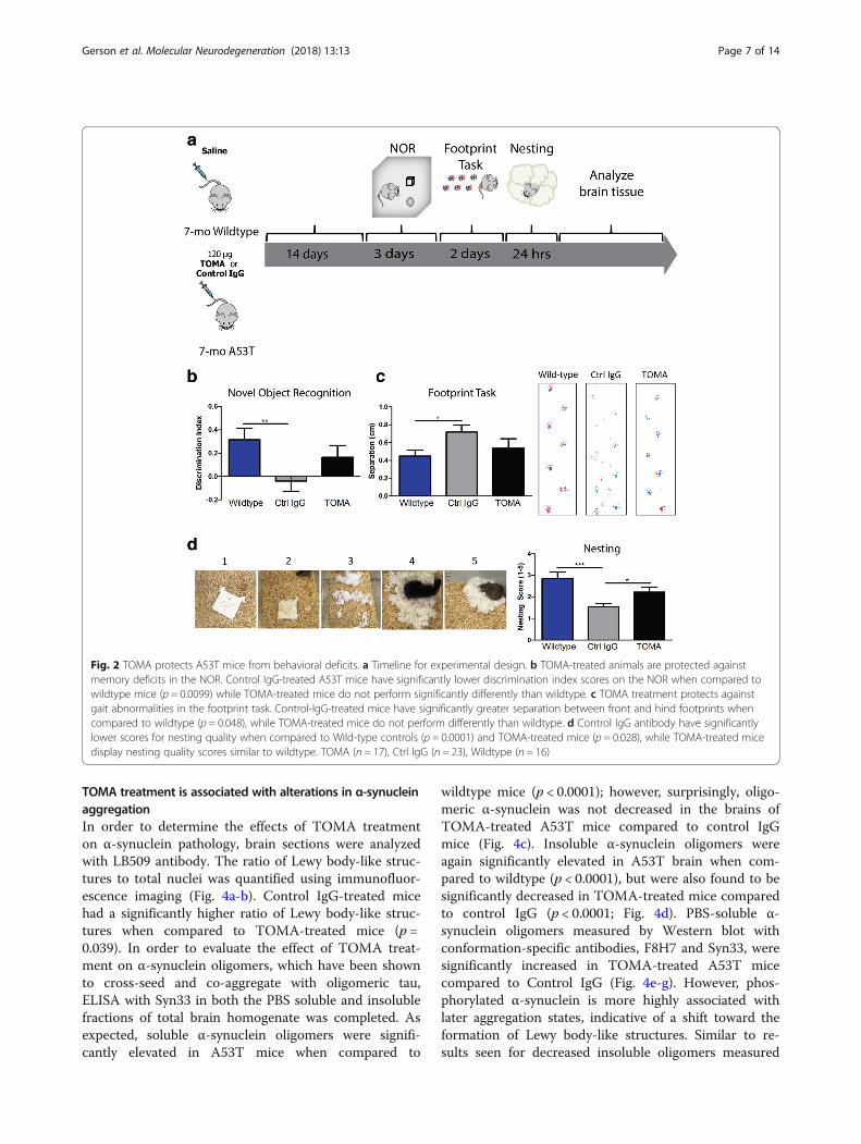
performed the footprint task in order to measure differ-ences in gait seen in the different treatment groups. Wefound no significant differences on all but one of theparameters. In the measure of front and hind paw overlap,however, only the control IgG-treated A53T mice showedgreater distance between the front and hind paws whencompared to Wild-type (p = 0.048), while TOMA-treatedmice were no different than Wild-type controls and werenot significantly different from control IgG mice (Fig. 2c).In order to detect more sensitive differences in motorbehavior that can be seen starting around 2 months of age[44], we performed the nesting test. Representative imagesfor each score on the task were recorded (Fig. 2d). Onehour after receiving a cotton nestlet, A53T mice treatedwith control IgG antibody had significantly lower scoresfor nesting quality when compared to Wild-type controls(p = 0.0001) and TOMA-treated mice (p = 0.028), whileTOMA-treated mice showed no differences from Wild-type (Fig. 2d).
TOMA specifically lowers levels of tau oligomersImmediately following the completion of behavioralexamination, mice were euthanized and brains were
extracted for analysis. By Western blot of PBS-solublewhole brain homogenate with T22 antibody, we foundthat TOMA-treated A53T mice had significantly lowerlevels of both low (p = 0.009) and high molecular weight(p = 0.036) tau oligomers when compared to A53T micetreated with a control IgG antibody (Fig. 3a-c). Using apan-tau antibody, Tau5, that recognizes the variousaggregation states of the protein, we showed that TOMAtreatment did not decrease levels of the functional,monomeric form of tau (Fig. 3d, f), while tau dimer wassignificantly decreased (p = 0.02; Fig. 3e-f ). Levels of a37kD tau fragment associated with neurodegeneration[56] that is recognized by T22 antibody were alsosignificantly decreased in TOMA-treated mice whencompared to Control-IgG (p = 0.003; Fig. 3f-g). Immuno-fluorescence analysis with T22 for tau oligomers(Fig. 3h-o) showed significantly decreased tau oligomersin cortex and hippocampus of TOMA-treated micewhen compared to Control IgG (p = 0.0089). Wild-type mice had significantly lower levels of tau oligo-mers when compared to Control IgG (p = 0.0041), butdid not show any differences with TOMA-treatedA53T mice.
Fig. 1 Tau oligomers are associated with α-synuclein toxicity. a Tau oligomers in A53T mice detected with T22 are elevated compared tonon-transgenic controls. b Tau oligomers with T22 (red) are found in the vicinity of α-synuclein oligomers (green) in A53T mouse brains.c Western blot with Tau5 shows elevated oligomeric tau in A53T mice compared to non-transgenic (WT) controls. Recombinant tau oligomers(TO). d Western blot with TOMA shows detection of tau oligomers in PBS soluble (sol) and Sarkosyl soluble (insol) A53T mouse brain. e ELISAanalysis of the PBS soluble fraction from whole brain shows that tau oligomer levels in the A53T mouse model are elevated compared towild-type mice and increase with age. f Elevated levels of tau oligomers are detected in the PBS soluble brain homogenate of PD patients byWestern blot with TOMA and a pan-tau antibody. g Non-denaturing filter trap assay demonstrates specificity of tau oligomer-specificantibodies. **p < 0.01
Gerson et al. Molecular Neurodegeneration (2018) 13:13 Page 6 of 14
TOMA treatment is associated with alterations in α-synucleinaggregationIn order to determine the effects of TOMA treatmenton α-synuclein pathology, brain sections were analyzedwith LB509 antibody. The ratio of Lewy body-like struc-tures to total nuclei was quantified using immunofluor-escence imaging (Fig. 4a-b). Control IgG-treated micehad a significantly higher ratio of Lewy body-like struc-tures when compared to TOMA-treated mice (p =0.039). In order to evaluate the effect of TOMA treat-ment on α-synuclein oligomers, which have been shownto cross-seed and co-aggregate with oligomeric tau,ELISA with Syn33 in both the PBS soluble and insolublefractions of total brain homogenate was completed. Asexpected, soluble α-synuclein oligomers were signifi-cantly elevated in A53T mice when compared to
wildtype mice (p < 0.0001); however, surprisingly, oligo-meric α-synuclein was not decreased in the brains ofTOMA-treated A53T mice compared to control IgGmice (Fig. 4c). Insoluble α-synuclein oligomers wereagain significantly elevated in A53T brain when com-pared to wildtype (p < 0.0001), but were also found to besignificantly decreased in TOMA-treated mice comparedto control IgG (p < 0.0001; Fig. 4d). PBS-soluble α-synuclein oligomers measured by Western blot withconformation-specific antibodies, F8H7 and Syn33, weresignificantly increased in TOMA-treated A53T micecompared to Control IgG (Fig. 4e-g). However, phos-phorylated α-synuclein is more highly associated withlater aggregation states, indicative of a shift toward theformation of Lewy body-like structures. Similar to re-sults seen for decreased insoluble oligomers measured
Fig. 2 TOMA protects A53T mice from behavioral deficits. a Timeline for experimental design. b TOMA-treated animals are protected againstmemory deficits in the NOR. Control IgG-treated A53T mice have significantly lower discrimination index scores on the NOR when compared towildtype mice (p = 0.0099) while TOMA-treated mice do not perform significantly differently than wildtype. c TOMA treatment protects againstgait abnormalities in the footprint task. Control-IgG-treated mice have significantly greater separation between front and hind footprints whencompared to wildtype (p = 0.048), while TOMA-treated mice do not perform differently than wildtype. d Control IgG antibody have significantlylower scores for nesting quality when compared to Wild-type controls (p = 0.0001) and TOMA-treated mice (p = 0.028), while TOMA-treated micedisplay nesting quality scores similar to wildtype. TOMA (n = 17), Ctrl IgG (n = 23), Wildtype (n = 16)
Gerson et al. Molecular Neurodegeneration (2018) 13:13 Page 7 of 14

Fig. 3 Levels of tau oligomers are specifically reduced in A53T mice treated with TOMA. a-c Western blot of PBS soluble total brain homogenatewith tau oligomer-specific antibody, T22, showed that levels of tau dimer (low molecular weight oligomers) were significantly lowered inTOMA-treated mice compared to control IgG-treated mice (A; p = 0.009), as well as 250 kD (high molecular weight) tau oligomers (B; p = 0.036).d-f Tau monomer levels measured by Western blot with Tau5 were unaffected by treatment, while tau dimer was significantly decreased withTOMA. LE (low exposure) HE (high exposure). *Denotes non-specific band. f-g Western blot of the PBS soluble fraction of whole brain homogenatewith T22 for tau oligomers showed that TOMA-treated mice had significantly lower levels of a 37 kD fragment compared to Control IgG (p = 0.003).h-m Immunofluorescence imaging detects significantly lower levels of tau oligomers in cortex (h-j, n) and hippocampi (k-m, o) of TOMA-treated mice.Control IgG-treated mice had significantly higher levels of T22 fluorescence intensity as a ratio to nuclei when compared to wildtype mice (p = 0.004)and TOMA-treated A53T mice (p = 0.009) in the cortex, as well as significantly higher levels in the hippocampus compared to wildtype mice (p = 0.001)and TOMA-treated A53T mice (p = 0.0044). Scale bar 50 μm
Gerson et al. Molecular Neurodegeneration (2018) 13:13 Page 8 of 14

Fig. 4 Lewy body-like structures are significantly decreased in TOMA-treated A53T mice. a α-synuclein structures were detected by immunofluorescencewith LB509 (red) and nuclei were labeled with DAPI (blue). b Control IgG-treated mice had a significantly higher ratio of Lewy body-like structures to totalnuclei when compared to TOMA-treated mice (p= 0.039). c Levels of soluble α-synuclein oligomers detected by ELISA with Syn33 were significantlyelevated in both Control IgG and TOMA-treated A53T mice compared to wildtype mice (p< 0.0001) d PBS insoluble oligomeric α-synuclein was significantlyincreased in A53T mice treated with TOMA and Control IgG (p < 0.0001), however TOMA-treated mice had significantly decreased α-synuclein oligomerscompared to Control IgG mice (p < 0.0001). e TOMA-treated mice had significantly higher soluble α-synuclein oligomers detected with (f) F8H7 (g) andSyn33 and (h) a trend toward lower phosphorylated α-synuclein by Western blot. i TOMA-treated mice showed elevated levels of α-synuclein oligomersdetected with Syn33 over Control IgG approaching significance (p= 0.059; left panel), but PK-resistant α-synuclein oligomers were found to be significantlyincreased in Control IgG mice compared to TOMA (p = 0.036; right panel). j-k Levels of total α-synuclein detected by ELISA with 4D6 were unchanged inboth soluble and insoluble fractions of total brain homogenate. l No differences were detected in total α-synuclein levels measured by fluorescence intensity(ratio to nuclei) with 4D6 between TOMA and Control IgG treatment groups. Scale bar 20 μm. TOMA (n= 6), Wildtype (n= 6), Ctrl IgG (n= 8)
Gerson et al. Molecular Neurodegeneration (2018) 13:13 Page 9 of 14

by ELISA, the phosphorylated α-synuclein oligomerstrended toward a decrease in TOMA-treated micecompared to Control IgG (Fig. 4e, h). Previous study ofα-synuclein toxicity has indicated that oligomers that arepartially Proteinase K (PK)-resistant are the most toxicform of the protein [57]. Thus, immunofluorescence ofbrain sections with Syn33 was completed with and with-out PK treatment and α-synuclein oligomers were notdecreased in the brain sections of TOMA-treated micethat were not treated with PK (Fig. 4i). However,proteinase K treatment showed that PK-resistant α-synuclein oligomers were significantly increased in Con-trol IgG-treated mice compared to TOMA-treated mice(p = 0.036; Fig. 4i). In order to confirm that total α-synuclein levels were not altered by antibody treatment,ELISA with 4D6 antibody in both the PBS soluble andinsoluble fractions of total brain homogenate wascompleted and no differences were found betweengroups (Fig. 4j-k). Additionally, measurement of fluores-cence corrected for number of nuclei in sections labeledwith 4D6 did not detect any differences between TOMAand Control IgG groups (Fig. 4l).
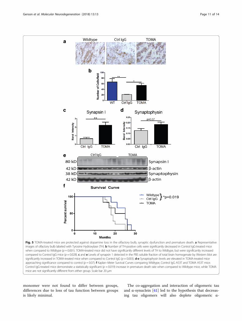
TOMA treatment protects against loss of synapticproteins and olfactory bulb dopamineIn order to evaluate effects of antibody treatment onParkinson’s disease-like toxicity, levels of catecholamineswere measured by immunohistochemistry with TyrosineHydroxylase (TH) in the olfactory bulb, substantia nigra,striatum and locus coeruleus. Olfaction deficits areknown to appear early in the course of disease in PDand are associated with a loss of dopamine in the olfac-tory bulb, as has also been seen previously in A53T mice[58–62]. While no differences were seen in the substan-tia nigra, striatum, or locus coeruleus (data not shown),Control IgG-treated mice had significantly decreasedlevels of the cells positive for the dopamine precursorwhen compared to Wildtype in the olfactory bulb (p =0.001). TOMA-treated mice did not have significantlydifferent levels of TH compared to Wildtype mice, butwere significantly increased compared to control IgGmice (p = 0.028; Fig. 5a-b).Rescue of synaptic toxicity was measured by Western
blot with antibodies specific to synaptic proteins. TOMAtreatment was associated with significantly elevatedlevels of synapsin 1 in the PBS soluble fraction of totalbrain homogenate when compared to Control IgG (p =0.003; Fig. 5c, e) and a trend toward elevated levels ofSynaptophysin (p = 0.07; Fig. 5d-e).
TOMA treatment does not show signs of toxicityIn order to assess potential effects of TOMA treatmenton survival, half of the treated mice in each group wereassessed for mortality using Kaplan-Meier survival
curves (Fig. 5f ). A significant difference in survival wasfound between Wildtype and control IgG A53T mice (p= 0.019, Log-rank Mantel-Cox test with Bonferroniadjustment for multiple comparisons), while TOMA-treated mice did not show differences to Wildtype mice.
DiscussionSynucleinopathies, including PD and DLB, have noeffective treatment and affect millions of people world-wide. Most therapeutic approaches, thus far, havefocused on targeting the symptoms or decreasing α-synuclein protein. Several studies have found evidencefor α-synuclein oligomers as the most toxic form of theprotein. Mouse models of synucleinopathy show deficitsindependent of the formation of fibrillar deposits andoligomeric α-synuclein induces toxicity in cell culturemodels, while fibrils do not [14–17]. However, researchfrom our laboratory and others suggests that tau proteinmay also have a critical pathological role in synucleino-pathies. We recently showed that the most toxic form ofboth tau and α-synuclein, oligomers, coexist and coag-gregate in PD and DLB [41]. These results combinedwith studies showing a genetic and molecular interactionbetween tau and α-synuclein [26, 36–38] and the abilityof α-synuclein oligomers to seed the aggregation of tau[27] suggest that the two proteins may have a synergisticinteraction in disease. Therefore, targeting tau oligomersmay prevent toxicity induced by tau, as well as affectsynergy and co-aggregation with α-synuclein.In order to inhibit toxicity of tau oligomers, we previ-
ously created a tau oligomer-specific antibody, TOMA,which we found to be effective in attenuating the diseasephenotype in three different mouse models of tauopathyand Alzheimer’s disease [54, 55]. Importantly, in a modelof Alzheimer’s disease with both Aβ and tau pathology,decreasing tau oligomers was not only effective at alter-ing cognitive and synaptic dysfunction, but also shiftedtoxic oligomeric Aβ to a non-toxic conformation [54].These results, combined with a number of other studiessuggesting that Aβ triggers tau toxicity [63] support thehypothesis that targeting toxic tau may also rid the brainof toxic upstream targets, such as α-synuclein. Thus far,no studies have investigated the therapeutic potential ofreducing tau oligomers in a synucleinopathy model.Here, we passively immunized a well-characterized synu-cleinopathy mouse model, the Prnp-SNCA*A53T model,which has been studied extensively and shown todevelop insoluble α-synuclein aggregates, α-synucleinoligomers and hyperphosphorylated tau [26, 64–66].The behavioral and pathological improvements seenin mice treated with TOMA suggested that loweringlevels of tau oligomers may be sufficient to reducetoxicity due to tau and possibly α-synuclein toxicityin synucleinopathies. As levels of functional tau
Gerson et al. Molecular Neurodegeneration (2018) 13:13 Page 10 of 14
monomer were not found to differ between groups,differences due to loss of tau function between groupsis likely minimal.
The co-aggregation and interaction of oligomeric tauand α-synuclein [41] led to the hypothesis that decreas-ing tau oligomers will also deplete oligomeric α-
Fig. 5 TOMA-treated mice are protected against dopamine loss in the olfactory bulb, synaptic dysfunction and premature death. a Representativeimages of olfactory bulb labeled with Tyrosine Hydroxylase (TH). b Number of TH-positive cells were significantly decreased in Control IgG-treated micewhen compared to Wildtype (p= 0.001). TOMA-treated mice did not have significantly different levels of TH to Wildtype, but were significantly increasedcompared to Control IgG mice (p= 0.028). c and e Levels of synapsin 1 detected in the PBS soluble fraction of total brain homogenate by Western blot aresignificantly increased in TOMA-treated mice when compared to Control IgG (p= 0.003). d-e Synaptophysin levels are elevated in TOMA-treated miceapproaching significance compared to control (p= 0.07). f Kaplan–Meier Survival Curves comparing Wildtype, Control IgG A53T and TOMA A53T mice.Control IgG-treated mice demonstrate a statistically significant (p = 0.019) increase in premature death rate when compared to Wildtype mice, while TOMAmice are not significantly different from either group. Scale bar 20 μm
Gerson et al. Molecular Neurodegeneration (2018) 13:13 Page 11 of 14
synuclein. However, surprisingly we found that decreasesin α-synuclein oligomers by TOMA were detected only inα-synuclein oligomers with fibril-like characteristics,including insoluble, phosphorylated and proteinase-K(PK)-resistant oligomers. As a recent study showed thatthe most toxic form of oligomeric α-synuclein is partiallyresistant to PK [57], our results suggest that highertoxicity oligomeric α-synuclein structures were alteredfollowing depletion of tau oligomers. Thus, the relation-ship between tau oligomer depletion and α-synuclein ag-gregation appears to be multi-faceted. While TOMAtreatment did not decrease total α-synuclein oligomerlevels, the insoluble and proteinase-resistant form waslowered, as were Lewy body-like structures. It is possiblethat targeting tau interacting with α-synuclein alters itsconformation or that changes may be dependent on tim-ing. More study is warranted to further interrogate thesynergy between the two aggregated proteins, how-ever, beneficial effects due to tau oligomer depletionmay initially occur independently of α-synucleinoligomer decline as tau toxicity lies downstream of α-synuclein. Thus, targeting tau alone could eliminatesome toxicity without affecting upstream α-synuclein.The most effective treatment throughout time willlikely require a combination of therapeutics againstaggregates of both proteins.While the effect of A53T expression in mice on dopa-
mine levels has not been conclusively determined, THlevels in the olfactory bulb are decreased in A53T mice[58, 59]. Olfactory dysfunction and decreased dopaminehas also been shown to be an important factor in bothAlzheimer’s and PD [60–62]. Changes to TH levels mayreflect a combination of both dopaminergic and nor-adrenergic effects, both of which may induce behav-ioral deficits in mouse models of synucleinopathy.Our results showing regulation of TH levels in the ol-factory bulb by TOMA suggest that tau oligomer tox-icity may impact dopamine and possibly othercatecholamine levels.
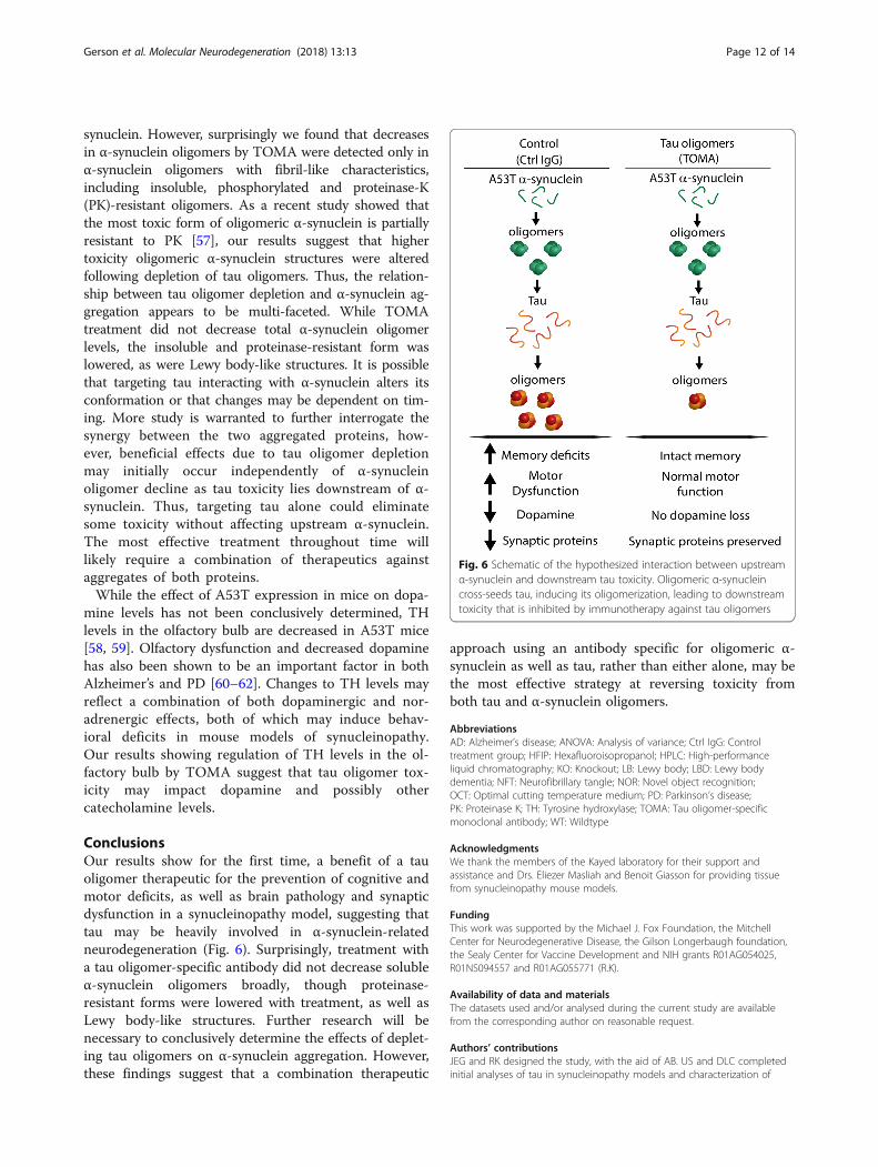
ConclusionsOur results show for the first time, a benefit of a tauoligomer therapeutic for the prevention of cognitive andmotor deficits, as well as brain pathology and synapticdysfunction in a synucleinopathy model, suggesting thattau may be heavily involved in α-synuclein-relatedneurodegeneration (Fig. 6). Surprisingly, treatment witha tau oligomer-specific antibody did not decrease solubleα-synuclein oligomers broadly, though proteinase-resistant forms were lowered with treatment, as well asLewy body-like structures. Further research will benecessary to conclusively determine the effects of deplet-ing tau oligomers on α-synuclein aggregation. However,these findings suggest that a combination therapeutic
approach using an antibody specific for oligomeric α-synuclein as well as tau, rather than either alone, may bethe most effective strategy at reversing toxicity fromboth tau and α-synuclein oligomers.
AbbreviationsAD: Alzheimer’s disease; ANOVA: Analysis of variance; Ctrl IgG: Controltreatment group; HFIP: Hexafluoroisopropanol; HPLC: High-performanceliquid chromatography; KO: Knockout; LB: Lewy body; LBD: Lewy bodydementia; NFT: Neurofibrillary tangle; NOR: Novel object recognition;OCT: Optimal cutting temperature medium; PD: Parkinson’s disease;PK: Proteinase K; TH: Tyrosine hydroxylase; TOMA: Tau oligomer-specificmonoclonal antibody; WT: Wildtype
AcknowledgmentsWe thank the members of the Kayed laboratory for their support andassistance and Drs. Eliezer Masliah and Benoit Giasson for providing tissuefrom synucleinopathy mouse models.
FundingThis work was supported by the Michael J. Fox Foundation, the MitchellCenter for Neurodegenerative Disease, the Gilson Longerbaugh foundation,the Sealy Center for Vaccine Development and NIH grants R01AG054025,R01NS094557 and R01AG055771 (R.K).
Availability of data and materialsThe datasets used and/or analysed during the current study are availablefrom the corresponding author on reasonable request.
Authors’ contributionsJEG and RK designed the study, with the aid of AB. US and DLC completedinitial analyses of tau in synucleinopathy models and characterization of
Fig. 6 Schematic of the hypothesized interaction between upstreamα-synuclein and downstream tau toxicity. Oligomeric α-synucleincross-seeds tau, inducing its oligomerization, leading to downstreamtoxicity that is inhibited by immunotherapy against tau oligomers
Gerson et al. Molecular Neurodegeneration (2018) 13:13 Page 12 of 14

α-synuclein antibodies. JEG and DLC performed immunotherapy treatment.JEG and NH performed all behavioral experiments. JEG, KF, MC and NHanalyzed tissue from treated mice. JEG completed all statistical analysis andwrote the manuscript with RK. All authors read and approved the finalmanuscript.
Ethics approvalThis study was conducted in a facility approved by the American Associationfor the Accreditation of Laboratory Animal Care, and all experiments wereperformed in accordance with the National Institutes of Health Guide for theCare and Use of Laboratory Animals and approved by the Institutional AnimalCare and Use Committee of the University of Texas Medical Branch (UTMB).
Consent for publicationNot applicable
Competing interestsRK has patent applications on the compositions and methods related to tauoligomers and antibodies. All other authors declare that they have nocompeting interests.
Publisher’s NoteSpringer Nature remains neutral with regard to jurisdictional claims inpublished maps and institutional affiliations.
Received: 13 November 2017 Accepted: 7 March 2018
References1. Sevigny J, et al. The antibody aducanumab reduces Aβ plaques in
Alzheimer’s disease. Nature. 2016;537(7618):50–6.2. Bachurin SO, Bovina EV, Ustyugov AA. Drugs in clinical trials for Alzheimer's
disease: the major trends. Med Res Rev. 2017;37(5):1186–225.3. Galpern WR, Lang AE. Interface between tauopathies and synucleinopathies:
a tale of two proteins. Ann Neurol. 2006;59(3):449–58.4. Goris A, et al. Tau and alpha-synuclein in susceptibility to, and dementia in,
Parkinson's disease. Ann Neurol. 2007;62(2):145–53.5. Piao YS, et al. Co-localization of alpha-synuclein and phosphorylated tau in
neuronal and glial cytoplasmic inclusions in a patient with multiple systematrophy of long duration. Acta Neuropathol. 2001;101(3):285–93.
6. Zhukareva V, et al. Loss of brain tau defines novel sporadic andfamilial tauopathies with frontotemporal dementia. Ann Neurol. 2001;49(2):165–75.
7. Muntané G, et al. Phosphorylation of tau and α-synuclein in synaptic-enrichedfractions of the frontal cortex in Alzheimer’s disease, and in Parkinson’s diseaseand related α-synucleinopathies. Neuroscience. 2008;152(4):913–23.
8. Wills J, et al. Elevated tauopathy and alpha synuclein pathology inpostmortem parkinson’s disease brains with and without dementia. ExpNeurol. 2010;225(1):210–8.
9. Parkkinen L, Pirttila T, Alafuzoff I. Applicability of current staging/categorization of alpha-synuclein pathology and their clinical relevance.Acta Neuropathol. 2008;115(4):399–407.
10. Jellinger KA. A critical reappraisal of current staging of Lewy-relatedpathology in human brain. Acta Neuropathol. 2008;116(1):1–16.
11. Weisman D, et al. In dementia with Lewy bodies, Braak stage determinesphenotype, not Lewy body distribution. Neurology. 2007;69(4):356–9.
12. Parkkinen L, et al. Widespread and abundant alpha-synuclein pathology in aneurologically unimpaired subject. Neuropathology. 2005;25(4):304–14.
13. Parkkinen L, et al. Alpha-synuclein pathology does not predictextrapyramidal symptoms or dementia. Ann Neurol. 2005;57(1):82–91.
14. Gosavi N, et al. Golgi fragmentation occurs in the cells with prefibrillaralpha-synuclein aggregates and precedes the formation of fibrillarinclusions. J Biol Chem. 2002;277:48984–92.
15. Danzer KM, et al. Different species of α-Synuclein oligomers induce calciuminflux and seeding. J Neurosci. 2007;27(34):9220–32.
16. Danzer K, et al. Exosomal cell-to-cell transmission of alpha synucleinoligomers. Mol Neurodegener. 2012;7(1):42.
17. Masliah E, et al. Dopaminergic loss and inclusion body formation in alpha-synuclein mice: implications for neurodegenerative disorders. Science.2000;287:1265–9.
18. El-Agnaf O, et al. Differential effects of immunotherapy with antibodiestargeting α-synuclein oligomers and fibrils in a transgenic model ofsynucleinopathy. Neurobiol Dis. 2017;104:85–96.
19. Charlesworth G, et al. Tau acts as an independent genetic risk factor inpathologically proven PD. Neurobiol Aging. 2012;33(4):838.e7–838.e11.
20. Kaul T, et al. Region-specific tauopathy and synucleinopathy in brain of thealpha-synuclein overexpressing mouse model of Parkinson's disease. BMCNeurosci. 2011;12:79.
21. Frasier M, et al. Tau phosphorylation increases in symptomatic miceoverexpressing A30P alpha-synuclein. Exp Neurol. 2005;192(2):274–87.
22. Emmer KL, et al. E46K human alpha-synuclein transgenic mice developLewy-like and tau pathology associated with age-dependent, detrimentalmotor impairment. J Biol Chem. 2011;286(40):35104–18.
23. Duka T, et al. Alpha-Synuclein contributes to GSK-3beta-catalyzed tauphosphorylation in Parkinson's disease models. FASEB J. 2009;23(9):2820–30.
24. Duka T, et al. Alpha-synuclein induces hyperphosphorylation of tau in theMPTP model of parkinsonism. FASEB J. 2006;20(13):2302–12.
25. Haggerty T, et al. Hyperphosphorylated tau in an alpha-synuclein-overexpressing transgenic model of Parkinson's disease. Eur J Neurosci.2011;33(9):1598–610.
26. Giasson BI, et al. Initiation and synergistic fibrillization of tau and alpha-synuclein. Science. 2003;300(5619):636–40.
27. Lasagna-Reeves CA, et al. Preparation and characterization of neurotoxic tauoligomers. Biochemistry. 2010;49(47):10039–41.
28. Jellinger KA. Interaction between alpha-synuclein and other proteins inneurodegenerative disorders. ScientificWorldJournal. 2011;11:1893–907.
29. Nubling G, et al. Synergistic influence of phosphorylation and metal ions ontau oligomer formation and coaggregation with alpha-synuclein at thesingle molecule level. Mol Neurodegener. 2012;7:35.
30. Spires TL, et al. Region-specific dissociation of neuronal loss andneurofibrillary pathology in a mouse model of tauopathy. Am J Pathol.2006;168(5):1598–607.
31. Berger Z, et al. Accumulation of pathological tau species and memoryloss in a conditional model of tauopathy. J Neurosci. 2007;27(14):3650–62.
32. Maeda S, et al. Granular tau oligomers as intermediates of tau filaments.Biochemistry. 2007;46(12):3856–61.
33. Kopeikina KJ, et al. Tau accumulation causes mitochondrial distributiondeficits in neurons in a mouse model of Tauopathy and in humanAlzheimer's disease brain. Am J Pathol. 2011;179(4):2071–82.
34. Cowan CM, Quraishe S, Mudher A. What is the pathological significance oftau oligomers? Biochem Soc Trans. 2012;40(4):693–7.
35. Sahara N, DeTure M, Ren Y, Ebrahim AS, Kang D, Knight J, Volbracht C,Pedersen JT, Dickson DW, Yen SH, Lewis J. Characteristics of TBS-extractablehyperphosphorylated tau species: aggregation intermediates in rTg4510mouse brain. J Alzheimers Dis. 2013;33(1):249–63.
36. Simon-Sanchez J, et al. Genome-wide association study reveals genetic riskunderlying Parkinson's disease. Nat Genet. 2009;41(12):1308–12.
37. Colom-Cadena M, et al. MAPT H1 haplotype is associated with enhanced α-synuclein deposition in dementia with Lewy bodies. Neurobiol Aging.2013;34(3):936–42.
38. Guo JL., et al., Distinct α-Synuclein strains differentially promote tauinclusions in neurons. Cell, 2013. 154(1): 103-117. do. https://doi.org/10.1016/j.cell.2013.05.057.
39. Attems J. Alzheimer's disease pathology in synucleinopathies. The LancetNeurology. 2017;16(1):22–3.
40. Magen I, et al. Intranasal NAP (davunetide) decreases tauhyperphosphorylation and moderately improves behavioral deficits inmice overexpressing α-synuclein. Pharmacol Res Perspect. 2014;2(5):e00065.
41. Sengupta U, et al. Pathological Interface between oligomeric alpha-Synuclein and tau in Synucleinopathies. Biol Psychiatry. 2015;78(10):672–83.
42. Wills J, et al. Tauopathic changes in the striatum of A53T alpha-synucleinmutant mouse model of Parkinson's disease. PLoS One. 2011;6(3):e17953.
43. Oaks AW, et al. Age-dependent effects of A53T alpha-synuclein on behaviorand dopaminergic function. PLoS One. 2013;8(4):e60378.
44. Paumier KL, et al. Behavioral characterization of A53T mice reveals earlyand late stage deficits related to Parkinson’s disease. PLoS One. 2013;8(8):e70274.
Gerson et al. Molecular Neurodegeneration (2018) 13:13 Page 13 of 14
45. Gerson JE, Sengupta U, Kayed R. Tau oligomers as pathogenic seeds:preparation and propagation in vitro and in vivo. In: Smet-Nocca C, editor.Tau protein: methods and protocols. New York: Springer New York; 2017.p. 141–57.
46. Lim Y, et al. α-Syn suppression reverses synaptic and memory defects in amouse model of dementia with Lewy bodies. J Neurosci. 2011;31(27):10076–87.
47. Graham DR, Sidhu A. Mice expressing the A53T mutant form of humanalpha-synuclein exhibit hyperactivity and reduced anxiety-like behavior. JNeurosci Res. 2010;88(8):1777–83.
48. Gispert S, et al. Transgenic mice expressing mutant A53T human alpha-synuclein show neuronal dysfunction in the absence of aggregateformation. Mol Cell Neurosci. 2003;24(2):419–29.
49. Carter RJ, et al. Characterization of progressive motor deficits in micetransgenic for the human Huntington’s disease mutation. J Neurosci. 1999;19(8):3248–57.
50. Deacon RMJ. Assessing nest building in mice. Nat Protocols. 2006;1(3):1117–9.51. Giasson BI, et al. Neuronal α-Synucleinopathy with severe movement
disorder in mice expressing A53T human α-Synuclein. Neuron. 2002;34(4):521–33.
52. SantaCruz K, et al. Tau suppression in a neurodegenerative mouse modelimproves memory function. Science. 2005;309(5733):476–81.
53. Castillo-Carranza DL, et al. Passive immunization with tau oligomer monoclonalantibody reverses Tauopathy phenotypes without affectingHyperphosphorylated neurofibrillary tangles. J Neurosci. 2014;34(12):4260–72.
54. Castillo-Carranza DL, et al. Tau immunotherapy modulates bothpathological tau and upstream amyloid pathology in an Alzheimer's diseasemouse model. J Neurosci. 2015;35(12):4857–68.
55. Castillo-Carranza DL, et al. Specific targeting of tau oligomers in Htaumice prevents cognitive impairment and tau toxicity followinginjection with brain-derived tau oligomeric seeds. J Alzheimers Dis.2014;40:S97–S111.
56. Zhao X, et al. Caspase-2 cleavage of tau reversibly impairs memory. NatMed. 2016;22(11):1268–76.
57. Roberts RF, Wade-Martins R, Alegre-Abarrategui J. Direct visualization ofalpha-synuclein oligomers reveals previously undetected pathology inParkinson’s disease brain. Brain. 2015;138(6):1642–57.
58. Sotiriou E, et al. Selective noradrenergic vulnerability in alpha-synucleintransgenic mice. Neurobiol Aging. 2010;31(12):2103–14.
59. Farrell KF, et al. Non-motor parkinsonian pathology in aging A53T α-Synuclein mice is associated with progressive synucleinopathy and alteredenzymatic function. J Neurochem. 2014;128(4):536–46.
60. Ubeda-Bañon I, et al. Staging of α-synuclein in the olfactory bulb in amodel of Parkinson's disease: cell types involved. Mov Disord. 2010;25(11):1701–7.
61. Kim S, et al. Alpha-synuclein interferes with cAMP/PKA-dependentupregulation of dopamine β-hydroxylase and is associated with abnormaladaptive responses to immobilization stress. Exp Neurol. 2014;252:63–74.
62. Mazzulli JR, et al. Cytosolic Catechols inhibit α-Synuclein aggregation andfacilitate the formation of intracellular soluble oligomeric intermediates. JNeurosci. 2006;26(39):10068–78.
63. Bloom GS. Amyloid-β and tau: the trigger and bullet in alzheimer diseasepathogenesis. JAMA Neurology. 2014;71(4):505–8.
64. Waxman EA, Giasson BI. Induction of intracellular tau aggregation ispromoted by alpha-synuclein seeds and provides novel insights into thehyperphosphorylation of tau. J Neurosci. 2011;31(21):7604–18.
65. Tsika E, et al. Distinct region-specific alpha-synuclein oligomers in A53Ttransgenic mice: implications for neurodegeneration. J Neurosci. 2010;30(9):3409–18.
66. Kotzbauer PT, et al. Fibrillization of alpha-synuclein and tau in familialParkinson's disease caused by the A53T alpha-synuclein mutation. ExpNeurol. 2004;187(2):279–88.
• We accept pre-submission inquiries
• Our selector tool helps you to find the most relevant journal
• We provide round the clock customer support
• Convenient online submission
• Thorough peer review
• Inclusion in PubMed and all major indexing services
• Maximum visibility for your research
Submit your manuscript atwww.biomedcentral.com/submit
Submit your next manuscript to BioMed Central and we will help you at every step:
Gerson et al. Molecular Neurodegeneration (2018) 13:13 Page 14 of 14