Targeted gene correction of FKRP by CRISPR/Cas9 restores ...
25
1 Targeted gene correction of FKRP by CRISPR/Cas9 restores functional glycosylation of α- dystroglycan in cortical neurons derived from human induced pluripotent stem cells Beatrice Lana 1,2,5 , Jihee Kim 1,2,5 , David Ryan 3 , Evangelos Konstantinidis 1,2, , Sandra Louzada 3 , Beiyuan Fu 3 , Fengtang Yang 3 , Derek L. Stemple 3 , Pentao Liu 3 , Francesco Muntoni 4 , Yung-Yao Lin 1,2, * 1 Centre for Genomics and Child Health, Blizard Institute, Barts and the London School of Medicine and Dentistry, Queen Mary University of London, 4 Newark Street, London E1 2AT, UK 2 Stem Cell Laboratory, National Bowel Research Centre, Blizard Institute, Barts and the London School of Medicine and Dentistry, Queen Mary University of London, 2 Newark Street, London E1 2AT, UK 3 Wellcome Trust Sanger Institute, Wellcome Trust Genome Campus, Hinxton, Cambridge CB10 1SA, UK 4 UCL Great Ormond Street Institute of Child Health, 30 Guilford Street, London WC1N 1EH, UK 5 Co-first author *Correspondence: [email protected] (Y.-Y.L.) Keywords: fukutin-related protein; FKRP; ribitol-5-phosphate transferase; dystroglycanopathy; α- dystroglycan; glycosylation; induced pluripotent stem cell; CRISPR/Cas9; genome editing; cortical differentiation Highlights • Generation of FKRP-iPSCs for modelling cortical abnormalities in dystroglycanopathies • Precise gene correction by CRISPR/Cas9-mediated genome editing • Directed differentiation of isogenic control and FKRP-iPSC to cortical neurons • Functional glycosylation of α-dystroglycan is restored in cortical neurons derived from CRISPR/Cas9-corrected iPSCs • Targeted gene mutation of FKRP disrupts functional glycosylation of α-dystroglycan in cortical neurons . CC-BY-NC-ND 4.0 International license not peer-reviewed) is the author/funder. It is made available under a The copyright holder for this preprint (which was . http://dx.doi.org/10.1101/101352 doi: bioRxiv preprint first posted online Jan. 18, 2017;
-
Upload
dangkhuong -
Category
Documents
-
view
222 -
download
2
Transcript of Targeted gene correction of FKRP by CRISPR/Cas9 restores ...
1
Targeted gene correction of FKRP by CRISPR/Cas9 restores functional glycosylation of α-
dystroglycan in cortical neurons derived from human induced pluripotent stem cells
Beatrice Lana1,2,5, Jihee Kim1,2,5, David Ryan3, Evangelos Konstantinidis1,2,, Sandra Louzada3,
Beiyuan Fu3, Fengtang Yang3, Derek L. Stemple3, Pentao Liu3, Francesco Muntoni4, Yung-Yao
Lin1,2,*
1Centre for Genomics and Child Health, Blizard Institute, Barts and the London School of Medicine
and Dentistry, Queen Mary University of London, 4 Newark Street, London E1 2AT, UK 2Stem Cell Laboratory, National Bowel Research Centre, Blizard Institute, Barts and the London
School of Medicine and Dentistry, Queen Mary University of London, 2 Newark Street, London E1
2AT, UK 3Wellcome Trust Sanger Institute, Wellcome Trust Genome Campus, Hinxton, Cambridge CB10
1SA, UK 4UCL Great Ormond Street Institute of Child Health, 30 Guilford Street, London WC1N 1EH, UK 5Co-first author
*Correspondence: [email protected] (Y.-Y.L.)
Keywords: fukutin-related protein; FKRP; ribitol-5-phosphate transferase; dystroglycanopathy; α-
dystroglycan; glycosylation; induced pluripotent stem cell; CRISPR/Cas9; genome editing; cortical
differentiation
Highlights
• Generation of FKRP-iPSCs for modelling cortical abnormalities in dystroglycanopathies
• Precise gene correction by CRISPR/Cas9-mediated genome editing
• Directed differentiation of isogenic control and FKRP-iPSC to cortical neurons
• Functional glycosylation of α-dystroglycan is restored in cortical neurons derived from
CRISPR/Cas9-corrected iPSCs
• Targeted gene mutation of FKRP disrupts functional glycosylation of α-dystroglycan in cortical
neurons
.CC-BY-NC-ND 4.0 International licensenot peer-reviewed) is the author/funder. It is made available under aThe copyright holder for this preprint (which was. http://dx.doi.org/10.1101/101352doi: bioRxiv preprint first posted online Jan. 18, 2017;
2
Summary
Mutations in genes required for functional glycosylation of α-dystroglycan cause a group of
congenital muscular dystrophies associated with brain malformations, referred to as
dystroglycanopathies. The lack of isogenic, physiology-relevant human cellular models has limited
our understanding of the cortical abnormalities in dystroglycanopathies. Here we generate induced
pluripotent stem cells (iPSCs) from a severe dystroglycanopathy patient with homozygous
mutations in the ribitol-5-phosphate transferase gene, FKRP. We carry out targeted gene correction
in FKRP-iPSCs using CRISPR/Cas9-mediated genome editing. We characterise the directed
differentiation of FKRP- and corrected-iPSCs to neural stem cells, cortical progenitors and cortical
neurons. Importantly, we show that targeted gene correction of FKRP restores functional
glycosylation of α-dystroglycan in iPSC-derived cortical neurons. We independently validate this
result by showing targeted gene mutation of FKRP disrupts functional glycosylation of α-
dystroglycan. This work demonstrates the feasibility of using CRISPR/Cas9-engineered human
iPSCs for modelling dystroglycanopathies and provides a foundation for therapeutic development.
.CC-BY-NC-ND 4.0 International licensenot peer-reviewed) is the author/funder. It is made available under aThe copyright holder for this preprint (which was. http://dx.doi.org/10.1101/101352doi: bioRxiv preprint first posted online Jan. 18, 2017;
3
Introduction
Post-translational processing of dystroglycan is critical for its function as a cell surface receptor in a
variety of fetal and adult tissues. The dystroglycan precursor is cleaved into non-covalently
associated α- and β-subunits, forming an integral component of a multiprotein complex. The mature
α-dystroglycan is a heavily glycosylated peripheral membrane protein whose molecular weight
ranges from 100 to 156 kDa, depending on the tissue-specific glycosylation. In contrast, the β-
dystroglycan is a 43 kDa transmembrane protein that links to the actin cytoskeleton via interaction
with dystrophin (Barresi and Campbell, 2006). Defective O-linked glycosylation of α-dystroglycan is
a common pathological hallmark associated with a number of genetic syndromes, encompassing
symptoms from muscular dystrophies to ocular defects, cognitive deficits, and cortical
malformations (cobblestone lissencephaly) in the central nervous system (CNS). This group of
autosomal recessive disorders are commonly referred to as secondary dystroglycanopathies.
(Godfrey et al., 2011). Currently there is no cure or effective treatment for dystroglycanopathies.
The effort of identifying causative gene mutations in dystroglycanopathies has shed light on a novel
mammalian glycosylation pathway (reviewed in Freeze, 2013; Praissman and Wells, 2014; Yoshida-
Moriguchi and Campbell, 2015).
To date an increasing number of genes have been implicated in dystroglycanopathies and their
products sequentially elaborate the functional glycosylation of α-dystroglycan that is required for
binding with extracellular matrix (ECM) ligands, e.g. laminins, perlecan and neurexin (Figure 1A)
(Praissman and Wells, 2014; Yoshida-Moriguchi and Campbell, 2015). These include genes
involved in the dolichol-phosphate-mannose synthesis: GMPPB, DPM1, DPM2, DPM3 and DOLK
(Barone et al., 2012; Carss et al., 2013; Fernandez et al., 2002; Lefeber et al., 2011; Lefeber et al.,
2009; Maeda et al., 2000; Ning and Elbein, 2000; Yang et al., 2013); genes required for O-
mannosylation and subsequent sugar addition: POMT1, POMT2, POMGNT1, POMGNT2/GTDC2
and B3GALNT2 (Beltran-Valero de Bernabe et al., 2002; Hiruma et al., 2004; Manya et al., 2004;
Manzini et al., 2012; Stevens et al., 2013; van Reeuwijk et al., 2005; Yoshida et al., 2001; Yoshida-
Moriguchi et al., 2013) and an O-mannose specific kinase gene: POMK/SGK196 (Jae et al., 2013;
Yoshida-Moriguchi et al., 2013). Recent studies have demonstrated the ISPD gene encodes a CDP-
ribitol pyrophosporylase that generates the reduced nucleotide sugar for the addition of tandem
ribitol-5-phosphate to α-dystroglycan by ribitol-5-phosphate transferases, encoded by the FKTN and
FKRP genes (Figure 1A) (Brockington et al., 2001; Gerin et al., 2016; Kanagawa et al., 2016;
Kobayashi et al., 1998; Praissman et al., 2016; Riemersma et al., 2015; Roscioli et al., 2012; Willer
et al., 2012). Furthermore, TMEM5 and B4GAT1/B3GNT1 genes encode enzymes to prime the
phospho-ribitol with xylose and then glucuronic acid (Buysse et al., 2013; Praissman et al., 2014;
Praissman et al., 2016; Vuillaumier-Barrot et al., 2012; Willer et al., 2014). LARGE encodes a
.CC-BY-NC-ND 4.0 International licensenot peer-reviewed) is the author/funder. It is made available under aThe copyright holder for this preprint (which was. http://dx.doi.org/10.1101/101352doi: bioRxiv preprint first posted online Jan. 18, 2017;
4
bifunctional enzyme to synthesize the subsequent extension of xylose-glucuronic acid disaccharide
repeats that function as the binding sites for ECM ligands (Figure 1A) (Inamori et al., 2012;
Longman et al., 2003).
Allelic FKRP mutations lead to the widest spectrum of clinical severities, ranging from the mild late-
onset limb-girdle muscular dystrophy without neurological deficits (e.g. LGMD2I) to congenital
muscular dystrophy with severe CNS abnormalities (e.g. Walker-Warburg syndrome) (Godfrey et
al., 2007; Godfrey et al., 2011). Specifically, malformations of cortical development are pathological
features at the severe end of the dystroglycanopathy spectrum (Barkovich et al., 2012; Beltran-
Valero de Bernabe et al., 2004; Devisme et al., 2012). The absence of physiology-relevant human
cellular models has hindered our understanding of mechanisms underlying CNS involvement in
dystroglycanopathies and our ability to test potential drug targets in a neural-specific context. In
addition, the patient’s neural tissue is not easily accessible and there is no appropriate isogenic
control for the patient’s tissue. Induced pluripotent stem cells (iPSCs), which are remarkably similar
to embryonic stem cells (ESCs), can be generated from human somatic tissues (Takahashi et al.,
2007). Recent studies have demonstrated success in using patient-specific iPSCs to model a
variety of neurodevelopment and neurodegenerative disease (An et al., 2012; Ryan et al., 2013; Shi
et al., 2012a). These studies suggest that patient-specific iPSC-derived cortical neurons will allow
us to study mechanisms underlying CNS involvement in the severe forms of dystroglycanopathies.
In this study we test the hypothesis that patient-specific iPSC-derived cortical neurons can
recapitulate pathological hallmarks in dystroglycanopathies. We generate the first human FKRP-
iPSCs from a congenital muscular dystrophy patient with severe CNS abnormalities. We carry out
CRISPR/Cas9-mediated genome editing (Cong et al., 2013; Mali et al., 2013) and differentiate
isogenic pairs of iPSCs to cortical neurons. We show that, for the first time, targeted gene correction
of FKRP restores α-dystroglycan functional glycosylation in iPSC-derived cortical neurons, whereas
targeted gene mutation of FKRP disrupts α-dystroglycan glycosylation. Taken together, our isogenic
pairs of iPSC-derived cellular models will further elucidate mechanisms underlying the CNS
involvement in FKRP-deficient dystroglycanopathies.
Results
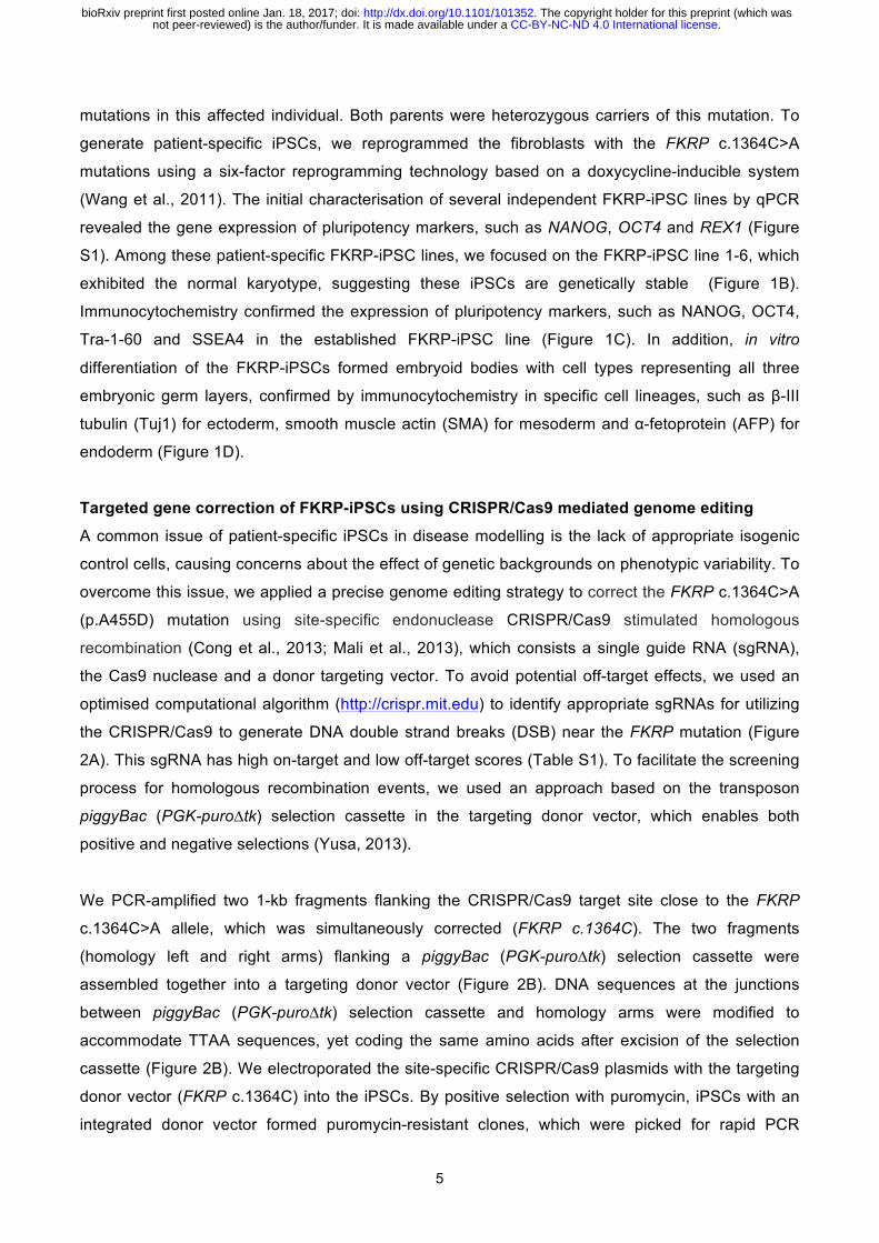
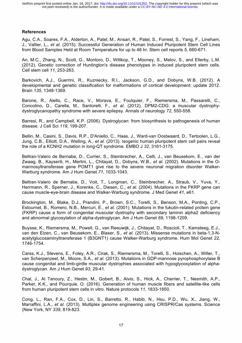
Generation and characterisation of FKRP-iPSCs derived from a dystroglycanopathy patient
with CNS abnormalities
We obtained dermal fibroblasts from a female individual, previously diagnosed congenital muscular
dystrophy with CNS abnormalities, including marked cognitive delay, microcephaly, cerebellar cysts
and cerebellar dysplasia. Genetic analysis revealed homozygous FKRP c.1364C>A (p.A455D)
.CC-BY-NC-ND 4.0 International licensenot peer-reviewed) is the author/funder. It is made available under aThe copyright holder for this preprint (which was. http://dx.doi.org/10.1101/101352doi: bioRxiv preprint first posted online Jan. 18, 2017;
5
mutations in this affected individual. Both parents were heterozygous carriers of this mutation. To
generate patient-specific iPSCs, we reprogrammed the fibroblasts with the FKRP c.1364C>A
mutations using a six-factor reprogramming technology based on a doxycycline-inducible system
(Wang et al., 2011). The initial characterisation of several independent FKRP-iPSC lines by qPCR
revealed the gene expression of pluripotency markers, such as NANOG, OCT4 and REX1 (Figure
S1). Among these patient-specific FKRP-iPSC lines, we focused on the FKRP-iPSC line 1-6, which
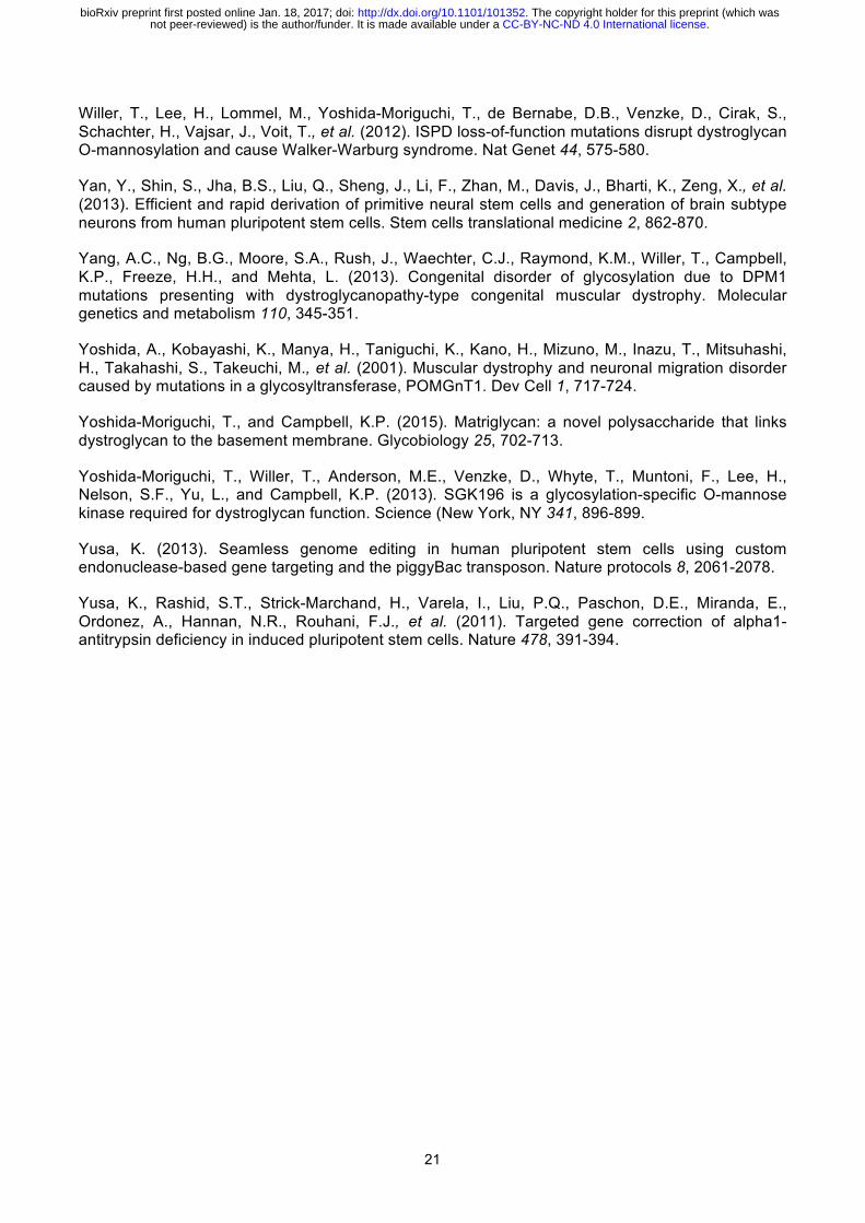
exhibited the normal karyotype, suggesting these iPSCs are genetically stable (Figure 1B).
Immunocytochemistry confirmed the expression of pluripotency markers, such as NANOG, OCT4,
Tra-1-60 and SSEA4 in the established FKRP-iPSC line (Figure 1C). In addition, in vitro
differentiation of the FKRP-iPSCs formed embryoid bodies with cell types representing all three
embryonic germ layers, confirmed by immunocytochemistry in specific cell lineages, such as β-III
tubulin (Tuj1) for ectoderm, smooth muscle actin (SMA) for mesoderm and α-fetoprotein (AFP) for
endoderm (Figure 1D).
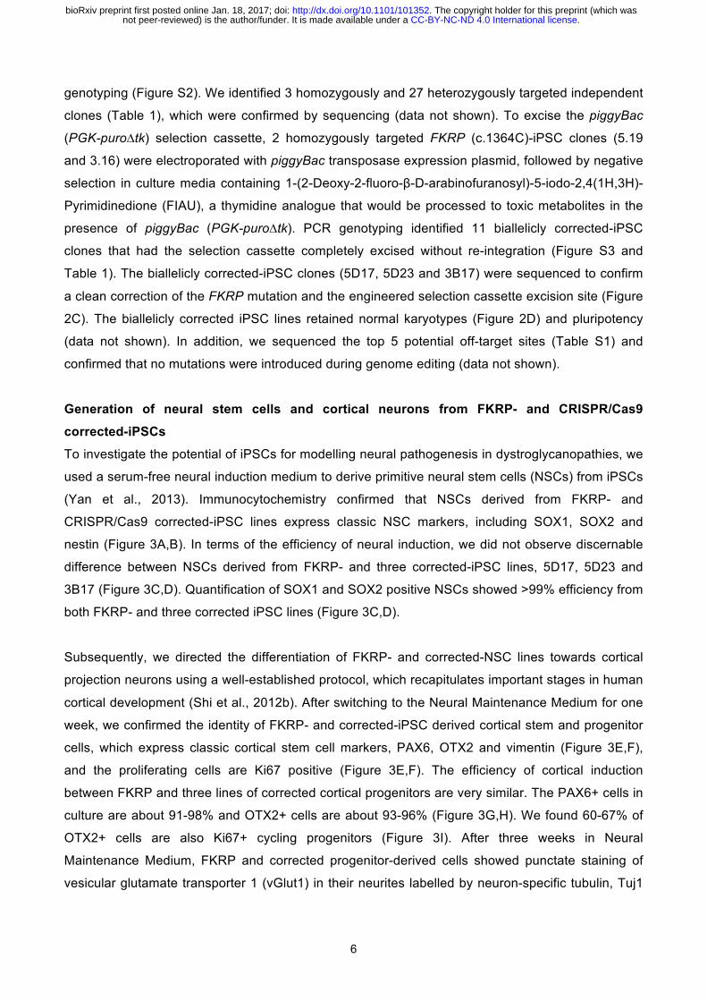
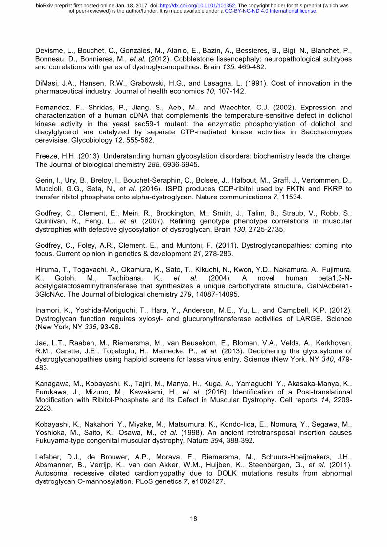
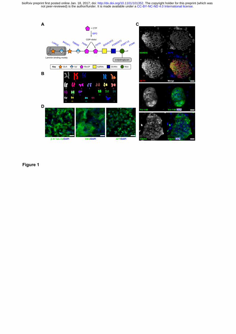
Targeted gene correction of FKRP-iPSCs using CRISPR/Cas9 mediated genome editing
A common issue of patient-specific iPSCs in disease modelling is the lack of appropriate isogenic
control cells, causing concerns about the effect of genetic backgrounds on phenotypic variability. To
overcome this issue, we applied a precise genome editing strategy to correct the FKRP c.1364C>A
(p.A455D) mutation using site-specific endonuclease CRISPR/Cas9 stimulated homologous
recombination (Cong et al., 2013; Mali et al., 2013), which consists a single guide RNA (sgRNA),
the Cas9 nuclease and a donor targeting vector. To avoid potential off-target effects, we used an
optimised computational algorithm (http://crispr.mit.edu) to identify appropriate sgRNAs for utilizing
the CRISPR/Cas9 to generate DNA double strand breaks (DSB) near the FKRP mutation (Figure
2A). This sgRNA has high on-target and low off-target scores (Table S1). To facilitate the screening
process for homologous recombination events, we used an approach based on the transposon
piggyBac (PGK-puro∆tk) selection cassette in the targeting donor vector, which enables both
positive and negative selections (Yusa, 2013).
We PCR-amplified two 1-kb fragments flanking the CRISPR/Cas9 target site close to the FKRP
c.1364C>A allele, which was simultaneously corrected (FKRP c.1364C). The two fragments
(homology left and right arms) flanking a piggyBac (PGK-puro∆tk) selection cassette were
assembled together into a targeting donor vector (Figure 2B). DNA sequences at the junctions
between piggyBac (PGK-puro∆tk) selection cassette and homology arms were modified to
accommodate TTAA sequences, yet coding the same amino acids after excision of the selection
cassette (Figure 2B). We electroporated the site-specific CRISPR/Cas9 plasmids with the targeting
donor vector (FKRP c.1364C) into the iPSCs. By positive selection with puromycin, iPSCs with an
integrated donor vector formed puromycin-resistant clones, which were picked for rapid PCR
.CC-BY-NC-ND 4.0 International licensenot peer-reviewed) is the author/funder. It is made available under aThe copyright holder for this preprint (which was. http://dx.doi.org/10.1101/101352doi: bioRxiv preprint first posted online Jan. 18, 2017;
6
genotyping (Figure S2). We identified 3 homozygously and 27 heterozygously targeted independent
clones (Table 1), which were confirmed by sequencing (data not shown). To excise the piggyBac
(PGK-puro∆tk) selection cassette, 2 homozygously targeted FKRP (c.1364C)-iPSC clones (5.19
and 3.16) were electroporated with piggyBac transposase expression plasmid, followed by negative
selection in culture media containing 1-(2-Deoxy-2-fluoro-β-D-arabinofuranosyl)-5-iodo-2,4(1H,3H)-
Pyrimidinedione (FIAU), a thymidine analogue that would be processed to toxic metabolites in the
presence of piggyBac (PGK-puro∆tk). PCR genotyping identified 11 biallelicly corrected-iPSC
clones that had the selection cassette completely excised without re-integration (Figure S3 and
Table 1). The biallelicly corrected-iPSC clones (5D17, 5D23 and 3B17) were sequenced to confirm
a clean correction of the FKRP mutation and the engineered selection cassette excision site (Figure
2C). The biallelicly corrected iPSC lines retained normal karyotypes (Figure 2D) and pluripotency
(data not shown). In addition, we sequenced the top 5 potential off-target sites (Table S1) and
confirmed that no mutations were introduced during genome editing (data not shown).
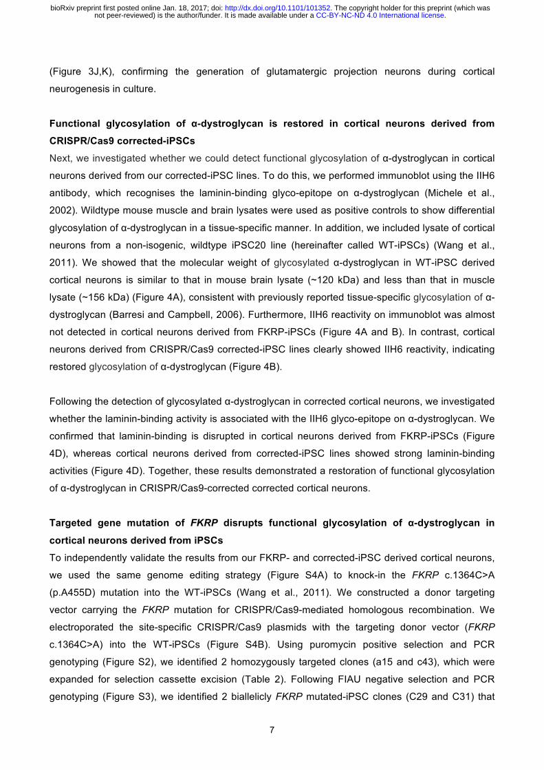
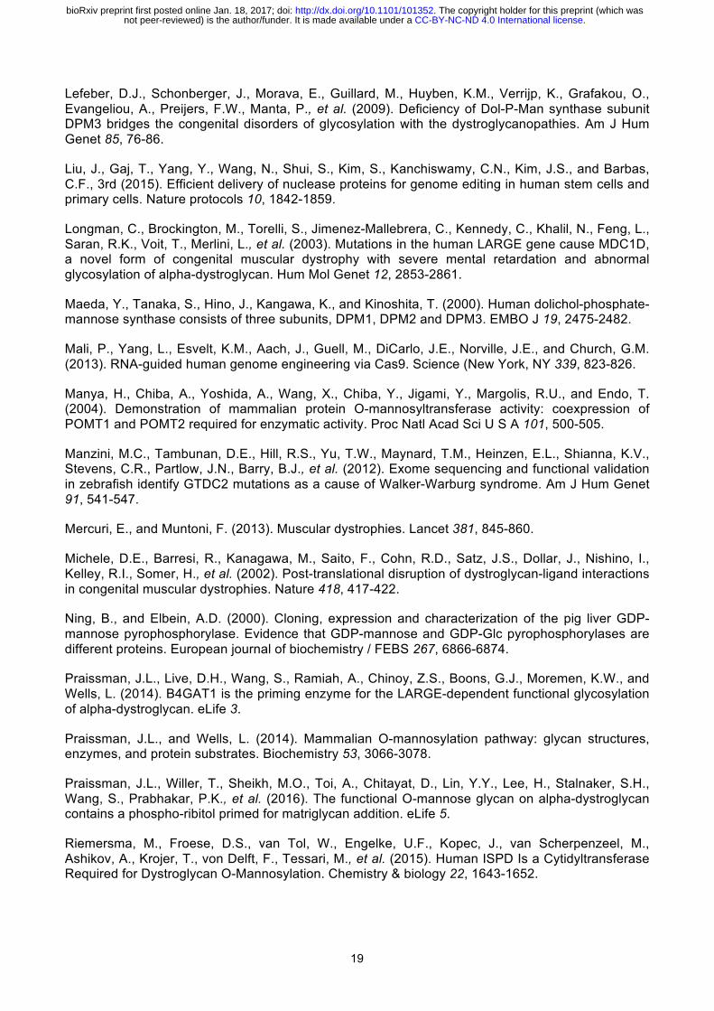
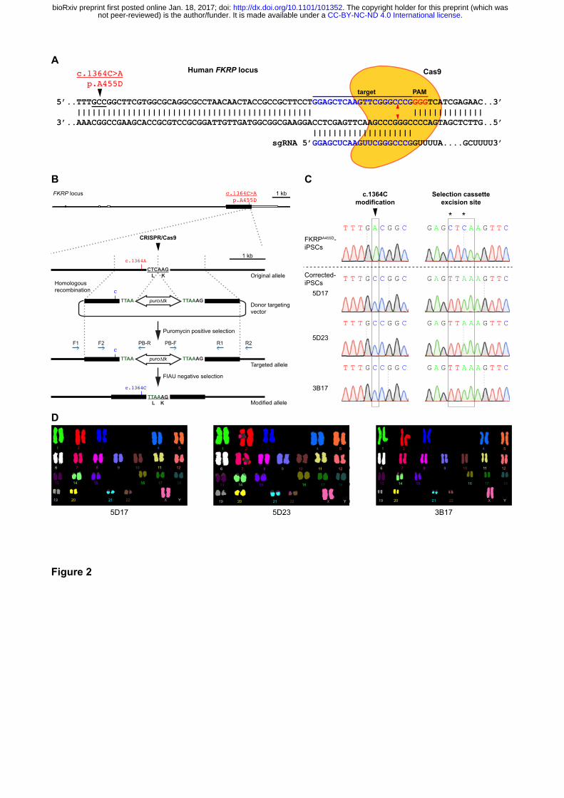
Generation of neural stem cells and cortical neurons from FKRP- and CRISPR/Cas9
corrected-iPSCs
To investigate the potential of iPSCs for modelling neural pathogenesis in dystroglycanopathies, we
used a serum-free neural induction medium to derive primitive neural stem cells (NSCs) from iPSCs
(Yan et al., 2013). Immunocytochemistry confirmed that NSCs derived from FKRP- and
CRISPR/Cas9 corrected-iPSC lines express classic NSC markers, including SOX1, SOX2 and
nestin (Figure 3A,B). In terms of the efficiency of neural induction, we did not observe discernable
difference between NSCs derived from FKRP- and three corrected-iPSC lines, 5D17, 5D23 and
3B17 (Figure 3C,D). Quantification of SOX1 and SOX2 positive NSCs showed >99% efficiency from
both FKRP- and three corrected iPSC lines (Figure 3C,D).
Subsequently, we directed the differentiation of FKRP- and corrected-NSC lines towards cortical
projection neurons using a well-established protocol, which recapitulates important stages in human
cortical development (Shi et al., 2012b). After switching to the Neural Maintenance Medium for one
week, we confirmed the identity of FKRP- and corrected-iPSC derived cortical stem and progenitor
cells, which express classic cortical stem cell markers, PAX6, OTX2 and vimentin (Figure 3E,F),
and the proliferating cells are Ki67 positive (Figure 3E,F). The efficiency of cortical induction
between FKRP and three lines of corrected cortical progenitors are very similar. The PAX6+ cells in
culture are about 91-98% and OTX2+ cells are about 93-96% (Figure 3G,H). We found 60-67% of
OTX2+ cells are also Ki67+ cycling progenitors (Figure 3I). After three weeks in Neural
Maintenance Medium, FKRP and corrected progenitor-derived cells showed punctate staining of
vesicular glutamate transporter 1 (vGlut1) in their neurites labelled by neuron-specific tubulin, Tuj1
.CC-BY-NC-ND 4.0 International licensenot peer-reviewed) is the author/funder. It is made available under aThe copyright holder for this preprint (which was. http://dx.doi.org/10.1101/101352doi: bioRxiv preprint first posted online Jan. 18, 2017;
7
(Figure 3J,K), confirming the generation of glutamatergic projection neurons during cortical
neurogenesis in culture.
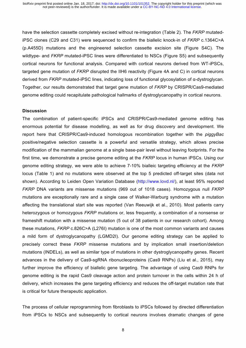
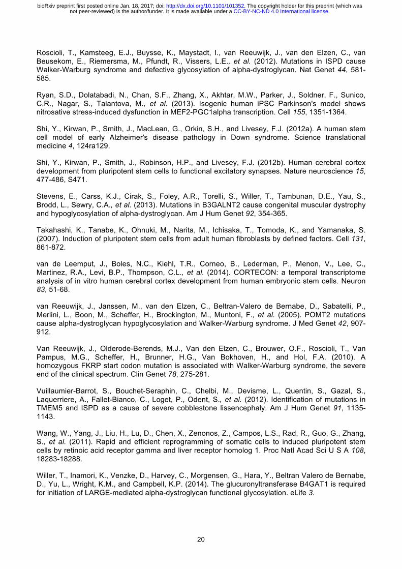
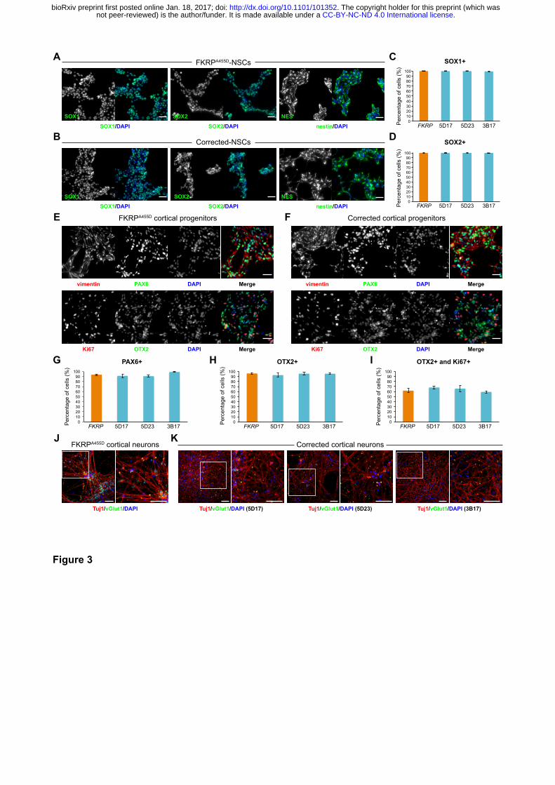
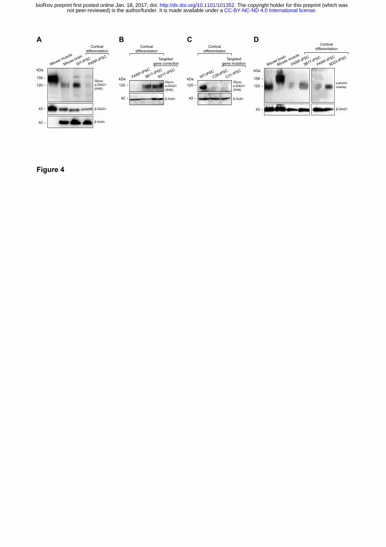
Functional glycosylation of α-dystroglycan is restored in cortical neurons derived from
CRISPR/Cas9 corrected-iPSCs
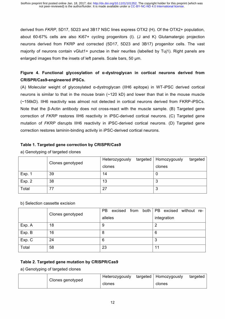
Next, we investigated whether we could detect functional glycosylation of α-dystroglycan in cortical
neurons derived from our corrected-iPSC lines. To do this, we performed immunoblot using the IIH6
antibody, which recognises the laminin-binding glyco-epitope on α-dystroglycan (Michele et al.,
2002). Wildtype mouse muscle and brain lysates were used as positive controls to show differential
glycosylation of α-dystroglycan in a tissue-specific manner. In addition, we included lysate of cortical
neurons from a non-isogenic, wildtype iPSC20 line (hereinafter called WT-iPSCs) (Wang et al.,
2011). We showed that the molecular weight of glycosylated α-dystroglycan in WT-iPSC derived
cortical neurons is similar to that in mouse brain lysate (~120 kDa) and less than that in muscle
lysate (~156 kDa) (Figure 4A), consistent with previously reported tissue-specific glycosylation of α-
dystroglycan (Barresi and Campbell, 2006). Furthermore, IIH6 reactivity on immunoblot was almost
not detected in cortical neurons derived from FKRP-iPSCs (Figure 4A and B). In contrast, cortical
neurons derived from CRISPR/Cas9 corrected-iPSC lines clearly showed IIH6 reactivity, indicating
restored glycosylation of α-dystroglycan (Figure 4B).
Following the detection of glycosylated α-dystroglycan in corrected cortical neurons, we investigated
whether the laminin-binding activity is associated with the IIH6 glyco-epitope on α-dystroglycan. We
confirmed that laminin-binding is disrupted in cortical neurons derived from FKRP-iPSCs (Figure
4D), whereas cortical neurons derived from corrected-iPSC lines showed strong laminin-binding
activities (Figure 4D). Together, these results demonstrated a restoration of functional glycosylation
of α-dystroglycan in CRISPR/Cas9-corrected corrected cortical neurons.
Targeted gene mutation of FKRP disrupts functional glycosylation of α-dystroglycan in
cortical neurons derived from iPSCs
To independently validate the results from our FKRP- and corrected-iPSC derived cortical neurons,
we used the same genome editing strategy (Figure S4A) to knock-in the FKRP c.1364C>A
(p.A455D) mutation into the WT-iPSCs (Wang et al., 2011). We constructed a donor targeting
vector carrying the FKRP mutation for CRISPR/Cas9-mediated homologous recombination. We
electroporated the site-specific CRISPR/Cas9 plasmids with the targeting donor vector (FKRP
c.1364C>A) into the WT-iPSCs (Figure S4B). Using puromycin positive selection and PCR
genotyping (Figure S2), we identified 2 homozygously targeted clones (a15 and c43), which were
expanded for selection cassette excision (Table 2). Following FIAU negative selection and PCR
genotyping (Figure S3), we identified 2 biallelicly FKRP mutated-iPSC clones (C29 and C31) that
.CC-BY-NC-ND 4.0 International licensenot peer-reviewed) is the author/funder. It is made available under aThe copyright holder for this preprint (which was. http://dx.doi.org/10.1101/101352doi: bioRxiv preprint first posted online Jan. 18, 2017;
8
have the selection cassette completely excised without re-integration (Table 2). The FKRP mutated-
iPSC clones (C29 and C31) were sequenced to confirm the biallelic knock-in of FKRP c.1364C>A
(p.A455D) mutations and the engineered selection cassette excision site (Figure S4C). The
wildtype- and FKRP mutated-iPSC lines were differentiated to NSCs (Figure S5) and subsequently
cortical neurons for functional analysis. Compared with cortical neurons derived from WT-iPSCs,
targeted gene mutation of FKRP disrupted the IIH6 reactivity (Figure 4A and C) in cortical neurons
derived from FKRP mutated-iPSC lines, indicating loss of functional glycosylation of α-dystroglycan.
Together, our results demonstrated that target gene mutation of FKRP by CRISPR/Cas9-mediated
genome editing could recapitulate pathological hallmarks of dystroglycanopathy in cortical neurons.
Discussion
The combination of patient-specific iPSCs and CRISPR/Cas9-mediated genome editing has
enormous potential for disease modelling, as well as for drug discovery and development. We
report here that CRISPR/Cas9-induced homologous recombination together with the piggyBac
positive/negative selection cassette is a powerful and versatile strategy, which allows precise
modification of the mammalian genome at a single base-pair level without leaving footprints. For the
first time, we demonstrate a precise genome editing at the FKRP locus in human iPSCs. Using our
genome editing strategy, we were able to achieve 7-10% bialleic targeting efficiency at the FKRP
locus (Table 1) and no mutations were observed at the top 5 predicted off-target sites (data not
shown). According to Leiden Open Variation Database (http://www.lovd.nl/), at least 95% reported
FKRP DNA variants are missense mutations (969 out of 1018 cases). Homozygous null FKRP
mutations are exceptionally rare and a single case of Walker-Warburg syndrome with a mutation
affecting the translational start site was reported (Van Reeuwijk et al., 2010). Most patients carry
heterozygous or homozygous FKRP mutations or, less frequently, a combination of a nonsense or
frameshift mutation with a missense mutation (5 out of 38 patients in our research cohort). Among
these mutations, FKRP c.826C>A (L276I) mutation is one of the most common variants and causes
a mild form of dystroglycanopathy (LGMD2I). Our genome editing strategy can be applied to
precisely correct these FKRP missense mutations and by implication small insertion/deletion
mutations (INDELs), as well as similar type of mutations in other dystroglycanopathy genes. Recent
advances in the delivery of Cas9-sgRNA ribonucleoproteins (Cas9 RNPs) (Liu et al., 2015), may
further improve the efficiency of biallelic gene targeting. The advantage of using Cas9 RNPs for
genome editing is the rapid Cas9 cleavage action and protein turnover in the cells within 24 h of
delivery, which increases the gene targeting efficiency and reduces the off-target mutation rate that
is critical for future therapeutic application.
The process of cellular reprogramming from fibroblasts to iPSCs followed by directed differentiation
from iPSCs to NSCs and subsequently to cortical neurons involves dramatic changes of gene
.CC-BY-NC-ND 4.0 International licensenot peer-reviewed) is the author/funder. It is made available under aThe copyright holder for this preprint (which was. http://dx.doi.org/10.1101/101352doi: bioRxiv preprint first posted online Jan. 18, 2017;
9
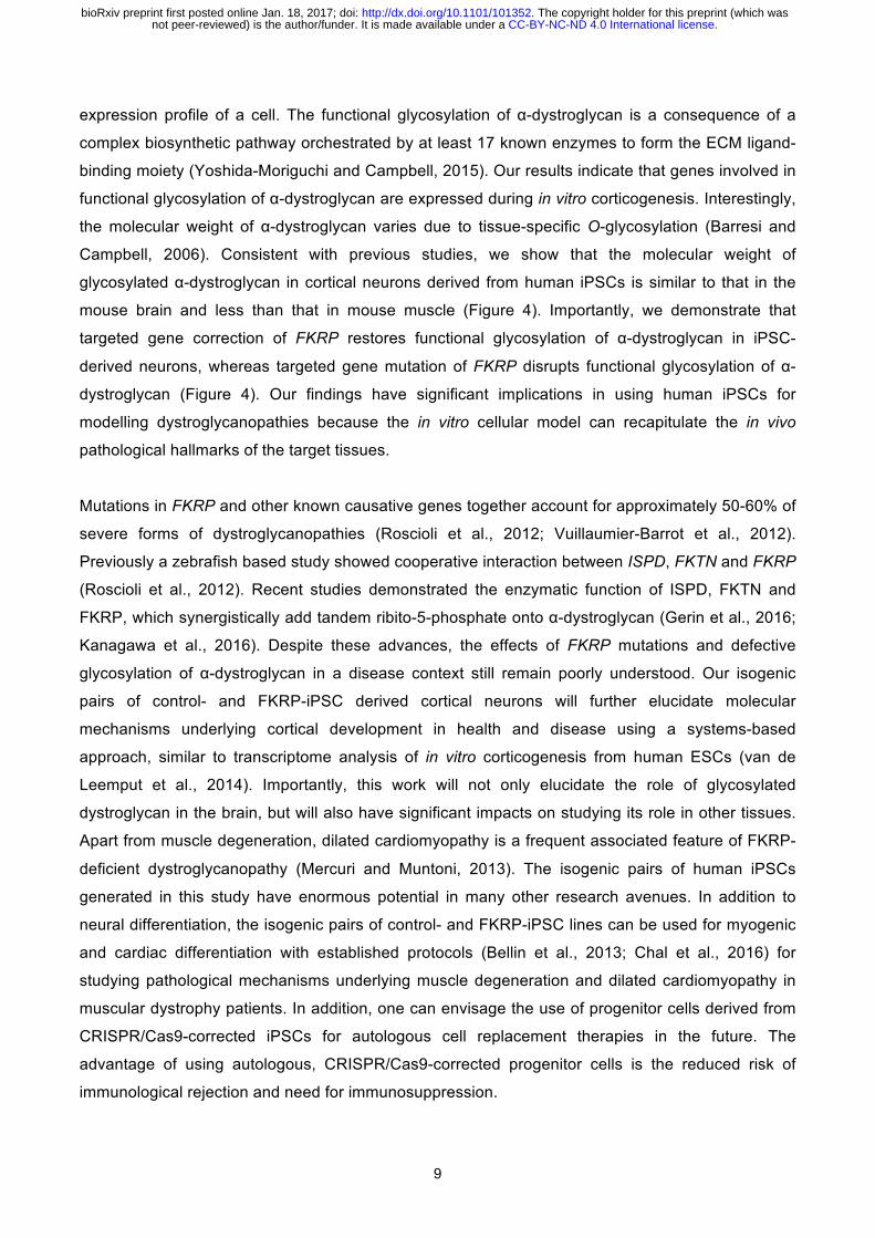
expression profile of a cell. The functional glycosylation of α-dystroglycan is a consequence of a
complex biosynthetic pathway orchestrated by at least 17 known enzymes to form the ECM ligand-
binding moiety (Yoshida-Moriguchi and Campbell, 2015). Our results indicate that genes involved in
functional glycosylation of α-dystroglycan are expressed during in vitro corticogenesis. Interestingly,
the molecular weight of α-dystroglycan varies due to tissue-specific O-glycosylation (Barresi and
Campbell, 2006). Consistent with previous studies, we show that the molecular weight of
glycosylated α-dystroglycan in cortical neurons derived from human iPSCs is similar to that in the
mouse brain and less than that in mouse muscle (Figure 4). Importantly, we demonstrate that
targeted gene correction of FKRP restores functional glycosylation of α-dystroglycan in iPSC-
derived neurons, whereas targeted gene mutation of FKRP disrupts functional glycosylation of α-
dystroglycan (Figure 4). Our findings have significant implications in using human iPSCs for
modelling dystroglycanopathies because the in vitro cellular model can recapitulate the in vivo
pathological hallmarks of the target tissues.
Mutations in FKRP and other known causative genes together account for approximately 50-60% of
severe forms of dystroglycanopathies (Roscioli et al., 2012; Vuillaumier-Barrot et al., 2012).
Previously a zebrafish based study showed cooperative interaction between ISPD, FKTN and FKRP
(Roscioli et al., 2012). Recent studies demonstrated the enzymatic function of ISPD, FKTN and
FKRP, which synergistically add tandem ribito-5-phosphate onto α-dystroglycan (Gerin et al., 2016;
Kanagawa et al., 2016). Despite these advances, the effects of FKRP mutations and defective
glycosylation of α-dystroglycan in a disease context still remain poorly understood. Our isogenic
pairs of control- and FKRP-iPSC derived cortical neurons will further elucidate molecular
mechanisms underlying cortical development in health and disease using a systems-based
approach, similar to transcriptome analysis of in vitro corticogenesis from human ESCs (van de
Leemput et al., 2014). Importantly, this work will not only elucidate the role of glycosylated
dystroglycan in the brain, but will also have significant impacts on studying its role in other tissues.
Apart from muscle degeneration, dilated cardiomyopathy is a frequent associated feature of FKRP-
deficient dystroglycanopathy (Mercuri and Muntoni, 2013). The isogenic pairs of human iPSCs
generated in this study have enormous potential in many other research avenues. In addition to
neural differentiation, the isogenic pairs of control- and FKRP-iPSC lines can be used for myogenic
and cardiac differentiation with established protocols (Bellin et al., 2013; Chal et al., 2016) for
studying pathological mechanisms underlying muscle degeneration and dilated cardiomyopathy in
muscular dystrophy patients. In addition, one can envisage the use of progenitor cells derived from
CRISPR/Cas9-corrected iPSCs for autologous cell replacement therapies in the future. The
advantage of using autologous, CRISPR/Cas9-corrected progenitor cells is the reduced risk of
immunological rejection and need for immunosuppression.
.CC-BY-NC-ND 4.0 International licensenot peer-reviewed) is the author/funder. It is made available under aThe copyright holder for this preprint (which was. http://dx.doi.org/10.1101/101352doi: bioRxiv preprint first posted online Jan. 18, 2017;
10
In conclusion, we have established two isogenic pairs of human iPSC models for a severe form of
FKRP-deficient dystroglycanopathy. A significant impact of this study is that other human iPSC-
based dystroglycanopathy models will be generated. Collectively, these iPSC models will provide
powerful in vitro platforms that can be exploited for setting up high-content drug screens using
quantifiable phenotypes as readouts. Importantly, while more than 90% of drugs fail during
development (DiMasi et al., 1991), our isogenic pairs of human iPSC lines will provide an invaluable
resource for systematic chemical and genetic screens capable of reverting pathophysiological
defects in physiology-relevant cellular models. This will facilitate the drug discovery and
development for treating dystroglycanopathies.
.CC-BY-NC-ND 4.0 International licensenot peer-reviewed) is the author/funder. It is made available under aThe copyright holder for this preprint (which was. http://dx.doi.org/10.1101/101352doi: bioRxiv preprint first posted online Jan. 18, 2017;
11
Figure legends
Figure 1. Functional glycosylation of α-dystroglycan and dystroglycanopathy patient-
specific iPSCs
(A) Current model of the core M3 functional glycan structure on α-dystroglycan and enzymes
involved in its synthesis. ECM ligands bind to the Xyl-GlucA disaccharide repeats. Man, Mannose;
GlcNAc, N-Acetylglucosamine; GalNAc, N-Acetylgalactosamine; Rbo5P, ribitol-5-phosphate; Xyl,
xylose; GlcA, glucuronic acid. (B) FKRPA455D-iPSCs have a normal karyotype. (C) Immunostaining
demonstrates FKRPA455D-iPSCs express specific pluripotency-associated markers, including
NANOG, OCT4, Tra-1-60 and SSEA4. (D) In vitro differentiation of FKRPA455D-iPSCs to cells
representing ectoderm (β-III Tubulin, Tuj1), mesoderm (SMA, smooth muscle actin) and endoderm
(α-fectoprotein). Scale bars, 50 µm.
Figure 2. Targeted gene correction of FKRPA455D-iPSCs by CRISPR/Cas9-mediated genome
editing
(A) Cas9 protein and the specific sgRNA targeting the human FKRP locus. The FKRP c.1364C>A
(p.A455D) mutation is 43 bases upstream of the sgRNA target sequences. Red arrowheads indicate
putative cleavage site. PAM, protospacer-adjacent motif. (B) A schematic diagram shows the
genome editing strategy based on CRISPR/Cas9-stimulated homologous recombination, followed
by positive selection with puromycin and negative selection with FIAU. Homology left and right arms
on the targeting donor vector are indicated as black boxes, flanking the piggyBac (PGK-puro∆tk)
selection cassette, which is under the control of PGK promoter. PCR genotyping primers are shown
as blue arrows. Note that the TTAA sequences are designed to accommodation the selection
cassette excision sites, yet code the same amino acids. (C) Sequence analysis shows precise
biallelic correction of the FKRP mutation in three independently corrected iPSC clones (5D17, 5D23
and 3B17), compared with their parental FKRPA455D-iPSCs. Selection cassette excision sites are
identified in the corrected-iPSC lines. (D) CRISPR/Cas9 corrected-iPSC lines show a normal
karyotype.
Figure 3. Characterisation of NSCs and cortical neurons derived from FKRP- and
CRISPR/Cas9 corrected-iPSCs
(A and B) Representative images of NSCs derived from FKRP- and corrected-iPSC lines
expressing SOX1, SOX2 and nestin. (C and D) Quantification of percentage of SOX1+ (C) and
SOX2+ (D) cells in culture. The efficiency of neural induction is more than 99% in FKRP- and
corrected-iPSC (5D17, 5D23 and 3B17) lines. (E and F) FKRP- and corrected NSC lines can be
further differentiated to cortical neural progenitor cells, expressing PAX6, OTX2 and vimentin. (G-I)
Quantification of percentage of PAX6+ (G) and OTX2+ (H) cells in culture. About 91-98% of cells
derived from FKRP, 5D17, 5D23 and 3B17 NSC lines express PAX6 (G). About 93-96% of cells
.CC-BY-NC-ND 4.0 International licensenot peer-reviewed) is the author/funder. It is made available under aThe copyright holder for this preprint (which was. http://dx.doi.org/10.1101/101352doi: bioRxiv preprint first posted online Jan. 18, 2017;
12
derived from FKRP, 5D17, 5D23 and 3B17 NSC lines express OTX2 (H). Of the OTX2+ population,
about 60-67% cells are also Ki67+ cycling progenitors (I). (J and K) Glutamatergic projection
neurons derived from FKRP and corrected (5D17, 5D23 and 3B17) progenitor cells. The vast
majority of neurons contain vGlut1+ punctae in their neurites (labelled by Tuj1). Right panels are
enlarged images from the insets of left panels. Scale bars, 50 µm.
Figure 4. Functional glycosylation of α-dystroglycan in cortical neurons derived from
CRISPR/Cas9-engineered iPSCs.
(A) Molecular weight of glycosylated α-dystroglycan (IIH6 epitope) in WT-iPSC derived cortical
neurons is similar to that in the mouse brain (~120 kD) and lower than that in the mouse muscle
(~156kD). IIH6 reactivity was almost not detected in cortical neurons derived from FKRP-iPSCs.
Note that the β-Actin antibody does not cross-react with the muscle sample. (B) Targeted gene
correction of FKRP restores IIH6 reactivity in iPSC-derived cortical neurons. (C) Targeted gene
mutation of FKRP disrupts IIH6 reactivity in iPSC-derived cortical neurons. (D) Targeted gene
correction restores laminin-binding activity in iPSC-derived cortical neurons.
Table 1. Targeted gene correction by CRISPR/Cas9
a) Genotyping of targeted clones
Clones genotyped Heterozygously targeted
clones
Homozygously targeted
clones
Exp. 1 39 14 0
Exp. 2 38 13 3
Total 77 27 3
b) Selection cassette excision
Clones genotyped PB excised from both
alleles
PB excised without re-
integration
Exp. A 18 9 2
Exp. B 16 8 6
Exp. C 24 6 3
Total 58 23 11
Table 2. Targeted gene mutation by CRISPR/Cas9
a) Genotyping of targeted clones
Clones genotyped Heterozygously targeted
clones
Homozygously targeted
clones
.CC-BY-NC-ND 4.0 International licensenot peer-reviewed) is the author/funder. It is made available under aThe copyright holder for this preprint (which was. http://dx.doi.org/10.1101/101352doi: bioRxiv preprint first posted online Jan. 18, 2017;
13
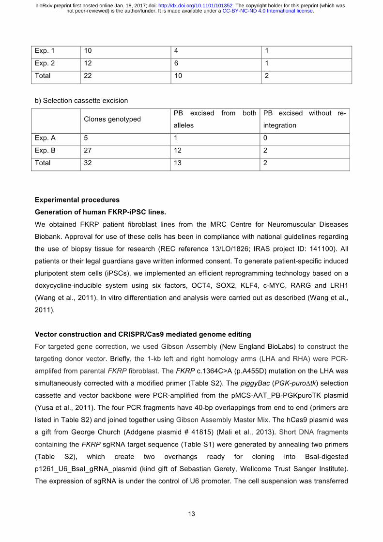
Exp. 1 10 4 1
Exp. 2 12 6 1
Total 22 10 2
b) Selection cassette excision
Clones genotyped PB excised from both
alleles
PB excised without re-
integration
Exp. A 5 1 0
Exp. B 27 12 2
Total 32 13 2
Experimental procedures
Generation of human FKRP-iPSC lines.
We obtained FKRP patient fibroblast lines from the MRC Centre for Neuromuscular Diseases
Biobank. Approval for use of these cells has been in compliance with national guidelines regarding
the use of biopsy tissue for research (REC reference 13/LO/1826; IRAS project ID: 141100). All
patients or their legal guardians gave written informed consent. To generate patient-specific induced
pluripotent stem cells (iPSCs), we implemented an efficient reprogramming technology based on a
doxycycline-inducible system using six factors, OCT4, SOX2, KLF4, c-MYC, RARG and LRH1
(Wang et al., 2011). In vitro differentiation and analysis were carried out as described (Wang et al.,
2011).
Vector construction and CRISPR/Cas9 mediated genome editing
For targeted gene correction, we used Gibson Assembly (New England BioLabs) to construct the
targeting donor vector. Briefly, the 1-kb left and right homology arms (LHA and RHA) were PCR-
amplifed from parental FKRP fibroblast. The FKRP c.1364C>A (p.A455D) mutation on the LHA was
simultaneously corrected with a modified primer (Table S2). The piggyBac (PGK-puro∆tk) selection
cassette and vector backbone were PCR-amplified from the pMCS-AAT_PB-PGKpuroTK plasmid
(Yusa et al., 2011). The four PCR fragments have 40-bp overlappings from end to end (primers are
listed in Table S2) and joined together using Gibson Assembly Master Mix. The hCas9 plasmid was
a gift from George Church (Addgene plasmid # 41815) (Mali et al., 2013). Short DNA fragments
containing the FKRP sgRNA target sequence (Table S1) were generated by annealing two primers
(Table S2), which create two overhangs ready for cloning into BsaI-digested
p1261_U6_BsaI_gRNA_plasmid (kind gift of Sebastian Gerety, Wellcome Trust Sanger Institute).
The expression of sgRNA is under the control of U6 promoter. The cell suspension was transferred
.CC-BY-NC-ND 4.0 International licensenot peer-reviewed) is the author/funder. It is made available under aThe copyright holder for this preprint (which was. http://dx.doi.org/10.1101/101352doi: bioRxiv preprint first posted online Jan. 18, 2017;
14
to a cuvette and electroporated using Amaxa Nucleofection 2b device with program B16. For
targeted gene mutation, the same strategy was used to construct the donor targeting vector carrying
the FKRP c.1364C>A (p.A455D) mutation on LHA using primers in Table S2.
Karyotyping analysis
As previously described (Agu et al., 2015), multiplex fluorescent in situ hybridization (M-FISH)
karyotype analysis was performed on iPSC lines with slight modification. Briefly, prior to metaphase
harvesting, iPSCs were grown in M15 medium (Knockout DMEM, 15% Fetal Bovine Serum, 1X
glutamine-penicillin-streptomycin, 1X nonessential amino acids and 1 ng/ml human recombinant
LIF) for 24 h and then treated with 10 µM Y-27632 dihydrochloride (Tocris) for 2–3 h.
Neural induction and cortical differentiation from human iPSCs
As described (Yan et al., 2013), induction of NSCs from human iPSCs was carried out using Gibco
Neural Induction Medium (Neurobasal medium and 2% Neural Induction Supplement) for 7days,
followed by passaging and expansion in Neural Expansion Medium (49% Neurobasal medium, 49%
Advanced DMDM/F-12 and 2% Neural Induction Supplement). Expanded NSCs were
cryopreserved or differentiated into cortical neurons following the established protocol (Shi et al.,
2012b). Briefly, tissue culture plates were pre-coated with 0.01% (w/v) poly-L-ornithin (Sigma,
P4957) for 4 h, followed by 20 µg/ml of laminins (Sigma, L2020) for 4h. NSCs were seeded on the
pre-coated plates (50,000/cm2) in Neural Expansion Medium with 5 µM Y-27632 dihydrochloride
(Tocris) for 24 h. Subsequently, Neural Expansion Medium was replaced with Neural Maintenance
Medium, containing 50% DMEM/F-12 (Gibco), 50% Neurobasal medium (Gibco), 0.5X N2
supplement (Gibco), 0.5X B27 supplement (Gibco), 1.5 mM GlutaMAX-I (Gibco), 0.5X Penicillin-
streptomycin (Gibco), 2.5 µg/ml Insulin (Sigma), 0.05 mM of 2-Mecaptoethanol (Gibco), 0.5% v/v of
Non-Essential Amino Acid (Gibco), 20 ng/ml BDNF (Peprotech) and 20 ng/ml GDNF (Peprotech).
When reaching 90% confluence (approximately within 3-4days), cells were re-seeded (60,000/cm2)
in Neural Maintenance Medium (with 5 µM Y-27632 dihydrochloride for 24 h), followed by media
change every other day. In vitro cortigenesis occurs in the following weeks. Cultured cells were then
harvested or fixed at specific time points for further analysis.
Immunocytochemistry
Cells were fixed with 4% PFA for 15 mins at room temperature (RT). Prior to immunocytochemistry,
cells were permeabilized with 0.1% Triton/phosphate-buffered saline (PBS) for 15min at RT and
blocked with either 10% goat serum/PBS or 10% horse serum/PBS. Primary antibodies used were:
OCT4 (1:100; Santa Cruz, sc-5279), NANOG (1:100; abcam, AB80892), Tra-1-60 (1:100; Santa
Cruz, sc-21705), SSEA4 (1:100; BD Bioscience, 560796), α-fetoprotein (1:150; R&D Systems,
MAB1368), α-smooth muscle actin (1:150; R&D Systems, MAB1420), Sox1 (1:100; R&D Systems,
.CC-BY-NC-ND 4.0 International licensenot peer-reviewed) is the author/funder. It is made available under aThe copyright holder for this preprint (which was. http://dx.doi.org/10.1101/101352doi: bioRxiv preprint first posted online Jan. 18, 2017;
15
AF3369), Sox2 (1:100; R&D Systems, 245610), Nestin (1:500; abcam, ab22035), Ki67 (1:100; BD
Pharmingen, 550609), OTX2 (1:250; Millipore, AB9566), Pax6 (1:300; Biolegend, 901301), vimentin
(1:100; abcam, ab28028), vGLUT1 (1:2000; Synaptic systems, 135303) and β-III tubulin (Tuj1)
(1:150; R&D System, MAB1195). All primary antibodies were diluted in blocking solution and
incubated overnight at 4°C, followed by washing in PBS 3 times for 15 mins. Subsequently,
appropriate Alexa Fluor 488 or 564 conjugated secondary antibodies were incubated for 1 h at RT.
For nuclei staining, samples were incubated with DAPI (Sigma-Aldrich) for 5 mins and washed in
PBS briefly. Finally, images were captured and analysed using IN Cell 2200.
Immunoblot analysis
Cells were harvested at specific time points and proteins were extracted in RIPA buffer consisting of
50 mM Tris-HCl (pH 7.5), 150 mM NaCl, 1 mM EDTA, 1% Triton X-100, 1% SDS and 1 mM azide
plus a cocktail of protease inhibitors (Roche). A total of 30-60 µg of soluble protein was resolved
using NuPage Novex 4-12% Bis-Tris protein gels (Invitrogen) and then electrophoretically
transferred to polyvinylidene difluoride (PVDF) membrane (Millipore). The PVDF membrane was
blocked in 3% bovine serum albumin in TBST (Tris-buffered saline with 0.1% Tween) and probed
with primary antibodies to glycosylated α-dystroglycan (IIH6 1:500; kind gift from Kevin Campbell)
and β-dystroglycan (1:100; Leica Biosystems, NCL-b-DG), followed by washing with TBST and
incubation with horseradish peroxidase (HRP) conjugated anti-mouse IgM (Millipore) and HRP
conjugated anti-mouse IgG (Jackson laboratories), respectively. Note that anti-beta actin antibody
(1:1000; abcam, ab8226) used in this study does not cross-react with skeletal muscle actin.
SuperSignal West Pico Chemiluminescent Substrate (Thermo Fisher) was applied to PVDF
membranes and signals were visualized using a Bio-Rad ChemiDoc MP imaging system.
Laminin overlay analysis
Protein samples were extracted, resolved and transferred to PVDF membranes as described above.
The PVDF membrane was then blocked with 5% skimmed milk in laminin binding buffer (LBB),
containing 10 mM triethanolamine, 140 mM NaCl, 1 mM MgCl2, 1 mM CaCl2 (pH adjusted to 7.6),
followed by incubation with laminins from Engelbreth-Holm-Swarm murine sarcoma basement
membrane (Sigma, L2020) in LBB with a final concentration of 5 µg/ml at 4oC overnight. The PVDF
membrane was then incubated with the pan-laminin antibody (1:1000; Sigma, L9393), washed with
1x LBB with 0.1% Tween and incubated with HRP conjugated anti-rabbit IgG (Jackson
laboratories). Signal detection using chemiluminescence substrate was performed as above.
Author Contribution
B.L., J.K., D.R., and E.K. carried out the experimental work, analyzed and interpreted the data. S.G,
B.F, and F.Y. performed and analyzed the karyotyping experiments. D.R., D.S., P.L., and F.M.
.CC-BY-NC-ND 4.0 International licensenot peer-reviewed) is the author/funder. It is made available under aThe copyright holder for this preprint (which was. http://dx.doi.org/10.1101/101352doi: bioRxiv preprint first posted online Jan. 18, 2017;
16
contributed to the conception and design of the project and edited the manuscript. Y.-Y.L. designed
the project, performed some of the experiments, analyzed and interpreted the data and supervised
the research. J.K., and Y.-Y.L. co-wrote the manuscript.
Acknowledgement
We thank Dr Wei Wang (Wellcome Trust Sanger Institute) for his critical advice on the generation of
human iPSCs. We are indebted to Dr Kosuke Yusa (Wellcome Trust Sanger Institute) for valuable
discussion about genome editing and providing plasmids. We would like to thank Dr Susan Brown
(Royal Veterinary College) for providing wildtype mouse muscle and brain lysates and critical
reading of the manuscript. We thank Dr Luke Gammon (Blizard Institute Screening Core Facility)
and Amaia Paredes Redondo (Blizard Institute) for their kind assistance in some experiments. F.M.
is supported by the National Institute for Health Research Biomedical Research Centre at Great
Ormond Street Hospital for Children NHS Foundation Trust and University College London. The
support of the Muscular Dystrophy UK to the Dubowitz Neuromuscular Centre and of the MRC
Neuromuscular Centre Biobank is also gratefully acknowledged. This work was supported in part by
Royal Society Research Grant (RG130417) and Newlife Research grant (SG/14-15/14) to Y.-Y.L.
.CC-BY-NC-ND 4.0 International licensenot peer-reviewed) is the author/funder. It is made available under aThe copyright holder for this preprint (which was. http://dx.doi.org/10.1101/101352doi: bioRxiv preprint first posted online Jan. 18, 2017;
17
References Agu, C.A., Soares, F.A., Alderton, A., Patel, M., Ansari, R., Patel, S., Forrest, S., Yang, F., Lineham, J., Vallier, L., et al. (2015). Successful Generation of Human Induced Pluripotent Stem Cell Lines from Blood Samples Held at Room Temperature for up to 48 hr. Stem cell reports 5, 660-671.
An, M.C., Zhang, N., Scott, G., Montoro, D., Wittkop, T., Mooney, S., Melov, S., and Ellerby, L.M. (2012). Genetic correction of Huntington's disease phenotypes in induced pluripotent stem cells. Cell stem cell 11, 253-263.
Barkovich, A.J., Guerrini, R., Kuzniecky, R.I., Jackson, G.D., and Dobyns, W.B. (2012). A developmental and genetic classification for malformations of cortical development: update 2012. Brain 135, 1348-1369.
Barone, R., Aiello, C., Race, V., Morava, E., Foulquier, F., Riemersma, M., Passarelli, C., Concolino, D., Carella, M., Santorelli, F., et al. (2012). DPM2-CDG: a muscular dystrophy-dystroglycanopathy syndrome with severe epilepsy. Annals of neurology 72, 550-558.
Barresi, R., and Campbell, K.P. (2006). Dystroglycan: from biosynthesis to pathogenesis of human disease. J Cell Sci 119, 199-207.
Bellin, M., Casini, S., Davis, R.P., D'Aniello, C., Haas, J., Ward-van Oostwaard, D., Tertoolen, L.G., Jung, C.B., Elliott, D.A., Welling, A., et al. (2013). Isogenic human pluripotent stem cell pairs reveal the role of a KCNH2 mutation in long-QT syndrome. EMBO J 32, 3161-3175.
Beltran-Valero de Bernabe, D., Currier, S., Steinbrecher, A., Celli, J., van Beusekom, E., van der Zwaag, B., Kayserili, H., Merlini, L., Chitayat, D., Dobyns, W.B., et al. (2002). Mutations in the O-mannosyltransferase gene POMT1 give rise to the severe neuronal migration disorder Walker-Warburg syndrome. Am J Hum Genet 71, 1033-1043.
Beltran-Valero de Bernabe, D., Voit, T., Longman, C., Steinbrecher, A., Straub, V., Yuva, Y., Herrmann, R., Sperner, J., Korenke, C., Diesen, C., et al. (2004). Mutations in the FKRP gene can cause muscle-eye-brain disease and Walker-Warburg syndrome. J Med Genet 41, e61.
Brockington, M., Blake, D.J., Prandini, P., Brown, S.C., Torelli, S., Benson, M.A., Ponting, C.P., Estournet, B., Romero, N.B., Mercuri, E., et al. (2001). Mutations in the fukutin-related protein gene (FKRP) cause a form of congenital muscular dystrophy with secondary laminin alpha2 deficiency and abnormal glycosylation of alpha-dystroglycan. Am J Hum Genet 69, 1198-1209.
Buysse, K., Riemersma, M., Powell, G., van Reeuwijk, J., Chitayat, D., Roscioli, T., Kamsteeg, E.J., van den Elzen, C., van Beusekom, E., Blaser, S., et al. (2013). Missense mutations in beta-1,3-N-acetylglucosaminyltransferase 1 (B3GNT1) cause Walker-Warburg syndrome. Hum Mol Genet 22, 1746-1754.
Carss, K.J., Stevens, E., Foley, A.R., Cirak, S., Riemersma, M., Torelli, S., Hoischen, A., Willer, T., van Scherpenzeel, M., Moore, S.A., et al. (2013). Mutations in GDP-mannose pyrophosphorylase B cause congenital and limb-girdle muscular dystrophies associated with hypoglycosylation of alpha-dystroglycan. Am J Hum Genet 93, 29-41.
Chal, J., Al Tanoury, Z., Hestin, M., Gobert, B., Aivio, S., Hick, A., Cherrier, T., Nesmith, A.P., Parker, K.K., and Pourquie, O. (2016). Generation of human muscle fibers and satellite-like cells from human pluripotent stem cells in vitro. Nature protocols 11, 1833-1850.
Cong, L., Ran, F.A., Cox, D., Lin, S., Barretto, R., Habib, N., Hsu, P.D., Wu, X., Jiang, W., Marraffini, L.A., et al. (2013). Multiplex genome engineering using CRISPR/Cas systems. Science (New York, NY 339, 819-823.
.CC-BY-NC-ND 4.0 International licensenot peer-reviewed) is the author/funder. It is made available under aThe copyright holder for this preprint (which was. http://dx.doi.org/10.1101/101352doi: bioRxiv preprint first posted online Jan. 18, 2017;
18
Devisme, L., Bouchet, C., Gonzales, M., Alanio, E., Bazin, A., Bessieres, B., Bigi, N., Blanchet, P., Bonneau, D., Bonnieres, M., et al. (2012). Cobblestone lissencephaly: neuropathological subtypes and correlations with genes of dystroglycanopathies. Brain 135, 469-482.
DiMasi, J.A., Hansen, R.W., Grabowski, H.G., and Lasagna, L. (1991). Cost of innovation in the pharmaceutical industry. Journal of health economics 10, 107-142.
Fernandez, F., Shridas, P., Jiang, S., Aebi, M., and Waechter, C.J. (2002). Expression and characterization of a human cDNA that complements the temperature-sensitive defect in dolichol kinase activity in the yeast sec59-1 mutant: the enzymatic phosphorylation of dolichol and diacylglycerol are catalyzed by separate CTP-mediated kinase activities in Saccharomyces cerevisiae. Glycobiology 12, 555-562.
Freeze, H.H. (2013). Understanding human glycosylation disorders: biochemistry leads the charge. The Journal of biological chemistry 288, 6936-6945.
Gerin, I., Ury, B., Breloy, I., Bouchet-Seraphin, C., Bolsee, J., Halbout, M., Graff, J., Vertommen, D., Muccioli, G.G., Seta, N., et al. (2016). ISPD produces CDP-ribitol used by FKTN and FKRP to transfer ribitol phosphate onto alpha-dystroglycan. Nature communications 7, 11534.
Godfrey, C., Clement, E., Mein, R., Brockington, M., Smith, J., Talim, B., Straub, V., Robb, S., Quinlivan, R., Feng, L., et al. (2007). Refining genotype phenotype correlations in muscular dystrophies with defective glycosylation of dystroglycan. Brain 130, 2725-2735.
Godfrey, C., Foley, A.R., Clement, E., and Muntoni, F. (2011). Dystroglycanopathies: coming into focus. Current opinion in genetics & development 21, 278-285.
Hiruma, T., Togayachi, A., Okamura, K., Sato, T., Kikuchi, N., Kwon, Y.D., Nakamura, A., Fujimura, K., Gotoh, M., Tachibana, K., et al. (2004). A novel human beta1,3-N-acetylgalactosaminyltransferase that synthesizes a unique carbohydrate structure, GalNAcbeta1-3GlcNAc. The Journal of biological chemistry 279, 14087-14095.
Inamori, K., Yoshida-Moriguchi, T., Hara, Y., Anderson, M.E., Yu, L., and Campbell, K.P. (2012). Dystroglycan function requires xylosyl- and glucuronyltransferase activities of LARGE. Science (New York, NY 335, 93-96.
Jae, L.T., Raaben, M., Riemersma, M., van Beusekom, E., Blomen, V.A., Velds, A., Kerkhoven, R.M., Carette, J.E., Topaloglu, H., Meinecke, P., et al. (2013). Deciphering the glycosylome of dystroglycanopathies using haploid screens for lassa virus entry. Science (New York, NY 340, 479-483.
Kanagawa, M., Kobayashi, K., Tajiri, M., Manya, H., Kuga, A., Yamaguchi, Y., Akasaka-Manya, K., Furukawa, J., Mizuno, M., Kawakami, H., et al. (2016). Identification of a Post-translational Modification with Ribitol-Phosphate and Its Defect in Muscular Dystrophy. Cell reports 14, 2209-2223.
Kobayashi, K., Nakahori, Y., Miyake, M., Matsumura, K., Kondo-Iida, E., Nomura, Y., Segawa, M., Yoshioka, M., Saito, K., Osawa, M., et al. (1998). An ancient retrotransposal insertion causes Fukuyama-type congenital muscular dystrophy. Nature 394, 388-392.
Lefeber, D.J., de Brouwer, A.P., Morava, E., Riemersma, M., Schuurs-Hoeijmakers, J.H., Absmanner, B., Verrijp, K., van den Akker, W.M., Huijben, K., Steenbergen, G., et al. (2011). Autosomal recessive dilated cardiomyopathy due to DOLK mutations results from abnormal dystroglycan O-mannosylation. PLoS genetics 7, e1002427.
.CC-BY-NC-ND 4.0 International licensenot peer-reviewed) is the author/funder. It is made available under aThe copyright holder for this preprint (which was. http://dx.doi.org/10.1101/101352doi: bioRxiv preprint first posted online Jan. 18, 2017;
19
Lefeber, D.J., Schonberger, J., Morava, E., Guillard, M., Huyben, K.M., Verrijp, K., Grafakou, O., Evangeliou, A., Preijers, F.W., Manta, P., et al. (2009). Deficiency of Dol-P-Man synthase subunit DPM3 bridges the congenital disorders of glycosylation with the dystroglycanopathies. Am J Hum Genet 85, 76-86.
Liu, J., Gaj, T., Yang, Y., Wang, N., Shui, S., Kim, S., Kanchiswamy, C.N., Kim, J.S., and Barbas, C.F., 3rd (2015). Efficient delivery of nuclease proteins for genome editing in human stem cells and primary cells. Nature protocols 10, 1842-1859.
Longman, C., Brockington, M., Torelli, S., Jimenez-Mallebrera, C., Kennedy, C., Khalil, N., Feng, L., Saran, R.K., Voit, T., Merlini, L., et al. (2003). Mutations in the human LARGE gene cause MDC1D, a novel form of congenital muscular dystrophy with severe mental retardation and abnormal glycosylation of alpha-dystroglycan. Hum Mol Genet 12, 2853-2861.
Maeda, Y., Tanaka, S., Hino, J., Kangawa, K., and Kinoshita, T. (2000). Human dolichol-phosphate-mannose synthase consists of three subunits, DPM1, DPM2 and DPM3. EMBO J 19, 2475-2482.
Mali, P., Yang, L., Esvelt, K.M., Aach, J., Guell, M., DiCarlo, J.E., Norville, J.E., and Church, G.M. (2013). RNA-guided human genome engineering via Cas9. Science (New York, NY 339, 823-826.
Manya, H., Chiba, A., Yoshida, A., Wang, X., Chiba, Y., Jigami, Y., Margolis, R.U., and Endo, T. (2004). Demonstration of mammalian protein O-mannosyltransferase activity: coexpression of POMT1 and POMT2 required for enzymatic activity. Proc Natl Acad Sci U S A 101, 500-505.
Manzini, M.C., Tambunan, D.E., Hill, R.S., Yu, T.W., Maynard, T.M., Heinzen, E.L., Shianna, K.V., Stevens, C.R., Partlow, J.N., Barry, B.J., et al. (2012). Exome sequencing and functional validation in zebrafish identify GTDC2 mutations as a cause of Walker-Warburg syndrome. Am J Hum Genet 91, 541-547.
Mercuri, E., and Muntoni, F. (2013). Muscular dystrophies. Lancet 381, 845-860.
Michele, D.E., Barresi, R., Kanagawa, M., Saito, F., Cohn, R.D., Satz, J.S., Dollar, J., Nishino, I., Kelley, R.I., Somer, H., et al. (2002). Post-translational disruption of dystroglycan-ligand interactions in congenital muscular dystrophies. Nature 418, 417-422.
Ning, B., and Elbein, A.D. (2000). Cloning, expression and characterization of the pig liver GDP-mannose pyrophosphorylase. Evidence that GDP-mannose and GDP-Glc pyrophosphorylases are different proteins. European journal of biochemistry / FEBS 267, 6866-6874.
Praissman, J.L., Live, D.H., Wang, S., Ramiah, A., Chinoy, Z.S., Boons, G.J., Moremen, K.W., and Wells, L. (2014). B4GAT1 is the priming enzyme for the LARGE-dependent functional glycosylation of alpha-dystroglycan. eLife 3.
Praissman, J.L., and Wells, L. (2014). Mammalian O-mannosylation pathway: glycan structures, enzymes, and protein substrates. Biochemistry 53, 3066-3078.
Praissman, J.L., Willer, T., Sheikh, M.O., Toi, A., Chitayat, D., Lin, Y.Y., Lee, H., Stalnaker, S.H., Wang, S., Prabhakar, P.K., et al. (2016). The functional O-mannose glycan on alpha-dystroglycan contains a phospho-ribitol primed for matriglycan addition. eLife 5.
Riemersma, M., Froese, D.S., van Tol, W., Engelke, U.F., Kopec, J., van Scherpenzeel, M., Ashikov, A., Krojer, T., von Delft, F., Tessari, M., et al. (2015). Human ISPD Is a Cytidyltransferase Required for Dystroglycan O-Mannosylation. Chemistry & biology 22, 1643-1652.
.CC-BY-NC-ND 4.0 International licensenot peer-reviewed) is the author/funder. It is made available under aThe copyright holder for this preprint (which was. http://dx.doi.org/10.1101/101352doi: bioRxiv preprint first posted online Jan. 18, 2017;
20
Roscioli, T., Kamsteeg, E.J., Buysse, K., Maystadt, I., van Reeuwijk, J., van den Elzen, C., van Beusekom, E., Riemersma, M., Pfundt, R., Vissers, L.E., et al. (2012). Mutations in ISPD cause Walker-Warburg syndrome and defective glycosylation of alpha-dystroglycan. Nat Genet 44, 581-585.
Ryan, S.D., Dolatabadi, N., Chan, S.F., Zhang, X., Akhtar, M.W., Parker, J., Soldner, F., Sunico, C.R., Nagar, S., Talantova, M., et al. (2013). Isogenic human iPSC Parkinson's model shows nitrosative stress-induced dysfunction in MEF2-PGC1alpha transcription. Cell 155, 1351-1364.
Shi, Y., Kirwan, P., Smith, J., MacLean, G., Orkin, S.H., and Livesey, F.J. (2012a). A human stem cell model of early Alzheimer's disease pathology in Down syndrome. Science translational medicine 4, 124ra129.
Shi, Y., Kirwan, P., Smith, J., Robinson, H.P., and Livesey, F.J. (2012b). Human cerebral cortex development from pluripotent stem cells to functional excitatory synapses. Nature neuroscience 15, 477-486, S471.
Stevens, E., Carss, K.J., Cirak, S., Foley, A.R., Torelli, S., Willer, T., Tambunan, D.E., Yau, S., Brodd, L., Sewry, C.A., et al. (2013). Mutations in B3GALNT2 cause congenital muscular dystrophy and hypoglycosylation of alpha-dystroglycan. Am J Hum Genet 92, 354-365.
Takahashi, K., Tanabe, K., Ohnuki, M., Narita, M., Ichisaka, T., Tomoda, K., and Yamanaka, S. (2007). Induction of pluripotent stem cells from adult human fibroblasts by defined factors. Cell 131, 861-872.
van de Leemput, J., Boles, N.C., Kiehl, T.R., Corneo, B., Lederman, P., Menon, V., Lee, C., Martinez, R.A., Levi, B.P., Thompson, C.L., et al. (2014). CORTECON: a temporal transcriptome analysis of in vitro human cerebral cortex development from human embryonic stem cells. Neuron 83, 51-68.
van Reeuwijk, J., Janssen, M., van den Elzen, C., Beltran-Valero de Bernabe, D., Sabatelli, P., Merlini, L., Boon, M., Scheffer, H., Brockington, M., Muntoni, F., et al. (2005). POMT2 mutations cause alpha-dystroglycan hypoglycosylation and Walker-Warburg syndrome. J Med Genet 42, 907-912.
Van Reeuwijk, J., Olderode-Berends, M.J., Van den Elzen, C., Brouwer, O.F., Roscioli, T., Van Pampus, M.G., Scheffer, H., Brunner, H.G., Van Bokhoven, H., and Hol, F.A. (2010). A homozygous FKRP start codon mutation is associated with Walker-Warburg syndrome, the severe end of the clinical spectrum. Clin Genet 78, 275-281.
Vuillaumier-Barrot, S., Bouchet-Seraphin, C., Chelbi, M., Devisme, L., Quentin, S., Gazal, S., Laquerriere, A., Fallet-Bianco, C., Loget, P., Odent, S., et al. (2012). Identification of mutations in TMEM5 and ISPD as a cause of severe cobblestone lissencephaly. Am J Hum Genet 91, 1135-1143.
Wang, W., Yang, J., Liu, H., Lu, D., Chen, X., Zenonos, Z., Campos, L.S., Rad, R., Guo, G., Zhang, S., et al. (2011). Rapid and efficient reprogramming of somatic cells to induced pluripotent stem cells by retinoic acid receptor gamma and liver receptor homolog 1. Proc Natl Acad Sci U S A 108, 18283-18288.
Willer, T., Inamori, K., Venzke, D., Harvey, C., Morgensen, G., Hara, Y., Beltran Valero de Bernabe, D., Yu, L., Wright, K.M., and Campbell, K.P. (2014). The glucuronyltransferase B4GAT1 is required for initiation of LARGE-mediated alpha-dystroglycan functional glycosylation. eLife 3.
.CC-BY-NC-ND 4.0 International licensenot peer-reviewed) is the author/funder. It is made available under aThe copyright holder for this preprint (which was. http://dx.doi.org/10.1101/101352doi: bioRxiv preprint first posted online Jan. 18, 2017;
21
Willer, T., Lee, H., Lommel, M., Yoshida-Moriguchi, T., de Bernabe, D.B., Venzke, D., Cirak, S., Schachter, H., Vajsar, J., Voit, T., et al. (2012). ISPD loss-of-function mutations disrupt dystroglycan O-mannosylation and cause Walker-Warburg syndrome. Nat Genet 44, 575-580.
Yan, Y., Shin, S., Jha, B.S., Liu, Q., Sheng, J., Li, F., Zhan, M., Davis, J., Bharti, K., Zeng, X., et al. (2013). Efficient and rapid derivation of primitive neural stem cells and generation of brain subtype neurons from human pluripotent stem cells. Stem cells translational medicine 2, 862-870.
Yang, A.C., Ng, B.G., Moore, S.A., Rush, J., Waechter, C.J., Raymond, K.M., Willer, T., Campbell, K.P., Freeze, H.H., and Mehta, L. (2013). Congenital disorder of glycosylation due to DPM1 mutations presenting with dystroglycanopathy-type congenital muscular dystrophy. Molecular genetics and metabolism 110, 345-351.
Yoshida, A., Kobayashi, K., Manya, H., Taniguchi, K., Kano, H., Mizuno, M., Inazu, T., Mitsuhashi, H., Takahashi, S., Takeuchi, M., et al. (2001). Muscular dystrophy and neuronal migration disorder caused by mutations in a glycosyltransferase, POMGnT1. Dev Cell 1, 717-724.
Yoshida-Moriguchi, T., and Campbell, K.P. (2015). Matriglycan: a novel polysaccharide that links dystroglycan to the basement membrane. Glycobiology 25, 702-713.
Yoshida-Moriguchi, T., Willer, T., Anderson, M.E., Venzke, D., Whyte, T., Muntoni, F., Lee, H., Nelson, S.F., Yu, L., and Campbell, K.P. (2013). SGK196 is a glycosylation-specific O-mannose kinase required for dystroglycan function. Science (New York, NY 341, 896-899.
Yusa, K. (2013). Seamless genome editing in human pluripotent stem cells using custom endonuclease-based gene targeting and the piggyBac transposon. Nature protocols 8, 2061-2078.
Yusa, K., Rashid, S.T., Strick-Marchand, H., Varela, I., Liu, P.Q., Paschon, D.E., Miranda, E., Ordonez, A., Hannan, N.R., Rouhani, F.J., et al. (2011). Targeted gene correction of alpha1-antitrypsin deficiency in induced pluripotent stem cells. Nature 478, 391-394.
.CC-BY-NC-ND 4.0 International licensenot peer-reviewed) is the author/funder. It is made available under aThe copyright holder for this preprint (which was. http://dx.doi.org/10.1101/101352doi: bioRxiv preprint first posted online Jan. 18, 2017;
C
Tra-1-60
SSEA4
NANOG
OCT4 Merge
DAPI
Tra-1-60 DAPI
SSEA4 DAPI
B1 2 3 4 5
6 7 8 9 10 11 12
13 14 15 16 17 18
19 20 21 22 X Y
D
β-III Tubulin/DAPI SMA/DAPI AFP/DAPI
A
FKRP FKTN
α-dystroglycan
nP
Laminin binding moiety
POMT1/2
POMKPOMGNT2
B3GALNT2TMEM5B4GAT1
LARGE
ManGlcNAcGalNAcRbo5PXylGlcAKey
CDP-ribitol
+ CTP
ISPD
Figure 1
.CC-BY-NC-ND 4.0 International licensenot peer-reviewed) is the author/funder. It is made available under aThe copyright holder for this preprint (which was. http://dx.doi.org/10.1101/101352doi: bioRxiv preprint first posted online Jan. 18, 2017;
ACas9
target PAM5’..TTTGCCGGCTTCGTGGCGCAGGCGCCTAACAACTACCGCCGCTTCCTGGAGCTCAAGTTCGGGCCCGGGGTCATCGAGAAC..3’ ||||||||||||||||||||||||||||||||||||||||||||||| ||||||||||||||3’..AAACGGCCGAAGCACCGCGTCCGCGGATTGTTGATGGCGGCGAAGGACCTCGAGTTCAAGCCCGGGCCCCAGTAGCTCTTG..5’ |||||||||||||||||||| sgRNA 5’GGAGCUCAAGUUCGGGCCCGGUUUUA....GCUUUU3’
Human FKRP locusc.1364C>Ap.A455D
B
C
C
c.1364C
c.1364C>Ap.A455D
1 kbFKRP locus
c.1364A
CRISPR/Cas9
CTCAAG L K
puroΔtkTTAA TTAAAG
puroΔtk
Original allele
Targeted allele
Modified allele
Puromycin positive selection
FIAU negative selection
Donor targetingvector
Homologousrecombination
TTAA TTAAAG
TTAAAG L K
1 kb
PB-FPB-RF1 F2 R1 R2
G A G T T A A A G T T CT T T G C C G G C
3B17
G A G T T A A A G T T CT T T G C C G G C
5D17
G A G T T A A A G T T CT T T G C C G G C
5D23
FKRPA455D-iPSCs
G A G C T C A A G T T CT T T G A C G G C* *
Cc.1364C
modificationSelection cassette
excision site
Corrected-iPSCs
1 2 3 4 5
6 7 8 9 10 11 12
13 14 15 16 17 18
19 20 21 22 X Y
3B17
D
5D17
1 2 3 4 5
6 7 8 9 10 11 12
13 14 15 16 17 18
19 20 21 22 X Y
5D23
1 2 3 4 5
6 7 8 9 10 11 12
13 14 15 16 17 18
19 20 21 22 X Y
Figure 2
.CC-BY-NC-ND 4.0 International licensenot peer-reviewed) is the author/funder. It is made available under aThe copyright holder for this preprint (which was. http://dx.doi.org/10.1101/101352doi: bioRxiv preprint first posted online Jan. 18, 2017;
E FKRPA455D cortical progenitors
vimentin PAX6 DAPI Merge
Ki67 OTX2 DAPI Merge
F Corrected cortical progenitors
vimentin PAX6 DAPI Merge
Ki67 OTX2 DAPI Merge
G
0 10 20 30 40 50 60 70 80 90
100
FKRP 5D17 5D23 3B17 Per
cent
age
of c
ells
(%)
PAX6+ H
0 10 20 30 40 50 60 70 80 90
100
FKRP 5D17 5D23 3B17 Per
cent
age
of c
ells
(%)
OTX2+ I
0 10 20 30 40 50 60 70 80 90
100
FKRP 5D17 5D23 3B17 Per
cent
age
of c
ells
(%)
OTX2+ and Ki67+
C
0 10 20 30 40 50 60 70 80 90
100
FKRP 5D17 5D23 3B17 Per
cent
age
of c
ells
(%)
SOX1+
D
0 10 20 30 40 50 60 70 80 90
100
FKRP 5D17 5D23 3B17 Per
cent
age
of c
ells
(%)
SOX2+
A
SOX1/DAPI
SOX1
SOX2/DAPI nestin/DAPI
FKRPA455D-NSCs
SOX2 NES
B
SOX1/DAPISOX1
SOX2/DAPI nestin/DAPI
Corrected-NSCs
SOX2 NES
FKRPA455D cortical neurons
Tuj1/vGlut1/DAPI
J
Tuj1/vGlut1/DAPI (5D23) Tuj1/vGlut1/DAPI (3B17)Tuj1/vGlut1/DAPI (5D17)
Corrected cortical neuronsK
Figure 3
.CC-BY-NC-ND 4.0 International licensenot peer-reviewed) is the author/funder. It is made available under aThe copyright holder for this preprint (which was. http://dx.doi.org/10.1101/101352doi: bioRxiv preprint first posted online Jan. 18, 2017;
Mouse muscle
Mouse brain
WT-iPSCFKRP-iPSC
156
120Glyco.α-DAG1(IIH6)
β-Actin
β-DAG1
Corticaldifferentiation
43
42
kDa
A
5D17-iPSC
Glyco.α-DAG1(IIH6)
β-Actin
FKRP-iPSC
3B17-iPSC
Corticaldifferentiation
Targeted gene correction
120
kDa
42
B
WT-iPSCC29-iPSC
C31-iPSC
Glyco.α-DAG1(IIH6)
β-Actin
Targetedgene mutation
Corticaldifferentiation
120
kDa
42
C
Mouse muscle
Mouse brain
3B17-iPSC
FKRP-iPSC
156
120
43
kDa5D23-iPSC
FKRP-iPSC
Lamininoverlay
β-DAG1
Corticaldifferentiation
D
Figure 4
.CC-BY-NC-ND 4.0 International licensenot peer-reviewed) is the author/funder. It is made available under aThe copyright holder for this preprint (which was. http://dx.doi.org/10.1101/101352doi: bioRxiv preprint first posted online Jan. 18, 2017;