Targeted control of kinetics of β-amyloid self-association by surface ...
27
Targeted control of kinetics of β-amyloid self-association by surface tension-modifying peptides J.R. Kim, T.J. Gibson and R. M. Murphy* Department of Chemical Engineering University of Wisconsin-Madison 1415 Engineering Drive Madison, WI 53706 *Address all correspondence to Dr. Regina M. Murphy, (608) 262-1587 FAX: (608) 262-5434, [email protected] Running title: Surface tension control of β-amyloid kinetics 1 Copyright 2003 by The American Society for Biochemistry and Molecular Biology, Inc. JBC Papers in Press. Published on August 13, 2003 as Manuscript M305466200 by guest on February 13, 2018 http://www.jbc.org/ Downloaded from
Transcript of Targeted control of kinetics of β-amyloid self-association by surface ...
Targeted control of kinetics of β-amyloid self-association
by surface tension-modifying peptides
J.R. Kim, T.J. Gibson and R. M. Murphy*Department of Chemical Engineering
University of Wisconsin-Madison1415 Engineering Drive
Madison, WI 53706
*Address all correspondence to Dr. Regina M. Murphy, (608) 262-1587 FAX: (608) 262-5434, [email protected]
Running title: Surface tension control of β-amyloid kinetics
1
Copyright 2003 by The American Society for Biochemistry and Molecular Biology, Inc.
JBC Papers in Press. Published on August 13, 2003 as Manuscript M305466200 by guest on February 13, 2018
http://ww
w.jbc.org/
Dow
nloaded from
Summary
Brain tissue from Alzheimers patients contains extracellular senile plaques composed primarily
of deposits of fibrillar aggregates of beta-amyloid peptide. Beta-amyloid aggregation is
postulated to be a major factor in the onset of this neurodegenerative disease. Recently proposed
is the hypothesis that oligomeric intermediates, rather than fully-formed insoluble fibrils, are
cytotoxic. Previously we reported the discovery of peptides that accelerate beta-amyloid
aggregation yet inhibit toxicity in vitro, in support of this hypothesis. These peptides contain two
domains: a recognition element designed to bind to beta-amyloid, and a disrupting element that
alters beta-amyloid aggregation kinetics. Here we show that the aggregation rate-enhancing
activity of the disrupting element correlates strongly with its ability to increase surface tension of
aqueous solutions. Using the Hofmeister series as a guide, we designed a novel peptide with
terminal side-chain trimethylammonium groups in the disrupting domain. The derivatized
peptide greatly increased solvent surface tension and accelerated beta-amyloid aggregation
kinetics by several-fold. Equivalent increases in surface tension in the absence of a recognition
domain had no effect on beta-amyloid aggregation. These results suggest a novel strategy for
targeting localized changes in interfacial energy to specific proteins, as a way to selectively alter
protein folding, stability, and aggregation.
2
by guest on February 13, 2018http://w
ww
.jbc.org/D
ownloaded from
Introduction
Beta-amyloid (Aβ)1 is a 40-42 amino acid fragment cleaved from membrane-bound amyloid
precursor protein (APP), containing sequences from both extracellular and transmembrane
regions of the parent protein. Post-mortem analysis of Alzheimers disease (AD) brains reveals
the presence of extracellular senile plaques composed primarily of deposits of Aβ fibrillar
aggregates. The amyloid hypothesis, that Aβ amyloid deposition is a causative factor in the onset
of AD, is supported by biochemical, genetic, and animal studies (1) .
Aβ self-association proceeds from the random coil monomer, through β-sheet structure
formation and oligomerization, filament (or protofibril) initiation and growth, and then fibril
assembly, growth, and deposition (2). The hypothesis, that Aβ is toxic only when aggregated into
fibrils, is supported by a substantial body of data (3-5). Recently, an alternative hypothesis has
been put forth: specifically, that a soluble intermediate in the fibrillogenesis pathway, rather than
the fully-formed fibrillar end product, is the most cytotoxic form of Aβ (2, 6-10). The issue of
the conformation and aggregation status of the toxic Aβ specie(s) remains controversial.
Several groups have reported the synthesis of compounds that interfere with Aβ aggregation and
________________________1 Abbreviations: Aβ, beta-amyloid peptide; AD, Alzheimers disease; Boc, t-butyloxycarbonyl; DCHA, dicyclohexamine; DIPEA; diisopropylethylamine; Fmoc, 9-fluorenylmethoxycarbonyl; HBTU, 2-(1H-benzotriazol-1-yl)-1,1,3,3,-tetramethyluronium hexafluorophosphaate; Mtt, 4-methyltrityl; PBSA, phosphate-buffered saline with azide; TFA, trifluoroacetic acid.
3
by guest on February 13, 2018http://w
ww
.jbc.org/D
ownloaded from
inhibit toxicity (11-20). Our group chose a strategy employing hybrid peptides as inhibitors;
these peptides contain a recognition domain, designed to bind specifically to Aβ, and a
disrupting domain, designed to interfere with normal Aβ aggregation (21). As recognition
domain, we chose residues 16-20 (KLVFF) of full-length Aβ this region was identified as
critical for Aβ self-association (22,23). Hybrid peptides with the strongest affinity for binding to
Aβ were the most effective at protecting against Aβ toxicity (24). We identified hybrid peptides
that, when mixed with Aβ, inhibit Aβ toxicity while promoting more rapid formation of larger
Aβ aggregates (21,25,26). If it proves to be true that intermediate oligomeric species in the Aβ
aggregation pathway are the toxic species, the cytoprotection afforded by these compounds
might result from their ability to reduce the concentration of toxic intermediate species.
The objective of the work reported here is to identify a plausible physicochemical basis for the
action of hybrid peptides in accelerating Aβ aggregation. We demonstrate a strong positive
correlation between the surface tension of aqueous solutions of active compounds and the ability
of these compounds to increase the rate of Aβ aggregation. This concept is used to rationally
design a modified peptide with markedly enhanced activity.
4
by guest on February 13, 2018http://w
ww
.jbc.org/D
ownloaded from
Experimental Procedures
Peptides: Aβ(1-40) was purchased from Anaspec, Inc. (San Jose, CA). Protected amino acids,
resin, and HBTU were purchased from Novabiochem (La Jolla, CA). Betaine,ethyl acetate, and
piperidine were purchased from Sigma Aldrich (St Louis, MZ). DIPEA was purchased from
Advanced Chemtech (Louisville, KY). KKKK was purchased from Bachem, Inc. (King of
Prussia, PA). All other materials were purchased from Fisher Scientific or Sigma-Aldrich.
KLVFFKKKKKK was synthesized by solid-phase peptide synthesis using Fmoc-protected
amino acids and purified by HPLC as described previously (26). The DCHA salt of double Boc-
protected lysine was converted to the free acid form using sulfuric acid and ethyl acetate
extraction. To make the betaine-modified peptide, LVFFKKKKKK was synthesized on a Wang
resin, but with Mtt-protection on the 6 C-terminal lysines. While still on the resin, the free acid
form of double-Boc-protected lysine was coupled at the N-terminus in the presence of HBTU
and DIPEA activators. Mtt protecting groups were cleaved using 1% TFA, followed by addition
of piperidine. Betaine was coupled to the free amines to form an amide linkage, in the presence
of HBTU and DIPEA. 95 % TFA was added to cleave the resulting peptide from the resin and
remove the Boc protecting groups. The cleaved peptides were purified by reverse-phase HPLC
(C4 column) using an acetonitrile/water gradient. Fractions were collected and analyzed by
MALDI mass spectrometry. Purified peptides were stored as lyophilized powders at –70° C.
Surface tension. The equilibrium surface tension of peptide solutions was measured using a
5
by guest on February 13, 2018http://w
ww
.jbc.org/D
ownloaded from
FTÅ200 pendant drop tensiometer (First Ten Angstroms, Portsmouth, VA). A droplet of
inhibitor solution was formed at the end of a blunt, 22-gauge stainless steel needle, and the
shape of the droplet was imaged. The surface tension was measured by fitting the Young-
Laplace equation to the contour of drop shape once equilibrium was reached.
Light scattering. Peptide or betaine solutions were prepared by dissolving the compounds in
double-filtered (0.22 µm) PBSA. Aβ was dissolved in double-filtered (0.22 µm) 8 M urea at a
concentration of 2.8 mM for 10 minutes and then diluted into double-filtered PBSA, or PBSA
containing test compound, to 140 µM Aβ. All samples were at pH 7.4 and contained 0.4 M urea.
Samples were quickly filtered through 0.45 µm filters directly into clean light scattering
cuvettes. Dynamic light scattering data as well as average scattered intensity at 90° scattering
angle were collected using a Coherent argon ion laser at 488 nm and a Malvern 4700 system, as
described in more detail elsewhere (26).
6
by guest on February 13, 2018http://w
ww
.jbc.org/D
ownloaded from
Results
Previously we proposed a strategy for generating hybrid peptide compounds that modulate Aβ
aggregation and inhibit Aβ toxicity (21). Effective hybrid peptides contain an N-terminal
recognition domain, KLVFF, homologous to residues 16-20 of Aβ, and a C-terminal disrupting
domain, a repeat sequence of non-homologous amino acids. We observed that
KLVFFKKKKKK and KLVFFEEEE, but not KLVFF or KLVFFSSSS, protected cells from Aβ
toxicity (25,26). Interestingly, protection from toxicity was accompanied invariably with an
increase in the rate of aggregation of Aβ.
Given that both cationic and anionic, but not polar uncharged, disrupting domains were capable
of accelerating Aβ aggregation, we hypothesized that the disrupting domain acted by altering
physical properties of the solvent. To test this, the surface tension of aqueous solutions of hybrid
peptides (without Aβ) was measured using a pendant drop method. Compounds with charged
disrupting domains increased surface tension in a concentration-dependent manner but
compounds with polar uncharged disrupting domains, or the recognition element alone with no
disrupting domain, had little effect on surface tension (Table 1). Increased surface tension of
aqueous solutions of the hybrid peptide correlated strongly with an increased rate of aggregation
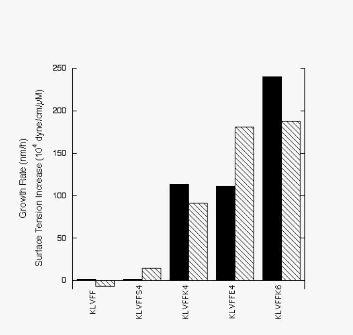
of mixtures of Aβ + hybrid peptide (Figure 1).
We next tested whether the disrupting domain alone was sufficient to accelerate Aβ aggregation.
7
by guest on February 13, 2018http://w
ww
.jbc.org/D
ownloaded from
KKKK at 140 and 280 µM increased solvent surface tension to 53.9 ± 0.8 and 57 ± 1 dyne/cm,
respectively, similar to that of KLVFFKKKKKK at the same concentrations (Table 1), but even
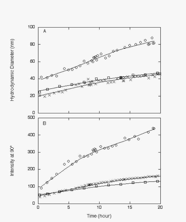
420 µM KKKK had no effect on Aβ aggregation whereas 140 µM KLVFFKKKKKK greatly
accelerated Aβ aggregation (Figure 2). These results indicate that modest increases in surface
tension alone are insufficient to cause acceleration of aggregation, and provide further support
for the notion that the Aβ-binding ability of the KLVFF recognition domain is required for
activity.
We reasoned that even greater acceleration of Aβ aggregation could be obtained with a KLVFF
recognition element coupled to a disrupting domain that produced a greater solvent surface
tension effect. To identify an appropriate candidate, we turned to the Hofmeister series, which
lists ions in order of their ability to stabilize protein folded structure (27). Protein structure
stabilization by co-solutes correlates strongly with the co-solute’s ability to increase the surface
tension of water (28). The Hofmeister cation series is N(CH3)4+ > NH2(CH3)2+ > NH4+> K+ >
Na+ > Cs+ > Li+ > Mg2+ > Ca2+ > Ba2+ , with the cations on the left classified as kosmotropes
(protein structure stabilizing, or “salting-out”) and those on the right as chaotropes (protein
structure destabilizing, or “salting-in”) (27),.
8
by guest on February 13, 2018http://w
ww
.jbc.org/D
ownloaded from
The lysine hexamer disrupting domain of our most effective hybrid peptide reported to date,
KLVFFKKKKKK, contains terminal amines that are protonated at neutral pH (-(CH2)4-
NH3+). We reasoned by analogy to the Hofmeister series that a side chain with a methyl-substituted
terminal amine group might have enhanced activity compared to lysine. Betaine
((CH3)3N+CH2COO-) is a naturally occurring compound that contains the requisite methyl-substituted amino
group, and also contains a free carboxyl group allowing facile coupling to a lysine side chain.
We modified the lysine side chains in the disrupting domain using the following strategy.
KLVFFKKKKKK was synthesized using standard Fmoc solid-phase synthesis techniques, but
with Mtt protection of the six C-terminal lysines and Boc protection of the N-terminal lysine.
While maintaining the peptide on the resin, the Mtt groups were cleaved, and betaine was
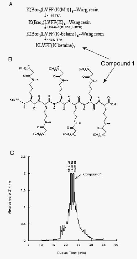
coupled to the lysine side chains via an amide linkage (Figure 3). Cleavage from the resin
produced a mixture of KLVFFKKKKKK derivatized with 4, 5 or 6 betaines, as confirmed by
mass spectroscopy analysis (Figure 3). A fraction highly enriched in the fully-derivatized
peptide, compound 1, was isolated by reverse-phase HPLC for further study (Figure 3).
Compound 1 was extremely water-soluble. Size-exclusion chromatographic analysis confirmed
that it was monomeric in PBSA (data not shown). Pendant drop measurements of aqueous
solutions of the purified compound demonstrated greatly increased surface tension compared to
KLVFFKKKKKK (Table 2).
9
by guest on February 13, 2018http://w
ww
.jbc.org/D
ownloaded from
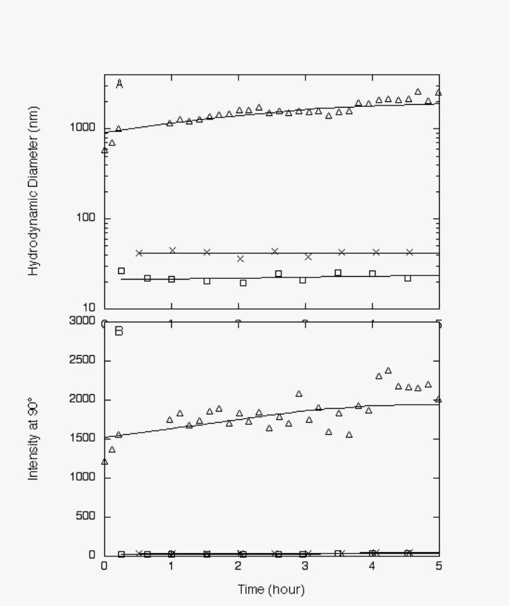
The effect of compound 1 on Aβ aggregation was remarkable (Figure 4). After four hours of
aggregation, the hydrodynamic diameter of Aβ in the presence of compound 1 was more than
100-fold greater than Aβ alone,(~2300 nm vs. ~20 nm) and nearly 50 times greater than Aβ with
KLVFFKKKKKK, which was our most active compound prior to discovery of compound 1.
Betaine at 1700 µM increased the surface tension of an aqueous solution to 61.4 ± 0.9 dyne/cm,
equivalent to 140 µM of compound 1, but betaine at 3400 µM had no measurable effect on Aβ
aggregation kinetics (Figure 4). These results show that surface tension can be used as a design
strategy for improving activity of the disrupting domain of hybrid peptides, and confirm that a
specific recognition domain is required for activity.
10
by guest on February 13, 2018http://w
ww
.jbc.org/D
ownloaded from
Discussion
Self-association of protein into large aggregates of β-sheet structure and fibrillar morphology is
a feature of a number of diseases, including the primary amyloidoses and neurodegenerative
diseases such as Alzheimers. Many, perhaps even most, proteins and peptides can be coaxed into
forming β-sheet fibrils under appropriate conditions of solvent, pH, and temperature (29),
Alarmingly, fibril-forming peptides and proteins appear to be toxic to a wide number of cell
types (29). The end product of aggregation – the fully-formed amyloid fibril – may be the
primary toxic species, but recent evidence suggests that rather it is a structured kinetic
intermediate that is killing cells (2-10). Indeed, our earliest studies provided the first published
data, to our knowledge, linking accelerated aggregation with inhibition of toxicity (21).
Our observations that hybrid peptides with either anionic or cationic disrupting domains
accelerate Aβ aggregation (26) led to the hypothesis that disrupting domains act by affecting
solvent properties. In particular, an increase in surface tension of aqueous solutions by addition
of co-solutes is strongly linked to changes in protein stability and protein aggregation (28).
Indeed, we observed that those hybrid peptides that accelerated Aβ aggregation also measurably
increased the surface tension of aqueous solutions (Figure 1). The increase in surface tension was
mediated solely through the disrupting domain (Table 1). We tested whether the disrupting
11
by guest on February 13, 2018http://w
ww
.jbc.org/D
ownloaded from
domain alone was capable of affecting solvent properties and/or Aβ aggregation. Although a
lysine tetramer at 420 µM increased the surface tension, it failed to alter Aβ aggregation kinetics.
This is not surprising, given that molar concentrations (>0.1 M – 1 M or higher) of co-solute are
generally required for a sufficient change in solvent properties to produce measurable changes in
protein folding and aggregation (27,30,31).
These encouraging results served as a basis for rational design of novel compounds with greater
efficacy. We used the Hofmeister series as a guide towards selecting functional groups with
strong surface-tension activity. The Hofmeister series has proven to be a reasonably reliable
predictor of co-solute effects on protein structure, aggregation, and activity (e.g., 32). The
strongest “salting-out” (kosmotropic) cations in the Hofmeister series are methylammonium ions
(27). Several related compounds, such as betaine ((CH3) 3N+CH2COO- ), sarcosine
(CH3)H2N+CH2COO-), and trimethylamine N-oxide ((CH3)3NO), are naturally-occurring intracellular
solutes that regulate osmotic pressure and modulate protein folding and enzyme function (e.g.,
33-35). These compounds also affect protein aggregation. For example, sarcosine stabilized the
native conformation of the serpin α1-antitrypsin and protected against thermal inactivation and
aggregation (30). Similarly, betaine partially inhibited light chain amyloid fibril formation from
immunoglobulin light chain (29). In contrast, trimethylamine N-oxide accelerated fibril
assembly from Aβ (36). This apparent contradiction can be resolved by noting that kosmotropes,
12
by guest on February 13, 2018http://w
ww
.jbc.org/D
ownloaded from
through a preferential exclusion mechanism, increase the surface tension (interfacial energy per
unit area) of aqueous solutions, thereby producing a thermodynamic driving force to reduce
surface area in order to minimize interfacial energy. Thus, kosmotropes drive the system
towards more compact protein structures. For α1-antitrypsin and immunoglobulin light chain the
most compact structure is the natively-folded monomer, but since Aβ monomer is random coil,
its most compact and folded structure is the β-sheet fibril.
Given these clues, we developed a method for synthesizing hybrid peptides with terminal
trimethylammonium groups in the disrupting domain. The betaine-derivatized
KLVFFKKKKKK was remarkably active at increasing surface tension, and was dramatically
more effective at accelerating Aβ aggregation than KLVFFKKKKKK. Although betaine alone
increased surface tension of aqueous solutions, the osmolyte did not measurably affect Aβ
aggregation. These results indicate the specificity of the action of compound 1.
It is well-established that co-solutes that increase solvent surface tension favor compact folded
and/or aggregated protein structures. What is unique about the compounds described here is that
the increase in surface tension is apparently localized via the recognition element to the solvent
near the aggregating peptides. We hypothesize that this leads to a much greater localized change
in solvent surface tension than that experienced in the bulk solvent, or in the absence of the
13
by guest on February 13, 2018http://w
ww
.jbc.org/D
ownloaded from
recognition element. This localized change in solvent properties then drives further growth of
highly-aggregated species. It is interesting to speculate that similar strategies could be used as
novel mechanism of targeting interfacial changes to individual proteins to influence protein
folding, stability, and aggregation in a highly specific manner.
Acknowledgments
This work was supported by a grant NS37728 from the National Institutes of Health.
14
by guest on February 13, 2018http://w
ww
.jbc.org/D
ownloaded from
References,
1. Hardy, J. and Selkoe, D. J. (2002) Science 297, 353-356.
2. Pallitto, M. M., and Murphy, R. M. (2001) Biophysical J. 81, 1805-1822.
3. Pike, C. J., Burdick, D., Walencewicz, A. .J, Glabe, C. G., and Cotman, C. W. (1993) J.
Neurosci. 13, 1676-1687.
4. Simmons, L. K., May, P. C., Tomaselli, K. J., Rydel, R. E., Fuson, K. S., Brigham, E. F.,
Wright, S., Lieberburg, I., Becker, G. W., Brems, D. N., and Li, W. (1994) Mol.
Pharmacol. 45, 373-379.
5. Seilheimer, B., Bohrman, B., Bondolfi, L., Muller, F., Stuber, D., and Dobeli H. (1997) J.
Struct. Biol. 119, 59-71.
6. Roher, A. E., Chang, M. O., Kuo, Y.-M., Webster, S. D., Stine, W. B., Haverkamp, L. J.,
Woods, A. S., Cotter, R. J., Tuohy, J. M., Krafft, G. A., Bonnell, B. S., and Emmerling, M.
R. (1996) J. Biol. Chem 271, 20631-2635.
7. Lambert, M. P., Barlow, A. K., Chromy, B. A., Edwards, C., Freed, R., Liosatos, M.,
Morgan, T. E., Rozovky, I., Trommer, B., Biola, K. L., Wals, P., Zhang, C., Finch, C. E.,
Krafft, G. A., and Klein, W. L. (1998) Proc. Natl. Acad. Sci. USA 95, 6448-6453.
8. Hartley, D. M., Walsh, D. M., Ye, C. P., Diehl, T., Vasquez, S., Vassilev, P. M., Teplow,
D. B., and Selkoe, D. S. (1999) J. Neurosci. 19, 8876-8884.
9. Ward, R. V., Jennings, K. H., Jepras, R., Neville, W., Owen, D. E., Hawkins, J., Christie,
15
by guest on February 13, 2018http://w
ww
.jbc.org/D
ownloaded from
G., Davis, J. B., George, A., Karran, E. H., and Howlett, D. R. (2000) Biochem. J. 348(Pt
1): 137-144.
10. Kayed, R., Head, E., Thompson, J. L., Mcintire, T. M., Milton, S. C., Cotman, C. W., and
Glabe, C. G. (2003) Science 300, 486-489.
11. Klunk, W. E., Debnath, M. L., Koros, A. M., and Pettegrew, J. W. (1998) Life Sci. 63,
1807-1814.
12. Soto, C., Sigurdsson, E. M., Morelli, L., Kumar, R. A., Castano, E. M., and Frangione, B.
(1998) Nat. Med. 4, 822-826.
13. Howlett, D. R., Perry, A. E., Godfrey F., Swatton, J. E., Jennings, K. H., Spitzfaden, C.,
Wadsworth, B., Wood, S. J., and Markwell, R. E. (1999) Biochem. J. 340, 283-289.
14. Findeis, M. A., Musso, G. M., Arico-Muendel, C. C., Benjamin, H. W., Hundal, A. M.,
Lee, J.-J., Chin, J., Kelley, M., Wakefield, J., Hayward, N. J., and Molineaux, S. M. (1999)
Biochemistry 38, 6791-6800.
15. Kuner, P., Bohrmann, B., Tjernberg, L. O., Naslund, J., Huber, G., Celenk, S., Gruninger-
Leitch, F., Richards, J. G., Jakob-Roetne, R., Kemp, J. A., and Nordstedt, C. (2000) J.
Biol. Chem. 275, 1673-1678.
16. Hughes, E., Burke, R. M., and Doig, A. J. (2000) J. Biol. Chem. 275, 25109-25115.
17. Gordon, D. J., Sciarretta, K. L., Meredith, S. C. (2001) Biochemistry 40, 8237-8245.
18. Nakagami, Y., Nishimura, S., Murasugi, T., Kaneko, I., Meguro, M. Marumoto, S., Kogen,
16
by guest on February 13, 2018http://w
ww
.jbc.org/D
ownloaded from
H., Koyama, K., and Oda, T. (2002) Br. J. Pharmacol. 137, 676-682.
19. Doig, A. J., Hughes, E., Burke, R. M., Su, T. J., Heenan, R. K., and Lu, J. (2002) Biochem.
Soc. Trans. 30, 537-542.
20. Gordon, D. J., and Meredith, S. C. (2003) Biochemistry 42, 475-485.
21. Ghanta, J., Shen, C.-L., Kiessling, L. L., and Murphy, R. M. (1996) J. Biol. Chem. 271,
29525-29528.
22. Tjernberg, L. O., Näslund, J., Lindqvist, F., Johansson, J., Karlström, A. R., Thyberg, J.,
Terenius, L., and Nordstedt (1996). J. Biol. Chem. 271, 8545-48.
23. Tjernberg, L. O., Lilliehöök, C., Callaway, D. J. E., Näslund, J., Hahne, S., Thyberg, J.,
Terenius, L., and Nordstedt (1997). J. Biol. Chem. 272, 12601-5.
24. Cairo, C. W., Strzelec, A., Murphy, R. M., Kiessling, L. L. (2002) Biochemistry 41, 8620-
8629.
25. Pallitto, M. M., Ghanta, J., Heinzelman, P., Kiessling, L. L., and Murphy, R. M. (1999)
Biochemistry 38, 3570-3578.
26. Lowe, T. L., Strzelec, A., Kiessling, L. L., and Murphy, R. M. (2001) Biochemistry 40,
7882-7889.
27. Cacace, M. G., Landau, E. M., and Ramsden, J. J. (1997) Quart. Rev. Biophys. 30, 241-
277.
28. Lin, T.-Y., and Timasheff, S. N. (1996) Protein Sci. 5, 372-381.
29. Bucciantini, M., Giamoni, E., Chiti, F., Baroni, F., Formigli, L., Zurdo, J., Taddie, N.,
17
by guest on February 13, 2018http://w
ww
.jbc.org/D
ownloaded from
Ramponi, G., Dobson, C. M. and Stefani, M. (2002) Nature, 416, 507-511.
30. Kim, Y.-S., Cape, S. P., Chi, E., Raffen, R., Wilkins-Stevens, P., Stevens, F. J., Manning,
M. C., Randolph, T. W., Solomon, A., and Carpenter, J. F. (2001) J. Biol. Chem. 276,
1626-1633.
31. Chow, M. K. M., Devlin, G. L., and Bottomley, S. P. (2001) Biol. Chem. 382, 1593-1599.
32. Ahmad. A., Akhtar, M. S., and Bhakuni, V. (2001) Biochemistry 40, 1945-1955.
33. Wang, A., and Bolen, D. W., (1997) Biochemistry 36, 9101-9108.
34. Ratnaparkhi, G. S., and Varadarajan, R. (2001) J. Biol. Chem. 276, 28789-28798.
35. Bourot, S., Sire, O., Trautwetter, A., Touze, T., Wu, L. F., Blanco, C., and Bernard, T.
(2000). J. Biol. Chem. 275, 1050-1056.
36. Yang, D.-S., Yip, C. M., Huang, T. H. J., Chakrabartty, A., and Fraser, P. E. (1999) J.
Biol. Chem. 274, 32970-32974.
18
by guest on February 13, 2018http://w
ww
.jbc.org/D
ownloaded from
Figure Legends
FIGURE 1: Correlation between the increase of Aβ aggregation rate by hybrid peptides (solid
bars) and the increase in surface tension of aqueous solutions of hybrid peptides (striped bars).
Aggregation rate is calculated as the average rate of increase in hydrodynamic diameter over the
duration of the experiment; data were taken from (25,26). Surface tension data are from Table 1.
FIGURE 2: Effect of KLVFFKKKKKK and KKKK on Aβ aggregation. Aβ was dissolved in 8
M urea and then diluted 20-fold into PBSA (o), PBSA with KLVFFKKKKKK at 1:1 molar ratio
(Ο), or PBSA with three-fold molar excess of KKKK(×). Final Aβ concentration was 140 µM.
Growth was followed by laser light scattering. A. Average hydrodynamic diameter vs. time, as
determined by least-squares fit of autocorrelation data. B. Average scattered intensity at 90°
scattering angle.
FIGURE 3. Synthesis of surface tension-modifying peptide. A. Schematic showing
protection/deprotection strategy for linking betaine to six C-terminal lysines. B. Structure of
synthesized peptide. C. Reverse-phase HPLC trace of cleavage product showing 3 major
products. Molar mass of each fraction as determined by MALDI mass spectroscopy, is indicated.
The molar masses correspond closely to calculated molar masses for KLVFFKKKKKK with 4,
19
by guest on February 13, 2018http://w
ww
.jbc.org/D
ownloaded from
5, or 6 betaine additions. Compound 1 was purified and used in subsequent studies.
FIGURE 4: Effect of compound 1 on Aβ aggregation. Aβ was dissolved in 8 M urea and then
diluted 20-fold into PBSA (o), PBSA with compound 1 at 1:1 molar ratio (∆), or PBSA with
24-fold molar excess of betaine(×). Final Aβ concentration was 140 µM. Growth was followed
by laser light scattering. A. Average hydrodynamic diameter vs. time, as determined by least-
squares fit of autocorrelation data. B. Average scattered intensity at 90° scattering angle.
20
by guest on February 13, 2018http://w
ww
.jbc.org/D
ownloaded from
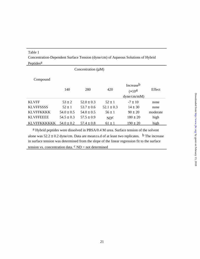
Table 1Concentration-Dependent Surface Tension (dyne/cm) of Aqueous Solutions of Hybrid
Peptidesa
Compound
Concentration (µM)
140 280 420Increaseb
(×104 dyne/cm/mM)
Effect
KLVFF 53 ± 2 52.0 ± 0.3 52 ± 1 -7 ± 10 noneKLVFFSSSS 52 ± 1 53.7 ± 0.6 52.1 ± 0.3 14 ± 30 noneKLVFFKKKK 54.0 ± 0.5 54.0 ± 0.5 56 ± 1 90 ± 20 moderateKLVFFEEEE 54.5 ± 0.3 57.5 ± 0.9 NDc 180 ± 20 high
KLVFFKKKKKK 54.0 ± 0.2 57.4 ± 0.8 61 ± 1 190 ± 20 high
a Hybrid peptides were dissolved in PBSA/0.4 M urea. Surface tension of the solvent
alone was 52.2 ± 0.2 dyne/cm. Data are mean±s.d of at least two replicates. b The increase in surface tension was determined from the slope of the linear regression fit to the surface
tension vs. concentration data. c ND = not determined
21
by guest on February 13, 2018http://w
ww
.jbc.org/D
ownloaded from
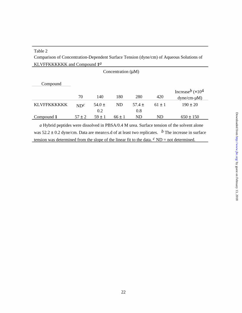
Table 2Comparison of Concentration-Dependent Surface Tension (dyne/cm) of Aqueous Solutions of
KLVFFKKKKKK and Compound 1a
Compound
Concentration (µM)
70 140 180 280 420Increaseb (×104
dyne/cm-µM)
KLVFFKKKKKK NDc 54.0 ± 0.2
ND 57.4 ± 0.8
61 ± 1 190 ± 20
Compound 1 57 ± 2 59 ± 1 66 ± 1 ND ND 650 ± 150
a Hybrid peptides were dissolved in PBSA/0.4 M urea. Surface tension of the solvent alone
was 52.2 ± 0.2 dyne/cm. Data are mean±s.d of at least two replicates. b The increase in surface
tension was determined from the slope of the linear fit to the data. c ND = not determined.
22
by guest on February 13, 2018http://w
ww
.jbc.org/D
ownloaded from
Jin Ryoun Kim, Todd J. Gibson and Regina M. Murphypeptides
-amyloid self-association by surface tension-modifyingβTargeted control of kinetics of
published online August 13, 2003J. Biol. Chem.
10.1074/jbc.M305466200Access the most updated version of this article at doi:
Alerts:
When a correction for this article is posted•
When this article is cited•
to choose from all of JBC's e-mail alertsClick here
by guest on February 13, 2018http://w
ww
.jbc.org/D
ownloaded from