T.-D. NGUYEN et al., Synthesis and Characterization of...
7
T.-D. NGUYEN et al., Synthesis and Characterization of β-Cyclodextrin/alginate…, Chem. Biochem. Eng. Q., 29 (3) 429–435 (2015) 429 Introduction In recent years, biomaterials have been em- ployed increasingly for sustained delivery applica- tions to enhance drug safety and efficacy as well as to improve patient compliance. Among biomateri- als, nanomaterials have received considerable inter- est in the pharmaceutical field. At the nanometer scale, these structures match typical sizes of natural functional units in living organisms, thus allowing them to interact with biomolecules 1 . Most of the nanomaterials used in controlled drug delivery in- volve non-toxicity, immunogenicity and biorecogni- tion, as well as the overcoming of obstacles arising from low drug solubility, degradation, fast clearance rates, relatively short-lasting biological activity and inability to cross biological barriers 2 . Nanoparticles have been widely exploited in the pharmaceutical industry for controlling drug re- lease due to fundamental changes related to the par- ticle surface and optical properties 3–5 . Nanoparticu- late delivery systems of small molecule drugs have the potential power to enhance drug stability and increase the duration of the therapeutic effect. Nanoparticles prepared from natural, synthetic polymers or lipids have been developed to over- come the enzymatic barrier 6–8 . Among them, algi- nate and β-cyclodextrin have provoked great interest because they satisfy almost all the necessary proper- ties of a material for drug delivery nanocarriers. Alginate (ALG) is a biodegradable polymer used as a suspending agent, a gelling agent, and a colloid stabilizer. It is extracted from brown sea weed and is composed of alternating blocks of 1–4 linked α-L-guluronic and β-D-mannuronic acid resi- due (Fig. 1a). A reversibly aqueous alginate gel may be formed in the presence of divalent ions at room temperature 9 . Alginate-based nanoparticles may be obtained easily by ionotropic gelation method. In this method, hydrogel beads, also called gelispheres, are formed from cross-linking between alginate solution and counter ions such as Ca 2+ . Drug mole- cules are commonly distributed into the gelispheres to form a three-dimensional lattice of ionically cross-linked moiety before the drug-loaded nanopar- ticles are generated 10 . Studies on alginate-nanoparti- cle materials have increased greatly over the past decade 11–15 . β-Cyclodextrin (β-CD) is a cyclic oligosaccha- ride consisting of seven (α-1,4)-linked α-D-glucopy- ranose units as shown in Fig. 1b, resulting in the formation of toroidal molecules with internal hy- drophobic cavities and external hydrophilic surface. The most common pharmaceutical application of β-CD has been to enhance the solubility, stability, safety and bioavailability of drug molecules 16 . In Synthesis and Characterization of β-Cyclodextrin/alginate Nanoparticle as a Novel Drug Delivery System T.-D. Nguyen, a,b,* T. Hong-Ngan Tran, a C.-H. Nguyen, a C. Im, b and C.-H. Dang a,* a Department of Pharmaceutical Chemistry Technology, Institute of Chemical Technology, Vietnam Academy of Science and Technology, 1 Mac Dinh Chi Street, District 1, Ho Chi Minh City, Vietnam b Department of Chemistry, Konkuk University, 1 Hwayang-dong, Gwangjin-gu, Seoul 143–701, South Korea The aim of this work was to study a novel nanoparticle system formed from alginate and β-cyclodextrin by ionotropic gelation method and to evaluate their potential for the association and delivery of drugs. The nanoparticles were prepared by electrostatic inter- actions between Ca 2+ /alginate gel and β-cyclodextrin. Morphology and structure charac- terization of nanoparticles was investigated by scanning electron micrographs (SEM), transmission electron microscope (TEM), Fourier transform infrared spectra (FTIR). The nanoparticle size was about 50 – 80 nm and their structure quite regular and consistent. Ketoprofen, chosen as a model drug, was incorporated into the nanocarriers with associ- ation efficiency of 50.7 % and loading efficiency of 5.7 %. Ketoprofen released from the nanoparticles was 57 % at pH 7.4 and 69 % at pH 5.0 for 4 h. Key words: nanoparticle, nanocarrier, drug delivery, β-cyclodextrin, alginate, ketoprofen, drug re- lease * Corresponding authors: Thanh-Danh Nguyen ([email protected]); Chi-Hien Dang ([email protected]). doi: 10.15255/CABEQ.2014.2092 Original scientific paper Received: August 11, 2014 Accepted: August 13, 2015
-
Upload
truongkhanh -
Category
Documents
-
view
235 -
download
0
Transcript of T.-D. NGUYEN et al., Synthesis and Characterization of...
T.-D. NGUYEN et al., Synthesis and Characterization of β-Cyclodextrin/alginate…, Chem. Biochem. Eng. Q., 29 (3) 429–435 (2015) 429
Introduction
In recent years, biomaterials have been em-ployed increasingly for sustained delivery applica-tions to enhance drug safety and efficacy as well as to improve patient compliance. Among biomateri-als, nanomaterials have received considerable inter-est in the pharmaceutical field. At the nanometer scale, these structures match typical sizes of natural functional units in living organisms, thus allowing them to interact with biomolecules1. Most of the nanomaterials used in controlled drug delivery in-volve non-toxicity, immunogenicity and biorecogni-tion, as well as the overcoming of obstacles arising from low drug solubility, degradation, fast clearance rates, relatively short-lasting biological activity and inability to cross biological barriers2.
Nanoparticles have been widely exploited in the pharmaceutical industry for controlling drug re-lease due to fundamental changes related to the par-ticle surface and optical properties3–5. Nanoparticu-late delivery systems of small molecule drugs have the potential power to enhance drug stability and increase the duration of the therapeutic effect. Nanoparticles prepared from natural, synthetic polymers or lipids have been developed to over-come the enzymatic barrier6–8. Among them, algi-
nate and β-cyclodextrin have provoked great interest because they satisfy almost all the necessary proper-ties of a material for drug delivery nanocarriers.
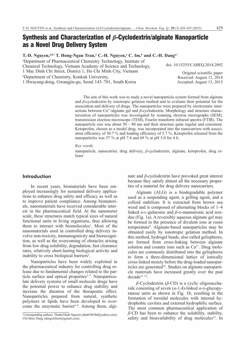
Alginate (ALG) is a biodegradable polymer used as a suspending agent, a gelling agent, and a colloid stabilizer. It is extracted from brown sea weed and is composed of alternating blocks of 1–4 linked α-L-guluronic and β-D-mannuronic acid resi-due (Fig. 1a). A reversibly aqueous alginate gel may be formed in the presence of divalent ions at room temperature9. Alginate-based nanoparticles may be obtained easily by ionotropic gelation method. In this method, hydrogel beads, also called gelispheres, are formed from cross-linking between alginate solution and counter ions such as Ca2+. Drug mole-cules are commonly distributed into the gelispheres to form a three-dimensional lattice of ionically cross-linked moiety before the drug-loaded nanopar-ticles are generated10. Studies on alginate-nanoparti-cle materials have increased greatly over the past decade11–15.
β-Cyclodextrin (β-CD) is a cyclic oligosaccha-ride consisting of seven (α-1,4)-linked α-D-glucopy-ranose units as shown in Fig. 1b, resulting in the formation of toroidal molecules with internal hy-drophobic cavities and external hydrophilic surface. The most common pharmaceutical application of β-CD has been to enhance the solubility, stability, safety and bioavailability of drug molecules16. In
Synthesis and Characterization of β-Cyclodextrin/alginate Nanoparticle as a Novel Drug Delivery System
T.-D. Nguyen,a,b,* T. Hong-Ngan Tran,a C.-H. Nguyen,a C. Im,b and C.-H. Danga,*
aDepartment of Pharmaceutical Chemistry Technology, Institute of Chemical Technology, Vietnam Academy of Science and Technology, 1 Mac Dinh Chi Street, District 1, Ho Chi Minh City, VietnambDepartment of Chemistry, Konkuk University, 1 Hwayang-dong, Gwangjin-gu, Seoul 143–701, South Korea
The aim of this work was to study a novel nanoparticle system formed from alginate and β-cyclodextrin by ionotropic gelation method and to evaluate their potential for the association and delivery of drugs. The nanoparticles were prepared by electrostatic inter-actions between Ca2+/alginate gel and β-cyclodextrin. Morphology and structure charac-terization of nanoparticles was investigated by scanning electron micrographs (SEM), transmission electron microscope (TEM), Fourier transform infrared spectra (FTIR). The nanoparticle size was about 50 – 80 nm and their structure quite regular and consistent. Ketoprofen, chosen as a model drug, was incorporated into the nanocarriers with associ-ation efficiency of 50.7 % and loading efficiency of 5.7 %. Ketoprofen released from the nanoparticles was 57 % at pH 7.4 and 69 % at pH 5.0 for 4 h.
Key words:nanoparticle, nanocarrier, drug delivery, β-cyclodextrin, alginate, ketoprofen, drug re-lease
*Corresponding authors: Thanh-Danh Nguyen ([email protected]); Chi-Hien Dang ([email protected]).
doi: 10.15255/CABEQ.2014.2092
Original scientific paper Received: August 11, 2014 Accepted: August 13, 2015
430 T.-D. NGUYEN et al., Synthesis and Characterization of β-Cyclodextrin/alginate…, Chem. Biochem. Eng. Q., 29 (3) 429–435 (2015)
aqueous solutions, β-CD can form inclusion com-plexes with many drugs by taking up the drug mol-ecule or some lipophilic moiety of the molecule, into the central cavity and no covalent bonds are formed or broken during complex formation. β-CD has been an attractive building block for various types of drug delivery systems due to its favorable toxicological profile, inherent ability to partly or completely retain drug molecules and protect them from the external environment17–19. In particular, Skiba et al. 20 demonstrated that β-CD derivatives could interact with Ca2+ ions in an electrostatic man-ner as non-specific adsorption. Therefore, Ca2+ ion can be used as a bridge ion between negatively charged elements and β-CD compounds.
Ketoprofen is one of the propionic acid classes of non-steroidal anti-inflammatory drugs with anal-gesic, antipyretic effects, and used in the treatment of rheumatoid arthritis21,22. Ketoprofen structure is shown in Fig. 1c. Solubility and bioavailability of ketoprofen, a poorly water-soluble drug, can be im-proved by complexation with β-CD as a drug carri-er23,24. However, studies on ketoprofen delivery nanomaterials are presently limited.
In recent years, β-CD and its derivatives encap-sulated into micro-systems of alginate/polymer have been widely investigated in drug delivery applica-tion12,25,26. However, nanoparticles containing both ALG and β-CD have not been reported in literature. In this paper, we describe the preparation and char-acterization of a new nanoparticle containing the two above components as a drug delivery system. In order to evaluate incorporation and delivery from such a system, ketoprofen was chosen as an exam-ple of BCS class II drug (according to WHO Guid-ance, low solubility and high permeability)27,28.
Experimental
Materials
β-Cyclodextrin, sodium alginate and ketopro-fen were purchased from Bodi Chemical Co. (Tian-jin, China), Shanghai Chemical Co. Ltd (China) and Merck (Germany), respectively. All other chemicals were of AR grade and used as received without fur-ther purification. Distilled water and magnetic stir-rer were used throughout.
Preparation of blank β-cyclodextrin/alginate nanoparticles
β-CD/ALG nanoparticles were spontaneously obtained via ionotropic gelation method between the positively charged ALG/Ca2+ gelispheres and the negatively charged β-CD. 0.42–1.47 mL of aqueous calcium chloride (10.05 mg mL–1) was added dropwise to 3.30 mL of aqueous sodium algi-nate (9.00 mg mL–1) under stirring for 30 minutes. Then, 2 mL of β-CD solution (0.99 – 14.85 mg mL–1) was added dropwise into the calcium alginate pre-gel and stirred for an additional hour. The gel solu-tion was equilibrated overnight to allow formation of nanoparticles in uniform size. The nanoparticles were separated by centrifugation (EBA20S-Hettich Germany, 8,000 g for 30 min, 30 °C) and then freeze-dried to yield the product.
The nanoparticle product yield was calculated as follows:
Nanoparticle product yieldNanoparticles weight
Total solids C
=
=( aa ALG CD weight2 100++ + −
⋅β )
%
Preparation of ketoprofen loading β-cyclodextrin/alginate nanoparticles
A constant volume (0.36 mL) of ketoprofen in an ethanol/water mixture (1:1, 1.10 mg mL–1) was added to 0.7 mL of calcium chloride solution (10.05 mg mL–1). Nanoparticles were prepared as method of blank β-CD/alginate nanoparticles with various con-centrations of β-CD solution (0.99 – 2.97 mg mL–1).
F i g . 1 – Chemical structures of sodium alginate (a), β-cyclo-dextrin (b) and ketoprofen (c)
T.-D. NGUYEN et al., Synthesis and Characterization of β-Cyclodextrin/alginate…, Chem. Biochem. Eng. Q., 29 (3) 429–435 (2015) 431
Morphology and structure characterization of nanoparticles
The morphology of nanospheres was studied by scanning electronic microscopy (SEM) using JSM7401F (Japan). The samples were lyophilized and then mounted on metal stubs, gold coated under vacuum before examination. The nanoparticles size was measured by transmission electron microscope (TEM, Hitachi H8100 with the acceleration 200 kV, LaB6 source of electrons, resolution 0.14 nm).
Fourier transform infrared (FTIR) spectra of blank and loaded nanoparticles were taken with KBr pellets on a FTIR spectrometer (Equinox 55 IR – Bruker (Germany)). The same was obtained for alginate, β-CD, ketoprofen as standards.
Drugs association efficiency and loading efficiency
The association efficiency was determined in-directly after separation of nanoparticles from the aqueous medium containing nonassociated drug. The drug was obtained from the separated solution by extraction with diethyl ether. The amount of drug associated with the particles was calculated by the difference between the total amount used to pre-pare the particles and the amount of drug present in the aqueous phase after centrifugation.
A difference between the total amount of drug and the amount of residual nonassociated drug as a percent of total nanoparticle dry mass is determined as loading efficiency. Dry mass was obtained by freeze-drying an aliquot of hydrated nanoparticles.
Quantitative analysis of ketoprofen
Ketoprofen was assayed by reversed-phase liq-uid chromatography (RP-HPLC) using an Agilent HPLC system (Agilent 1100, USA) equipped with UV detector and a ZORBAX SB – C18. For prepara-tion of standard solutions, a standard solution was prepared by diluting a stock solution at a concentra-tion of 10 mg mL–1 in methanol. The standard solu-tions were prepared at concentrations of 4.0, 2.0, 1.0 and 0.5 mg mL–1. For preparation of sample solu-tions, the supernatant which was obtained by centri-fuging nanoparticle solutions was extracted with di-ethyl ether three times and the residue was dissolved in methanol after evaporation of diethyl ether.
Association efficiencyTotal amount of drug free drug in
=
=− supernattant
total amount of drug⋅100 %
Loading efficiencyTotal amount of drug free drug in rn
=
=− supe atant
ttotal dry weight of nanoparticles⋅100 %
Drug release profiles
Ketoprofen release studies were performed by stirring 10 mg dried nanoparticles in 10 mL of phos-phate buffer solutions (pH 5.0 and 7.4) at 37 °C. At appropriate intervals, the samples were separated by centrifugation (8,000g for 30 min, 30 °C) and the amount of drug released from the nanoparticles was evaluated by HPLC as described above. The release was quantified as follows:
ReleRel
aseeased ketoprofenTotal ketoprofen
(%)
To determine the mechanism of ketoprofen re-lease, we analysed the release data applying the Ridger-Peppas model29:
MM
ktt n
∞
=
Where Mt/M∞ is the fractional drug release, t is the release time, k is a kinetic constant characteris-tic of the system, and n is the diffusional exponent which is indicative of the transport mechanism. This equation is useful for a generalized analysis of release data, although it may only be used for up to 60 % of the drug released30. The fitting model was conducted with OriginPro 8.5 software.
Results and discussion
Preparation and structure characterization β-cyclodextrin/alginate nanoparticles
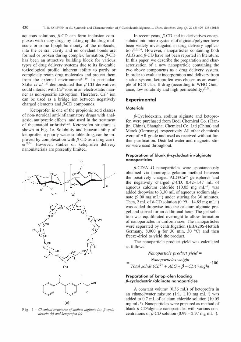
Preparation of β-CD/ALG nanoparticles was carried out by simple ionotropic gelation method. The hydrogel beads were formed between Ca2+ solu-tion and aqueous alginate solution. β-CD drops were diffused into a three-dimensional lattice of the ioni-cally crosslinking gel. In order to study structure of formation of nanoparticles, prior to incorporation of the gel and β-CD suspension, morphological infor-mation of the distinct suspension was observed by SEM (Fig. 2). The images confirmed that both indi-vidual components, the alginate/Ca2+ gel and β-CD, had micrometer size with a disjointed composition. After the incorporation was performed, the distinct spherical particles with solid dense structure were observed. Nanoparticle sizes were less than 100 nm according to SEM image (Fig. 2c).
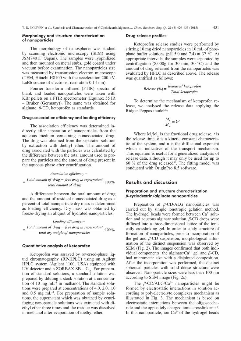
The β-CD/ALG/Ca2+ nanoparticles might be formed by electrostatic interactions in solution ac-cording to polyelectrolyte complexes mechanism as illustrated in Fig. 3. The mechanism is based on electrostatic interactions between the oligosaccha-ride and the oppositely charged ionic crosslinker31,32. In this nanoparticle, ion Ca2+ of the hydrogel beads
432 T.-D. NGUYEN et al., Synthesis and Characterization of β-Cyclodextrin/alginate…, Chem. Biochem. Eng. Q., 29 (3) 429–435 (2015)
played a role as a bridge ion interacting with the β-CD compounds by virtue of its charge in an elec-trostatic manner20. These complexes resemble ionic cross-linking, since non-permanent networks are formed that are more sensitive to changes in envi-ronmental conditions.
In order to determine conditions suitable for the formation of nanoparticles, in the first step, we studied formation of nanoparticles with different Ca2+/ALG w/w ratios in the presence of β-CD. Dif-ferent volumes of calcium chloride (10.07 mg mL–1) were added into the constant volume of ALG solu-tion (9.0 mg mL–1) to obtain Ca2+/ALG ratios from 1:2 to 1:7 (w/w). In the presence of β-CD, a high amount of Ca2+ ion with 1:2 to 1:3 w/w ratios gave the unstable nanoparticles the tendency to agglom-erate due to increased electrostatic interaction be-tween Ca2+ ion and β-CD compounds28. Meanwhile, the high amount of ALG with 1:6 to 1:7 w/w ratios could not generate the formation of nanoparticles. As a result, Ca2+/ALG ratios for formation of nanoparticles in the presence of β-CD were opti-
mized from 1:4 to 1:5. Alternatively, 1:4.2 w/w Ca2+/ALG ratio was finally chosen for further study.
The aim of the next step was to investigate the effect of β-CD concentration on size and yield of product nanoparticles. The results showed that β-CD concentration influenced the size of the re-sulting nanoparticles (Table 1). In the case of high β-CD concentration (14.85 mg mL–1), due to ag-glomeration of individual nanoparticles, the ob-served nanoparticles possessing size less than 100 nm could not be obtained. At 1/4.2/0.42 ratio, a highest product yield obtained with a nanoparticle size less than 100 nm was 90.1 %. Therefore, these ratios would be used for further studies on the drug loading nanoparticles.
Ta b l e 1 – Effect of β-CD concentration on nanoparticle product size and yield (mean ± SD, n = 3)
β-CD concentration
(mg mL–1)
Ca2+/ALG/β-CD w/w/w ratio
Size (nm)
Nanoparticle product yield
(%)0 1/4.2/0 >1000 –
1.0 1/4.2/0.28 < 100 84.7 ± 2.5
1.49 1/4.2/0.42 < 100 90.1 ± 0.8
2.97 1/4.2/0.84 < 100 75.0 ± 5.1
14.85 1/4.2/4.2 > 100 41.7 ± 1.5
Preparation and structure characterization of drug loading β-cyclodextrin/alginate nanoparticles
To investigate whether these nanocarriers could be used advantageously for hydrophobic drug deliv-ery, we used ketoprofen as a model drug for studies of nanoparticle structure and drug release. The preparation of ketoprofen-loaded β-CD/ALG nano-
F i g . 2 – Scanning electron micrographs of β-cyclodextrin (a), alginate/Ca2+ (b), and ALG/ β-CD nanoparticles with Ca2+/ALG/β-CD w/w/w ratio of 1/4.2/0.28 (c)
F i g . 3 – Mechanism for formation of β-cyclodextrin/alginate nanoparticles
T.-D. NGUYEN et al., Synthesis and Characterization of β-Cyclodextrin/alginate…, Chem. Biochem. Eng. Q., 29 (3) 429–435 (2015) 433
particles is actually difficult because ketoprofen is a hydrophobic drug, while the β-CD/ALG nanoparti-cles are hydrophilic systems. Precipitation process of water-miscible organic solvents could occur in an aqueous phase as discrete spherical particles. However, a successful entrapment was achieved by dissolving the drug in an alcohol/water mixture (1:1) prior to its incorporation into the calcium chloride solution.
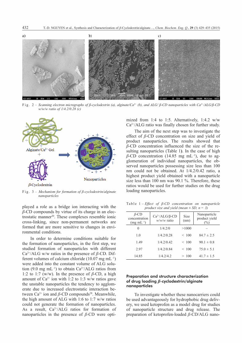
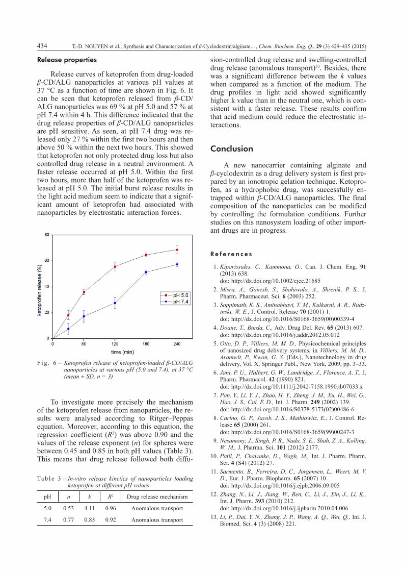
Electron microscopy analysis confirmed the presence of nanoparticles and provided morpholog-ical information of ketoprofen-loaded β-CD/ALG nanoparticles (Fig. 4). Thus, the SEM image (Fig. 4a) showed the morphology of the drug-loaded nanoparticle to be solid dense. Additionally, nanoparticles possessing a bunch-like structure with sizes 50 – 80 nm were seemingly formed from ag-glomeration of smaller nanosized units.
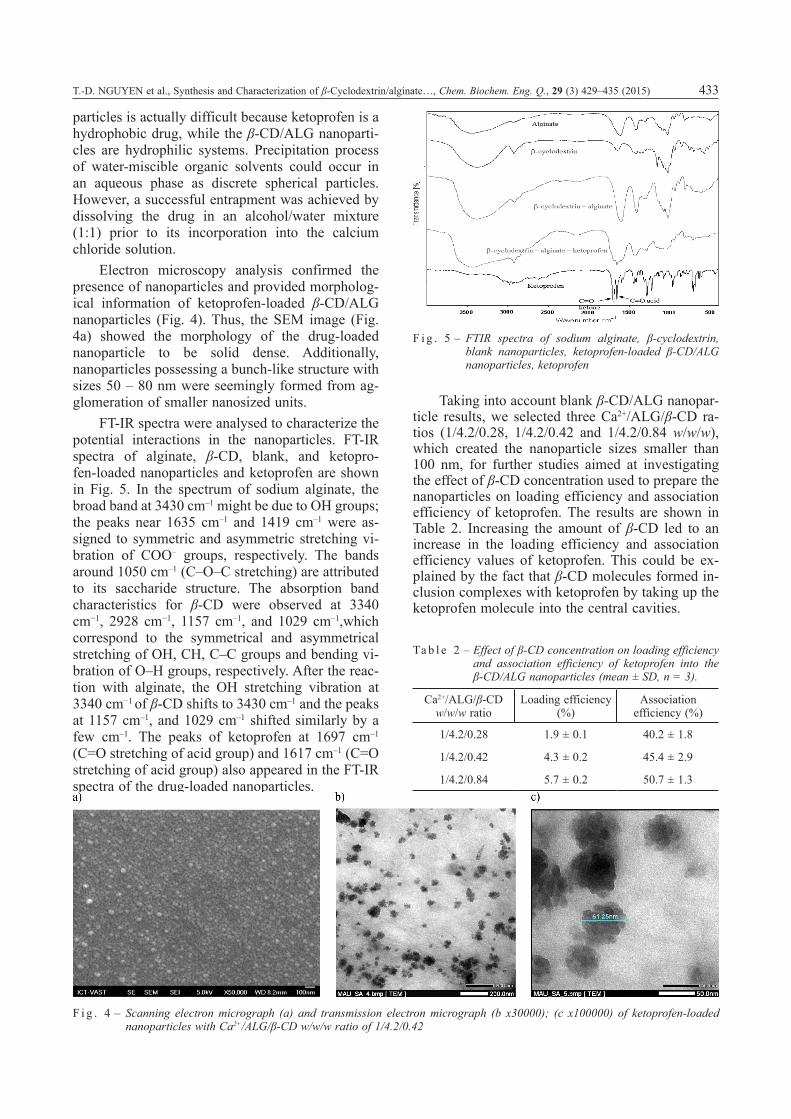
FT-IR spectra were analysed to characterize the potential interactions in the nanoparticles. FT-IR spectra of alginate, β-CD, blank, and ketopro-fen-loaded nanoparticles and ketoprofen are shown in Fig. 5. In the spectrum of sodium alginate, the broad band at 3430 cm–1 might be due to OH groups; the peaks near 1635 cm–1 and 1419 cm–1 were as-signed to symmetric and asymmetric stretching vi-bration of COO– groups, respectively. The bands around 1050 cm–1 (C–O–C stretching) are attributed to its saccharide structure. The absorption band characteristics for β-CD were observed at 3340 cm−1, 2928 cm−1, 1157 cm–1, and 1029 cm–1,which correspond to the symmetrical and asymmetrical stretching of OH, CH, C–C groups and bending vi-bration of O–H groups, respectively. After the reac-tion with alginate, the OH stretching vibration at 3340 cm−1 of β-CD shifts to 3430 cm–1 and the peaks at 1157 cm–1, and 1029 cm–1 shifted similarly by a few cm–1. The peaks of ketoprofen at 1697 cm–1 (C=O stretching of acid group) and 1617 cm–1 (C=O stretching of acid group) also appeared in the FT-IR spectra of the drug-loaded nanoparticles.
Taking into account blank β-CD/ALG nanopar-ticle results, we selected three Ca2+/ALG/β-CD ra-tios (1/4.2/0.28, 1/4.2/0.42 and 1/4.2/0.84 w/w/w), which created the nanoparticle sizes smaller than 100 nm, for further studies aimed at investigating the effect of β-CD concentration used to prepare the nanoparticles on loading efficiency and association efficiency of ketoprofen. The results are shown in Table 2. Increasing the amount of β-CD led to an increase in the loading efficiency and association efficiency values of ketoprofen. This could be ex-plained by the fact that β-CD molecules formed in-clusion complexes with ketoprofen by taking up the ketoprofen molecule into the central cavities.
Ta b l e 2 – Effect of β-CD concentration on loading efficiency and association efficiency of ketoprofen into the β-CD/ALG nanoparticles (mean ± SD, n = 3).
Ca2+/ALG/β-CD w/w/w ratio
Loading efficiency (%)
Association efficiency (%)
1/4.2/0.28 1.9 ± 0.1 40.2 ± 1.8
1/4.2/0.42 4.3 ± 0.2 45.4 ± 2.9
1/4.2/0.84 5.7 ± 0.2 50.7 ± 1.3
F i g . 4 – Scanning electron micrograph (a) and transmission electron micrograph (b x30000); (c x100000) of ketoprofen-loaded nanoparticles with Ca2+/ALG/β-CD w/w/w ratio of 1/4.2/0.42
F i g . 5 – FTIR spectra of sodium alginate, β-cyclodextrin, blank nanoparticles, ketoprofen-loaded β-CD/ALG nanoparticles, ketoprofen
434 T.-D. NGUYEN et al., Synthesis and Characterization of β-Cyclodextrin/alginate…, Chem. Biochem. Eng. Q., 29 (3) 429–435 (2015)
Release properties
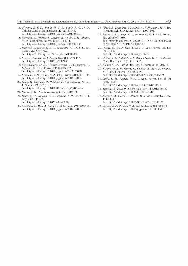
Release curves of ketoprofen from drug-loaded β-CD/ALG nanoparticles at various pH values at 37 °C as a function of time are shown in Fig. 6. It can be seen that ketoprofen released from β-CD/ALG nanoparticles was 69 % at pH 5.0 and 57 % at pH 7.4 within 4 h. This difference indicated that the drug release properties of β-CD/ALG nanoparticles are pH sensitive. As seen, at pH 7.4 drug was re-leased only 27 % within the first two hours and then above 50 % within the next two hours. This showed that ketoprofen not only protected drug loss but also controlled drug release in a neutral environment. A faster release occurred at pH 5.0. Within the first two hours, more than half of the ketoprofen was re-leased at pH 5.0. The initial burst release results in the light acid medium seem to indicate that a signif-icant amount of ketoprofen had associated with nanoparticles by electrostatic interaction forces.
To investigate more precisely the mechanism of the ketoprofen release from nanoparticles, the re-sults were analysed according to Ritger–Peppas equation. Moreover, according to this equation, the regression coefficient (R2) was above 0.90 and the values of the release exponent (n) for spheres were between 0.45 and 0.85 in both pH values (Table 3). This means that drug release followed both diffu-
sion-controlled drug release and swelling-controlled drug release (anomalous transport)33. Besides, there was a significant difference between the k values when compared as a function of the medium. The drug profiles in light acid showed significantly higher k value than in the neutral one, which is con-sistent with a faster release. These results confirm that acid medium could reduce the electrostatic in-teractions.
Conclusion
A new nanocarrier containing alginate and β-cyclodextrin as a drug delivery system is first pre-pared by an ionotropic gelation technique. Ketopro-fen, as a hydrophobic drug, was successfully en-trapped within β-CD/ALG nanoparticles. The final composition of the nanoparticles can be modified by controlling the formulation conditions. Further studies on this nanosystem loading of other import-ant drugs are in progress.
R e f e r e n c e s
1. Kiparissides, C., Kammona, O., Can. J. Chem. Eng. 91 (2013) 638.doi: http://dx.doi.org/10.1002/cjce.21685
2. Misra, A., Ganesh, S., Shahiwala, A., Shrenik, P. S., J. Pharm. Pharmaceut. Sci. 6 (2003) 252.
3. Soppimath, K. S., Aminabhavi, T. M., Kulkarni, A. R., Rudz-inski, W. E., J. Control. Release 70 (2001) 1.doi: http://dx.doi.org/10.1016/S0168-3659(00)00339-4
4. Doane, T., Burda, C., Adv. Drug Del. Rev. 65 (2013) 607.doi: http://dx.doi.org/10.1016/j.addr.2012.05.012
5. Otto, D. P., Villiers, M. M. D., Physicochemical principles of nanosized drug delivery systems, in Villiers, M. M. D., Aramwit, P., Kwon, G. S. (Eds.), Nanotechnology in drug delivery, Vol. X, Springer Publ., New York, 2009, pp. 3–33.
6. Jani, P. U., Halbert, G. W., Landridge, J., Florence, A. T., J. Pharm. Pharmacol. 42 (1990) 821.doi: http://dx.doi.org/10.1111/j.2042-7158.1990.tb07033.x
7. Pan, Y., Li, Y. J., Zhao, H. Y., Zheng, J. M., Xu, H., Wei, G., Hao, J. S., Cui, F. D., Int. J. Pharm. 249 (2002) 139.doi: http://dx.doi.org/10.1016/S0378-5173(02)00486-6
8. Carino, G. P., Jacob, J. S., Mathiowitz, E., J. Control. Re-lease 65 (2000) 261.doi: http://dx.doi.org/10.1016/S0168-3659(99)00247-3
9. Nesamony, J., Singh, P. R., Nada, S. E., Shah, Z. A., Kolling, W. M., J. Pharma. Sci. 101 (2012) 2177.
10. Patil, P., Chavanke, D., Wagh, M., Int. J. Pharm. Pharm. Sci. 4 (S4) (2012) 27.
11. Sarmento, B., Ferreira, D. C., Jorgensen, L., Weert, M. V. D., Eur. J. Pharm. Biopharm. 65 (2007) 10.doi: http://dx.doi.org/10.1016/j.ejpb.2006.09.005
12. Zhang, N., Li, J., Jiang, W., Ren, C., Li, J., Xin, J., Li, K., Int. J. Pharm. 393 (2010) 212.doi: http://dx.doi.org/10.1016/j.ijpharm.2010.04.006
13. Li, P., Dai, Y. N., Zhang, J. P., Wang, A. Q., Wei, Q., Int. J. Biomed. Sci. 4 (3) (2008) 221.
F i g . 6 – Ketoprofen release of ketoprofen-loaded β-CD/ALG nanoparticles at various pH (5.0 and 7.4), at 37 °C (mean ± SD, n = 3)
Ta b l e 3 – In-vitro release kinetics of nanoparticles loading ketoprofen at different pH values
pH n k R2 Drug release mechanism
5.0 0.53 4.11 0.96 Anomalous transport
7.4 0.77 0.85 0.92 Anomalous transport
T.-D. NGUYEN et al., Synthesis and Characterization of β-Cyclodextrin/alginate…, Chem. Biochem. Eng. Q., 29 (3) 429–435 (2015) 435
14. Oliveira, E. F. D., Paula, H. C. B., Paula, R. C. M. D., Colloids Surf. B Biointerfaces 113 (2014) 146.doi: http://dx.doi.org/10.1016/j.colsurfb.2013.08.038
15. Martínez, A., Iglesias, I., Lozano, R., Teijón, J. M., Blanco, M. D., Carbohydr. Polym. 83 (2011) 1311.doi: http://dx.doi.org/10.1016/j.carbpol.2010.09.038
16. Rasheed, A., Kumar, C. K. A., Sravanthi, V. V. N. S. S., Sci. Pharm. 76 (2008) 567.doi: http://dx.doi.org/10.3797/scipharm.0808-05
17. Irie, E., Uekama, K., J. Pharm. Sci. 86 (1997) 147.doi: http://dx.doi.org/10.1021/js960213f
18. Moya-Ortega, M. D., Alvarez-Lorenzo, C., Concheiro, A., Loftsson, T., Int. J. Pharm. 428 (2012) 152.doi: http://dx.doi.org/10.1016/j.ijpharm.2012.02.038
19. Krauland, A. H., Alonso, M. J., Int. J. Pharm. 340 (2007) 134.doi: http://dx.doi.org/10.1016/j.ijpharm.2007.03.005
20. Skiba, M., Duchene, D., Puisieux, F., Wouessidjewe, D., Int. J. Pharm. 129 (1996) 113.doi: http://dx.doi.org/10.1016/0378-5173(95)04272-5
21. Kantor, T. G., Pharmacotherapy 6 (3) (1986) 93.22. Dang, C. H., Nguyen, C. H., Nguyen, T. D., Im, C., RSC
Adv. 4 (2014) 6239.doi: http://dx.doi.org/10.1039/c3ra46007j
23. Maestrelli, F., Mari, A., Mura, P., Int. J. Pharm. 298 (2005) 55.doi: http://dx.doi.org/10.1016/j.ijpharm.2005.03.033
24. Vikesh, S., Rajashree, M., Ashok, A., Fakkirappa, M. V., Int. J. Pharm. Sci. & Drug Res. 1 (3) (2009) 195.
25. Moses, L. R. Dileep, K. J., Sharma, C. P., J. Appl. Polym. Sci. 75 (2000) 1089.doi: http://dx.doi.org/10.1002/(SICI)1097-4628(20000228)75:9<1089::AID-APP1>3.0.CO;2-5
26. Huang, L., Xin, J., Guo, Y., Li J., J. Appl. Polym. Sci. 115 (2010) 1371.doi: http://dx.doi.org/10.1002/app.30775
27. Shohin, I. E., Kulinich, J. I., Ramenskaya, G. V., Vasilenko, G. F., Dis. Tech. 18 (1) (2011) 26.
28. Kumar, K. M., Anil, B., Int. Res. J. Pharm. 3 (3) (2012) 5.29. Korsmeyer, R. W., Gurny, R., Doelker, E., Buri, P., Peppas,
N. A., Int. J. Pharm. 15 (1983) 25.doi: http://dx.doi.org/10.1016/0378-5173(83)90064-9
30. Lucht, L. M., Peppas, N. A., J. Appl. Polym. Sci. 33 (5) (1987) 1557.doi: http://dx.doi.org/10.1002/app.1987.070330511
31. Mizrahy, S., Peer, D., Chem. Soc. Rev. 41 (2012) 2623.doi: http://dx.doi.org/10.1039/C1CS15239D
32. Janes, K. A., Calvo, P., Alonso, M. J., Adv. Drug Del. Rev. 47 (2001) 83.doi: http://dx.doi.org/10.1016/S0169-409X(00)00123-X
33. Siepmann, J., Peppas, N. A., Int. J. Pharm. 418 (2011) 6.doi: http://dx.doi.org/10.1016/j.ijpharm.2011.03.051