Systemic administration of a novel immune-stimulatory...
50
1 Systemic administration of a novel immune-stimulatory pseudovirion suppresses lung metastatic melanoma by regionally enhancing IFN-γ production Kotaro Saga, Katsuto Tamai, Takehiko Yamazaki and Yasufumi Kaneda* Division of Gene Therapy Science, Graduate School of Medicine, Osaka University Suita, Osaka 565-0871, Japan *To whom correspondence and reprint requests should be addressed: Yasufumi Kaneda Division of Gene Therapy Science, Osaka University Graduate School of Medicine 2-2 Yamada-oka, Suita, Osaka 565-0871, Japan Tel: +81 6 6879 3900; Fax: +81 6 6879 3909 E-mail: [email protected] There are no conflicts of interest to be disclosed. on April 2, 2020. © 2012 American Association for Cancer Research. clincancerres.aacrjournals.org Downloaded from Author manuscripts have been peer reviewed and accepted for publication but have not yet been edited. Author Manuscript Published OnlineFirst on December 18, 2012; DOI: 10.1158/1078-0432.CCR-12-1947
Transcript of Systemic administration of a novel immune-stimulatory...
1
Systemic administration of a novel immune-stimulatory pseudovirion suppresses lung
metastatic melanoma by regionally enhancing IFN-γ production
Kotaro Saga, Katsuto Tamai, Takehiko Yamazaki and Yasufumi Kaneda*
Division of Gene Therapy Science, Graduate School of Medicine, Osaka University
Suita, Osaka 565-0871, Japan
*To whom correspondence and reprint requests should be addressed:
Yasufumi Kaneda
Division of Gene Therapy Science, Osaka University Graduate School of Medicine
2-2 Yamada-oka, Suita, Osaka 565-0871, Japan
Tel: +81 6 6879 3900; Fax: +81 6 6879 3909
E-mail: [email protected]
There are no conflicts of interest to be disclosed.
on April 2, 2020. © 2012 American Association for Cancer Research.clincancerres.aacrjournals.org Downloaded from
Author manuscripts have been peer reviewed and accepted for publication but have not yet been edited. Author Manuscript Published OnlineFirst on December 18, 2012; DOI: 10.1158/1078-0432.CCR-12-1947

2
Running Title
Immunostimulatory pseudovirion for cancer therapy
Statement of translational relevance
Sendai virus-envelope (HVJ-E) has been shown to induce immunity to multiple
types of tumors, and clinical trials to test its safety and anti-tumor immunity are ongoing
in Japan. However, HVJ-E is not able to stimulate the production of interferon (IFN)-γ,
which is important for anti-tumor immunity. Therefore, to develop a second
generation HVJ-E with enhanced anti-tumor activity, we constructed interleukin
(IL)-12-conjugated, HN-depleted HVJ-E (IL-12-HVJ-E).
Although IL-12 has a robust anti-tumor effect, the systemic administration of
IL-12 is prohibited by the critical side effects resulting from systemic elevation in IFN-γ
levels. By contrast, IL-12-HVJ-E accumulated in the lung following intravenous
administration, induced local IFN-γ expression without increasing the serum IFN-γ level
and significantly reduced metastatic melanoma in the lung. Thus, IL-12-HVJ-E is a
promising tool to treat metastatic lung cancers as well as regional cancers.
on April 2, 2020. © 2012 American Association for Cancer Research.clincancerres.aacrjournals.org Downloaded from
Author manuscripts have been peer reviewed and accepted for publication but have not yet been edited. Author Manuscript Published OnlineFirst on December 18, 2012; DOI: 10.1158/1078-0432.CCR-12-1947
3
Abstract
Purpose
Cancer immunotherapy has encountered many difficulties in the face of the
expectation to eradicate cancer, and new breakthroughs are required.
We have previously shown that UV-inactivated Sendai virus particles
(hemagglutinating virus of Japan envelope; HVJ-E) induce immunity against multiple
tumor types. In this study, a novel pseudovirion that stimulates more robust anti-tumor
immunity was designed for cancer treatment.
Experimental Design
First, we found that culturing murine splenocytes with HVJ-E in combination
with IL-12 resulted in a remarkable increase in IFN-γ production compared with that
observed in splenocytes cultured with IL-12 alone. The synergistic effects of HVJ-E
and IL-12 on IFN-γ production were caused by viral F proteins independently of HVJ-E
fusion activity and not by hemagglutination from HN proteins. We next constructed
HN-depleted HVJ-E expressing the Fc region of IgG on the envelope and single chain
IL-12 containing the ZZ domain of protein A to produce an IL-12-conjugated HVJ-E
particle without hemagglutinating activity.
Results
on April 2, 2020. © 2012 American Association for Cancer Research.clincancerres.aacrjournals.org Downloaded from
Author manuscripts have been peer reviewed and accepted for publication but have not yet been edited. Author Manuscript Published OnlineFirst on December 18, 2012; DOI: 10.1158/1078-0432.CCR-12-1947
4
IL-12-conjugated HVJ-E dramatically enhanced the amount of IFN-γ produced by
immune cells. Intratumoral injection of IL-12-conjugated HVJ-E eradicated murine
melanomas more effectively than injection of wild-type HVJ-E through increased
production of melanoma-specific cytotoxic T cells (CTLs). IL-12-conjugated HVJ-E
preferentially accumulated in the lungs after systemic administration. When small
metastatic melanoma foci were formed in the lungs, systemic administration of
IL-12-conjugated HVJ-E significantly reduced the number of metastatic foci by
inducing local production of IFN-γ in the lungs and generating large numbers of
melanoma-specific CTLs.
Conclusion
IL-12-conjugated HVJ-E is a promising tool for the treatment of cancers,
including lung metastasis.
on April 2, 2020. © 2012 American Association for Cancer Research.clincancerres.aacrjournals.org Downloaded from
Author manuscripts have been peer reviewed and accepted for publication but have not yet been edited. Author Manuscript Published OnlineFirst on December 18, 2012; DOI: 10.1158/1078-0432.CCR-12-1947
5
Introduction
Cancer tissues employ several systems to induce immunotolerance, including the
activation of FoxP3+CD4+CD25+ regulatory T cells (Tregs) (1). Therefore, although
many types of anti-cancer drugs have been developed, the cure for cancer remains
elusive. In recent years, much attention has been paid to cancer immunotherapy
because it may suppress tumor metastasis and recurrence by activating immune cells to
target cancer cells. Recently, several cancer immunotherapy systems (Provenge,
Ipilimumab and anti-PD1) were developed. Provenge induces the activation of
effector lymphocytes specific for cancer cells, and Ipilimumab (anti-CTLA4 antibody)
and the anti-PD1 antibody inhibit the down-regulation of effector lymphocyte activity;
these systems have shown beneficial effects for the treatment of cancer (2-6).
Therefore, more effective immunotherapy should result from the activation of
cancer-targeting effector lymphocytes and the suppression of immunosuppressive
factors (7-9).
The Sendai virus (hemagglutinating virus of Japan; HVJ) belongs to the
paramyxovirus family and has a negative-sense, single-strand RNA genome (10, 11) and
two membrane glycoproteins. One of the glycoproteins is
hemagglutinin-neuraminidase (HN), which binds to cell surface receptors, and the other
on April 2, 2020. © 2012 American Association for Cancer Research.clincancerres.aacrjournals.org Downloaded from
Author manuscripts have been peer reviewed and accepted for publication but have not yet been edited. Author Manuscript Published OnlineFirst on December 18, 2012; DOI: 10.1158/1078-0432.CCR-12-1947

6
is fusion protein (F), which allows for membrane fusion after binding to the receptors
(10, 11). We recently reported that UV-inactivated HVJ (HVJ-envelope; HVJ-E)
suppresses murine colon carcinoma (CT26) tumors by activating cytotoxic T
lymphocytes (CTLs) and eradicates murine renal cancer by activating natural killer
(NK) cells (12, 13). Several cytokines and chemokines, such as interferon (IFN)-β,
CXCL10 and interleukin (IL)-6, are produced by dendritic cells (DCs) in tumor tissues
and are associated with the anti-tumor immunity induced by HVJ-E. IL-6 plays a
major role in tumor elimination. The anti-tumor activity of HVJ-E is abrogated by the
suppression of IL-6 signaling with anti-IL-6 receptor antibodies in mice bearing CT26
cell-derived tumors (12), and HVJ-E fails to eliminate tumors in IL-6 knockout mice.
HVJ-E inhibits Treg-mediated immunosuppression by inducing IL-6 secretion by
mature DCs (12). IL-6 most likely inhibits Tregs through increased methylation of the
enhancer region of FoxP3, a key transcription factor of Tregs (14, 15). The F protein,
one of the HVJ envelope proteins, has been found to be necessary for Toll-like
receptor-independent IL-6 production in DCs.
Clinical studies to examine the safety and efficacy of HVJ-E have been conducted
in patients with melanomas and prostate cancers at Osaka University Hospital since
2009. To achieve more effective cancer immunotherapy, HVJ-E needs to be improved
on April 2, 2020. © 2012 American Association for Cancer Research.clincancerres.aacrjournals.org Downloaded from
Author manuscripts have been peer reviewed and accepted for publication but have not yet been edited. Author Manuscript Published OnlineFirst on December 18, 2012; DOI: 10.1158/1078-0432.CCR-12-1947
7
in the following ways. First, the inability of HVJ-E to directly induce IFN-γ
production by immune cells must be addressed. IFN-γ is an important factor for
anti-tumor activities, including the activation of cytotoxic T lymphocytes (CTLs) and
NK cells (16), the induction of chemokines that mediate T cell infiltration into the tumor
(17, 18) and the up-regulation of major histocompatibility complex class I expression in
tumor cells (19, 20). Previous reports have shown the importance of IFN-γ in cancer
immunotherapy (21), and several clinical trials have demonstrated the positive effect of
IFN-γ treatment in cancer therapy (22-25). Second, HVJ-E should be improved to
enable systemic administration. The HVJ-E currently used cannot be systemically
administered because the HN protein induces hemagglutination in the blood. In this
study, we attempted to overcome these limitations by developing a high-performance
HVJ-E for more robust cancer immunotherapy.
IL-12 is a heterodimeric cytokine that is composed of p40 and p35 subunits and
exhibits anti-tumor activity by stimulating IFN-γ secretion and promoting Th1
differentiation (26, 27). It has also been reported that single-chain (sc) IL-12, which
connects p40 and p35, maintains the bioactivity of IL-12 (28, 29). Although systemic
administration of IL-12 suppresses tumor growth (30), severe side effects are induced
by the high serum levels of IFN-γ (31, 32). Thus, systemic administration of IL-12 has
on April 2, 2020. © 2012 American Association for Cancer Research.clincancerres.aacrjournals.org Downloaded from
Author manuscripts have been peer reviewed and accepted for publication but have not yet been edited. Author Manuscript Published OnlineFirst on December 18, 2012; DOI: 10.1158/1078-0432.CCR-12-1947
8
not been used clinically. Considering the advantages and disadvantages of IL-12 for
cancer therapy, we used a combination of IL-12 and HVJ-E to enhance the anti-tumor
activity of HVJ-E.
In this report, we found that IL-12 and HVJ-E acted synergistically to enhance
IFN-γ production in a fusion-independent manner. We then showed that combination
treatment with HN-depleted HVJ-E and scIL-12 induced much higher levels of IFN-γ
secretion from cultured splenocytes than treatment with scIL-12 alone and eradicated
tumors more effectively than treatment with HVJ-E alone. Systemic administration of
scIL-12-conjugated HN-depleted HVJ-E successfully reduced the number of pulmonary
metastatic foci in murine melanomas by regionally enhancing IFN-γ production in the
lungs without elevating the serum IFN-γ levels.
on April 2, 2020. © 2012 American Association for Cancer Research.clincancerres.aacrjournals.org Downloaded from
Author manuscripts have been peer reviewed and accepted for publication but have not yet been edited. Author Manuscript Published OnlineFirst on December 18, 2012; DOI: 10.1158/1078-0432.CCR-12-1947

9
Materials and methods
Virus
HVJ (VR-105 parainfluenza1 Sendai/52, Z strain) was purchased from the
American Type Culture Collection (Manassas, VA, USA), amplified in the
chorioallantoic fluid of 10- to 14-day-old chick eggs and purified using centrifugation,
as previously described (12, 13).
Mice
Female C57BL/6N mice were purchased from Japan Clea (Tokyo, Japan) and
maintained in a temperature-controlled, pathogen-free room. All animals were
handled according to the approved protocols and guidelines of the Animal Committee
of Osaka University.
Cell culture
Monkey kidney cells (LLCMK2) and F10 melanoma cells were purchased from
the American Type Culture Collection (Rockville, MD, USA), and Chinese hamster
ovary cells (CHO-K1) were purchased from the European Collection of Cell Cultures
(Wiltshire, UK). LLCMK2, CHO-K1, F10 melanoma cells and murine splenocytes
were maintained in minimum essential medium (MEM) (Gibco-BRL, Rockville, MD,
on April 2, 2020. © 2012 American Association for Cancer Research.clincancerres.aacrjournals.org Downloaded from
Author manuscripts have been peer reviewed and accepted for publication but have not yet been edited. Author Manuscript Published OnlineFirst on December 18, 2012; DOI: 10.1158/1078-0432.CCR-12-1947
10
USA), Ham’s F-12 medium (F-12) (MP Biomedicals, Irvine, CA, USA), Dulbecco's
modified Eagle's medium (DMEM) (Nacalai Tesque Inc., Tokyo, Japan) and Roswell
Park Memorial Institute (RPMI) 1640 medium (Nacalai Tesque Inc.), respectively. All
media were supplemented with 10% fetal bovine serum (FBS) (Biowest, Nuaillé,
France), 100 units/ml penicillin and 0.1 mg/ml streptomycin (Penicillin-Streptomycin
Mixed Solution, Nacalai Tesque Inc.). β-Mercaptoethanol (4 nl/ml) was added to the
media for the culture of splenocytes.
Plasmids
To construct the HN deletion mutants, Fc-HN and full-length HN, HN cDNA,
which was kindly provided by H. Taira (Iwate University), was used as a template for
amplification. Several HN deletion mutants (ecto-400aa-HN, ecto-300aa-HN,
ecto-200aa-HN, ecto-100aa-HN and ecto-0aa-HN) were generated using PCR. The Fc
(CH2-CH3) domain of murine IgG2a cDNA was amplified from murine B cell cDNA,
and Fc-HN was generated by connecting Fc and ecto-100aa-HN.
Murine IL-12 p40 and p35 cDNAs were amplified from murine splenocyte cDNA,
and scIL-12 was generated by connecting p40 with the 5’-terminus of p35 without
including the first 66 nucleotides (the signal peptide sequence) via a (GGGGS)3 linker.
on April 2, 2020. © 2012 American Association for Cancer Research.clincancerres.aacrjournals.org Downloaded from
Author manuscripts have been peer reviewed and accepted for publication but have not yet been edited. Author Manuscript Published OnlineFirst on December 18, 2012; DOI: 10.1158/1078-0432.CCR-12-1947
11
ZZ domain cDNA was amplified from pEZZ18 (GE Healthcare UK Ltd, Little Chalfont,
England) and fused with an HA-tag sequence at the 5’-terminus (HA-ZZ) using PCR.
ZZ-scIL-12 and scIL-12 were generated by introducing the HA-ZZ or HA-tag sequence
into the downstream signal peptide sequence of the p40 region.
To construct expression plasmids for the recombinant proteins, the coding regions
were introduced into the CAGIpuro Gateway vector.
Gene transfer
The recombinant HN expression vector was transferred to LLCMK2 cells using
LipofectamineTM (Invitrogen, California, USA) and Plus Reagent (Invitrogen)
according to the manufacturer’s instructions.
Stable transformation
To generate a stable transformant of Fc-HN, LLCMK2 cells were transfected with
CAGIpuro/Fc-HN using electroporation. A stable transformant was isolated by adding
puromycin (4 μg/ml) to the culture medium. To generate a stable transformant of
scIL-12 and ZZ-scIL-12, CHO-K1 cells were transfected with CAGIpuro/scIL-12 and
ZZ-scIL-12 using electroporation. The respective stable transformants were isolated
on April 2, 2020. © 2012 American Association for Cancer Research.clincancerres.aacrjournals.org Downloaded from
Author manuscripts have been peer reviewed and accepted for publication but have not yet been edited. Author Manuscript Published OnlineFirst on December 18, 2012; DOI: 10.1158/1078-0432.CCR-12-1947
12
by adding puromycin (27 μg/ml) to the culture medium.
Production of ZZ-scIL-12 and scIL-12
The culture supernatant of CHO-K1 cells stably expressing scIL-12 and
ZZ-scIL-12 was passed through a filter (pore size, 1.2 μm), and protease inhibitor
cocktail tablets (Roche, Indianapolis, USA) were added to inhibit protein degradation.
ScIL-12 and ZZ-scIL-12 were purified from the supernatant with EZviewTM Red
Anti-HA Affinity Gel (Sigma, Saint Louis, MO, USA).
Generation of wt-HVJ-E, ΔHN-HVJ-E and Fc-HVJ-E
For wt-HVJ-E, the culture medium of HVJ-infected LLCMK2 cells was passed
through a filter (pore size, 1.2 μm) and then centrifuged at 100,000 x g for two hours at
4°C to precipitate the wt-HVJ particles. wt-HVJ was inactivated by UV irradiation (99
mJ/cm2).
For ΔHN-HVJ-E, LLCMK2 cells were transfected with 100 pmol/ml HN-siRNA
using Lipofectamine and Plus Reagent. Twenty-four hours after the transfection, the
cells were infected with HVJ (1.5 particles/cell) for one hour. ΔHN-HVJ-E was
collected using the same protocol as wt-HVJ-E.
on April 2, 2020. © 2012 American Association for Cancer Research.clincancerres.aacrjournals.org Downloaded from
Author manuscripts have been peer reviewed and accepted for publication but have not yet been edited. Author Manuscript Published OnlineFirst on December 18, 2012; DOI: 10.1158/1078-0432.CCR-12-1947
13
Fc-HVJ-E was collected from LLCMK2 cells stably expressing Fc-HN using the
same protocol as ΔHN-HVJ-E.
Generation of F1/F2-formed wt-HVJ-E and F-degraded ΔHN-HVJ-E
F1/F2-formed wt-HVJ-E was prepared by treating wt-HVJ-E with 5 μg/ml trypsin
(Nacalai Tesque Inc.) for 30 minutes at 37°C. F-degraded ΔHN-HVJ-E was prepared
by treating ΔHN-HVJ-E with 2.5 mg/ml trypsin for 24 hours at 37°C.
Production of scIL-12-HVJ-E
Fc-HVJ-E was treated with Factor Xa (5 μg/ml) for 2.5 hours at 23°C to activate
the F protein, and then ZZ-scIL-12 (10 μg) was added. The mixture was placed in a
30% sucrose liquid solution (1.2 ml in a 1.5-ml tube) and centrifuged at 20,000 x g for 1
hour at 4°C. The pellets of scIL-12-HVJ-E were re-suspended in PBS.
Sucrose density gradient centrifugation
A 25%-50% sucrose gradient was created using the Gradient Master system
(Towa Kagaku, Tokyo, Japan). A mixture of ZZ-scIL-12 and Fc-HVJ-E was placed in
the sucrose gradient and centrifuged at 100,000 x g for 11 hours at 4°C.
on April 2, 2020. © 2012 American Association for Cancer Research.clincancerres.aacrjournals.org Downloaded from
Author manuscripts have been peer reviewed and accepted for publication but have not yet been edited. Author Manuscript Published OnlineFirst on December 18, 2012; DOI: 10.1158/1078-0432.CCR-12-1947
14
Co-precipitation of Fc-HN with protein A-Sepharose
LLCMK2 cells transiently expressing Fc-HN and ecto-100aa-HN were
solubilized with RIPA buffer and protease inhibitor tablets, and the supernatant was
mixed with protein A-Sepharose (GE Healthcare). The mixture was then centrifuged
at 2,300 × g for five minutes at 4°C, and the protein that co-precipitated with protein
A-Sepharose was solubilized with sample buffer for SDS-PAGE.
Western blotting analysis
The samples were subjected to SDS-PAGE on 12% gels, and the proteins were
transferred to Immobilon-P Transfer Membranes (Millipore Co, Billerica, MA, USA).
To detect proteins, anti-HN (Scrum Inc., Tokyo, Japan), anti-F (Scrum), anti-M
(Hokkaido System Science Co., Ltd., Sapporo, Japan), anti-Myc tag (Medical &
Biological Laboratories Co., Ltd., Nagoya, Japan), anti-HA tag (Sigma) or anti-β-actin
(Sigma) IgG were used as primary antibodies. ECLTM horseradish
peroxidase-conjugated donkey anti-rabbit IgG (GE Healthcare UK, Ltd.) was used as a
secondary antibody for HN, F and M detection, and ECLTM horseradish
peroxidase-conjugated sheep anti-mouse IgG (GE Healthcare UK, Ltd.) was used as a
on April 2, 2020. © 2012 American Association for Cancer Research.clincancerres.aacrjournals.org Downloaded from
Author manuscripts have been peer reviewed and accepted for publication but have not yet been edited. Author Manuscript Published OnlineFirst on December 18, 2012; DOI: 10.1158/1078-0432.CCR-12-1947
15
secondary antibody for Myc tag, HA tag and β-actin detection. ECL Western Blotting
Detection Reagent (GE Healthcare UK Ltd) was used to detect the signals for each
protein.
Immunostaining
Recombinant HN-transfected LLCMK2 cells were stained with an anti-Myc tag
IgG primary antibody and an Alexa Fluor 488-conjugated goat anti-mouse IgG
secondary antibody. The cells were mounted in VECTASHIELD mounting medium
(Vector Laboratories, Burlingame, CA, USA) and imaged with a confocal laser
microscope (Radiance 2100; Bio-Rad Japan, Tokyo, Japan) equipped with the Laser
Sharp 2000 software program.
Preparation of splenocytes
Spleens were isolated from female C57BL/6N mice, and the cells derived from
the spleens were filtered through a 40-μm mesh sieve. These cells were hemolyzed
with HLB solution (Immno-biological Laboratories Co., Ltd., Gunma, Japan), and the
splenocytes were isolated.
on April 2, 2020. © 2012 American Association for Cancer Research.clincancerres.aacrjournals.org Downloaded from
Author manuscripts have been peer reviewed and accepted for publication but have not yet been edited. Author Manuscript Published OnlineFirst on December 18, 2012; DOI: 10.1158/1078-0432.CCR-12-1947
16
In vitro measurement of IFN-γ
Splenocytes (2 x 10⁵ cells/100 μl/well) were seeded on 96-well plates. scIL-12
or ZZ-scIL-12 (10, 20 or 2000 pg) and HVJ-E [1.5 or 3 x 10⁷ particles (F1/F2: fusion
competent wild-type, F0: fusion-incompetent wild-type and ΔHN: HN-deleted type)]
were added to the splenocytes in each experiment in a total volume of 100 μl of culture
medium. The culture medium was collected 24 hours after treatment. The IFN-γ
concentration of the culture medium was measured using an IFN-γ ELISA (R&D
Systems, Inc., Minneapolis, MN, USA).
In vivo tumor volume measurement and depletion of CD4, CD8 and NK cells
Viable F10 melanoma cells (5 x 10⁵ cells) were re-suspended in 50 μl PBS and
intradermally injected into the backs of female C57BL/6N mice. When each tumor
had grown to 3-5 mm in diameter, the mice were treated with an intratumoral injection
of wt-HVJ-E, scIL-12-HVJ-E (3 x 10⁸ particles in a total volume of 100 μl) or 100 μl
PBS on days 5, 7 and 9. Tumor volume was measured in a blind manner with slide
calipers using the following formula: tumor volume (mm3) = length × (width)2/2.
Anti-CD4 (clone GK1.5) and anti-CD8 (clone 53-7.62) antibodies were kindly
provided by Dr. Murakami (Osaka University, Suita, Japan), and the anti-asialo GM1
on April 2, 2020. © 2012 American Association for Cancer Research.clincancerres.aacrjournals.org Downloaded from
Author manuscripts have been peer reviewed and accepted for publication but have not yet been edited. Author Manuscript Published OnlineFirst on December 18, 2012; DOI: 10.1158/1078-0432.CCR-12-1947
17
antibody was purchased from Wako Pure Chemical Industries, Ltd. (Osaka, Japan).
To deplete the CD4+ T cells, CD8+ T cells or NK cells, each antibody [anti-CD4 (200
μg), anti-CD8 (500 μg) and anti-asialo GM1 (20 μg)] was administered
intraperitoneally on days 4, 5, 6, 7, 9 and 11, and the anti-asialo GM1 antibody (40 μg)
was also administered intratumorally at the time of the scIL-12-HVJ-E administration.
Rat IgG (Sigma) was used as a control for the anti-CD4 and anti-CD8 antibodies, and
rabbit IgG (R&D Systems) was used as a control for the anti-asialo GM1 antibody.
Labeling of scIL-12-HVJ-E with 125I
HVJ-E (wt-HVJ-E or scIL-12-HVJ-E) was labeled with iodine-125 radionuclide
(125I) (PerkinElmer) using lactoperoxidase, and the 125I-labeled HVJ-E was suspended in
saline to a concentration of 6 × 10⁸ particles/200 μl. The 125I-labeled HVJ-E (200 μl)
was intravenously injected into the tail veins of the mice, and tissues (brain, lungs, heart,
liver, kidneys, spleen, muscle and blood) were harvested from the mice after 24 hours.
The 125I level in the tissues was measured using γ-scintillation counting.
Systemic administration of scIL-12-HVJ-E
A viable F10 melanoma cell suspension (5 x 10⁵ cells/200 μl PBS) was
on April 2, 2020. © 2012 American Association for Cancer Research.clincancerres.aacrjournals.org Downloaded from
Author manuscripts have been peer reviewed and accepted for publication but have not yet been edited. Author Manuscript Published OnlineFirst on December 18, 2012; DOI: 10.1158/1078-0432.CCR-12-1947
18
intravenously injected into the tail veins of female C57BL/6N mice followed by an
injection of 300 μl PBS to avoid embolization of the vessels by the F10 melanomas.
Beginning five days after the F10 melanoma injection, the mice received three
intravenous injections of wt-HVJ-E, scIL-12-HVJ-E (6 x 10⁸ particles/200 μl PBS),
ZZ-scIL-12 (500 pg/200 μl PBS) or 200 μl PBS via the tail vein every other day. On
day 14, the lungs were isolated from the mice after the last injection was administered,
and the number of metastatic foci was counted.
51Cr release CTL assays and ELISpot assays
wt-HVJ-E, scIL-12-HVJ-E, ZZ-scIL-12 or PBS was injected locally into the
intradermal tumor masses or systemically via the tail vein in the mice bearing
pulmonary metastases. The spleens were isolated from the mice 10 days after the last
injection was administered. Splenocytes were isolated from the spleens as described
above. F10 melanoma cells were treated with mitomycin C (15 μg/ml) for 45 minutes.
For the 51Cr release CTL assays, the splenocytes and mitomycin C-treated F10
melanoma cells were mixed at a 10:1 ratio and cultured with culture medium that
included 10 ng/ml recombinant mouse IL-2 (R&D Systems). Four days later, culture
medium containing 5 ng/ml recombinant mouse IL-2 was added to the cultured cells,
on April 2, 2020. © 2012 American Association for Cancer Research.clincancerres.aacrjournals.org Downloaded from
Author manuscripts have been peer reviewed and accepted for publication but have not yet been edited. Author Manuscript Published OnlineFirst on December 18, 2012; DOI: 10.1158/1078-0432.CCR-12-1947
19
and the cells were cultured for another three days. Non-adherent splenocytes were
collected, and serial two-fold dilutions of splenocytes (20, 10, 5, 2.5, 1.25 and 0.625 x
105/100 μl/well) were made in 96-well plates. As positive and negative controls, 1%
NP-40 and culture medium were added to the wells, respectively. The F10 melanoma
cells were treated with the chromium-51 radionuclide (51Cr) (PerkinElmer Japan Co.,
Ltd., Kanagawa, Japan) (1.85 MBq/250 μl) for 45 minutes at 37°C and washed three
times with RPMI1640. A 51Cr-labeled F10 melanoma suspension (2 x 10⁴ cells/100
μl) was added to each well of the 96-well plate, which contained a two-fold dilution of
splenocytes, and the cells were incubated for four hours at 37°C. The supernatant of
each well was collected after incubation, and the amount of 51Cr released from the
labeled F10 melanoma cells was determined using γ-scintillation counting.
For the ELISpot assay, the splenocytes and mitomycin C-treated F10 melanoma
cells were mixed at a ratio of 10:1. Forty-eight hours later, non-adherent splenocytes
were collected, and the ELISpot assay was performed using the Mouse IFN-γ
Development Module (R&D Systems) and the ELISpot Blue Color Module (R&D
Systems). The number of IFN-γ-secreting cells was subsequently counted.
Real-time RT-PCR
on April 2, 2020. © 2012 American Association for Cancer Research.clincancerres.aacrjournals.org Downloaded from
Author manuscripts have been peer reviewed and accepted for publication but have not yet been edited. Author Manuscript Published OnlineFirst on December 18, 2012; DOI: 10.1158/1078-0432.CCR-12-1947

20
On day 5 after the intravenous injection of the F10 melanoma cells, wt-HVJ-E,
scIL-12-HVJ-E (6 x 10⁸ particles/200 μl PBS), ZZ-scIL-12 (500 pg/200 μl PBS) or 200
μl PBS was intravenously injected into the tail veins of the mice once a day for three
consecutive days. The lungs were isolated from the mice 24 hours after the last
injection was administered and homogenized in RLT buffer from the RNeasy Mini Kit
(Invitrogen) using the Multi-Beads Shocker cell disruption system (Yasui Kikai co.,
Osaka, Japan). Total RNA was isolated from the homogenized lung specimens using
the RNeasy Mini Kit, and cDNA was synthesized from the RNA using High-Capacity
cDNA Reverse Transcription Kits (Applied Biosystems Japan, Ltd., Tokyo, Japan).
IFN-γ and NKG2D mRNA were quantified with real-time RT-PCR using the Realtime
PCR Master Mix (Toyobo Co., Ltd., Osaka, Japan) and TaqMan® Probes (Applied
Biosystems Japan, Ltd.) for murine IFN-γ and NKG2D. In the same manner, GAPDH
mRNA was quantified as a control.
Statistical analysis
The statistical analyses were performed using the Tukey-Kramer test or Student’s
unpaired t-test, and P < 0.05 was considered to be statistically significant.
on April 2, 2020. © 2012 American Association for Cancer Research.clincancerres.aacrjournals.org Downloaded from
Author manuscripts have been peer reviewed and accepted for publication but have not yet been edited. Author Manuscript Published OnlineFirst on December 18, 2012; DOI: 10.1158/1078-0432.CCR-12-1947
21
Results
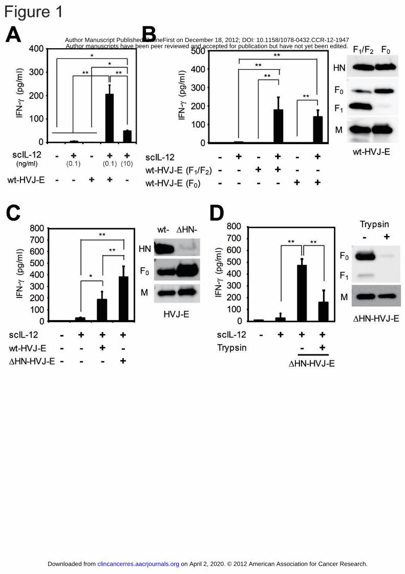
The HVJ-E F protein enhanced the IL-12-induced IFN-γ secretion in a
fusion-independent manner
We have described the construction of the murine scIL-12 expression vector in a
previous report (28). In this study, we used this vector to produce scIL-12 proteins in
CHO cells. Stimulation of splenocytes from normal C57BL/6N mice with HVJ-E or
scIL-12 (0.1 ng/ml) induced only a low level of IFN-γ secretion. However, we
demonstrated that the combining HVJ-E and scIL-12 dramatically enhanced the
secretion of IFN-γ by the splenocytes (Fig. 1A). In addition, administration of a high
dose of scIL-12 (10 ng/ml) induced IFN-γ secretion, but scIL-12 was significantly less
effective than combined treatment with scIL-12 and HVJ-E (Fig. 1A). Next, we
investigated which cells of the heterogeneous splenocyte population produced IFN-γ in
response to scIL-12 and HVJ-E and found that IFN-γ was mainly produced by CD3+ T
cells (Supplementary Fig. S1).
The envelope of HVJ-E is composed of two different membrane proteins, the
fusion protein (F) and hemagglutinin-neuraminidase (HN). F exists in either the
inactive F0 form on fusion-incompetent HVJ-E or the enzymatically cleaved, active
F1/F2 form on fusion-competent HVJ-E (33). Splenocytes were thus treated with either
on April 2, 2020. © 2012 American Association for Cancer Research.clincancerres.aacrjournals.org Downloaded from
Author manuscripts have been peer reviewed and accepted for publication but have not yet been edited. Author Manuscript Published OnlineFirst on December 18, 2012; DOI: 10.1158/1078-0432.CCR-12-1947
22
F0- or F1/F2-HVJ-E in combination with scIL-12, and both types of HVJ-E induced
robust IFN-γ secretion (Fig. 1B). Therefore, the HVJ-E-mediated enhancement of
IFN-γ secretion was observed to be independent of fusion competence. Next, we
investigated which membrane glycoproteins, HN or F, were responsible for the
enhanced IFN-γ production. When administered with scIL-12, HN-depleted HVJ-E
(ΔHN-HVJ-E), which displays increased F expression on the envelope, enhanced IFN-γ
secretion more potently than wild-type HVJ-E (wt-HVJ-E) (Fig. 1C), suggesting that F,
but not HN, may be involved in the induction of IFN-γ production. Although both the
F and HN proteins are present on the surface of the HVJ-E particle, the F protein is
predominant. To generate F-depleted HVJ-E, the F protein of ΔHN-HVJ-E was
enzymatically degraded by trypsin overtreatment (2.5 mg/ml for 24 hours at 37°C).
F-depleted ΔHN-HVJ-E induced a significantly reduced level of IFN-γ secretion (Fig.
1D), and moreover, the anti-F antibody inhibited the induction of IFN-γ
(Supplementary Fig. S2A), suggesting that the activity of the F protein is required for
IFN-γ induction.
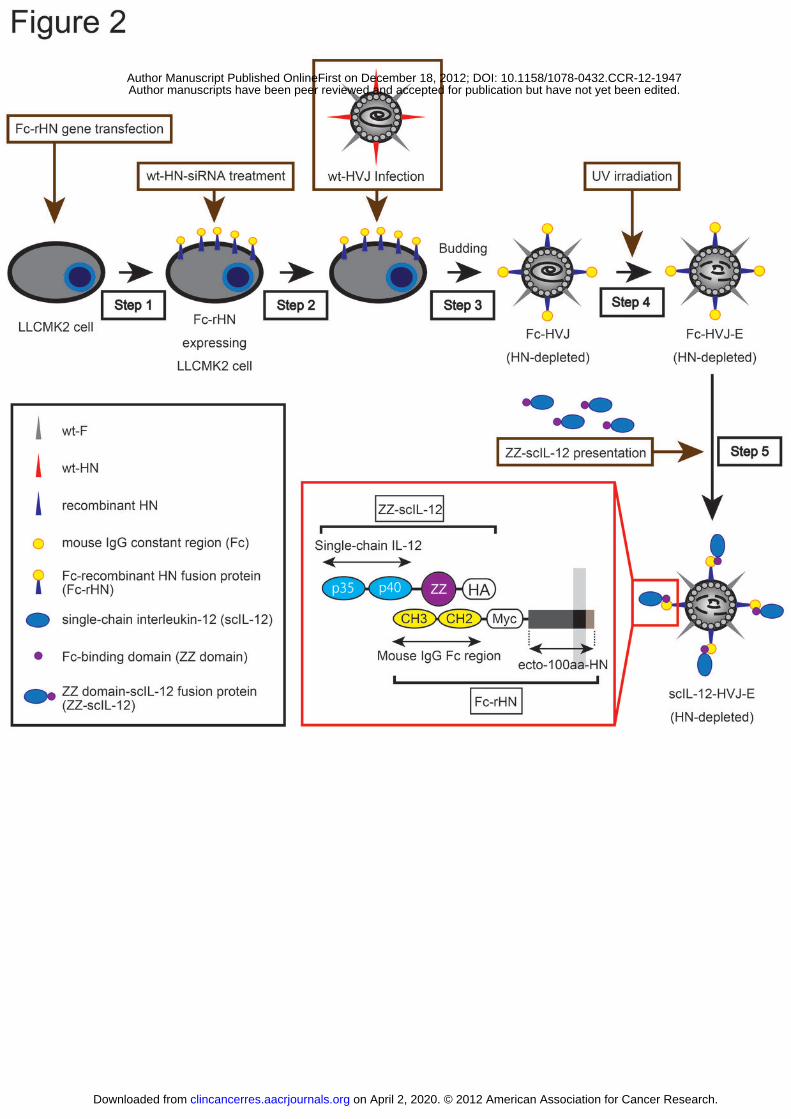
Overall strategy for the presentation of IL-12 on the surface of HVJ-E
These results prompted us to hypothesize that a close association between HVJ-E
on April 2, 2020. © 2012 American Association for Cancer Research.clincancerres.aacrjournals.org Downloaded from
Author manuscripts have been peer reviewed and accepted for publication but have not yet been edited. Author Manuscript Published OnlineFirst on December 18, 2012; DOI: 10.1158/1078-0432.CCR-12-1947
23
and IL-12 in the tumor microenvironment in vivo may synergistically enhance
anti-tumor immunity. To prove this hypothesis, we generated scIL-12-conjugated
HVJ-E (scIL-12-HVJ-E) that presents scIL-12 on the surface of ΔHN-HVJ-E by
binding to the IgG constant region (Fc) and the protein A-Fc binding domain (ZZ) (34)
using viral gene engineering technology (35-37). This technology allowed for the
manipulation of the HVJ-E membrane proteins by inducing the expression of
recombinant envelope proteins on HVJ-infected cells (Fig. 2).
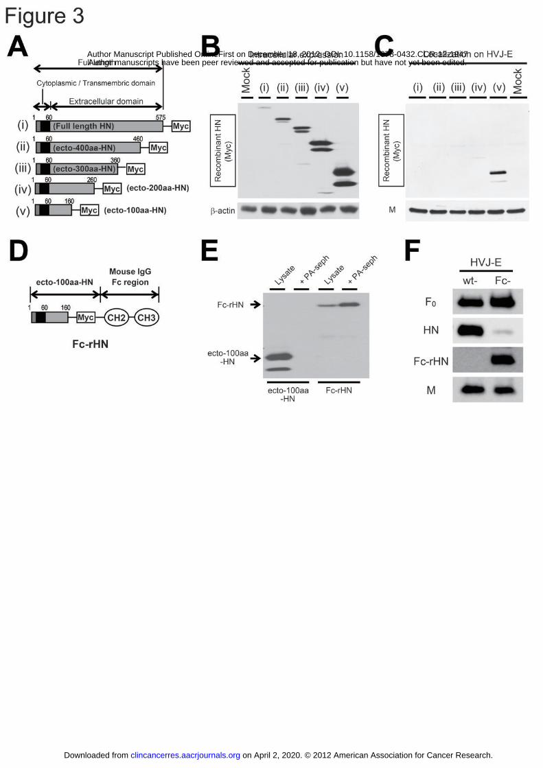
Generation of Fc-rHN and Fc-HVJ-E
To generate scIL-12-HVJ-E, we aimed to localize Fc-HN fusion proteins on the
surface of ΔHN-HVJ-E. We constructed expression plasmids of HN deletion mutants
(full length-, ecto-400aa-, ecto-300aa-, ecto-200aa- and ecto-100aa-HN) (Fig. 3A).
Although all recombinant HN (rHN) proteins were expressed in the LLC-MK2 cells
transfected with these plasmids (Fig. 3B), only ecto-100aa-HN was incorporated into
the cell-derived progeny of HVJ by infecting rHN-expressing cells with live HVJ (Fig.
3C). Therefore, we constructed Fc-rHN, in which murine Fc was fused to the
C-terminus of ecto-100aa-HN (Fig. 3D). We confirmed that Fc-rHN had the ability to
bind to protein A using a co-precipitation assay with protein A-Sepharose (Fig. 3E).
on April 2, 2020. © 2012 American Association for Cancer Research.clincancerres.aacrjournals.org Downloaded from
Author manuscripts have been peer reviewed and accepted for publication but have not yet been edited. Author Manuscript Published OnlineFirst on December 18, 2012; DOI: 10.1158/1078-0432.CCR-12-1947

24
We then generated Fc-HVJ-E, which included Fc-rHN with depleted wt-HN (Fig. 3F).
Generation of ZZ-scIL-12 and scIL-12-HVJ-E
To present scIL-12 on the surface of Fc-HVJ-E, we constructed ZZ-scIL-12 by
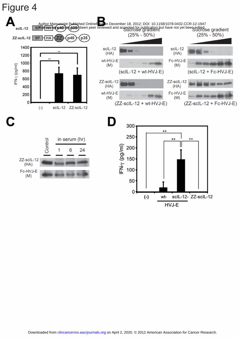
inserting the ZZ domain into the downstream scIL-12 signal peptide (Fig. 4A).
ZZ-scIL-12 and scIL-12 induced IFN-γ secretion by splenocytes (Fig. 4A). Next, we
investigated the ability of ZZ-scIL-12 to bind to Fc-rHN on Fc-HVJ-E using sucrose
density gradient centrifugation. When analyzed separately, ZZ-scIL-12 was detected
in the upper layer, and HVJ-E was detected in the lower layer of the 25%-50% sucrose
gradients (Supplementary Fig. S3). However, ZZ-scIL-12 was shifted to the lower
layer in the sedimentation of the ZZ-scIL-12 and Fc-HVJ-E mixture (Fig. 4B),
suggesting that ZZ-scIL-12 binds to Fc-rHN on Fc-HVJ-E. Moreover,
ZZ-scIL-12-conjugated Fc-HVJ-E (scIL-12-HVJ-E) was incubated in murine serum,
and ZZ-scIL-12 was maintained in Fc-HVJ-E for 24 hours (Fig. 4C). We calculated
the amount of ZZ-scIL-12 on Fc-HVJ-E by comparing the density of the bands
corresponding to ZZ-scIL-12 by Western blotting analysis and estimated that
approximately 6.83 molecules of ZZ-scIL-12 were loaded onto one particle of
Fc-HVJ-E (Supplementary Fig. S4). Finally, scIL-12-HVJ-E displayed much
on April 2, 2020. © 2012 American Association for Cancer Research.clincancerres.aacrjournals.org Downloaded from
Author manuscripts have been peer reviewed and accepted for publication but have not yet been edited. Author Manuscript Published OnlineFirst on December 18, 2012; DOI: 10.1158/1078-0432.CCR-12-1947
25
stronger IFN-γ-inducing activity on splenocytes and DCs than wt-HVJ-E or scIL-12
alone (Fig. 4D and Supplementary Fig. S2B).
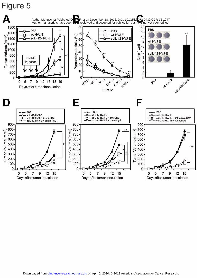
Anti-tumor effects against intradermal F10 melanomas induced by the
intratumoral administration of scIL-12-HVJ-E
We next investigated the anti-tumor activity of scIL-12-HVJ-E in vivo. A
murine intradermal tumor model was generated through intradermal inoculation of F10
melanoma cells (5 x 10⁵ cells) into the backs of female C57BL/6N mice. When each
tumor had grown to ~3-5 mm in diameter, scIL-12-HVJ-E, wt-HVJ-E or PBS was
injected into the tumor for a total of three times every other day. In these experiments,
scIL-12-HVJ-E caused a much more robust tumor suppression than wt-HVJ-E (Fig. 5A).
Moreover, the anti-tumor immune responses against the F10 melanomas were examined
using 51Cr release CTL assays (Fig. 5B) and ELISPOT assays (Fig. 5C). The data
revealed that scIL-12-HVJ-E remarkably enhanced cytotoxic T cell activity against F10
melanomas. Next, to investigate the roles of CD4+ T cells, CD8+ T cells and NK cells
in the therapeutic effect, scIL-12-HVJ-E-mediated tumor growth inhibition was
assessed in mice depleted of CD4+ T cells, CD8+ T cells and NK cells by administrating
a neutralizing antibody specific for each cell type (13, 38). The depletion of CD8 and
on April 2, 2020. © 2012 American Association for Cancer Research.clincancerres.aacrjournals.org Downloaded from
Author manuscripts have been peer reviewed and accepted for publication but have not yet been edited. Author Manuscript Published OnlineFirst on December 18, 2012; DOI: 10.1158/1078-0432.CCR-12-1947
26
NK cells, but not CD4 cells, significantly decreased the scIL-12-HVJ-E-mediated effect
(Fig. 5D, E and F), demonstrating that the anti-tumor effect of scIL-12-HVJ-E was
dependent on CD8+ T cells and NK cells but not CD4+ T cells.
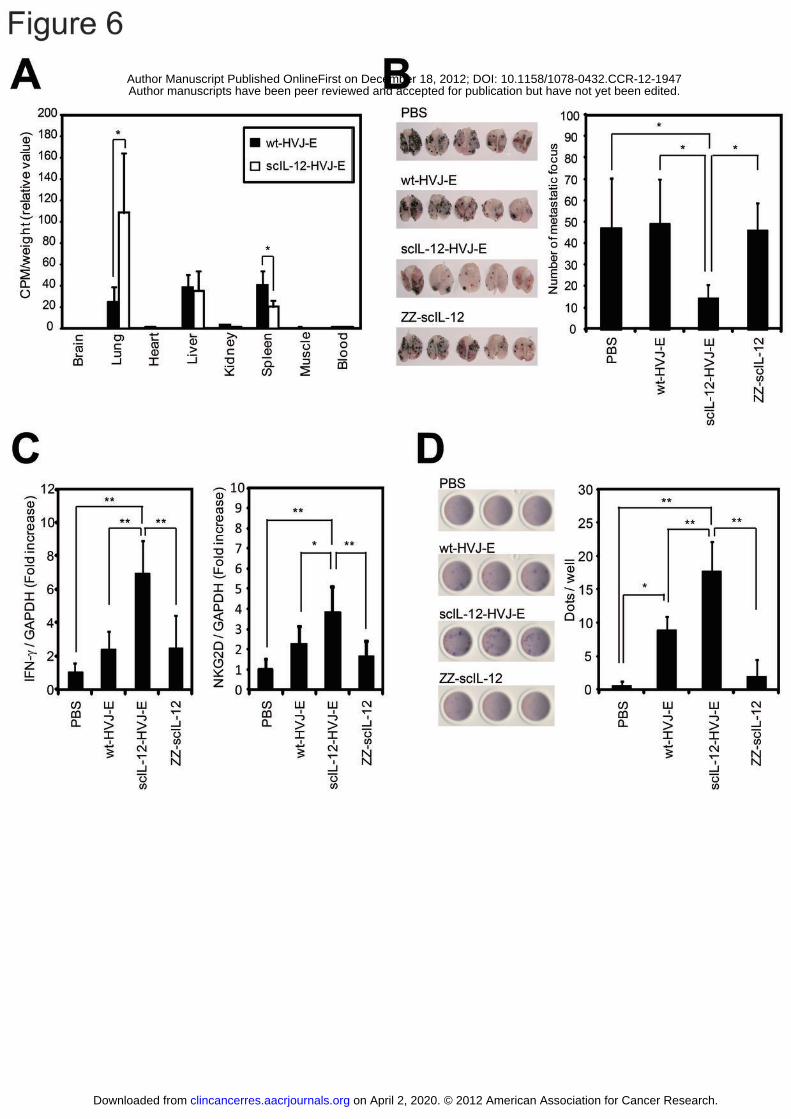
Anti-tumor effects against metastatic lung F10 melanomas induced by the systemic
administration of scIL-12-HVJ-E
We then investigated the tissue distribution of scIL-12-HVJ-E following
intravenous injection. 125I-labeled HVJ-E was systemically administered via tail vein
injection, and the level of 125I in the tissues (brain, lungs, heart, liver, kidneys, spleen,
muscles and blood) was measured using a γ-scintillation counter 24 hours after
injection. Interestingly, scIL-12-HVJ-E and ΔHN-HVJ-E, but not wt-HVJ-E,
preferentially accumulated in the lungs (Fig. 6A and Supplementary Fig. S5A),
implying that HN depletion likely affected the localization of HVJ-E.
We next investigated the therapeutic effects of intravenous scIL-12-HVJ-E
administration in a murine F10 melanoma model of lung metastasis. The mice were
inoculated with F10 melanoma cells (5 x 10⁵ cells) by intravenous injection. Multiple
metastatic foci appeared on the surface of the lungs five days after the F10 melanoma
injection (Supplementary Fig. S5B). We began intravenous administration of
on April 2, 2020. © 2012 American Association for Cancer Research.clincancerres.aacrjournals.org Downloaded from
Author manuscripts have been peer reviewed and accepted for publication but have not yet been edited. Author Manuscript Published OnlineFirst on December 18, 2012; DOI: 10.1158/1078-0432.CCR-12-1947
27
scIL-12-HVJ-E, wt-HVJ-E or ZZ-scIL-12 when lung metastasis was confirmed. The
injections were repeated for a total of three times every other day. As expected, only
the scIL-12-HVJ-E-treated mice exhibited a significant reduction in the number of
metastatic foci in the lungs (Fig. 6B). Based on our microarray analysis of the lung
transcripts (data not shown), we focused on IFN-γ and NKG2D, which showed
increased expression upon scIL-12-HVJ-E treatment. Real-time RT-PCR analysis
confirmed that IFN-γ and NKG2D expression were both increased in lungs treated
with scIL-12-HVJ-E (Fig. 6C). Although IFN-γ expression was found to be elevated
in the lungs, the serum IFN-γ level was unchanged by scIL-12-HVJ-E administration
(Supplementary Fig. S6A). However, the co-administration of scIL-12 and
Fc-HVJ-E increased the serum IFN-γ level (Supplementary Fig. S6B). Moreover,
scIL-12-HVJ-E significantly induced NKG2D expression in the lungs, suggesting that
NK cells and CD8+ T lymphocytes were activated in the lesions (Fig. 6C). Systemic
administration of scIL-12-HVJ-E induced robust activation of CTLs specific for
melanoma (Fig. 6D), and no evident adverse effects were observed in the treated mice.
The CTL activation was significantly reduced by an anti-IFN-γ antibody, indicating
that the CTL activation was dependent on IFN-γ (Supplementary Fig. S7A).
Moreover, the CTLs targeted melanoma cells specifically, as the cytotoxic effects were
on April 2, 2020. © 2012 American Association for Cancer Research.clincancerres.aacrjournals.org Downloaded from
Author manuscripts have been peer reviewed and accepted for publication but have not yet been edited. Author Manuscript Published OnlineFirst on December 18, 2012; DOI: 10.1158/1078-0432.CCR-12-1947
28
not detected in other cells (Supplementary Fig. S7B).
on April 2, 2020. © 2012 American Association for Cancer Research.clincancerres.aacrjournals.org Downloaded from
Author manuscripts have been peer reviewed and accepted for publication but have not yet been edited. Author Manuscript Published OnlineFirst on December 18, 2012; DOI: 10.1158/1078-0432.CCR-12-1947

29
Discussion
In this study, we demonstrated that HVJ-E dramatically enhances IL-12 activity
and induces IFN-γ production in splenocytes. The systemic administration of
IL-12-conjugated HVJ-E significantly increases the level of IFN-γ expression in the
lungs without elevating the serum IFN-γ level and effectively induces anti-tumor
activity against metastatic lung melanomas.
Previously, our group reported that several components of HVJ-E stimulate tumor
cells and DCs to secrete various cytokines. RNA genome fragments of HVJ-E, which
are taken into host cells through membrane fusion, are recognized by RIG-I but not
TLRs (39), and type-I IFN is induced in tumor cells and DCs. However, F0-formed
HVJ-E, which is unable to fuse with host cells, enhances scIL-12-induced IFN-γ
secretion (Fig. 1B). Therefore, the activation of RIG-I by RNA fragments is unlikely
to be correlated with IFN-γ enhancement. In this study, we demonstrated that F is
responsible for enhancing IFN-γ secretion (Fig. 1C and 1D). Although our previous
report shows that F induces IL-6 secretion in DCs in a fusion-independent manner (40),
F-mediated IL-6 secretion does not mediate IFN-γ enhancement because IL-6 does not
enhance IL-12 activity (41, 42). These results suggest that putative F receptors on
splenocytes may transmit signals to enhance IFN-γ and IL-12 production.
on April 2, 2020. © 2012 American Association for Cancer Research.clincancerres.aacrjournals.org Downloaded from
Author manuscripts have been peer reviewed and accepted for publication but have not yet been edited. Author Manuscript Published OnlineFirst on December 18, 2012; DOI: 10.1158/1078-0432.CCR-12-1947
30
ΔHN-HVJ-E enhanced IFN-γ induction more robustly than wt-HVJ-E because the
expression levels of F in ΔHN-HVJ-E were increased by knocking down HN (37 and
Fig. 1C). Because HN induces hemagglutination by binding to sialic acid on the
surface receptors of red blood cells (43), the administration of wt-HVJ-E, especially
intravenously, has limitations for cancer therapy. ΔHN-HVJ-E shows very low
hemagglutinating activity (37) and robustly enhances IFN-γ and IL-12 production in
vitro.
In the experiments shown in Fig. 5D, E and F, although CD8+ T cells and NK
cells were required for scIL-12-HVJ-E-mediated anti-tumor effects, the depletion of
CD4+ T cells did not affect the suppression of tumor growth by scIL-12-HVJ-E.
Previous reports have indicated that the IL-12-mediated anti-tumor response is
maintained in CD4+ T cell-depleted mice (44) and that IL-12 stimulates antigen-specific
CD8+ T cells in CD4+ T cell-knockout mice (45). Therefore, we suggest that
scIL-12-HVJ-E-mediated anti-tumor immunity was independent of CD4+ T cells.
Moreover, in Fig. 5D, E and F and Supplementary Fig. S7, the peritoneal administration
of a large amount of control IgG (more than 200 μg) induced a small decrease in
scIL-12-HVJ-E-mediated anti-tumor immunity. Although the precise mechanism is
unclear, this result might be due to the immunosuppressive effect of the administration
on April 2, 2020. © 2012 American Association for Cancer Research.clincancerres.aacrjournals.org Downloaded from
Author manuscripts have been peer reviewed and accepted for publication but have not yet been edited. Author Manuscript Published OnlineFirst on December 18, 2012; DOI: 10.1158/1078-0432.CCR-12-1947

31
of a large amount of IgG (IVIG) (46).
In previous reports, the Fc-ZZ binding system has been applied to various types
of targeting vectors (47-49), and we hypothesized that scIL-12-HVJ-E generated using
this system could be functional upon systemic administration in mice. In fact, the
Fc-ZZ binding of scIL-12-HVJ-E was stable in serum for at least 24 hours (Fig. 4C),
and intravenous administration of scIL-12-HVJ-E resulted in elevated IFN-γ expression
levels in the lungs due to pulmonary accumulation of the virions (Fig. 6A and 6C).
scIL-12-HVJ-E and ΔHN-HVJ-E, but not wt-HVJ-E, preferentially accumulated
in the lungs (Fig. 6A and Supplementary Fig. S5A). A previous report has indicated
that HVJ-E preferentially accumulates in spleen after intravenous administration (50).
This accumulation in the spleen most likely occurs because HN on HVJ-E induces
hemagglutination by binding to the sialic acid on the erythrocyte surface, causing the
erythrocyte and HVJ-E complex to be transported to the spleen and degraded.
Therefore, the accumulation of scIL-12-HVJ-E and ΔHN-HVJ-E in the lung may result
from the loss of hemagglutinating activity upon HN knockdown and the subsequent
escape from the “spleen-trap”; however, the precise mechanism by which this occurs
remains unclear.
A previous study has shown that only high doses (approximately 0.5 ~ 1.0 μg) of
on April 2, 2020. © 2012 American Association for Cancer Research.clincancerres.aacrjournals.org Downloaded from
Author manuscripts have been peer reviewed and accepted for publication but have not yet been edited. Author Manuscript Published OnlineFirst on December 18, 2012; DOI: 10.1158/1078-0432.CCR-12-1947

32
systemic IL-12 suppress tumor growth in mice by inducing a significant elevation in the
serum IFN-γ level (30). However, such increases in serum IFN-γ have been shown to
induce serious side effects, such as stomatitis, gastrointestinal bleeding, colitis and
diarrhea (31, 32). Therefore, the systemic administration of IL-12 has not been applied
for clinical use thus far. We herein demonstrated that systemically administered
scIL-12-HVJ-E, which contains approximately 500 pg scIL-12, preferentially
accumulated in the lungs (Fig. 6A) and mediated the synergistic anti-tumor effects of
HVJ-E and IL-12 in the tumor microenvironment (Fig. 6B and 6C) without elevating
the serum IFN-γ levels (Supplementary Fig. S6A). By contrast, the co-administration
of scIL-12 and Fc-HVJ-E increased the serum IFN-γ level (Supplementary Fig. S6B).
Therefore, the conjugation of IL-12 and HVJ-E is important to inhibit the elevation of
serum IFN-γ, which may cause severe side effects (31, 32). The data shown here
provide a basis for the future clinical application of scIL-12-HVJ-E, which appears to
have a higher anti-cancer activity and lower toxicity than IL-12 alone.
on April 2, 2020. © 2012 American Association for Cancer Research.clincancerres.aacrjournals.org Downloaded from
Author manuscripts have been peer reviewed and accepted for publication but have not yet been edited. Author Manuscript Published OnlineFirst on December 18, 2012; DOI: 10.1158/1078-0432.CCR-12-1947
33
Acknowledgements
This work was supported by the Northern Osaka (Saito) Biomedical
Knowledge-Based Cluster Creation Project, Special Coordination Funds for Promoting
Science from the Ministry of Education, Culture, Sports, Science and Technology of
Japan and grants from the Ministry of Health, Labor and Welfare of Japan.
on April 2, 2020. © 2012 American Association for Cancer Research.clincancerres.aacrjournals.org Downloaded from
Author manuscripts have been peer reviewed and accepted for publication but have not yet been edited. Author Manuscript Published OnlineFirst on December 18, 2012; DOI: 10.1158/1078-0432.CCR-12-1947
34
References
1. Vignali DAA, Collison LW, Workman CJ. How regulatory T cells work. Nature
Reviews Immunology. 2008;8:523–32.
2. Burch PA, Croghan GA, Gastineau DA, Jones LA, Kaur JS, Kylstra JW, et al.
Immunotherapy (APC8015, Provenge®) targeting prostatic acid phosphatase can
induce durable remission of metastatic androgen-independent prostate cancer: A
phase 2 trial. The Prostate. 2004;60:197–204.
3. Cheever MA, Higano CS. PROVENGE (Sipuleucel-T) in Prostate Cancer: The
First FDA-Approved Therapeutic Cancer Vaccine. Clin Cancer Res.
2011;17:3520–6.
4. Hodi FS, O’Day SJ, McDermott DF, Weber RW, Sosman JA, Haanen JB, et al.
Improved Survival with Ipilimumab in Patients with Metastatic Melanoma. New
England Journal of Medicine. 2010;363:711–23.
5. Prieto PA, Yang JC, Sherry RM, Hughes MS, Kammula US, White DE, et al.
CTLA-4 Blockade with Ipilimumab: Long-term Follow-up of 177 Patients with
Metastatic Melanoma. Clin Cancer Res. 2012;18:2039–47.
6. Topalian SL, Hodi FS, Brahmer JR, Gettinger SN, Smith DC, McDermott DF, et
al. Safety, Activity, and Immune Correlates of Anti–PD-1 Antibody in Cancer.
New England Journal of Medicine. 2012;366:2443–54.
7. Dudley ME, Yang JC, Sherry R, Hughes MS, Royal R, Kammula U, et al.
Adoptive Cell Therapy for Patients With Metastatic Melanoma: Evaluation of
Intensive Myeloablative Chemoradiation Preparative Regimens. JCO.
2008;26:5233–9.
8. Kirkwood JM, Tarhini AA, Panelli MC, Moschos SJ, Zarour HM, Butterfield LH,
et al. Next Generation of Immunotherapy for Melanoma. JCO. 2008;26:3445–55.
9. Mellman I, Coukos G, Dranoff G. Cancer immunotherapy comes of age. Nature.
2011;480:480–9.
10. Lamb RA, Kolakofsky D. Paramyxoviridae: The Viruses and Their Replicaiton.
Fields Virology. 2001:1305-40.
on April 2, 2020. © 2012 American Association for Cancer Research.clincancerres.aacrjournals.org Downloaded from
Author manuscripts have been peer reviewed and accepted for publication but have not yet been edited. Author Manuscript Published OnlineFirst on December 18, 2012; DOI: 10.1158/1078-0432.CCR-12-1947

35
11. Curran J, Kolakofsky D. Replication of paramyxoviruses. Adv. Virus Res.
1999;54:403–22.
12. Kurooka M, Kaneda Y. Inactivated Sendai Virus Particles Eradicate Tumors by
Inducing Immune Responses through Blocking Regulatory T Cells. Cancer Res.
2007;67:227–36.
13. Fujihara A, Kurooka M, Miki T, Kaneda Y. Intratumoral injection of inactivated
Sendai virus particles elicits strong antitumor activity by enhancing local CXCL10
expression and systemic NK cell activation. Cancer Immunol. Immunother.
2008;57:73–84.
14. Pasare C, Medzhitov R. Toll Pathway-Dependent Blockade of CD4+CD25+ T
Cell-Mediated Suppression by Dendritic Cells. Science. 2003;299:1033–6.
15. Lal G, Zhang N, Van Der Touw W, Ding Y, Ju W, Bottinger EP, et al. Epigenetic
Regulation of Foxp3 Expression in Regulatory T Cells by DNA Methylation. J
Immunol. 2009;182:259–73.
16. Benveniste EN. Cytokine Actions in the Central Nervous System. Cytokine &
Growth Factor Reviews. 1998;9:259–75.
17. Tannenbaum CS, Tubbs R, Armstrong D, Finke JH, Bukowski RM, Hamilton TA.
The CXC Chemokines IP-10 and Mig Are Necessary for IL-12-Mediated
Regression of the Mouse RENCA Tumor. J Immunol. 1998;161:927–32.
18. Bukowski RM, Rayman P, Molto L, Tannenbaum CS, Olencki T, Peereboom D, et
al. Interferon-γ and CXC Chemokine Induction by Interleukin 12 in Renal Cell
Carcinoma. Clin Cancer Res. 1999;5:2780–9.
19. Seliger B, Hammers S, Höhne A, Zeidler R, Knuth A, Gerharz CD, et al.
IFN-gamma-mediated coordinated transcriptional regulation of the human TAP-1
and LMP-2 genes in human renal cell carcinoma. Clin Cancer Res. 1997;3:573–8.
20. Weber JS, Rosenberg SA. Modulation of Murine Tumor Major Histocompatibility
Antigens by Cytokines in Vivo and in Vitro. Cancer Res. 1988;48:5818–24.
21. Nakajima C, Uekusa Y, Iwasaki M, Yamaguchi N, Mukai T, Gao P, et al. A Role
of Interferon-γ (IFN-γ) in Tumor Immunity T Cells with the Capacity to Reject
on April 2, 2020. © 2012 American Association for Cancer Research.clincancerres.aacrjournals.org Downloaded from
Author manuscripts have been peer reviewed and accepted for publication but have not yet been edited. Author Manuscript Published OnlineFirst on December 18, 2012; DOI: 10.1158/1078-0432.CCR-12-1947

36
Tumor Cells Are Generated But Fail to Migrate to Tumor Sitesin IFN-γ-deficient
Mice. Cancer Res. 2001;61:3399–405.
22. Pujade-Lauraine E, Guastalla JP, Colombo N, Devillier P, François E, Fumoleau P,
et al. Intraperitoneal recombinant interferon gamma in ovarian cancer patients with
residual disease at second-look laparotomy. JCO. 1996;14:343–50.
23. Windbichler GH, Hausmaninger H, Stummvoll W, Graf AH, Kainz C, Lahodny J,
et al. Interferon-gamma in the first-line therapy of ovarian cancer: a randomized
phase III trial. Br J Cancer. 2000;82:1138–44.
24. Giannopoulos A, Constantinides C, Fokaeas E, Stravodimos C, Giannopoulou M,
Kyroudi A, et al. The Immunomodulating Effect of Interferon-γ Intravesical
Instillations in Preventing Bladder Cancer Recurrence. Clin Cancer Res.
2003;9:5550–8.
25. Marth C, Windbichler G h., Hausmaninger H, Petru E, Estermann K, Pelzer A, et
al. Interferon-gamma in combination with carboplatin and paclitaxel as a safe and
effective first-line treatment option for advanced ovarian cancer: results of a phase
I/II study. International Journal of Gynecological Cancer. 2006;16:1522–8.
26. Colombo MP, Trinchieri G. Interleukin-12 in anti-tumor immunity and
immunotherapy. Cytokine & Growth Factor Reviews. 2002;13:155–68.
27. Watford WT, Moriguchi M, Morinobu A, O’Shea JJ. The biology of IL-12:
coordinating innate and adaptive immune responses. Cytokine & Growth Factor
Reviews. 2003;14:361–8.
28. Lieschke GJ, Rao PK, Gately MK, Mulligan RC. Bioactive murine and human
interleukin-12 fusion proteins which retain antitumor activity in vivo. Nat Biotech.
1997;15:35–40.
29. Foss, Moody, Murphy Jr, Pazmany, Zilliox, Murtaugh. In Vitro and In Vivo
Bioactivity of Single‐Chain Interleukin‐12. Scandinavian Journal of
Immunology. 1999;50:596–604.
30. Nastala CL, Edington HD, McKinney TG, Tahara H, Nalesnik MA, Brunda MJ, et
al. Recombinant IL-12 administration induces tumor regression in association with
IFN-gamma production. J Immunol. 1994;153:1697–706.
on April 2, 2020. © 2012 American Association for Cancer Research.clincancerres.aacrjournals.org Downloaded from
Author manuscripts have been peer reviewed and accepted for publication but have not yet been edited. Author Manuscript Published OnlineFirst on December 18, 2012; DOI: 10.1158/1078-0432.CCR-12-1947
37
31. Atkins MB, Robertson MJ, Gordon M, Lotze MT, DeCoste M, DuBois JS, et al.
Phase I evaluation of intravenous recombinant human interleukin 12 in patients
with advanced malignancies. Clin Cancer Res. 1997;3:409–17.
32. Leonard JP, Sherman ML, Fisher GL, Buchanan LJ, Larsen G, Atkins MB, et al.
Effects of single-dose interleukin-12 exposure on interleukin-12-associated
toxicity and interferon-gamma production. Blood. 1997;90:2541–8.
33. Tashiro M, McQueen NL, Seto JT. Determinants of organ tropism of sendai virus.
Front. Biosci. 1999;4:D642–645.
34. Nilsson B, Moks T, Jansson B, Abrahmsén L, Elmblad A, Holmgren E, et al. A
synthetic IgG-binding domain based on staphylococcal protein A. Protein Eng.
1987;1:107–13.
35. Kawachi M, Tamai K, Saga K, Yamazaki T, Fujita H, Shimbo T, et al.
Development of tissue-targeting hemagglutinating virus of Japan envelope vector
for successful delivery of therapeutic gene to mouse skin. Hum. Gene Ther.
2007;18:881–94.
36. Shimbo T, Kawachi M, Saga K, Fujita H, Yamazaki T, Tamai K, et al.
Development of a transferrin receptor-targeting HVJ-E vector. Biochem. Biophys.
Res. Commun. 2007;364:423–8.
37. Saga K, Tamai K, Kawachi M, Shimbo T, Fujita H, Yamazaki T, et al. Functional
modification of Sendai virus by siRNA. Journal of Biotechnology.
2008;133:386–94.
38. Lee J, Nakagiri T, Oto T, Harada M, Morii E, Shintani Y, et al. IL-6 Amplifier,
NF-κB–Triggered Positive Feedback for IL-6 Signaling, in Grafts Is Involved in
Allogeneic Rejection Responses. J Immunol. 2012;189:1928–36.
39. López CB, Moltedo B, Alexopoulou L, Bonifaz L, Flavell RA, Moran TM.
TLR-Independent Induction of Dendritic Cell Maturation and Adaptive Immunity
by Negative-Strand RNA Viruses. J Immunol. 2004;173:6882–9.
40. Suzuki H, Kurooka M, Hiroaki Y, Fujiyoshi Y, Kaneda Y. Sendai virus F
glycoprotein induces IL-6 production in dendritic cells in a fusion-independent
manner. FEBS Letters. 2008;582:1325–9.
on April 2, 2020. © 2012 American Association for Cancer Research.clincancerres.aacrjournals.org Downloaded from
Author manuscripts have been peer reviewed and accepted for publication but have not yet been edited. Author Manuscript Published OnlineFirst on December 18, 2012; DOI: 10.1158/1078-0432.CCR-12-1947
38
41. Rincón M, Anguita J, Nakamura T, Fikrig E, Flavell RA. Interleukin (IL)-6
Directs the Differentiation of IL-4–producing CD4+ T Cells. J Exp Med.
1997;185:461–70.
42. Frassanito MA, Cusmai A, Dammacco F. Deregulated cytokine network and
defective Th1 immune response in multiple myeloma. Clinical & Experimental
Immunology. 2001;125:190–7.
43. Portner A, Scroggs RA, Metzger DW. Distinct functions of antigenic sites of the
HN glycoprotein of sendai virus. Virology. 1987;158:61–8.
44. Brunda MJ, Luistro L, Warrier RR, Wright RB, Hubbard BR, Murphy M, et al.
Antitumor and antimetastatic activity of interleukin 12 against murine tumors. J.
Exp. Med. 1993;178:1223–30.
45. Schmidt CS, Mescher MF. Peptide Antigen Priming of Naive, But Not Memory,
CD8 T Cells Requires a Third Signal That Can Be Provided by IL-12. J Immunol.
2002;168:5521–9.
46. Ephrem A, Chamat S, Miquel C, Fisson S, Mouthon L, Caligiuri G, et al.
Expansion of CD4+CD25+ regulatory T cells by intravenous immunoglobulin: a
critical factor in controlling experimental autoimmune encephalomyelitis. Blood.
2008;111:715–22.
47. Morizono K, Bristol G, Xie Y, Kung SK-P, Chen ISY. Antibody-Directed
Targeting of Retroviral Vectors via Cell Surface Antigens. J. Virol.
2001;75:8016–20.
48. Henning P, Andersson KME, Frykholm K, Ali A, Magnusson MK, Nygren P-A, et
al. Tumor cell targeted gene delivery by adenovirus 5 vectors carrying knobless
fibers with antibody-binding domains. Gene Ther. 2004;12:211–24.
49. Morizono K, Xie Y, Ringpis G-E, Johnson M, Nassanian H, Lee B, et al.
Lentiviral vector retargeting to P-glycoprotein on metastatic melanoma through
intravenous injection. Nat Med. 2005;11:346–52.
50. Kaneda Y, Nakajima T, Nishikawa T, Yamamoto S, Ikegami H, Suzuki N, et al.
Hemagglutinating Virus of Japan (HVJ) Envelope Vector as a Versatile Gene
Delivery System. Molecular Therapy. 2002;6:219–26.
on April 2, 2020. © 2012 American Association for Cancer Research.clincancerres.aacrjournals.org Downloaded from
Author manuscripts have been peer reviewed and accepted for publication but have not yet been edited. Author Manuscript Published OnlineFirst on December 18, 2012; DOI: 10.1158/1078-0432.CCR-12-1947

39
Figure legends
Figure 1: The synergistic effect of HVJ-E on IL-12-induced IFN-γ secretion.
(A) - (D) scIL-12 and/or various types of HVJ-E [(A) wt-HVJ-E (F1/F2), (B) F1/F2 or F0
wt-HVJ-E, (C) wt-HVJ-E or ΔHN-HVJ-E and (D) ΔHN-HVJ-E or F-degraded
ΔHN-HVJ-E] were added to splenocytes, and 24 hours later, the IFN-γ concentrations of
the supernatants were measured by ELISA. To confirm the protein composition of
various types of HVJ-E, several viral proteins (HN, F0, F1 and M) were detected by
Western blotting: (B) F1/F2 and F0 wt-HVJ-E, (C) wt-HVJ-E and ΔHN-HVJ-E and (D)
ΔHN-HVJ-E and F-degraded ΔHN-HVJ-E. All data are presented as the mean ± SD
(n = 4). * P < 0.05, ** P < 0.01, Tukey-Kramer test.
Figure 2: The construction of scIL-12-HVJ-E. LLCMK2 cells stably expressing Fc-HN
were transfected with HN-siRNA. In LLCMK2 cells infected with live wt-HVJ,
Fc-HVJ (HN depleted) was produced as a viral progeny. Fc-HVJ-E (HN depleted) was
obtained after UV irradiation of the progeny. scIL-12-HVJ-E (HN depleted) was
constructed by conjugating ZZ-scIL-12 and Fc-HVJ-E (HN depleted).
on April 2, 2020. © 2012 American Association for Cancer Research.clincancerres.aacrjournals.org Downloaded from
Author manuscripts have been peer reviewed and accepted for publication but have not yet been edited. Author Manuscript Published OnlineFirst on December 18, 2012; DOI: 10.1158/1078-0432.CCR-12-1947
40
Figure 3: The construction of Fc-rHN and the generation of Fc-HVJ-E.
(A) The design of the recombinant deletion mutants of HN. (i) Full length-, (ii)
ecto-400aa-, (iii) ecto-300aa-, (iv) ecto-200aa- and (v) ecto-100aa-HN. (B) The
various recombinant HN (rHN) genes were transferred to LLC-MK2 cells, and their
intracellular expression was detected using Western blotting analysis. (C) HVJ was
used to infect rHN-expressing LLC-MK2 cells, and the rHN in the cell-derived HVJ-E
progeny was detected using Western blotting analysis. (D) Fc-rHN, (E) Fc-rHN or
ecto-100aa-HN was mixed with protein A-Sepharose, and the rHN that co-precipitated
with protein A-Sepharose was detected using Western blotting analysis. (F) The
expression of the viral proteins (HN, F, M and Fc-rHN) of Fc-HVJ-E and wt-HVJ-E
was examined using Western blotting analysis.
Figure 4: The construction of ZZ-scIL-12 and the generation of scIL-12-HVJ-E.
(A) Splenocytes were incubated with 10 ng/ml scIL-12 or ZZ-scIL-12 (2 ng/100 μl for
each) for 48 hours, and the IFN-γ concentrations of the supernatants were measured by
ELISA. (B) scIL-12 or ZZ-scIL-12 was mixed with wt-HVJ-E or Fc-HVJ-E, and each
mixture was treated to fractionation by 25%-50% sucrose gradient. The scIL-12 or
ZZ-scIL-12 and wt-HVJ-E or Fc-HVJ-E in each fraction was detected using Western
on April 2, 2020. © 2012 American Association for Cancer Research.clincancerres.aacrjournals.org Downloaded from
Author manuscripts have been peer reviewed and accepted for publication but have not yet been edited. Author Manuscript Published OnlineFirst on December 18, 2012; DOI: 10.1158/1078-0432.CCR-12-1947
41
blotting analysis. An anti-HA antibody was used to detect scIL-12 and ZZ-scIL-12,
and an anti-M antibody was used to detect wt-HVJ-E and Fc-HVJ-E. (C) Fc-HVJ-E
bound with ZZ-scIL-12 was incubated in murine serum at 37°C for 1, 6 or 24 hours and
then subjected to sucrose gradient centrifugation. After isolating the Fc-HVJ-E
fraction, ZZ-scIL-12 and Fc-HVJ-E were detected using Western blotting analysis.
(D) wt-HVJ-E, scIL-12-HVJ-E (1.5 x 10⁷ particles) or ZZ-scIL-12 (12.5 pg) in 100 μl
PBS was added to the splenocytes for 24 hours, and the IFN-γ concentrations of the
supernatants were measured by ELISA. All data are presented as the mean ± SD (n =
4). ** P < 0.01, Tukey-Kramer test.
Figure 5: The anti-tumor effects induced by the intratumoral administration of
scIL-12-HVJ-E.
F10 melanoma cells (5 x 10⁵ cells) were inoculated into the intradermal spaces in the
backs of mice (day 0), and wt-HVJ-E, scIL-12-HVJ-E or PBS was intratumorally
administered on days 5, 7 and 9. (A) Tumor volume was assessed daily. (B) The
CTL activity against the F10 melanomas was measured using a 51Cr release assay. (C)
F10 melanoma-specific IFN-γ secretion from splenocytes was measured using an
ELISpot assay. (D-F) F10 melanoma-bearing mice were generated by inoculating the
on April 2, 2020. © 2012 American Association for Cancer Research.clincancerres.aacrjournals.org Downloaded from
Author manuscripts have been peer reviewed and accepted for publication but have not yet been edited. Author Manuscript Published OnlineFirst on December 18, 2012; DOI: 10.1158/1078-0432.CCR-12-1947
42
tumor cells into their backs (day 0). PBS or scIL-12-HVJ-E was intratumorally
administered on days 5, 7 and 9, and tumor volume was assessed daily. For the
depletion of CD4+ (D) or CD8+ (E) T cells, anti-CD4 (200 μg) or anti-CD8 (500 μg)
antibodies were intraperitoneally administered on days 4, 5, 6, 7, 9 and 11. For the
depletion of NK cells (F), the anti-asialo GM1 antibody was administered
intraperitoneally (20 μg) on days 4, 5, 6, 7, 9 and 11 and intratumorally (40 μg) on days
5, 7 and 9. All data are presented as the mean ± SD (n = 3). * P < 0.05, ** P < 0.01,
Tukey-Kramer test.
Figure 6: Treatment of lung metastatic F10 melanomas with systemically administered
scIL-12-HVJ-E
(A) 125I-labeled wt-HVJ-E or scIL-12-HVJ-E was injected into the murine tail vein, and
24 hours later, the 125I level in various tissues (brain, lungs, heart, liver, kidneys, spleen,
muscles and blood) was measured using a γ-scintillation counter (n = 5). (B-D)
wt-HVJ-E, scIL-12-HVJ-E, ZZ-scIL-12 or PBS was systemically administered three
times into the tail veins of mice bearing metastatic F10 lung melanomas. (B) The
lungs were isolated, and the number of pulmonary metastatic foci was counted (n = 5).
(C) The expression levels of IFN-γ and NKG2D in the lungs were measured using
on April 2, 2020. © 2012 American Association for Cancer Research.clincancerres.aacrjournals.org Downloaded from
Author manuscripts have been peer reviewed and accepted for publication but have not yet been edited. Author Manuscript Published OnlineFirst on December 18, 2012; DOI: 10.1158/1078-0432.CCR-12-1947
43
real-time RT-PCR (n = 4). (D) The level of F10 melanoma-specific IFN-γ secretion by
splenocytes was assessed using an ELISpot assay (PBS and ZZ-scIL-12, n = 3;
wt-HVJ-E and scIL-12-HVJ-E, n = 4). All data are presented as the mean ± SD. * P
< 0.05, ** P < 0.01, Tukey-Kramer test.
on April 2, 2020. © 2012 American Association for Cancer Research.clincancerres.aacrjournals.org Downloaded from
Author manuscripts have been peer reviewed and accepted for publication but have not yet been edited. Author Manuscript Published OnlineFirst on December 18, 2012; DOI: 10.1158/1078-0432.CCR-12-1947
Published OnlineFirst December 18, 2012.Clin Cancer Res Kotaro Saga, Katsuto Tamai, Takehiko Yamazaki, et al.
productionγregionally enhancing IFN-pseudovirion suppresses lung metastatic melanoma by Systemic administration of a novel immune-stimulatory
Updated version
10.1158/1078-0432.CCR-12-1947doi:
Access the most recent version of this article at:
Material
Supplementary
http://clincancerres.aacrjournals.org/content/suppl/2012/12/18/1078-0432.CCR-12-1947.DC1
Access the most recent supplemental material at:
Manuscript
Authoredited. Author manuscripts have been peer reviewed and accepted for publication but have not yet been
E-mail alerts related to this article or journal.Sign up to receive free email-alerts
Subscriptions
Reprints and
To order reprints of this article or to subscribe to the journal, contact the AACR Publications
Permissions
Rightslink site. Click on "Request Permissions" which will take you to the Copyright Clearance Center's (CCC)
.http://clincancerres.aacrjournals.org/content/early/2012/12/18/1078-0432.CCR-12-1947To request permission to re-use all or part of this article, use this link
on April 2, 2020. © 2012 American Association for Cancer Research.clincancerres.aacrjournals.org Downloaded from
Author manuscripts have been peer reviewed and accepted for publication but have not yet been edited. Author Manuscript Published OnlineFirst on December 18, 2012; DOI: 10.1158/1078-0432.CCR-12-1947