SYNTHESIS OF SITE SPECIFIC DNA METHYLATING COMPOUNDS ...
142
SYNTHESIS OF SITE SPECIFIC DNA METHYLATING COMPOUNDS TARGETING PANCREATIC β-CELLS Lacie Marie Smith A Thesis Submitted to the University of North Carolina Wilmington in Partial Fulfillment Of the Requirements for the Degree of Master of Science Department of Chemistry and Biochemistry University of North Carolina Wilmington 2008 Approved by Advisory Committee Dr. Pamela Seaton Dr. John Tyrell Dr. Sridhar Varadarajan Chair Accepted by ______________________________ Dean, Graduate School
Transcript of SYNTHESIS OF SITE SPECIFIC DNA METHYLATING COMPOUNDS ...
SYNTHESIS OF SITE SPECIFIC DNA METHYLATING COMPOUNDS TARGETING PANCREATIC β-CELLS
Lacie Marie Smith
A Thesis Submitted to the University of North Carolina Wilmington in Partial Fulfillment
Of the Requirements for the Degree of Master of Science
Department of Chemistry and Biochemistry
University of North Carolina Wilmington
2008
Approved by
Advisory Committee Dr. Pamela Seaton Dr. John Tyrell
Dr. Sridhar Varadarajan Chair
Accepted by
______________________________ Dean, Graduate School

This thesis has been prepared in the style and format consistent with the journal
Journal of Organic Chemistry
ii
TABLE OF CONTENTS
ABSTRACT.........................................................................................................................v ACKNOWLEDGEMENTS............................................................................................... vi LIST OF TABLE .............................................................................................................. vii LIST OF FIGURES ......................................................................................................... viii CHAPTER 1: INTRODUCTION........................................................................................1 CHAPTER 2: BACKGROUND..........................................................................................6 2.1. Background.......................................................................................................7
2.1.1. DNA Structure and Reactivity .......................................................................7
2.1.2. Reactivity of DNA towards alkylating agents .............................................11
2.1.3. Biological consequences of DNA methylation............................................13
2.1.4. Structure and Properties of Me-lex ..............................................................14
2.1.5. Biological consequences of 3-MeA formation by Me-lex...........................19
2.1.6. DNA methylation of Streptozotocin ............................................................20 2.2. Design of molecules........................................................................................24 CHAPTER 3: SYNTHESIS OF FINAL COMPOUNDS..................................................26
3.1. Synthetic Strategy ...........................................................................................27
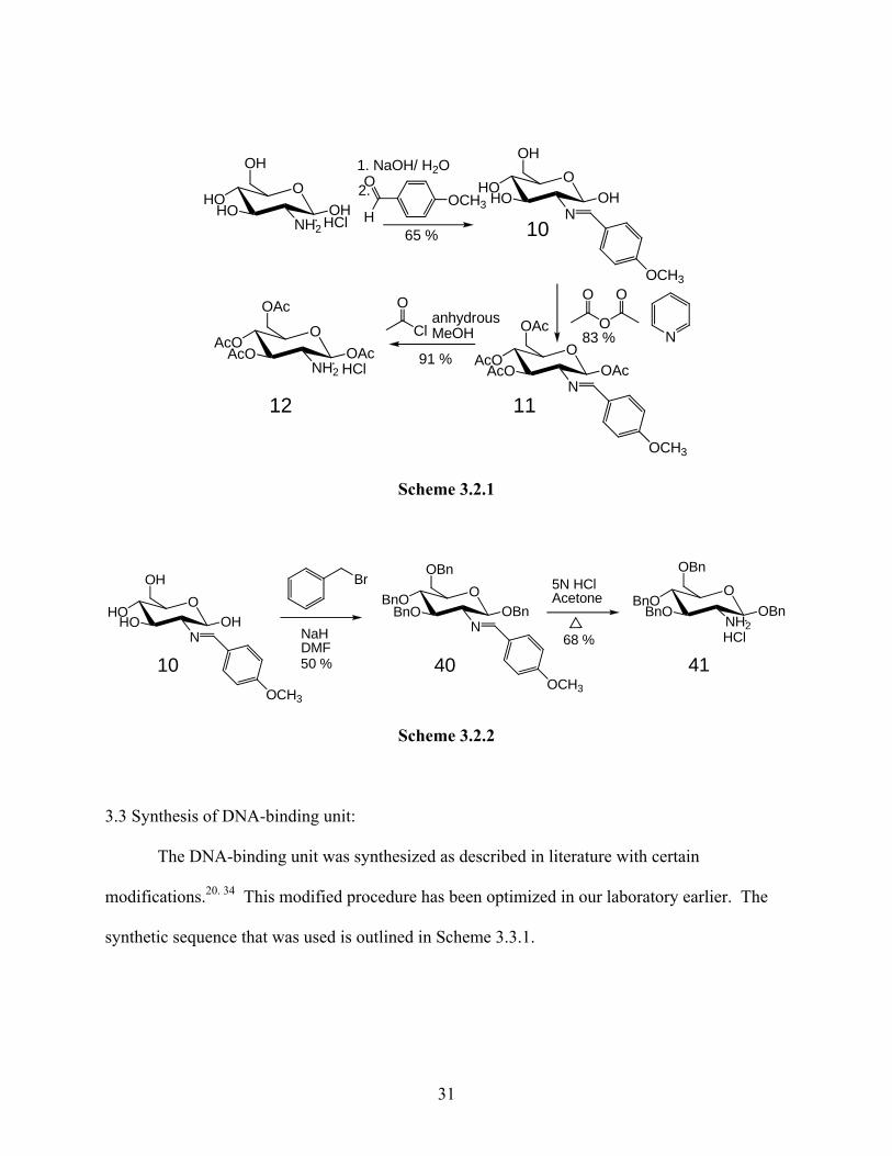
3.2. Synthesis of cell targeting ligand....................................................................30
3.3. Synthesis of DNA-binding unit ......................................................................31
3.4. Assembly of DNA-binding unit and cell targeting unit..................................33
CHAPTER 4: SYNTHESIS OF ANALOG COMPOUNDS.............................................47
4.1. Significance.....................................................................................................48
4.2. Synthesis of stable analogs for DNA-binding experiments............................50
iii
4.3. Synthesis of model compounds for cell transport studies...............................54
CHAPTER 5: EXPERIMENTAL......................................................................................60
5.1. General............................................................................................................61
5.2. Synthesis .........................................................................................................62
CHAPTER 6: RESULTS AND DISCUSSION...............................................................101
REFERENCES ................................................................................................................112
APPENDIX......................................................................................................................118
iv
ABSTRACT
This project involves synthesizing compounds that are capable of producing cytotoxic
N3-methyladenine DNA adducts in pancreatic β-cells. The compounds will consist of three
units: 1) A cell targeting ligand, glucose, which is specific to the insulin producing pancreatic β-
cells, 2) a DNA methylating unit, Me-lex, which has been shown to selectively make N3-
methyladenine adducts, and 3) linker component which will connect the two units previously
mentioned. The linker portion is critical in the binding of the compounds to DNA as well as the
ability of the compounds to be transported into the pancreatic β-cells. Several stable analog
compounds were synthesized to test the ability of the compounds to be transported selectively
into pancreatic β-cells and test their binding to DNA.
v
ACKNOWLEGEMENTS
I would first like to thank my mentor Dr. Sridhar Varadarajan for his guidance and
encouragement through out my undergraduate and graduate career. He has exposed and
educated me to a great deal of organic chemistry that I would have never of learned by reading a
book or taking a class. Because of him, I know that I am a going to be a better scientist and
person after I leave UNCW. I hope to make him proud by continuing to grow as a scientist and
pushing myself to be the best I can be.
I would also like to thank my committee members: Dr. John Tyrell and Dr. Pamela
Seaton for their support and guidance. Whenever I had a question you both were willing to help
me, which I greatly appreciated. I would like to thank Dr. Jeffrey Wright for his continued
support as an undergraduate and graduate student. I would like to thank Dr. Jeremy Morgan for
his help and guidance in the lab as well as introducing me to new chemistry. All of you are
wonderful people and I will never forget how you helped me during my collegiate years.
Last but not least, I would like to thank my family. We have been through a lot as a
family and you were all very understanding of how important my research was to me. I know at
times I seemed stressed, but you all were there guaranteeing me that one day it will pay off and
that I will succeed. This thesis proves that I have done just that and I thank you for pushing me
to the very end.
vi
LIST OF TABLES
Table Page
3.1 Comparison of reaction conditions for removal of acetyl groups on glucose ...........40
vii
LIST OF FIGURES
Figure Page
1.1 Structure of Streptozotocin (STZ) .............................................................................4
2.1 Structure of B-DNA ..................................................................................................8
2.2 Hydrogen bonding interactions between the B-DNA base pairs .............................10 2.3 Methylation/alkylation sites in the major and minor groove of DNA are indicated by arrows.......................................................................................................................12 2.4 a) Structure of Me-lex. b) Computational model showing Me-lex bound within the minor groove of
DNA...................................................................................................................15 2.5 a) Me-lex’s hydrogen bonding interactions with adenine in the minor groove. b) Steric hindrance between Me-lex and guanine in the minor groove ..................16 2.6 DNA-methylation at A/T rich regions in the minor groove of DNA ......................18
2.7 Structure of Streptozotocin (STZ) ...........................................................................21 2.8 a) Chlorozotocin is a small molecule that can be transported by glucose transporters b) Structure of Pyro-2DG........................................................................................23 2.9 Design of molecules for this project ........................................................................25 3.1 Synthetic strategy.....................................................................................................29 3.2 a) DNA-binding unit 15 functionalized as a alkene-carboxylic acid. b) Acetyl protected glucosamine hydrochloride, 12. c) Benzyl protected glucosamine hydrochloride, 41...............................................34 3.3 a) Model compound for acetyl removal..................................................................38 3.4 a) Anomeric forms of compound 27. b) 1H-NMR of both anomers...................................................................................42 4.1 Compounds designed for: a) DNA-methylation studies. b) DNA-binding studies. c) Cell transport studies with NBD d) Cell transport studies with coumarin ..................................................................49
viii

4.2 Compounds for cell transport studies a) NBD. b) Coumarin ............................................................................................................55 6.1 Structure of compounds being synthesized for this project ...................................103 6.2 Structure of compound 5........................................................................................105 6.3 Molecular mechanics model of methyl sulfonate bound to minor groove of DNA at A/T rich regions.............................................................................................................110
ix

CHAPTER 1: INTRODUCTION
DNA-alkylating drugs are the earliest and most common treatment for cancer
chemotherapy. DNA-alkylating drugs work by damaging (alkylating) DNA, thereby killing
cells.1,2 Normal cells are able to overcome the damage caused by DNA-alkylating agents by
going into cell cycle arrest until the damage gets repaired. Cancer cells on the other hand divide
rapidly and so the DNA damage in these cells does not get repaired. Therefore cancer cells
succumb to the treatment while normal cells do not. But, some normal cells like hair cells, and
cells of the gastric lining normally divide rapidly (like cancer cells) and therefore get destroyed
as a result of this treatment.2 This is the reason why cancer patients receiving the DNA
alkylating drugs for treatment often lose their hair and develop gastric problems. These side
effects result because DNA-alkylating drugs cannot be targeted only to cancer cells.
A more serious problem with DNA-alkylating drugs is that they can cause mutations.
Mutations are a result of the wide spread damage inflicted on DNA. These mutations can
develop into a secondary cancer, which is a serious side effect for cancer chemotherapy
patients.2,3,4 It would be very advantageous to develop new drugs that do not cause mutations
and lead only to cell death (cytotoxicity).
Me-lex, a molecule well described in literature5-8 is a DNA-alkylating (methylating)
compound that produces only one kind of damage to DNA. This particular damage leads to cell
death and not to mutations, due to the formation of a DNA adduct known as N3-methyladenine
(3-MeA).1 Therefore, Me-lex could be used to destroy cancer cells. However, Me-lex has no
cell specificity. If Me-lex could be targeted to a particular cell (cancer cells) it would be possible
to destroy those cells without mutations. The ability to target a specific cell requires a cell-
specific ligand to be attached to Me-lex.
2
In order to target a cell, a cell-specific ligand has to be identified. The cell-targeting
ligand has to be easy to synthesize, and should be able to maintain its cell-targeting ability upon
modification. Streptozotocin (STZ), Figure 1, is a potent DNA-alkylating drug that targets the
insulin producing pancreatic β-cells because of its specific transport through the GLUT-2
glucose transporter.9-10 The glucose unit in STZ is believed to be responsible for its selective
transport. The high levels of the GLUT-2 transporter on the surface of pancreatic β-cells makes
STZ very toxic to these cells.11 Streptozotocin was developed to study diabetes in mice by
analyzing the sequence of events in pancreatic β-cell destruction (type-1 diabetes).12-15 There is
evidence that large molecules, such as porphoryin rings connected to glucose can be transported
by glucose transporters into the pancreatic β-cell.16 Therefore it is likely that Me-lex attached to
glucose would be transported into the pancreatic β-cells too.
3
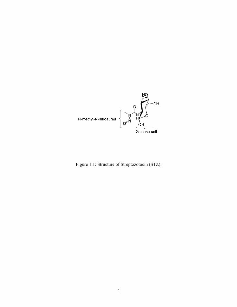
Figure 1.1: Structure of Streptozotocin (STZ).
4
The ability to target Me-lex to pancreatic β-cells by attaching it to glucose will provide
proof that this strategy can be used to destroy unwanted cells by targeting Me-lex to those cells.
An added advantage of targeting Me-lex to the pancreatic β-cells is that it could be used as a new
model for studying type-1 diabetes.11-13 STZ is currently used to induce diabetes in animals.
However, due to the extensive DNA damage, and consequent mutations caused by STZ, animals
treated with STZ develop tumors. DNA damage caused by Me-lex would not cause such tumors.
The goal of this thesis is to synthesize new compounds that incorporate the pancreatic β-
cells-targeting abilities of glucose and the selective DNA-methylating abilities of Me-lex into a
single molecule. Such molecules would have the ability to selectively deliver cytotoxic damage
only to pancreatic β-cells.
5
CHAPTER 2: BACKGROUND
2.1. Background:
The molecules designed and synthesized for this project should be able to target the
pancreatic β-cells via the GLUT-2 cells, bind to the minor groove of DNA at A/T rich regions
within those cells, and methylate the N3 position of adenine on DNA. Therefore designing
compounds that have these three properties requires a good understanding of DNA structure and
reactivity, and of the characteristics that enable transport through the GLUT-2 glucose
transporter.
2.1.1. DNA Structure and Reactivity:
DNA (deoxyribose nucleic acid) is composed of two strands held together through
hydrogen bonding. Each strand has a phosphate, sugar, and base. The phosphate and sugar form
the backbone. The bases are adenine (A), thymine (T), guanine (G) and cytosine (C). Hydrogen
bonding between the bases brings the two complementary strands together to form a double
helix; A and T are paired together by two hydrogen bonds and G and C are paired together by
three hydrogen bonds. Figure 2.1 shows the structure of B-DNA. This double helix is a right-
handed spiral which results in two distinct grooves, the minor groove and the major groove. The
minor groove is deep and narrow and therefore sites within the minor groove are more difficult
to access. The major groove is wide and shallow making the sites within the major groove easier
to access. Typically, most proteins and enzymes interact with DNA in the major groove.
7
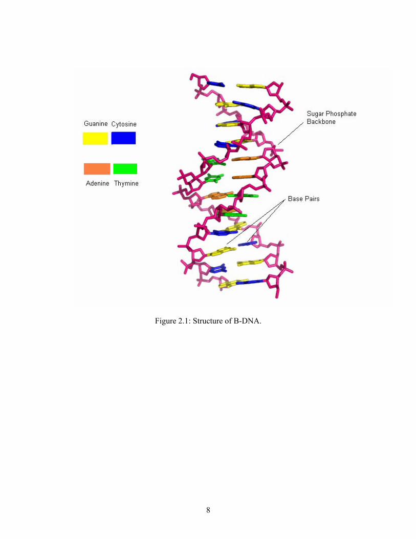
Figure 2.1: Structure of B-DNA.
8
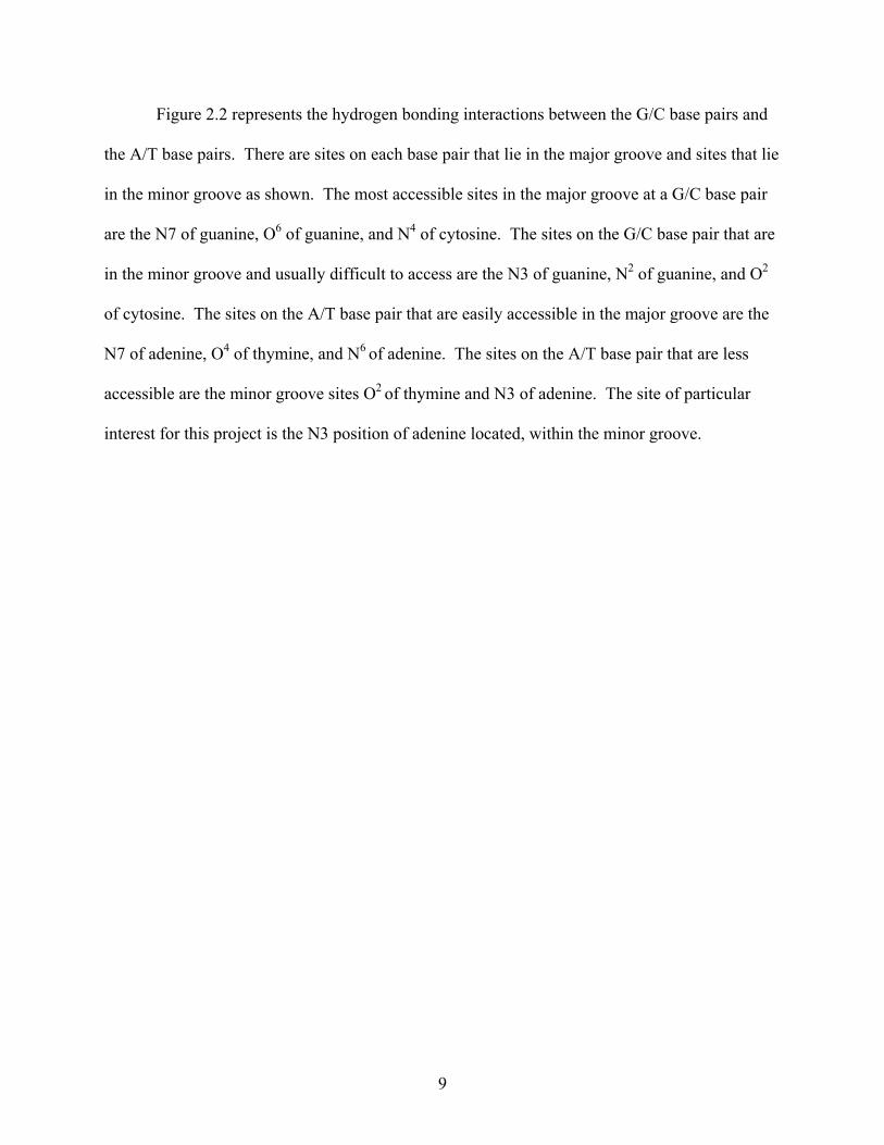
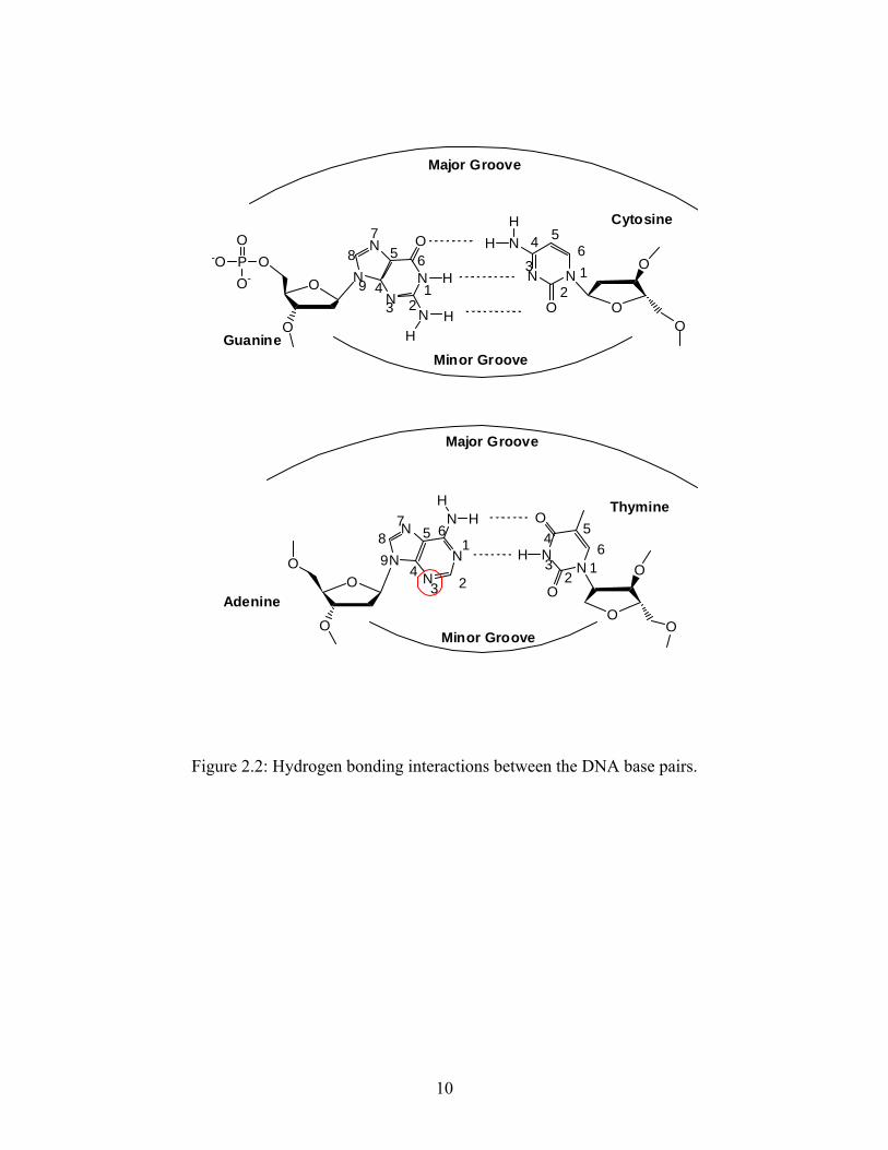
Figure 2.2 represents the hydrogen bonding interactions between the G/C base pairs and
the A/T base pairs. There are sites on each base pair that lie in the major groove and sites that lie
in the minor groove as shown. The most accessible sites in the major groove at a G/C base pair
are the N7 of guanine, O6 of guanine, and N4 of cytosine. The sites on the G/C base pair that are
in the minor groove and usually difficult to access are the N3 of guanine, N2 of guanine, and O2
of cytosine. The sites on the A/T base pair that are easily accessible in the major groove are the
N7 of adenine, O4 of thymine, and N6 of adenine. The sites on the A/T base pair that are less
accessible are the minor groove sites O2 of thymine and N3 of adenine. The site of particular
interest for this project is the N3 position of adenine located, within the minor groove.
9
NN
N O
N
NOOP-O
O
O- H
HH
Guanine
NN
N
NN
O
N N
O
NN
O
O
O
N
O
HH
O
O
O
Major Groove
Minor Groove
Cytosine
Major Groove
123
49
87
5 616
54
3
2
O
O
HH
HO
OMinor Groove
Adenine
Thymine
3 2
165
789
4 12
3
45
6
Figure 2.2: Hydrogen bonding interactions between the DNA base pairs.
10
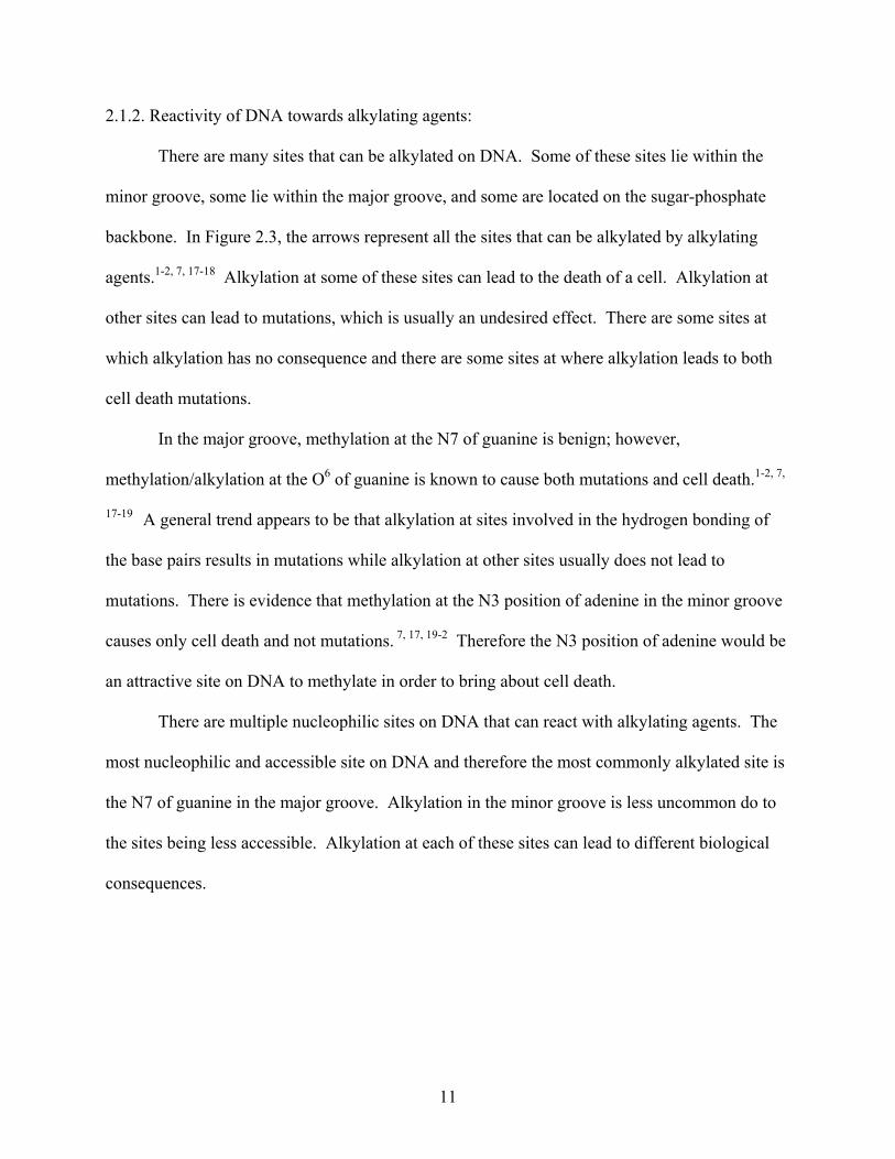
2.1.2. Reactivity of DNA towards alkylating agents:
There are many sites that can be alkylated on DNA. Some of these sites lie within the
minor groove, some lie within the major groove, and some are located on the sugar-phosphate
backbone. In Figure 2.3, the arrows represent all the sites that can be alkylated by alkylating
agents.1-2, 7, 17-18 Alkylation at some of these sites can lead to the death of a cell. Alkylation at
other sites can lead to mutations, which is usually an undesired effect. There are some sites at
which alkylation has no consequence and there are some sites at where alkylation leads to both
cell death mutations.
In the major groove, methylation at the N7 of guanine is benign; however,
methylation/alkylation at the O6 of guanine is known to cause both mutations and cell death.1-2, 7,
17-19 A general trend appears to be that alkylation at sites involved in the hydrogen bonding of
the base pairs results in mutations while alkylation at other sites usually does not lead to
mutations. There is evidence that methylation at the N3 position of adenine in the minor groove
causes only cell death and not mutations. 7, 17, 19-2 Therefore the N3 position of adenine would be
an attractive site on DNA to methylate in order to bring about cell death.
There are multiple nucleophilic sites on DNA that can react with alkylating agents. The
most nucleophilic and accessible site on DNA and therefore the most commonly alkylated site is
the N7 of guanine in the major groove. Alkylation in the minor groove is less uncommon do to
the sites being less accessible. Alkylation at each of these sites can lead to different biological
consequences.
11
Figure 2.3: Methylation/alkylation sites in the major and minor groove of DNA are
indicated by arrows.
12
2.1.3. Biological consequences of DNA methylation:
The biological consequences of DNA alkylation depends upon the site of alkylation and
the alkylating unit. Methylation at the N7 of guanine in the major groove appears to have no
biological consequence.1, 7, 17-18 The O6 of guanine, also in the major groove, and involved in
hydrogen bonding interactions with cytosine is another commonly methylated site. Methylation
at this site can result in both mutations and cell-toxicity.1, 7, 17-18
Methylation at other sites that can result in mutations are the N2 of guanine, the O2 and O4
of thymine, and O2 of cytosine.1, 7, 17 However, methylation at the N3 site of adenine, in the
minor groove, has shown to result in cytotoxicity, without leading to mutations.7, 17, 19-21 Thus
compounds that can exclusively make 3-MeA adducts can be used to kill cells without any risk
of mutations.
13
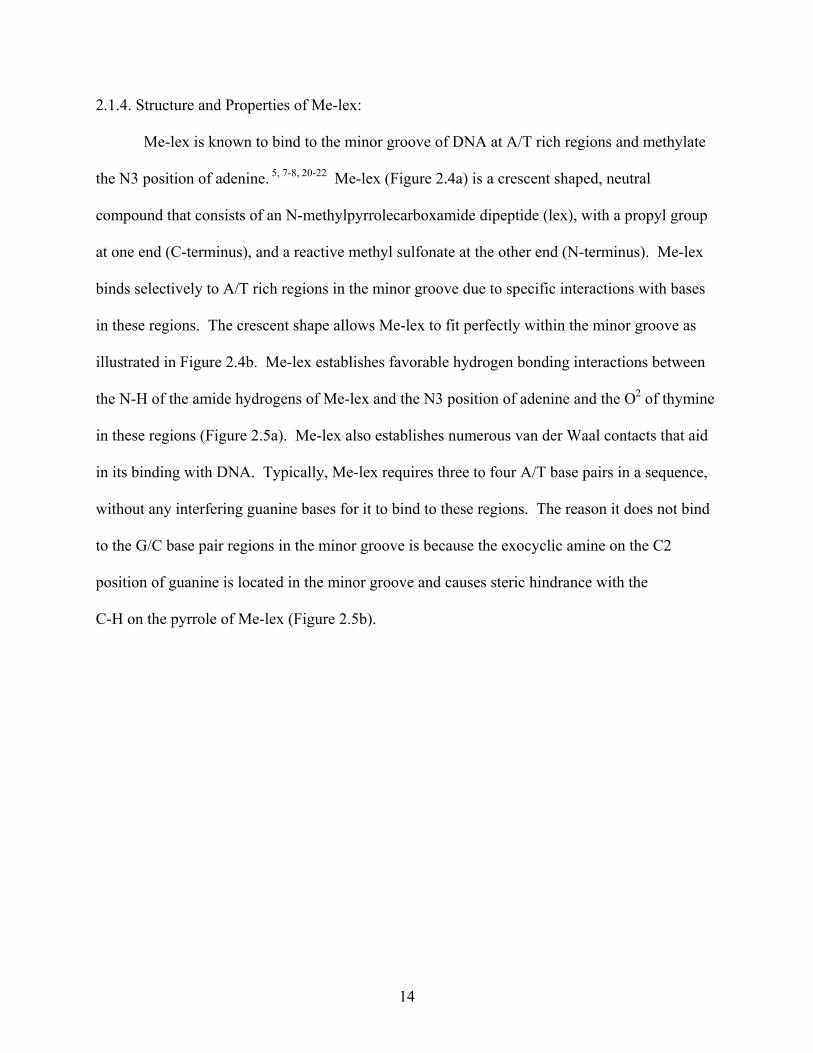
2.1.4. Structure and Properties of Me-lex:
Me-lex is known to bind to the minor groove of DNA at A/T rich regions and methylate
the N3 position of adenine. 5, 7-8, 20-22 Me-lex (Figure 2.4a) is a crescent shaped, neutral
compound that consists of an N-methylpyrrolecarboxamide dipeptide (lex), with a propyl group
at one end (C-terminus), and a reactive methyl sulfonate at the other end (N-terminus). Me-lex
binds selectively to A/T rich regions in the minor groove due to specific interactions with bases
in these regions. The crescent shape allows Me-lex to fit perfectly within the minor groove as
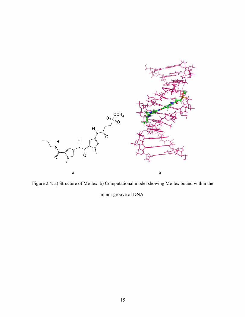
illustrated in Figure 2.4b. Me-lex establishes favorable hydrogen bonding interactions between
the N-H of the amide hydrogens of Me-lex and the N3 position of adenine and the O2 of thymine
in these regions (Figure 2.5a). Me-lex also establishes numerous van der Waal contacts that aid
in its binding with DNA. Typically, Me-lex requires three to four A/T base pairs in a sequence,
without any interfering guanine bases for it to bind to these regions. The reason it does not bind
to the G/C base pair regions in the minor groove is because the exocyclic amine on the C2
position of guanine is located in the minor groove and causes steric hindrance with the
C-H on the pyrrole of Me-lex (Figure 2.5b).
14
a b
Figure 2.4: a) Structure of Me-lex. b) Computational model showing Me-lex bound within the
minor groove of DNA.
15
a)N
NHN
NNH2
1
23
4
5 678
9
N O
H
N
O
HHydrogenbondinginteraction
NH
NNH
N
O
N
N ON
O
HH H
H StericHindrance
1
2
34
5 678
9
b)
Figure 2.5: a) Me-lex’s hydrogen bonding interactions with adenine in the minor groove.
b) Steric hindrance between Me-lex and guanine in the minor groove.
16
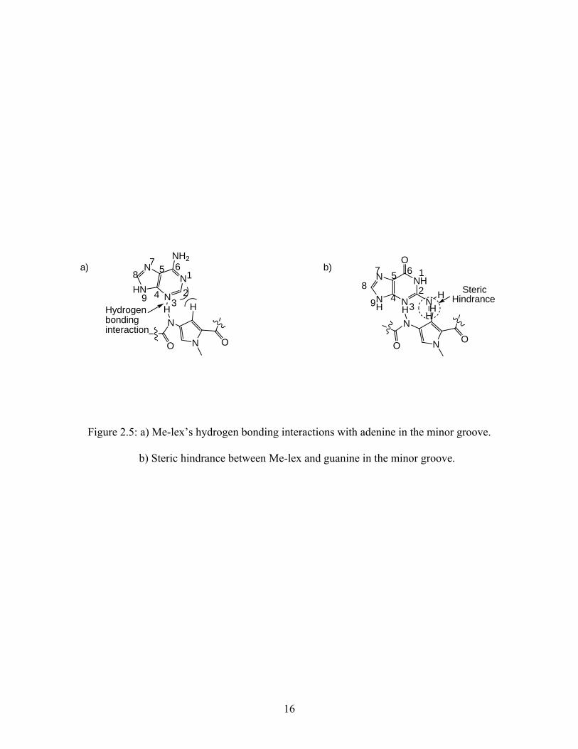
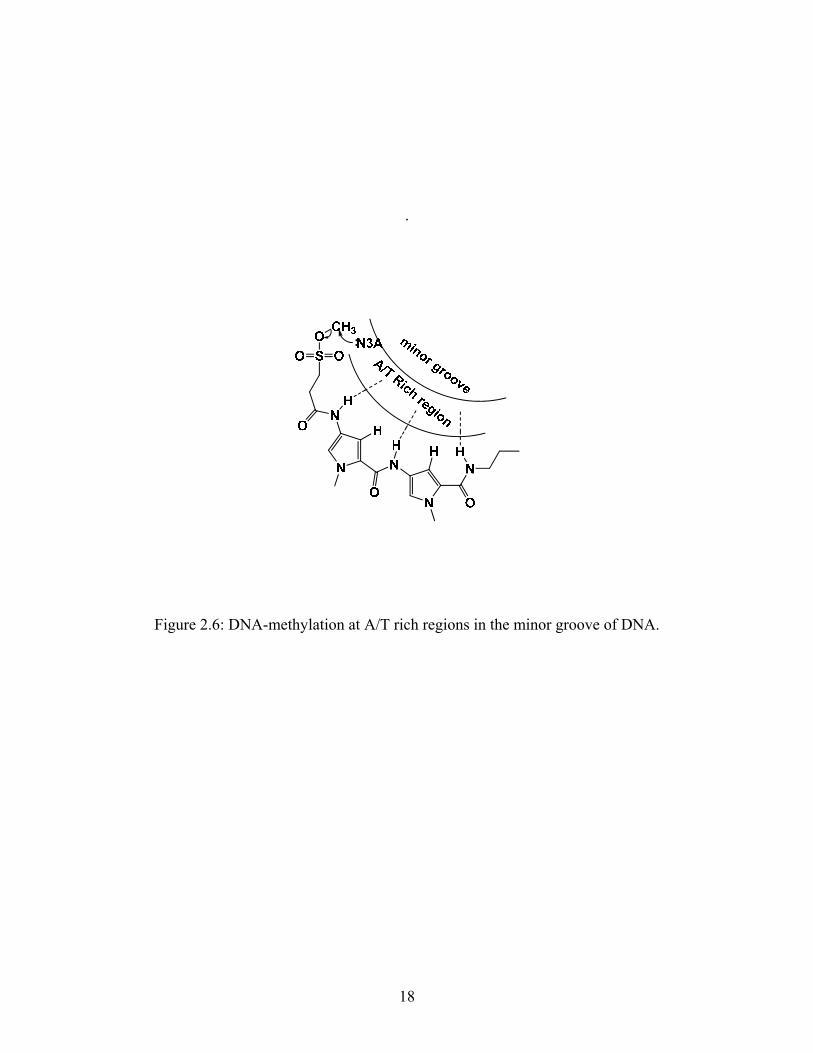
When Me-lex binds selectively to the A/T rich regions in the minor groove; the molecule
places the reactive methyl sulfonate group at these A/T rich regions as shown in Figure 2.6. In
these regions the N3 position of adenine is the most nucleophilic site. Therefore, Me-lex
exclusively methylates DNA at this site; resulting in > 95 % of the methylated DNA-adducts
formed by Me-lex being 3-MeA adducts.5-8 Once the reactive methyl group is transferred to the
N3 position of adenine, the resulting sulfonate anion with a negative charge, gets eliminated
from DNA due to electrostatic repulsion with the negatively charged phosphate backbone of
DNA. 7, 39 Thus, this molecule functions as an efficient delivery agent for the methyl group and
does not have any further biological consequences of its own by staying associated with the
DNA.
17
.
Figure 2.6: DNA-methylation at A/T rich regions in the minor groove of DNA.
18
2.1.5. Biological consequences of 3-MeA formation by Me-lex:
The 3-MeA adducts formed by Me-lex have been shown to be cytotoxic in yeast, E. coli,
and mammalian cells.19-23 It has also been shown that the extent of cell death correlates to the
levels of 3-MeA adducts formed. 19-23 The 3-MeA adducts formed by Me-lex have also been
shown to be non-mutagenic. While Me-lex has attractive properties in terms of cell toxicity, its
development into a useful drug is limited by the fact that it has no cell specificity. The ability to
deliver Me-lex to a particular cell would aid in the development of drugs for cancer
chemotherapy. The goal of this project is to develop new compounds which can use the
selective DNA-methylating property of Me-lex and deliver it to a cell of interest.
19
2.1.6. DNA methylation of Streptozotocin:
Streptozotocin (STZ, Figure 2.7) is a potent DNA-methylating agent that is known to
methylate DNA at multiple sites and has been shown to have selective pancreatic β-cell
toxicity.9-11, 13-15 STZ has a glucose unit to which it is attached to a N-methyl-N-nitrosurea unit
The nitrosurea unit methylates the DNA and it is believed that the glucose unit is responsible for
the ability of STZ to target pancreatic β-cells due to selective transport through the low affinity
glucose transporter, GLUT-2, that is present on the surface of the pancreatic β-cells.24, 25, 27
Methylation by STZ occurs at multiple sites on DNA. The sites that can be methylated
by STZ include: N7 of guanine, O6 of guanine, N7 of adenine, and N3 of adenine.9 Over 70 % of
the DNA adducts formed by STZ is N7-methylguanine, which is a begin adduct. 1,7, 9, 17, 18 STZ
also forms the O6 –methylguanine adduct in the major groove, which is known to cause both
mutations and cell death.1, 7, 17, 18 The formation of O6-MeG adduct by STZ is believed to result
in tumors. It is likely that STZ kills pancreatic β-cells due to the formation of 3-MeA adducts
and O6- MeG.7, 14
STZ has been used for studying type-1 diabetes by killing pancreatic β-cells in rodent
(mice) models.12, 14 However, in these cases the study of type-1 diabetes is complicated by the
fact that STZ causes extensive damage to DNA at multiple sites resulting in mutations which
cause tumors.
20
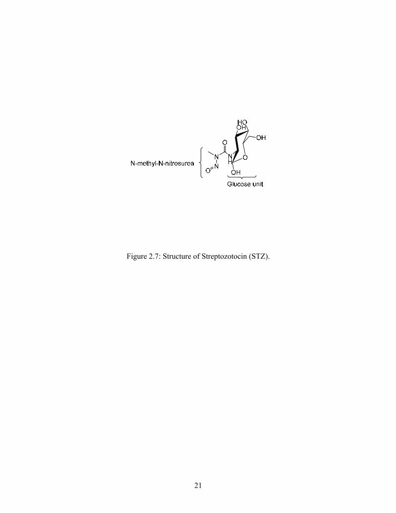
Figure 2.7: Structure of Streptozotocin (STZ).
21
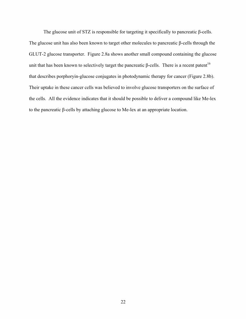
The glucose unit of STZ is responsible for targeting it specifically to pancreatic β-cells.
The glucose unit has also been known to target other molecules to pancreatic β-cells through the
GLUT-2 glucose transporter. Figure 2.8a shows another small compound containing the glucose
unit that has been known to selectively target the pancreatic β-cells. There is a recent patent16
that describes porphoryin-glucose conjugates in photodynamic therapy for cancer (Figure 2.8b).
Their uptake in these cancer cells was believed to involve glucose transporters on the surface of
the cells. All the evidence indicates that it should be possible to deliver a compound like Me-lex
to the pancreatic β-cells by attaching glucose to Me-lex at an appropriate location.
22
a)
NN
O
O
HOOH
NH
OH
OH
O
Chlorozotocin (CTZ)
Cl
b)
Figure 2.8: a) Chlorozotocin is a small molecule that can be transported by glucose transporters.
b) Structure of Pyro-2DG.
23
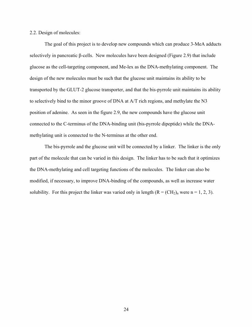
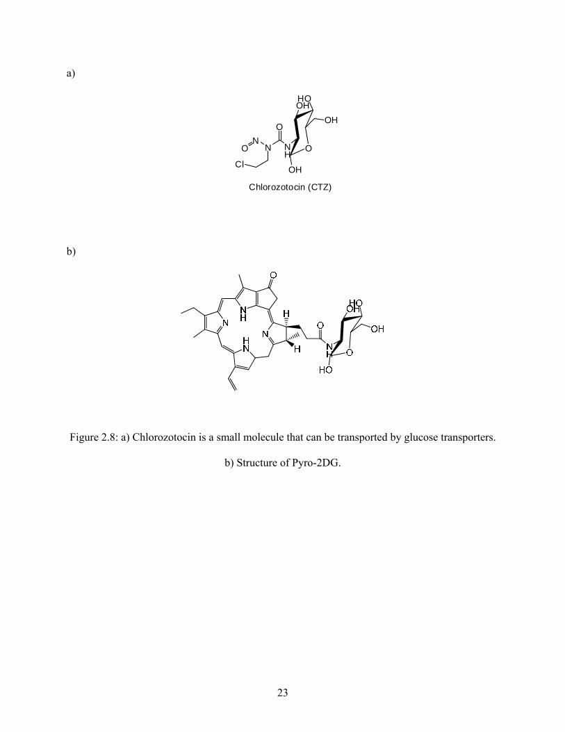
2.2. Design of molecules:
The goal of this project is to develop new compounds which can produce 3-MeA adducts
selectively in pancreatic β-cells. New molecules have been designed (Figure 2.9) that include
glucose as the cell-targeting component, and Me-lex as the DNA-methylating component. The
design of the new molecules must be such that the glucose unit maintains its ability to be
transported by the GLUT-2 glucose transporter, and that the bis-pyrrole unit maintains its ability
to selectively bind to the minor groove of DNA at A/T rich regions, and methylate the N3
position of adenine. As seen in the figure 2.9, the new compounds have the glucose unit
connected to the C-terminus of the DNA-binding unit (bis-pyrrole dipeptide) while the DNA-
methylating unit is connected to the N-terminus at the other end.
The bis-pyrrole and the glucose unit will be connected by a linker. The linker is the only
part of the molecule that can be varied in this design. The linker has to be such that it optimizes
the DNA-methylating and cell targeting functions of the molecules. The linker can also be
modified, if necessary, to improve DNA-binding of the compounds, as well as increase water
solubility. For this project the linker was varied only in length (R = (CH2)n were n = 1, 2, 3).
24
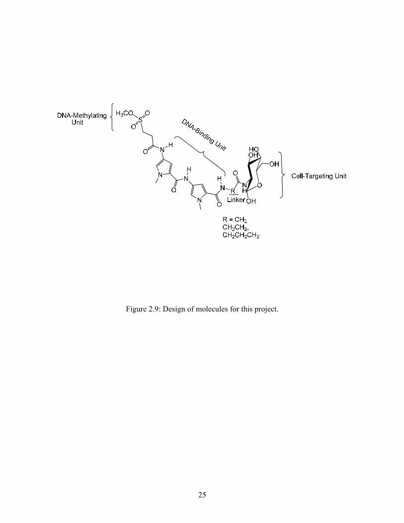
Figure 2.9: Design of molecules for this project.
25
CHAPTER 3: SYNTHESIS OF FINAL COMPOUNDS
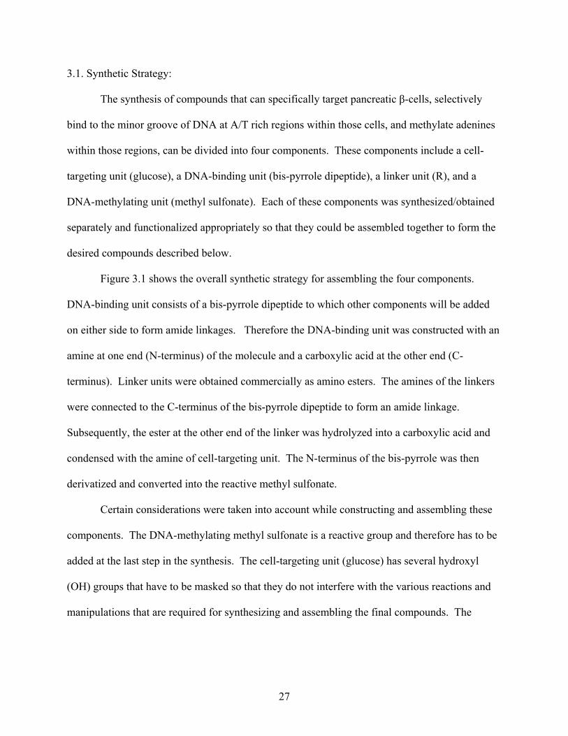
3.1. Synthetic Strategy:
The synthesis of compounds that can specifically target pancreatic β-cells, selectively
bind to the minor groove of DNA at A/T rich regions within those cells, and methylate adenines
within those regions, can be divided into four components. These components include a cell-
targeting unit (glucose), a DNA-binding unit (bis-pyrrole dipeptide), a linker unit (R), and a
DNA-methylating unit (methyl sulfonate). Each of these components was synthesized/obtained
separately and functionalized appropriately so that they could be assembled together to form the
desired compounds described below.
Figure 3.1 shows the overall synthetic strategy for assembling the four components.
DNA-binding unit consists of a bis-pyrrole dipeptide to which other components will be added
on either side to form amide linkages. Therefore the DNA-binding unit was constructed with an
amine at one end (N-terminus) of the molecule and a carboxylic acid at the other end (C-
terminus). Linker units were obtained commercially as amino esters. The amines of the linkers
were connected to the C-terminus of the bis-pyrrole dipeptide to form an amide linkage.
Subsequently, the ester at the other end of the linker was hydrolyzed into a carboxylic acid and
condensed with the amine of cell-targeting unit. The N-terminus of the bis-pyrrole was then
derivatized and converted into the reactive methyl sulfonate.
Certain considerations were taken into account while constructing and assembling these
components. The DNA-methylating methyl sulfonate is a reactive group and therefore has to be
added at the last step in the synthesis. The cell-targeting unit (glucose) has several hydroxyl
(OH) groups that have to be masked so that they do not interfere with the various reactions and
manipulations that are required for synthesizing and assembling the final compounds. The
27

hydroxyl groups would be regenerated just before the introduction of the reactive methyl
sulfonate.
28
Figure 3.1: Synthetic strategy.
29
3.2. Synthesis of cell targeting ligand:
The cell-targeting ligand, glucose, has four nucleophilic OH groups and one nucleophilic
amine group. The amine of glucose has to be condensed with the carboxylic acid at the C-
terminus of the DNA-binding unit. In order to prevent interference from the OH groups during
the condensation they have be protected first. Two different protecting groups were considered.
Protection with acetyl groups can be easily accomplished in high yields using acetic anhydride,
and the resulting compounds are easily purified and characterized. Subsequently, deprotection
can be achieved using basic conditions, but it is also possible to deprotect the acetyl groups using
strong acid. Therefore strongly acidic or basic conditions used during chemical manipulations
may result in premature deprotection of the OH groups. Protection with benzyl groups resulted
in lower yields than the acetyl protection, and resulted in compounds that are more difficult to
characterize by NMR due to the overwhelming number of aromatic hydrogens in the spectrum.
However, the benzyl groups are stable to acidic and basic conditions, can be removed in a single
hydrogentation step, and the only byproduct formed upon deprotection is toluene which can be
easily removed by rotary evaportation.
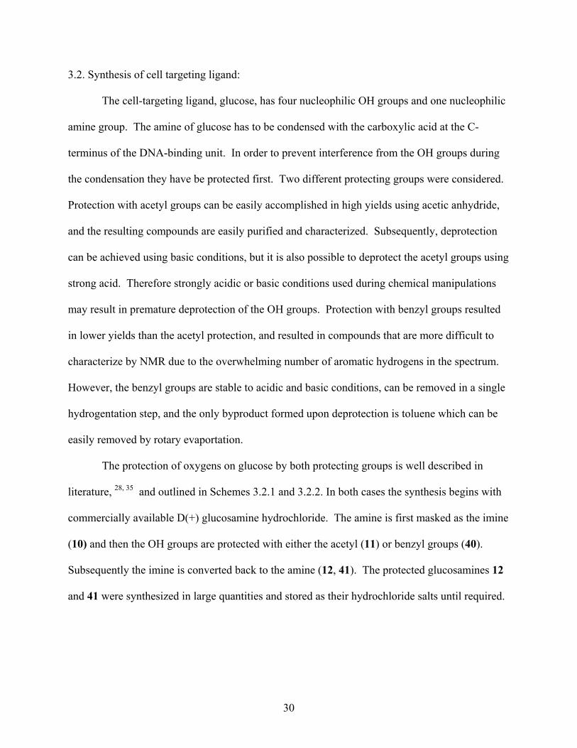
The protection of oxygens on glucose by both protecting groups is well described in
literature, 28, 35 and outlined in Schemes 3.2.1 and 3.2.2. In both cases the synthesis begins with
commercially available D(+) glucosamine hydrochloride. The amine is first masked as the imine
(10) and then the OH groups are protected with either the acetyl (11) or benzyl groups (40).
Subsequently the imine is converted back to the amine (12, 41). The protected glucosamines 12
and 41 were synthesized in large quantities and stored as their hydrochloride salts until required.
30
OHO
HONH2
OH
OH
HClOCH3
O
H
OHO
HON
OH
OH
OAcO
AcON
OAc
OAc
O
O
OO
ClOAcO
AcONH2
OAc
OAc
HCl
1. NaOH/ H2O
2.
anhydrousMeOH
65 %
83 %91 %
OCH3
OCH3
N
10
12 11
Scheme 3.2.1
OHO
HON
OH
OH
OCH3
10
Br
NaHDMF
OBnOBnO
NOBn
OBn
OCH3
40
OBnO
BnONH2
OBn
OBn
HCl
5N HClAcetone
4150 %
68 %
Scheme 3.2.2
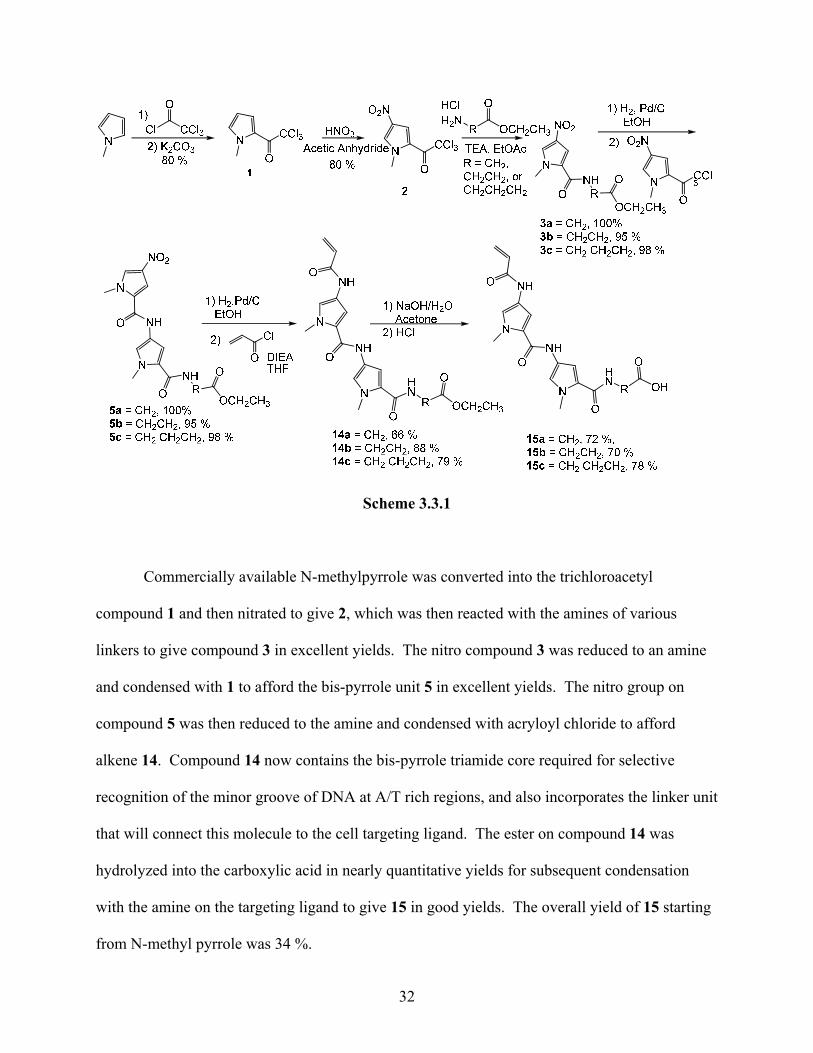
3.3 Synthesis of DNA-binding unit:
The DNA-binding unit was synthesized as described in literature with certain
modifications.20. 34 This modified procedure has been optimized in our laboratory earlier. The
synthetic sequence that was used is outlined in Scheme 3.3.1.
31
Scheme 3.3.1
Commercially available N-methylpyrrole was converted into the trichloroacetyl
compound 1 and then nitrated to give 2, which was then reacted with the amines of various
linkers to give compound 3 in excellent yields. The nitro compound 3 was reduced to an amine
and condensed with 1 to afford the bis-pyrrole unit 5 in excellent yields. The nitro group on
compound 5 was then reduced to the amine and condensed with acryloyl chloride to afford
alkene 14. Compound 14 now contains the bis-pyrrole triamide core required for selective
recognition of the minor groove of DNA at A/T rich regions, and also incorporates the linker unit
that will connect this molecule to the cell targeting ligand. The ester on compound 14 was
hydrolyzed into the carboxylic acid in nearly quantitative yields for subsequent condensation
with the amine on the targeting ligand to give 15 in good yields. The overall yield of 15 starting
from N-methyl pyrrole was 34 %.
32
3.4. Assembly of DNA-binding unit and cell targeting unit:
The DNA-binding unit in the form of compound 15 (Figure 3.2a), has an alkene at the N-
terminus that can be converted into the methyl sulfonate, and has a carboxylic acid at the C-
terminus which can be combined with the amines of 12 and 41 (Figure 3.2 b & c, respectively)
by forming amide linkages.
33
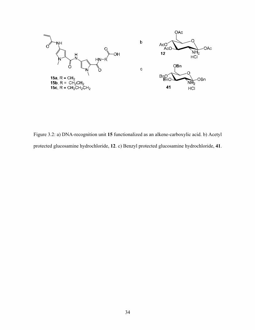
Figure 3.2: a) DNA-recognition unit 15 functionalized as an alkene-carboxylic acid. b) Acetyl
protected glucosamine hydrochloride, 12. c) Benzyl protected glucosamine hydrochloride, 41.
34
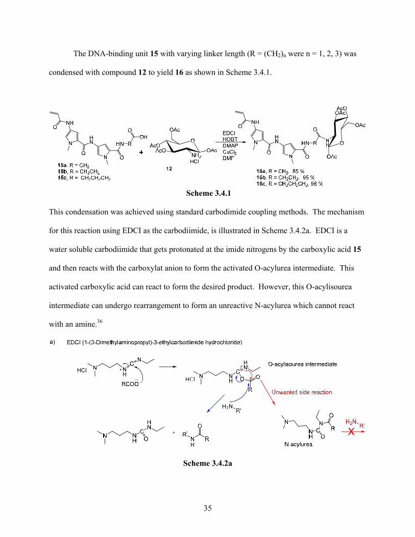
The DNA-binding unit 15 with varying linker length (R = (CH2)n were n = 1, 2, 3) was
condensed with compound 12 to yield 16 as shown in Scheme 3.4.1.
Scheme 3.4.1
This condensation was achieved using standard carbodimide coupling methods. The mechanism
for this reaction using EDCI as the carbodiimide, is illustrated in Scheme 3.4.2a. EDCI is a
water soluble carbodiimide that gets protonated at the imide nitrogens by the carboxylic acid 15
and then reacts with the carboxylat anion to form the activated O-acylurea intermediate. This
activated carboxylic acid can react to form the desired product. However, this O-acylisourea
intermediate can undergo rearrangement to form an unreactive N-acylurea which cannot react
with an amine.36
Scheme 3.4.2a
35
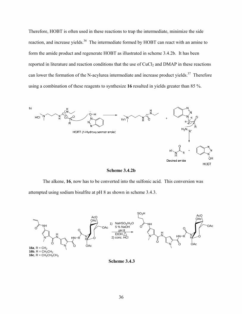
Therefore, HOBT is often used in these reactions to trap the intermediate, minimize the side
reaction, and increase yields.36 The intermediate formed by HOBT can react with an amine to
form the amide product and regenerate HOBT as illustrated in scheme 3.4.2b. It has been
reported in literature and reaction conditions that the use of CuCl2 and DMAP in these reactions
can lower the formation of the N-acylurea intermediate and increase product yields.37 Therefore
using a combination of these reagents to synthesize 16 resulted in yields greater than 85 %.
Scheme 3.4.2b
The alkene, 16, now has to be converted into the sulfonic acid. This conversion was
attempted using sodium bisulfite at pH 8 as shown in scheme 3.4.3.
N
N
NHO
O
HN
O
HN R
O
O
AcOOAc
NH
OAc
OAc
16a, R = CH216b, R = CH2CH216c, R = CH2CH2CH2
NaHSO3/H2O5 % NaOH
pH 8EtOH N
N
NHO
O
HN
O
HN R
O
O
AcOOAc
NH
OAc
OAc
SO3H
1)
2) conc. HCl
Scheme 3.4.3
36
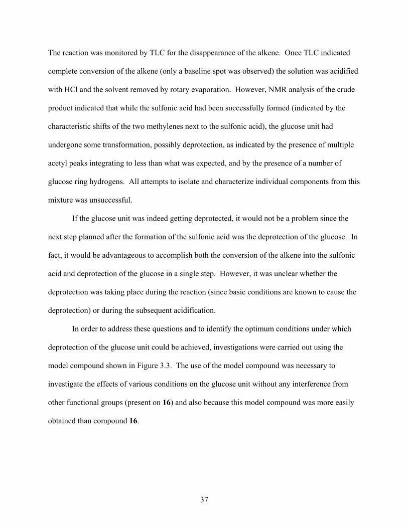
The reaction was monitored by TLC for the disappearance of the alkene. Once TLC indicated
complete conversion of the alkene (only a baseline spot was observed) the solution was acidified
with HCl and the solvent removed by rotary evaporation. However, NMR analysis of the crude
product indicated that while the sulfonic acid had been successfully formed (indicated by the
characteristic shifts of the two methylenes next to the sulfonic acid), the glucose unit had
undergone some transformation, possibly deprotection, as indicated by the presence of multiple
acetyl peaks integrating to less than what was expected, and by the presence of a number of
glucose ring hydrogens. All attempts to isolate and characterize individual components from this
mixture was unsuccessful.
If the glucose unit was indeed getting deprotected, it would not be a problem since the
next step planned after the formation of the sulfonic acid was the deprotection of the glucose. In
fact, it would be advantageous to accomplish both the conversion of the alkene into the sulfonic
acid and deprotection of the glucose in a single step. However, it was unclear whether the
deprotection was taking place during the reaction (since basic conditions are known to cause the
deprotection) or during the subsequent acidification.
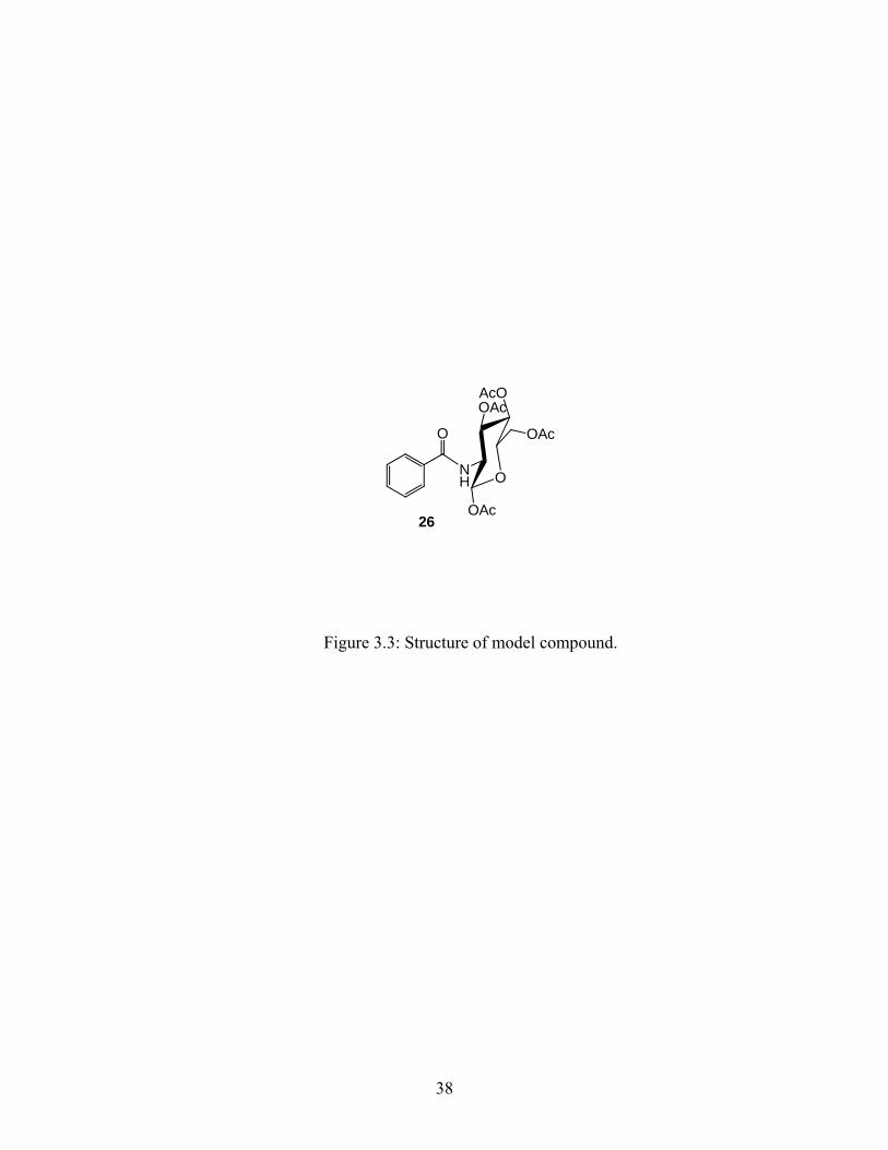
In order to address these questions and to identify the optimum conditions under which
deprotection of the glucose unit could be achieved, investigations were carried out using the
model compound shown in Figure 3.3. The use of the model compound was necessary to
investigate the effects of various conditions on the glucose unit without any interference from
other functional groups (present on 16) and also because this model compound was more easily
obtained than compound 16.
37
O
O
AcOOAc
NH
OAc
OAc
26
Figure 3.3: Structure of model compound.
38
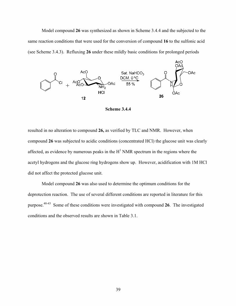
Model compound 26 was synthesized as shown in Scheme 3.4.4 and the subjected to the
same reaction conditions that were used for the conversion of compound 16 to the sulfonic acid
(see Scheme 3.4.3). Refluxing 26 under these mildly basic conditions for prolonged periods
Scheme 3.4.4
resulted in no alteration to compound 26, as verified by TLC and NMR. However, when
compound 26 was subjected to acidic conditions (concentrated HCl) the glucose unit was clearly
affected, as evidence by numerous peaks in the H1 NMR spectrum in the regions where the
acetyl hydrogens and the glucose ring hydrogens show up. However, acidification with 1M HCl
did not affect the protected glucose unit.
Model compound 26 was also used to determine the optimum conditions for the
deprotection reaction. The use of several different conditions are reported in literature for this
purpose.40-43 Some of these conditions were investigated with compound 26. The investigated
conditions and the observed results are shown in Table 3.1.
39
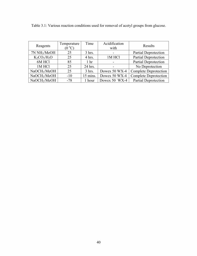
Table 3.1: Various reaction conditions used for removal of acetyl groups from glucose.
Reagents Temperature(0 oC)
Time
Acidification with Results
7N NH3/MeOH 25 3 hrs. - Partial Deprotection K2CO3/H2O 25 4 hrs. 1M HCl Partial Deprotection
6M HCl 85 1 hr - Partial Deprotection 1M HCl 25 24 hrs. - No Deprotection
NaOCH3/MeOH 25 3 hrs. Dowex 50 WX-4 Complete DeprotectionNaOCH3/MeOH -10 15 mins. Dowex 50 WX-4 Complete DeprotectionNaOCH3/MeOH -78 1 hour Dowex 50 WX-4 Partial Deprotection
40
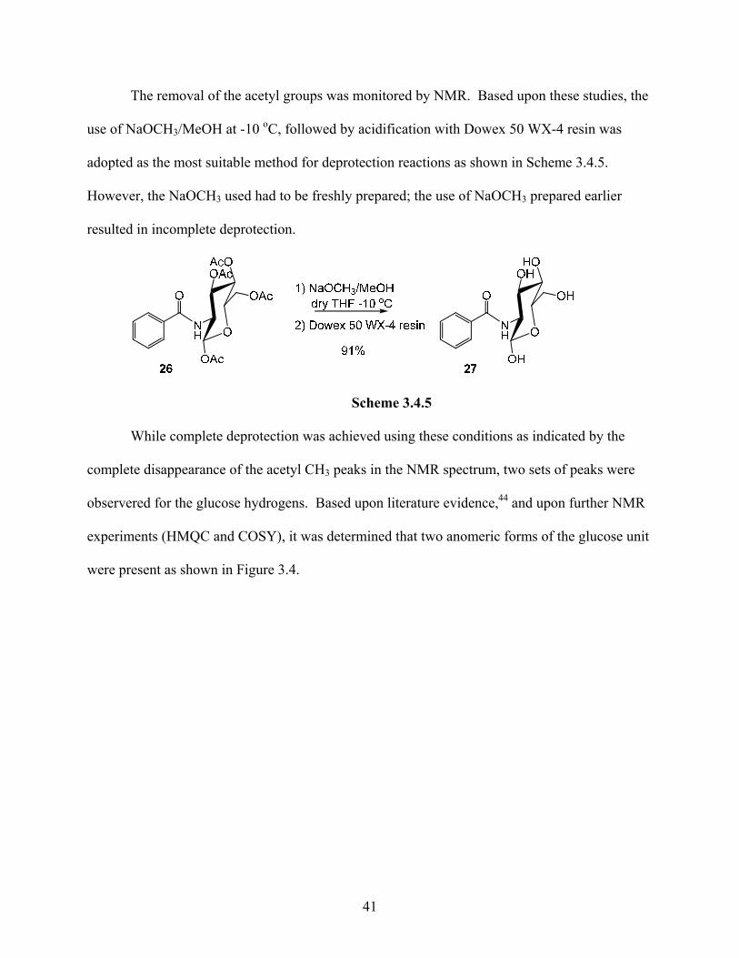
The removal of the acetyl groups was monitored by NMR. Based upon these studies, the
use of NaOCH3/MeOH at -10 oC, followed by acidification with Dowex 50 WX-4 resin was
adopted as the most suitable method for deprotection reactions as shown in Scheme 3.4.5.
However, the NaOCH3 used had to be freshly prepared; the use of NaOCH3 prepared earlier
resulted in incomplete deprotection.
Scheme 3.4.5
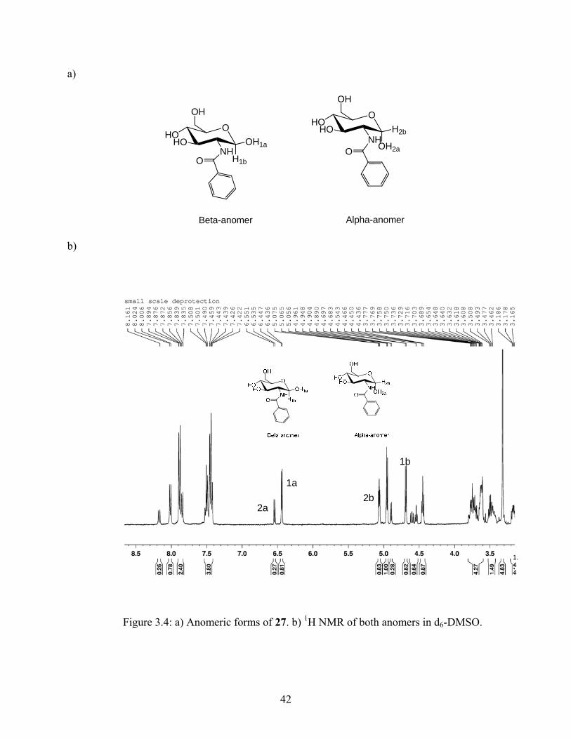
While complete deprotection was achieved using these conditions as indicated by the
complete disappearance of the acetyl CH3 peaks in the NMR spectrum, two sets of peaks were
observered for the glucose hydrogens. Based upon literature evidence,44 and upon further NMR
experiments (HMQC and COSY), it was determined that two anomeric forms of the glucose unit
were present as shown in Figure 3.4.
41
a)
O
Beta-anomer
O
Alpha-anomer
OHO
HO
H1bNH
OH1a
OH OHO
HO
OH2aNH
H2b
OH
b)
1.44
2a
1a2b
1b
Figure 3.4: a) Anomeric forms of 27. b) 1H NMR of both anomers in d6-DMSO.
42
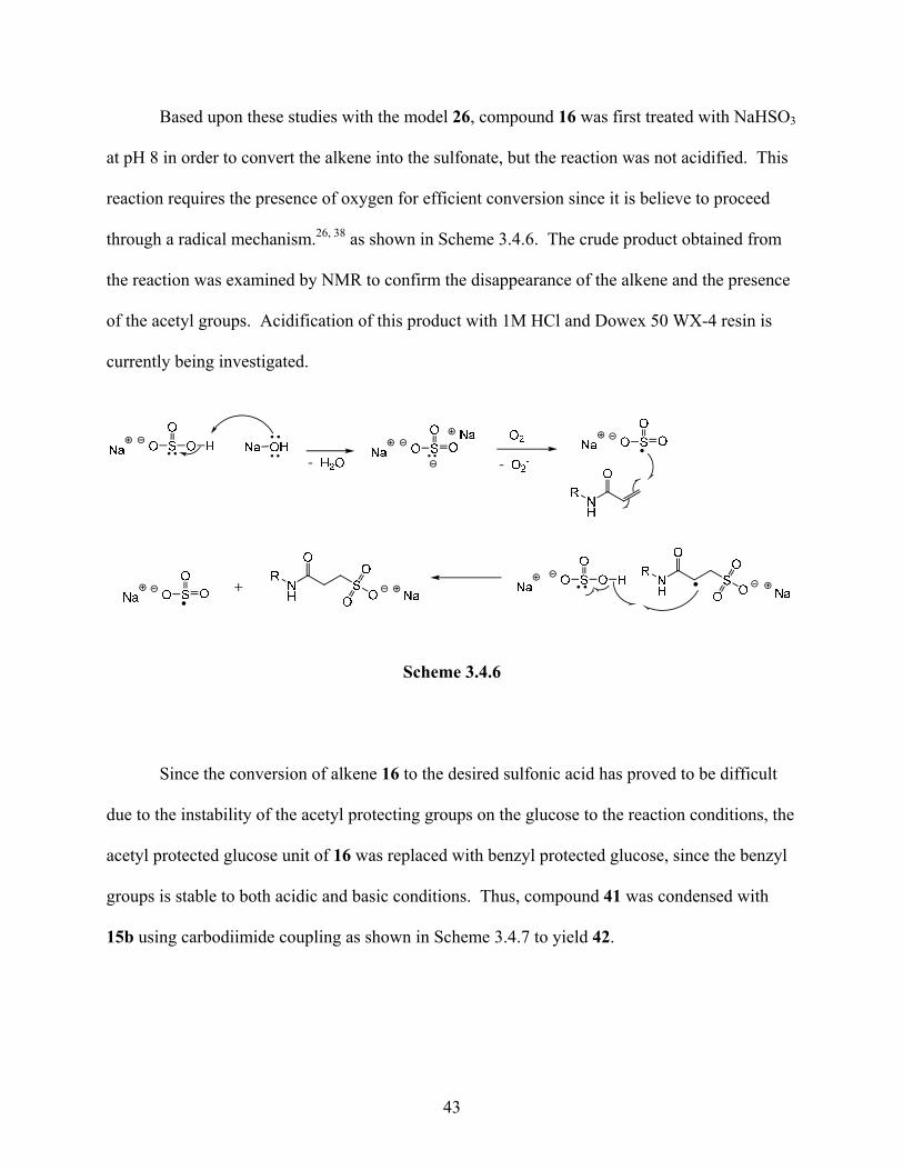
Based upon these studies with the model 26, compound 16 was first treated with NaHSO3
at pH 8 in order to convert the alkene into the sulfonate, but the reaction was not acidified. This
reaction requires the presence of oxygen for efficient conversion since it is believe to proceed
through a radical mechanism.26, 38 as shown in Scheme 3.4.6. The crude product obtained from
the reaction was examined by NMR to confirm the disappearance of the alkene and the presence
of the acetyl groups. Acidification of this product with 1M HCl and Dowex 50 WX-4 resin is
currently being investigated.
Scheme 3.4.6
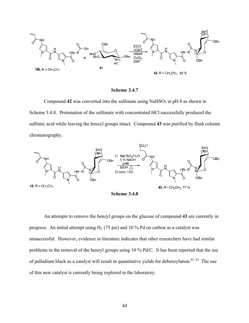
Since the conversion of alkene 16 to the desired sulfonic acid has proved to be difficult
due to the instability of the acetyl protecting groups on the glucose to the reaction conditions, the
acetyl protected glucose unit of 16 was replaced with benzyl protected glucose, since the benzyl
groups is stable to both acidic and basic conditions. Thus, compound 41 was condensed with
15b using carbodiimide coupling as shown in Scheme 3.4.7 to yield 42.
43
Scheme 3.4.7
Compound 42 was converted into the sulfonate using NaHSO3 at pH 8 as shown in
Scheme 3.4.8. Protonation of the sulfonate with concentrated HCl successfully produced the
sulfonic acid while leaving the benzyl groups intact. Compound 43 was purified by flash column
chromatography.
Scheme 3.4.8
An attempts to remove the benzyl groups on the glucose of compound 43 are currently in
progress. An initial attempt using H2 (75 psi) and 10 % Pd on carbon as a catalyst was
unsuccessful. However, evidence in literature indicates that other researchers have had similar
problems in the removal of the benzyl groups using 10 % Pd/C. It has been reported that the use
of palladium black as a catalyst will result in quantitative yields for debenzylation.41, 43 The use
of this new catalyst is currently being explored in the laboratory.
44
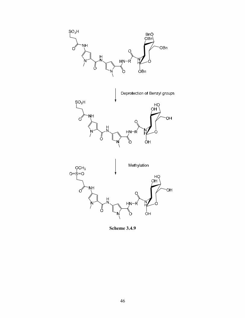
Once the benzyl groups can be successfully removed the next step is the methylation of
the sulfonic acid to the methyl sulfonate as shown in Scheme 3.4.9. This methylation will be
attempted using either trimethyl ortho formate or methyl-p-tolytriazine as the methylating agent.
It is known that these methylating agents can methylate sulfonic acids of similar compounds, and
it has been verified that they do not methylate the hydroxyl groups on the glucose. Once the
sulfonic acid is methylated the compounds will be used in experiments with DNA to characterize
its ability to methylate DNA.
45
Scheme 3.4.9
46

CHAPTER 4: SYNTHESIS OF STABLE ANALOG COMPOUNDS
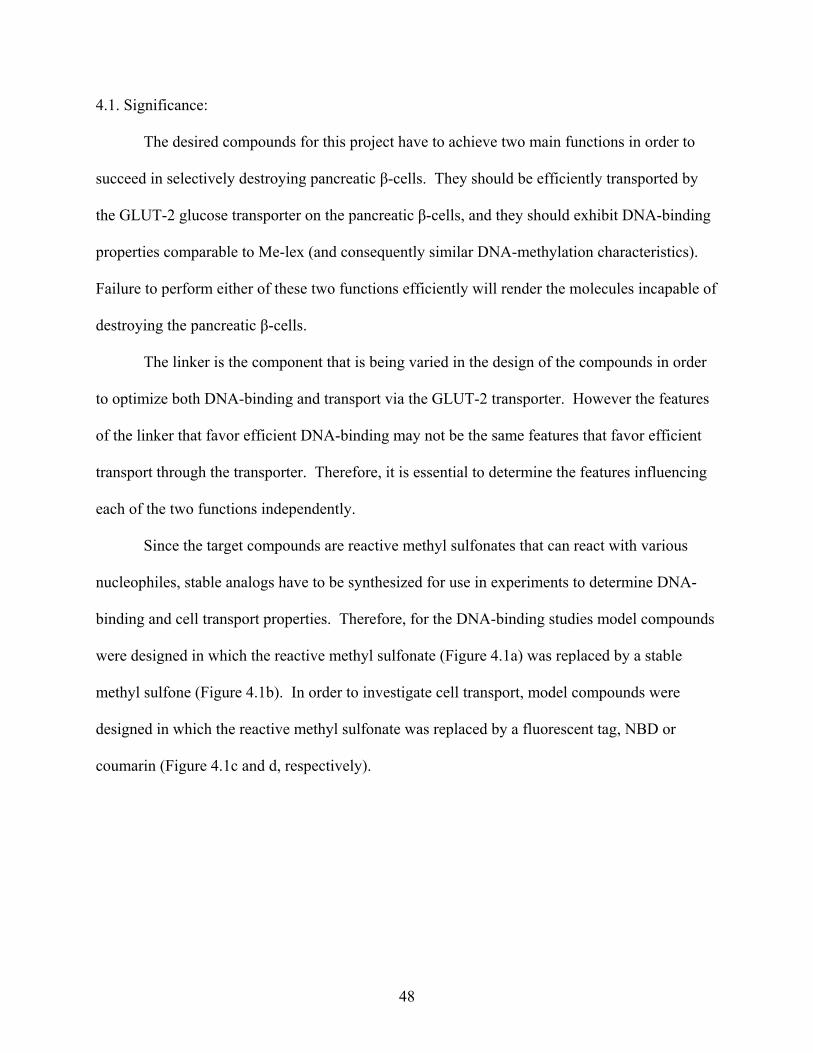
4.1. Significance:
The desired compounds for this project have to achieve two main functions in order to
succeed in selectively destroying pancreatic β-cells. They should be efficiently transported by
the GLUT-2 glucose transporter on the pancreatic β-cells, and they should exhibit DNA-binding
properties comparable to Me-lex (and consequently similar DNA-methylation characteristics).
Failure to perform either of these two functions efficiently will render the molecules incapable of
destroying the pancreatic β-cells.
The linker is the component that is being varied in the design of the compounds in order
to optimize both DNA-binding and transport via the GLUT-2 transporter. However the features
of the linker that favor efficient DNA-binding may not be the same features that favor efficient
transport through the transporter. Therefore, it is essential to determine the features influencing
each of the two functions independently.
Since the target compounds are reactive methyl sulfonates that can react with various
nucleophiles, stable analogs have to be synthesized for use in experiments to determine DNA-
binding and cell transport properties. Therefore, for the DNA-binding studies model compounds
were designed in which the reactive methyl sulfonate (Figure 4.1a) was replaced by a stable
methyl sulfone (Figure 4.1b). In order to investigate cell transport, model compounds were
designed in which the reactive methyl sulfonate was replaced by a fluorescent tag, NBD or
coumarin (Figure 4.1c and d, respectively).
48
NH
N
NO
HN
O
HN R
O
NN
HN
O2N
O
O
OHHO
NHOH
OH
O
N
NHO
N
NO
H
N
O
HR
O
SO
O
H3CO
O
HOOH
NH
OH
OH
R = CH2R = CH2CH2R = CH2CH2CH2
N
NHO
N
NO
H
N
O
HR
O
SO
O
H3C
O
HOOH
NH
OH
OH
Reactivemethylsulfonate
Non- reactivemethyl sulfone
a)
b)
c)
NH
N
NO
HN
O
HN R
OO
OHHO
NHOH
OH
O
NHO
OO
d)
Coumarin
NBD
Figure 4.1: Compounds designed for a) DNA-methylation studies. b) DNA-binding studies.
c) Cell transport studies with NBD d) Cell transport studies with coumarin.
49
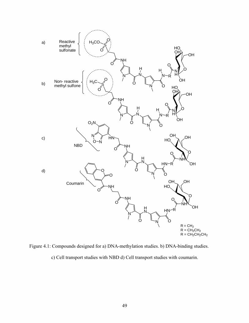
4.2. Synthesis of stable analogs for DNA-binding experiments:
A general synthetic route was first designed (scheme 4.2.1) that would result in a
molecule having the methyl sulfone unit attached to the bis-pyrrole triamide with the linker
attached at the other end and functionalized as a carboxylic acid. This molecule could then be
attached to various targeting ligands for different projects.
Scheme 4.2.1
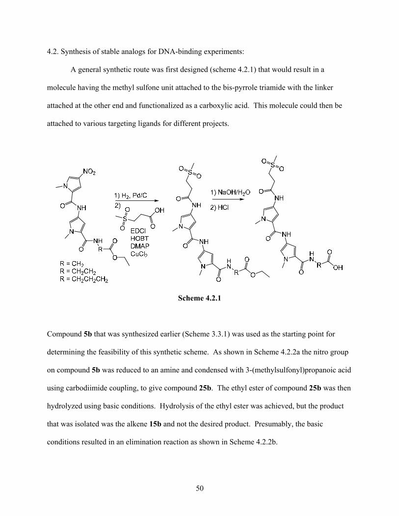
Compound 5b that was synthesized earlier (Scheme 3.3.1) was used as the starting point for
determining the feasibility of this synthetic scheme. As shown in Scheme 4.2.2a the nitro group
on compound 5b was reduced to an amine and condensed with 3-(methylsulfonyl)propanoic acid
using carbodiimide coupling, to give compound 25b. The ethyl ester of compound 25b was then
hydrolyzed using basic conditions. Hydrolysis of the ethyl ester was achieved, but the product
that was isolated was the alkene 15b and not the desired product. Presumably, the basic
conditions resulted in an elimination reaction as shown in Scheme 4.2.2b.
50
Scheme 4.2.2a
Scheme 4.2.2b
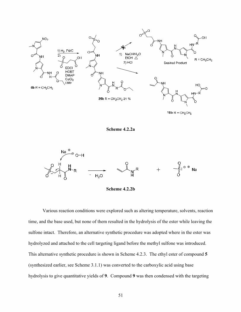
Various reaction conditions were explored such as altering temperature, solvents, reaction
time, and the base used, but none of them resulted in the hydrolysis of the ester while leaving the
sulfone intact. Therefore, an alternative synthetic procedure was adopted where in the ester was
hydrolyzed and attached to the cell targeting ligand before the methyl sulfone was introduced.
This alternative synthetic procedure is shown in Scheme 4.2.3. The ethyl ester of compound 5
(synthesized earlier, see Scheme 3.1.1) was converted to the carboxylic acid using base
hydrolysis to give quantitative yields of 9. Compound 9 was then condensed with the targeting
51
ligand 12 using carbodiimide coupling to form compound 13. This nitro group on compound 13
was reduced to the amine and condensed with 3-(methylsulfonyl)propanoyl chloride to give 18.
Scheme 4.2.3
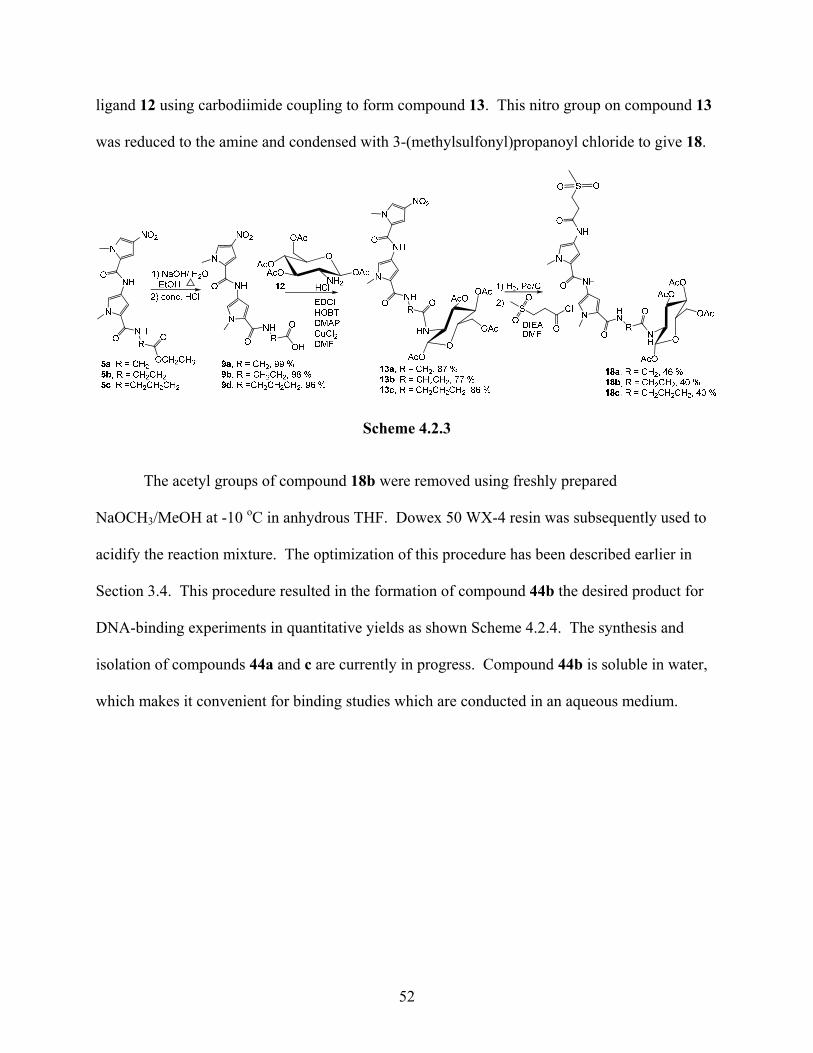
The acetyl groups of compound 18b were removed using freshly prepared
NaOCH3/MeOH at -10 oC in anhydrous THF. Dowex 50 WX-4 resin was subsequently used to
acidify the reaction mixture. The optimization of this procedure has been described earlier in
Section 3.4. This procedure resulted in the formation of compound 44b the desired product for
DNA-binding experiments in quantitative yields as shown Scheme 4.2.4. The synthesis and
isolation of compounds 44a and c are currently in progress. Compound 44b is soluble in water,
which makes it convenient for binding studies which are conducted in an aqueous medium.
52
Scheme 4.2.4
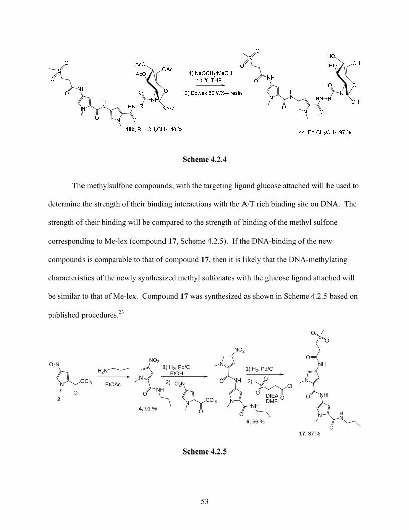
The methylsulfone compounds, with the targeting ligand glucose attached will be used to
determine the strength of their binding interactions with the A/T rich binding site on DNA. The
strength of their binding will be compared to the strength of binding of the methyl sulfone
corresponding to Me-lex (compound 17, Scheme 4.2.5). If the DNA-binding of the new
compounds is comparable to that of compound 17, then it is likely that the DNA-methylating
characteristics of the newly synthesized methyl sulfonates with the glucose ligand attached will
be similar to that of Me-lex. Compound 17 was synthesized as shown in Scheme 4.2.5 based on
published procedures.23
CCl3
ON
O2N
2
H2N
EtOAcNH
O
N
NO2
4, 91 %
1) H2, Pd/CEtOH
2)
CCl3
ON
O2N
N
NO2
O NH
NNH
O6, 56 %
2)S
O
OO
Cl
DIEADMF
1) H2, Pd/C
N
NH
N HN
O
NH
O
O
SO
O
17, 37 %
Scheme 4.2.5
53
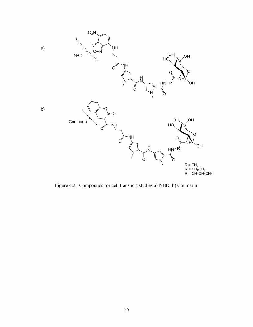
4.3. Synthesis of model compounds cell transport studies:
Model compounds containing fluorophores will be used to determine if the glucose unit
is capable of transporting the DNA-binding bis-pyrrole unit into cells that contain the GLUT-2
transports. A comparison of the transport of fluorescently tagged compounds into cells
expressing the transporter and cells lacking the transporter (available with collaborators) will
provide the desired information. Therefore, the synthesis of compounds containing the
fluorophores NBD or coumarin was attempted (Figure 4.2).
54
NH
N
NO
HN
O
HN R
O
NN
NH
O2N
O
O
OHHO
NHOH
OH
O
R = CH2R = CH2CH2R = CH2CH2CH2
a)
NH
N
NO
HN
O
HN R
OO
OHHO
NHOH
OH
O
NHO
OO
b)
Coumarin
NBD
Figure 4.2: Compounds for cell transport studies a) NBD. b) Coumarin.
55
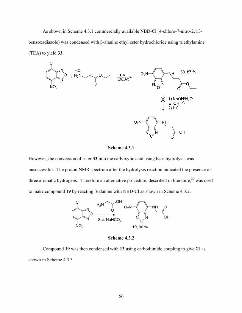
As shown in Scheme 4.3.1 commercially available NBD-Cl (4-chloro-7-nitro-2,1,3-
benzoxadiazole) was condensed with β-alanine ethyl ester hydrochloride using triethylamine
(TEA) to yield 33.
Scheme 4.3.1
However, the conversion of ester 33 into the carboxylic acid using base hydrolysis was
unsuccessful. The proton NMR spectrum after the hydrolysis reaction indicated the presence of
three aromatic hydrogens. Therefore an alternative procedure, described in literature,29 was used
to make compound 19 by reacting β-alanine with NBD-Cl as shown in Scheme 4.3.2.
N
N
Cl
NO2
O
H2NOH
O
Sat. NaHCO3N N
NHO2N
O
O
OH
19, 90 %
Scheme 4.3.2
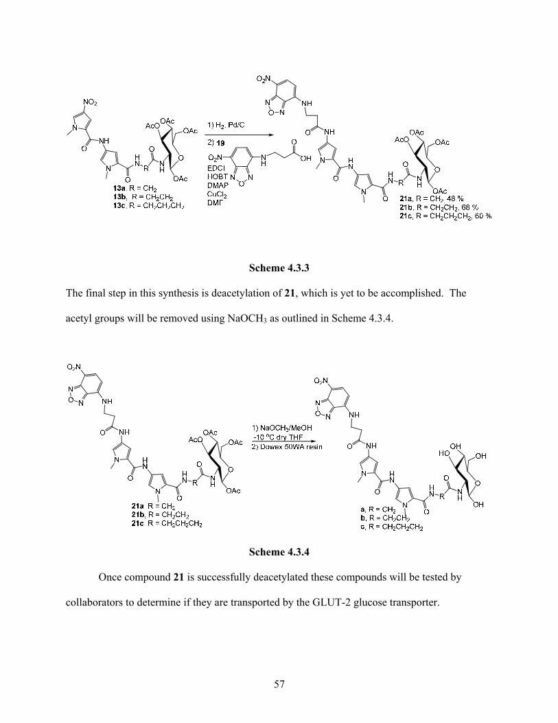
Compound 19 was then condensed with 13 using carbodiimide coupling to give 21 as
shown in Scheme 4.3.3.
56
Scheme 4.3.3
The final step in this synthesis is deacetylation of 21, which is yet to be accomplished. The
acetyl groups will be removed using NaOCH3 as outlined in Scheme 4.3.4.
Scheme 4.3.4
Once compound 21 is successfully deacetylated these compounds will be tested by
collaborators to determine if they are transported by the GLUT-2 glucose transporter.
57
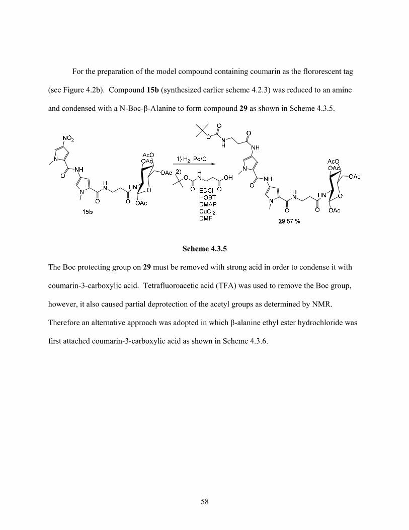
For the preparation of the model compound containing coumarin as the flororescent tag
(see Figure 4.2b). Compound 15b (synthesized earlier scheme 4.2.3) was reduced to an amine
and condensed with a N-Boc-β-Alanine to form compound 29 as shown in Scheme 4.3.5.
Scheme 4.3.5
The Boc protecting group on 29 must be removed with strong acid in order to condense it with
coumarin-3-carboxylic acid. Tetrafluoroacetic acid (TFA) was used to remove the Boc group,
however, it also caused partial deprotection of the acetyl groups as determined by NMR.
Therefore an alternative approach was adopted in which β-alanine ethyl ester hydrochloride was
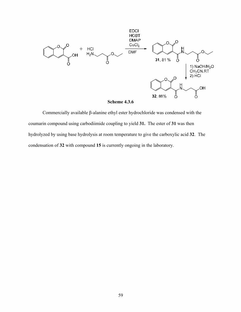
first attached coumarin-3-carboxylic acid as shown in Scheme 4.3.6.
58
Scheme 4.3.6
Commercially available β-alanine ethyl ester hydrochloride was condensed with the
coumarin compound using carbodiimide coupling to yield 31. The ester of 31 was then
hydrolyzed by using base hydrolysis at room temperature to give the carboxylic acid 32. The
condensation of 32 with compound 15 is currently ongoing in the laboratory.
59
CHAPTER 5: EXPERIMENTAL
5.1. General:
All solvents and reagents were purchased with the highest grade available from VWR
International (West Chester, Pennsylvania) or Sigma-Aldrich (Atlanta, Georgia). Flash
chromatography was performed with silica gel 60 Geduran® (40-63 µm mesh, Merck). TLC was
performed on aluminum plates coated with silica gel 60 (F254, Merck) that had a fluorescence
indicator and were detected by UV visualization. All rotary evaporations were carried out using
a Buchi R-3000 or a Buchi R-114 rotary evaporator equipped with a Brinkman model B-16
vacuum aspirator. Hydrogenations were performed using a Parr Hydrogenation Apparatus in a
500 mL Parr jar. Melting points were determined using a Mel-Temp II.
All anhydrous reactions were carried out under positive pressure argon or nitrogen.
Glassware for anhydrous reactions were dried overnight in the oven set at 150 oC, assembled
while still hot, and cooled to room temperature under argon or nitrogen. Solvent and reagents for
anhydrous reactions were purchased in sure-seal capped bottles and transferred to reactions by
oven dried needles and glass syringes.
All 1H-NMR and 13C-NMR spectra were obtained with a Bruker 400MHz NMR
spectrometer, using deuterated D6-DMSO, deuterated chloroform, or deuterated methanol as the
solvent. The deuterated D6-DMSO was obtained in sealed ampoules and 100 mL bottles from
Sigma-Aldrich. The deuterated chloroform was obtained from Alfa Aesar. The deuterated
methanol was obtained from Sigma-Aldrich. The spectra were reported in ppm and referenced
to deuterated D6-DMSO (2.49 ppm for 1H, 39.5 ppm for 13C) or referenced to chloroform (7.26
ppm for 1H, 77 ppm for 13C). The NMR tubes were 5 mm Pyrex glass obtained from Wilmad-
LabGlass, Buena, New Jersey.
61
5.2. Synthesis:
2,2,2-Trichloro-1-(1-methyl-1H-pyrrol-2-yl)ethanone (1). Trichloro acetyl chloride
(50 mL) was added to 250 mL of dry dichloromethane in a 1000 mL flask flushed with argon. In
a dropping funnel, N-methyl pyrrole (50 mL) was dissolved 100 mL of dry dichloromethane.
This mixture was then added drop wise to the trichloro acetyl chloride and stirred overnight. To
quench the reaction a solution of potassium carbonate (32 g in 250 mL of DI water) was added to
the empty drop funnel and added over a two hour period. The mixture was then extracted with
DI water (2 x 300 mL) and dichloromethane (1 x 300mL). The organic layer was dried over
MgSO4, filtered, and the filtrate concentrated under rotary evaporation to produce an oily
substance. A pure seed was added and the flask placed under vacuum until dry to yield a dark
brown solid 1 (80.35 g, 80%): mp 51-57 oC. TLC (1:1 EtOAc/Hexane) Rf = 0.56. 1H NMR data
(CDCl3): δ 7.45 (dd, J = 1.6, 6 Hz, 1H), 6.94 (s, 1H), 6.17 (dd, J = 2.4, 6 Hz), 3.93 (s, 3H). 13C
NMR data: δ 172.34, 135.80, 124.19, 121.18, 109.60, 96.55, 38.45.
2,2,2-Trichloro-1-(1-methyl-4-nitro-1H-pyrrol-2-yl)ethanone (2). Compound 1 (25 g)
was dissolved in acetic anhydride (175 mL) in a 500 mL round bottom flask and cooled to -40 oC
in a dry ice/acetone bath. After maintaining the temperature for 20 minutes fuming nitric acid
(12.3 mL) was added slowly in a addition funnel for one hour with constant stirring. Stirring
continued for an additional 45 minutes while keeping the temperature at -40 oC. The solution
was then allowed to warm to room temperature over two hours. The flask was then immersed in
an ice water bath as cold DI water (101 mL) was added slowly in portions over one hour. This
mixture was then allowed to stir overnight at which point a brown-orange precipitate was formed
and filtered under vacuum until dryness to give 2 (23.48 g, 80%): mp 112-120oC. TLC (1:1
62
EtOAc/Hexane) Rf = 0.84. 1H NMR data (CDCl3): δ 7.94 (d, J = 1.6 Hz, 1H), 7.75 (d, J = 1.6
Hz, 4.05 (s, 3H). 13C NMR data: δ 173.31, 134.74, 133.08, 121.10, 116.81, 95.03,
Ethyl 2-(1-methyl-4-nitro-1H-pyrrole-2-carboxamido)acetate (3a). The nitro pyrrole
2 (30.01 g, 11.1 mmol) was added to a 500 mL round bottom flask followed by glycine ethyl
ester hydrochloride (23.55 g, 25.5 mmol) and took up in EtOAc dried over sieves (100 mL). A
drop funnel was fitted to reaction flask and filled with TEA (35.5 mL) dissolved in EtOAc dried
over sieves (50 mL). Argon was bubbled through this solution, and this mixture was added drop
wise over a period of 15 hours under argon. Once all of the TEA mixture was added, the
reaction was allowed to stir for and additional 48 hours under argon. The white precipitate
formed was removed by filtration and the filtrate was extracted with 1M HCl (2 x 100 mL) and
DI water (1 x 100 mL). The organic layer was dried over MgSO4, filtered and the solution
concentrated by rotary evaporation to give a yellow solid 3a (28.38 g, 100%): mp 98-103 oC.
TLC (1:1 EtOAc /Hexane) Rf = 0.52. 1H NMR data (CDCl3): δ 7.51 (d, J = 1.6 Hz, 1H), 7.12 (d,
J = 2 Hz, 1H), 6.48 (s, 1H), 4.20 (q, J = 6.8, 7.2 Hz, 2H), 4.08 (d, J = 5.2, 2H), 3.90 (s, 3H), 1.26
(t, J = 7.2 Hz, 3H). 13C NMR data: δ 170.12, 160.71, 134.32, 128.67, 126.11, 108.33, 60.99,
41.16, 37.89, 14.55.
Ethyl 3-(1-methyl-4-nitro-1H-pyrrole-2-carboxamido)propanoate (3b). Compound
3b was synthesized by a procedure similar to the one described above for 3a using (30.02 g, 11.1
mmol) of 2, (17.05 g, 25.5 mmol) of β-alanine ethyl ester hydrochloride and (8.5 mL) of TEA to
obtain 3b (27.85 g, 95%): mp 120-124 oC. TLC (1:1 EtOAc/Hexane) Rf = 0.42. 1H NMR data
(CDCl3): δ 7.47 (d, J = 1.2 Hz, 1H), 6.99 (d, J = 1.6, 1H), 6.62 (s, 1H), 4.12 (q, J = 7.2, 2H), 3.91
(s, 3H), 3.58 (q, J = 6 Hz, 2H), 2.54 (t, J = 7.2 Hz, 2H), 1.22 (t, J = 7.2 Hz, 3H). 13C NMR data
(CDCl3): δ 172.64, 160.21, 134.98, 126.64, 126.24, 106.98, 61.00, 37.86, 34.82, 33.81, 14.19.
63
Ethyl 4-(1-methyl-4-nitro-1H-pyrrole-2-carboxamido)butanoate (3c). Compound 3c
was synthesized by a procedure similar to the one described above for 3a using (30.02 g, 11.1
mmol) of 2, (28.05 g, 25.5 mmol) of aminobutyrate hydrochloride and (35.5 mL) of TEA to
obtain 3c (30.56 g, 98%): mp 54-59 oC. TLC (1:1 EtOAc/Hexane) Rf = 0.36. 1H NMR data
(CDCl3): δ 7.54 (d, J = 1.6 Hz, 1H), 7.13 (d, J = 1.6 Hz, 1H), 6.81 (s, 1H), 4.11 (q, J = 3.6 Hz,
7.2 Hz, 2H), 3.96 (s, 3H), 3.41 (q, J = 6.8 Hz, 6 Hz, 2H), 2.41 (t, J = 6.8 Hz, 2H), 1.92 (pentet, J
= 6.8 Hz, 2H), 1.23 (t, J = 7.2 Hz, 3H). 13C NMR data (CDCl3): δ 173.88, 160.53, 134.84,
126.69, 126.42, 107.03, 60.84, 39.25, 37.91, 31.98, 24.22, 21.07, 14.17.
1-Methyl-4-nitro-N-propyl-1H-pyrrole-2-carboxamide (4). In a flask flushed with Ar,
the nitro pyrrole 2 (0.642 g, 2.37 mmol) was dissolved in EtOAc dried over sieves (5 mL),
propylamine (870 µL) was added while stirring at room temperature for two days to yield 4
(0.455 g, 91%): mp 124-130 oC. TLC (1:1 Hexane/EtOAc) Rf = 0.53. 1H NMR data (CDCl3): δ
7.54 (d, J = 1.6 Hz, 1H), 7.06 (d, J = 1.6 Hz, 1H), 6.09 (s, 1H), 3.98 (s, 3H), 3.35 (q, J = 6.8 Hz,
7.2 Hz, 2H), 1.61 (m, 2H), 0.97 (t, J = 7.6 Hz, 3H). 13C NMR data (CDCl3): δ 160.37, 134.91,
126.54, 106.59, 41.28, 37.81, 22.85, 11.36.
Ethyl 2-(1-methyl-4-(1-methyl-4-nitro-1H-pyrrole-2-carboxamido)-1H-pyrrole-2-
carboxamido acetate (5a). The nitro pyrrole ester 3a (13.61 g, 53 mmol) was dissolved in
ethanol (50 mL) in a 500 mL Parr jar, 10% water wet Pd/C (2.50 g) was added to this and the
mixture was shaken on a hydrogenator under pressurized hydrogen (70 psi) until reaction was
complete by TLC (100% EtOAc). The Pd/C was filtered through celite and the filtrate
concentrated by rotary evaporation and kept under vacuum overnight to yield a pale yellow solid.
The amine was dissolved in EtOAc (50 mL) dried over sieves and stirred with a mechanical
stirrer. Once dissolved, compound 1 (14.47 g, 53 mmol) was added and allowed to stir for four
64
days. The mixture was concentrated until minimal amount of EtOAc was present. At which
time the flask was placed in refrigerator over night to give yellow solid 5a (12.84 g, 79 %): mp
222-226 oC. TLC (100% EtOAc) Rf = 0.63. 1H NMR data (D6-DMSO): δ 10.26, 8.45 (t, J = 5.6
Hz, 1H), 8.18 (s, 1H), 7.58 (d, J = 2 Hz, 1H), 7.27 (s, 1H), 6.92 (s, 1H), 4.11 (q, J = 6.8 Hz, 7.2
Hz, 2H), 3.96 (s, 3H), 3.88 (d, J = 5.6 Hz, 2H), 3.81 (s, 3H), 1.21 (t, J = 7.2 Hz, 3H). 13C NMR
data (D6-DMSO): δ 170.65, 161.84, 157.36, 134.24, 128.69, 126.73, 122.80, 121.97, 119.06,
108.07, 104.96, 60.81, 41.15, 37.93 36.60, 14.59.
Ethyl 3-(1-methyl-4-(1-methyl-4-nitro-1H-pyrrole-2-carboxamido)-1H-pyrrole-2-
carboxamido) propanoate (5b). The nitro pyrrole ester 3b (13.0 g, 48.3 mmol) was dissolved
in ethanol (60 mL) in a 500 mL Parr jar, 10 % water wet Pd/C (1.0 g) was added to this and the
mixture was shaken on a hydrogenator under pressurized hydrogen (70 psi) until reaction was
complete by TLC (EtOAc). The mixture was then acidified with concentrated HCl (4.10 mL).
The Pd/C was filtered through celite and the filtrate concentrated by rotary evaporation and kept
under vacuum overnight to yield a yellow solid. In a 500 mL round bottom flask, this solid and
nitro pyrrole 1 was dissolved in EtOAc (250 mL) dried over sieves. TEA (17.00 mL) dissolved
in EtOAc was added drop wise using a drop funnel. The mixture was then allowed to stir 48
hours at which time a yellow precipitate formed. After the reaction was complete by TLC
(EtOAc), the yellow precipitate was filtered then stirred in DI water for 1 hour and filtered again
to give pure 5b (28.18 g 94%): mp 187-190 oC. TLC (EtOAc) Rf = 0.55. 1H NMR data (D6-
DMSO): δ 10.22, 8.17 (s, 1H), 8.08 (t, J = 6 Hz, 1H), 7.57 (d, J = 2 Hz, 1H), 7.21 (s, 1H), 6.83
(d, J = 1.6 Hz, 1H), 4.06 (q, J = 6.8 Hz, 7.2 Hz, 2H), 3.95 (s, 3H), 3.81 (s, 3H), 3.40 (m, 2H),
1.18 (t, J = 6.8 Hz, 3H). 13C NMR data (D6-DMSO): δ 171.84, 161.64, 157.31, 134.23, 128.68,
126.74, 123.42, 121.81, 118.57, 108.01, 104.58, 60.36, 37.93, 36.50, 35.28, 34.46, 14.56.
65
Ethyl 4-(1-methyl-4-(1-methyl-4-nitro-1H-pyrrole-2-carboxamido)-1H-pyrrole-2-
carboxamido) butanoate (5c). Compound 5c was synthesized by a procedure similar to the one
described above for 5b using (31.26 g, 0.11 mol) of pyrrole 3c, (29.25 g, 0.11 mol) of 1, and
(37.6 mL) of TEA to obtain 5c (31.15 g, 80%): mp 134-137 oC. TLC (EtOAc) Rf = 0.54. 1H
NMR data (CDCl3): δ 7.82 (s, 1H), 7.60 (d, J = 1.6 Hz, 1H), 7.22 (d, J = 2 Hz, 1H), 7.18 (d, J =
1.6 Hz, 1H), 6.54 (d, J = 1.6 Hz, 1H), 6.26 (t, J = 8 Hz, 1H), 4.13 (q, J = 7.2 Hz, 2H), 4.03 (s,
3H), 3.92 (s, 3H), 3.42 (q, J = 6.4 Hz, 2H), 1.92 (pentet, 6.8 Hz, 2H), 1.25 (t, J = 6.8 Hz, 3H).
13C NMR data (CDCl3): δ 173.19, 161.65, 157.31, 134.24, 128.68, 126.76, 123.63, 121.78,
118.45, 108.00, 104.49, 60.23, 38.19, 37.94, 36.49, 31.51, 25.15, 14.58.
1-Methyl-4-(1-methyl-4-nitro-1H-pyrrole-2-carboxamido)-N-propyl-1H-pyrrole-2-
carboxamide (6). Compound 4 (9.75 g 46.2 mmol) was dissolved in ethanol (60 mL) in a 500
mL Parr jar, 10 % water wet Pd/C (5.00 g) was added to this and the mixture was shaken on a
hydrogenator under pressurized hydrogen (70 psi) until reaction was complete by TLC (6:1
CHCl3/MeOH). The Pd/C was filtered through celite and the filtrate concentrated by rotary
evaporation and kept under vacuum overnight. This amine and nitro pyrrole 1 (12.67 g, 46.2
mmol) was dissolved in EtOAc (20 mL) dried over sieves. The reaction was stirred for 48 hours
at room temperature at which point a yellow precipitate formed. After reaction was complete by
TLC (6:1 CHCl3/MeOH) the yellow precipitate was filtered to give pure 6 (8.78 g, 56 %): mp
220-222 oC. TLC (6:1 CHCl3/MeOH) Rf = 0.51. 1H NMR data (CDCl3): δ 10.21 (s, 1H), 8.16
(s, 1H), 8.03 (s, 1H), 7.56 (s, 1H), 7.19 (s, 1H), 6.83 (s, 1H), 3.94 (s, 3H), 3.79 (s, 3H), 3.11 (m,
2H), 1.48 (m, 2H), 0.86 (t, J = 3.2 Hz, 3H). 13C NMR data (CDCl3): δ 161.57, 157.11, 134.26,
128.64, 126.79, 123.86, 121.75, 118.33, 107.99, 104.41, 40.61, 37.92, 36.44, 23.03, 11.89 (2C).
66
Methyl 1-methyl-4-(1-methyl-4-nitro-1H-pyrrol-2carboxamido)-1H-pyrrole-2-
carboxylate (7). Argon was bubbled through 100 mL of EtOAc containing 8.6 mL of DIEA in a
250 mL round bottom flask. Methyl 4-amino-1-methyl-1H-pyrrole-2-carboxylate, HCl (4.98 g,
0.03 mol) was then added to the solution followed by nitro compound 1 (7.10 g, 0.026 mol). The
solution was allowed to stir under Ar for 48 hours. The yellow precipitate that was formed was
filtered and washed with cold DI H2O and dried under vacuum to yield yellow solid 7 (4.67 g, 58
%): mp 235-239 oC. TLC (1:1 Hexane/EtOAc) Rf = 0.31. 1H NMR data (D6-DMSO): δ 10.26
(s, 1H), 8.18 (d, J = 2Hz, 1H), 7.54 (d, J = 2 Hz, 1H), 7.45 (s, 1H), 6.88 (d, J = 2 Hz, 1H), 3.94
(s, 3H), 3.84 (s, 3H), 3.74 (s, 3H). 13C NMR data (D6-DMSO): δ 161.20, 157.41, 134.26,
128.76, 126.56, 122.58, 121.31, 119.31, 108.77, 108.09, 51.50, 37.90, 36.72.
1-Methyl-4-(1-methyl-4-nitro-1H-pyrrole-2-carboxamido)-1-H-pyrrole-2-carboxylic
acid (8). The nitro ester 7 (1.00 g, 3.27 mmol) was suspended in EtOH (6 mL), and a solution of
NaOH (0.654g, 5eq.) in H2O was added. This suspension was allowed to reflux at 70 oC until
the disappearance of 7 was indicated by TLC (EtOAc). This solution was concentrated by rotary
evaporation to dryness to produce a yellow solid. The yellow solid was then dissolved in 2 mL
of H2O. This solution was then cooled to 0 oC and acidified with concentrated HCl until the pH
was 1. At this point a yellow precipitate fell out of solution and was filtered to yield 8 (0.789 g,
83%): mp 215-218 oC. TLC (6:1 CHCl3:MeOH) Rf = 0.42. 1H NMR data (D6-DMSO): δ 10.23
(s, 1H), 8.18 (d, J = 1.6 Hz, 1H), 7.54 (d, J = 2 Hz, 1H), 7.41 (d, J = 2 Hz, 1H), 6.82 (d, J = 2 Hz,
1H), 3.94 (s, 3H), 3.82 (s, 3H). 13C NMR data (D6-DMSO): δ 162.34, 1.57.35, 134.25, 128.73,
126.61, 122.36, 120.87, 120.26, 108.82, 108.19, 37.94, 36.69.
2-(1-Methyl-4-(1-methyl-4-nitro-1H-pyrrole-2-carboxamido)-1H-pyrrole-2-
carboxamido)acetic acid (9a). Compound 5a (3.03 g, 7.95 mmol) was dissolved in EtOH (20
67
mL) in a 100 mL round bottom flask to this NaOH (1.27 g, 4 eq.) and H2O (5 mL) solution was
added and allowed to reflux until the disappearance of 5a by TLC (6:1 CHCl3/MeOH). This
solution was then concentrated by the removal of the EtOH. The aqueous solution was then
cooled in an ice/water bath and the medium was acidified by concentrated HCl until the pH was
1. A yellow solid was produce and filtered to give yellow solid 9a (2.61g, 94%): mp 262-264
oC. TLC (1:1 CHCl3/MeOH) RF = 0.27. 1H NMR data (D6-DMSO): δ 12.5 (s, 1H), 10.30 (s,
1H), 8.20 (d, J = 2Hz, 1H), 7.60 (d, J = 2 Hz, 1H), 7.29 (d, J = 2Hz, 1H), 6.92 (d, J = 1.6 Hz,
1H), 3.97 (s, 3H), 3.82 (d, J = 3.6 Hz, 5H). 13C NMR data (D6-DMSO): δ 172.10, 161.77,
157.35, 134.23, 128.69, 126.74, 122.95, 121.95, 118.94, 108.10, 104.87, 37.94, 36.60.
3-(1-Methyl-4-(1-methyl-4-nitro-1H-pyrrole-2-carboxamido)-1H-pyrrole-2-
carboxamido)propanoic acid (9b). Compound 9b was synthesized using a procedure similar to
the ones described above for 9a using (2.01 g, 5.7 mmol) of 5b and NaOH (0.92 g, 4 eq.) to give
yellow solid 9b (1.81 g, 98 %): mp 226-230 oC. TLC (5:2 CHCl3/MeOH) Rf = 0.38. 1H NMR
data (D6-DMSO): δ 12.2 (s, 1H), 8.47, (s, 1H), 8.07 (s, 1H), 7.57 (s, 1H), 7.20 (s, 1H), 6.84 (s,
1H), 3.95 (s, 3H), 3.80 (s, 3H). 13C NMR data (D6-DMSO): δ 173.69, 161.61, 157.32, 134.21,
128.66, 126.73, 123.44, 121.87, 118.56, 108.19, 104.62, 37.97, 36.52, 35.41, 34.72.
4-(1-Methyl-4-(1-methyl-4-nitro-1H-pyrrole-2-carboxamido)-1H-pyrrole-2-
carboxamido)butanoic acid (9c). Compound 9c was synthesized using a procedure similar to
the ones described above for 9a using (2.00 g, 4.9 mmol) of 5c and NaOH (0.789 g, 4 eq.) to
give yellow solid 9c (1.78 g, 96 %): mp 240-245 oC. TLC (5:2 CHCl3/MeOH) Rf = 0.39. 1H
NMR data (D6-DMSO): δ 10.29 (s, 1H), 8.37 (s, 1H), 8.15 (d, J = 2 Hz, 1H), 7.57 (d, J = 2 Hz,
1H), 7.19 (d, J = 1.6 Hz, 1H), 6.83 (d, J = 1.6 Hz, 1H), 3.93 (s, 3H), 3.80 (s, 3H), 3.13 (q, J = 5.6
Hz, 6.4 Hz, 2H), 2.46 (t, J = 7.2 Hz, 2H), 1.66 (t, J = 7.2 Hz, 3H). 13C NMR data (D6-DMSO): δ
68
174.75, 161.63, 157.29, 134.23, 128.67, 126.75, 123.67, 121.77, 118.41, 108.01, 104.46, 38.29,
37.94, 36.48, 31.60, 25.19.
2-Deoxy-2-(4-methoxybenzylidene)amino-β-D-glucopyranose. (IUPAC: (E)-6-
(hydroxymethyl)-3-(3-methoxybenzylideneamino)tetrahydro-2H-pyran-2,4,5-triol) (10).
Synthesized from D-glucosamine hydrochloride as described in literature.28 In a 500 mL round
bottom flask cooled to 0 oC sodium hydroxide (11.18 g, 0.279 mols) was dissolved in 234 mL of
DI water while stirring. D-glucosamine (50.00 g, 0.232 mols) was then added and in 5 minutes
the solution turned clear. 4-methoxybenzaldehyde (31.0 mL, 0.254 mols) was added to form a
two layer mixture which was stirred for 30 minutes at 0 oC. The mixture was then allowed to sit
in the refrigerator over night to yield a white precipitate that was filtered under vacuum until
dryness to give 9 (42.83 g, 62%): mp 153-154 oC. 1H NMR data (D6-DMSO): δ 8.10 (s, 1H),
7.67 (d, J = 8.4 Hz, 2H), 6.97 (d, J = 8.8 Hz, 2H), 6.52 (s, 1H), 4.93 (d, J = 5.2 Hz, 1H), 4.91 (d,
J = 5.2 Hz, 1H), 4.67 (d, J = 8.8 Hz, 1H), 4.55 (t, J = 5.6 Hz, 1H), 3.79 (s, 3H), 3.71 (dd, J = 3
Hz, 7.7 Hz, 1H), 3.42 (m, 2H), 3.21 (m, 1H), 3.13 (m, 1H), 2.77 (t, J = 8 Hz, 1H). 13C NMR data
(D6-DMSO): δ 161.66, 161.49, 130.07, 129.56 (2C), 114.35, 96.08, 79.64, 78.65, 77.32, 75.04,
70.80, 61.72, 55.72.
1,3,4,6-Tetra-O-actyl-2-deoxy-2-(4-mehtoxybenzylidene)amino-β-D-glucopyranose.
(IUPAC: (E)-6-(acetoxymethyl)-3-(3-methoxybenzylideneamino)tetrahydro-2H-pyran-
2,4,5-triyl triacetate) (11). Compound 11 was synthesized as described in literature.28 Pyridine
(250 mL) was dried over sieves. The imine 10 (48.02g, 0.142 mmol) was dissolved in pyridine
(236 mL) at 0oC in an ice water bath. Once dissolved acetic anhydride (142 mL) was added and
the flask was immediately removed from ice water bath. The temperature was allowed to reach
room temperature overnight while stirring. The volume of the solution was reduced to half by
69
distillation under vacuum. The solution was then poured into ice water (~2000 mL) forming a
white precipitate. The solution was allowed to stir for 1 hour and then placed in refrigerator
overnight. The white precipitate was then filtered under vacuum to give 11 (54.47 g, 83%): mp
169-171oC. 1H NMR data (D6-DMSO): δ 8.29 (s, 1H), 7.64 (d, J = 8.8 Hz, 2H), 6.97 (d, J = 8.4
Hz, 2H), 6.05 (d, J = 8.4, 1H), 5.43 (t, J = 9.6 Hz, 1H), 4.96 (t, J = 9.6 Hz, 1H), 4.26 (m, 2H),
4.00 (d, J = 10.81, 1H), 3.78 (s, 3H), 3.44 (t, J = 8.4 Hz, 2H), 2.06 (s,3H), 1.97 (s, 3H), 1.96 (s,
3H), 1.81 (s, 3H). 13C NMR data (D6-DMSO): δ 170.50, 169.89, 169.43, 169.05, 164.90,
162.27, 130.37 (2C), 128.71, 114.65 (2C), 92.97, 72.78, 72.68, 71.96, 68.24, 62.10, 55.82, 20.99,
20.91, 20.90, 20.65.
1,3,4,6-Tetra-O-acetyl-2-amino-2-deoxy-β-D-glucopyranose hydrochloride. (IUPAC:
6-(acetoxymethyl)-3-aminotetrahydro-2H-pyran-2,4,5-triyl triacetate hydrochloride) (12).
Compound 12 was synthesized as described in literature.28 Acetyl chloride (3.95 mL) was added
to anhydrous methanol (27.38 mL) in a 100 mL round bottom flask at 0 oC with constant stirring
for 1 hour. This solution was then added to a stirred solution of imine 10 (25.04 g, 50.7 mmol)
in acetone (770.6 mL) at room temperature. This mixture was stirred for 1 hour and then cooled
to 0 oC. To the cooled solution ether (273.8 mL) was added and stirred for an additional 45 min.
at 0 oC. A white precipitate was filtered under vacuum to give 11 (18.92 g, 97%): mp 120 oC-
decomposition. 1H NMR data (D6-DMSO): δ 8.42 (s, 3H), 5.85 (d, J = 8.8, 1H), 5.31 (t, J = 9.6
Hz, 1H), 4.96 (t, J = 9.6 Hz, 1H), 4.26 (m, 2H), 4.00 (d, J = 10.8 Hz, 1H), 3.78 (s, 3H), 3.44 (t, J
= 8.4 Hz, 2H), 2.15 (s, 3H), 2.02 (s,3H), 1.99 (s, 3H), 1.97 (s, 3H). 13C NMR data (D6-DMSO):
δ 170.41, 170.26, 169.76, 169.09, 90.56, 72.06, 70.78, 68.23, 61.70, 52.55, 21.37, 21.29, 20.94,
20.80.
70
(2S,3R,4R,5S,6R)-6-(Acetoxymethyl-3-(2-(1-methyl-4-(1-methyl-4-nitro-1H-
pyrrole-2-carboxamido)-1H-pyrrole-2-carboxamido)acetamido)tetrahydro-2H-pyran-2,4,5-
triyl triacetate (13a). In a flask flushed with Ar, 12 (2.03g, 53 mmol) was dissolved in 8 mL
anhydrous DMF along with EDCI (1.57 g, 1.5 eq.), DMAP (1.62 g, 2.5 eq.), HOBT (1.27 g, 1.57
eq.), and CuCl2 (71 mg, 0.01 eq.). Once in solution, the carboxylic acid 9a (2.00 g, 53 mmol)
was then added and allowed to stir at room temperature over 48 hours until the disappearance of
starting material by TLC (3:1 EtOAc/MeOH). This solution was diluted with DCM (100 mL)
and extracted with H2O (150 mL, 2x), sat. NaHCO3 (100 mL, 2x), and 1M HCl (100mL, 2x).
The organic layer was dried over MgSO4. The solution was filtered and concentrated by rotary
evaporation until a solid began to fall out of solution. The solution was warmed up again until
the solid redissolved and then allowed to slowly reach room temperature overnight at which
point an orange-brown solid fell out of solution. The flask was then placed in the refrigerator to
cool further for 24 hours. The crystals were then filtered and dried to give 13a (3.18 g, 87%):
mp 118-121 oC. TLC (6:1 CHCl3/MeOH) Rf = 0.52. 1H NMR data (D6-DMSO): δ 10.27 (s, 1H),
8.4 (t, J = 8.4 Hz, J = 10 Hz, 1H), 8.18 (d, J = 2 Hz, 1H), 7.83 (d, J = 9.6, 1H), 7.59 (d, J = 1.6,
1H), 7.26 (d, J = 2 Hz, 1H), 6.88 (d, J = 2 Hz, 1H), 5.73 (d, J = 8.8 Hz, 1H), 5.24 (t, J = 9.6, 1H),
4.86 (t, J = 9.6 Hz, 1H), 4.18 (dd, J = 4.4 Hz, 4Hz, 8.4 Hz, 1H), 4.03 (m, 2H), 3.98 (s, 3H), 3.77
(3H), 3.62 (d, J = 6 Hz, 2H), 2.05 (s, 3H) ,1.99 (s, 3H), 1.94 (s, 3H), 1.94 (s, 3H). 13C NMR data
(D6-DMSO): δ 171.25, 170.47, 170.05, 169.71, 169.31, 161.31, 161.59, 157.32, 134.24, 128.66,
126.75, 123.48, 121.81, 118.58, 108.02, 104.43, 92.15, 72.62, 71.99, 68.52, 61.96, 52.35, 37.92,
36.48, 36.01, 35.84, 20.95 (2C), 20.85, 20.68.
(2S,3R,4R,5S,6R)-6-(Acetoxymethyl-3-(3-(1-methyl-4-(1-methyl-4-nitro-1H-pyrrole-
2-carboxamido)-1H-pyrrole-2-carboxamido)propanamido)tetrahydro-2H-pyran-2,4,5-triyl
71
triacetate (13b). Compound 13b was synthesized using a procedure similar to the one described
for 13a using (4.44 g, 12.2 mmol) of 9b and (4.69 g, 12.2 mmol) of 12 to give a yellow solid 13b
(6.62 g, 77 %): mp 175-179 oC. TLC (6:1 CHCl3/MeOH) Rf = 0.46. 1H NMR data (D6-DMSO):
δ 10.23 (s, 1H), 8.18 (d, J = 2 Hz, 1H), 8.10 (d, J = 8.8 Hz, 1H), 8.03 (t, J = 5.2 Hz, 1H), 7.57 (d,
J = 4 Hz, 1H), 7.22 (d, J = 1.6 Hz, 1H), 6.81 (d, J = 1.6 Hz, 1H), 5.72 (d, J = 8.8 Hz, 1H), 5.18 (t,
J = 9.6 Hz, 1H), 4.88 (t, J = 9.6 Hz, 1H), 4.19 (dd, J = 4.4 Hz, J = 4.8 Hz, J = 7.6 Hz, 1H), 3.98
(m, 3H), 3.96 (s, 3H), 3.80 (s, 3H), 2.31 (t, J = 7.2 Hz, 2H), 2.0 (s, 3H), 1.99 (s, 3H), 1.97 (s,
3H), 1.88 (s, 3H). 13C NMR data (D6-DMSO): δ 171.25, 170.47, 170.05, 169.71, 169.31,
161.59, 157.32, 134.24, 128.66, 126.75, 123.48, 121.81, 118.58, 104.43, 92.15, 72.62, , 71.99,
68.52, 61.96, 52.35, 37.92, 36.48, 36.01, 35.84, 20.95 (2C), 20.85, 20.68.
(2S,3R,4R,5S,6R)-6-(Acetoxymethyl-3-(4-(1-methyl-4-(1-methyl-4-nitro-1H-pyrrole-
2-carboxamido)-1H-pyrrole-2-carboxamido)butanamido)tetrahydro-2H-pyran-2,4,5-triyl
triacetate (13c). Compound 13c was synthesized using a procedure similar to the one described
for 13a using (3.25 g, 8.6 mmol) of 9c and of 12 (3.00 g, 7.82 mmol) to give to give a yellow
solid 13c (4.88g, 86 %): mp 171-176 oC. TLC (6:1 CHCl3/MeOH) Rf = 0.44. 1H NMR data
(D6-DMSO): δ 10.23 (s, 1H), 8.17 (s, 1H), 8.05 (m, 2H), 7.19 (s, 1H), 6.84 (s, 1H), 5.70 (d, J =
8Hz, 1H), 5.17 (t, J = 9.6 Hz, 1H), 4.88 (t, J = 9.6 Hz, 1H), 4.17 (dd, J = 4.4 Hz, J = 8.4 Hz, 1H),
4.0 (m, 3H), 3.96 (s, 3H), 3.79 (s, 3H), 3.10 (s, 3H), 2.06 (s, 3H), 2.03 (s, 3H), 2.01 (s, 3H), 1.89
(s, 3H), 1.64 (m, 2H). 13C NMR data (D6-DMSO): δ 172.67, 170.48, 170.04, 169.34, 161.56,
157.30, 134.23, 128.67, 126.75, 123.64, 121.77, 118.43, 108.00, 104.43, 92.18, 72.62, 71.97,
68.52, 61.93, 52.23, 38.42, 37.93, 36.48, 33.71, 26.13, 20.96 (2C), 20.86, 20.77.
Ethyl 2-(4-(4-acrylamido-1-methyl-1H-pyrrole-2-carboxamido)1-methyl-1H-
pyrrole-2-carboxamido)acetate (14a). Compound 5a (2.04 g, 5.4 mmol) was dissolved in 50
72
mL of ethanol and 1.72 g 10 % water wet Pd/C was added in a Parr jar. The mixture was shaken
under pressurized hydrogen (70 psi) until 5a had disappeared by TLC (EtOAc). The Pd/C was
filtered through celite and the filtrate was concentrated by rotary evaporation. The residue was
placed under high vacuum overnight. The flask was flushed with N2 and the residue was
dissolved in anhydrous THF (20 mL) and DIEA (3.13 mL, 24 mmol). The solution was bubbled
with N2 and cooled to -40 oC for 1 hour. Acryloyl chloride (489 µL, 0.5 mmol) was the added to
the cooled solution drop wise. The reaction temperature was maintained between -40 oC to -20
oC and protected from light. The reaction continued until it was complete by TLC (6:1
CHCl3/MeOH). The solution was concentrated by rotary evaporation, re-dissolved in DCM (50
mL) and extracted with DI water (2 x 60 mL). The organic layer was dried over MgSO4. The
solution was concentrated by rotary evaporation until dry to give pure 14a (2.11 g, 97 %): mp
101-106 oC. TLC (6:1 CHCl3/MeOH) Rf = 0.66. 1H NMR data (D6-DMSO): δ 10.12 (s, 1H),
9.94 (s, 1H), 8.40 (t, J = 5.6 Hz, 1H), 7.24 (d, J = 9.6 Hz, 2H), 6.9 (s, 2H), 6.35 (m, 1H), 6.17
(dd, J = 1.6 Hz, 1H), 5.66 (dd, J = 1.6 Hz, 1H), 4.10 (q, J = 6.8 Hz, 2H), 3.87 (d, J = 5.7 Hz, 2H),
3.83 (s, 3H), 3.78 (s, 3H), 1.18 (t, J = 8 Hz, 3H). 13C NMR data (D6-DMSO): δ 170.68, 162.14,
161.95, 158.81, 131.92, 126.11, 123.45, 122.62, 122.53, 122.14, 118.95, 105.14, 104.51, 60.82,
36.63, 36.52, 14.59.
Ethyl 3-(4-(4-acrylamido-1-methyl-1H-pyrrole-2-carboxamido)1-methyl-1H-
pyrrole-2-carboxamido)propanoate (14b). Compound 14b was synthesized using a procedure
similar to the one described for 14a using 351 mg, (0.90 mmol) of 5b, 81 µL (0.98 mmol) of
acryoloyl chloride and 515 µL (3.1 mmol) of DIEA to yield 14b as a brown solid (0.329 g,
88%): mp 66-71 oC. TLC (6:1 CHCl3/MeOH) Rf = 0.39. 1H NMR data (D6-DMSO): δ 10.10 (s,
1H), 9.88 (s, 1H), 8.03 (t, J = 4.9 Hz, 1H), 7.23 (d, J = 1.6 Hz, 2H), 7.16 (d, J = 1.6 Hz, 1H), 6.88
73
(d, J = 1.6 Hz, 1H), 6.81 (d, J = 1.6 Hz, 1H), 6.35 (m, 1H), 6.15 (dd, J = 2 Hz, 1H), 5.63 (dd, J =
5 Hz, 1H), 4.03 (q, J = 6.5 Hz, 7.2 Hz 2H), 3.81 (s, 3H), 3.76 (s, 3H), 1.15 (t, J = 7.2 Hz, 3H).
13C NMR (D6-DMSO): δ 171.86, 162.10, 161.75, 158.76, 131.97, 123.45, 123.12, 122.50,
122.15, 118.91, 118.45, 104.74, 104.45, 60.36, 36.64, 36.43, 35.26, 34.48, 31.17, 14.56.
Ethyl 4-(4-(4-acrylamido-1-methyl-1H-pyrrole-2-carboxamido)1-methyl-1H-
pyrrole-2-carboxamido)butanoate (14c). Compound 14c was synthesized using a procedure
similar to the one described for 14a using 421 mg, (1.04 mmol) of 5c, 94 µL (1.14 mmol) of
acryoloyl chloride and 603 µL (3.6 mmol) of DIEA to yield 14c as a brown solid (0.350 g, 79%):
mp 75-80 oC. TLC (6:1 CHCl3/MeOH) Rf = 0.63. 1H NMR data (D6-DMSO): δ 10.11 (s, 1H),
9.89 (s, 1H), 8.03 (t, J = 5.5 Hz, 1H), 7.26 (d, J = 1.6 Hz, 1H), 7.17 (d, J = 1.6 Hz, 1H), 6.90 (d, J
= 1.6 Hz, 1H), 6.84 (d, J =1.6 Hz, 1H), 6.35 (m, 1H), 6.17 (dd, J = 2 Hz, 14.8 Hz, 1H), 5.65 (dd,
J = 2 Hz, 8 Hz, 1H), 4.03 (q, J = 6.8 Hz, 7.2 Hz), 3.83 (s, 3H), 3.78 (s, 3H), 3.17 (q, J = 6.4 Hz, 6
Hz, 2H), 2.31 (t, J = 7.6 Hz, 2H), 1.73 (m 2H), 1.17 (t, J = 6.8 Hz, 3H). 13C NMR (D6-DMSO):
δ 173.18, 162.09, 161.75, 158.74, 131.95, 126.06, 123.48, 123.33, 122.45, 122.14, 118.89,
118.32, 104.63, 104.41, 60.22, 38.16, 36.65, 36.42, 31.50, 25.17, 14.58.
2-(4-(4-Acrylamido-1-methyl-1H-pyrrole-2-carboxamido)-1-methyl-1H-pyrrole-2-
carboxamido)acetic acid (15a). Compound 14a (1.28 g, 3.18 mmol) was dissolved in reagent
acetone (16 mL). NaOH (0.501 g, 1.2 mmol) dissolved in 4 mL of DI H2O was added to the
reaction flask. After 1 hour no starting material was left as indicated by TLC (6:1
CHCl3:MeOH). The acetone was removed by rotary evaporation completely leaving the DI
H2O. The flask was then cooled in an ice/water bath at which point the pH of the medium was
adjusted to 1 with concentrated HCl producing a brown precipitate. The brown precipitate was
filtered by vacuum filtration to give pure 15a (0.894, 72 %): mp 130-133 oC. TLC 2:1
74
(CHCl3:MeOH) Rf = 0.52. 1H NMR data (D6-DMSO): δ 10.15 (s, 1H), 9.94 (s, 1H), 8.312 (t, J =
5 Hz, 1H), 7.26 (s, 1H), 7.23 (s, 3H), 6.91 (d, J = 5.8 Hz, 2H), 6.37 (m, 1H), 6.17 (dd, J = 2 Hz,
1H), 5.65 (dd, 2.4 Hz, 8 Hz, 1H), 3.91 (d, J = 6 Hz, 2H), 3.84 (s, 3H), 3.83 (s, 3H). 13C NMR
(D6-DMSO): δ 172.21, 162.12, 161.88, 131.98, 126.02, 123.49, 122.73, 122.17, 118.82, 105.07,
104.52, 65.38, 36.62, 36.50, 15.63.
3-(4-(4-Acrylamido-1-methyl-1H-pyrrole-2-carboxamido)-1-methyl-1H-pyrrole-2-
carboxamido)propanoic acid (15b). Compound 15b was synthesized using a procedure similar
to the one described for 15a using (250 mg, 0.602 mmol) of 14b and 0.096 g of NaOH (2 mmol)
to yield 15b as a brown solid (0.163 g, 70 %): mp 107-110 oC. TLC 5:2 (CHCl3:MeOH) Rf =
0.30. 1H NMR data (D6-DMSO): δ 10.08 (s, 1H), 9.85 (s, 1H), 7.98 (t, J = 5 Hz, 1H), 7.21 (s,
1H), 7.13 (s, 1H), 6.85 (s, 1H), 6.78 (s, 1H), 6.31 (m, 1H), 6.12 (d, J = 1.6 Hz, 1H), 3.82 (s, 3H),
3.78 (s, 3H), 3.31 (t, J = 6.4 Hz, 2H). 13C NMR (D6-DMSO): δ 173.48, 162.11, 161.72, 158.75,
131.98, 123.46, 123.16, 122.49, 122.15, 118.91, 118.42, 104.72, 104.45, 36.65, 36.44, 35.30,
34.49, 21.54.
4-(4-(4-Acrylamido-1-methyl-1H-pyrrole-2-carboxamido)-1-methyl-1H-pyrrole-2-
carboxamido)butanoic acid (15c). Compound 15c was synthesized using a procedure similar
to the one described for 15a using (285 mg, 0.66 mmol) of 14c and 0.106 g of NaOH (2.6 mmol)
to yield 15c as a brown solid (0.208 g, 78%): mp 71-76oC. TLC 5:2 (CHCl3:MeOH) Rf = 0.63.
1H NMR data (D6-DMSO): δ 10.16 (s, 1H), 9.90 (s, 1H), 8.04 (t, J= 4.8 Hz, 1H), 7.25 (s, 1H),
7.17 (s, 1H), 6.90 (s, 1H), 6.84 (s, 1H), 6.37 (m, 1H), 6.18 (dd, J =1.3 Hz, 1H), 5.66 (dd, J = 1.2
Hz, 8.8 Hz, 1H), 3.82 (s, 3H), 3.80 (s, 3H), 3.17 (t, J = 6.2 Hz, 2H), 2.23 (t, J = 7.2 Hz, 2H), 1.70
(pentet, J = 6.5 Hz, 6.8 Hz, 7.32 Hz, 2H). 13C NMR (D6-DMSO): δ 175.23, 174.80, 162.16,
75
161.78, 158.77, 131.90, 126.02, 123.47, 123.36, 122.42, 122.12, 118.85, 118.34, 104.66, 104.46,
38.27, 36.64, 36.41, 31.60, 25.20.
6-(Acetoxymethyl)-3-(2-(4-(4-acrylamido-1-methyl-1H-pyrrole-2-carboxamido)-1-
methyl-1H-pyrrole-2-carboxamido)acetamido)tetrahydro-2H-pyran-2,4,5-triyl triacetate
(16a). In a flask flushed with N2, 15a (0.849 g, 2.27 mmol) was dissolved in 2 mL of anhydrous
DMF along with EDCI (0.657 g, 1.5 eq.), DMAP (0.701 g, 2.5 eq.), HOBT (0.926 g, 3.0 eq,),
CuCl2 (0.031 g, 0.1 eq.), and the amine hydrochloride 12 (0.960 g, 2.5 mmol) was added and the
solution was allowed to stir at room temperature over a period of two days. The solution was
diluted with 100 mL DCM and the organic layer extracted with H2O (2 x, 150 mL), sat. NaHCO3
(2 x, 150 mL), and 1M HCl (2 x, 150 mL). The organic layer was then dried over MgSO4. The
resulting solution was rotary evaporated until dry to yield the product, 16a as brown-orange
solid. (1.36 g, 85 %): mp 135-138 oC. TLC (6:1 CHCl3/MeOH) Rf = 0.74. 1H NMR (D6-
DMSO): δ 10.09 (s, 1H), 9.93 (s, 1H), 8.31 (t, J = 2.8 Hz, 1H), 7.81 (d, J = 9.2 Hz, 1H), 7.24 (d,
J = 10 Hz, 2H), 6.91 (d, J = 14 Hz, 2H), 6.37 (m, 1H), 6.17 (dd, J = 2.8 Hz, 15.6 Hz, 5.74 (d, 8.8
Hz, 1H), 5.65 (dd, J = 3.2 Hz, 8 Hz, 1H), 4.18 (dd, 8.8 Hz, 7.6 Hz, 1H), 3.93 (s, 3H), 3.82 (s,
3H), 3.62 (d, J = 5.6 Hz, 2H), 2.06 (s, 3H), 2.05 (s, 3H), 1.99 (s, 3H), 1.95 (s, 3H). 13C NMR
(D6-DMSO): δ 170.49, 170.26, 170.09, 169.72, 169.42, 162.12, 162.02, 158.81, 131.97, 123.48,
122.87, 122.15, 118.91, 118.75, 105.03, 104.55, 92.20, 72.31, 72.01, 68.77, 61.96, 52.07, 43.08,
36.62, 36.42, 21.02, 20.95, 20.86, 20.84.
6-(Acetoxymethyl)-3-(3-(4-(4-acrylamido-1-methyl-1H-pyrrole-2-carboxamido)-1-
methyl-1H-pyrrole-2-carboxamido) propanamido)tetrahydro-2H-pyran-2,4,5-triyl
triacetate (16b). Compound 16b was synthesized similar to the procedure described above for
16a using 0.147 g (0.379 mmol) of 15b, 2 mL anhydrous DMF along with, EDCI (0.108 g, 1.5
76
eq.), DMAP (0.112 g, 2.5 eq.), HOBT (0.151 g, 3.0 eq,), and CuCl2 (0.007 g, 0.1 eq.) and 0.149
g (0.388 mmol) of 12 to yield 16b as a brown-orange solid (0.260 g, 95 %): mp 131-136 oC.
TLC (6:1 CHCl3/MeOH) Rf = 0.50. 1H NMR (D6-DMSO): δ 10.09 (s, 1H), 9.93 (s, 1H), 8.31 (t,
J = 4.8 Hz, 1H), 7.81 (d, J = 9.2 Hz, 1H), 7.24 (d, J = 10 Hz, 2H), 6.91 (d, J = 14 Hz, 2H), 6.80
(d, J = 1.6 Hz, 1H), 6.36 (m, 1H), 6.18 (dd, J = 2 Hz, 15 Hz, 1H), 5.65 (dd, J = 2 Hz, 8 Hz, 1H),
5.17 (t, J = 10 Hz, 1H), 4.88 (t, J = 9.9 Hz, 1H), 4.18 (dd, J = 5.2 Hz, 8Hz, 1H), 3.97 (m, 2H),
3.82 (s, 3H), 3.78 (s, 3H), 2.54 (t, J = 1.6 Hz, 2H), 2.30 (q, J = 1.6 Hz, 7.2 Hz, 2H), 2.00 (s, 3H),
1.99 (s, 3H), 1.96 (s, 3H), 1.87 (s, 3H). 13C NMR (D6-DMSO): δ 171.26, 170.50, 170.07,
169.73, 169.34, 162.09, 161.68, 158.74, 131.96, 126.05, 123.45, 123.15, 122.48, 122.12, 118.86,
118.41, 104.58, 104.32, 92.12, 72.61, 71.96, 68.46, 61.93, 52.29, 36.46, 36.42, 36.26, 36.03,
35.83, 20.97, 20.87 (2C), 20.70.
6-(Acetoxymethyl)-3-(4-(4-(4-acrylamido-1-methyl-1H-pyrrole-2-carboxamido)-1-
methyl-1H-pyrrole-2-carboxamido)butanamido)tetrahydro-2H-pyran-2,4,5-triyl triacetate
(16c). Compound 16c was synthesized similarly to the procedure described above for 16a using
0.240 g (0.598 mmol) of 15c, 2 mL anhydrous DMF along with, EDCI (0.174 g, 1.5 eq.), DMAP
(0.183 g, 2.5 eq.), HOBT (0.243 g, 3.0 eq,), and CuCl2 (0.010 g, 0.1 eq.) and 0.230 g (0.599
mmol) of 12 to yield 16c as a brown-orange solid (0.432 g, 98 %): mp 136-139 oC. TLC (6:1
CHCl3/MeOH) Rf = 0.40. 1H NMR (D6-DMSO): δ 10.09 (s, 1H), 9.89 (s, 1H), 8.02 (t, J = 9.2
Hz, 1H), 7.26 (d, J = 1.2 Hz, 1H), 7.17 (d, J = 1.2 Hz, 1H), 6.90 (d, J = 1.6 Hz, 2H), 6.83 (d, J =
1.6 Hz, 1H), 6.33 (m, 1H), 6.15 (dd, J = 1.6 Hz, 15.2 Hz, 1H), 5.70 (d, J = 8.8 Hz, 1H), 5.65 (dd,
J = 1.6 Hz, 8.4 Hz, 1H), 5.16 (t, J = 10 Hz, 1H), 4.88 (t, J = 9.6 Hz, 1H), 4.17 (dd, J = 4.4 Hz, 8
Hz, 1H), 4.00 (m, 4H), 3.82 (s, 3H), 3.78 (s, 3H), 3.11 (q, J = 5.2 Hz, 6 Hz, 2H), 2.04 (s, 3H),
2.00 (s, 3H), 1.97 (s, 3H), 1.91 (s, 3H), 1.64 (pentet, J = 7.5 Hz, 6.9 Hz, 14 Hz, 2H). 13C NMR
77
(D6-DMSO): δ 172.70, 170.52, 170.07, 169.74, 169.38, 162.10, 161.69, 158.75, 131.93, 126.10,
123.47, 123.34, 122.43, 122.13, 118.32, 105.03, 104.60, 92.17, 72.61, 71.96, 68.49, 61.91, 52.24,
49.06, 36.65, 36.42, 33.72, 26.17, 20.97, 20.87 (2C), 20.78.
1-Methyl-4-(1-methyl-4-(3-methylsulfonyl)propanamido)-1H-pyrrole-2-
carboxamido)-N-propyl-1H-pyrrole-2-carboxamide (17). Compound 6 (0.166 g, 0.410
mmol) was dissolved in 50 mL of ethanol and 0.120 g 10% water wet Pd/C was added in a Parr
jar. The mixture was shaken under pressurized hydrogen (70 psi) until 6 had disappeared by
TLC (6:1 CHCl3/MeOH). The Pd/C was filtered through celite and the filtrate was concentrated
by rotary evaporation. The residue was placed under high vacuum overnight. The flask
containing the reduced compound 6 was flushed with N2 and dissolved with 4 mL of anhydrous
DMF and anhydrous DIEA (289 µL, 1.7 mmol). The mixture was cooled to -40 0C for 45
minutes. The 3-(methylsulfonyl) propanoyl chloride (0.094 g, 0.55 mmol) was then added to the
cooled mixture and the temperature was allowed to slowly reach RT until TLC
(6:1CHCl3/MeOH) indicated the disappearance of the reduced compound 6. The solvent was
then removed by rotary evaporation and the solid was took up in 100 mL of DCM and extracted
with HCl (2x, 100 mL) and DI H2O (2x, 100 mL). The organic layer was then dried over
MgSO4. The resulting solution was concentrated by rotary evaporation to yield the crude
product and purified with flash column chromatography (9:1 EtOAc/MeOH) to give pure 17
(0.055 g, 25 %): mp > 150 oC. TLC (9:1 EtOAc/MeOH) Rf = 0.36. 1H NMR (D6-DMSO): δ
10.0 (s, 1H), 9.83 (s, 1H), 7.96 (t, J = 5.2 Hz, 1H), 7.15 (d, J = 1.6 Hz, 2H), 6.83 (d, J = 9.6 Hz,
2H), 3.81 (s, 3H), 3.77 (s, 3H), 3.10 (m, 2H), 2.99 (s, 3H), 2.72 (t, J = 7.6 Hz, 2H), 1.47 (q, J =
7.2 Hz, 2H), 0.85 (t, J = 7.2 Hz, 3H). 13C NMR (D6-DMSO): δ 166.57, 161.70, 158.78, 123.55,
123.33, 122.39, 122.12, 118.65, 118.22, 104.59, 104.33, 50.22, 36.59, 36.36, 28.68, 23.03, 11.89.
78
6-(Acetoxymethyl)-3-(2-(1-methyl-4-(1-methyl-4-(3-(methylsulfonyl)propanamido)-
1H-pyrrole-2-carboxamido)acetamido)tetrahydro-2H-pyran-2,4,5-triyl triacetate (18a).
Compound 16a (0.317 g, 0.467 mmol) was dissolved in 52 mL of ethanol and 0.412 g 50 %
water wet Pd/C was added in a Parr jar. The mixture was shaken under pressurized hydrogen (70
psi) until 16a had disappeared by TLC (6:1 CHCl3/MeOH). The Pd/C was filtered through celite
and the filtrate was concentrated by rotary evaporation. The residue was placed under high
vacuum overnight. The flask with flushed with N2 and the reduced compound 16a was dissolved
in 2.5 mL of anhydrous DMF along anhydrous DIEA (193 µL, 1.1 mmol) and cooled to
-40 oC in a dry ice acetone bath for 45 minutes. 3-(methylsulfonyl)propanoyl chloride (0.081 g,
0.467 mmol) was then added and temperature was allowed to reach room temperature over night.
The reaction stirred for two days at room temperature until the disappearance of the reduced
compound 16a was indicated by TLC (6:1 CHCl3/MeOH). The solution was diluted with 50 mL
DCM and the organic solution was extracted with H2O (2x, 60 mL), 1 M HCl (2x, 50 mL), and
sat. NaHCO3 (2x, 50 mL). The organic layer was then dried over MgSO4. The resulting
solution was rotary evaporated until dry to yield the product, 18a as an orange solid (0.170 g, 46
%): mp 94-97 oC. TLC (6:1 CHCl3/MeOH) Rf = 0.64. 1H NMR (D6-DMSO): δ 10.06 (s, 1H),
9.92 (s, 1H), 8.33 (t, J = 12 Hz, 1H), 7.23 (d, J = 8 Hz, 2H), 6.87 (dd, J = 1.6 Hz, 2 Hz, 2H), 5.73
(d, J = 8.8 Hz, 1H), 5.24 (t, J = 10 Hz, 1H), 4.85 (t, J = 9.2 Hz, 1H), 4.17 (dd, J = 4.4 Hz, 8.3 Hz,
1H), 3.95 (m, 3 H), 3.63 (d, J = 5.6 Hz, 2H), 3.39 (t, J = 11.2 Hz, 2H), 3.00 (s, 3H), 2.058 (s,
3H), 1.99 (s, 3H), 1.95 (s, 3H), 1.94 (s, 3H). 13C NMR (D6-DMSO): δ 170.52, 170.28, 170.11,
169.75, 169.46, 166.56, 162.02, 132.17, 123.27, 122.83, 122.61, 122.24, 118.65, 104.42, 118.73,
105.01, 104.42, 92.18, 72.28, 71.98, 68.71, 61.93, 52.02, 20.21, 43.06, 36.62, 36.44, 28.68,
20.96, 20.88 (2C), 20.85
79
6-(Acetoxymethyl)-3-(3-(1-methyl-4-(1-methyl-4-(3-(methylsulfonyl)propanamido)-
1H-pyrrole-2-carboxamido)-1H-pyrrole carboxamido)propanamido)tetrahydro-2H-pyran-
2,4,5-triyl triacetate (18b). Compound 18b (0.383 g, 0.540 mmol) was dissolved in 50 mL of
ethanol and 0.333 g 10 % water wet Pd/C was added in a Parr jar. The mixture was shaken
under pressurized hydrogen (70 psi) until 16b had disappeared by TLC (6:1 CHCl3/MeOH). The
Pd/C was filtered through celite and the filtrate was concentrated by rotary evaporation. The
residue was placed under high vacuum overnight. The flask was flushed with N2 and the
reduced compound 6 along with 3-(methylsulfonyl) propanoic acid (0.0824 g, 0.542 mmol),
EDCI (0.160 g, 1.5 eq.), HOBT (0.218 g, 3.0 eq,), DMAP (0.166 g, 2.5 eq.) and CuCl2 (0.008 g,
0.1 eq.) were dissolved in 3 mL of anhydrous DMF and allowed to stir at room temperature for
two days until the disappearance of starting material as indicated by TLC (6:1 CHCl3/MeOH).
The solution was diluted with 50 mL DCM and the organic solution was extracted with H2O (2
x, 60 mL), 1 M HCl (2 x, 50 mL), and sat. NaHCO3 (2 x, 50 mL). The organic layer was then
dried over MgSO4. The resulting solution was rotary evaporated until dry to yield the crude
product. The crude product was purified by flash column chromatography (6:1 CHCl3/MeOH)
yield 18b as a brown solid (0.168 g, 40 %): mp 94-97 oC. TLC (6:1 CHCl3/MeOH) Rf = 0.40.
1H NMR (D6-DMSO): δ 10.06 (s, 1H), 9.87 (s, 1H), 8.09 (d, J = 9.2 Hz, 1H), 7.95 (t, J = 8.4 Hz,
1H), 7.16 (d, J = 1.2 Hz, 2H), 6.85 (d, J = 1.6 Hz, 1H), 6.79 (d, J = 1.6 Hz, 2H), 5.72 (d, J = 8.8
Hz, 1H), 5.17 (t, J = 10.4 Hz, 1H), 4.88 (t, J = 10 Hz, 1H), 4.16 (dd, J = 8.4 Hz, 4 Hz, 1 H), 4.08
(m, 3H), 3.80 (s, 3H), 3.77 (s, 3H), 3.01 (s, 3H), 2.30 (t, J = 7.2 Hz, 2H), 2.00 (s, 3H), 1.99 (s,
3H), 1.96 (s, 3H), 1.87 (s, 3H). 13C NMR (D6-DMSO): δ 171.28, 170.76, 170.49, 170.06,
169.73, 169.33, 166.55, 162.78, 161.69, 158.76, 123.29, 123.17, 122.49, 122.14, 118.63, 118.44,
104.58, 104.34, 92.14, 72.62, 71.99, 61.95, 61.04, 52.33, 50.22, 49.63, 20.95, 20.86 (2C), 20.69.
80
6-(Acetoxymethyl)-3-(4-(1-methyl-4-(1-methyl-4-(3-(methylsulfonyl)propanamido)-
1H-pyrrole-2-carboxamido)-1H-pyrrole carboxamido)butanamido)tetrahydro-2H-pyran-
2,4,5-triyl triacetate (18c). Using the procedure described for 18a, 0.737 g, (1.0 mmol) of 16c,
603 µL (3.6 mmol) of DIEA and 0.195 g of 3-(methylsulfonyl)propanoyl chloride to yield 18c as
a brown solid (0.385 g, 46 %): mp 154-160 oC. TLC (4:1 EtOAc/MeOH) Rf = 0.63. 1H NMR
data (D6-DMSO): δ 10.02 (s, 1H), 9.83 (s, 1H), 7.97 (t, J = 9.2 Hz, 1H), 7.14 (dd, J = 1.6 Hz, 2
Hz, 2H), 6.83 (dd, J = 1.6 Hz, 6.4 Hz, 2H), 5.70 (d, J = 8.8 Hz, 1H), 5.15 (t, J = 9.6 Hz, 1H), 4.87
(t, J = 9.6 Hz, 1H), 4.16 (dd, J = 8 Hz, 4Hz, 1H), 3.97 (m, 3H), 3.80 (s, 3H), 3.76 (s, 3H), 3.10
(q, J = 6.4 Hz, 2H), 2.99 (s, 3H), 2.02 (s, 3H), 2.01 (s, 3H), 1.98 (s, 3H), 1.94 (s, 3H). 13C NMR
data (D6-DMSO): δ 172.69, 170.50, 170.06, 169.73, 169.36, 166.54, 161.69, 158.76, 123.35,
123.29, 122.44, 122.14, 118.62, 118.31, 104.62, 104.31, 92.19, 72.62, 71.97, 68.52, 52.25, 50.21,
36.61, 36.40, 36.25, 33.72, 28.66, 26.16, 20.96, 20.87 (2C), 20.78.
3-[N-(7’-Nitrobenz-2’-oxa-1’, 3’diazol-4’yl)amino]propanoic. (IUPAC: 2-(7-
nitrobenzo[c][1,2,5]oxodiazol-4-ylamino)acetic acid) (19). Compound 19 was synthesized
from 4-chloro-7-nitro-benzofurazane as described in literature.29 β-Alanine (6.24 g, 70 mmol,
7eq.) and NaHCO3 (5.88 g, 70 mmol, 7eq) were dissolved in 20 mL of D.I water (20 mL). After
addition of 4-chloro-7-nitro-benzofurazane (2.0 g, 10 mmol) in methanol (80 mL) the mixture
was stirred for 1.5 hours at 50 oC until TLC (4:1 CHCl3/MeOH) indicated disappearance of
starting material. The methanol was then removed by rotary evaporation leaving behind the
water. The mixture was then cooled in an ice water bath while adjusting the pH to 1 with 1M
HCl producing a brown precipitate. The brown precipitate was then filtered to give pure 19
(2.14 g, 90 %): mp > 200 oC. TLC 4:1 (CHCl3/MeOH) Rf = 0.20. 1H NMR data (D6-DMSO): δ
81
12.43 (s, 1H), 9.45 (s, 1H), 8.51 (d, J = 8 Hz, 1H), 6.44 (d, J = 8.8 Hz, 1H), 2.71 (t, J = 2.4 Hz,
2H). 13C NMR (D6-DMSO): δ 172.87, 145.33, 144.84, 144.62, 138.38, 121.49, 99.86, 32.79.
1-Methyl-4-(1-methyl-4-(2-(7-nitrobenzo[c][1,2,5,]oxodiazol-4-ylamino)acetamido-
1H-pyrrole-2-carboxamido N-propyl-1H-pyrrole-2-carboxamide (20). Compound 6 (0.140
g, 0.419 mmol) was dissolved in 50 mL of ethanol and of 0.1184 g 10 % water wet Pd/C was
added in a Parr jar. The mixture was shaken under pressurized hydrogen (70 psi) until 6 had
disappeared by TLC (6:1 CHCl3/MeOH). The Pd/C was filtered through celite and the filtrate
was concentrated by rotary evaporation. The residue was placed under high vacuum overnight.
The flask was flushed with N2, the reduced compound 20 was dissolved in anhydrous DMF (3.5
mL) along with carboxylic acid 19 (0.111 g, 0.466 mmol), EDCI (0.123 g, 1.5 eq.), DMAP
(0.130 g, 2.5 eq.), HOBT (0.1750 g, 3eq), and CuCl2 (0.008g, 0.1 eq.). The mixture was allowed
to stir for two days until the disappearance of the starting material was indicated by TLC (6:1
CHCl3/MeOH). The solution was diluted with 100 mL DCM and the organic solution was
extracted with H2O (2x, 150 mL), 1 M HCl (2x, 100 mL), and sat. NaHCO3 (2x, 100 mL). The
organic layer was then dried over MgSO4. The resulting solution was rotary evaporated until dry
to yield the crude product. Flash column chromatography (6:1 CHCl3/MeOH) yielded pure 20 as
an orange solid (0.067 g, 37%): mp 152-157 oC. TLC (6:1 CHCl3/MeOH) Rf = 0.36. 1H NMR
data (D6-DMSO): δ 9.99 (s, 1H), 9.83 (s, 1H), 9.52 (s, 1H), 8.54 (t, J = 8 Hz, 1H), 7.98 (t, J = 4
Hz, 1H), 7.17 (d, J = 8 Hz, 2H), 6.84 (d, J = 8.4 Hz, 2H), 6.48 (d, J = 8.8 Hz, 2H), 3.82 (s, 3H),
3.78 (s, 3H), 3.10 (d, J = 8 Hz, 2H), 2.72 (d, J = 6.8 Hz, 2H), 1.46 (m, 2H), 0.85 (t, J = 7.2 Hz,
3H). 13C NMR (D6-DMSO): δ 167.45, 161.66, 158.76, 123.52, 123.25, 122.39, 122.13, 118.61,
118.20, 104.54, 104.26, 41.67, 40.77, 36.61, 36.37, 23.04, 11.90.
82
6-(Acetoxymethyl)-3-(2-(1-methyl-4-(1-methyl-4-(2-(7-nitrobenzo[c][1,2,5]oxadiazol-
4-ylamino)acetamido)-1H-pyrrole-2-carboxamido)-1H-pyrrole-2-
carboxamido)acetamido)tetrahydro-2H-pyran-2,4,5-triyl triacetate (21a). Compound 13a
(0.344 g, 0.495 mmol) was dissolved in 30 mL of ethanol and of 0.278 g 50 % water wet Pd/C
was added in a Parr jar. The mixture was shaken under pressurized hydrogen (60 psi) until 6 had
disappeared by TLC (6:1 CHCl3/MeOH). The Pd/C was filtered through celite and the filtrate
was concentrated by rotary evaporation then further dried under high vacuum overnight. The
flask was flushed with N2, the reduced compound 13a was dissolved in anhydrous DMF (3mL)
along with carboxylic acid 19 (0.118 g, 0.495 mmol), EDCI (0.147 g, 1.5 eq.), DMAP (0.151 g,
2.5 eq.), HOBT (0.200 g, 3eq), and CuCl2 (0.009 g, 0.1 eq.). The mixture was allowed to stir for
two days until the disappearance of the starting material was indicated by TLC (6:1
CHCl3/MeOH). The solution was diluted with 150 mL DCM and the organic solution was
extracted with H2O (2x, 200 mL), 1 M HCl (2x, 200 mL), and sat. NaHCO3 (2x, 200 mL). The
organic layer was then dried over MgSO4. The resulting solution was rotary evaporated until dry
to yield the crude product. Flash column chromatography (6:1 CHCl3/MeOH) yielded pure 21a
as an orange solid (0.200 g, 48%): mp 144 oC to decomp. TLC (6:1 CHCl3/MeOH) Rf = 0.36.
1H NMR data (D6-DMSO): δ 9.92 (s, 1H), 9.90 (s, 1H), 9.52 (s, 1H), 8.52 (t, J = 5.2 Hz, 1H),
8.32 (t, J = 5.2 Hz, 1H), 7.82 (d, J = 9.6 Hz, 1H), 7.22 (s, 1H), 7.17 (d, J = 1.6 Hz, 1H), 6.87 (s,
2H), 6.46 (d, J = 9.2 Hz, 1H), 5.74 (d, J = 8.8 Hz, 1H), 5.24 (t, J = 9.6 Hz, 1H), 4.85 (t, J = 9.6
Hz, 1H), 4.18 (dd, J = 4.8 Hz, 8 Hz, 2H), 3.40 (m, 3H), 3.81 (s, 3H), 3.76 (s, 3H), 2.05 (s, 3H),
1.99 (s, 3H), 1.95 (s, 3H), 1.94 (s, 3H). 13C NMR (D6-DMSO): δ 170.51, 170.27, 170.11,
169.74, 169.45, 167.46, 162.01, 158.80, 132.16, 132.08, 129.1, 123.19, 122.82, 122.55, 122.24,
83
118.73, 104.96, 92.17, 72.28, 71.98, 68.72, 67.86, 61.94, 52.02, 36.63, 36.44, 30.25, 28.82,
23.69, 22.87, 21.03, 20.96, 20.88, 20.85, 14.38, 11.27.
6-(Acetoxymethyl)-3-(3-(1-methyl-4-(1-methyl-4-(2-(7-nitrobenzo[c][1,2,5]oxadiazol-
4-ylamino)acetamido)-1H-pyrrole-2-carboxamido)-1H-pyrrole-2-
carboxamido)propanamido)tetrahydro-2H-pyran-2,4,5-triyl triacetate (21b). Compound
21b was synthesized similar to the procedure described above for 20a, reducing 0.4067 g (0.573
mmol) of 15b to the amino and dissolving in 4 mL anhydrous DMF along with, compound 20
(0.152 g, 0.64 mmol), EDCI (0.164 g, 1.5 eq.), DMAP (0.179 g, 2.5 eq.), HOBT (0.234 g, 3.0
eq,), and CuCl2 (0.008 g, 0.1 eq.) to yield 21b as a dark brown solid (0.349 g, 68 %): mp TLC
(6:1 CHCl3/MeOH) Rf = 0.27. 1H NMR data (D6-DMSO): δ 10.01 (s, 1H), 9.84 (s, 1H), 9.50 (s,
1H), 8.50 (s, 1H), 8.07 (d, J = 8.8 Hz, 1H), 7.93 (t, J = 6 Hz, 1H), 7.13 (s, 1H), 6.83 (s, 1H), 6.75
(s, 1H), 6.46 (d, J = 8.4 Hz, 1H), 5.68 (d, J = 8.8 Hz, 1H), 5.15 (t, J = 9.6 Hz, 1H), 4.85 (t, J = 10
Hz, 1H), 4.15 (dd, J = 4.4 Hz, 9.2 Hz, 2H), 3.96 (m, 3H), 3.76 (s, 3H), 3.73 (s, 3H), 2.71 (q, J =
6.8 Hz, 2H), 2.27 (t, J = 6.8 Hz, 2H), 1.95 (s, 6H), 1.92 (s, 3H), 1.83 (s, 3H). 13C NMR (D6-
DMSO): δ 171.21, 170.50, 170.06, 169.74, 169.34, 167.45, 161.68, 158.77, 145.44, 144.92,
138.43, 123.20, 123.13, 12..49, 122.26, 118.62, 118.44, 104.58, 104.32, 99.93, 92.13, 72.61,
71.96, 68.47, 61.93, 52.30, 36.62, 36.42, 36.03, 35.82, 20.97, 20.87, 20.70.
6-(Acetoxymethyl)-3-(4-(1-methyl-4-(1-methyl-4-(2-(7-nitrobenzo[c][1,2,5]oxadiazol-
4-ylamino)acetamido)-1H-pyrrole-2-carboxamido)-1H-pyrrole-2-
carboxamido)butanamido)tetrahydro-2H-pyran-2,4,5-triyl triacetate (21c). Compound 21c
was synthesized similar to the procedure described above for 20a, reducing 0.5424 g (0.751
mmol) of 15c to the amino and dissolving in 4 mL anhydrous DMF along with, compound 20
(0.197 g, 0.8 mmol), EDCI (0.213 g, 1.5 eq.), DMAP (0.243 g, 2.5 eq.), HOBT (0.307 g, 3.0
84
eq,), and CuCl2 (0.015 g, 0.1 eq.) to yield 21c as a dark brown solid (0.403 g, 60 %): mp TLC
(6:1 CHCl3/MeOH) Rf = 0.38. 1H NMR data (D6-DMSO): δ 10.05 (s, 1H), 9.86 (s, 1H), 9.52 (s,
1H), 8.55 (t, J = 5.6 Hz, 1H), 8.05 (t, J = 5.6 Hz, 1H), 7.16 (s, 2H), 6.86 (d, J = 6.6 Hz, 2H), 6.49
(m, 1H), 5.72 (d, J = 8.45 Hz, 1H), 5.18 (t, J = 6 Hz, 1H), 4.88 (t, J = 6.4 Hz, 1H), 4.20 (dd, J =
3.2 Hz, 6 Hz, 2H), 4.18 (t, J = 2 Hz, 3H), 3.83 (s, 3H), 3.79 (s, 3H), 3.12 (t, J = 2 Hz, 2H), 2.06
(s, 3H), 2.02 (s, 3H), 1.98 (s, 3H), 1.93 (s, 3H), 1.65 (pentet, J = 1.2 Hz, 4.8 Hz, 2H). 13C NMR
(D6-DMSO): δ172.70, 170.50, 170.05, 169.74, 169.36, 167.45, 162.78, 161.67, 158.76, 144.84,
144.55, 138.38, 123.20, 122.42, 122.24, 118.61, 118.29, 104.60, 104.25, 99.88, 92.17, 72.61,
71.95, 68.48, 61.90, 60.68, 52.22, 38.35, 36.61, 36.40, 36.24, 33.71, 31.22, 26.16, 20.95, 20.86,
20.77.
Methyl 4-(4-acrylamido-1-methyl-1H-pyrrole-2-carboxamido)-1-methyl-1H-
pyrrole-2-carboxylate (22). Compound 7 (4.10 g, 19.6 mmol) was dissolved in 50 mL of EtOH
and 0.50 g 10 % water wet Pd/C was added in a parr jar. The mixture was shaken under
pressurized hydrogen (70 psi) until 7 had disappeared as indicated by TLC (EtOAc). The Pd/C
was filtered through celite and the filtrate was concentrated by rotary evaporation and the residue
was kept under high vacuum overnight. The flask with the residue was flushed with Ar and the
residue was dissolved in anhydrous THF (8 mL) and anhydrous DIEA (3.32 mL) was added to
the solution. The solution was bubbled with Ar and cooled to -40 oC. Acryloyl chloride (1.20
mL) was added drop wise to the solution which was then allowed to stir at -20 oC, protected from
light, until the reaction was complete as indicated by TLC (6:1 CHCl3/MeOH). The solvent was
removed by rotary evaporation and the resulting solid dissolved in 50 mL of EtOAc and
extracted with DI H2O (2x, 60 mL). The organic layer was then dried over MgSO4. The
resulting solution was rotary evaporated until dry to yield the product, 22 as an orange solid
85
(4.18 g, 96 %): mp 179-184 oC. TLC (6:1 CHCl3/MeOH) Rf = 0.58. 1H NMR data (D6-DMSO):
δ 10.11 (s, 1H), 9.93 (s, 1H), 7.44 (d, J = 1.6 Hz, 1H), 7.25 (d, J = 1.6 Hz, 1H), 6.90 (d, J = 2.0
Hz, 1H), 6.87 (d, J = 2.4 Hz, 1H), 6.33 (m, 1H), 6.16 (dd, J = 2.0 Hz, 15.2 Hz, 1H), 5.65 (dd, J =
2.0 Hz, 8 Hz, 1H), 3.82 (s, 3H), 3.81 (s, 3H), 3.72 (s, 3H). 13C NMR (D6-DMSO): δ 162.10,
161.26, 158.77, 131.91, 126.10, 125.70, 123.29, 122.21, 121.22, 119.03, 118.97, 108.77, 104.54,
51.44, 36.62 (2C).
3-(5-(5-(Methoxycarbonyl)-1-methyl-1H-pyrrol-3-ylcarbamoyl)-1-methyl-1H-
pyrrol-3-ylamino)-3-oxopropane-1-sulfonic acid (23). To a solution of alkene 22 (1.68 g, 5.1
mmol) dissolved in 4 mL EtOH, NaHSO3 (1.06 g, 10 mmol) dissolved in 1mL of DI H2O was
added all at once. The pH was adjusted to 8 with 5 % NaOH and the mixture was refluxed until
22 had disappeared and a baseline spot appeared by TLC (6:1 CHCl3/MeOH). The solution was
then rotovaped until a minimal amount of water remained. This solution was then cooled in an
ice/water bath and the pH adjusted to 1 with concentrated HCl. A pale yellow precipitate fell out
of solution, which was vacuum filtered to give pure 23 as a pale yellow solid (2.10 g, 81 %): mp
decomposition 197 oC. TLC (5:2 CHCl3/MeOH) Rf = 0.27. 1H NMR data (D6-DMSO): δ 9.97
(s, 1H), 9.89 (s, 1H), 7.44 (s, 1H), 7.14 (s, 1H), 6.88 (s, 1H), 6.84 (s, 1H), 3.82 (s, 3H), 3.80 (s,
3H), 3.72 (s, 3H), 2.64 (d, J = 8.4 Hz, 2H), 2.54 (d, J = 8.8 Hz, 2H). 13C NMR (D6-DMSO): δ
169.02, 161.28, 158.89, 123.40, 122.89, 121.21, 118.97, 118.74, 51.41, 48.03, 36.62, 36.50,
33.00, 31.16.
1-Methyl-4-(1-methyl-4-(3-sulfopropanamido)-1H-pyrrole-2-carboxamido)-1H-
pyrrole-2-carboxylic acid (24). To a solution of sulfonic acid 23 (33 mg, 79 µmol) suspended
in 4 mL of EtOH, NaOH (19 mg, 4.8 mmol) dissolved in 1 mL of DI H2O was added all at once.
The mixture was refluxed until 23 had disappeared and a baseline spot appeared by TLC (6:1
86
CHCl3/MeOH). The solution was then rotovaped until a minimal amount of water remained.
This solution was then cooled in an ice/water bath and the pH adjusted to 1 with concentrated
HCl. A pale yellow precipitate fell out of solution, which was vacuum filtered to give pure 24 as
a pale yellow solid (25 mg, 72 %): mp decomposition 169 oC. TLC 3:1 CHCl3/MeOH Rf = 0.20.
1H NMR data (D6-DMSO): δ 9.95 (s, 1H), 9.70 (s, 1H), 7.11 (d, J = 2.0 Hz, 1H), 7.07 (d, J = 1.6
Hz, 1H), 6.80 (d, J = 1.6 Hz, 1H), 6.50 (d, J = 1.6 Hz, 1H), 6.04 (s, 1H), 3.79 (s, 3H), 3.77 (s,
3H), 2.66 (d, J = 8.8 Hz, 2H), 2.52 (d, J = 7.2 Hz, 2H). 13C NMR (D6-DMSO): δ 168.57,
158.42, 123.89, 123.47, 122.40, 119.63, 118.24, 112.25, 104.02, 101.01, 47.95, 36.47, 36.30,
32.55.
Ethyl 3-(1-methyl-4-(1-methyl-4-(3-(methylsulfonyl)propanamido)-1H-pyrrole-2-
carboxamido)1H-pyrrole-2-carboxamido)propanoate (25). Compound 5b (1.03 g, 2.65
mmol) was dissolved in 50 mL of ethanol and 0.500 g 50 % water wet Pd/C was added in a Parr
jar. The mixture was shaken under pressurized hydrogen (70 psi) until 5b had disappeared by
TLC (EtOAc). The Pd/C was filtered through celite and the filtrate was concentrated by rotary
evaporation. The residue was placed under high vacuum overnight. The 3-(methylsulfonyl)
propanoic acid (0.500 g, 3.3 mmol) was dissolved in 4 mL of anhydrous DMF along with, EDCI
(0.785 g, 1.5 eq.), DMAP (0.818 g, 2.5 eq.), HOBT (1.11 g, 3.0 eq,), and CuCl2 (0.042 g, 0.1
eq.). After stirring for 1 hour the reduced compound 5b was canulated into the mixture and
stirred at room temperature for two days until the disappearance of starting material as indicated
by TLC (6:1 CHCl3/MeOH). The solution was diluted with 100 mL of DCM and the organic
solution was extracted with H2O (2x, 200 mL), sat. NaHCO3 (2x, 200 mL), and 1 M HCl (2x,
200 mL). The organic layer was then dried over MgSO4. The resulting solution was rotary
evaporated until dry to yield a red-brown solid 17 (0.052g, 40%): mp 75-80 oC. TLC (6:1
87
CHCl3/MeOH) Rf = 0.64. 1H NMR data (D6-DMSO): δ 10.06 (s, 1H), 9.87 (s, 1H), 8.05 (t, J =
Hz, 1H), 7.16 (d, J = 8 Hz, 2H), 6.85 (d, J = Hz, 2H), 4.04 (q, J = 6.8 Hz, 7.2 Hz, 3H), 3.81 (s,
3H), 3.78 (s, 3H), 3.00 (s, 3H), 2.72 (t, J = 8 Hz, 2H), 1.17 (t, J = 7.2 Hz, 3H). 13C NMR data
(D6-DMSO): δ 171.84, 170.76, 161.74, 158.76, 123.14, 122.49, 122.14, 118.63, 118.44, 104.73,
104.33, 61.03, 60.34, 49.63, 36.40, 35.26, 34.49, 27.58, 14.55, 14.47.
(2S,3R,4R,5S,6R)-6-(Acetoxymethyl)-3-benzamidotetrahydro-2H-pyran-2,4,5-triyl
triacetate (26). Compound 12 (4.12 g, 11 mmol) was suspended in 100 mL of DCM. While
vigorously stirring the solution was cooled to 0 oC in an ice cold water bath. Saturated NaHCO3
(100 mL) was added to the cooled solution making a heterogeneous system. Benzoyl chloride (2
mL, 17 mmol) mixed with DCM (10 mL) was added to the heterogeneous solution drop wise in a
drop funnel. After 45 minutes of stirring at 0 oC a white precipitate formed, the reaction was
allowed to reach room temperature before filtering the pure white solid 26 (2.78 g, 56 %): mp >
200 oC. TLC (6:1 CHCl3/MeOH) Rf = 0.67. 1H NMR data (D6-DMSO): δ 8.56 (d, J = 9.2 Hz,
1H), 7.71 (dd, J = 1.2 Hz, 8 Hz, 2H), 7.50 (m, 3H), 5.88 (d, J = 8.8 Hz, 1H), 5.34 (t, J = 9.6 Hz,
1H), 4.97 (t, J = 9.6 Hz, 1H), 4.22 (m, 3H), 2.02 (s, 3H), 2.00 (s, 3H), 1.98 (s, 3H), 1.85 (s,3H).
13 C NMR data (D6-DMSO) δ: 170.54, 170.13, 169.76, 169.35, 167.06, 134.52, 132.01, 128.92,
127.54, 92.32, 72.83, 72.14, 68.39, 61.93, 53.01, 20.99, 20.95, 20.89, 20.72.
N-((2R,3R,4R,5S,6R)-2,4,5-trihydroxy-6-(hydroxymethyl)tetrahydro-2H-pyran-3-
yl)benzamide (27). Compound 27 was deacetylated using a combo of procedures from
literature.30,31 Compound 26 (0.052 g, 0.12 mmol) was dissolved in 1.1 mL of dry THF and
cooled to -10 oC. Freshly prepared 1 M NaOCH3 (416 μL, 0.461 mmol) was added to the cooled
solution and deprotection was monitored by TLC (6:1CHCl3/MeOH) until reaction was complete
in 20 minutes. The mixture was treated with Dowex 50WA resin, filtered, and the solvent
88
removed by rotary evaporation to give 27 as a white solid (0.029 g, 91 %, 4:1 diasteromers): mp
159-163 oC. TLC (3:1 CHCl3/MeOH) Rf = 0.34. 1H NMR data (D6-DMSO): δ 8.17 (d, J = 8.9
Hz, 1H), 8.03 (d, J = 7.2 Hz, 1H), 7.87 (m, 4H), 7.49 (m, 6H), 6.55 (d, J = 6 Hz, 1H), 6.45 (d, J =
4.4 Hz, 1H), 5.07 (t, J = 4 Hz, 1H), 4.96 (d, J = 5.2 Hz, 1H), 4.70 (d, J = 5.2 Hz, 1H), 4.46 (t, J =
5.9 Hz, 1H), 3.74 (m, 3H), 3.62 (m, 3 H), 3.51 (m, 1H). 13 C NMR data (D6-DMSO) δ: 166.88,
134.95, 131.57, 128.59, 127.92, 127.78, 90.87, 72.61, 71.46, 70.49, 61.58, 55.83.
1-Methyl-4-(1-methyl-4-(1-methyl-4-nitro-1H-pyrrole-2-carboxamido)-1H-pyrrole-
2-carboxamido)-N-propyl-1H-pyrrole-2-carboxamide (28). Compound 28 was synthesized as
described in literature.32 Compound 6 (0.359 g, 1.1 mmol) was dissolved in 50 mL of EtOH and
0.340 g 50 % water wet Pd/C was added in a parr jar. The mixture was shaken under pressurized
hydrogen (70 psi) until 6 had disappeared as indicated by TLC (6:1CHCl3/MeOH). The Pd/C
was filtered through celite and the filtrate was concentrated by rotary evaporation to yield 0.330
g (0.99 mmol) of reduced compound 6 and stored under high vacuum overnight. Compound 2
was added to a solution of reduced compound 6 (0.330 g, 0.99 mmol) dissolved in anhydrous
acetonitrile at 0 oC. The mixture was then allowed to reach room temperature and stir for two
days or until TLC (EtOAc) indicated the disappearance of both starting compounds. The solvent
was evaporated under rotary evaporation and the brown residue was recrystallized from
methanol to afford 0.138 g (28%) of 28 as a yellow powder: mp > 200 oC. TLC (EtOAc) Rf =
0.36. 1H NMR data (D6-DMSO): δ 10.29 (s, 1H), 9.94 (s, 1H), 8.19 (d, J = 1.6 Hz, 1H), 8.00 (t,
J = 5.6, 1H), 7.58 (d, J = 1.2 Hz, 1H), 7.26 (d, J = 1.6 Hz, 1H), 7.17 (d, J = 1.6 Hz, 1H), 7.02 (d,
J = 1.6 Hz, 1H), 6.84 (d, J = 2.0 Hz, 1H), 3.95 (s, 3H), 3.85 (s, 3H), 3.78 (s, 3H), 3.10 (m, 2H),
1.47 (m, 2H), 0.85 (t, J = 7.2 Hz, 3H). 13C NMR data (D6-DMSO): δ 161.67, 158.77, 157.36,
89
134.24, 128.74, 126.75, 123.49, 122.45, 121.86, 119.04, 118.20, 108.06, 104.89, 104.54, 79.44,
37.97, 36.67, 23.07, 11.92.
6-(Acetoxymethyl)-2-(3-(4-(4-(3-(tert-butoxycarbonylamino)propanamido)-1-
methyl-1H-pyrrole-2-carboxamido)-1-methyl-1H-pyrrole-2-
carboxamido)propanamido)tetrahydro-2H-pyran-2,4,5-tryl triacetate (29). Compound 13b
(0.313 g, 0.44 mmol) was dissolved in 30 mL of EtOH and 0.200 g 50 % water wet Pd/C was
added in a parr jar. The mixture was shaken under pressurized hydrogen (70 psi) until 13b had
disappeared as indicated by TLC (6:1CHCl3/MeOH). The Pd/C was filtered through celite and
the filtrate was concentrated by rotary evaporation and the reduced compound 13b was stored
under high vacuum overnight. The reduced compound 13b and N-Boc-beta-alanine (0.170 g,
0.89 mmol) was dissolved in 3 mL of anhydrous DMF along with, EDCI (0.143 g, 1.5 eq.),
DMAP (0.140 g, 2.5 eq.), HOBT (0.179 g, 3.0 eq,), and CuCl2 (0.005 g, 0.1 eq.). The mixture
was allowed to stir for two days until the disappearance of starting material as indicated by TLC
(6:1 CHCl3/MeOH). The solution was diluted with 100 mL of DCM and the organic solution
was extracted with H2O (2x, 100 mL), sat. NaHCO3 (2x, 100 mL), and 1 M HCl (2x, 100 mL).
The organic layer was then dried over MgSO4. The resulting solution was rotary evaporated
until dry to yield a red-brown solid 29 (0.211 g, 57%): mp 106-110 oC. TLC (6:1 CHCl3/MeOH)
Rf = 0.65. 1H NMR data (D6-DMSO): δ. 9.87 (s, 1H), 8.10 (d, J = 8.8 Hz, 2H), 7.19 (dd, J = 1.6
Hz, 10.4 Hz, 2H), 6.83 (dd, J = 1.6 Hz, 9.2 Hz, 2H), 5.74 (d, J = 9.2 Hz, 1H), 5.20 (t, J = 9.2 Hz,
1H), 4.90 (t, J = 9.2 Hz, 1H), 4.19 (dd, J = 4.4 Hz, 8 Hz, 2H), 4.01 (m, 3H), 3.83 (s, 3H), 3.80 (s,
3H), 2.02 (s, 3H), 2.01 (s, 3H), 1.98 (s, 3H), 1.89 (s, 3H), 1.39 (s, 9H). 13C NMR data (D6-
DMSO): δ171.28, 171.02, 170.47, 170.05, 169.71, 169.30, 161.65, 158.52, 124.90, 123.33,
90
122.26, 119.58, 118.42, 113.11, 104.53 (2C), 92.15, 72.63, 72.00, 68.52, 61.96, 52.36, 36.99,
36.43, 28.67, 21.08, 20.95, 20.86, 20.69.
Tert-butyl 3-(1-methyl-5-(1-methyl-5-(propylcarbamoyl)-1H-pyrrole-3-
ylcarbamoyl)-1H-pyrrole-3-ylamino)3-oxopropylcarbamate (30). Compound 30 was
synthesized using a procedure similar to the one described for 29 using 0.2110 g (0.633 mmol) of
6, 3 mL anhydrous DMF along with, EDCI (0.190 g, 1.5 eq.), DMAP (0.194 g, 2.5 eq.), HOBT
(0.258 g, 3.0 eq,), CuCl2 (0.01 g, 0.1 eq.) and 0.121 g (0.633 mmol) N-Boc-beta-alanine to yield
30 as a red-brown solid (0.227 g, 74 %): mp 171-176 oC. TLC (6:1 CHCl3/MeOH) Rf = 0.53. 1H
NMR (D6-DMSO): δ 9.84 (d, J = 4.8 Hz, 1H), 7.97 (t, J = 5.6 Hz, 1H), 7.15 (t, J = 1.6 Hz, 2H),
6.83 (dd, J = 1.6 Hz, 4 Hz, 3H), 3.81 (s, 3H), 3.78 (s, 3H), 3.14 (m, 4H), 2.49 (q, J = 6 Hz, 6.8
Hz, 2H), 1.47 (m, 2H), 1.36 (s, 10 H), 0.86 (t, J = 7.2 Hz, 3H). 13C NMR (D6-DMSO) δ: 171.62,
168.05, 161.69, 158.82, 123.55, 123.21, 122.46, 122.38, 18.58, 118.18, 204.57, 104.34, 78.08,
60.31, 36.54, 36.35, 28.71, 23.04, 14.52, 11.89.
Ethyl 3-(2-oxo-2H-3-carboxamido)propanoate (31). In a flask flushed with Ar,
coumarin-3-carboxylic acid (0.503 g, 0.26 mmol) was dissolved in 4 mL of DMF along with
EDCI (0.774 g, 1.5 eq.), DMAP (0.799 g, 2.5 eq.), HOBT (1.06 g, 3.0 eq,), CuCl2 (0.037 g, 0.1
eq.), and β-Alanine ethyl ester hydrochloride (0.413 g, 0.27 mmol). The reaction was allowed to
stir for two days until TLC (3:1 EtOAc/MeOH) indicated the disappearance of starting
carboxylic acid. The mixture was diluted with 100 mL of DCM and the organic solution was
extracted with H2O (2x, 100 mL), sat. NaHCO3 (2x, 100 mL), and 1 M HCl (2x, 100 mL). The
organic layer was then dried over MgSO4. The resulting solution was rotary evaporated until dry
to yield a white solid 29 (0.607 g, 80%): mp 116-118 oC. TLC (3:1 EtOAc/MeOH) Rf = 0.67.
1H NMR (D6-DMSO): δ 8.88 (s, 2H), 7.98 (d, J = 8 Hz, 1H), 7.74 (t, J = 7.6 Hz, 1H), 7.50 (d, J
91
= 8 Hz, 1H), 7.44 (t, J = 7.6 Hz, 1H), 4.08 (q, J = 6.8 Hz, 7.2 Hz, 2H), 3.56 (q, J = 6.4 Hz, 6 Hz,
2H), 2.58 (t, J = 2.4 Hz, 3H), 1.19 (t, J = 7.2 Hz, 3H). 13C NMR (D6-DMSO): δ 171.92, 161.54,
160.82, 154.37, 148.16, 134.61, 130.78, 125.60, 119.90, 118.91, 116.59, 60.56, 35.54, 34.13,
14.52.
3-(2-Oxo-2H-chromene-3-carboxamido)propanoic acid (32). To a solution of ester 31
(0.493 g, 1.7 mmol) suspended in 12 mL of acetonitrile, NaOH (0.277 g, 6.9 mmol) dissolved in
3 mL of DI H2O was added all at once. The mixture was stirred at room temperature until 31
had disappeared and a baseline spot appeared by TLC (3:1 EtOAc/MeOH). The solution was
then rotary evaporated until a minimal amount of water remained. This solution was then cooled
in an ice/water bath and the pH adjusted to 1 with concentrated HCl. A white precipitate fell out
of solution, which was vacuum filtered to give pure 32 as a white solid (0.397 g, 88 %): mp 204-
206 oC. TLC (3:1 EtOAc/MeOH) Rf = 0.73. 1H NMR data (D6-DMSO): δ 12.36 (s, 1H), 8.88
(s, 2H), 7.98 (d, J = 8 Hz, 1H), 7.74 (t, J = 7.6 Hz, 1H), 7.49 (d, J = 8.4 Hz, 1H), 7.43 (t, J = 7.6
Hz, 1H), 3.52 (d, J = 6.4 Hz, 2H). 13C NMR (D6-DMSO): δ 173.58, 161.44, 160.85, 154.34,
148.12, 134.56, 130.75, 125.57, 119.06, 118.90, 116.57, 35.56, 34.14.
Ethyl 3-(7-nitrobenzo[c][1,2,5]oxadiazol-4-ylamino)propanoate (33). In a flask
flushed with Ar, 4-chloro-7-nitro-2,1,3-benzoxadiazole (0.102 g, 0.50 mmol) and β-Alanine
ethyl ester hydrochloride (0.085 g, 0.51 mmol) were dissolved in 2 mL of EtOAc dried over
sieves. To this mixture TEA (175 μL, 0.13 mmol, 2.5 eq.) dried over sieves was added causing
the color of the reaction to change from yellow to orange. The reaction was monitored by TLC
(3:1 EtOAc/MeOH) until complete disappearance of 4-chloro-7-nitro-2,1,3-benzoxadiazole. The
solution was rotary evaporated to dryness and then dissolved in 50 mL of DCM and the organic
92
solution was extracted with 1M HCl (2x, 60 mL). The organic layer was then dried over MgSO4.
The resulting solution was rotary evaporated until dry to yield a red-brown solid 33 (0.163 g,
87 %): mp decomposition at 170 oC. TLC (3:1 EtOAc/MeOH) Rf = 0.65. 1H NMR data (D6-
DMSO): δ 9.43 (s, 1H), 8.52 (d, J = 8.4 Hz, 1H), 6.43 (d, J = 8.8 Hz, 1H), 4.05 (q, J = 7.2 Hz,
2H), 3.67 (s, 2H), 2.75 (t, 6.8 Hz, 2H), 1.15 (t, J = 7.2 Hz, 3H). 13C NMR (D6-DMSO): δ
171.29, 145.23, 144.76, 144.20, 138.27, 121.46, 99.85, 60.67, 14.50.
N-(2-bromoethyl)-1-mthyl-4-nitro-1H-pyrrole-2-carboxamide (34). Compound 2
(4.92 g, 18 mmol) was dissolved in 40 mL of EtOAc dried over sieves. 2-bromoethylamine
hydrobromide (9.28 g, 45 mmol) was then added to solution resulting in a suspension. TEA
(7.53 mL, 54 mmol) dissolved in 10 mL of EtOAc dried over sieves was added drop wise to the
mixture via a drop funnel. The mixture stirred for two days at which time a brown precipitate
formed. The precipitate was filtered and the filtrate extracted with 1M HCl (2x, 60 mL) and DI
H2O (2x, 60 mL). The organic layer was then dried over MgSO4. The resulting solution was
rotary evaporated until dry to yield a pale orange solid 34 (3.23 g, 64 %): mp 130-135 oC. TLC
(6:1 CHCl3/MeOH) Rf = 0.52. 1H NMR data (D6-DMSO): δ 8.69 (s, 1H), 8.14 (d, J = 1.6 Hz,
1H), 7.44 (d, J = 2 Hz, 1H), 3.89 (s, 3H). 13C NMR (D6-DMSO): δ 160.38, 134.24, 128.60,
162.40, 108.10, 41.15, 37.90, 32.46. 1H NMR data (CDCl3): δ 7.58 (d, J = 1.6 Hz, 1H), 7.14 (d,
J = 1.6 Hz, 1H), 6.40 (s, 1H), 3.99 (s, 3H), 3.80 (q, J = 6 Hz, 5.6 Hz, 2H), 3.56 (t, J = 5.6 Hz,
2H). 13C NMR (D6-DMSO): δ 160.32, 135.00, 126.95, 125.80, 107.36, 41.03, 37.95, 32.13.
6-(Acetoxymethyl)-3-(3-(1-methyl-4-propionamido-1H-pyrrole-2-carboxamido)-1H-
pyrrole-2-carboxamido)propanamido)tetrahydro-2H-pyran-2,4,5-tryl triacetate (35a).
Compound 16b (0.110 g, 0.15 mmol) was dissolved in 20 mL of 200 proof EtOH and 0.200 g 50
% water wet Pd/C was added in a parr jar. The mixture was shaken under pressurized hydrogen
93
(80 psi) until 16b had disappeared as indicated by TLC (6:1CHCl3/MeOH). The Pd/C was
filtered through celite and the filtrate was concentrated by rotary evaporation to yield pure 35a as
a white solid (0.108g, 98 %): mp 141-148 oC. TLC (6:1CHCl3/MeOH) Rf = 0.47. 1H NMR data
(D6-DMSO): δ 9.85 (s, 1H), 9.75 (s, 1H), 8.09 (d, J = 9.2 Hz, 1H), 7.96 (t, J = 6.0 Hz, 1H), 7.17
(dd, J = 1.2 Hz, 13.2 Hz, 2H), 6.82 (dd, J = 1.6 Hz, 14.8 Hz, 2H), 5.72 (d, J = 1.2 Hz, 1H), 5.18
(t, J = 10 Hz, 1H), 4.88 (t, J = 10 Hz, 1H), 4.20 (dd, J = 4.4 Hz, 8.0 Hz, 1H), 3.98 (m, 3H), 3.81
(s, 3H), 3.78 (s, 3H), 2.30 (t, J = 7.2 Hz, 2H), 2.24 (q, J = 7.6 Hz, 2H), 2.001 (s, 3H), 1.99 (s,
3H), 1.97 (s, 3H), 1.88 (s, 3H), 1.05 (t, J = 7.2 Hz, 3H). 13C NMR (D6-DMSO): δ 171.27,
170.72, 170.50, 170.07, 169.74, 169.34, 161.69, 158.83, 123.12, 122.54, 118.52, 118.42, 104.56,
104.38, 92.13, 72.61, 71.96, 68.47, 61.93, 52.31, 36.54, 36.41, 36.04, 35.81, 29.21, 20.97, 20.87,
20.70, 10.48.
6-(Acetoxymethyl)-3-(4-(1-methyl-4-propionamido-1H-pyrrole-2-carboxamido)-1H-
pyrrole-2-carboxamido)butanamido)tetrahydro-2H-pyran-2,4,5-tryl triacetate (35b).
Compound 35b was synthesized similar to the procedure described above for 35a, using 0.100 g
(0.13 mmol) of 16c and 0.100 g of 50 % water wet Pd/C to yield pure 35b as a white solid (0.087
g, 87 %): mp 141-147 oC. TLC (6:1CHCl3/MeOH) Rf = 0.44. 1H NMR data (D6-DMSO): δ
9.84 (s, 1H), 9.75 (s, 1H), 8.01 (m, 2H), 7.16 (d, J = 8.8 Hz, 2H), 6.84 (s, 2H), 5.71 (d, J = 8.8
Hz, 1H), 5.17 (t, J = 10 Hz, 1H), 4.90 (t, J = 10 Hz, 1H), 4.18 (dd, J = 4.8 Hz, 8 Hz, 1H), 3.99
(m, 3H), 3.81 (s, 3H), 3.78 (s, 3H), 3.43 (m, 2H), 3.11 (q, J = 6 Hz, 2H), 2.22 (q, J = 7.6 Hz, 2H),
2.05 (s, 3H), 2.00 (s, 3H), 1.97 (s, 3H), 1.92 (s, 3H), 1.65 (t, J = 7.2 Hz, 2H), 1.06 (t, J = 7.2 Hz,
3H). 13C NMR (D6-DMSO): δ 172.71, 170.76, 170.48, 170.04, 169.72, 169.33, 161.72, 158.88,
123.37, 123.18, 122.57, 118.54, 118.31, 104.66, 104.43, 92.22, 72.66, 72.00, 68.57, 52.30, 38.41,
36.52, 36.38, 33.74, 29.22, 26.18, 20.95, 20.85, 20.77, 10.46.
94
1-Methyl-4-propionamido-1H-pyrrole-2-carboxamido)-N-(3-oxo-3-(2,4,5-
trihydroxy-6-(hydroxymethyl)tetrahydro-2H-pyran-3-ylamino)propyl)-1H-pyrrole-2-
carboxamide (36). Compound 35a (0.055 g, 0.077 mmol) was deacetylated with anhydrous 7N
methanolic ammonia (3mL). The reaction was monitored by TLC (6:1CHCl3/MeOH) until
compound 35a condensed to a baseline spot. Once a baseline spot, the solvent system was
changed to 100 % MeOH to ensure complete deprotection if the acetyl groups. When complete
by TLC (MeOH) the mixture was rotovaped to dryness and placed under vacuum to yield crude
35a. The crude compound was then recrytallized in EtOH to yield 36 as a white solid (0.10 g, 24
%, mixture of diasteromers 2:1): mp 215-219 oC. TLC (3:2 CHCl3/MeOH) Rf = 0.57. 1H NMR
data (D6-DMSO): δ 9.85 (s, 1H), 9.75 (s, 1H), 8.09 (d, J = 9.2 Hz, 1H), 7.96 (t, J = 5.2 Hz, 1H),
7.79 (d, J = 7.8 Hz, 1H, minor) 7.75 (d, J = 8.8 Hz, 1H, major), 7.18 (d, J = 16 Hz, 2H), 6.83 (d, J
= 14 Hz, 2H), 6.51 (d, J = 6 Hz, 1H, major), 6.42 (d, J = 4.4 Hz, 1H, minor), 4.89 (m, 2H), 4.84
(d, J = 4 Hz, 1H, minor), 4.62 (d, J = 5.2 Hz, 1H, major). 13C NMR (D6-DMSO): δ 171.3,
170.9, 161.6, 158.7, 123.29, 123.12, 122.58, 121.75, 118.54, 118.36, 104.54, 104.37, 91.04,
72.57, 71.60, 70.93, 61.60, 54.73, 36.57, 36.42, 35.81, 29.21, 10.29.
Ethyl 4-(4-(4-(3-bromopropanamido)1-methyl-1H-pyrrole-2-carboxamido)-1-
methyl-1H-pyrrole-2-carboxamido)butanoate (37). Compound 5c (0.203 g, 0.501 mmol) was
dissolved in 50 mL of EtOH and 0.250 g 50 % water wet Pd/C was added in a parr jar. The
mixture was shaken under pressurized hydrogen (70 psi) until 5c had disappeared as indicated by
TLC (6:1CHCl3/MeOH). The Pd/C was filtered through celite and the filtrate was concentrated
by rotary evaporation and the reduced compound 5c was stored under high vacuum overnight.
The reduced compound 5c was dissolved in DCM (15 mL) and cooled to 0 oC in an ice/water
bath. Saturated NaHCO3 (15 mL) was added to forma two-phase system, while stirring
95
vigorously. 3-Bromopropionyl chloride (38 μL, 0.37 mmol) was dissolved in DCM (5 mL) and
added drop wise via a drop funnel for over 5 minutes. The mixture stirred vigorously for 30
minutes at 0 oC then allowed to stir at room temperature for 2 hours. When the reaction was
complete by TLC (6:1CHCl3/MeOH) the organic layer was removed by transferring the solution
to a separatory funnel. The organic layer was then dried with MgSO4, filtered, and rotary
evaporated to dryness to yield a pure brown solid 37 (0.221 g, 84 %): mp 75-79 oC. TLC
(6:1CHCl3/MeOH) Rf = 0.64. 1H NMR data (D6-DMSO): δ 10.09 (s, 1H), 9.89 (s, 1H), 8.03 (t, J
= 6 Hz, 1H), 7.18 (s, 2H), 6.86 (d, J = 11.2 Hz, 2H), 4.06(q, J = 6.8 Hz, 7.2 Hz, 2H), 3.83 (s,
3H), 3.79 (s, 3H), 3.74 (t, J= 6.4 Hz, 2H), 3.18 (t, J = 5.6 Hz, 2H), 2.88 (t, J = 6.4 Hz, 1H), 2.32
(t, J = 7.2 Hz, 2H), 1.73 (t, J = 6.8 Hz, 3H), 1.20 (m, 2H). 13C NMR (D6-DMSO): δ 173.18,
166.84, 166.62, 161.76, 158.78, 12.32, 123.27, 122.48, 122.15, 118.65, 118.30, 104.64, 104.37,
60.22, 38.16, 36.58, 36.41, 31.50, 30.14, 25.17, 14.56.
3-(5-(5-(4-Ethoxy-4-oxobutylcarbamoyl)-1-methyl-1H-pyrrol-3-ylcarbamoyl)-1-
methyl-1H-pyrrol-3-ylamino)-3-oxopropane-1-sulfonic acid (38). To a solution of compound
37 (0.093 g, 0.18 mmol) dissolved in 4 mL EtOH, 15 % aqueous Na2SO3 (4mL) was added and
the mixture refluxed until 37 disappeared and a baseline spot appeared by TLC (6:1
CHCl3/MeOH). The solvent was then removed by rotary evaporation until a minimal amount of
water remained. This solution was then cooled in an ice/water bath and the pH adjusted to 1
with concentrated HCl. A brown precipitate fell out of solution, which was vacuum filtered to
give pure 38 as a brown solid (0.087 g, 94 %): mp 165-170 oC. TLC (5:2 CHCl3/MeOH) Rf =
0.32. 1H NMR data (D6-DMSO): δ 9.94 (s, 1H0, 9.82 (s, 1H), 8.00 (t, J = 5.6 Hz, 1H), 7.15 (d, J
= 10 Hz, 2H), 6.82 (s, 2H), 4.02 (q, J = 7.2 Hz, 2H), 3.78 (s, 3H), 3.76 (s, 3H), 2.69 (t, J = 7.2
Hz, 2H), 2.58 (t, J = 8.4 Hz, 2H), 2.22 (t, J = 7.2 Hz, 2H), 1.69 (pentet, J = 3.6 Hz, 7.2 Hz, 6.8
96
Hz, 2H), 1.17 (t, J = 7.2 Hz, 3H). 13C NMR (D6-DMSO): δ 173.28, 168.96, 161.76, 158.81,
123.28, 123.08, 122.54, 118.55, 118.25, 104.61, 104.32, 60.21, 48.03, 38.15, 36.54, 36.39, 32.98,
31.50, 25.16, 14.58
6-(Acetoxymethyl)-3-(3-(4-(4-(3-bromopropanamido)-1-methyl-1H-pyrrole-2-
carboxamido)-1-methyl-1H-pyrrole-2-carboxamido)propanamido)tetrahydro-2H-pyran-
2,4,5-triyl triacetate (39). Compound 39 was synthesized using a procedure similar to
compound 37, using 0.0536 g (0.077 mmol) of 13a and 10.7 μL (0.11 mmol) of 3-
Bromopropionyl chloride to yield a brown precipitate that was vacuum filtered to give pure 39 as
a brown solid (0.013 g, 22 %): mp 140-144 oC. TLC (6:1 CHCl3/MeOH) RF = 0.67. 1H NMR
data (D6-DMSO): δ 9.98 (s, 3H), 9.92 (s, 3H), 8.35 (t, J = 5.6 Hz, 1H), 7.83 (d, J = 9.2 Hz, 1H),
7.22 (s, 1H), 7.17 (s, 1H), 6.89 (s, 2H), 5.75 (d, J = 8.8 Hz, 1H), 5.24 (t, J = 9.6 Hz, 1H), 4.85 (t,
J = 9.6 Hz, 1H), 4.17 (dd, J = 4.8 Hz, 8 Hz, 2H), 3.98 (m, 3H), 3.82 (s, 3H), 3.76 (s, 3H), 2.05 (s,
3H), 1.99 (s, 3H), 1.94 (s, 3H), 1.93 (s, 3H). 13C NMR (D6-DMSO): δ 170.50, 170.27, 170.10,
169.73, 169.44, 166.83, 166.61, 162.01, 158.81, 123.24,122.83, 122.58, 122.13, 118.71, 118.64,
104.97, 104.47, 92.19, 72.29, 71.98, 68.73, 61.95, 52.02, 43.08, 36.59, 36.44, 30.17, 21.04,
20.96, 20.88, 20.86.
(E)-2,4,5-tris(benzyloxy)-6-(benzyloxymethyl)-N-(4-methoxybenzylidene)tetrahydro-
2H-pyran-3-amine (40). Compound 40 was synthesized as described in literature. A mixture of
10 (0.787 g, 2.6 mmol) and benzyl bromide (2.53 mL, 21.1 mmol) in dry DMF (6 mL) was
stirred vigorously in an ice/salt bath while NaH (57-63 % in oil, 0.887 g, 36.9 mmol) was added
in four portions. The mixture was then allowed to reach room temperature overnight. EtOAc
(50 mL) was added drop wise while stirring and then the solvent was removed by rotary
evaporation. The residue was taken up in EtOAc (50 mL) and DI H2O (50 mL), the organic
97
layer was separated, dried with MgSO4, and the solvent removed by rotary evaporation. The
compound was then dry loaded onto a column by dissolving the residue in EtOAc and adding
about 5 g of SiO2 gel, then removing the solvent by rotary evaporation to give a solid. Flash
column chromatography of the solid (4:1 Hexane/EtOAc) afforded 0.870 g (50 %) of a yellow
oil. TLC (4:1 EtOAc/Hexane) Rf = 0.29. The NMR obtained was not well resolved to identify
each peak due to the large number of phenyl groups present on the molecule, the ranges of
signals were: 1H NMR data (D6-DMSO): δ 8.33 (s, 1H), 7.75 (d, J = 8.8 Hz, 2H), 7.42 – 7.14
(phenyl, 30H), 7.0 (d, J = 8.8 Hz, 2H), 4.92 – 4.59 (10H), 3.93 (s, 3H). 13C NMR (D6-DMSO):
δ 186.03, 133.64, 133.38, 133.12, 127.24, 125.27, 123.61, 123.57, 123.50, 12.36, 123.23, 12.11,
123.01, 122.84, 122.72, 122.34, 109.58, 93.85, 73.50, 69.07, 67.63, 66.19, 64.10, 50.84.
2-Amino-1,3,4,6-tetra-O-2deoxy-β-D-glucose Hydrochloride (41). Compound 40
(0.870 g, 1.32 mmol) was dissolved in acetone (10 mL), treated with HCl (5N, 1.5 mL), refluxed
for 30 minutes, and then refrigerated overnight after having cooled to room temperature. The
white mass was broken, filtered using vacuum filtration, washed with acetone, and recrystallized
with 190 proof EtOH to yield 41 (0.52 g, 68 %) as white crystals: mp decomposition 200 oC.
The NMR obtained was not well resolved to identify each peak due to the large number of
phenyl groups present on the molecule, the ranges of signals were: 1H NMR data (D6-DMSO): δ
8.20 (s, 3H), 7.47 -7.15 (phenyl groups), 4.85 - 4.51, 3.71 -3.04. 13C NMR (D6-DMSO): δ
138.61, 138.54, 138.19, 137.45, 128.76, 128.69, 128.64, 128.56, 128.28, 128.20, 128.15, 128.10,
127.99, 127.97, 98.99, 80.15, 78.72, 74.81, 74.40, 74.16, 72.84, 71.02, 68.73, 55.24.
4-Acrylamido-1-methyl-N-(1-methyl-5-(3-oxo-3(2,4,5-tris(benzyloxy)-6-
(benzyloxymethyl)tetrahydro-2H-pyran-3-ylamino)propylcarbamoyl)-1H-pyrrol-3-yl)-1H-
pyrrole-2-carboxamide (42). In a flask flushed with N2, 15b (0.103 g, 0.26 mmol) was
98
dissolved in 4 mL of anhydrous DMF along with EDCI (0.105 g, 2.0 eq.), DMAP (0.106 g, 3.5
eq.), HOBT (0.105 g, 3.0 eq,), CuCl2 (0.009 g, 0.3 eq.) and the amine hydrochloride 41 (0.152 g,
0.26 mmol) was added and the solution was allowed to stir at room temperature over a period of
two days. The solution was diluted with 50 mL DCM and the organic layer extracted with H2O
(2 x, 50 mL), 5 % NaHCO3 (2 x, 50 mL), and 1M HCl (2 x, 50 mL). The organic layer was then
dried over MgSO4. The resulting solution was rotary evaporated until dry to yield the crude
product, 16a which was purified using flash column chromatography (6:1 CHCl3/MeOH) as pale
yellow solid ( 0.150 g, 62 %): mp 50-55 oC. TLC (6:1 CHCl3/MeOH) Rf = 0.67. The NMR
obtained was not well resolved to identify each peak due to the large number of phenyl groups
present on the molecule, the ranges of signals were: 1H NMR (D6-DMSO): δ 10.08 (s, 1H), 9.6
(s, 1H), 8.0 (m, 3H), 7.36- 7.17 (m), 6.9 (s, 1H), 6.83 (d, J = 1.2 Hz, 1H), 6.36 (m, 1H), 6.19 (dd,
J = 1.6 Hz, 1H), 5.64 (dd, J = 2 Hz, 1H), 4.8-4.49 (m). 13C NMR (D6-DMSO): δ 142.32,
162.15, 161.79, 158.80, 138.80, 138.70, 138.37, 132.08, 128.73, 128.69, 128.66, 128.60, 128.33,
128.21, 128.04, 127.91, 127.75, 125.86, 123.50, 123.24, 122.22, 118.49, 104.56,
3-(1-Methyl-5-(1-methyl-5-(3-oxo-3(2,4,5-tris(benzyloxy)-6-
(benzyloxymethyl)tetrahydro-2H-pyran-3-ylamino)propylcarbamoyl)-1H-pyrrol-3-
ylcarbamoyl)-1H-pyrrol-3-ylamino)-3-oxopropane-1-sulfonic acid (43). To a solution of
alkene 42 (47.7 mg, 0.052 mmol) dissolved in 12 mL EtOH, NaHSO3 (0.105 g, 1 mmol)
dissolved in 4 mL of DI H2O was added all at once. The pH was adjusted to 8 with 5 % NaOH
and the mixture was refluxed until 42 had disappeared and a baseline spot appeared by TLC (6:1
CHCl3/MeOH). The EtOH was then removed by rotary evaporation until a minimal amount of
water remained. This solution was then cooled in an ice/water bath and the pH adjusted to 1
with concentrated HCl. A pale yellow precipitate fell out of solution, which was vacuum filtered
99
to give pure 43 as a pale yellow solid (0.04 g, 77 %): mp 116-120oC. TLC (2:1 EtOAc/MeOH)
Rf = 0.48. The NMR obtained was not well resolved to identify each peak due to the large
number of phenyl groups present on the molecule, the ranges of signals were: 1H NMR data (D6-
DMSO): δ 10.08 (s, 1H), 9.6 (s, 1H), 8.0 (m, 3H), 7.36- 7.17 (m).
1-Methyl-4-(1-methyl-4-(3-(methylsulfonyl)propananmide)-1H-pyrrole-2-
carboxamid)-N-(3-oxo-3-[(2R,3R,4R,5S,6R)-2,4,5-trihydroxy-6-(hydromthyl)tetrahydro-
2H-pyran-3-ylamino)propyl)-1H-pyrrole-2-carboxamide (44). Compound 18b (0.052 g,
0.065 mmol) was dissolved in 1.5 mL of anhydrous THF and cooled to – 10 oC in a dry
ice/acetone bath. Freshly prepared NaOCH3/MeOH (1M, 261 μL, 0.26 mmol) was added to the
cooled solution. The reaction was monitored by TLC (6:1 CHCl3/MeOH) initially until all spots
collapsed to a baseline spot then TLC solvent was switched to MeOH and reaction was
monitored until one spot was indicated by TLC, approx. 15 minutes. Once complete by TLC
Dowex 50-WA acidic resin was added to the cooled solution while stirring for 10 minutes. The
Dowex was filtered and washed with 1:1 DI H2O/MeOH. The solvent was removed by rotary
evaportation to give pure 44 as a brown solid (0.04 g, 97 %, mix of diastereomers): mp 172-180
oC. TLC (MeOH) Rf = 0.62. 1H NMR data (D6-DMSO): δ 10.06 (s, 1H), 9.87 (s, 1H), 7.97 (t, J
= 4.8 Hz, 1H), 7.77 (d, J = 8.4 Hz, 1H), 7.74 (d, J = 9.6 Hz, 1H), 7.17 (dd, J = 4 Hz, 5.6 Hz, 2H),
6.85 (d, J = 1.6 Hz, 2H), 6.42 (s, 1H), 4.93 (d, J = 3.2 Hz, 1H), 4.45 (d, J = 4 Hz, 1H), 3.82 (s,
1H), 3.79 (s, 1H), 3.01 (s, 1H), 2.72 (t, J = 9.2 Hz, 2H). 13C NMR (D6-DMSO): δ 171.22,
166.55, 161.65, 158.75, 123.26, 122.15, 118.63, 118.34, 104.54, 104.29, 91.04, 72.55, 71.58,
70.92, 54.70, 50.20, 36.62, 36.43, 35.85, 28.65.
100

CHAPTER 6: RESULTS AND DISCUSSION
The goals of this project were to develop compounds that can deliver a specific DNA-
methylating agent to pancreatic β-cells and that can be used to study the structural features that
govern selective cell-targeting and DNA-damage. These compounds will be used to demonstrate
that a molecules that can form cytotoxic N3-methyladenine DNA-adducts exclusively can be
directed to a particular cell-type by attaching it to targeting ligand and that this strategy can be
used to destroy the targeting cell.
This thesis describes the synthesis of three categories of compounds for this project
(Figure 6.1). All three categories of compounds included a glucose unit at one end for achieving
selective targeting of the pancreatic β-cells via the GLUT-2 glucose transporter. They also
contain a bis-pyrrole triamide unit that can achieve site-specific DNA-binding in the minor
groove of DNA at A/T rich regions. The difference between the three categories lies at the N-
terminus of these peptide molecules. One set of compounds have fluorescent tags at this
terminus and are designed for use in experiments to characterize cell-transport. Another set of
compounds bear a methyl sulfone unit at the N-terminus and will be used to characterize the
DNA-binding properties of this class of compounds. Finally, the third category of compounds
have a reactive methyl-sulfonate at the N-terminus. These compounds will be used to form the
cytotoxic N3-methyl adenine DNA-adducts in pancreatic β-cells.
102
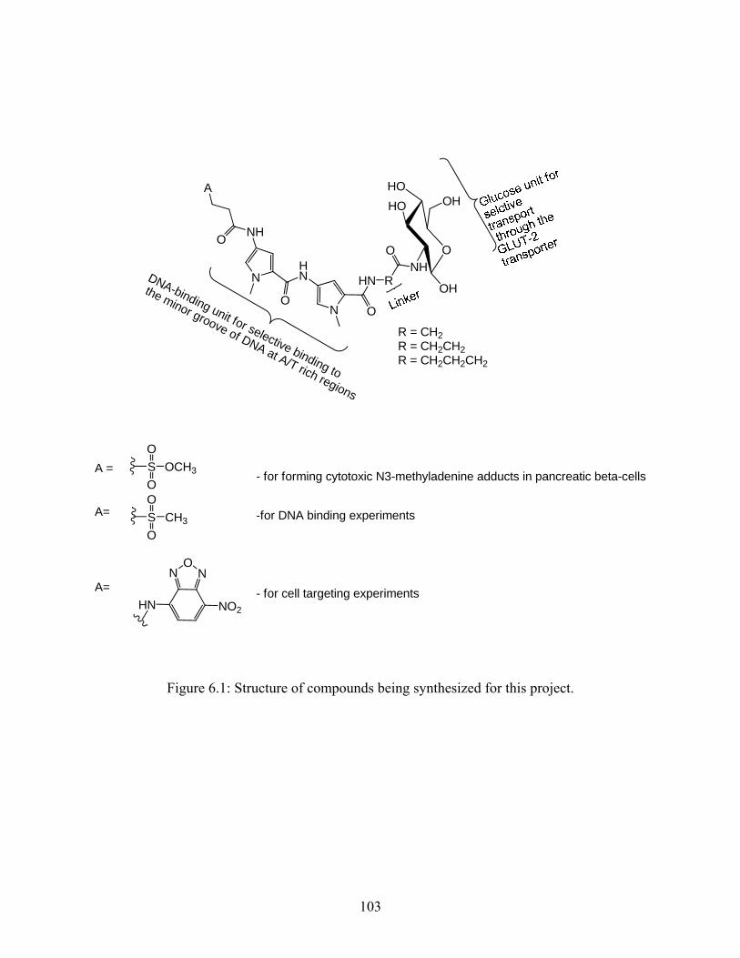
NHN
N
HN R
O
ONH
O
O
A
O
HOHO
NH
OH
OH
R = CH2R = CH2CH2R = CH2CH2CH2
A =
A=
A=
SO
OOCH3
SO
OCH3
NO
N
NO2HN
- for forming cytotoxic N3-methyladenine adducts in pancreatic beta-cells
-for DNA binding experiments
- for cell targeting experiments
DNA-binding unit for selective binding to
the minor groove of DNA at A/T rich regions
Figure 6.1: Structure of compounds being synthesized for this project.
103
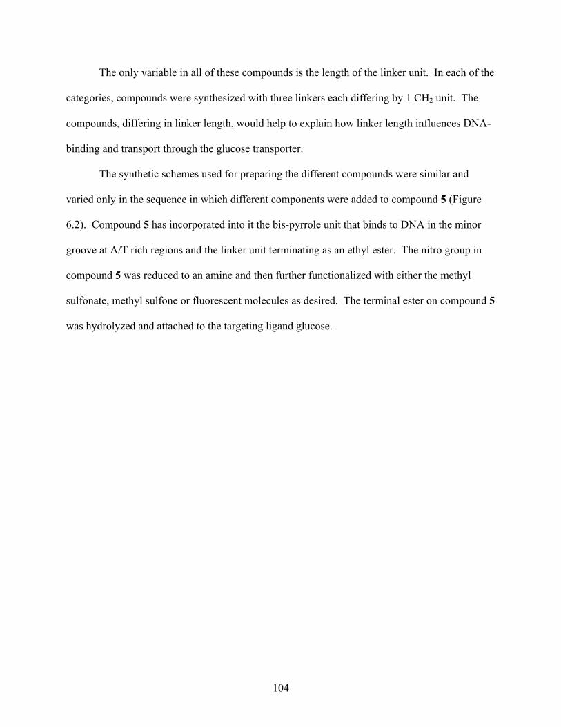
The only variable in all of these compounds is the length of the linker unit. In each of the
categories, compounds were synthesized with three linkers each differing by 1 CH2 unit. The
compounds, differing in linker length, would help to explain how linker length influences DNA-
binding and transport through the glucose transporter.
The synthetic schemes used for preparing the different compounds were similar and
varied only in the sequence in which different components were added to compound 5 (Figure
6.2). Compound 5 has incorporated into it the bis-pyrrole unit that binds to DNA in the minor
groove at A/T rich regions and the linker unit terminating as an ethyl ester. The nitro group in
compound 5 was reduced to an amine and then further functionalized with either the methyl
sulfonate, methyl sulfone or fluorescent molecules as desired. The terminal ester on compound 5
was hydrolyzed and attached to the targeting ligand glucose.
104
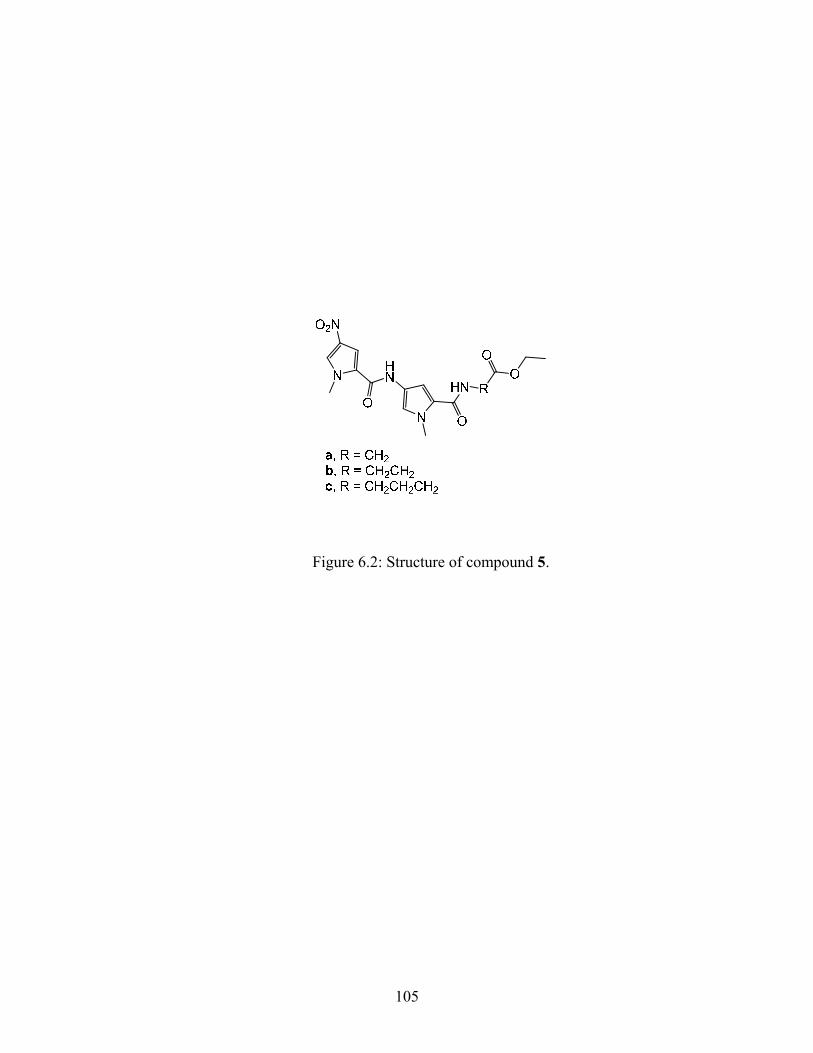
Figure 6.2: Structure of compound 5.
105
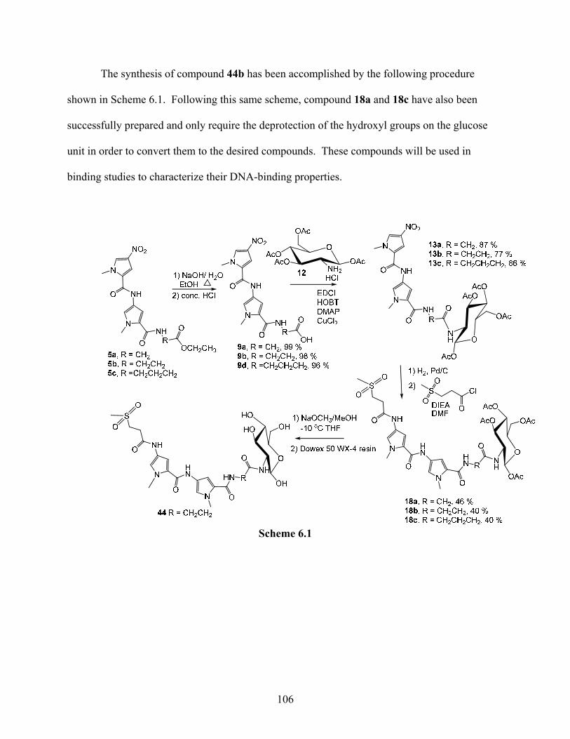
The synthesis of compound 44b has been accomplished by the following procedure
shown in Scheme 6.1. Following this same scheme, compound 18a and 18c have also been
successfully prepared and only require the deprotection of the hydroxyl groups on the glucose
unit in order to convert them to the desired compounds. These compounds will be used in
binding studies to characterize their DNA-binding properties.
Scheme 6.1
106
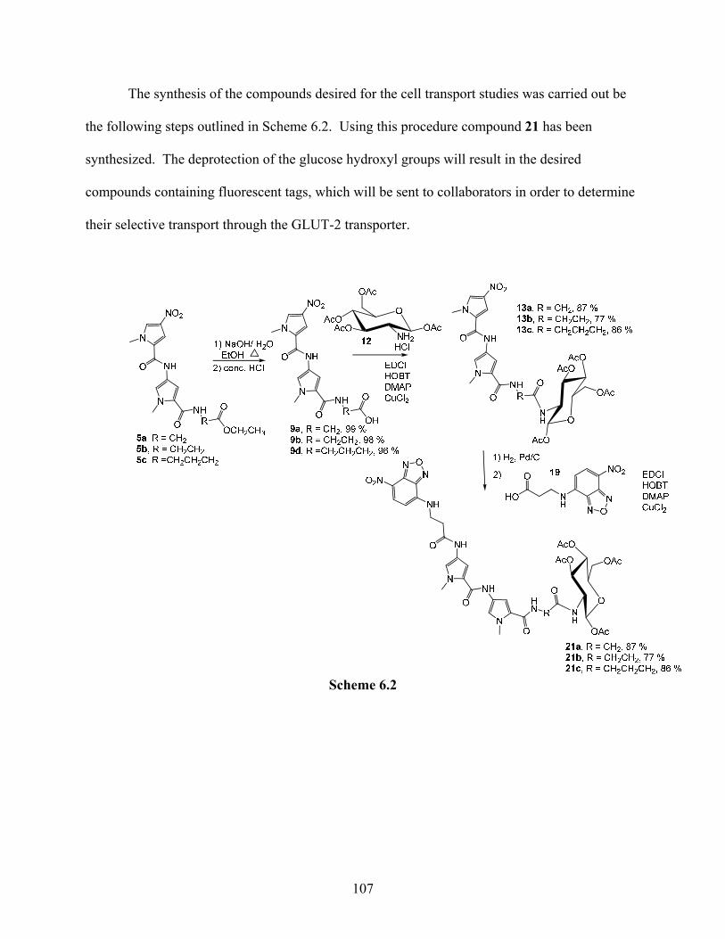
The synthesis of the compounds desired for the cell transport studies was carried out be
the following steps outlined in Scheme 6.2. Using this procedure compound 21 has been
synthesized. The deprotection of the glucose hydroxyl groups will result in the desired
compounds containing fluorescent tags, which will be sent to collaborators in order to determine
their selective transport through the GLUT-2 transporter.
Scheme 6.2
107
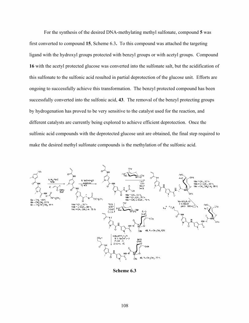
For the synthesis of the desired DNA-methylating methyl sulfonate, compound 5 was
first converted to compound 15, Scheme 6.3. To this compound was attached the targeting
ligand with the hydroxyl groups protected with benzyl groups or with acetyl groups. Compound
16 with the acetyl protected glucose was converted into the sulfonate salt, but the acidification of
this sulfonate to the sulfonic acid resulted in partial deprotection of the glucose unit. Efforts are
ongoing to successfully achieve this transformation. The benzyl protected compound has been
successfully converted into the sulfonic acid, 43. The removal of the benzyl protecting groups
by hydrogenation has proved to be very sensitive to the catalyst used for the reaction, and
different catalysts are currently being explored to achieve efficient deprotection. Once the
sulfonic acid compounds with the deprotected glucose unit are obtained, the final step required to
make the desired methyl sulfonate compounds is the methylation of the sulfonic acid.
Scheme 6.3
108
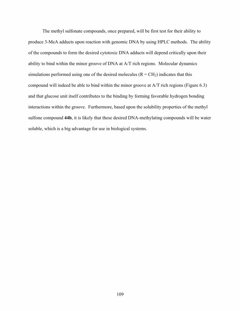
The methyl sulfonate compounds, once prepared, will be first test for their ability to
produce 3-MeA adducts upon reaction with genomic DNA by using HPLC methods. The ability
of the compounds to form the desired cytotoxic DNA adducts will depend critically upon their
ability to bind within the minor groove of DNA at A/T rich regions. Molecular dynamics
simulations performed using one of the desired molecules (R = CH2) indicates that this
compound will indeed be able to bind within the minor groove at A/T rich regions (Figure 6.3)
and that glucose unit itself contributes to the binding by forming favorable hydrogen bonding
interactions within the groove. Furthermore, based upon the solubility properties of the methyl
sulfone compound 44b, it is likely that these desired DNA-methylating compounds will be water
soluble, which is a big advantage for use in biological systems.
109
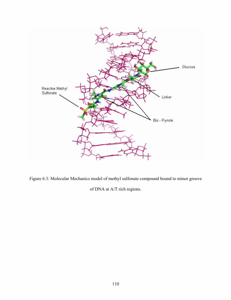
Figure 6.3: Molecular Mechanics model of methyl sulfonate compound bound to minor groove
of DNA at A/T rich regions.
110
Precursor compounds in all three categories have been synthesized with three different
linkers each differing by 1 CH2 group. The differences between the properties of these
compounds varying in linker-length will be used to identify the optimum length required for
DNA-binding, cell-transport, and overall cell-toxicity. If desired, the strength of DNA-binding
can be improved by introducing a third pyrrole unit into the molecule. The decreased water
solubility due to this third pyrrole is not expected to be critical since the glucose unit provides
significant water solubility.
In transport experiments, the presence of the fluorescent tags may interfere with the
passage of the compounds through the GLUT-2 transporter. This is one of the reasons why two
different fluorescent tags are being explored. If the fluorescently tagged compounds are unable
to get into cells via the transporter, it does not necessarily imply that the methyl sulfonate
compounds will be unable to traverse through these transporters. However, if these compounds
with the tags do selectively get into cells containing the GLUT-2 glucose transporter, then it is
likely that the methyl sulfonate compounds too will be able to pass through these transporters.
111
References
1. Lawley, P.D, et al. “Carcinogenesis by Alkylating Agents.” ChemicalCarcinogens.
1984, 1, 325-484, American Chemical Society, Washington, DC.
2. Pratt, W.B.; Ruddon, R. W.; Ensminger, W.D.; Maybaum, J.; et al. The Anitcancer
Drugs. 1994, 2nd ed.Oxford Press, New York.
3. Henry-Amar M, Dietrich PY. Hematoglogy/Onocology Clinks of North America:
Therapy-Related Second Malignancies. 1993, Vol. 7. Philadelphia: W.B. Saunders.
4. Smith, M.A., McCaffrey, R.P., and Karp J.E.. J. Natl. Cancer. Inst. 1996, 88, 407-418.
5. Encell, L.; Shuker, D.E.G.; Foiles, P.G; Gold, B. “The in Vitro Methylation of DNA by a
Minor Groove Binding Methyl Sulfonate Ester.” Chem. Res. Tox. 1996, 9(3), 563-567.
6. Zhang, Y.; Chen, F.; Mehta, P.; Gold, B. “Groove- and Sequence-Selective Alkylation of
DNA by Sulfonate Esters Tethered to Lexitropsin.” Biochemistry. 1993, 32, 7954-7965.
7. Fronza, G.: Gold, B. “The Biological Effects of N3-Methyladenine.” Journal of Cellular
Biochemistry. 2004, 91, 250-257.
8. Encell, L; Shuker, DEG; Folies P.G., Gold, B. “The In Vitro Methylation of DNA By A
Minor Groove Binding Methyl Sulfonate Ester.” Chem. Res. Toxicol. 1996, 9, 563-567.
9. Bolzán, A; Bianchi, M. “Genotoxicity of Streptozotocin.” Mutation Research. 2002, 512
121-134.
10. Elsner, M; Guldbakke, B; Tiedge M; Munday, R; Lenzen, S. “Relative Importance of
Transport and Alkylation for Pancreatic Beta-Cell Toxicity of Streptozotocin.”
Diabetologia. 2000, 43, 1528-1533.
11. Uldry, M., Ibberson, M., Hosokawa, B.T. “GLUT-2 is a high affinity glucosamine
transporter.” FEBS Letters. 2002, 524, 199-203.
112
12. Yang, H.; Jr. Wright, J.R. “Human β Cells Are Exceedingly Resistant to Streptozotocin
in Vitro.” Endocrinology. 2002. 143(7), 2491-2495.
13. Greiner, D.L; Rossini, A.A.; Mordes, J.P. “Translating Data from Animal Models into
Methods for Prevention Human Autoimmune Diabetes Mellitus: Caveat Emptor and
Primum non Nocere.” Clinical Immunology. 2001, 100(2). 134-143.
14. Wang, Z; Gleichmann, H.; “Glucose Transporter 2 Expression: Prevention of
Streptozotocin-Induced Reduction in Beta-Cells With 5-Thio-D-Glucose.” Exp. Clin.
Endocrinology & Diabetes. 1995, 103(2), 83-87.
15. Cardinal, J.W.; Margison, G.P.; Mynett, K.J.; Yates, A.P.; Cameron, D.P.; Elder R.H.
“Increased Susceptibility to Streptozotocin-Induced β-cell Apoptosis and Delayed
Autoimmune Diabetes in Alkylpurine-DNA-N-glycosylase-deficient Mice.” Mol. Cell.
Biol. 2001, 21, 5605-5613.
16. Zheng, G.; Glickson, J.D.; Chance, B. “Preparation of 2-aminodeoxy-gluose deriviatives
as antieoplastic agents targeted via GLUT transporters.” PCT Int. Appl. 2004, 71.
CODEN:PIXXD2 WO 2004110255 A2 20041223 CAN 142:56618 AN 2004:1124548
CAPLUS.
17. Tentori, L.; Olindo, F.; Fossile, E.; Muzi, A.; Vergati, M.; Portarena, I; Amiei, C.; Gold,
B.; Graziani, G. “N3-Methyladenine Induces Early Poly(ADP-Ribosylation), Reduciton
of Nuclear Factor-κB DNA Binding Ability, and Nuclear Up-Regulation of Telomerase
Activity.” Molcular Pharmacology. 2004. 67, 572-581.
18. Beranek, D.T.; Weis, C.C.; Swenson, D.H. “A Comprehensive Quantitative Analysis of
Methylated and Ethylated DNA Using HPLC.” Carcinogenesis. 1980, 1, 595-605.
113
19. Shah, D.; Barry, G. “Evidence in Escherichia coli that N3-Methyladenien Lesions and
Cytotoxicity Induced by a Minor Groove Binding Methyl Sulfonate Ester Can Be
Modulated in Vivo by Netropsin.” Biochemistry. 2003, 42, 12610-12626.
20. Varadarajan, S.; Shah, D.; Dande, P.; Settles, S.; Chen, F.; Fronza, G.; Gold, B. “DNA
Damage and Cytotoxicity Induced by Minor Groove Binding Methyl Sulfonate Esters.”
Biochemistry. 2003, 42, 14318-14327.
21. Monti, P.; Iannone, R.; Campomenosi, P; Ciribilli, Y.; Varadarajan, S.; Shah, D.:
Menichini, P.; Gold, B.; Fronza, G. “Nucleotide Excision Repair Defect
Influences Lethality and Mutagenicity Induced by Me-Lex, a Sequence-Selective N3-
Adenine Methylating Agent in the Absence of Base Excision Repair.” Biochemistry.
2004, 43, 5592-5599.
22. Engelward, B.P.; Allan, J.M.; Dreslin, A.J.; Kelly, J.D.; Gold, B.; Samson, L.D. “A
Chemical and Genetic Approach Together Define the Biological Consequence of 3-
Methyladenine Lesion in the Mammalian Genome.” J. Biolo. Chem. 1998, 273, 5412-
5418.
23. Shah, D.; Kelly, J.; Zhang, Y.; Prasad, D.; Martinez, J.;Ortiz, G.; Fronza, G.; Tran, H.;
Soto, A.; Marky, L.; Gold, B.; “Evidence in Escherichia coli that N3-Methyladenine
Lesions Induced by a Minor Groove Binding Methyl Sulfonate Ester Can Be Processed
by both Base and Nucleotide Excision Repair.” Biochemistry. 2001, 40, 1796-1803.
24. Ito, Mikio. “Streptozocin-induced diabetes animal models.” Sogo Rinshu. 2004, 53 (4),
1479-1481.
25. Schnedl, W.J.; Ferber, S.; Johnson, J.H.; Newgard, C.B. “STZ transport and cytotoxicity.
Specific enhancement in GLUT2-expressing cells.” Diabetes. 1994, 43(11), 1326-33.
114
26. Gilbert, E. Sulfonation and Related Reactions. “Alkenes and Alkynes with Bisulfite.”
John Wiley & Sons, Inc. 1965, 148-149.
27. Wang, Z.; Gleichmann, H. “GLUT2 in pancreatic islets: crucial target molecule in
Diabetes induced with multiple low doses of Streptozotocin in mice.” Diabetes. 1998,
47(1), 50-6.
28. Medeyes, A; Farkas, E; Lipták, A; Pozsgay, V. “Synthesis of the Monosaccharide Units
of the O-Specific Polysaccharide of Shigella Sonnei.” Tetrahedron. 1997, 53(12), 4159-
4178.
29. Mayer, T.G; Weingart, R; Münstermann, F; Kawada, T; Kurzchalia, T; Schmidt, R.
“Synthesis of Labeled Glycosyl Phosphatidyl Inositiol (GPI) Anchors.” Eur. J. Org.
Chem. 1999, 2563-2571.
30. Giordano. C.; Gallina, C.; Consalvi, V.; Scandurra, R. “Synthesis of properties of D-
glucosamine N-peptidyl derivatives as substrate analog inhibitor of papain and cathepsin
B.” Eur. J. Med. Chem. 1991. 26, 753-762.
31. Chauviére, G.; Bouteille, B.; Enanga, B.; Albuquerque, C.; Croft, S.; Dumas, M.; Périé, J.
“Synthesis and Biological Activity of Nitro Heterocyles Analogous to Megazol, a
Trypanocidal Lead.” J. Med. Chem. 2003. 46(3), 427-440.
32. Kanaoka, Y.; Ikeuchi, Y.; Kawamoto, T.; Bessho, K.; Akimoto, N.; Mikata, Y.; Mishida,
M.; Yano, S.; Sasaki, T.; Fumio, Y. “Synthesis and Evaluation of Nitro 5-Deazaflavin-
pyrrolecarboxamide(s) Hybrid Molecules as Novel DNA Targeted Bioreductive
Antitumor Agents.” Bioorg. Med. Chem. 1998. 6, 301-314.
34. Xia, J.; Yuan, G.; Huang.; Chan, A.; Lee, D. “A Convenient Method for the Synthesis of
DNA-Recognizing Polyamides in Solution.” J. Org. Chem. 2000, 65, 5506-5513.
115
35. Aly, M.R.E; Schmidt, R. “New Diacylamino Protecting Groups for Glucosamine.” Eur.
J. Org. Chem. 2005, 4382-4392.
36. Grayson, I. “Water-soluble carbodiimide - an efficient agent for synthesis.”
Pharmaceutical Intermediates. 2000, 86-88.
37. Miyazawa, T.; Otomatsu, T.; Fukui, Y.; Yamada, T.; Kuwata, S. “Racemization – free
and Efficient Peptide Synthesis by the Carbodiimide Method using 1-
Hydroxybenzotriazole and Copper (II) Chloride simultaneously as Additives.” J. Chem.
Soc., Chem. Commun. 1988, 419-420.
38. Fuhrhop, J.; David, H.; Mathiew, J.; Liman, U.; Winter, H.; Boekema, E.
“Bolaamiphiphiles and Monolayer Lipid Membranes Made from 1, 6, 19, 24-Tetraoxa-3,
21-cyclohexatriacontadiene-2, 5, 20, 23-tetrone.” J. Am. Chem. Soc. 1986, 108, 1785-
1791.
39. Zhang, Y.; Chen, F.; Mehta, P.; Gold, B. “Groove- and Sequence-Selective Alkylation of
DNA by Sulfonate Esters Tethered to Lexitropsin.” Biochemistry. 1993, 32, 7954-7965.
40. Niknam, K.; Kiasat, A.R.; Karimi, S. “Dowex Polymer-Mediated Protection of Carbonyl
Groups.” Synthetic Communications. 2005. 35, 2231-2236.
41. Wang, D.; Chen, C. “Synthesis and Biological Evaluation of L-α-Phosphatidyl-D-3-
deoxy-3-heteromethyl-myo-inositols as Phosphoinositide 3-Kinase Inhibitors.”
Bioorganic & Med. Chem. 2001. 9, 3165-3172.
42. Plattner, J.J.; Gless, R.D.; Rapoport, H. “Synthesis of Some DE and CDE Ring Analogs
of Camptothecin.” J. Am. Chem. Soc. 1972. 94 (24), 8613-8615.
43. Richtmyer, N,K. “The Cleavage of Glycosides by Catalytic Hydrogenation.” 1934. 56,
1633-1635.
116
44. Liberek, B.; Melcer, A.; Osuch, A.; Wakieć, R.; Milewski, S.; Wiśniewski, A. “N-Alkyl
derivative of 2-amino-2deoxy-D-glucose.” Carbohydrate Research. 2005, 340, 1876-
1884.
117
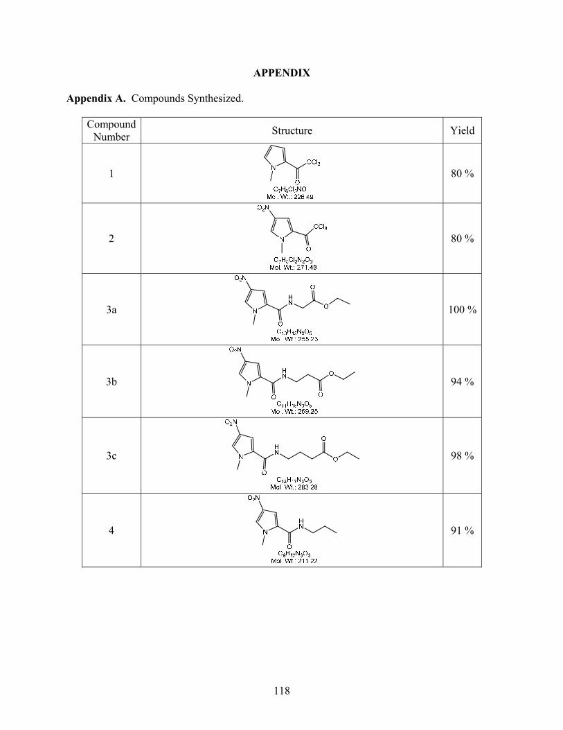
APPENDIX
Appendix A. Compounds Synthesized.
Compound Number Structure Yield
1
80 %
2
80 %
3a
100 %
3b
94 %
3c
98 %
4
91 %
118
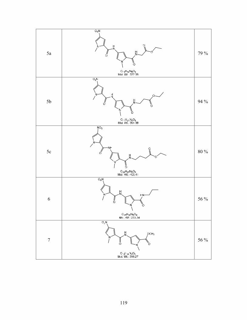
5a
79 %
5b
94 %
5c
80 %
6
56 %
7
56 %
119
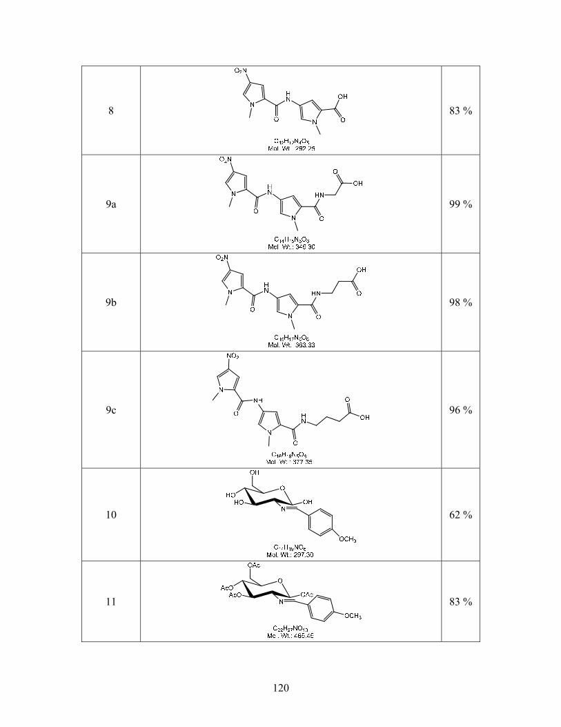
8
83 %
9a
99 %
9b
98 %
9c
96 %
10
62 %
11
83 %
120
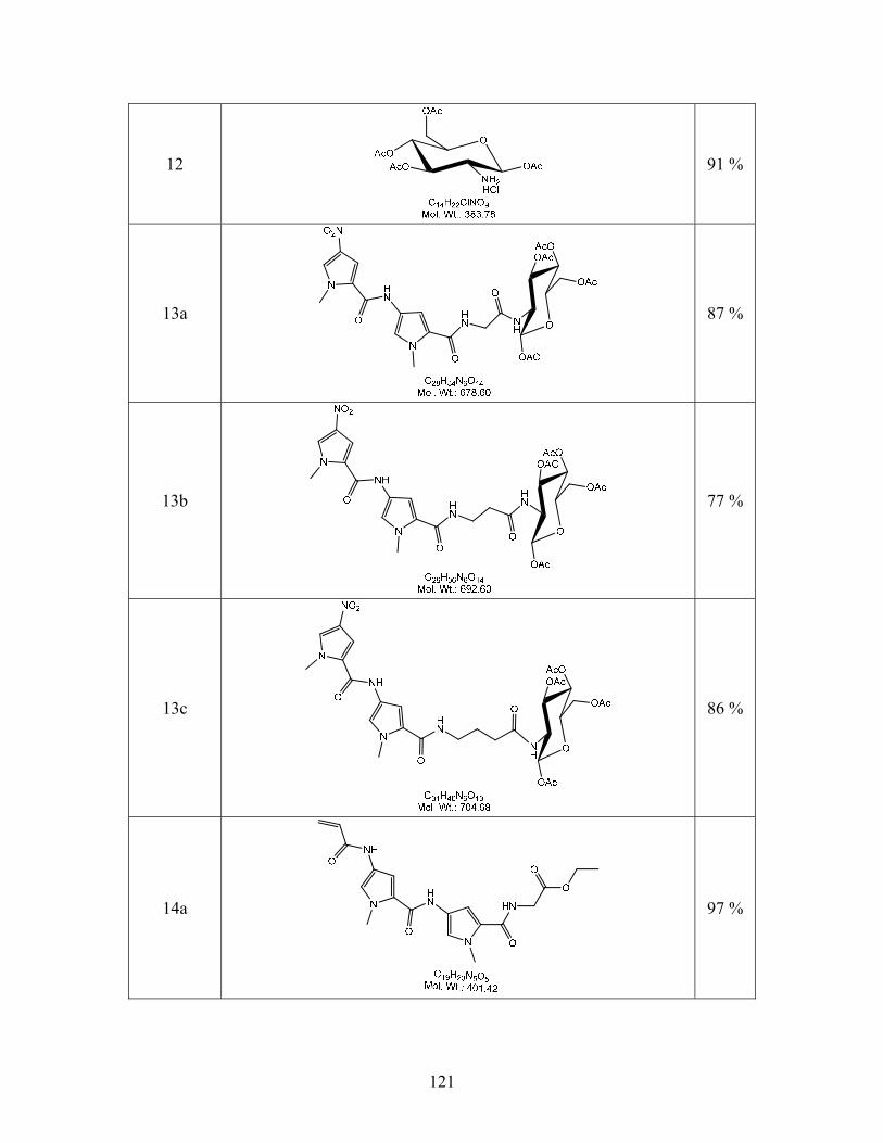
12
91 %
13a
87 %
13b
77 %
13c
86 %
14a
97 %
121
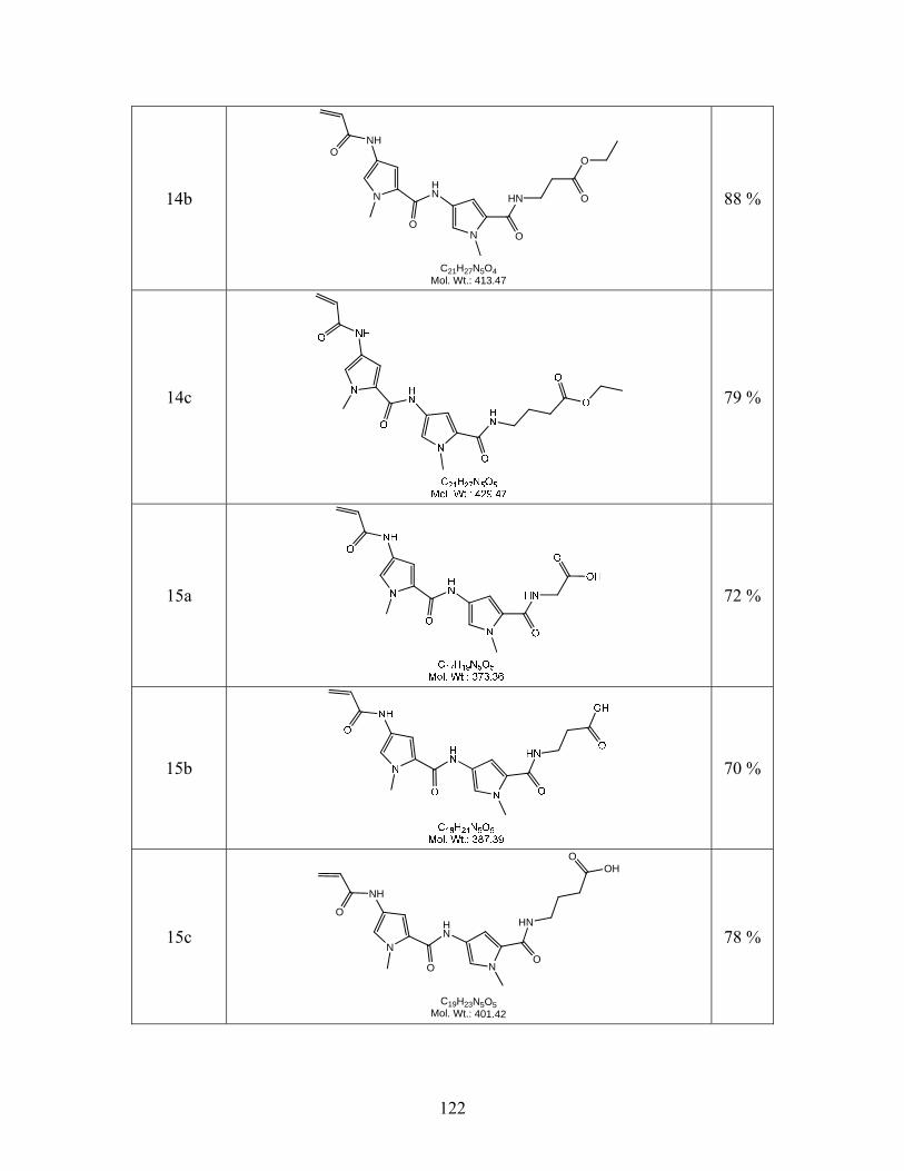
14b N
N
NHO
O
HN
O
HN
O
O
C21H27N5O4Mol. Wt.: 413.47
88 %
14c
79 %
15a
72 %
15b
70 %
15c N
N
NH
O
O
HN
O
HN
OHO
C19H23N5O5Mol. Wt.: 401.42
78 %
122
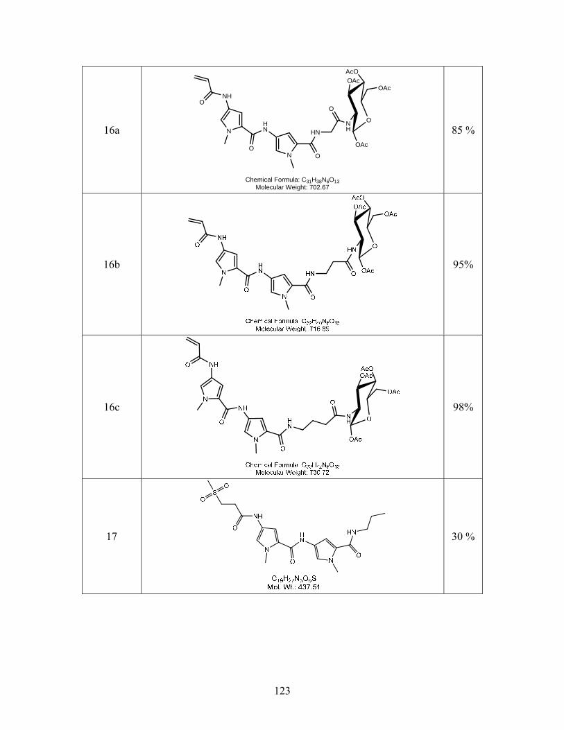
16a N
N
NHO
O
HN
O
HN
O
O
AcOOAc
NH
OAc
OAc
Chemical Formula: C31H38N6O13Molecular Weight: 702.67
85 %
16b
95%
16c
98%
17
30 %
123
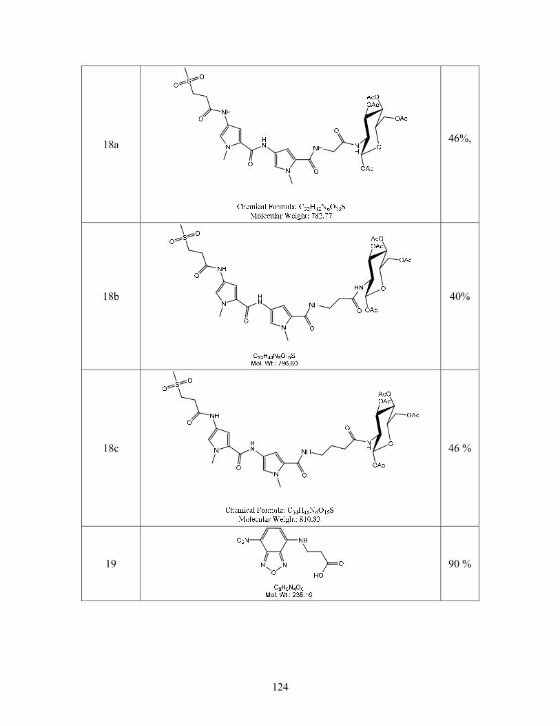
18a
46%,
18b
40%
18c
46 %
19
90 %
124
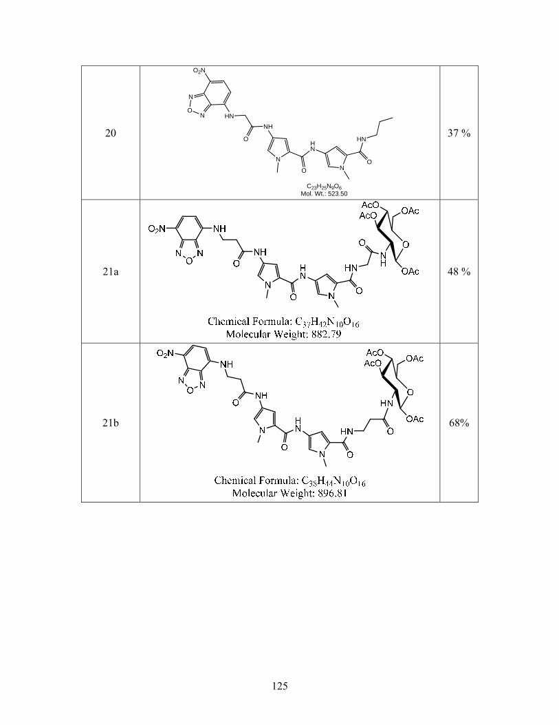
20
N
ON HN
O2N
O
NH
N
O
HN
NO
HN
C23H25N9O6Mol. Wt.: 523.50
37 %
21a
48 %
21b
68%
125
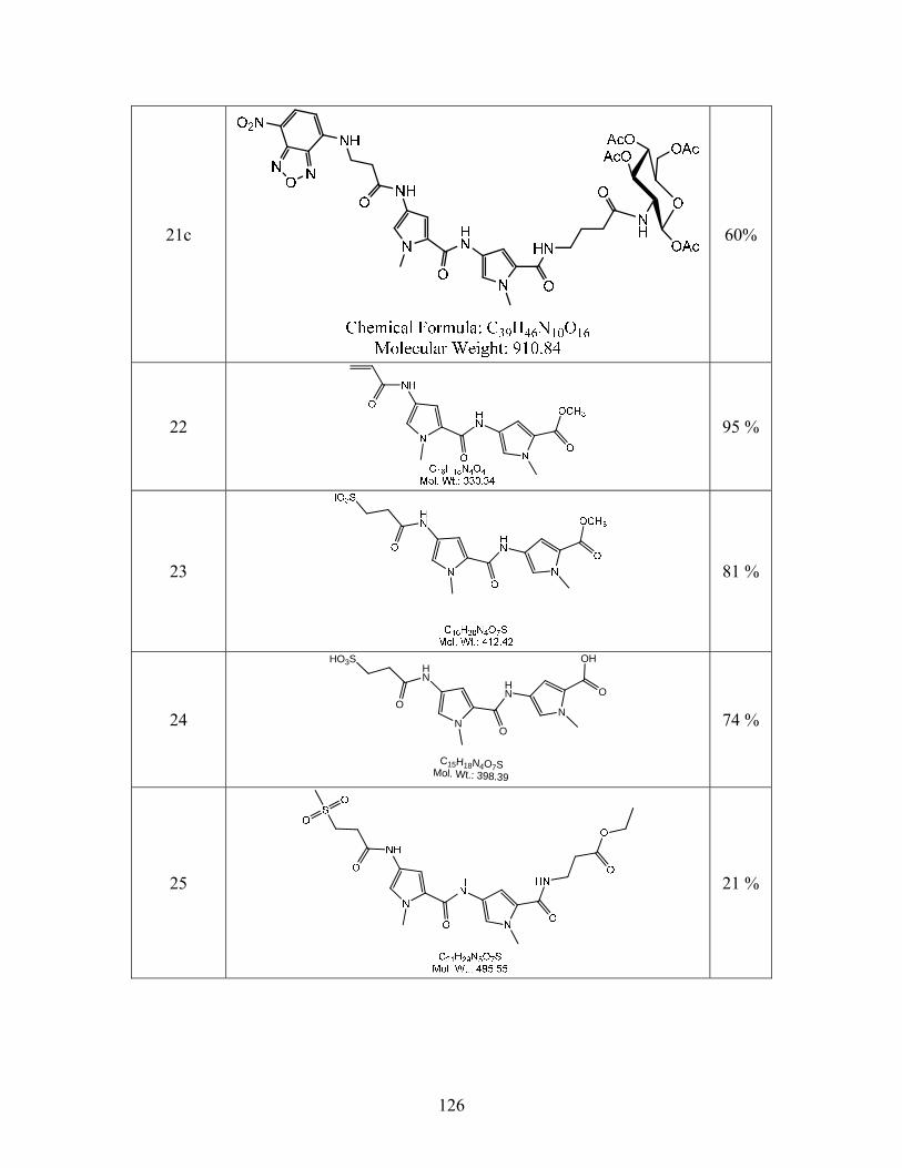
21c
60%
22
95 %
23
81 %
24 N
HN
O
HN
N
O
OHHO3S
O
C15H18N4O7SMol. Wt.: 398.39
74 %
25
21 %
126
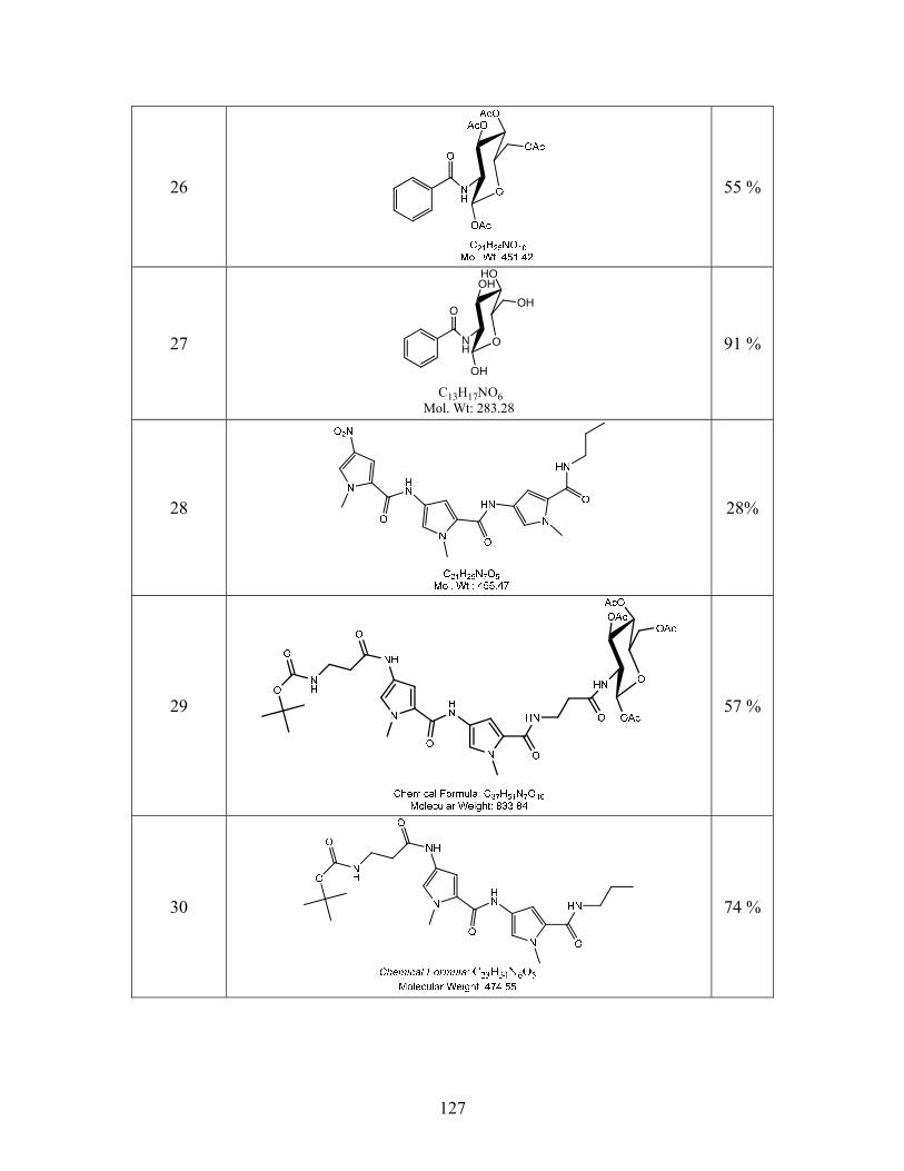
26
55 %
27
O
O
HOOH
NH
OH
OH
C13H17NO6Mol. Wt: 283.28
91 %
28
28%
29
57 %
30
74 %
127
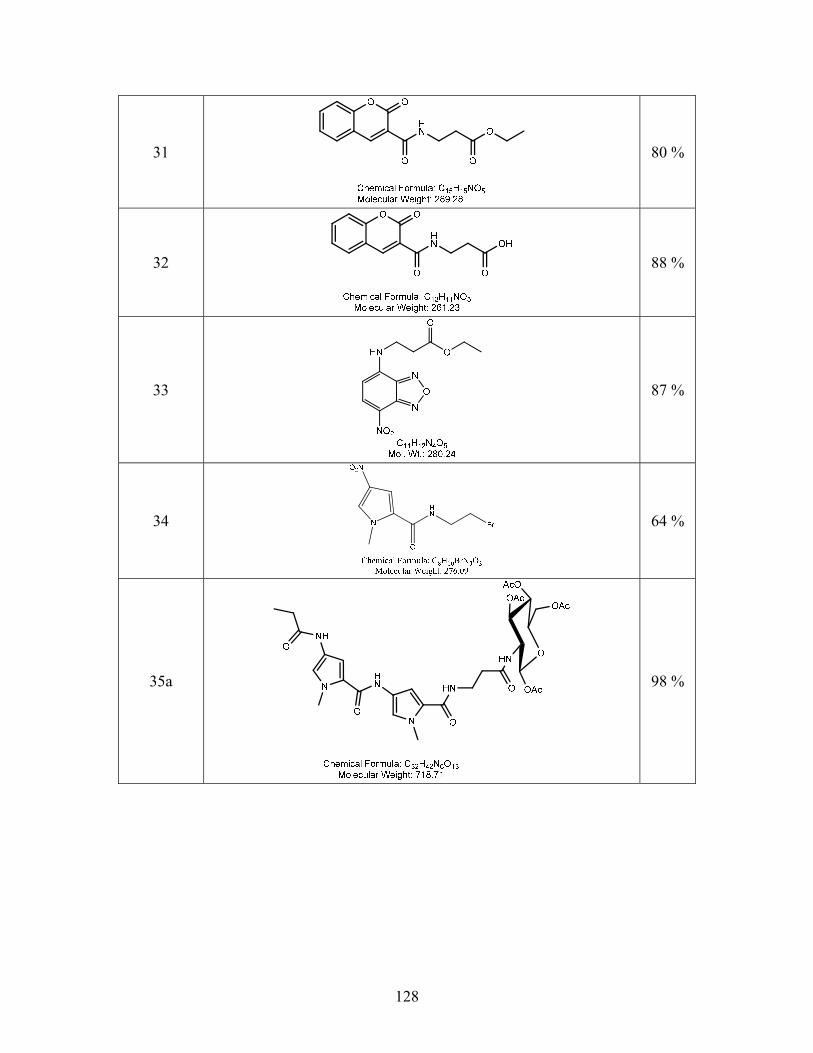
31
80 %
32
88 %
33
87 %
34
64 %
35a
98 %
128
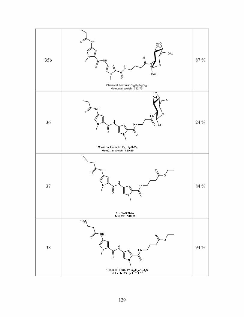
35b N
N
NHO
O
NH
O
HN
O
AcOOAc
NH
OAc
OAcO
Chemical Formula: C33H44N6O13Molecular Weight: 732.73
87 %
36
24 %
37
84 %
38
94 %
129
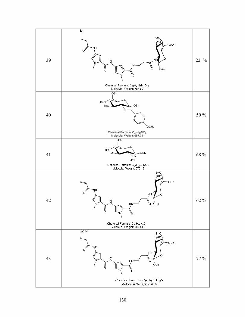
39
22 %
40
OBnO
BnON
OBn
OBn
OCH3
Chemical Formula: C42H43NO6Molecular Weight: 657.79
50 %
41
68 %
42
62 %
130
43
77 %
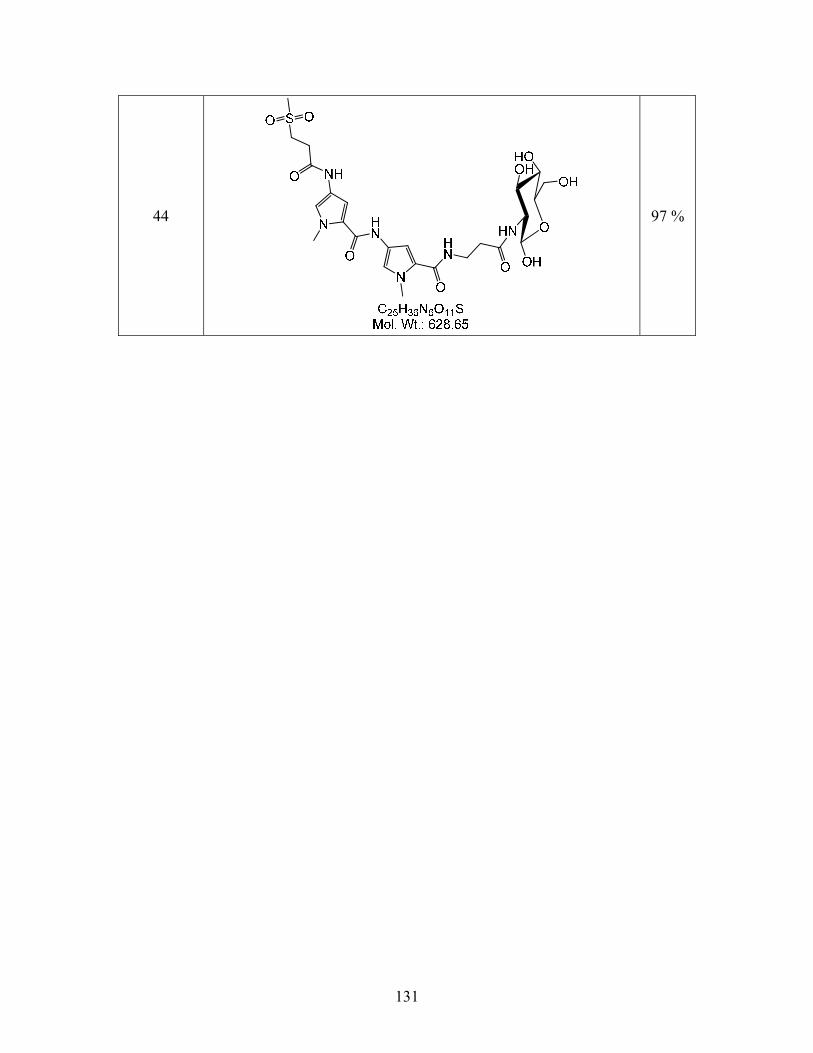
44
97 %
131
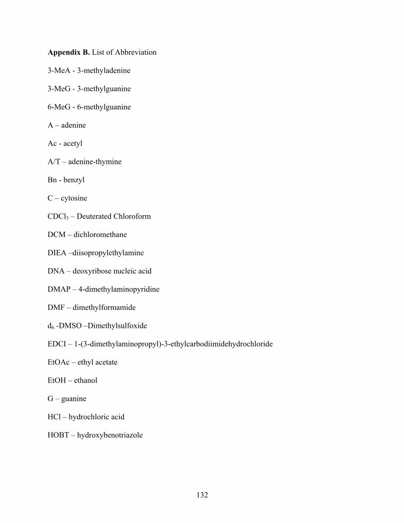
Appendix B. List of Abbreviation
3-MeA - 3-methyladenine
3-MeG - 3-methylguanine
6-MeG - 6-methylguanine
A – adenine
Ac - acetyl
A/T – adenine-thymine
Bn - benzyl
C – cytosine
CDCl3 – Deuterated Chloroform
DCM – dichloromethane
DIEA –diisopropylethylamine
DNA – deoxyribose nucleic acid
DMAP – 4-dimethylaminopyridine
DMF – dimethylformamide
d6 -DMSO –Dimethylsulfoxide
EDCI – 1-(3-dimethylaminopropyl)-3-ethylcarbodiimidehydrochloride
EtOAc – ethyl acetate
EtOH – ethanol
G – guanine
HCl – hydrochloric acid
HOBT – hydroxybenotriazole
132
Me-lex – methyl 3-(1-methyl-5-(1-mehtyl-5-(propylcarbomyl)-1H-pyrrol-3-ylcarbamoyl)-1H-
pyrrol-3-ylamino)-3oxopropane-1-sulfonate
MeOH – methanol
Mp – melting point
NaOCH3 – sodium methoxide
NaOH –sodium hydroxide
NMR – nuclear magnetic resonance
Pd/C – palladium on carbon
Rf – retention factor
TEA – triethylamine
THF – tetrahydrofuran
TLC – thin layer chromatography
T - thymine
133