Synthesis of platinum nanoparticle chains based on α-chymotrpsin fibrils
4
Synthesis of platinum nanoparticle chains based on α-chymotrpsin fibrils Yifen Xian, Faming Gao n , Bin Cai Key Laboratory of Applied Chemistry, Yanshan University, Qinhuangdao 066004, PR China article info Article history: Received 25 July 2013 Accepted 10 August 2013 Available online 17 August 2013 Keywords: Pt nanoparticle chains Nanocrystalline materials α-chymotrpsin amyloid fibrils Electronic microscopy Methanol oxidation Oxygen reduction reaction abstract Pt nanoparticle chains (PtNPCS) with diameter ranging from 15–20 nm and length to micrometers were fabricated using α-chymotrpsin amyloid fibrils (α-CTAFS) as a sacrificial template. The PtNPCs can be simply obtained by reducing the salt precursors using two different reducing agents, borane-dimethyl- amine complex (DMAB) and sodium borohydride (NaBH 4 ), in aqueous environment. Electrochemical measurements were used to study the properties of the PtNPCs that achieved from different reducing ways. Results indicates that DMAB is a facile reducing agent to prepare PtNPCs with a more uniform particle size and higher catalytic activity on methanol oxidation (MOR) and oxygen reduction reaction (ORR) than that obtained by using NaBH 4 as reductant. & 2013 Elsevier B.V. All rights reserved. 1. Introduction One-dimensional (1-D) platinum nanostructures have drawn increased attention from the research community due to their critical application as catalysts in many industrial processes, such as electromechanical resonator [1] and especially enhanced elec- trocatalysts in fuel cells [2]. Compared to the conventional zero- dimensional (0-D) Pt nanoparticles serving as the electrocatalysts, 1-D structures possess (a) higher aspect ratios, (b) fewer defect sites, (c) fewer lattice boundaries, and (d) higher numbers of surface atoms, all of which are desirable attributes for fuel cell catalysts [3,4]. In addition, 1-D structures maintain improved electron transport characteristics as a result of the path-directing effects of the structural anisotropy [5,6]. Up to now, the general methods employed for the preparation of 1-D Pt nanostructures are usually based on template-directed synthesis [7–10]. Recent year has seen an explosive development in the amyloid fibers-template metal nanostructures. Amyloid-like fibrils are elongated, unbranched protein fibrils with diameters ranging from 5 to 14 nm [11]. The amyloid fibrils have very high stability and attractive mechanical properties with comparable stiffness to some of the strongest known materials [12]. Thomas Scheibel and co-workers [13] used the N-terminal and middle region (NM) of Sup35p from Saccharomyces cerevisiae to produce 10-nm-wide protein fibers and link it to colloidal gold particles to obtain 100 nm wide silver and gold nanowires. Meital Reches and co-workers [14] used NH 2 –Phe–Phe–COOH dipeptide to form fibrillar structures and cast silver nanowires. Furthermore, our group [15–17] synthesized highly uniform single-crystal ultrathin Pt nanowires based on insulin amyloid fibrils. In this paper, we use α-chymotrpsin amyloid fibrils (α-CTAFS) as a scaffold to conduct pt nanoparticle chains (PtNPCs). 2. Experimental section The α-CTAFS were prepared from bovine α-CT powder as a modification protocol described by Nasrollah Rezaei-Ghaleh et al. [18]. In brief, the α-CT protein concentration was 1.5 mg/mL and Glycine-HCl (10 mM, pH 2.5) was used as buffer, the TFE was added at final concentration of 12% (v/v) and the resultant protein solution was incubated at 25 1C for specific time with gentle agitation. Immediately following the formation of α-CTAFS, PtCl 4 was dissolved in Glycine-HCl buffer (10 mM, pH 2.5) to form [PtCl x ] 4 x complex solution. Then a certain quantity of [PtCl x ] 4 x (5 mM, 80 mL) was added into the α-CTAFS (1.5 mg/mL, 200 μL) and the mixture was gently stirred at 15 1C for 10 h. Subsequently, reduction process was carried out by adding excess of fresh DMAB or NaBH 4 (5 mM) solution, drop by drop with the interval of 2 min. 3. Results and discussion Thioflavin-T (ThT) is widely used to detect the formation of amyloid fibril. When there is amyloid or amyloid like fibril exists, it will manifest a dramatic increase in fluorescence intensity [19]. Fig. 1(a) shows that at 483 nm, ThT fluorescence was substantially Contents lists available at ScienceDirect journal homepage: www.elsevier.com/locate/matlet Materials Letters 0167-577X/$- see front matter & 2013 Elsevier B.V. All rights reserved. http://dx.doi.org/10.1016/j.matlet.2013.08.051 n Corresponding author. Tel.: þ86 335 8387552; fax: þ86 335 8061569. E-mail address: [email protected] (F. Gao). Materials Letters 111 (2013) 39–42
Transcript of Synthesis of platinum nanoparticle chains based on α-chymotrpsin fibrils
Synthesis of platinum nanoparticle chains basedon α-chymotrpsin fibrils
Yifen Xian, Faming Gao n, Bin CaiKey Laboratory of Applied Chemistry, Yanshan University, Qinhuangdao 066004, PR China
a r t i c l e i n f o
Article history:Received 25 July 2013Accepted 10 August 2013Available online 17 August 2013
Keywords:Pt nanoparticle chainsNanocrystalline materialsα-chymotrpsin amyloid fibrilsElectronic microscopyMethanol oxidationOxygen reduction reaction
a b s t r a c t
Pt nanoparticle chains (PtNPCS) with diameter ranging from 15–20 nm and length to micrometers werefabricated using α-chymotrpsin amyloid fibrils (α-CTAFS) as a sacrificial template. The PtNPCs can besimply obtained by reducing the salt precursors using two different reducing agents, borane-dimethyl-amine complex (DMAB) and sodium borohydride (NaBH4), in aqueous environment. Electrochemicalmeasurements were used to study the properties of the PtNPCs that achieved from different reducingways. Results indicates that DMAB is a facile reducing agent to prepare PtNPCs with a more uniformparticle size and higher catalytic activity on methanol oxidation (MOR) and oxygen reduction reaction(ORR) than that obtained by using NaBH4 as reductant.
& 2013 Elsevier B.V. All rights reserved.
1. Introduction
One-dimensional (1-D) platinum nanostructures have drawnincreased attention from the research community due to theircritical application as catalysts in many industrial processes, suchas electromechanical resonator [1] and especially enhanced elec-trocatalysts in fuel cells [2]. Compared to the conventional zero-dimensional (0-D) Pt nanoparticles serving as the electrocatalysts,1-D structures possess (a) higher aspect ratios, (b) fewer defectsites, (c) fewer lattice boundaries, and (d) higher numbers ofsurface atoms, all of which are desirable attributes for fuel cellcatalysts [3,4]. In addition, 1-D structures maintain improvedelectron transport characteristics as a result of the path-directingeffects of the structural anisotropy [5,6].
Up to now, the general methods employed for the preparationof 1-D Pt nanostructures are usually based on template-directedsynthesis [7–10].
Recent year has seen an explosive development in the amyloidfibers-template metal nanostructures. Amyloid-like fibrils areelongated, unbranched protein fibrils with diameters ranging from5 to 14 nm [11]. The amyloid fibrils have very high stabilityand attractive mechanical properties with comparable stiffnessto some of the strongest known materials [12]. Thomas Scheibeland co-workers [13] used the N-terminal and middle region (NM)of Sup35p from Saccharomyces cerevisiae to produce 10-nm-wideprotein fibers and link it to colloidal gold particles to obtain100 nm wide silver and gold nanowires. Meital Reches and
co-workers [14] used NH2–Phe–Phe–COOH dipeptide to formfibrillar structures and cast silver nanowires. Furthermore, ourgroup [15–17] synthesized highly uniform single-crystal ultrathinPt nanowires based on insulin amyloid fibrils. In this paper, we useα-chymotrpsin amyloid fibrils (α-CTAFS) as a scaffold to conduct ptnanoparticle chains (PtNPCs).
2. Experimental section
The α-CTAFS were prepared from bovine α-CT powder as amodification protocol described by Nasrollah Rezaei-Ghaleh et al.[18]. In brief, the α-CT protein concentration was 1.5 mg/mL andGlycine-HCl (10 mM, pH 2.5) was used as buffer, the TFE wasadded at final concentration of 12% (v/v) and the resultant proteinsolution was incubated at 25 1C for specific time with gentleagitation. Immediately following the formation of α-CTAFS, PtCl4was dissolved in Glycine-HCl buffer (10 mM, pH 2.5) to form[PtClx]4�x complex solution. Then a certain quantity of [PtClx]4�x
(5 mM, 80 mL) was added into the α-CTAFS (1.5 mg/mL, 200 μL) andthe mixture was gently stirred at 15 1C for 10 h. Subsequently,reduction process was carried out by adding excess of fresh DMABor NaBH4 (5 mM) solution, drop by drop with the interval of 2 min.
3. Results and discussion
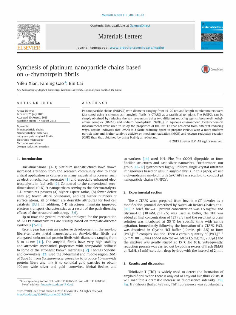
Thioflavin-T (ThT) is widely used to detect the formation ofamyloid fibril. When there is amyloid or amyloid like fibril exists, itwill manifest a dramatic increase in fluorescence intensity [19].Fig. 1(a) shows that at 483 nm, ThT fluorescence was substantially
Contents lists available at ScienceDirect
journal homepage: www.elsevier.com/locate/matlet
Materials Letters
0167-577X/$ - see front matter & 2013 Elsevier B.V. All rights reserved.http://dx.doi.org/10.1016/j.matlet.2013.08.051
n Corresponding author. Tel.: þ86 335 8387552; fax: þ86 335 8061569.E-mail address: [email protected] (F. Gao).
Materials Letters 111 (2013) 39–42
enhanced after the protein was incubated in 12% TTE at pH 2.5 for50 h. Furthermore, Fig. 1(b) shows that these fibrils produced a redshift of the maximum light absorption of the Congo red (CR). Allthese indicate the formation of amyloid fibrils [20].
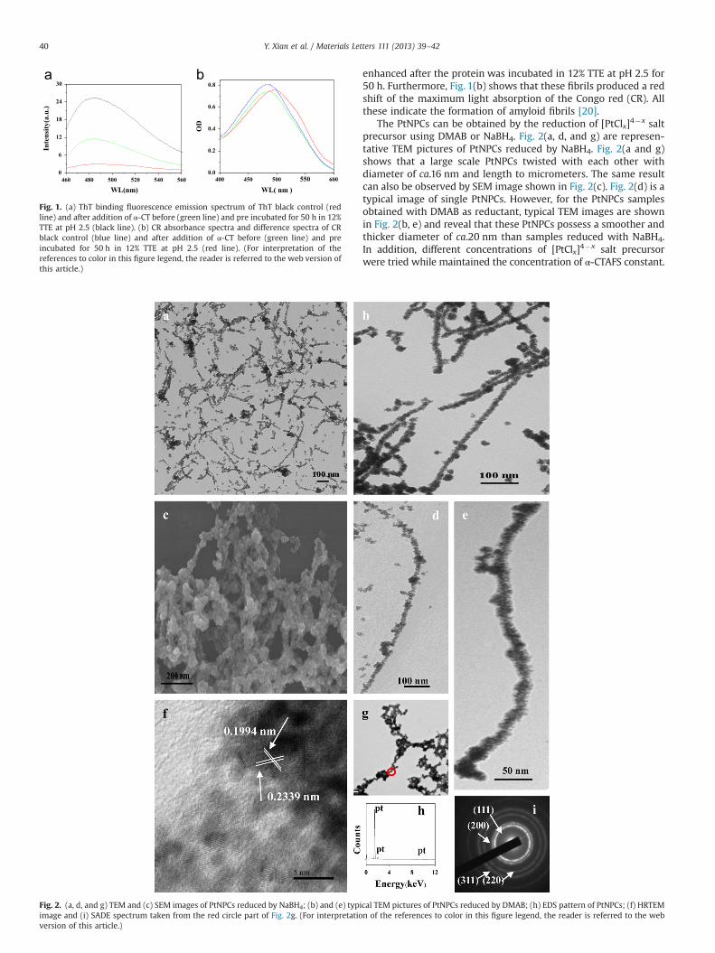
The PtNPCs can be obtained by the reduction of [PtClx]4�x saltprecursor using DMAB or NaBH4. Fig. 2(a, d, and g) are represen-tative TEM pictures of PtNPCs reduced by NaBH4. Fig. 2(a and g)shows that a large scale PtNPCs twisted with each other withdiameter of ca.16 nm and length to micrometers. The same resultcan also be observed by SEM image shown in Fig. 2(c). Fig. 2(d) is atypical image of single PtNPCs. However, for the PtNPCs samplesobtained with DMAB as reductant, typical TEM images are shownin Fig. 2(b, e) and reveal that these PtNPCs possess a smoother andthicker diameter of ca.20 nm than samples reduced with NaBH4.In addition, different concentrations of [PtClx]4�x salt precursorwere tried while maintained the concentration of α-CTAFS constant.
460 480 500 520 540 5600
6
12
18
24
30
Inte
nsity
(a.u
.)
WL(nm)400 450 500 550 600
0.0
0.2
0.4
0.6
0.8
OD
WL( nm )
Fig. 1. (a) ThT binding fluorescence emission spectrum of ThT black control (redline) and after addition of α-CT before (green line) and pre incubated for 50 h in 12%TTE at pH 2.5 (black line). (b) CR absorbance spectra and difference spectra of CRblack control (blue line) and after addition of α-CT before (green line) and preincubated for 50 h in 12% TTE at pH 2.5 (red line). (For interpretation of thereferences to color in this figure legend, the reader is referred to the web version ofthis article.)
Fig. 2. (a, d, and g) TEM and (c) SEM images of PtNPCs reduced by NaBH4; (b) and (e) typical TEM pictures of PtNPCs reduced by DMAB; (h) EDS pattern of PtNPCs; (f) HRTEMimage and (i) SADE spectrum taken from the red circle part of Fig. 2g. (For interpretation of the references to color in this figure legend, the reader is referred to the webversion of this article.)
Y. Xian et al. / Materials Letters 111 (2013) 39–4240
Results show that in some degree, the higher concentration of[PtClx]4�x the thicker in diameter of PtNPCs. Finally, an appro-priate concentration of [PtClx]4�x was confirmed. A large numberof repeated and contrast experiments prove that both DMAB andNaBH4 could be used to prepare long and uniform PtNPCs withdiameter ranging from 16–20 nm and length can reach to micro-meters. The overall crystal structure of the sample was elaboratedusing high-resolution TEM (HRTEM) and selected-area electrondiffraction (SAED). Fig. 2(f) and (i) are HRTEM image and SAEDpattern taken from the red circle part of Fig. 2(g). The latticespacings of 0.2339 and 0.1994 nm in the HRTEM are correspondto the {111} and {200} lattice spacings of Pt, respectively. Thecontinuous rings in the SAED pattern from inner to outer index tothe (111), (200), (220), and (311) planes of Pt, respectively. Both theHRTEM and SAED patterns reveal that the PtNPCs are polycrystal-line structure. Besides, the composite of samples was investigatedusing energy-dispersive X-ray spectroscopy (EDS) (Fig. 2h), whichexhibits the presence of Pt.
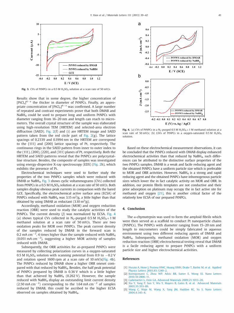
Electrochemical techniques were used to further study theproperties of the two PtNPCs samples which were reduced withDMAB or NaBH4. Fig. 3 shows cyclic voltammograms (CVs) collectedfrom PtNPCs in a 0.5 M H2SO4 solution at a scan rate of 50 mV/s. Bothsamples display obvious peak currents in comparison with the baredGCE. Specifically, the electrochemical active surface area (ECSA) ofPtNPCs reduced with NaBH4 was 3.15 m2/g, a little higher than thatobtained by using DMAB as reductant (3.10 m2/g).
Accordingly, methanol oxidation (MOR) and oxygen reductionreaction (ORR) were used to study the catalytic activities of thePtNPCs. The current density (J) was normalized by ECSA. Fig. 4(a) shows typical CVs collected in N2-purged 0.5 M H2SO4þ1 Mmethanol solution at a scan rate of 50 mV/s. There are twooxidation peaks for MOR over PtNPCs. The peak current densityof the samples reduced by DMAB in the forward scan is0.2 mA cm�2, 4 times higher than the sample reduced with NaBH4
(0.051 mA cm�2), suggesting a higher MOR activity of samplesreduced with DMAB.
Subsequently, the ORR activities for as-prepared PtNPCs weremeasured by collecting polarization curves in a oxygen-saturated0.5 M H2SO4 solution with scanning potential from 0.9 to �0.2 Vand rotation speed 1600 rpm at a scan rate of 10 mV/s(Fig. 4b).The PtNPCs reduced by DMAB show a higher ORR oneset com-pared with that reduced by NaBH4. Besides, the half peak potentialof PtNPCs prepared by DMAB is 0.36 V which is a little higherthan that achieved by NaBH4 (0.262 V). However, the samplereduced with NaBH4 display an outstanding limit current density(2.50 mA cm�2) corresponding to the 1.64 mA cm�2 of samplesreduced by DMAB, this could be ascribed to the higher ECSAobserved on samples obtained by NaBH4.
Based on these electrochemical measurement observations, it canbe concluded that the PtNPCs reduced with DMAB display enhancedelectrochemical activities than that reduced by NaBH4, such differ-ences can be attributed to the distinctive surface properties of thetwo PtNPCs samples. DMAB is a weak and facile reducing agent andthe obtained PtNPCs have a uniform particle size which is preferableto MOR and ORR activities. However, NaBH4 is a strong and rapidreducing agent and the obtained PtNPCs have inhomogenous particlesizes which lower the in fact catalytic activity on MOR and ORR. Inaddition, our protein fibrils templates are not conductive and theirprior adsorption on platinum may occupy the in fact active site formethanol and oxygen [21], this is another critical factor of therelatively low ECSA of our prepared PtNPCs.
4. Conclusion
The α-chymotrpsin was used to form the amyloid fibrils whichwere then served as a scaffold to conduct Pt nanoparticle chains(PtNPCs). The PtNPCs with diameter ranging from 15–20 nm andlength to micrometers could be simply fabricated in aqueousenvironment using two different reducing agents of DMAB andNaBH4. Subsequently, methanol oxidation (MOR) and oxygenreduction reaction (ORR) electrochemical testing reveal that DMABis a facile reducing agent to prepare PtNPCs with a uniformparticle size and higher electrochemical activities.
References
[1] Husain A, Hone J, Postma HWC, Huang XMH, Drake T, Barbic M, et al. AppliedPhysics Letters 2003;83:1240–2.
[2] Koenigsmann C, Zhou WP, Adzic RR, Sutter E, Wong SS. Nano Letters2010;10:2806–11.
[3] Cademartiri L, Ozin GA. Advanced Materials 2009;21:1013–20.[4] Xia Y, Yang P, Sun Y, Wu Y, Mayers B, Gates B, et al. Advanced Materials
2003;15:353–89.[5] Wang C, Waje M, Wang X, Tang JM, Haddon RC, Yu S. Nano Letters
2004;4:345–8.
-0.4 -0.2 0.0 0.2 0.4 0.6 0.8 1.0 1.2-15
-10
-5
0
5
10
Cur
rent
(µA
)
E / V (SCE)
PtNWS reduced by DMABPtNWS reduced by NaBH4GCE
Fig. 3. CVs of PtNPCs in a 0.5 M H2SO4 solution at a scan rate of 50 mV/s.
-0.2 -0.1 0.0 0.1 0.2 0.3 0.4 0.5 0.6 0.7 0.8
-2.5
-2.0
-1.5
-1.0
-0.5
0.0
J (m
A c
m-2
)
E (V) vs.Ag / AgCl
PtNWS reduced by DMABPtNWS reduced by NaBH4
-0.4 -0.2 0.0 0.2 0.4 0.6 0.8 1.0 1.2-0.6
-0.4
-0.2
0.0
0.2
0.4
0.6
J (m
A c
m-2
)
E / V (SCE)
PtNWS reduced by DMABPtNWS reduced by NaBH4
Fig. 4. (a) CVs of PtNPCs in a N2-purged 0.5 M H2SO4þ1 M methanol solution at ascan rate of 50 mV/s; (b) LSVs of PtNPCs in a oxygen-saturated 0.5 M H2SO4
solution.
Y. Xian et al. / Materials Letters 111 (2013) 39–42 41

[6] Chen ZW, Waje M, Li WZ, Yan YS. Angewandte Chemie Internatinal Edition2007;46:4060–3.
[7] Han YJ, Kim JM, Stucky GD. Chemistry of Materials 2000;12:2068–9.[8] Sakamoto Y, Fukuoka A, Higuchi T, Shimomura N, Inagaki S, Ichikawa M.
Journal of Physical Chemistry B 2004;108:853–8.[9] Song YJ, Garcia RM, Dorin RM, et al. Nano Letters 2007;7:3650–5.[10] Kijima T, Yoshimura T, Uota M, Ikeda T, Fujikawa D, Mouri S, Uoyama S.
Angewandte Chemie International Edition 2004;43:228–32.[11] Dobson CM. Nature 2002;418:729–30.[12] Tang Q, Solin N, Lu J, Inganas O. Chemical Communications 2010;46:4157–9.[13] Scheibel T, Parthasarathy R, Sawicki G, Lin XM, Jaeger H, Lindquist SL.
Proceedings of the National Academy of Sciences USA 2003;100:4527–32.
[14] Reches M, Gazit E. Science 2003;300:625–7.[15] Zhang LG, Gao FM. Journal of Nanoparticle Research 2012;14:855.[16] Zhang LG, Li N, Gao FM, Hou L, Xu ZM. Journal of the American Chemical
Society 2012;134:11326–9.[17] Gao FM, Xian YF, Zhang LG. Journal of Yanshan University 2013;37:95–101.[18] Rezaei-Ghaleh N, Zweckstetter M, Morshedi D, Ebrahim-Habibi A, Nemat-
Gorgani M. Biopolymers 2009;91:28–36.[19] Nilsson RM. Methods 2004;34:151–60.[20] Sunde M, Blake C. Advances in Protein Chemistry 1997;50:123–59.[21] Sanchez-Sanchez CM, Solla-Gullon J, Vidal-Iglesias FJ, Aldaz A, Montiel V,
Herrero E. Journal of the American Chemical Society 2010;132:5622–4.
Y. Xian et al. / Materials Letters 111 (2013) 39–4242