
Synthesis, Biological Evaluation, and Three-Dimensional in ... · Synthesis, Biological Evaluation,...
14
pubs.acs.org/jmc Published on Web 08/12/2009 r 2009 American Chemical Society 5380 J. Med. Chem. 2009, 52, 5380–5393 DOI: 10.1021/jm900366z Synthesis, Biological Evaluation, and Three-Dimensional in Silico Pharmacophore Model for σ 1 Receptor Ligands Based on a Series of Substituted Benzo[d ]oxazol-2(3H )-one Derivatives Daniele Zampieri, ‡ Maria Grazia Mamolo,* ,‡ Erik Laurini, ‡ Chiara Florio, † Caterina Zanette, † Maurizio Fermeglia, § Paola Posocco, § Maria Silvia Paneni, § Sabrina Pricl,* ,§ and Luciano Vio ‡ ‡ Department of Pharmaceutical Sciences, Piazzale Europa 1, University of Trieste, 34127 Trieste, Italy, † Department of Life Sciences, Section of Pharmacology, University of Trieste, 34127 Trieste, Italy, and § Department of Chemical, Environmental, and Raw Materials Engineering (DICAMP), Molecular Simulation Engineering (MOSE) Laboratory, Piazzale Europa 1, University of Trieste, 34127 Trieste, Italy Received March 23, 2009 Novel benzo[d ]oxazol-2(3H )-one derivatives were designed and synthesized, and their affinities against σ receptors were evaluated. On the basis of 31 compounds, a three-dimensional pharmacophore model for the σ 1 receptor binding site was developed using the Catalyst 4.9 software package. The best 3D pharmacophore hypothesis, consisting of one positive ionizable, one hydrogen bond acceptor, two hydrophobic aromatic, and one hydrophobic features provided a 3D-QSAR model with a correlation coefficient of 0.89. The best hypothesis was also validated by three independent methods, i.e., the Fisher randomization test included in the CatScramble functionality of Catalyst, the leave-one-out test, and activity prediction of an additional test set. The achieved results will allow researchers to use this 3D pharmacophore model for the design and synthesis of a second generation of high affinity σ 1 ligands, as well as to discover other lead compounds for this class of receptors. Introduction The σ binding sites were originally defined and classified as opioid receptor subtypes. 1 Later investigations demonstrated that σ receptors were distinct from opioid and phencyclidine analogues, and since then, at least two distinct σ receptor subtypes, designated σ 1 and σ 2 , 2 have been pharmacologically characterized. 3-5 The cellular and anatomical distribution of σ receptors is not restricted to the central nervous system (CNS a ), 6-8 but extends to peripheral tissues such as blood vessels, adrenal glands, testicles, ovaries, and immune sys- tem. 9 There is increasing evidence that σ receptors have a neuromodulatory role in the CNS and are involved in the etiology of various psychiatric diseases 10 such as anxiety, schizophrenia, and depression. The correlation between the extent of toxic effects on CNS of some antipsychotic com- pounds and their σ receptor affinity is also documented. Furthermore, σ receptors may also have a potential role in neuromotor diseases such as dystonia and dyskinesia; 11 indeed, σ receptors are highly expressed in brain areas asso- ciated with the control of movement of facial muscles. 11 The σ 1 receptor subtype has been purified and cloned from several animal species and man. 12,13 Its primary sequence is now available and shows remarkable homology with sterol C8-C7 isomerase from fungi. The σ 1 receptors exert a modulatory role on neurotransmitter systems such as dopa- minergic, serotoninergic, and muscarinic systems 5,14,15 and on the NMDA-stimulated neurotransmitters’ release. 16 More- over, σ 1 receptors are involved in neuroprotective and antiamnesic activities, 17 modulation of opioid analgesia, 18 and attenuation of cocaine-induced locomotor activity and toxicity. 19 In addition, σ 1 antagonists have been shown to be effective against negative symptoms of schizophrenia without producing extrapyramidal side effects typical of traditional neuroleptics. 20,21 On the other hand, the molecular identity of the σ 2 receptor subtype has not been fully determined, 12,13 although a number of studies have presented evidence linking σ 2 receptors to potassium channels and intracellular calcium release in NCB- 20 cells. 22,23 Unlike the σ 1 subtype, σ 2 receptors may contribute to the acute side effects of typical neuroleptic drugs, and σ 2 antagonists are known to attenuate extrapyramidal effects, dystonic reactions, and tardive dyskinesia, 2,14,22,24,25 suggesting their potential use in the treatment of psychoses. 20,21 Further- more, σ 2 receptors are involved in the regulation of cell proliferation and maintenance of cell viability. They are highly expressed in several tumoral cell lines, 26,27 where σ 2 agonists produce morphological changes and apoptosis. The σ 2 recep- tor agonists promote Ca 2þ release from endoplasmatic reticu- lum and mitochondrial stores, 28 with subsequent cell death by *To whom correspondence should be addressed. For M.G.M. (chemistry and biological sections): phone, þ390405583719; fax, þ3904052572; e-mail, [email protected]. For S.P. (computational and molecular modeling sections): phone, þ390405583750; fax,þ39040569823; e-mail, [email protected]. a Abbreviations: 3D-QSAR, three-dimensional quantitative struc- ture-activity relationship; CNS, central nervous system; NMDA, N- methyl D-aspartate; NCB-20, mouse neuroblastoma Chinese hamster brain hybrid cell line; PET, positron-emission tomography; SPECT, single photon emission computed tomography; PTZ, (þ)-pentazocine; NANM, (þ)-N-allylnormetazocine; DMAP, 4-dimethylaminopyridine; TFA, trifluoroacetic acid; PC, positive charge; NC, negative charge; HBD, hydrogen bond donor; HBA, hydrogen bond acceptor; HBAl, hydrogen bond acceptor lipid; HYAr, hydrophobic aromatic; HY, generic hydrophobic; Ar, ring aromatic; PI, positive ionizable; rmsd, root-mean-square deviation; ERG2, ether-a-go-go related gene 2; SEM, standard error of the mean. Downloaded by UNIVERSITA STUDI TRIESTE on September 9, 2009 | http://pubs.acs.org Publication Date (Web): August 12, 2009 | doi: 10.1021/jm900366z
Transcript of Synthesis, Biological Evaluation, and Three-Dimensional in ... · Synthesis, Biological Evaluation,...
pubs.acs.org/jmc Published on Web 08/12/2009 r 2009 American Chemical Society
5380 J. Med. Chem. 2009, 52, 5380–5393
DOI: 10.1021/jm900366z
Synthesis, Biological Evaluation, and Three-Dimensional in Silico Pharmacophore Model for
σ1 Receptor Ligands Based on a Series of Substituted Benzo[d ]oxazol-2(3H )-one Derivatives
Daniele Zampieri,‡ Maria Grazia Mamolo,*,‡ Erik Laurini,‡ Chiara Florio,† Caterina Zanette,† Maurizio Fermeglia,§
Paola Posocco,§ Maria Silvia Paneni,§ Sabrina Pricl,*,§ and Luciano Vio‡
‡Department of Pharmaceutical Sciences, Piazzale Europa 1, University of Trieste, 34127 Trieste, Italy, †Department of Life Sciences,Section of Pharmacology, University of Trieste, 34127 Trieste, Italy, and §Department of Chemical, Environmental,and Raw Materials Engineering (DICAMP), Molecular Simulation Engineering (MOSE) Laboratory, Piazzale Europa 1,University of Trieste, 34127 Trieste, Italy
Received March 23, 2009
Novel benzo[d ]oxazol-2(3H )-one derivatives were designed and synthesized, and their affinities againstσ receptors were evaluated. On the basis of 31 compounds, a three-dimensional pharmacophore modelfor the σ1 receptor binding site was developed using the Catalyst 4.9 software package. The best 3Dpharmacophore hypothesis, consisting of one positive ionizable, one hydrogen bond acceptor, twohydrophobic aromatic, and one hydrophobic features provided a 3D-QSAR model with a correlationcoefficient of 0.89. The best hypothesis was also validated by three independentmethods, i.e., the Fisherrandomization test included in the CatScramble functionality of Catalyst, the leave-one-out test, andactivity prediction of an additional test set. The achieved results will allow researchers to use this 3Dpharmacophore model for the design and synthesis of a second generation of high affinity σ1 ligands, aswell as to discover other lead compounds for this class of receptors.
Introduction
The σ binding sites were originally defined and classified asopioid receptor subtypes.1 Later investigations demonstratedthat σ receptors were distinct from opioid and phencyclidineanalogues, and since then, at least two distinct σ receptorsubtypes, designated σ1 and σ2,
2 have been pharmacologicallycharacterized.3-5 The cellular and anatomical distributionof σ receptors is not restricted to the central nervous system(CNSa),6-8 but extends to peripheral tissues such as bloodvessels, adrenal glands, testicles, ovaries, and immune sys-tem.9 There is increasing evidence that σ receptors have aneuromodulatory role in the CNS and are involved in theetiology of various psychiatric diseases10 such as anxiety,schizophrenia, and depression. The correlation between theextent of toxic effects on CNS of some antipsychotic com-pounds and their σ receptor affinity is also documented.Furthermore, σ receptors may also have a potential role
in neuromotor diseases such as dystonia and dyskinesia;11
indeed, σ receptors are highly expressed in brain areas asso-ciated with the control of movement of facial muscles.11
The σ1 receptor subtype has been purified and cloned fromseveral animal species and man.12,13 Its primary sequence isnow available and shows remarkable homology with sterolC8-C7 isomerase from fungi. The σ1 receptors exert amodulatory role on neurotransmitter systems such as dopa-minergic, serotoninergic, andmuscarinic systems5,14,15 and onthe NMDA-stimulated neurotransmitters’ release.16 More-over, σ1 receptors are involved in neuroprotective andantiamnesic activities,17 modulation of opioid analgesia,18
and attenuation of cocaine-induced locomotor activity andtoxicity.19 In addition, σ1 antagonists have been shown to beeffective against negative symptoms of schizophrenia withoutproducing extrapyramidal side effects typical of traditionalneuroleptics.20,21
On the other hand, the molecular identity of the σ2 receptorsubtype has not been fully determined,12,13 although a numberof studies have presented evidence linking σ2 receptors topotassium channels and intracellular calcium release in NCB-20 cells.22,23Unlike the σ1 subtype, σ2 receptorsmay contributeto the acute side effects of typical neuroleptic drugs, andσ2 antagonists are known to attenuate extrapyramidal effects,dystonic reactions, and tardivedyskinesia,2,14,22,24,25 suggestingtheir potential use in the treatment of psychoses.20,21 Further-more, σ2 receptors are involved in the regulation of cellproliferation andmaintenance of cell viability. They are highlyexpressed in several tumoral cell lines,26,27 where σ2 agonistsproduce morphological changes and apoptosis. The σ2 recep-tor agonists promote Ca2þ release from endoplasmatic reticu-lum and mitochondrial stores,28 with subsequent cell death by
*To whom correspondence should be addressed. For M.G.M.(chemistry and biological sections): phone, þ390405583719; fax,þ3904052572; e-mail, [email protected]. For S.P. (computational andmolecularmodeling sections): phone,þ390405583750; fax,þ39040569823;e-mail, [email protected].
aAbbreviations: 3D-QSAR, three-dimensional quantitative struc-ture-activity relationship; CNS, central nervous system; NMDA, N-methyl D-aspartate; NCB-20, mouse neuroblastoma Chinese hamsterbrain hybrid cell line; PET, positron-emission tomography; SPECT,single photon emission computed tomography; PTZ, (þ)-pentazocine;NANM, (þ)-N-allylnormetazocine; DMAP, 4-dimethylaminopyridine;TFA, trifluoroacetic acid; PC, positive charge; NC, negative charge;HBD, hydrogen bond donor; HBA, hydrogen bond acceptor; HBAl,hydrogen bond acceptor lipid; HYAr, hydrophobic aromatic; HY,generic hydrophobic; Ar, ring aromatic; PI, positive ionizable; rmsd,root-mean-square deviation; ERG2, ether-a-go-go related gene 2; SEM,standard error of the mean.
Dow
nloa
ded
by U
NIV
ER
SIT
A S
TU
DI
TR
IEST
E o
n Se
ptem
ber
9, 2
009
| http
://pu
bs.a
cs.o
rg
Pub
licat
ion
Dat
e (W
eb):
Aug
ust 1
2, 2
009
| doi
: 10.
1021
/jm90
0366
z
Article Journal of Medicinal Chemistry, 2009, Vol. 52, No. 17 5381
caspase-independent apoptosis.27 Apoptosis may also be in-duced in tumoral cells by regulation of the sphingolipid path-way.29 Therefore, σ2 agonistsmay be useful as novel anticanceragents and as imaging agents in cancer diagnosis by positronemission tomography (PET)30 and single photon emissioncomputed tomography (SPECT).31,32
To date, well-known compounds characterized by a certaindegree of selectivity for the σ1 receptor subtype include (þ)-benzomorphans such as (þ)-pentazocine (PTZ) and (þ)-N-allylnormetazocine (NANM, SKF-10047), while haloperidoland 1,3-di-(2-tolyl)guanidine show high affinity for bothreceptor subtypes.22
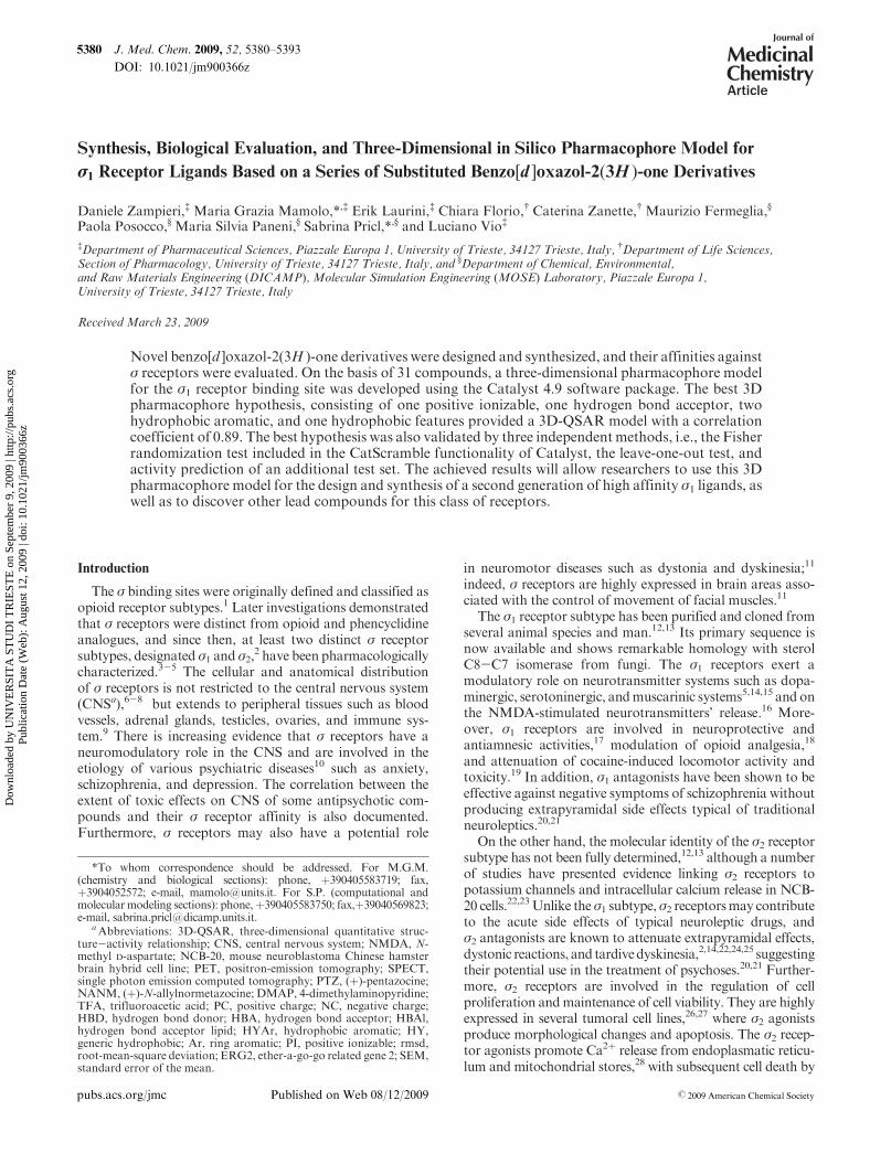
In our previous work33 we described the synthesis and theaffinities for the σ receptors of a series of substituted benzo-oxazolone derivatives 1a-j (Figure 1), in which the benzylgroup was variously substituted at the phenyl ring. Thebenzooxazolone derivatives 1a-j have been designed accord-ing to the σ1 receptormodel proposed byGlennon,34-36 underthe following assumptions: (i) the benzooxazolone moietymay interact with a primary hydrophobic site correspondingto Glennon’s phenyl “B” region;34-36 (ii) the 4-methylpiper-idin-1-yl spacer links the basic nitrogen atom to the benzoox-azolone moiety; (iii) the substituted N-benzyl moiety maybind the secondary hydrophobic phenyl “A” region of theσ1 receptorial model, modulating the binding affinity of thecompounds for the σ receptors.
The relevant results indicated that the substituents on thephenyl ring can modulate the σ1 and σ2 binding affinities ofthese compounds. Indeed, all molecules show, to variousdegrees, a preference for σ1 receptor sites, with the unsubsti-tuted derivative and the corresponding para-substituted deri-vatives exhibiting the highest affinity. Particularly, the bestresult was reached with the 4-chloro substituted compound,with a Ki(σ1) value of 0.1 nM and a Ki(σ2)/Ki(σ1) selectivityratio of 4270.
In this work we synthesized (Scheme 1) a series of 3-[(N-benzyl-N-ethylamine)alkyl]benzo[d ]oxazol-2(3H )-one deri-vatives 2a-k and 3a-k (Table 1), with various substitutionsat the benzene ring, to generate an adequate number ofcompounds for the purpose of developinga three-dimensional(3D) pharmacophore model for σ1 receptor ligands.
Since the benzooxazolone moiety is considered to form agood interaction with a putative hydrophobic region in thesereceptors, this group has been maintained in the new mole-cular series. Moreover, the electronegative atoms of theoxazolone moiety may further contribute to the bindingaffinity. Effectively, electronegative atoms such as O or Sare frequently present in very potent σ1 ligands, bridging thearomatic component and the classical alkyl or cycloalkylintermediate spacer linked to the basic nitrogen atom.13,36
On the basis of a molecular modeling study of σ1 receptorligands, Gund et al.37 also concluded that there could be asecondary binding region that may surround the oxygen orsulfur atomof themolecules. In the other half of themolecule,the piperidin-4-ylmethyl core has been replaced by an alkyl
spacer, endowed with greater conformal freedom. The choiceof a propyl or butyl chain is dictated by the fact that both fallwithin the range of optimal distances between the basicnitrogen atom and hydrophobic primary site in the receptor
Figure 1. Structures of compounds 1a-j.
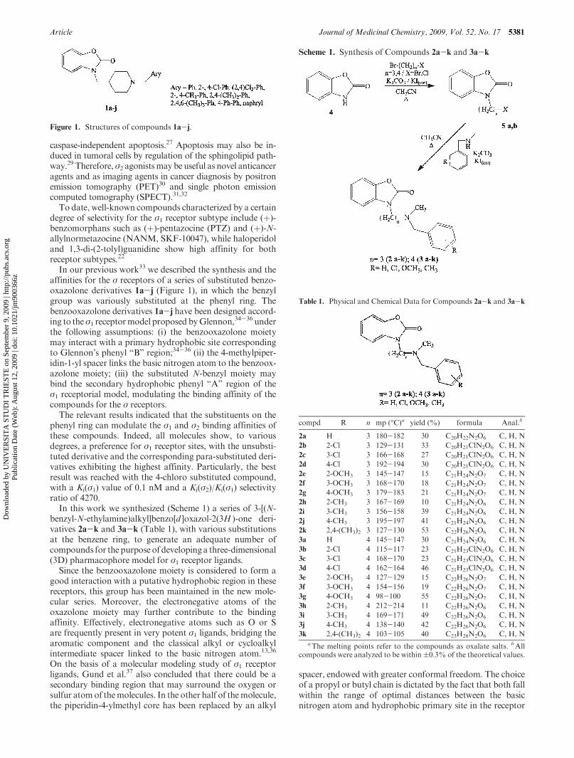
Scheme 1. Synthesis of Compounds 2a-k and 3a-k
Table 1. Physical and Chemical Data for Compounds 2a-k and 3a-k
compd R n mp (�C)a yield (%) formula Anal.b
2a H 3 180-182 30 C20H22N2O6 C, H, N
2b 2-Cl 3 129-131 33 C20H21ClN2O6 C, H, N
2c 3-Cl 3 166-168 27 C20H21ClN2O6 C, H, N
2d 4-Cl 3 192-194 30 C20H21ClN2O6 C, H, N
2e 2-OCH3 3 145-147 15 C21H24N2O7 C, H, N
2f 3-OCH3 3 168-170 18 C21H24N2O7 C, H, N
2g 4-OCH3 3 179-183 21 C21H24N2O7 C, H, N
2h 2-CH3 3 167-169 10 C21H24N2O6 C, H, N
2i 3-CH3 3 156-158 39 C21H24N2O6 C, H, N
2j 4-CH3 3 195-197 41 C21H24N2O6 C, H, N
2k 2,4-(CH3)2 3 127-130 53 C22H26N2O6 C, H, N
3a H 4 145-147 30 C21H24N2O6 C, H, N
3b 2-Cl 4 115-117 23 C21H23ClN2O6 C, H, N
3c 3-Cl 4 168-170 23 C21H23ClN2O6 C, H, N
3d 4-Cl 4 162-164 46 C21H23ClN2O6 C, H, N
3e 2-OCH3 4 127-129 15 C22H26N2O7 C, H, N
3f 3-OCH3 4 154-156 19 C22H26N2O7 C, H, N
3g 4-OCH3 4 98-100 55 C22H26N2O7 C, H, N
3h 2-CH3 4 212-214 11 C22H26N2O6 C, H, N
3i 3-CH3 4 169-171 49 C22H26N2O6 C, H, N
3j 4-CH3 4 138-140 42 C22H26N2O6 C, H, N
3k 2,4-(CH3)2 4 103-105 40 C23H28N2O6 C, H, NaThe melting points refer to the compounds as oxalate salts. bAll
compounds were analyzed to be within(0.3% of the theoretical values.
Dow
nloa
ded
by U
NIV
ER
SIT
A S
TU
DI
TR
IEST
E o
n Se
ptem
ber
9, 2
009
| http
://pu
bs.a
cs.o
rg
Pub
licat
ion
Dat
e (W
eb):
Aug
ust 1
2, 2
009
| doi
: 10.
1021
/jm90
0366
z
5382 Journal of Medicinal Chemistry, 2009, Vol. 52, No. 17 Zampieri et al.
model of Glennon.34-36 All compounds obtained were sub-sequently tested in order to assess their affinity and theirselectivity toward σ receptors.
As mentioned previously, the crystal structure of bothσ1 and σ2 receptors remains unsolved to date. In the absenceof a reliable 3D model of the target structure, ligand-based
molecular modeling tools can be successfully employedto devise structural requirements crucial for receptor bind-ing. The 3D quantitative structure-activity relationship(3D-QSAR) pharmacophore modeling is such an approachand constitutes a consolidated technique in drug design anddiscovery.
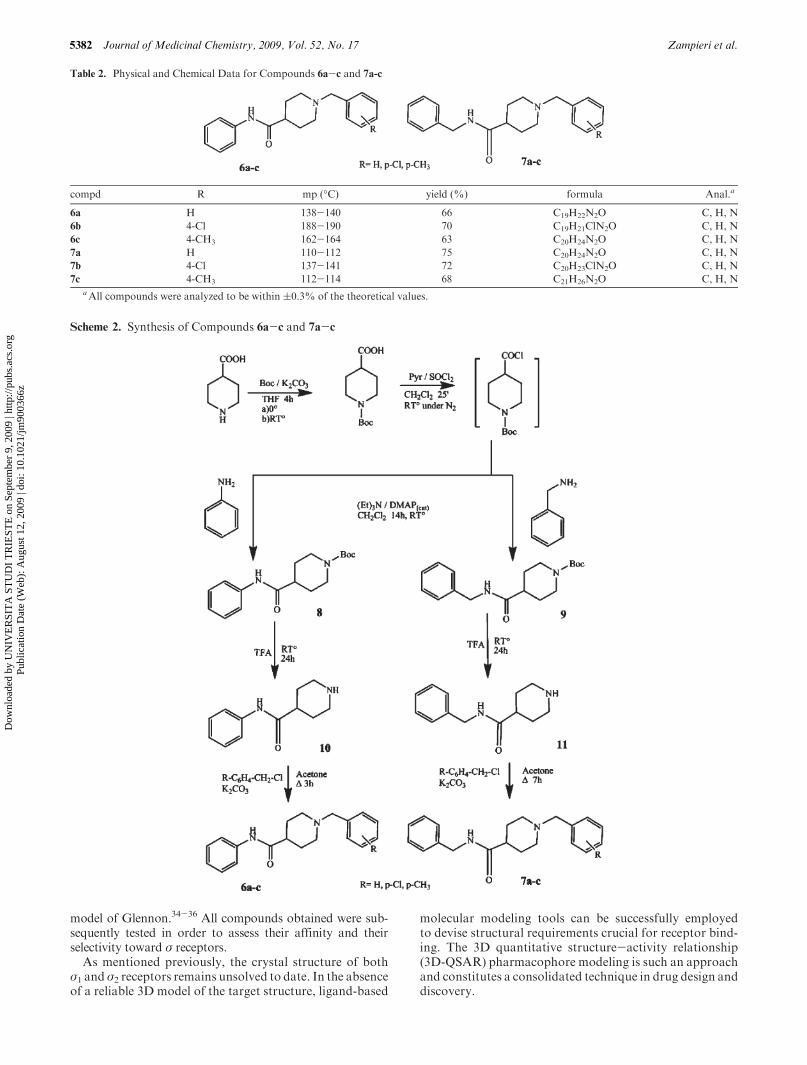
Table 2. Physical and Chemical Data for Compounds 6a-c and 7a-c
compd R mp (�C) yield (%) formula Anal.a
6a H 138-140 66 C19H22N2O C, H, N
6b 4-Cl 188-190 70 C19H21ClN2O C, H, N
6c 4-CH3 162-164 63 C20H24N2O C, H, N
7a H 110-112 75 C20H24N2O C, H, N
7b 4-Cl 137-141 72 C20H23ClN2O C, H, N
7c 4-CH3 112-114 68 C21H26N2O C, H, NaAll compounds were analyzed to be within (0.3% of the theoretical values.
Scheme 2. Synthesis of Compounds 6a-c and 7a-c
Dow
nloa
ded
by U
NIV
ER
SIT
A S
TU
DI
TR
IEST
E o
n Se
ptem
ber
9, 2
009
| http
://pu
bs.a
cs.o
rg
Pub
licat
ion
Dat
e (W
eb):
Aug
ust 1
2, 2
009
| doi
: 10.
1021
/jm90
0366
z
Article Journal of Medicinal Chemistry, 2009, Vol. 52, No. 17 5383
The major goal this paper is the generation of a predictivepharmacophore model for σ1 ligands. To develop the model,we resorted to the HypoGen method implemented in theCatalyst software package.38 Starting with a training set of31 σ1 ligands, a pharmacophore model (also called ahypothesis) able to quantitatively correlate the estimatedaffinities with the corresponding measured values was gener-ated. The model was then validated by statistical means andby its ability to predict the affinity of a further ensemble ofdifferent compounds (test set). Overall, we verified that our3D-QSAR pharmacophore model was predictive not onlywithin the training set but also for the test set compounds,withacceptable errors.
Chemistry
The substituted 3-[3-(N-benzyl-N-methylamino)propyl]-benzo[d ]oxazol-2(3H)-one derivatives2a-k and3-[4-(N-ben-zyl-N-methylamino)butyl]benzo[d ]oxazol-2(3H)-one deriva-tives 3a-k were synthesized (Scheme 1) by treating anacetonitrile solution of benzo[d ]oxazol-2(3H)-one 4 with1-bromo-3-chloropropane and 1,4-dibromobutane, respec-tively, in the presence of K2CO3 and a catalytic amount ofKI. From the obtained 3-(3-chloropropyl) and 4-(4-bro-mobutyl)benzo[d ]oxazol-2(3H)-one intermediates 5a,b, thesubstituted N-benzyl derivatives were obtained by heatingat reflux with N-methylbenzylamines in acetonitrile in thepresence of K2CO3 and a catalytic amount of KI.
The substituted 1-benzyl-N-phenylpiperidine-4-carbox-amide derivatives 6a-c and the correspondingN-benzyl deri-vatives 7a-c were prepared starting from the commerciallyavailable 4-piperidinecarboxylic acid which was transformedinto the correspondingN-boc-4-piperidinecarboxylic chlorideby treatment with SOCl2 under N2 (Table 2 and Scheme 2).A solution of aniline and triethylamine in CH2Cl2 was addeddirectly to the reaction mixture with a catalytic amount ofDMAP. The reaction was carried out at room temperatureunder N2 flux to afford N-phenyl-1-(tert-butoxycarbonyl)-piperidine-4-carboxamide 8, which was deprotected withTFA to yield the N-phenylpiperidine-4-carboxamide 10.
Following the same route described above and usingbenzylamine, N-benzyl-1-(tert-butoxycarbonyl)piperidine-4-carboxamide 9was prepared, fromwhichN-benzylpiperidine-4-carboxamide 11was obtained after deprotection with TFA.From the piperidine-4-carboxamides 10 and 11, the corre-sponding 1-benzyl derivatives 6a-c and 7a-c were obtainedby alkylation with the opportune benzyl chlorides.
Results and Discussion
On the basis of the interesting Ki(σ1) values of a series ofbenzo[d ]oxazolone derivatives 1a-j (Figure 1) we described inpreviouswork,33we synthesized a series of newbenzooxazolonederivatives 2a-k and 3a-k (Table 1) in which the 4-methylpi-peridin-1-yl spacer linking the benzooxazolone moiety to thebenzyl group was replaced by the 3-(N-methylamino)propyl
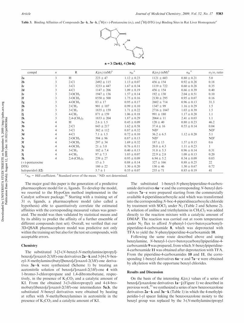
Table 3. Binding Affinities of Compounds 2a-k, 3a-k, [3H]-(þ)-Pentazocine (σ1), and [3H]-DTG (σ2) Binding Sites in Rat Liver Homogenatea
compd n R Ki(σ1) (nM)b nHb Ki(σ2) (nM)b nH
b σ2/σ1 ratio
2a 3 H 223 ( 47 1.12 ( 0.23 1121 ( 603 0.80 ( 0.21 5.0
2b 3 2-Cl 2492 ( 115 1.13 ( 0.07 456 ( 154 0.92 ( 0.28 0.18
2c 3 3-Cl 3233 ( 447 1.67 ( 0.34 1119 ( 722 0.60 ( 0.28 0.35
2d 3 4-Cl 1147 ( 206 1.09 ( 0.19 456 ( 154 0.66 ( 0.39 0.40
2e 3 2-OCH3 1947 ( 136 1.57 ( 0.14 192 ( 130 2.04 ( 0.51 0.10
2f 3 3-OCH3 8550 ( 500 1.75 ( 0.16 2130 ( 295 0.95 ( 0.07 0.16
2g 3 4-OCH3 83 ( 17 0.95 ( 0.17 2602 ( 714 0.96 ( 0.13 31.3
2h 3 2-CH3 901 ( 107 0.99 ( 0.10 1347 ( 99 1.16 ( 0.29 1.5
2i 3 3-CH3 1835 ( 159 1.71 ( 0.22 2716 ( 1847 1.03 ( 0.39 1.5
2j 3 4-CH3 871 ( 159 1.06 ( 0.18 991 ( 180 1.17 ( 0.20 1.1
2k 3 2,4-(CH3)2 1833 ( 204 1.87 ( 0.29 2064 ( 11 2.41 ( 0.03 1.1
3a 4 H 2.6 ( 1.5 0.45 ( 0.09 120 ( 40 0.80 ( 0.23 46.2
3b 4 2-Cl 843 ( 217 1.62 ( 0.58 37.4 ( 16 0.55 ( 0.14 0.04
3c 4 3-Cl 302 ( 112 0.87 ( 0.32 NDc NDc
3d 4 4-Cl 7.1 ( 1.5 0.72 ( 0.10 36.2 ( 6.5 1.12 ( 0.20 5.1
3e 4 2-OCH3 594 ( 96 0.87 ( 0.13 NDc NDc
3f 4 3-OCH3 297 ( 34 1.69 ( 0.32 187 ( 13 1.57 ( 0.15 0.6
3g 4 4-OCH3 21 ( 3.8 0.76 ( 0.11 20.8 ( 4.3 1.11 ( 0.23 1
3i 4 3-CH3 102 ( 7.4 0.40 ( 0.13 31.8 ( 5.3 0.96 ( 0.14 0.31
3j 4 4-CH3 97 ( 7.3 1.03 ( 0.07 22.9 ( 2.8 1.08 ( 0.13 0.20
3k 4 2,4-(CH3)2 239 ( 27 0.95 ( 0.09 6.94 ( 5.2 0.34 ( 0.09 0.03
(þ)-pentazocine 15 ( 3 0.88 ( 0.14 327 ( 166 0.88 ( 0.25 22
DTG 180 ( 22 1.30 ( 0.20 130 ( 46 1.50 ( 0.67 0.72
haloperidol (12) 5.7 ( 1 0.55 ( 0.07 235 ( 71 0.85 ( 0.19 41a nH = Hill coefficient. b Standard error of the mean. cND: not determined.
Dow
nloa
ded
by U
NIV
ER
SIT
A S
TU
DI
TR
IEST
E o
n Se
ptem
ber
9, 2
009
| http
://pu
bs.a
cs.o
rg
Pub
licat
ion
Dat
e (W
eb):
Aug
ust 1
2, 2
009
| doi
: 10.
1021
/jm90
0366
z
5384 Journal of Medicinal Chemistry, 2009, Vol. 52, No. 17 Zampieri et al.
and 4-(N-methylamino)butyl groups, respectively. The aimwasto verify if these modifications might produce compoundsretaining affinity forσ receptor sites.All compoundswere testedto evaluate their Ki values against both σ1 and σ2 subtypes(Table 3).
From the results obtained so far (Table 3), it appears thatthe substituents on the phenyl ring strongly modulate theσ1 and σ2 binding affinity of these compounds, butylenederivatives 3a-k having higher affinity than the correspond-ing propylene derivatives 2a-k, as in the case of compounds1a-j.33 The butylene intermediate chain determines the opti-mal distance between the primary B and secondary A hydro-phobic centers proposed by the Glennon’s model.34-36 Thecompound with the highest affinity is the butylene derivative3a (R=H), with Ki(σ1) value of 2.6 nM and an interestingselectivity ratio Ki(σ2)/Ki(σ1) = 46.2. The para-substitutedcompound 3d (R=4-Cl), instead, still maintains an appreci-able σ1 affinity but has a selectivity ratio (Ki(σ2)/Ki(σ1)=5.1)lower than those of the unsubstituted derivatives.
Unlike the derivatives previously described, compound 3g
(R=4-OCH3) has no selectivity, showing a moderate affinitytoward both receptors.On the other hand, compounds 3b and3i-k show preference toward the σ2 receptor. Specifically,compound 3k (R=2,4-(CH3)2) has the highest σ2 affinity(Ki(σ2)=6.94 nM) and selectivity ratio (Ki(σ2)/Ki(σ1)=0.03)of the series.
For the development of a 3D quantitative structure-activity relationship (3D-QSAR) pharmacophore model,
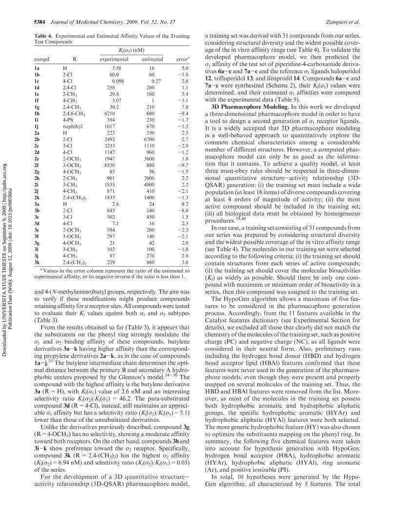
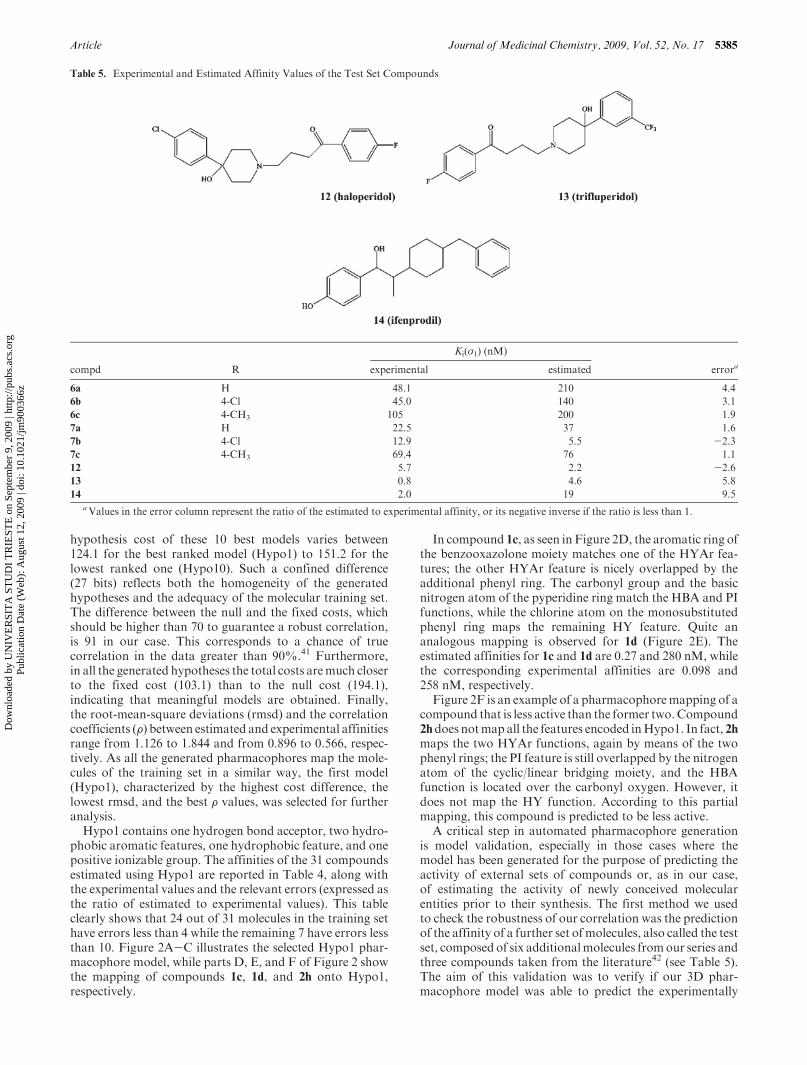
a training set was derived with 31 compounds from our series,considering structural diversity and the widest possible cover-age of the in vitro affinity range (see Table 4). To validate thedeveloped pharmacophore model, we then predicted theσ1 affinity of the test set of piperidine-4-carboxamide deriva-tives 6a-c and 7a-c and the reference σ1 ligands haloperidol12, trifluperidol 13, and ifenprodil 14. Compounds 6a-c and7a-c were synthesized (Scheme 2), their Ki(σ1) values weredetermined, and their estimated σ1 affinities were comparedwith the experimental data (Table 5).
3D Pharmacophore Modeling. In this work we developeda three-dimensional pharmacophore model in order to havea tool to design a second generation of σ1 receptor ligands.It is a widely accepted that 3D pharmacophore modelingis a well-behaved approach to quantitatively explore thecommon chemical characteristics among a considerablenumber of different structures. However, a computed phar-macophore model can only be as good as the informa-tion that it contains. To achieve a quality model, at leastthree must-obey rules should be respected in three-dimen-sional quantitative structure-activity relationship (3D-QSAR) generation: (i) the training set must include a widepopulation (at least 16 items) of diverse compounds coveringat least 4 orders of magnitude of activity; (ii) the mostactive compound should be included in the training set;(iii) all biological data must be obtained by homogeneousprocedures.39,40
In our case, a training set consisting of 31 compounds fromour series was prepared by considering structural diversityand the widest possible coverage of the in vitro affinity range(see Table 4). The molecules in our training set were selectedaccording to the following criteria: (i) the training set shouldcontain structures from each series of active compounds;(ii) the training set should cover the molecular bioactivities(Ki) as widely as possible. Should there be only one com-pound with maximum or minimum order of bioactivity in aseries, then this compound was assigned to the training set.
The HypoGen algorithm allows a maximum of five fea-tures to be considered in the pharmacophore generationprocess. Accordingly, from the 11 features available in theCatalyst features dictionary (see Experimental Section fordetails), we excluded all those that clearly did not match thechemistry of themolecules of the training set, such as positivecharge (PC) and negative charge (NC), as all ligands wereconsidered in their neutral form. Also, preliminary runsincluding the hydrogen bond donor (HBD) and hydrogenbond acceptor lipid (HBAl) features confirmed that thesefeatures were never used in the generation of the pharmaco-phore models, even though they were present and properlymapped on several molecules of the training set. Thus, theHBD and HBAl features were removed from the list. More-over, as most of the molecules in the training set possessboth hydrophobic aromatic and hydrophobic aliphaticgroups, the specific hydrophobic aromatic (HYAr) andhydrophobic aliphatic (HYAl) features were both selected.Themore generic hydrophobic feature (HY) was also chosento optimize the substituents mapping on the phenyl ring. Insummary, the following five chemical features were takeninto account for hypothesis generation with HypoGen:hydrogen bond acceptor (HBA), hydrophobic aromatic(HYAr), hydrophobic aliphatic (HYAl), ring aromatic(Ar), and positive ionizable (PI).
In total, 10 hypotheses were generated by the Hypo-Gen algorithm, all characterized by 5 features. The total
Table 4. Experimental and Estimated Affinity Values of the TrainingTest Compounds
Ki(σ1) (nM)
compd R experimental estimated errora
1a H 3.58 18 5.0
1b 2-Cl 60.9 60 -1.0
1c 4-Cl 0.098 0.27 2.8
1d 2,4-Cl 258 280 1.1
1e 2-CH3 29.8 160 5.4
1f 4-CH3 3.07 1 -3.1
1g 2,4-CH3 30.2 210 7.0
1h 2,4,6-CH3 6210 660 -9.4
1i 4-Ph 394 230 -1.7
1j naphthyl 1017 670 -1.5
2a H 223 550 2.5
2b 2-Cl 2492 6700 2.7
2c 3-Cl 3233 1110 -2.9
2d 4-Cl 1147 960 -1.2
2e 2-OCH3 1947 3600 1.8
2f 3-OCH3 8550 880 -9.7
2g 4-OCH3 83 56 -1.5
2h 2-CH3 901 2000 2.2
2i 3-CH3 1835 4000 2.2
2j 4-CH3 871 410 -2.1
2k 2,4-(CH3)2 1833 1400 -1.3
3a H 2.6 24 9.2
3b 2-Cl 843 140 -6.0
3c 3-Cl 302 450 1.5
3d 4-Cl 7.1 16 2.3
3e 2-OCH3 594 260 -2.3
3f 3-OCH3 297 140 -2.1
3g 4-OCH3 21 42 2.0
3i 3-CH3 102 100 -1.0
3j 4-CH3 97 270 2.8
3k 2,4-(CH3)2 239 860 3.6aValues in the error column represent the ratio of the estimated to
experimental affinity, or its negative inverse if the ratio is less than 1.
Dow
nloa
ded
by U
NIV
ER
SIT
A S
TU
DI
TR
IEST
E o
n Se
ptem
ber
9, 2
009
| http
://pu
bs.a
cs.o
rg
Pub
licat
ion
Dat
e (W
eb):
Aug
ust 1
2, 2
009
| doi
: 10.
1021
/jm90
0366
z
Article Journal of Medicinal Chemistry, 2009, Vol. 52, No. 17 5385
hypothesis cost of these 10 best models varies between124.1 for the best ranked model (Hypo1) to 151.2 for thelowest ranked one (Hypo10). Such a confined difference(27 bits) reflects both the homogeneity of the generatedhypotheses and the adequacy of the molecular training set.The difference between the null and the fixed costs, whichshould be higher than 70 to guarantee a robust correlation,is 91 in our case. This corresponds to a chance of truecorrelation in the data greater than 90%.41 Furthermore,in all the generated hypotheses the total costs aremuch closerto the fixed cost (103.1) than to the null cost (194.1),indicating that meaningful models are obtained. Finally,the root-mean-square deviations (rmsd) and the correlationcoefficients (F) between estimated and experimental affinitiesrange from 1.126 to 1.844 and from 0.896 to 0.566, respec-tively. As all the generated pharmacophores map the mole-cules of the training set in a similar way, the first model(Hypo1), characterized by the highest cost difference, thelowest rmsd, and the best F values, was selected for furtheranalysis.
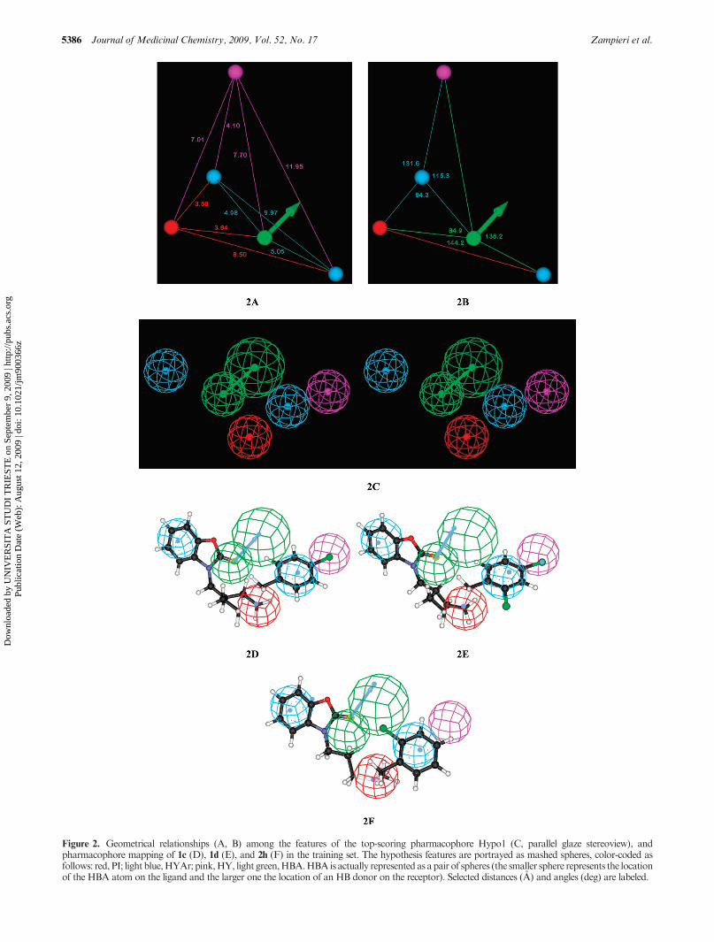
Hypo1 contains one hydrogen bond acceptor, two hydro-phobic aromatic features, one hydrophobic feature, and onepositive ionizable group. The affinities of the 31 compoundsestimated using Hypo1 are reported in Table 4, along withthe experimental values and the relevant errors (expressed asthe ratio of estimated to experimental values). This tableclearly shows that 24 out of 31 molecules in the training sethave errors less than 4 while the remaining 7 have errors lessthan 10. Figure 2A-C illustrates the selected Hypo1 phar-macophore model, while parts D, E, and F of Figure 2 showthe mapping of compounds 1c, 1d, and 2h onto Hypo1,respectively.
In compound 1c, as seen in Figure 2D, the aromatic ring ofthe benzooxazolone moiety matches one of the HYAr fea-tures; the other HYAr feature is nicely overlapped by theadditional phenyl ring. The carbonyl group and the basicnitrogen atom of the pyperidine ring match the HBA and PIfunctions, while the chlorine atom on the monosubstitutedphenyl ring maps the remaining HY feature. Quite ananalogous mapping is observed for 1d (Figure 2E). Theestimated affinities for 1c and 1d are 0.27 and 280 nM, whilethe corresponding experimental affinities are 0.098 and258 nM, respectively.
Figure 2F is an example of a pharmacophoremapping of acompound that is less active than the former two.Compound2hdoes notmap all the features encoded inHypo1. In fact, 2hmaps the two HYAr functions, again by means of the twophenyl rings; the PI feature is still overlapped by the nitrogenatom of the cyclic/linear bridging moiety, and the HBAfunction is located over the carbonyl oxygen. However, itdoes not map the HY function. According to this partialmapping, this compound is predicted to be less active.
A critical step in automated pharmacophore generationis model validation, especially in those cases where themodel has been generated for the purpose of predicting theactivity of external sets of compounds or, as in our case,of estimating the activity of newly conceived molecularentities prior to their synthesis. The first method we usedto check the robustness of our correlation was the predictionof the affinity of a further set ofmolecules, also called the testset, composed of six additionalmolecules fromour series andthree compounds taken from the literature42 (see Table 5).The aim of this validation was to verify if our 3D phar-macophore model was able to predict the experimentally
Table 5. Experimental and Estimated Affinity Values of the Test Set Compounds
Ki(σ1) (nM)
compd R experimental estimated errora
6a H 48.1 210 4.4
6b 4-Cl 45.0 140 3.1
6c 4-CH3 105 200 1.9
7a H 22.5 37 1.6
7b 4-Cl 12.9 5.5 -2.3
7c 4-CH3 69.4 76 1.1
12 5.7 2.2 -2.6
13 0.8 4.6 5.8
14 2.0 19 9.5aValues in the error column represent the ratio of the estimated to experimental affinity, or its negative inverse if the ratio is less than 1.
Dow
nloa
ded
by U
NIV
ER
SIT
A S
TU
DI
TR
IEST
E o
n Se
ptem
ber
9, 2
009
| http
://pu
bs.a
cs.o
rg
Pub
licat
ion
Dat
e (W
eb):
Aug
ust 1
2, 2
009
| doi
: 10.
1021
/jm90
0366
z
5386 Journal of Medicinal Chemistry, 2009, Vol. 52, No. 17 Zampieri et al.
Figure 2. Geometrical relationships (A, B) among the features of the top-scoring pharmacophore Hypo1 (C, parallel glaze stereoview), andpharmacophore mapping of 1c (D), 1d (E), and 2h (F) in the training set. The hypothesis features are portrayed as mashed spheres, color-coded asfollows: red, PI; light blue,HYAr; pink,HY, light green,HBA.HBAis actually represented as apair of spheres (the smaller sphere represents the locationof the HBA atom on the ligand and the larger one the location of an HB donor on the receptor). Selected distances (A) and angles (deg) are labeled.
Dow
nloa
ded
by U
NIV
ER
SIT
A S
TU
DI
TR
IEST
E o
n Se
ptem
ber
9, 2
009
| http
://pu
bs.a
cs.o
rg
Pub
licat
ion
Dat
e (W
eb):
Aug
ust 1
2, 2
009
| doi
: 10.
1021
/jm90
0366
z
Article Journal of Medicinal Chemistry, 2009, Vol. 52, No. 17 5387
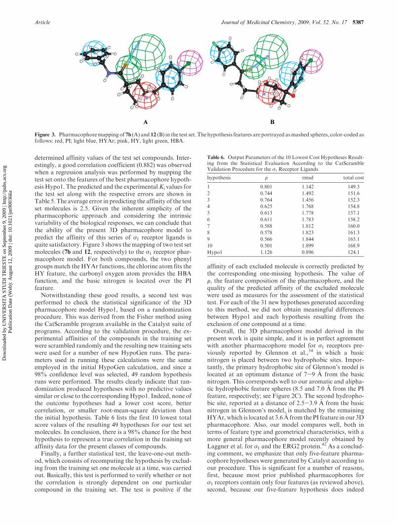
determined affinity values of the test set compounds. Inter-estingly, a good correlation coefficient (0.882) was observedwhen a regression analysis was performed by mapping thetest set onto the features of the best pharmacophore hypoth-esis Hypo1. The predicted and the experimentalKi values forthe test set along with the respective errors are shown inTable 5. The average error in predicting the affinity of the testset molecules is 2.5. Given the inherent simplicity of thepharmacophoric approach and considering the intrinsicvariability of the biological responses, we can conclude thatthe ability of the present 3D pharmacophore model topredict the affinity of this series of σ1 receptor ligands isquite satisfactory. Figure 3 shows themapping of two test setmolecules (7b and 12, respectively) to the σ1 receptor phar-macophore model. For both compounds, the two phenylgroups match the HYAr functions, the chlorine atom fits theHY feature, the carbonyl oxygen atom provides the HBAfunction, and the basic nitrogen is located over the PIfeature.
Notwithstanding these good results, a second test wasperformed to check the statistical significance of the 3Dpharmacophore model Hypo1, based on a randomizationprocedure. This was derived from the Fisher method usingthe CatScramble program available in the Catalyst suite ofprograms. According to the validation procedure, the ex-perimental affinities of the compounds in the training setwere scrambled randomly and the resulting new training setswere used for a number of new HypoGen runs. The para-meters used in running these calculations were the sameemployed in the initial HypoGen calculation, and since a98% confidence level was selected, 49 random hypothesisruns were performed. The results clearly indicate that ran-domization produced hypotheses with no predictive valuessimilar or close to the correspondingHypo1. Indeed, none ofthe outcome hypotheses had a lower cost score, bettercorrelation, or smaller root-mean-square deviation thanthe initial hypothesis. Table 6 lists the first 10 lowest totalscore values of the resulting 49 hypotheses for our test setmolecules. In conclusion, there is a 98% chance for the besthypothesis to represent a true correlation in the training setaffinity data for the present classes of compounds.
Finally, a further statistical test, the leave-one-out meth-od, which consists of recomputing the hypothesis by exclud-ing from the training set one molecule at a time, was carriedout. Basically, this test is performed to verify whether or notthe correlation is strongly dependent on one particularcompound in the training set. The test is positive if the
affinity of each excluded molecule is correctly predicted bythe corresponding one-missing hypothesis. The value ofF, the feature composition of the pharmacophore, and thequality of the predicted affinity of the excluded moleculewere used as measures for the assessment of the statisticaltest. For each of the 31 new hypotheses generated accordingto this method, we did not obtain meaningful differencesbetween Hypo1 and each hypothesis resulting from theexclusion of one compound at a time.
Overall, the 3D pharmacophore model derived in thepresent work is quite simple, and it is in perfect agreementwith another pharmacophore model for σ1 receptors pre-viously reported by Glennon et al.,34 in which a basicnitrogen is placed between two hydrophobic sites. Impor-tantly, the primary hydrophobic site of Glennon’s model islocated at an optimum distance of 7-9 A from the basicnitrogen. This corresponds well to our aromatic and alipha-tic hydrophobic feature spheres (8.5 and 7.0 A from the PIfeature, respectively; see Figure 2C). The second hydropho-bic site, reported at a distance of 2.5-3.9 A from the basicnitrogen in Glennon’s model, is matched by the remainingHYAr,which is located at 3.6 A from the PI feature in our 3Dpharmacophore. Also, our model compares well, both interms of feature type and geometrical characteristics, with amore general pharmacophore model recently obtained byLaggner et al. for σ1 and the ERG2 protein.42 As a conclud-ing comment, we emphasize that only five-feature pharma-cophore hypotheses were generated by Catalyst according toour procedure. This is significant for a number of reasons,first, because most prior published pharmacophores forσ1 receptors contain only four features (as reviewed above),second, because our five-feature hypothesis does indeed
Figure 3. Pharmacophoremapping of 7b (A) and 12 (B) in the test set. The hypothesis features are portrayed asmashed spheres, color-coded asfollows: red, PI; light blue, HYAr; pink, HY, light green, HBA.
Table 6. Output Parameters of the 10 Lowest Cost Hypotheses Result-ing from the Statistical Evaluation According to the CatScrambleValidation Procedure for the σ1 Receptor Ligands
hypothesis F rmsd total cost
1 0.801 1.142 149.3
2 0.744 1.492 151.6
3 0.764 1.456 152.3
4 0.625 1.768 154.8
5 0.613 1.778 157.1
6 0.611 1.783 158.2
7 0.588 1.812 160.0
8 0.578 1.823 161.3
9 0.566 1.844 165.1
10 0.501 1.899 168.9
Hypo1 1.126 0.896 124.1
Dow
nloa
ded
by U
NIV
ER
SIT
A S
TU
DI
TR
IEST
E o
n Se
ptem
ber
9, 2
009
| http
://pu
bs.a
cs.o
rg
Pub
licat
ion
Dat
e (W
eb):
Aug
ust 1
2, 2
009
| doi
: 10.
1021
/jm90
0366
z
5388 Journal of Medicinal Chemistry, 2009, Vol. 52, No. 17 Zampieri et al.
represent a more stringent and significant model, and last,because it illustrates that Catalyst identified sufficient evi-dence in this structural data set to add or distinguish anadditional feature or nuance not seen previously.
Conclusions
In this work we discussed how, from three series of newlysynthesized compounds characterized by a broad range ofaffinity toward σ1 receptors, we derived a three-dimensionalpharmacophoremodel with quantitative predictive ability forthese classes ofmolecules. The best generated pharmacophoremodel (Hypo1) consists of five features: two hydrophobicaromatic, one hydrophobic aliphatic, one hydrogen bondacceptor, and onepositive ionizable group.Hypo1 reasonablypredicts the affinity of the test set molecules with a correlationcoefficient of 0.896 and shows the best statistical significanceamong all the generated models. Two validation tests, theFisher test and the leave-one-out test, confirmed the statisticalvalidity of our simple but effective 3D pharmacophore,excluding any possibility of a chance correlation betweenexperimental and predicted affinity values. Finally, the modelwas predictive not only for the training set compounds butalso for a test set of nine additional molecules, three of whichwere taken from the literature and not structurally related toour series.
Compared with other drug discovery tools, the pharmaco-phore approachhas the significant advantage that it is fast andable to predict the activity of quite a large number ofmolecules in a relatively short time. Given the reasonablepredictive ability of our model, we expect to exploit it in thedevelopment and optimization of our promising series ofcompounds. In particular, wewill use this 3Dpharmacophoreto estimate the potential affinity of virtual libraries of newlydesigned, second generation σ1 receptor ligands prior tosynthesis and biological testing.
Experimental Section
Unless otherwise noted, starting materials and reagents wereobtained from commercial suppliers and were used withoutpurification. Melting points were determined with a Buchi 510capillary apparatus and are uncorrected. Infrared spectra inNujolmullswere recordedona JaskoFT200 spectrophotometer.Proton nuclear magnetic resonance (1H NMR) spectra weredetermined on a Varian Gemini 200 spectrometer, and thechemical shifts are reported as δ (ppm) in CDCl3 solution.Coupling constants J are expressed in hertz (Hz). Reactioncourses and product mixtures were routinely monitored bythin-layer chromatography (TLC) on silica gel precoated F254
Merck plates. ESI-MS spectra were obtained on a PE-API Ispectrometer by infusion of a solution of the sample in MeOH.Elemental analyses (C, H, N) were performed on a Carlo Erba1106 analyzer and were within (0.3 of the theoretical value.
Synthesis of Benzooxazolone Derivatives. 3-(3-Chloropropyl)-benzo[d ]oxazol-2(3H)-one (5a).43 A mixture of benzo[d ]oxazol-2(3H)-one 4 (3.0 g, 22.22 mmol) and K2CO3 (7.7 g, 55.55 mmol)was dissolved in 50 mL of CH3CN and the solution was refluxedfor 10 min. 1-Bromo-3-chloropropane (8.7 g, 55.55 mmol) and acatalytic amountofKIwere added, and themixturewas stirred foran additional 3 h at reflux temperature. The inorganic salts werefiltered off, and the solvent was evaporated under reduced pres-sure. Distilled water (50 mL) was added, and the residue wasextracted 3 times with CHCl3 (3 � 100 mL). The organic phasewas separated and dried over anhydrous Na2SO4. The filteredsolution was concentrated under reduced pressure, and the re-maining oil was crystallized from n-hexane to afford a light-yellow
solid. Yield 4.01 g (85%); mp 62-64 �C. IR (Nujol): 1791 cm-1.1H NMR (CDCl3-TMS) ppm (δ): 2.30 (m, 2H, CH2-CH2-CH2,J=5.9-6.6 Hz); 3.63 (t, N-CH2, J=6.6Hz); 4.00 (t, 2H,CH2-Cl,J=5.9 Hz); 7.00-7.30 (m, 4H arom). MS: m/z 212 [MHþ] 214[MHþ þ 2].
3-(4-Bromobutyl)benzo[d ]oxazol-2(3H)-one (5b).44 This inter-mediate was obtained in an analogous way using 1,4-dibromo-butane. Yield 2.99 g (60%); mp 42-45 �C. IR (Nujol): 1747cm-1. 1H NMR (CDCl3-TMS) ppm (δ): 1.80 (m, 4H, CH2-CH2-CH2-CH2); 3.63 (m, N-CH2); 4.00 (m, 2H, CH2-Br);7.10-7.40 (m, 4H arom). MS: m/z 270 [MHþ] 272 [MHþ þ 2].
3-[3-(N-Benzyl-N-methylamino)propyl]benzo[d ]oxazol-2(3H)-one (2a). A mixture of 3-(4-chloropropyl)benzo[d ]ossazol-2(3H)-one (0.60 g, 2.84 mmol), N-methylbenzylamine (0.28 g,2.27 mmol), anhydrous K2CO3 (2.19 g, 15.9 mmol), and acatalytic amount of KI was dissolved in 50 mL of CH3CN, andthe solution was refluxed andmonitored by TLC. The inorganicsalts were filtered off, and the solvent was evaporated underreduce pressure. Distilled water (50 mL) was added, and theresidue was extracted 3 times with CHCl3 (3 � 100 mL). Theorganic phase was separated and dried over anhydrous Na2SO4.The filtered solution was concentrated under reduced pressureand the remaining oil was treated with an equimolar amount ofoxalic acid in absolute ethanol to yield the oxalate salt,whichwasfiltered andwashed with cold ethanol. Yield 0.26 g (0.20 g of freebase, 30%); mp 180-182 �C. IR (Nujol): 1772, 2697 cm-1. 1HNMR (free base, CDCl3-TMS) ppm (δ): 2.01 (m, 2H, CH2-CH2-CH2, J=6.7-7.3 Hz); 2.21 (s, 3H, N-CH3); 2.49 (t, 2H,CH2-N(CH3)-CH2-Ar, J=6.7Hz); 3.51 (s, 2H,N-CH2-Ar); 3.93(t, 2H, N-CH2-(CH2)2-, J=7.3 Hz); 6.95-7.40 (m, 9H, arom).MS: m/z 297 [MHþ].
Compounds 2b-kwere synthesized following the same routedescribed above for compound 2a.
3-[3-[N-(2-Chlorobenzyl)-N-methylamino]propyl]benzo[d ]oxazol-2(3H)-one (2b). IR (Nujol): 1764, 2721 cm-1. 1H NMR (free base,CDCl3-TMS) ppm (δ): 1.97 (m, 2H, CH2-CH2-CH2, J=6.6-7.3 Hz); 2.15 (s, 3H, CH3); 2.46 (t, 2H, CH2-N(CH3)-CH2-Ar, J=6.6 Hz); 3.51 (s, 2H, N-CH2-Ar); 3.84 (t, 2H, N-CH2-(CH2)2-,J=7.3 Hz); 6.89-7.37 (m, 8H, arom). MS: m/z 331 [MHþ] 333[MHþ þ 2].
3-[3-[N-(3-Chlorobenzyl)-N-methylamino]propyl]benzo[d ]oxazol-2(3H)-one (2c). IR (Nujol): 1767, 2673 cm-1. 1H NMR (free base,CDCl3-TMS) ppm (δ): 1.95 (m, 2H, CH2-CH2-CH2, J=6.6-7.3 Hz); 2.15 (s, 3H, CH3); 2.45 (t, 2H, CH2-N(CH3)-CH2-Ar,J=6.6 Hz); 3.43 (s, 2H, N-CH2-Ar); 3.86 (t, 2H, N-CH2-(CH2)2-,J=7.3 Hz); 6.92-7.30 (m, 8H, arom). MS: m/z 331 [MHþ] 333[MHþ þ 2].
3-[3-[N-(4-Chlorobenzyl)-N-methylamino]propyl]benzo[d ]oxazol-2(3H)-one (2d). IR (Nujol): 1770, 2677 cm-1. 1H NMR (free base,CDCl3-TMS) ppm (δ): 2.05 (m, 2H, CH2-CH2-CH2, J=6.6-7.3 Hz); 2.17 (s, 3H, N-CH3); 2.45 (t, 2H, CH2-N(CH3)-CH2-Ar,J=6.6 Hz); 3.46 (s, 2H, N-CH2-Ar); 3.90 (t, 2H, N-CH2-(CH2)2-,J=7.3 Hz); 6.95-7.30 (m, 8H, arom). MS: m/z 331 [MHþ] 333[MHþ þ 2].
3-[3-[N-(2-Methoxybenzyl)-N-methylamino]propyl]benzo[d ]oxa-zol-2(3H)-one (2e). IR (Nujol): 1771, 2673 cm-1. 1H NMR (freebase, CDCl3-TMS) ppm (δ): 1.87-2.00 (m, 2H, CH2-CH2-CH2,J=6.6-7.3Hz); 2.14 (s, 3H,CH3); 2.43 (t, 2H,CH2-N(CH3)-CH2-Ar,J=6.6Hz); 3.45 (s, 2H,N-CH2-Ar); 3.75 (s, 3H,OCH3); 3.87 (t,2H,N-CH2-(CH2)2-,J=7.3Hz); 6.79-7.25 (m, 8H, arom).MS:m/z 327 [MHþ].
3-[3-[N-(3-Methoxybenzyl)-N-methylamino]propyl]benzo-[d ]oxazol-2(3H )-one (2f). IR (Nujol): 1770, 2676 cm-1. 1HNMR (free base, CDCl3-TMS) ppm (δ): 1.83-1.97 (m, 2H,CH2-CH2-CH2, J=6.6-7.3 Hz); 2.12 (s, 3H, CH3); 2.40 (t,2H, CH2-N(CH3)-CH2-Ar, J=6.6 Hz); 3.39 (s, 2H, N-CH2-Ar); 3.74 (s, 3H, OCH3); 3.84 (t, 2H, N-CH2-(CH2)2-, J=7.3Hz); 6.70-7.21 (m, 8H, arom). MS: m/z 327 [MHþ].
3-[3-[N-(4-Methoxybenzyl)-N-methylamino]propyl]benzo[d ]-oxazol-2(3H )-one (2g). IR (Nujol): 1771, 2685 cm-1. 1H NMR
Dow
nloa
ded
by U
NIV
ER
SIT
A S
TU
DI
TR
IEST
E o
n Se
ptem
ber
9, 2
009
| http
://pu
bs.a
cs.o
rg
Pub
licat
ion
Dat
e (W
eb):
Aug
ust 1
2, 2
009
| doi
: 10.
1021
/jm90
0366
z
Article Journal of Medicinal Chemistry, 2009, Vol. 52, No. 17 5389
(free base, CDCl3-TMS) ppm (δ): 1.98 (m, 2H, CH2-CH2-CH2, J=6.7-7.3 Hz); 2.20 (s, 3H, CH3); 2.43 (t, 2H, CH2-N(CH3)-CH2-Ar, J=6.7 Hz); 3.45 (s, 2H, N-CH2-Ar); 3.84(s, 3H, OCH3); 3.91 (t, 2H, N-CH2-(CH2)2-, J=7.3 Hz); 6.85-7.30 (m, 8H, arom). MS: m/z 327 [MHþ].
3-[3-[N-Methylamino-N-(2-methylbenzyl)]propyl]benzo[d ]oxazol-2(3H)-one (2h). IR (Nujol): 1766, 2723 cm-1. 1H NMR (freebase, CDCl3-TMS) ppm (δ): 1.82-1.96 (m, 2H, CH2-CH2-CH2, J=6.6-7.3 Hz); 2.14 (s, 3H,CH3); 2.30 (s, 3H,CH3); 2.44(t, 2H, CH2-N(CH3)-CH2-Ar, J=6.6 Hz); 3.40 (s, 2H, N-CH2-Ar); 3.78 (t, 2H, N-CH2-(CH2)2-, J=7.3Hz); 6.86-7.23 (m, 8H,arom). MS: m/z 311 [MHþ].
3-[3-[N-Methylamino-N-(3-methylbenzyl)]propyl]benzo[d ]oxazol-2(3H)-one (2i). IR (Nujol): 1770, 2676 cm-1. 1H NMR (freebase, CDCl3-TMS) ppm (δ): 1.84-1.98 (m, 2H, CH2-CH2-CH2, J=6.6-7.3 Hz); 2.12 (s, 3H,CH3); 2.26 (s, 3H,CH3); 2.40(t, 2H, CH2-N(CH3)-CH2-Ar, J=6.6 Hz); 3.38 (s, 2H, N-CH2-Ar); 3.84 (t, 2H, N-CH2-(CH2)2-, J=7.3Hz); 6.91-7.18 (m, 8H,arom). MS: m/z 311 [MHþ].
3-[3-[N-Methylamino-N-(4-methylbenzyl)]propyl]benzo[d ]oxazol-2(3H)-one (2j). IR (Nujol): 1766, 2675 cm-1. 1H NMR (freebase, CDCl3-TMS) ppm (δ): 1.85-1.99 (m, 2H, CH2-CH2-CH2, J=6.6-7.3 Hz); 2.14 (s, 3H,CH3); 2.27 (s, 3H,CH3); 2.42(t, 2H, CH2-N(CH3)-CH2-Ar, J=6.6 Hz); 3.43 (s, 2H, N-CH2-Ar); 3.82 (t, 2H, N-CH2-(CH2)2-, J=7.3Hz); 6.92-7.20 (m, 8H,arom). MS: m/z 311 [MHþ].
3-[3-[N-(2,4-Dimethylbenzyl)-N-methylamino]propyl]benzo[d ]-oxazol-2(3H)-one (2k). IR (Nujol): 1777, 2725 cm-1. 1H NMR(free base, CDCl3-TMS) ppm (δ): 1.97 (m, 2H, CH2-CH2-CH2,J=6.7-7.3 Hz); 2.21 (s, 3H, N-CH3); 2.33 (s, 3H, CH3); 2.35(s, 3H, CH3); 2.50 (t, 2H, CH2-N(CH3)-CH2-Ar, J=6.7 Hz);3.43 (s, 2H, N-CH2-Ar); 3.86 (t, 2H, N-CH2-(CH2)2-, J=7.3Hz); 6.95-7.30 (m, 7H, arom). MS: m/z 325 [MHþ].
3-[4-(N-Benzyl-N-methylamino)butyl]benzo[d ]oxazol-2(3H )-one (3a).Amixture of 3-(4-bromobutyl)benzo[d ]ossazol-2(3H)-one (0.25 g, 0.926 mmol), N-methylbenzylamine (0.089 g,0741 mmol), anhydrous K2CO3 (0.72 g, 5.19 mmol), and acatalytic amount of KI was dissolved in 50 mL of ACN, andthe solution was refluxed andmonitored by TLC. The inorganicsalts were filtered off, and the solvent was evaporated underreduce pressure. Distilled water (50 mL) was added, and theresidue was extracted 3 times with CHCl3 (3 � 100 mL). Theorganic phase was separated and dried over anhydrousNa2SO4.The filtered solution was concentrated under reduced pressureand the remaining oil was treated with an equimolar amount ofoxalic acid in absolute ethanol to afford the oxalate salt, whichwas filtered andwashedwith cold ethanol. Yield 0.12 g (0.09 g offree base, 30%); mp 145-150 �C. IR (Nujol): 1772, 2668 cm-1.1HNMR (free base, CDCl3-TMS) ppm (δ): 1.50-2.00 (m, 4H,CH2-CH2-CH2-CH2, J=6.7-7.3 Hz); 2.20 (s, 3H, N-CH3); 2.40(t, 2H, CH2-N(CH3)-CH2-Ar, J=6.7 Hz); 3.45 (s, 2H, N-CH2-Ar); 3.85 (t, 2H, N-CH2-(CH2)3-, J=7.3Hz); 6.90-7.40 (m, 9H,arom). MS: m/z 311 [MHþ].
Compounds 3b-kwere synthesized following the same routedescribe above for compound 3a.
3-[4-[N-(2-Chlorobenzyl)-N-methylamino]butyl]benzo[d ]oxazol-2(3H)-one (3b). IR (Nujol): 1770, 2674 cm-1. 1H NMR(CDCl3-TMS) ppm (δ): 1.49-1.90 (m, 4H, CH2-CH2-CH2-CH2); 2.13 (s, 3H, CH3); 2.39 (t, 2H, CH2-N(CH3)-CH2-Ar, J=6.6Hz); 3.40 (s, 2H,N-CH2-Ar); 3.81 (t, 2H,N-CH2-(CH2)3-,J=7.3Hz); 6.90-7.25 (m, 8H, arom).MS:m/z345 [MHþ] 347 [MHþþ 2].
3-[4-[N-(3-Chlorobenzyl)-N-methylamino]butyl]benzo[d ]oxazol-2(3H)-one (3c). IR (Nujol): 1766, 2670 cm-1. 1H NMR(CDCl3-TMS) ppm (δ): 1.49-1.90 (m, 4H, CH2-CH2-CH2-CH2); 2.14 (s, 3H, CH3); 2.39 (t, 2H, CH2-N(CH3)-CH2-Ar, J=6.6Hz); 3.40 (s, 2H,N-CH2-Ar); 3.82 (t, 2H,N-CH2-(CH2)3-,J=7.3Hz); 6.91-7.27 (m, 8H, arom).MS:m/z345 [MHþ] 347 [MHþþ 2].
3-[4-[N-(4-Chlorobenzyl)-N-methylamino]butyl]benzo[d ]oxazol-2(3H)-one (3d). IR (Nujol): 1768, 2676 cm-1. 1H NMR(CDCl3-TMS) ppm (δ): 1.50-1.65 (m, 2H, CH2-CH2-CH2);
1.75-1.90 (m, 2H, CH2-CH2-CH2); 2.15 (s, 3H, CH3);2.36-2.43 (t, 2H, CH2-N(CH3)-CH2-Ar, J=6.6 Hz); 3.42 (s,2H, N-CH2-Ar); 3.79-3.86 (t, 2H, N-CH2-(CH2)3-, J=7.3 Hz);6.93-7.29 (m, 8H, arom). MS: m/z 345 [MHþ] 347 [MHþ þ 2].
3-[4-[N-(2-Methoxybenzyl)-N-methylamino]butyl]benzo[d ]oxazol-2(3H)-one (3e). IR (Nujol): 1770, 2673 cm-1. 1H NMR(CDCl3-TMS) ppm (δ): 1.53-1.90 (m, 4H, CH2-CH2-CH2-CH2); 2.20 (s, 3H, CH3); 2.45 (t, 2H, CH2-N(CH3)-CH2-Ar, J=6.6 Hz); 3.48 (s, 2H, N-CH2-Ar); 3.79 (s, 3H, CH3); 3.87 (t, 2H,N-CH2-(CH2)3-, J=7.3Hz); 6.83-7.30 (m, 8H, arom).MS:m/z341 [MHþ].
3-[4-[N-(3-Methoxybenzyl)-N-methylamino]butyl]benzo[d ]oxa-zol-2(3H)-one (3f). IR (Nujol): 1769, 2674 cm-1. 1H NMR(CDCl3-TMS) ppm (δ): 1.54-1.91 (m, 2H, CH2-CH2-CH2-CH2); 2.19 (s, 3H, CH3); 2.44 (t, 2H, CH2-N(CH3)-CH2-Ar, J=6.6 Hz); 3.49 (s, 2H, N-CH2-Ar); 3.79 (s, 3H, CH3); 3.86 (t, 2H,N-CH2-(CH2)3-, J=7.3 Hz); 6.81-7.28 (m, 8H, arom). MS: m/z341 [MHþ].
3-[3-[N-(4-Methoxybenzyl)-N-methylamino]butyl]benzo[d ]oxa-zol-2(3H)-one (3g). IR (Nujol): 1774, 2724 cm-1. 1H NMR (freebase, CDCl3-TMS) ppm (δ): 1.53-1.90 (m, 4H, CH2-CH2-CH2-CH2, J=6.7-7.3 Hz); 2.18 (s, 3H, CH3); 2.40 (t, 2H, CH2-N(CH3)-CH2-Ar, J=6.7 Hz); 3.42 (s, 2H, N-CH2-Ar); 3.86 (m,5H, OCH3 and N-CH2-(CH2)3-, J=7.3 Hz); 6.84-7.30 (m, 8H,arom). MS: m/z 341 [MHþ].
3-[4-(N-Methylamino-N-(2-methylbenzyl)]benzo[d ]oxazol-2(3H)-one (3h). IR (Nujol): 1769, 2668 cm-1. 1H NMR (CDCl3-TMS)ppm (δ): 1.49-1.90 (m, 4H, CH2-CH2-CH2-CH2); 2.15 (s, 3H,CH3); 2.33 (s, 3H, CH3); 2.41 (t, 2H, CH2-N(CH3)-CH2-Ar, J=6.6 Hz); 3.41 (s, 2H, N-CH2-Ar); 3.79 (t, 2H, N-CH2-(CH2)3-, J=7.3 Hz); 6.89-7.25 (m, 8H, arom). MS:m/z 325 [MHþ].
3-[4-(N-Methylamino-N-(3-methylbenzyl)]benzo[d ]oxazol-2(3H)-one (3i). IR (Nujol): 1770, 2666 cm-1. 1H NMR (CDCl3-TMS)ppm (δ): 1.50-1.91 (m, 4H, CH2-CH2-CH2-CH2); 2.15 (s, 3H,CH3); 2.31 (s, 3H, CH3); 2.38 (t, 2H, CH2-N(CH3)-CH2-Ar, J=6.6 Hz); 3.40 (s, 2H, N-CH2-Ar); 3.81 (t, 2H, N-CH2-(CH2)3-, J=7.3 Hz); 6.92-7.22 (m, 8H, arom). MS:m/z 325 [MHþ].
3-[4-(N-Methylamino-N-(4-methylbenzyl)]benzo[d ]oxazol-2(3H)-one (3j). IR (Nujol): 1769, 2674 cm-1. 1H NMR (CDCl3-TMS)ppm (δ): 1.51-1.92 (m, 4H, CH2-CH2-CH2-CH2); 2.16 (s, 3H,CH3); 2.33 (s, 3H, CH3); 2.39 (t, 2H, CH2-N(CH3)-CH2-Ar, J=6.6Hz); 3.43 (s, 2H,N-CH2-Ar); 3.82 (t, 2H,N-CH2-(CH2)3-,J=7.3Hz); 6.93-7.23 (m, 8H, arom). MS:m/z 325 [MHþ].
3-[4-[N-(2,4-Dimethylbenzyl)-N-methylamino]butyl]benzo[d ]oxa-zol-2(3H)-one (3k). IR (Nujol): 1779, 2724 cm-1. 1H NMR (freebase, CDCl3-TMS) ppm (δ): 1.50-1.88 (m, 4H, CH2-CH2-CH2-CH2, J=6.7-7.3 Hz); 2.17 (s, 3H, N-CH3); 2.32 (s, 6H, 2� CH3);2.42 (t, 2H,CH2-N(CH3)-CH2-Ar, J=6.7Hz); 3.40 (s, 2H,N-CH2-Ar); 3.82 (t, 2H, N-CH2-(CH2)3-, J=7.3 Hz); 6.90-7.30 (m, 7H,arom). MS: m/z 341 [MHþ].
Synthesis of Carboxamide Derivatives. N-Phenyl-1-(tert-but-oxycarbonyl)piperidine-4-carboxamide (8).45 To a mixture ofN-Boc-4-piperidinecarboxylic acid (2.12 g, 9.22mmol), pyridine(1.90mL, 23,6mmol) andCH2Cl2 (15mL) and SOCl2 (0.80mL,11.0 mmol) were added, under N2 at room temperature, whilestirring. After 25 min, a solution of aniline (0.95 g, 10.2 mmol),Et3N (4.50mL, 32.3mmol), and a catalytic amount ofDMAP inCH2Cl2 (15 mL) was added dropwise. The reaction was mon-itored by TLC. After 14 h, the organic phase was washed with1 NHCl (2� 20 mL) and distilled water (2� 20 mL), dried withNa2SO4, and concentrated in vacuum to give 1.70 g (60.1%) ofa light-brown solid; mp 140-142C. IR (Nujol): 1656, 1685,3257 cm-1. 1H NMR (CDCl3-TMS) ppm (δ): 1.44 (s 9H, CH3,Boc); 1.60-1.93 (m, 4H, H3,30-H5,50, pip.); 2.36 (m, 1H, H4, pip.);2.76 (m, 2H,H2-H6, pip.); 4.17 (m, 2H,H20-H60, pip.); 7.05-7.52 (m,6H,aromþNHdisappearingondeuteration).MS:m/z305 [MHþ].
Compound 9 was synthesized in analogous way using ben-zylamine instead of aniline. Yield: 1.52 g (52%).
N-Benzyl-1-(tert-butoxycarbonyl)piperidine-4-carboxamide (9).IR (Nujol): 1634, 1681, 3251 cm-1. 1H NMR (CDCl3-TMS)
Dow
nloa
ded
by U
NIV
ER
SIT
A S
TU
DI
TR
IEST
E o
n Se
ptem
ber
9, 2
009
| http
://pu
bs.a
cs.o
rg
Pub
licat
ion
Dat
e (W
eb):
Aug
ust 1
2, 2
009
| doi
: 10.
1021
/jm90
0366
z
5390 Journal of Medicinal Chemistry, 2009, Vol. 52, No. 17 Zampieri et al.
ppm(δ): 1.38 (s, 9H,CH3,Boc); 1.45-1.77 (m,4H,H3,30-H5,50, pip.);2.21 (m, 1H,H4, pip.); 2.65 (m, 2H,H2-H6, pip.); 4.06 (m, 2H,H20-H60, pip.); 4.36 (s, 2H, Ar-CH2-N-CO); 5.90 (s broad, 1H, NH,disappearing on deuteration); 7.08-7.30 (m, 5H, arom). MS: m/z319 [MHþ].
N-Phenylpiperidine-4-carboxamide (10).46 Compound 8 (1.00 g,3.24 mmol) was deprotected with 2 mL of trifluoroacetic acid atroom temperature for 24 h. The solution was concentrated atreduced pressure, and the residue was poured into water andbasified with NaOH 10% solution (pH 10). The mixture wasextracted with AcOEt (3 � 25 mL). The organic phase was driedwith Na2SO4 and concentrated. The obtained solid was usedwithout further purification. Yield: 0.60 g (89%); mp 105-107 �C. IR (Nujol): 1655, 3258 cm-1. 1H NMR (CDCl3-TMS)ppm (δ): 1.57-1.96 (m, 5H,H3,30-H5,50, pip.þNHdisappearingondeuteration); 2.33 (m, 1H,H4, pip.); 2.57 (m, 2H,H2-H6, pip.); 3.13(m, 2H, H20-H60, pip.); 7.04-7.50 (m, 6H, aromþ NH disappear-ing on deuteration). MS: m/z 205 [MHþ].
Compound 11 was synthesized starting from 9 (1.2 g) usingthe same procedure. Yield: 0.75 g (91%); mp115-117 �C.
N-Benzylpiperidine-4-carboxamide (11).47 IR (Nujol): 1634,3248 cm-1. 1H NMR (CDCl3-TMS) ppm (δ): 1.50-1.78 (m,4H, H3,30-H5,50, pip.); 2.11-2.26 (m, 2H, H4, pip. þ NH dis-appearing on deuteration); 2.52 (m, 2H, H2-H6, pip.); 3.05 (m,2H, H20-H60, pip.); 4.32 (s, 2H, Ar-CH2-N-CO); 6.13 (s.broad,1H, NH, disappearing on deuteration); 7.10-7.34 (m, 5H,arom). MS: m/z 219 [MHþ].
1-Benzyl-N-phenylpiperidine-4-carboxamide (6a). A solutionof 10 (0.17 g, 0.84mmol),K2CO3 (0.14 g, 1.00mmol), andbenzylchloride (0.11 g, 0.84 mmol) in 50 mL of acetone was stirredunder reflux for 5 h. After the mixture was cooled, the inorganicsalt was filtered and the solvent evaporated at reduced pressure.The residue was washed with distilled water and then with ethylether to afford 6a as a chromatographically pure solid: yield0.18 g (66%); mp 138-140 �C. IR (Nujol): 1657, 3318 cm-1.1H NMR (CDCl3-TMS) ppm (δ): 1.80-2.32 (m, 7H, H2-H3,30-H5,50-H6-H4, pip.); 3.02 (m, 2H, H20-H60, pip.); 3.57 (s, 2H,N-CH2-Ar); 7.06-7.55 (m, 11H, arom þ NH disappearing ondeuteration). MS: m/z 295 [MHþ].
Compounds 6b and 6c were synthesized according to theprocedure reported above. Compounds 7a, 7b, and 7cwere alsosynthesized following the same route but starting from 11.
N-Phenyl-1-(4-chlorobenzyl)piperidine-4-carboxamide (6b).IR (Nujol): 1656, 3301 cm-1. 1HNMR (CDCl3-TMS) ppm (δ):1.63-2.26 (m, 7H, H2-H3,30-H5,50-H6-H4, pip.); 2.93 (m, 2H,H20-H60, pip.); 3.47 (s, 2H, N-CH2-Ar); 7.06-7.53 (m, 10H,arom þ NH disappearing on deuteration). MS: m/z 329 [MHþ]331 [MHþ þ 2].
N-Phenyl-1-(4-methylbenzyl)piperidine-4-carboxamide (6c).IR (Nujol): 1654, 3292 cm-1. 1H NMR (CDCl3-TMS) ppm(δ): 1.62-2.29 (m, 7H, H2-H3,30-H5,50-H6-H4, pip.); 2.34 (s,3H,CH3); 2.97 (m, 2H, H20-H60, pip.); 3.48 (s, 2H, N-CH2-Ar);7.10-7.53 (m, 10H, aromþNH disappearing on deuteration).MS: m/z 309 [MHþ].
N,1-Dibenzylpiperidine-4-carboxamide (7a).48 IR (Nujol):1634, 3248 cm-1. 1H NMR (CDCl3-TMS) ppm (δ):1.65-2.13 (m, 7H, H2-H3,30-H5,50-H6-H4, pip.); 2.84 (m, 2H,H20-H60, pip.); 3.40 (s, 2H, N-CH2-Ar); 4.40 (s, 2H, Ar-CH2-N-CO); 5.75 (s.broad, 1H, NH, disappearing on deuteration);7.13-7.36 (m, 10H, arom). MS: m/z 309 [MHþ].
N-Benzyl-1-(4-chlorobenzyl)piperidine-4-carboxamide (7b).IR (Nujol): 1633, 3247 cm-1. 1H NMR (CDCl3-TMS) ppm(δ): 1.62-2.20 (m, 7H, H2-H3,30-H5,50-H6-H4, pip.); 2.87(m, 2H, H20-H60, pip.); 3.43 (s, 2H, N-CH2-Ar); 4.37 (s, 2H,Ar-CH2-N-CO); 5.78 (s broad, 1H, NH, disappearing ondeuteration); 7.14-7.32 (m, 9H, arom). MS: m/z 343 [MHþ]345 [MHþ þ 2].
N-Benzyl-1-(4-methylbenzyl)piperidine-4-carboxamide (7c).IR (Nujol): 1636, 3251 cm-1. 1H NMR (CDCl3-TMS) ppm(δ): 1.80-2.29 (m, 7H,H2-H3,30-H5,50-H6-H4, pip.); 2.41 (s, 3H,
CH3); 3.00 (m, 2H,H20-H60, pip.); 3.53 (s, 2H, N-CH2-Ar); 4.51(s, 2H, Ar-CH2-N-CO); 5.84 (s broad, 1H, NH, disappearingon deuteration); 7.18-7.45 (m, 9H, arom). MS: m/z 323[MHþ].
Pharmacology. Radioligand Binding Assays. Binding assayswere performed on rat liver membranes according to themethods of Hellewell23 and were slightly modified as pre-viously described.33 Briefly, for the σ1 receptor assay 250 μg ofrat liver homogenate was incubated for 120 min at 37 �C with1 nM [3H]-(þ)-pentazocine (PerkinElmer, specific activity34.9 Ci/mmol) in 50mMTris-HCl, pH 8.0, 0.5mL final volume.Nonspecific binding was defined in the presence of 10 μMhaloperidol. The reaction was stopped by vacuum filtrationthrough GF/B glass-fiber filters presoacked with 0.5% poly-ethylenimine, followed by rapid washing with 2 mL of ice-coldbuffer. The filters were placed in 3 mL of scintillation cocktail,and the radioactivity was determined by liquid scintillationcounting.
For the σ2 receptor assay, 150 μg of rat liver homogenate wasincubated for 120min at room temperature with 3 nM [3H]DTG(PerkinElmer, specific activity 58.1 Ci/mmol) in 50 mM Tris-HCl, pH 8.0, 0.5 mL final volume. (þ)-Pentazocine (100 nM)and haloperidol (10 μM) were used to mask σ1 receptors and todefine nonspecific binding, respectively.
Competition studies were done using at least 11 differentconcentrations of the ligand under investigation. As an internalcontrol, three increasing concentrations of unlabeled (þ)-pent-azocine (σ1 receptors) or DTG (σ2 receptors) were alwaysincluded. The compounds were prepared as 10 mM stocksolutions in 100% DMSO and diluted with Tris-HCl buffer onthe day of the experiment. The final DMSO concentration in theincubation tubes was maintained at 0.1%.
The competition data for two to four separate determinationsperformed in duplicate were averaged by fitting to a four-parameter curve by means of the SigmaPlot software. Calcu-lated IC50 values and Hill’s coefficients (nH) are reportedas mean values ( SEM. The corresponding Ki values wereobtained by the Cheng-Prusoff equation, as previouslyreported.49
Molecular Modeling. Training and Test Sets. The modelstructures of all compounds were built using the Catalyst 2D-3D sketcher.38 High quality conformational models are crucialfor the development of predictive pharmacophore models.Accordingly, in this study we employed an ad hoc procedureto derive molecular conformations, instead of using thosegenerated by Catalyst, for better quality in covering the low-energy conformational space. Each molecular structure wassubjected to energy minimization using the generalizedCHARMM force field50 until the gradient dropped below0.05. The minimized structures were used as the starting pointfor subsequent conformational searches. A 10000-step MonteCarlo torsional sampling conformational search was conductedfor each compound. Unique low-energy conformations within20 kcal/mol of the corresponding global energy minimum werecollected for each molecule. A conformation was consideredunique only when the maximum displacement of at leastone heavy atom was greater than 0.5 A. A maximum of 250unique conformations were recovered for each compound. Theclassical conformational search was also carried out usingthe Poling algorithm51-53 and the CHARMM force field51
as implemented in the Catalyst program for comparison. The“best quality” generation option was adopted to select repre-sentative conformers over a 0-20 kcal/mol interval above thecomputed global energy minimum in the conformational space,and again, the number of conformers generated for each com-pound was limited to a maximum of 250. Comparing the resultsof the two conformational searches, we verified the existence ofconsiderable differences between the two approaches in gener-ating conformations for saturated six-member rings suchas piperidine. This group is quite common in drug molecules
Dow
nloa
ded
by U
NIV
ER
SIT
A S
TU
DI
TR
IEST
E o
n Se
ptem
ber
9, 2
009
| http
://pu
bs.a
cs.o
rg
Pub
licat
ion
Dat
e (W
eb):
Aug
ust 1
2, 2
009
| doi
: 10.
1021
/jm90
0366
z
Article Journal of Medicinal Chemistry, 2009, Vol. 52, No. 17 5391
and constitutes a popular molecular scaffold. A survey of thecrystal structures of druglike molecules and protein/ligandcomplexes available in the literature and in public databasesreveals that this saturated ring overwhelmingly adopts low-energy chair conformations. The conformational search con-ducted with the typical Catalyst settings described above, how-ever, generated predominantly twisted conformations thatmight lead to an incorrect mapping of this functionally impor-tant group. In comparison, the alternative procedure of con-formational search produced a considerable number of chairconformations for this heterocyclic moiety. The main drawbackof this technique, however, is that it takes considerable longer togenerate the relevant conformational models. Nonetheless,as the spirit of the work was the generation of a predictive3D pharmacophore model for these classes of compounds,we considered it worthwhile to use more accurate conforma-tional models.
On the basis of the conformations for each compound, theHypoGenmodule of the Catalyst 4.9 software packagewas usedto generate three-dimensional pharmacophore models. Duringhypotheses generation, the software attempts to minimize a costfunction containing two main terms: the first penalizes thedeviation between the estimated affinities of the training setmolecules and their experimental values, while the secondpenalizes the complexity of the hypothesis. The uncertaintyfactor for each compound represents the ratio range of uncer-tainty in the affinity value based on the expected statisticalirregularity of biological data collection. Uncertainty influencesthe first step (also called the constructive phase) of the hypoth-esis generating process. In this work, an uncertainty of 1.1 waspreferred over the default factor of 3.0, as the experimentalaffinities of our compounds barely span the required 4 orders ofmagnitude.
Briefly, a pharmacophore captures the three-dimensionalarrangement of the structural features shared by all activemolecules that are presumably essential for the desired phar-macological activity. These features include hydrogen bonddonors (HBD) and acceptors (HBA), hydrophobic groups(HY), aromatic rings (RA), positively charged/ionizable groups(PC/PI), and negatively charged/ionizable (NC/NI) moieties. Inaddition, shape restraints and excluded volume effects can alsobe incorporated in the 3D-QSAR pharmacophores to accountfor the framework of the target active site. These popularmodeling techniques find many practical applications in drugdesign. For instance, they can be used to align structurallyunrelated lead compounds, thus identifying the groups ineach molecular structure that play an important role for thecorresponding biological activity. Moreover, the nonessentialparts of the molecules can be altered to improve their physico-chemical or pharmacokinetic properties, and new molecularscaffolds can be designed to establish novel patent space.Last but not least, pharmacophore models have been appliedwith success in virtual screening to extract molecular entities forbiological activity testing from large, proprietary, or publicdatabases.54
An analysis of the functional groups characterizing ourcompounds suggested that hydrophobic aromatic (HYAr) andaliphatic (HYAl) features, hydrogen bond acceptors (HBA),positive ionizable (PI), and ring aromatic (RAr) features couldeffectively map the critical chemical features and hence describethe σ1 receptor affinity of our compounds. Accordingly, thesefive features were selected to constitute the essential informationin the automated hypothesis generation process.
Three validation procedures were used to determine thestatistical relevance and the validity of the proposed 3Dpharmacophore models: the test set prediction method, theCatScramble method, and the leave-one-out procedure. Inthis work, the first procedure consisted of a collection offurther, different compounds into a test set and of performinga regression analysis by mapping the test set molecules onto
the best pharmacophore hypothesis. The high correlationcoefficients obtained using the test set compounds revealedthe good correlation between the actual and estimatedaffinities and hence the predictive validity of the correspond-ing 3D hypothesis. The CatScramble validation procedureis based on Fisher’s randomization test.55 The goal of thistype of validation is to check whether there is a strongcorrelation between the chemical structures and the biologi-cal activity. This is done by randomizing the affinity dataassociated with the training set compounds, generating phar-macophore hypotheses using the same features and para-meters employed to develop the original pharmacophoremodel. The statistical significance is calculated according tothe following formula:
significance ¼ 100� ½1-ð1þx=yÞ�where x is the total number of hypotheses having a total costlower than the original (best) hypothesis and y is the totalnumber of HypoGen runs (initial þ random runs). Thus,49 random spreadsheets (i.e., 49 HypoGen runs) have to begenerated to obtain a 98% confidence level. Should anyrandomized data set result in the generation of a 3D pharma-cophore with similar or even better cost values, root-mean-square deviations, and correlation coefficients, then itis likely that the original hypothesis does reflect a chancecorrelation.
Finally, the leave-one-out test checks if the correlation bet-ween experimental and computed affinities is heavily dependenton one particular molecule of the training set by recomputingthe pharmacophore model with the exclusion of onemolecule ata time. Accordingly, 31 new training sets were derived, eachcomposed of 30 molecules, and 31 HypoGen calculations wereperformed under the same conditions. For each run, the hypoth-esis characterized by the lowest total cost was employed topredict the affinity of the excluded compound and to estimatethe new correlation coefficient.
All PDB structures and 3D hypotheses generated in this workare available from the authors upon request.
Acknowledgment. Financial support from the ItalianMinistry of University and Scientific Research (MIUR,Rome) (Project PRIN-2005) is gratefully acknowledged.The authors thank Dr. Tien Luu for thoroughly reading themanuscript and her critical comments.
Supporting Information Available: Parameters for the 10 tophypotheses generated, elemental analysis results of compounds2, 3, 6, and 7, and table with Hill coefficients for compounds6a-c and 7a-c. This material is available free of charge via theInternet at http://pubs.acs.org.
References
(1) Martin, W. R.; Eades, C. E.; Thompson, J. A.; Huppler, R. E.;Gilbert, P. E. The effects ofmorphine- and nalorphine-like drugs inthe nondependent and morphine-dependent chronic spinal dog.J. Pharmacol. Exp. Ther. 1976, 197, 517–532.
(2) Bowen, W. D. Sigma receptors: recent advances and new clinicalpotentials. Pharm. Acta Helv. 2000, 74, 211–218.
(3) Hellewell, S. B.; Bowen, W. D. A sigma-like binding site in ratpheochromocytoma (PC12) cells: decreased affinity for (þ)-benzo-morphans and lower molecular weight suggest a different sigmareceptor form from that of guinea pig brain. Brain Res. 1990, 527,235–236.
(4) Itzhak, Y.; Stein, I. Regulation of sigma receptors and responsive-ness to guanine nucleotides following repeated exposure of rats tohaloperidol: further evidence for multiple sigma binding sites.Brain Res. 1991, 566, 166–172.
(5) Quirion, R; Bowen, W. D.; Itzhak, Y.; Junien, J. L.; Mustacchio,J. M.; Rothman, R. B.; Su, T. P.; Tam, S. W.; Taylor, D. P. Aproposal for the classification of sigma binding sites. TrendsPharmacol. Sci. 1992, 13, 85–86.
Dow
nloa
ded
by U
NIV
ER
SIT
A S
TU
DI
TR
IEST
E o
n Se
ptem
ber
9, 2
009
| http
://pu
bs.a
cs.o
rg
Pub
licat
ion
Dat
e (W
eb):
Aug
ust 1
2, 2
009
| doi
: 10.
1021
/jm90
0366
z
5392 Journal of Medicinal Chemistry, 2009, Vol. 52, No. 17 Zampieri et al.
(6) McLean, S.; Weber, E. Autoradiographic visualization of haloper-idol-sensitive sigma receptors in guinea-pig brain. Neuroscience1988, 25, 259–269.
(7) Matsumoto, R. R.; Hemstreet, M. K.; Lai, N. L.; Thurkauf, A.;De Costa, B. R.; Rice, K. C.; Helleweel, S. B.; Bowen, W. D.;Walker, J. M. Drug specificity of pharmacological dystonia.Pharmacol., Biochem. Behav. 1990, 36, 151–155.
(8) Wolfe, S. A., Jr.; Culp, S. G.; De Souza, E. B. Sigma-receptors inendocrine organs: identification, characterization, and autoradio-graphic localization on rat pituitary, adrenal, testis, and ovary.Endocrinology 1989, 124, 1160–1172.
(9) Su, T. P.; London, E. D.; Jaffe, J. H. Steroid binding at sigmareceptors suggests a link between endocrine, nervous, and immunesystems. Science 1988, 240, 219–221.
(10) Marrazzo, A.; Prezzavento, O.; Pappalardo, M. S.; Bousquet, E.;Iadanza, M.; Pike, V. W.; Ronsisvalle, G. Synthesis of (þ)- and(-)-cis-2-((1-adamantylamino)-methyl)-1-phenylcyclopropane de-rivatives as high affinity probes for sigma1 and sigma2 bindingsites. Farmaco 2002, 57, 45–53.
(11) Patrick, S. L.;Walker, J.M.; Perkel, J.M.; Lockwood,M.; Patrick,R. L. Increases in rat striatal extracellular dopamine and vacuouschewing produced by two sigma receptor ligands. Eur. J. Pharma-col. 1993, 231, 243–249.
(12) Hanner, M.; Moebius, F. F.; Flandorfer, A.; Knaus, H. G.;Striessnig, J.; Kempner, E.; Glossmann, H. Purification, molecularcloning, and expression of the mammalian sigma1-binding site.Proc. Natl. Acad. Sci. U.S.A. 1996, 93, 8072–8077.
(13) Kekuda, R.; Prasad, P. D.; Fei, J.-Y.; Leibach, F. H.; Ganapathy,V. Cloning and functional expression of the human type 1 sigmareceptor (hSigmaR1). Biochem. Biophys. Res. Commun. 1996, 229,553–558.
(14) Wyrick, S. D.; Booth, R. C. Progress in sigma receptor research.Drugs Future 1995, 20, 1033–1044.
(15) Debonnel, G.; de Montigny, C. Modulation of nmda and dopa-minergic neurotransmissions by sigma ligands: possible implica-tions for the treatment of psychiatric disorders. Life Sci. 1996, 58,721–734.
(16) Monnet, F. P.; Debonnel, G.; de Montigny, C. In vivo electro-physiological evidence for a selective modulation of N-methyl-D-aspartate-induced neuronal activation in rat CA3 dorsal hippo-campus by sigma ligands. J. Pharmacol. Exp. Ther. 1992, 261, 123–130.
(17) Maurice, T.; Lockhart, B. P. Neuroprotective and anti-amnesicpotentials of sigma (σ) receptor ligands. Prog. Neuro-Psychophar-macol. Biol. Psychiatry 1997, 21, 69–102.
(18) King, M.; Pan, X. Y.; Mei, J.; Chang, A.; Xu, J.; Pasternak, G. W.Enhanced K-opioid receptor-mediated analgesia by antisensetargeting the σ1 receptor. Eur. J. Pharmacol. 1997, 331, R5–R6.
(19) McCracken, K. A.; Bowen, W. D.; Walker, F. O.; de Costa, B.;Matsumoto, R. R. Two novel σ receptor ligands, BD1047 andLR172, attenuate cocaine-induced toxicity and locomotor activity.Eur. J. Pharmacol. 1999, 370, 225–232.
(20) Modell, S.; Naber, D.; Holzbach, R. Efficacy and safety of anopiate sigma-receptor antagonist (SL 82.0715) in schizophrenicpatients with negative symptoms: an open dose-range study.Pharmacopsychiatry 1996, 29, 63.
(21) Huber, M. T.; Gotthardt, U.; Schreiber, W.; Krieg, J. C. Efficacyand safety of the sigma receptor ligand EMD 57445 (panamesine)in patients with schizophrenia: an open clinical trial. Pharmaco-psychiatry 1999, 32, 68–72.
(22) Walker, J.M.; Bowen,W.D.;Walker, F.O.;Matsumoto, R.R.; deCosta, B.; Rice, K. C. Sigma receptors: biology and function.Pharmacol. Rev. 1990, 42, 355–402.
(23) Hellewell, S. B.; Bruce, A.; Feinstein, G.; Orringer, J.; Williams,W.; Bowen, W. D. Rat liver and kidney contain high densitiesof sigma 1 and sigma 2 receptors: characterization by ligandbinding and photoaffinity labeling. Eur. J. Pharmacol. 1994, 268,9–18.
(24) Bastianetto, S.; Perrault, G.; Sanger, D. J. Pharmacologicalevidence for the involvement of sigma sites in DTG-inducedcontralateral circling in rats. Neuropharmacology 1995, 34, 107–114.
(25) Jeanjean, A. P.; Laterre, E. C.; Maloteaux, J. M. Neurolepticbinding to sigma receptors: possible involvement in neuro-leptic-induced acute dystonia. Biol. Psychiatry 1997, 41, 1010–1019.
(26) Vilner, B. J.; John, C. S.; Bowen, W. D. Sigma-1 and sigma-2receptors are expressed in a wide variety of human and rodenttumor cell lines. Cancer Res. 1995, 55, 408–413.
(27) Crawford,K.W.; Bowen,W.D. Sigma-2 receptor agonists activatea novel apoptotic pathway and potentiate antineoplastic drugs inbreast tumor cell lines. Cancer Res. 2002, 62, 313–322.
(28) Vilner, B. J.; Bowen, W. D. Modulation of cellular calcium bysigma-2 receptors: release from intracellular stores in human SK-N-SH neuroblastoma cells. Pharmacol. Exp.Ther. 2000, 292, 900–911.
(29) Crawford, K. W.; Coop, A.; Bowen, W. D. σ2 receptors regulatechanges in sphingolipid levels in breast tumor cells. Eur. J. Phar-macol. 2002, 443, 207–209.
(30) Caveliers, V.; Everaert, H.; Lahoutte, T.; Dierickx, L. O.; John, C.S.; Bossuyt, A. Labelled sigma receptor ligands: can their role inneurology and oncology be extended? Eur. J. Nucl.Med. Mol.Imaging 2001, 28, 133–135.
(31) Friebe, M.; Mahmood, A.; Bolzati, C.; Drews, A.; Johannsen, B.;Eisenhut, M.; Kraemer, D.; Davison, A.; Jones, A. G. 99mTcoxotechnetium(V) complexes amine-amide-dithiol chelates withdialkylaminoalkyl substituents as potential diagnostic probes formalignant melanoma. J. Med. Chem. 2001, 44, 3132–3140.
(32) Choi, S. R.; Yang, B.; Plossl, K.; Chumpradit, S.; Wey, S. P.;Acton, P. D.; Wheeler, K. T.; Mach, R. H.; Kung, H. F. Develop-ment of a Tc-99m labeled sigma-2 receptor-specific ligand as apotential breast tumor imaging agent. Nucl. Med. Biol. 2001, 28,657–666.
(33) Zampieri, D.; Mamolo,M. G.; Laurini, E.; Zanette, C.; Florio, C.;Collina, S.; Urbano, M.; Azzolina, O.; Vio, L. Substituted benzo-(d)oxazol-2(3H)-one derivatives with preference for the sigma1binding site. Eur. J. Med. Chem. 2009, 44, 124–30.
(34) Glennon, R. A.; Ablordeppey, S. Y.; Ismaiel, A.M.; El-Ashmawy,M.B.; Fischer, J. B.;Howie,K.B. Structural features important forsigma.1 receptor binding. J. Med. Chem. 1994, 37, 1214–19.
(35) Ablordeppey, S. Y.; Fischer, J. B.; Law, H.; Glennon, R. A.Probing the proposed phenyl-A region of the sigma-1 receptor.Bioorg. Med. Chem. 2002, 10, 2759–2765.
(36) Glennon, R. A. Pharmacophore identification for sigma-1 (σ1)receptor binding: application of the “deconstruction-recon-struction-elaboration” approach. Mini-Rev. Med. Chem. 2005,5, 927–940.
(37) Gund, T. M.; Floyd, J.; Jung, D. Molecular modeling of σ1receptor ligands: a model of binding conformational and electro-static considerations. J. Mol. Graphics Modell. 2004, 22, 221–230.
(38) Catalyst, version 4.9; Accelrys Inc.: San Diego, CA.(39) Wang, H.; Duffy, R. A.; Boykow, G. C.; Chackalamannil, S.;
Madison, V. S. Identification of novel cannabinoid CB1 receptorantagonist by using virtual screeningwith apharmacophoremodel.J. Med. Chem. 2008, 51, 2493–2446.
(40) Guner, O.; Clement, O.; Kurogi, Y. Pharmacophore modelingand three-dimensional data bases searching for drug desingusing Catalyst: recent advances. Curr. Med. Chem. 2004, 11,2991–3005.
(41) Sutter, J.; Guner, O. F.; Hoffman, R. D.; Li, H.; Wadman, M.Effect of Variable Weight and Tolerances on Predictive ModelGeneration. In Pharmacophore Perception, Development, and Usein Drug Design; Guner, O. F., Ed.; International University Line: LaJolla, CA, 1999; pp 501-511.
(42) Laggner, C.; Schieferer, C.; Fiechtner, B.; Poles, G.; Hoffmann, R.D.; Glossmann, H.; Langer, T.; Moebius, F. Discovery of high-affinity ligands of s1 receptor, ERG2, and emopamil bindingprotein by pharmacophore modeling and virtual screening.J. Med. Chem. 2005, 48, 4754–4764.
(43) Sam, J.; Valentine, J. L.; Aboul-Enein, M. N. Reaction of3-(choroalkyl)-2-benzoxazolinones with amines. Formation of3-(aminoalkyl)-2-benzoxazolinones and 5-substituted 2,3,4,5-tetra-hydro-1,5-benzoxazepines. J. Pharm. Sci. 1971, 60, 1370–1375.
(44) Mokrosz,M. J.;Kowalski, P.;Kowalska, T.;Majka, Z.;Duszynska,B.; Bojarski, A. J.; Fruzinski, A.; Karolak-Wojciechowska, J.;Wesolowska, A.; Klodzinska, A.; Tatarczynska, E.; Chojnacka-Wojcik, E. Structure-activity relationship studies of CNS agents.Part 38. Novel 1,4-benzoxazin-3(4H)-one, 1,2-benzoxazolin-3-oneand 1,3-benzoxazolin-2,4-dione arylpiperazine derivatives with dif-ferent 5-HT1A and antagonistic 5-HT2A activities. Arch. Pharm.1999, 332, 373–379.
(45) Freund,R.;Mederski,W.W.K.R.A convenient synthetic route tospiro(indole-3,40-piperidin)-2-ones. Helv. Chim. Acta 2000, 83,1247–1255.
(46) Lunn, G.; Sansone, E. B. Facile reduction of pyridines withnickel-aluminum alloy. J. Org. Chem. 1986, 51, 513–517.
(47) Alabaster, V. A.; Campbell, S. F.; Danilewicz, J. C.; Greengrass,C. W.; Plews, R. M. 2,4-Diamino-6,7-dimethoxyquinazolines.2. 2-(4-Carbamoylpiperidino) derivatives as alpha 1-adrenoceptorantagonists and antihypertensive agents. J. Med. Chem. 1987, 30,999–1003.
(48) Atrash, B.; Bradley, M.; Kobylecki, R.; Cowell, D.; Reader,J. Revolutionizing resin handling for combinatorial synthesis.Angew. Chem., Int. Ed. 2001, 40, 938–941.
Dow
nloa
ded
by U
NIV
ER
SIT
A S
TU
DI
TR
IEST
E o
n Se
ptem
ber
9, 2
009
| http
://pu
bs.a
cs.o
rg
Pub
licat
ion
Dat
e (W
eb):
Aug
ust 1
2, 2
009
| doi
: 10.
1021
/jm90
0366
z
Article Journal of Medicinal Chemistry, 2009, Vol. 52, No. 17 5393
(49) Mamolo, M. G.; Zampieri, D.; Zanette, C.; Florio, C.; Collina, S.;Urbano, M.; Azzolina, O.; Vio, L. Substituted benzylaminoalk-ylindoleswith preference for the σ2 binding site.Eur. J.Med. Chem.2008, 43, 2073–2081.
(50) Brooks, B. R.; Bruccoleri, R. E.; Olafson, B. D.; States, D. J.;Swaminathan, S.; Karplus, M. J. CHARMM: a program formacromolecular energy, minimization, and dynamics calculations.J. Comput. Chem. 1983, 4, 187–217.
(51) Smellie, A.; Teig, S. L.; Towbin, P. Poling: promoting conforma-tional variation. J. Comput. Chem. 1994, 16, 171–187.
(52) Smellie, A.; Kahn, S. D.; Teig, S. L. Analysis of conformationalcoverage. 1. Validation and estimation of coverage. J. Chem. Inf.Comput. Sci. 1995, 35, 285–294.
(53) Smellie, A.; Kahn, S. D.; Teig, S. L. Analysis of conformationalcoverage. 2. Applications of conformational models. J. Chem. Inf.Comput. Sci. 1995, 35, 295–304.
(54) Langer, T.; Krovat, E. M. Chemical-feature based pharmaco-phores and virtual library screening for discovery of new leads.Curr. Opin. Drug Discovery Dev. 2003, 6, 370–376.
(55) Fisher,R.TheDesignofExperiments;HafnerPublishing:NewYork,1966.
Dow
nloa
ded
by U
NIV
ER
SIT
A S
TU
DI
TR
IEST
E o
n Se
ptem
ber
9, 2
009
| http
://pu
bs.a
cs.o
rg
Pub
licat
ion
Dat
e (W
eb):
Aug
ust 1
2, 2
009
| doi
: 10.
1021
/jm90
0366
z