Synaptic and Endosomal Localization of Active γ-Secretase in Rat Brain
10
Synaptic and Endosomal Localization of Active c-Secretase in Rat Brain Susanne Frykman 1 *, Ji-Yeun Hur 1 , Jenny Fra ˚nberg 2 , Mikio Aoki 1¤ , Bengt Winblad 1 , Jarmila Nahalkova 1 , Homira Behbahani 1 , Lars O. Tjernberg 1 1 Department of Neurobiology, Care Sciences and Society (NVS), Karolinska Institutet Dainippon Sumitomo Pharma Alzheimer Center (KASPAC), Novum, Huddinge, Sweden, 2 Department of Neurobiology, Care Sciences and Society (NVS), Karolinska Institutet Alzheimer’s Disease Research Center (KI-ADRC), Karolinska Institutet, Novum, Stockholm, Sweden Abstract Background: A key player in the development of Alzheimer’s disease (AD) is the c-secretase complex consisting of at least four components: presenilin, nicastrin, Aph-1 and Pen-2. c-Secretase is crucial for the generation of the neurotoxic amyloid b-peptide (Ab) but also takes part in the processing of many other substrates. In cell lines, active c-secretase has been found to localize primarily to the Golgi apparatus, endosomes and plasma membranes. However, no thorough studies have been performed to show the subcellular localization of the active c-secretase in the affected organ of AD, namely the brain. Principal Findings: We show by subcellular fractionation of rat brain that high c-secretase activity, as assessed by production of Ab40, is present in an endosome- and plasma membrane-enriched fraction of an iodixanol gradient. We also prepared crude synaptic vesicles as well as synaptic membranes and both fractions showed high Ab40 production and contained high amounts of the c-secretase components. Further purification of the synaptic vesicles verified the presence of the c-secretase components in these compartments. The localization of an active c-secretase in synapses and endosomes was confirmed in rat brain sections and neuronal cultures by using a biotinylated c-secretase inhibitor together with confocal microscopy. Significance: The information about the subcellular localization of c-secretase in brain is important for the understanding of the molecular mechanisms of AD. Furthermore, the identified fractions can be used as sources for highly active c-secretase. Citation: Frykman S, Hur J-Y, Fra ˚nberg J, Aoki M, Winblad B, et al. (2010) Synaptic and Endosomal Localization of Active c-Secretase in Rat Brain. PLoS ONE 5(1): e8948. doi:10.1371/journal.pone.0008948 Editor: Rafael Linden, Universidade Federal do Rio de Janeiro (UFRJ), Brazil Received August 27, 2009; Accepted January 5, 2010; Published January 28, 2010 This is an open-access article distributed under the terms of the Creative Commons Public Domain declaration which stipulates that, once placed in the public domain, this work may be freely reproduced, distributed, transmitted, modified, built upon, or otherwise used by anyone for any lawful purpose. Funding: The study was supported by Dainippon Sumitomo Pharma (DSP) and DSP had a role in reading the manuscript and decision to publish. Apart from that DSP had no role in study design, data collection and analysis and have no financial interest in the manuscript. JF was supported by The Foundation for Alzheimer’s Disease and Dementia and this funder had no role in study design, data colection and analysis, decision to publish or preparation of the manuscript. Competing Interests: Dainippon Sumitomo Pharma supported the study by salaries and materials but have no role in the study design or performance of the study and have no financial interest of the results. * E-mail: [email protected] ¤ Current address: Functional Genomics Group, Genomic Science Laboratories, Dainippon Sumitomo Pharma, Osaka, Japan Introduction Alzheimer’s disease (AD) is the most common neurodegener- ative disease and the prevalence is increasing with the longer life span of the human population. The disease is characterized by memory loss and other cognitive deficits as well as the pathological hallmarks amyloid plaques and neurofibrillar tangles. The amyloid plaques consist of fibrils of amyloid b-peptide (Ab) that also can form soluble, neurotoxic oligomers. Ab is produced from the amyloid precursor protein (APP) through two sequential cleavages performed by b- and c-secretase. In addition, APP can also be processed by a non-amyloidogenic pathway by a-secretase and c- secretase which results in the non-toxic P3 peptide. In both cases, the APP intracellular domain (AICD) is released into the cytosol upon c-secretase cleavage. c-Secretase is a transmembrane complex consisting of at least four proteins; presenilin (PS), nicastrin, anterior pharynx defective-1 (Aph-1) and presenilin enhancer-2 (Pen-2). During maturation of the complex, presenilin is endoproteolytically cleaved to form an N-terminal and a C- terminal fragment (NTF and CTF respectively). There are two isoforms of presenilin (PS1 and PS2) and three isoforms of Aph-1 (Aph-1aL, Aph-1aS and Aph1b) in humans. In addition to APP, c- secretase cleaves more than 60 other substrates which results in a high risk of side-effects when targeting c-secretase as a therapeutic strategy [1]. To fully understand the mechanism of how Ab causes Alzheimer’s disease it is important to localize the subcellular compartments in which Ab is produced. The subcellular localization could also play a role in substrate selectivity and detailed knowledge on the processing would be helpful in designing therapeutic compounds that specifically inhibits Ab production. Several studies have explored the subcellular localization of c-secretase in different cell lines and come to the conclusion that the mature c-secretase complex is mainly present in the late secretory and/or endosomal pathways [2,3,4,5] and that the assembly of the complex appears to be PLoS ONE | www.plosone.org 1 January 2010 | Volume 5 | Issue 1 | e8948
Transcript of Synaptic and Endosomal Localization of Active γ-Secretase in Rat Brain
Synaptic and Endosomal Localization of Activec-Secretase in Rat BrainSusanne Frykman1*, Ji-Yeun Hur1, Jenny Franberg2, Mikio Aoki1¤, Bengt Winblad1, Jarmila Nahalkova1,
Homira Behbahani1, Lars O. Tjernberg1
1 Department of Neurobiology, Care Sciences and Society (NVS), Karolinska Institutet Dainippon Sumitomo Pharma Alzheimer Center (KASPAC), Novum, Huddinge,
Sweden, 2 Department of Neurobiology, Care Sciences and Society (NVS), Karolinska Institutet Alzheimer’s Disease Research Center (KI-ADRC), Karolinska Institutet,
Novum, Stockholm, Sweden
Abstract
Background: A key player in the development of Alzheimer’s disease (AD) is the c-secretase complex consisting of at leastfour components: presenilin, nicastrin, Aph-1 and Pen-2. c-Secretase is crucial for the generation of the neurotoxicamyloid b-peptide (Ab) but also takes part in the processing of many other substrates. In cell lines, active c-secretase hasbeen found to localize primarily to the Golgi apparatus, endosomes and plasma membranes. However, no thoroughstudies have been performed to show the subcellular localization of the active c-secretase in the affected organ of AD,namely the brain.
Principal Findings: We show by subcellular fractionation of rat brain that high c-secretase activity, as assessed byproduction of Ab40, is present in an endosome- and plasma membrane-enriched fraction of an iodixanol gradient. We alsoprepared crude synaptic vesicles as well as synaptic membranes and both fractions showed high Ab40 production andcontained high amounts of the c-secretase components. Further purification of the synaptic vesicles verified the presence ofthe c-secretase components in these compartments. The localization of an active c-secretase in synapses and endosomeswas confirmed in rat brain sections and neuronal cultures by using a biotinylated c-secretase inhibitor together withconfocal microscopy.
Significance: The information about the subcellular localization of c-secretase in brain is important for the understanding ofthe molecular mechanisms of AD. Furthermore, the identified fractions can be used as sources for highly active c-secretase.
Citation: Frykman S, Hur J-Y, Franberg J, Aoki M, Winblad B, et al. (2010) Synaptic and Endosomal Localization of Active c-Secretase in Rat Brain. PLoS ONE 5(1):e8948. doi:10.1371/journal.pone.0008948
Editor: Rafael Linden, Universidade Federal do Rio de Janeiro (UFRJ), Brazil
Received August 27, 2009; Accepted January 5, 2010; Published January 28, 2010
This is an open-access article distributed under the terms of the Creative Commons Public Domain declaration which stipulates that, once placed in the publicdomain, this work may be freely reproduced, distributed, transmitted, modified, built upon, or otherwise used by anyone for any lawful purpose.
Funding: The study was supported by Dainippon Sumitomo Pharma (DSP) and DSP had a role in reading the manuscript and decision to publish. Apart from thatDSP had no role in study design, data collection and analysis and have no financial interest in the manuscript. JF was supported by The Foundation forAlzheimer’s Disease and Dementia and this funder had no role in study design, data colection and analysis, decision to publish or preparation of the manuscript.
Competing Interests: Dainippon Sumitomo Pharma supported the study by salaries and materials but have no role in the study design or performance of thestudy and have no financial interest of the results.
* E-mail: [email protected]
¤ Current address: Functional Genomics Group, Genomic Science Laboratories, Dainippon Sumitomo Pharma, Osaka, Japan
Introduction
Alzheimer’s disease (AD) is the most common neurodegener-
ative disease and the prevalence is increasing with the longer life
span of the human population. The disease is characterized by
memory loss and other cognitive deficits as well as the pathological
hallmarks amyloid plaques and neurofibrillar tangles. The amyloid
plaques consist of fibrils of amyloid b-peptide (Ab) that also can
form soluble, neurotoxic oligomers. Ab is produced from the
amyloid precursor protein (APP) through two sequential cleavages
performed by b- and c-secretase. In addition, APP can also be
processed by a non-amyloidogenic pathway by a-secretase and c-
secretase which results in the non-toxic P3 peptide. In both cases,
the APP intracellular domain (AICD) is released into the cytosol
upon c-secretase cleavage. c-Secretase is a transmembrane
complex consisting of at least four proteins; presenilin (PS),
nicastrin, anterior pharynx defective-1 (Aph-1) and presenilin
enhancer-2 (Pen-2). During maturation of the complex, presenilin
is endoproteolytically cleaved to form an N-terminal and a C-
terminal fragment (NTF and CTF respectively). There are two
isoforms of presenilin (PS1 and PS2) and three isoforms of Aph-1
(Aph-1aL, Aph-1aS and Aph1b) in humans. In addition to APP, c-
secretase cleaves more than 60 other substrates which results in a
high risk of side-effects when targeting c-secretase as a therapeutic
strategy [1].
To fully understand the mechanism of how Ab causes
Alzheimer’s disease it is important to localize the subcellular
compartments in which Ab is produced. The subcellular
localization could also play a role in substrate selectivity and
detailed knowledge on the processing would be helpful in
designing therapeutic compounds that specifically inhibits Abproduction. Several studies have explored the subcellular
localization of c-secretase in different cell lines and come to
the conclusion that the mature c-secretase complex is mainly
present in the late secretory and/or endosomal pathways
[2,3,4,5] and that the assembly of the complex appears to be
PLoS ONE | www.plosone.org 1 January 2010 | Volume 5 | Issue 1 | e8948
initiated in the endoplasmatic reticulum (ER) [6]. In addition,
lysosomal [7] and autophagosomal localization of the complex
has been reported [8] and a small proportion of the c-secretase
activity can be found at the cell surface [9] and in the
mitochondria [10]. To our knowledge, however, no extensive
study has been performed to elucidate the subcellular localiza-
tion of an active c-secretase in brain. Neurons differ consider-
ably from other cells and it is plausible that also the subcellular
localization of proteins is different in these cells. For example, it
was shown that Ab was mainly found in intracellular
compartments in neurons, whereas the majority of Ab was
secreted in COS-7 cells [11]. In addition, in some of the studies
the c-secretase components were overexpressed which can affect
the subcellular localization. Although the subcellular localiza-
tion of Ab in brain tissue has been found to be mainly
endosomal [12,13], it cannot be ruled out that this pool of Abwas produced in another subcellular compartment and/or
endocytosed from the extracellular space. The presenilins were
the first c-secretase components to be discovered and their
subcellular localization has been determined in brain
[14,15,16,17,18,19]. Interestingly, besides the localization to
different cell body compartments, presenilin was also found in
synaptic compartments [14,15,17,18]. Synaptic degeneration is
one of the first hallmarks of Alzheimer’s disease and thus the
potential production of the toxic Ab at this site is likely to be of
importance for the disease progression. Naturally, synaptic
compartments can only be studied in brain or cultured neurons
and are thus not included in the earlier studies where the
subcellular localization of the active c-secretase complex was
determined using non-neuronal cells.
We have performed an extensive subcellular fractionation to
elucidate the subcellular localization of active c-secretase in rat
brain. In addition to monitoring all the c-secretase components in
the subcellular fractions, we have also studied the c-secretase
activity, measuring AICD and Ab40 production in these fractions.
In addition we used confocal microscopy and a biotinylated c-
secretase active site inhibitor to label c-secretase in rat brain
sections or mouse primary neurons. Both methods indicate an
endosomal and synaptic localization of active c-secretase,
suggesting that these compartments to a large degree contribute
to the c-secretase activity in neurons.
Materials and Methods
AntibodiesThe following antibodies were used in this study: nicastrin,
(MAB5556, Chemicon); PS1-CTF (MAB5232, Chemicon); PS2-
CTF (Calbiochem); Aph-1aL (BioSite); UD1 raised against Pen-
2 (a gift from Dr. Jan Naslund, Karolinska Institutet, Sweden);
C1/6.1, raised against the C-terminus of APP (a gift from Dr.
Paul M. Mathews, Nathan Kline Institute, NY, USA); N-
cadherin (BD Biosciences); syntaxin 13 (Stressgen); c-Adaptin
(BD Biosciences); GM130 (clone: 35, BD Biosciences); ERGIC-
53 (Affinity Bioreagents) and KDEL (BioSite); PSD-95 (K28/43,
Upstate Cell Signaling Solutions); Synaptohysin (Chemicon),
Rab5 (ab18211, Abcam), synapsin1 (Invitrogen, ZYMED
Laboratories).
AnimalsMale Sprague Dawley rats and pregnant C57BL/6 mice were
obtained from Scanbur AB or Taconic. The rats used in this study
were treated according to the Karolinska Institutet as well as
national guidelines and the study was approved by the Animal
research ethical committee of southern Stockholm. No experi-
ments were performed on live animals.
Iodixanol Gradient CentrifugationMale Sprague Dawley rats were sacrificed by carbon dioxide
and the cerebellum, white matter and blood vessels were removed.
One half of a brain was homogenized in 1.5 ml Buffer I (130 mM
KCl, 25 mM Tris-HCl, pH 7.4, 1 mM EGTA) including
Complete protease inhibitor cocktail (Roche). The homogenates
were centrifuged at 1 0006g for 10 min to remove nuclei and at
10 0006g for 15 min to remove mitochondria and synaptosomes.
The 10 0006g supernatant was layered on an iodixanol gradient
consisting of 1 ml each of 30, 25, 20, 15, 12.5, 10, 7.5, 5 and 2.5%
(w/v) iodixanol (Sigma) in Buffer I and centrifuged at 126 0006g
for 40 min. 1 ml fractions was collected from the bottom of the
tube, diluted 4 times with Buffer I and centrifuged at 126 0006g
for 40 min. The pellets were resuspended in Buffer H (150 mM
NaCl, 20 mM Hepes-KOH, pH 7.0, 5 mM EDTA and protease
inhibitor cocktail) with 0.4% CHAPSO.
Preparation of Synaptic Membranes and VesiclesSynaptic membranes and vesicles were prepared according to
Cohen et al [20]. Briefly, rat brains were obtained as above and
homogenized in Buffer A (0.32 M sucrose, 1 mM NaHCO3,
1 mM MgCl2, 0.5 mM CaCl2). The P2 (17 3006g) pellet was
resuspended in Buffer B (1 mM NaHCO3, 0.32 M sucrose) and
layered on a sucrose gradient to purify synaptosomes. The
synaptosomes were lysed in 6 mM Tris-HCl pH 8.1 and the
lysate was centrifuged at 48 2506g to separate membranes
(LP1) from synaptic vesicles. Synaptic membranes were further
purified from LP1 on a second sucrose gradient to remove
synaptic mitochondria, whereas the synaptic vesicles were
pelleted at 100 0006g for 2 h. The 17 3006g supernatant
was centrifuged at 100 0006g for 1 h to obtain a reference
pellet (P3). In addition, highly pure synaptic vesicles were kindly
provided by Dr. Matthew Holt, Goettingen, Germany. The
vesicles were prepared according to Huttner et al [21], including
controlled pored glass chromatography (CPG). Protein
concentrations were determined by BCATM protein assay kit
(Pierce).
Gel Electrophoresis and ImmunoblottingThe fractions were loaded onto 4–12% polyacrylamide bis-tris
gels or 10–20% polyacrylamide tricine gels and transferred to
PVDF membranes which were incubated with primary antibodies
followed by HRP-coupled secondary antibodies (GE Healthcare)
and SuperSignal substrate (Pierce). For quantification of PSD-95
and synaptophysin, 0.02, 0.1, 0.5 and 2,5 mg of protein of the
homogenate, synaptic membranes and synaptic vesicles were
loaded on the gel.The signals were quantified either by using a
CCD-camera (Fuji LAS3000) (synaptic fractions) or by exposure to
film which was scanned on a flat-bed scanner and the image was
quantified by a Fluor-S Max CCD camera (BioRad) (iodixanol
fractions). In the latter case, precautions were taken not to use
overexposed films.
Electron MicroscopyThe fractions were fixed in 2% glutaraldehyde in 0.1 M
sodiumcacodylate, pH 7.4, 0.1 M sucrose, 3 mM CaCl2, at 4uCover night, and centrifuged to a pellet. The pellet was rinsed in
0.15 M sodiumcacodylate buffer containing 3 mM CaCl2, pH 7.4
followed by postfixation in 2% osmium tetroxide in 0.07 M
sodiumcacodylate buffer containing 1.5 mM CaCl2, pH 7.4 at
Localization of c-Secretase
PLoS ONE | www.plosone.org 2 January 2010 | Volume 5 | Issue 1 | e8948
4uC for 2 hours, dehydrated in ethanol followed by acetone and
embedded in LX-112 (Ladd). Sections were contrasted with uranyl
acetate followed by lead citrate and examined in a Leo 906
transmission electron microscope at 80 kV. Digital images were
taken by using a Morada digital camera (Soft Imaging System,
GmbH).
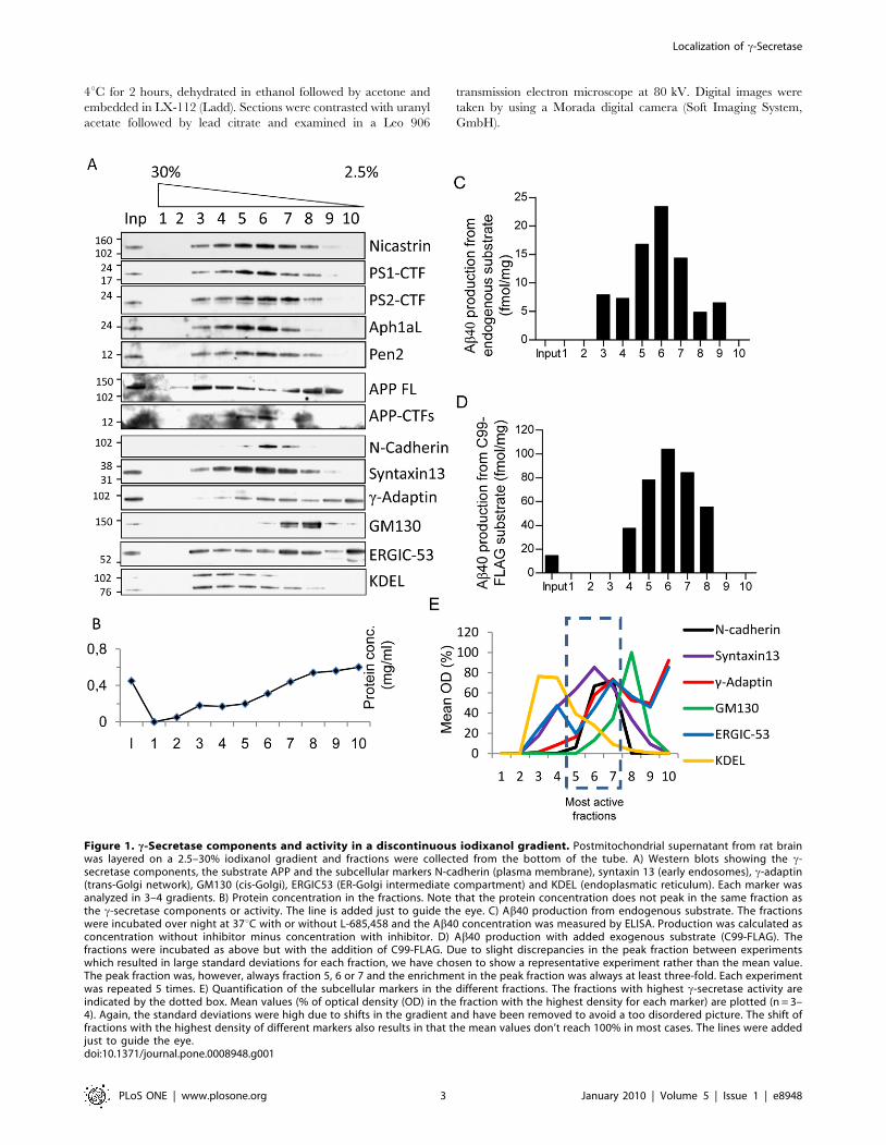
Figure 1. c-Secretase components and activity in a discontinuous iodixanol gradient. Postmitochondrial supernatant from rat brainwas layered on a 2.5–30% iodixanol gradient and fractions were collected from the bottom of the tube. A) Western blots showing the c-secretase components, the substrate APP and the subcellular markers N-cadherin (plasma membrane), syntaxin 13 (early endosomes), c-adaptin(trans-Golgi network), GM130 (cis-Golgi), ERGIC53 (ER-Golgi intermediate compartment) and KDEL (endoplasmatic reticulum). Each marker wasanalyzed in 3–4 gradients. B) Protein concentration in the fractions. Note that the protein concentration does not peak in the same fraction asthe c-secretase components or activity. The line is added just to guide the eye. C) Ab40 production from endogenous substrate. The fractionswere incubated over night at 37uC with or without L-685,458 and the Ab40 concentration was measured by ELISA. Production was calculated asconcentration without inhibitor minus concentration with inhibitor. D) Ab40 production with added exogenous substrate (C99-FLAG). Thefractions were incubated as above but with the addition of C99-FLAG. Due to slight discrepancies in the peak fraction between experimentswhich resulted in large standard deviations for each fraction, we have chosen to show a representative experiment rather than the mean value.The peak fraction was, however, always fraction 5, 6 or 7 and the enrichment in the peak fraction was always at least three-fold. Each experimentwas repeated 5 times. E) Quantification of the subcellular markers in the different fractions. The fractions with highest c-secretase activity areindicated by the dotted box. Mean values (% of optical density (OD) in the fraction with the highest density for each marker) are plotted (n = 3–4). Again, the standard deviations were high due to shifts in the gradient and have been removed to avoid a too disordered picture. The shift offractions with the highest density of different markers also results in that the mean values don’t reach 100% in most cases. The lines were addedjust to guide the eye.doi:10.1371/journal.pone.0008948.g001
Localization of c-Secretase
PLoS ONE | www.plosone.org 3 January 2010 | Volume 5 | Issue 1 | e8948
c-Secretase Activity AssayThe fractions were incubated in buffer H (20 mM Hepes,
150 mM NaCl, 5 mM EDTA, pH 7.0) including 0.4% CHAPSO
and a protease inhibitor cocktail (Roche) at 37uC for 16 h with or
without 1 mM of the c-secretase inhibitor L-685,458 (Bachem).
0.4% CHAPSO was used since we have earlier found this to be the
optimal detergent concentration [22]. For iodixanol fractions one
third of each fraction was used for each sample. For synaptic
fractions 100 mg of sample was used. For some samples, 20 ng of
C99-FLAG (a kind gift from Dr. Takeshi Nishimura, Dainippon
Sumitomo Pharma) solubilized in tri-fluoro-ethanol (TFE), was
added prior to incubation. Production of AICD was analyzed by
immunoblotting and production of Ab40 was analyzed by a
commercial ELISA system (Wako chemicals). Prior to ELISA, the
reactions were stopped by the addition of RIPA buffer, heated to
95uC for 5 min and centrifuged at 16 0006g for 5 min. The
supernatants were subjected to ELISA according to the manufac-
turer’s protocol. Production of Ab40 was calculated as Ab40 levels
without L-685,458 minus Ab40 levels with added L-685,458. For
AICD degradation, 0.2 ng of synthetic AICD (Calbiochem) and
1 mM of L-685,458 were added to the fractions prior to
incubation. Synthetic AICD without any fraction was used as a
control.
Statistical AnalysisThe Ab40 production in the different synaptic fractions was
compared with the production in homogenates using unpaired t-test.
Labeling of Rat Brain Sections with a Biotinylatedc-Secretase Inhibitor
Cryopreserved rat brain sections from frontal cortex embedded
in OCT-compound (Tissue-TEK), were cut in 12 mm thick
sections, mounted on Hypertema Teflon-coated (HTC) glass slides
(Novakemi) and air dried. Primary cortical neuron cultures were
established from cortices dissected from E17 mice (C57BL/6) and
prepared as previously described [23]. The rat brain tissues or
mouse primary neurons were fixed in buffered 4% (v/v)
formaldehyde for 5 min at room temperature (RT). The tissues
or cells were permeabilized with 0.2% Triton X-100 for 20 min
following blocking with a Avidin/Biotin blocking Kit (Vector
Laboratories, Inc.) for 15 min each. Blocking was done with
DAKO protein block serum-free for 30 min at RT after washing
with phosphate buffered saline (PBS). The tissues and cells were
preincubated with 50 mM L-685,458 for 5 min at RT, followed by
incubation with 500 nM GCB (c-secretase inhibitor with cleavable
biotin group, [24]) for 10 min, RT. Subsequently, the tissues or
cells were incubated with Streptavidin-Alexa 488 (Invitrogen,
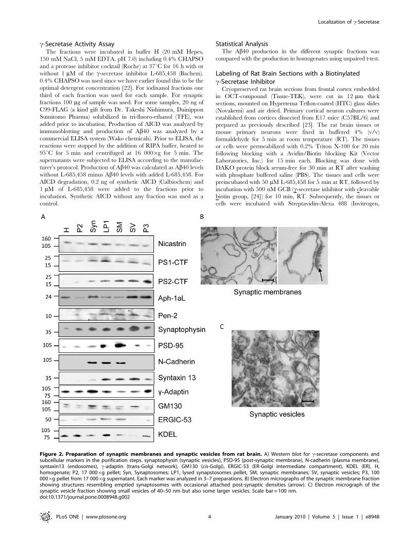
Figure 2. Preparation of synaptic membranes and synaptic vesicles from rat brain. A) Western blot for c-secretase components andsubcellular markers in the purification steps. synaptophysin (synaptic vesicles), PSD-95 (post-synaptic membrane), N-cadherin (plasma membrane),syntaxin13 (endosomes), c-adaptin (trans-Golgi network), GM130 (cis-Golgi), ERGIC-53 (ER-Golgi intermediate compartment), KDEL (ER), H,homogenate; P2, 17 0006g pellet; Syn, Synaptosomes; LP1, lysed synapstosomes pellet, SM, synaptic membranes; SV, synaptic vesicles; P3, 1000006g pellet from 17 0006g supernatant. Each marker was analyzed in 3–7 preparations. B) Electron micrographs of the synaptic membrane fractionshowing structures resembling emptied synaptosomes with occasional attached post-synaptic densities (arrow). C) Electron micrograph of thesynaptic vesicle fraction showing small vesicles of 40–50 nm but also some larger vesicles. Scale bar = 100 nm.doi:10.1371/journal.pone.0008948.g002
Localization of c-Secretase
PLoS ONE | www.plosone.org 4 January 2010 | Volume 5 | Issue 1 | e8948
Molecular Probes Inc.) for 30 min at 37uC. Later, incubations with
primary antibodies were performed at 37uC, 1 h for brain tissues
and at 4uC, over night for primary neurons. After washing with
PBS, the sections or cells were incubated with secondary
antibodies; anti-rabbit or anti-mouse AlexaFluor 546-conjugated
IgG (Invitrogen, Molecular Probes Inc.) diluted in 2% normal goat
serum for 15 min at 37uC. To reduce the background of staining
in tissues, we used autofluorescence eliminator reagent (Chemicon
International). All samples were visualized using an inverted laser
scanning microscope (LSM 510 META; Zeiss).
Results
c-Secretase Co-Fractionates with Endosomes and thePlasma Membrane
We have previously shown that the c-secretase activity is highly
enriched in a 100 0006g pellet prepared from rat brain containing
Golgi, ER and endosomes [22]. In order to further determine the
subcellular compartment with the highest c-secretase activity we
performed a density gradient centrifugation. To improve the
separation of organelles, we used a 10 0006g supernatant instead
of the 100 0006g pellet as starting material and we noted that
iodixanol was superior to sucrose in this matter (data not shown).
We prepared a 10 0006g supernatant from rat brain and
loaded this fraction on a discontinuous 2.5 to 30% iodixanol
gradient. The c-sectretase components nicastrin, PS1-CTF, PS2-
CTF, Aph-1aL and Pen-2 were enriched in fraction 5–7 of this
gradient, corresponding to an iodixanol concentration of 7.5–15%
(Figure 1A), while the highest protein concentration was found in
lighter fractions (Figure 1B). The direct substrate for c-secretase
cleavage, the APP-CTFs co-fractionated with the c-secretase
components whereas the full-length APP was more widely
distributed (Figure 1A). To measure c-secretase activity, the
fractions were incubated at 37uC with or without the c-secretase
inhibitor L-685,458 and assayed for endogenous Ab40 production
by ELISA (Figure 1C). Since it is possible that the substrate
concentration is a limiting factor, we also assayed for total c-
secretase activity by the addition of the exogenous substrate C99-
FLAG which corresponds to b-secretase cleaved APP (Figure 1D).
To calculate the c-secretase dependent Ab40 production, the
Ab40 levels found in the presence of the c-secretase inhibitor L-
685,458 were subtracted from the levels found in the absence of
the L-685,458. Unfortunately, we were not able to detect Ab42 in
this experimental setup. Both the endogenous Ab40 production
and the total c-secretase activity were enriched in fractions 5–7,
although the peak activity fraction varied slightly between
experiments. We quantified the levels of the subcellular markers
by western blotting in the different fractions and found that the
marker that showed the best correlation with c-secretase activity
was the early endosomal marker syntaxin 13 (Figure 1E). In
addition, the plasma membrane marker N-cadherin correlated
well with c-secretase activity. The ER-Golgi intermediate
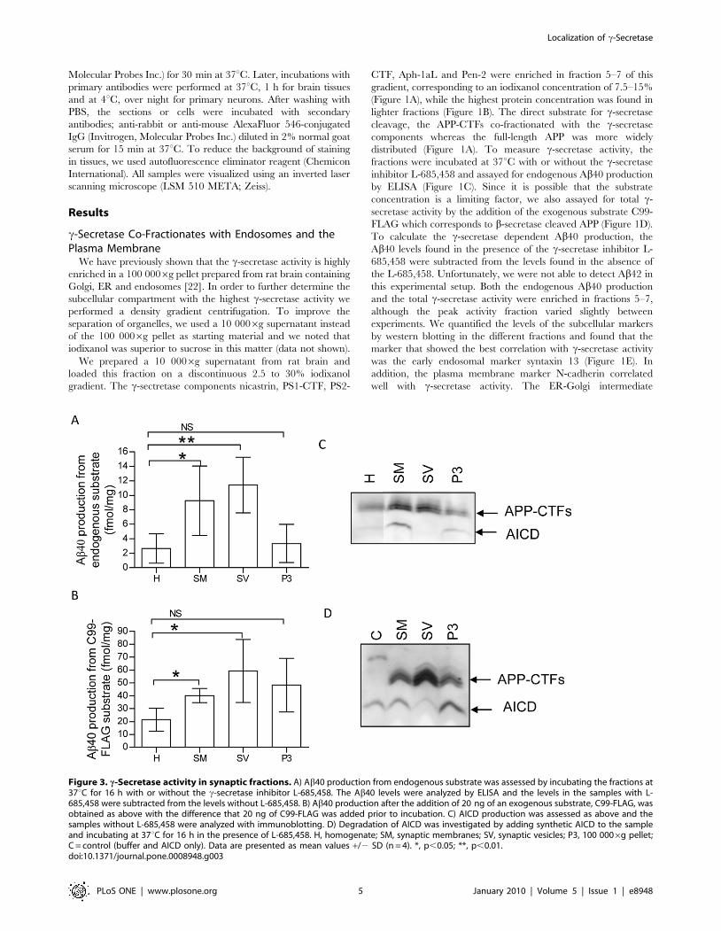
Figure 3. c-Secretase activity in synaptic fractions. A) Ab40 production from endogenous substrate was assessed by incubating the fractions at37uC for 16 h with or without the c-secretase inhibitor L-685,458. The Ab40 levels were analyzed by ELISA and the levels in the samples with L-685,458 were subtracted from the levels without L-685,458. B) Ab40 production after the addition of 20 ng of an exogenous substrate, C99-FLAG, wasobtained as above with the difference that 20 ng of C99-FLAG was added prior to incubation. C) AICD production was assessed as above and thesamples without L-685,458 were analyzed with immunoblotting. D) Degradation of AICD was investigated by adding synthetic AICD to the sampleand incubating at 37uC for 16 h in the presence of L-685,458. H, homogenate; SM, synaptic membranes; SV, synaptic vesicles; P3, 100 0006g pellet;C = control (buffer and AICD only). Data are presented as mean values +/2 SD (n = 4). *, p,0.05; **, p,0.01.doi:10.1371/journal.pone.0008948.g003
Localization of c-Secretase
PLoS ONE | www.plosone.org 5 January 2010 | Volume 5 | Issue 1 | e8948
compartment marker, ERGIC-53, and the trans-Golgi network
marker c-adaptin had two peaks of which one correlated with c-
secretase activity and the other one was found in lighter fractions.
The ER marker KDEL was found in heavier fractions whereas the
cis-Golgi marker GM130 was found in slightly lighter fractions
although some overlap with c-secretase occurred also for these
markers (Figure 1E). Thus, active c-secretase co-fractionates with
an endosomal/plasma membrane enriched fraction in an
iodixanol gradient prepared from rat brain.
c-Secretase Localize to Synaptic Membrane and SynapticVesicle Fractions
Since synaptic loss is one of the first pathological hallmarks of
AD [25] and high local concentration of Ab could induce this
degeneration, we were interested in whether active c-secretase was
present at the synapse. In order to find out, we used a sucrose
gradient protocol, followed by hypotonic lysis of synaptosomes to
prepare synaptic membranes and synaptic vesicles [20]. The
synaptic membrane markers PSD-95 and N-cadherin was
enriched in the synaptic membrane fraction and of the synaptic
vesicle marker synaptophysin was enriched in the synaptic vesicle
fraction compared to the homogenate (Figure 2A). To get a
quantitative measurement of synaptophysin and PSD-95, we
loaded different amounts of homogenate, synaptic membranes and
synaptic vesicles on a gel (data not shown) and came to the
conclusion that synaptophysin was enriched 37612 times (mean
6 SD, n = 3) in the synaptic vesicle fraction compared to
homogenate and PSD-95 was enriched 3 to 33 times in the
synaptic membrane fraction depending on which gel-system that
was used. The endosomal marker syntaxin 13 was also enriched in
the synaptic membrane and synaptic vesicle fractions whereas the
concentration of the trans-Golgi network marker c-adaptin and the
ER marker KDEL was decreased. In addition, the cis-Golgi-
marker GM130 and the ER-Golgi intermediate compartment
marker ERGIC-53 were sometimes found in the synaptic fractions
but these apparent contaminations varied between experiments.
Electron micrographs of the synaptic membrane fraction showed
large membrane structures, probably representing emptied
synaptosomes, with occasional attached post-synaptic densities
(Figure 2B). The synaptic vesicle fraction indeed contained small
vesicles of the expected size (,50 nm) but also several larger
vesicles, indicating that this was not a pure synaptic vesicle fraction
(Figure 2C).The c-secretase components were present in both the
synaptic membrane and synaptic vesicle fractions, but the degree
of enrichment varied between experiments.
We found high Ab40 production, assessed as above, from
endogenous APP-derived substrates (Figure 3A) as well as from
added C99-FLAG (Figure 3B) in both synaptic membranes and
synaptic vesicles. The activity was clearly enriched as compared to
the homogenate and the endogenous production was higher than
in P3 (100 0006g pellet). P3 showed low Ab40 production in this
experiment but the AICD production was increased compared to
homogenate (Figure 3C). Intriguingly, when we investigated the
AICD production, we could not detect any AICD in synaptic
vesicles whereas the AICD production in synaptic membranes was
high (Figure 3C). There were no detectable AICD levels in the
samples treated with L-685,458 (data not shown). To investigate
whether the absence of AICD was due to AICD degradation, we
incubated synthetic AICD with synaptic membranes, synaptic
vesicles or the P3 pellet in presence of L-685,458. Whereas there
was no degradation of AICD in synaptic membranes or in P3, the
levels of AICD in synaptic vesicles was indeed dramatically
decreased (Figure 3D). Regarding the intermediate purification
steps, the production of both Ab and AICD was low in the P2 and
synaptosome fraction and similar to synaptic membranes in the
LP1 fraction (data not shown).
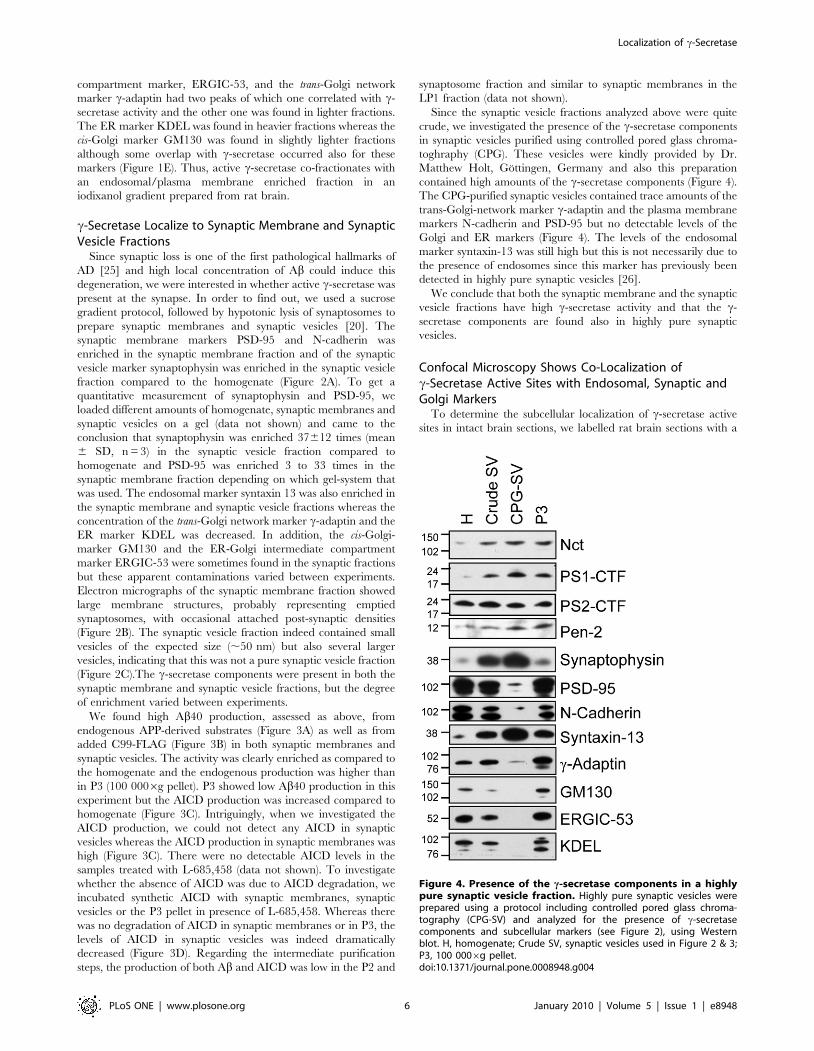
Since the synaptic vesicle fractions analyzed above were quite
crude, we investigated the presence of the c-secretase components
in synaptic vesicles purified using controlled pored glass chroma-
toghraphy (CPG). These vesicles were kindly provided by Dr.
Matthew Holt, Gottingen, Germany and also this preparation
contained high amounts of the c-secretase components (Figure 4).
The CPG-purified synaptic vesicles contained trace amounts of the
trans-Golgi-network marker c-adaptin and the plasma membrane
markers N-cadherin and PSD-95 but no detectable levels of the
Golgi and ER markers (Figure 4). The levels of the endosomal
marker syntaxin-13 was still high but this is not necessarily due to
the presence of endosomes since this marker has previously been
detected in highly pure synaptic vesicles [26].
We conclude that both the synaptic membrane and the synaptic
vesicle fractions have high c-secretase activity and that the c-
secretase components are found also in highly pure synaptic
vesicles.
Confocal Microscopy Shows Co-Localization ofc-Secretase Active Sites with Endosomal, Synaptic andGolgi Markers
To determine the subcellular localization of c-secretase active
sites in intact brain sections, we labelled rat brain sections with a
Figure 4. Presence of the c-secretase components in a highlypure synaptic vesicle fraction. Highly pure synaptic vesicles wereprepared using a protocol including controlled pored glass chroma-tography (CPG-SV) and analyzed for the presence of c-secretasecomponents and subcellular markers (see Figure 2), using Westernblot. H, homogenate; Crude SV, synaptic vesicles used in Figure 2 & 3;P3, 100 0006g pellet.doi:10.1371/journal.pone.0008948.g004
Localization of c-Secretase
PLoS ONE | www.plosone.org 6 January 2010 | Volume 5 | Issue 1 | e8948
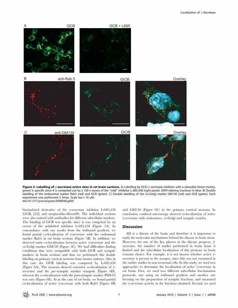
biotinylated derivative of the c-secretase inhibitor L-685,458
(GCB, [24]) and streptavidin-Alexa488. The individual sections
were also stained with antibodies for different subcellular markers.
The binding of GCB was specific since it was competed by an
excess of the unlabeled inhibitor L-685,458 (Figure 5A). In
concordance with our results from the iodixanol gradient, we
found partial co-localization of c-secretase with the endosomal
marker Rab5 in rat brain sections (Figure 5B). In addition, we
detected some co-localization between active c-secretase and the
cis-Golgi marker GM130 (Figure 5C). We had difficulties finding
conditions that were compatible with both GCB and synaptic
markers in brain sections and thus we performed this double
labeling on primary cortical neurons from mouse embryo. Also in
this case the GCB labeling was competed by L-685,458
(Figure 6A). The neurons showed extensive co-localization of c-
secretase and the pre-synaptic marker synapsin (Figure 6D),
whereas the co-localization with the post-synaptic marker PSD-95
was rare (Figure 6E). As in the case of rat brain, we found partial
co-localization of active c-secretase with both Rab5 (Figure 6B)
and GM130 (Figure 6C) in the primary cortical neurons. In
conclusion, confocal microscopy showed co-localization of active
c-secretase with endosomes, cis-Golgi and synaptic vesicles.
Discussion
AD is a disease of the brain and therefore it is important to
study the molecular mechanisms behind the disease in brain tissue.
However, for one of the key players in the disease progress, c-
secretase, the number of studies performed in brain tissue is
limited and the subcellular localization of this protease in brain
remains elusive. For example, it is not known whether active c-
secretase is present at the synapse, since this was not examined in
the earlier studies in non-neuronal cells. In this study, we used two
approaches to determine the localization of active c-secretase in
rat brain. First, we used two different subcellular fractionation
protocols; one using an iodixanol gradient and another one
focusing on the preparation of synaptic fractions, and measured
the c-secretase activity in the fractions obtained. Second, we used
Figure 5. Labelling of c-secretase active sites in rat brain sections. A) Labelling by GCB (c-secretase inhibitor with a cleavable biotin moiety,green) is specific since it is competed out by a 100 x excess of the ‘‘cold’’ inhibitor L-685,458 (right panel). DAPI-staining (nucleus) in blue. B) Doublelabelling of the endosomal marker Rab5 (red) and GCB (green). C) Double labelling of the cis-Golgi marker GM130 (red) and GCB (green). Eachexperiment was performed 3 times. Scale bar = 10 mM.doi:10.1371/journal.pone.0008948.g005
Localization of c-Secretase
PLoS ONE | www.plosone.org 7 January 2010 | Volume 5 | Issue 1 | e8948
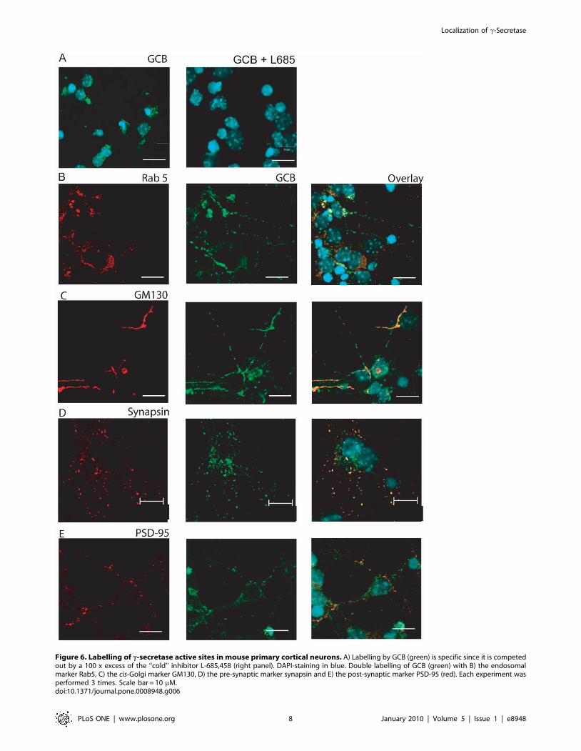
Figure 6. Labelling of c-secretase active sites in mouse primary cortical neurons. A) Labelling by GCB (green) is specific since it is competedout by a 100 x excess of the ‘‘cold’’ inhibitor L-685,458 (right panel). DAPI-staining in blue. Double labelling of GCB (green) with B) the endosomalmarker Rab5, C) the cis-Golgi marker GM130, D) the pre-synaptic marker synapsin and E) the post-synaptic marker PSD-95 (red). Each experiment wasperformed 3 times. Scale bar = 10 mM.doi:10.1371/journal.pone.0008948.g006
Localization of c-Secretase
PLoS ONE | www.plosone.org 8 January 2010 | Volume 5 | Issue 1 | e8948
a biotinylated c-secretase inhibitor (GCB) to label c-secretase
active sites in rat brain sections. Both approaches indicated
enrichment of active c-secretase in endosomes and synaptic
compartments.
Our results indicate endosomes as a major compartment for
Ab40 production in brain, since the endosomal marker syntaxin
13 was enriched in the same fractions as the c-secretase activity
in the iodixanol gradient. In addition, another endosomal
marker, Rab5, partially co-localized with GCB-labelled c-
secretase in rat brain sections and primary cortical neurons.
This is in agreement with earlier studies in cell lines, showing
that endocytosis of APP is required for Ab production
[27,28,29], and that the c-secretase components nicastrin, PS1
and Pen-2 co-fractionates with syntaxin 13 in a sucrose gradient
[2]. Ab produced in the endosomes can either remain in the
endosomes [12,13] or be secreted in exosomes via trafficking to
multi-vesicular bodies [30].
We also found high c-secretase activity in crude preparations
of synaptic vesicles and synaptic membranes and these results
were confirmed by the enrichment of the c-secretase compo-
nents in a highly pure synaptic vesicle preparation. In addition,
confocal microscopy showed co-localization of the pre-synaptic
marker synapsin and c-secretase active sites in mouse embryonic
cortical neurons. Furthermore, earlier studies have shown the
localization of PS1 in different synaptic structures [14,15,17,18].
Synaptic degeneration is an early event in AD and the
pathological feature that correlate best with cognitive decline
[25] and Ab has been found to be synaptotoxic and to affect
synaptic plasticity [31]. Our results suggest that the synaptotoxic
Ab is produced locally. c-Secretase localized to the synapse
could also have important physiological functions since
conditional PS1/PS2 double deficient mice show impaired
synaptic plasticity and memory followed by extensive neurode-
generation [32],
The endosomal and synaptic localization of active c-secretase
could be connected to each other since synaptic vesicle recycling
is highly dependent on endocytosis [33] and it has recently been
shown that increased Ab production in response to increased
synaptic activity is dependent on clathrin-mediated endocytosis
[34]. We found the endosomal marker syntaxin 13 also in our
highly pure synaptic vesicle preparations indicating either that
also this preparation was contaminated by endosomes or that
this marker are present in synaptic vesicles. In favour of the
latter hypothesis, this marker were found in synaptic vesicle
preparations where the purity was confirmed using electron
microscopy [26]. These data together with our histochemical
labelling of c-secretase active sites, that indicates a pre-synaptic
rather than post-synaptic localization in primary embryonic
mouse neurons, strongly suggest synaptic vesicle and/or pre-
synaptic endosomes as a main subcellular localization of c-
secretase.
Along with the finding of high c-secretase activity in synaptic
membranes, the plasma membrane marker N-cadherin also
correlated well with activity in the iodixanol gradient. Labelling
of active c-secretase in an approach similar to ours has
demonstrated the existence of c-secretase on the cell surface in
cell culture [35], and Chyung et al [9] showed that around 6% of
the total c-secretase is present at the cell surface.
In addition to endosomes and synaptic structures, we detected
sparse co-localization of the cis-Golgi marker GM130 with the
c-secretase in rat brain sections and this co-localization was
more pronounced in mouse primary neurons. In accordance
with this, we found some c-secretase activity in the Golgi-
enriched fractions in the iodixanol gradient although the peak
fractions of c-secretase activity did not correlate with GM130
expression and high c-secretase activity in Golgi/trans-
Golgi network has earlier been observed in different cell lines
[3,4].
It is possible that other subcellular compartments than the ones
mentioned above contribute to the c-secretase activity since some
other markers overlapped with c-secretase activity in the iodixanol
gradient and the synaptic fractions contained traces of non-
synaptic markers. In addition, the GCB labelling shows that all c-
secretase cannot be attributed to a single subcellular compartment.
In line with this, it has been shown that different substrates could
be processed at different subcellular locations. For example,
whereas most APP processing occurs inside the cells, Notch
processing mainly occurs at the cell-surface [36].
We also investigated the levels of the different c-secretase
components in the different fractions. In the iodixanol gradient
the components correlated well with c-secretase activity. In
contrast, although the c-secretase activity was highly enriched in
the synaptic fractions, the enrichment of the components was less
pronounced. The high activity is not solely explained by the
enrichment of the substrate levels (APP CTFs) since the c-
secretase activity was enriched also when an exogenous substrate
was added to the reaction. One explanation could be that the c-
secretase components only partially are assembled into an active
complex and that the individual components are more widely
distributed.
Interestingly, the AICD levels were very low in the synaptic
vesicle fraction despite high Ab levels. This was probably due to
degradation since synthetic AICD was degraded to a higher
degree in synaptic vesicles than in other fractions. Insulin
degrading enzyme (IDE) is the main candidate for AICD
degradation [37], and it can also degrade Ab [38] but no Abdegradation was observed in the synaptic vesicle fractions.
However, AICD and Ab are released to the opposite sides of
the membrane, and it is possible that IDE in this case is
compartmentalized in such a way that it only degrades AICD.
Alternatively, there might be other AICD degrading enzymes that
are enriched in synaptic vesicles.
In summary, our study indicates endosomes and/or synaptic
structures as the main compartments of active c-secretase in brain,
and that other subcellular compartments contribute to a minor
degree. The knowledge about the subcellular localization of Ab40
production may help to develop drugs that intervene with this
process. In addition, the fractions found to be enriched in c-
secretase activity can be used for further studies requiring highly
active c-secretase.
Acknowledgments
We are extremely grateful for the preparation of controlled pored glass
chromatography purified synaptic vesicles by Dr. Matthew Holt, Max
Planck Institute for Biophysical Chemistry, Goettingen, Germany. We are
also very thankful for the technical assistance from Dr. Kjell Hultenby
(electron microscopy) and Birgitta Wiehager (preparation of mouse
embryonic cortical neurons). In addition, we are thankful for the antibodies
provided by Dr. Paul Mathews, Nathan Kline Institute, NY (C1/6.1), Dr.
Jan Naslund, Karolinska Institutet (UD1) and the C99-FLAG provided by
Dr. Takeshi Nishimura, Dainippon Sumitomo Pharma.
Author Contributions
Conceived and designed the experiments: SF BW LOT. Performed the
experiments: SF JYH JF MA JN HB. Analyzed the data: SF JYH JF MA
BW JN HB LOT. Contributed reagents/materials/analysis tools: BW.
Wrote the paper: SF. Critically read the manuscript: JYH JF MA BW JN
HB LOT.
Localization of c-Secretase
PLoS ONE | www.plosone.org 9 January 2010 | Volume 5 | Issue 1 | e8948
References
1. McCarthy JV, Twomey C, Wujek P (2009) Presenilin-dependent regulated
intramembrane proteolysis and gamma-secretase activity. Cell Mol Life Sci.2. Vetrivel KS, Cheng H, Lin W, Sakurai T, Li T, et al. (2004) Association of
gamma-secretase with lipid rafts in post-Golgi and endosome membranes. J BiolChem 279: 44945–44954.
3. Baulac S, LaVoie MJ, Kimberly WT, Strahle J, Wolfe MS, et al. (2003)Functional gamma-secretase complex assembly in Golgi/trans-Golgi network:
interactions among presenilin, nicastrin, Aph1, Pen-2, and gamma-secretase
substrates. Neurobiol Dis 14: 194–204.4. Siman R, Velji J (2003) Localization of presenilin-nicastrin complexes and
gamma-secretase activity to the trans-Golgi network. J Neurochem 84:1143–1153.
5. Kaether C, Schmitt S, Willem M, Haass C (2006) Amyloid precursor protein
and Notch intracellular domains are generated after transport of their precursorsto the cell surface. Traffic 7: 408–415.
6. Capell A, Beher D, Prokop S, Steiner H, Kaether C, et al. (2005) Gamma-secretase complex assembly within the early secretory pathway. J Biol Chem
280: 6471–6478.
7. Pasternak SH, Bagshaw RD, Guiral M, Zhang S, Ackerley CA, et al. (2003)Presenilin-1, nicastrin, amyloid precursor protein, and gamma-secretase activity
are co-localized in the lysosomal membrane. J Biol Chem 278: 26687–26694.8. Yu WH, Cuervo AM, Kumar A, Peterhoff CM, Schmidt SD, et al. (2005)
Macroautophagy-a novel Beta-amyloid peptide-generating pathway activated inAlzheimer’s disease. J Cell Biol 171: 87–98.
9. Chyung JH, Raper DM, Selkoe DJ (2005) Gamma-secretase exists on the
plasma membrane as an intact complex that accepts substrates and effectsintramembrane cleavage. J Biol Chem 280: 4383–4392.
10. Hansson CA, Frykman S, Farmery MR, Tjernberg LO, Nilsberth C, et al.(2004) Nicastrin, presenilin, APH-1, and PEN-2 form active gamma-secretase
complexes in mitochondria. J Biol Chem 279: 51654–51660.
11. Hartmann T, Bieger SC, Bruhl B, Tienari PJ, Ida N, et al. (1997) Distinct sites ofintracellular production for Alzheimer’s disease A beta40/42 amyloid peptides.
Nat Med 3: 1016–1020.12. Takahashi RH, Milner TA, Li F, Nam EE, Edgar MA, et al. (2002)
Intraneuronal Alzheimer abeta42 accumulates in multivesicular bodies and isassociated with synaptic pathology. Am J Pathol 161: 1869–1879.
13. Cataldo AM, Petanceska S, Terio NB, Peterhoff CM, Durham R, et al. (2004)
Abeta localization in abnormal endosomes: association with earliest Abetaelevations in AD and Down syndrome. Neurobiol Aging 25: 1263–1272.
14. Lah JJ, Heilman CJ, Nash NR, Rees HD, Yi H, et al. (1997) Light and electronmicroscopic localization of presenilin-1 in primate brain. J Neurosci 17:
1971–1980.
15. Efthimiopoulos S, Floor E, Georgakopoulos A, Shioi J, Cui W, et al. (1998)Enrichment of presenilin 1 peptides in neuronal large dense-core and
somatodendritic clathrin-coated vesicles. J Neurochem 71: 2365–2372.16. Annaert WG, Levesque L, Craessaerts K, Dierinck I, Snellings G, et al. (1999)
Presenilin 1 controls gamma-secretase processing of amyloid precursor protein inpre-golgi compartments of hippocampal neurons. J Cell Biol 147: 277–294.
17. Beher D, Elle C, Underwood J, Davis JB, Ward R, et al. (1999) Proteolytic
fragments of Alzheimer’s disease-associated presenilin 1 are present in synapticorganelles and growth cone membranes of rat brain. J Neurochem 72:
1564–1573.18. Ribaut-Barassin C, Dupont JL, Haeberle AM, Bombarde G, Huber G, et al.
(2003) Alzheimer’s disease proteins in cerebellar and hippocampal synapses
during postnatal development and aging of the rat. Neuroscience 120: 405–423.19. Torp R, Ottersen OP, Cotman CW, Head E (2003) Identification of neuronal
plasma membrane microdomains that colocalize beta-amyloid and presenilin:
implications for beta-amyloid precursor protein processing. Neuroscience 120:
291–300.
20. Cohen RS, Blomberg F, Berzins K, Siekevitz P (1977) The structure of
postsynaptic densities isolated from dog cerebral cortex. I. Overall morphology
and protein composition. J Cell Biol 74: 181–203.
21. Huttner WB, Schiebler W, Greengard P, De Camilli P (1983) Synapsin I
(protein I), a nerve terminal-specific phosphoprotein. III. Its association with
synaptic vesicles studied in a highly purified synaptic vesicle preparation. J Cell
Biol 96: 1374–1388.
22. Franberg J, Welander H, Aoki M, Winblad B, Tjernberg LO, et al. (2007) Rat
brain gamma-secretase activity is highly influenced by detergents. Biochemistry
46: 7647–7654.
23. Behbahani H, Rickle A, Concha H, Ankarcrona M, Winblad B, et al. (2005)
Flow cytometry as a method for studying effects of stressors on primary rat
neurons. J Neurosci Res 82: 432–441.
24. Teranishi Y, Hur JY, Welander H, Franberg J, Aoki M, et al. (2009) Affinity
pulldown of gamma-secretase and associated proteins from human and rat
brain. J Cell Mol Med. Epub ahead of print.
25. Scheff SW, Price DA (2003) Synaptic pathology in Alzheimer’s disease: a review
of ultrastructural studies. Neurobiol Aging 24: 1029–1046.
26. Takamori S, Holt M, Stenius K, Lemke EA, Gronborg M, et al. (2006)
Molecular anatomy of a trafficking organelle. Cell 127: 831–846.
27. Golde TE, Estus S, Younkin LH, Selkoe DJ, Younkin SG (1992) Processing of
the amyloid protein precursor to potentially amyloidogenic derivatives. Science
255: 728–730.
28. Koo EH, Squazzo SL (1994) Evidence that production and release of amyloid
beta-protein involves the endocytic pathway. J Biol Chem 269: 17386–17389.
29. Ehehalt R, Keller P, Haass C, Thiele C, Simons K (2003) Amyloidogenic
processing of the Alzheimer beta-amyloid precursor protein depends on lipid
rafts. J Cell Biol 160: 113–123.
30. Rajendran L, Honsho M, Zahn TR, Keller P, Geiger KD, et al. (2006)
Alzheimer’s disease beta-amyloid peptides are released in association with
exosomes. Proc Natl Acad Sci U S A 103: 11172–11177.
31. Knobloch M, Mansuy IM (2008) Dendritic spine loss and synaptic alterations in
Alzheimer’s disease. Mol Neurobiol 37: 73–82.
32. Saura CA, Choi SY, Beglopoulos V, Malkani S, Zhang D, et al. (2004) Loss of
presenilin function causes impairments of memory and synaptic plasticity
followed by age-dependent neurodegeneration. Neuron 42: 23–36.
33. Shupliakov O (2009) The synaptic vesicle cluster: a source of endocytic proteins
during neurotransmitter release. Neuroscience 158: 204–210.
34. Cirrito JR, Kang JE, Lee J, Stewart FR, Verges DK, et al. (2008) Endocytosis is
required for synaptic activity-dependent release of amyloid-beta in vivo. Neuron
58: 42–51.
35. Chun J, Yin YI, Yang G, Tarassishin L, Li YM (2004) Stereoselective synthesis
of photoreactive peptidomimetic gamma-secretase inhibitors. J Org Chem 69:
7344–7347.
36. Tarassishin L, Yin YI, Bassit B, Li YM (2004) Processing of Notch and amyloid
precursor protein by gamma-secretase is spatially distinct. Proc Natl Acad
Sci U S A 101: 17050–17055.
37. Edbauer D, Willem M, Lammich S, Steiner H, Haass C (2002) Insulin-
degrading enzyme rapidly removes the beta-amyloid precursor protein
intracellular domain (AICD). J Biol Chem 277: 13389–13393.
38. Kurochkin IV, Goto S (1994) Alzheimer’s beta-amyloid peptide specifically
interacts with and is degraded by insulin degrading enzyme. FEBS Lett 345:
33–37.
Localization of c-Secretase
PLoS ONE | www.plosone.org 10 January 2010 | Volume 5 | Issue 1 | e8948