Suppression of the SDF‑1/CXCR4/β‑catenin axis contributes ...
9
ONCOLOGY REPORTS 40: 1666-1674, 2018 Abstract. Previous studies have found that the activation of stromal cell-derived factor-1 (SDF-1)/CXC chemokine receptor-4 (CXCR4)/β-catenin signaling is associated with biological malignant potential in cancers. However, its func- tion has been rarely reported in the progression of bladder cancer (BCa). The aim of the present study was to investigate the association of SDF-1/CXCR4 signaling and β-catenin in regards to BCa cell proliferation, colony formation, migration and invasion. The methods used were MTS, colony forma- tion, and Transwell migration and invasion assays which were performed in SW780 cells following treatment with the CXCR4 antagonist AMD3465, SDF-1, the β-catenin antago- nist FH535, AMD3465+SDF-1 or FH535+SDF-1. The mRNA and protein levels were assayed by RT-qPCR and western blotting, respectively. The effect of AMD3465 on SW780 cell xenograft growth in vivo was evaluated using a nude mouse model. According to our results, human BCa SW780 cells were identified as having high expression of CXCR4 and β-catenin. Subsequently, we found that both CXCR4 and β‑catenin antagonists could significantly inhibit the prolif- eration, colony formation, migration and invasion of SW780 cells. Notably, SDF-1 could reverse the inhibitory effects of AMD3465 and FH535 on proliferation, colony formation, migration and invasion in SW780 cells. In AMD3465-treated SW780 cells, the expression of c‑myc was significantly upreg- ulated, and E-cadherin was downregulated in the presence of SDF-1. Furthermore, the tumor volume and average weight in the AMD3465-treated group were evidently less than these parameters in the control group, indicating that AMD3465 can inhibit SW780 cell growth in vivo . In conclusion, targeting the SDF-1/CXCR4/β-catenin axis may be a potential therapeutic target for suppressing BCa progression. Introduction Bladder cancer (BCa) is one of the most commonly diagnosed urologic malignant tumor and remains life-threatening due to its high occurrence of metastases. The 5-year BCa survival rate is <60% (1). Each year ~80,500 patients are diagnosed with BCa, and 32,900 cancer-related deaths were reported to a population-based cancer registry (2009-2011) in China (2). Cisplatin-based chemotherapy combined with radical cystec- tomy is the recommended treatment strategy in routine clinical practice (3,4); however, the outcome of BCa patients is usually ineffective or poorly tolerated. Therefore, it is imperative to explore new potential molecular mechanisms that may provide better therapeutic targets and achieve better therapeutic effi- cacy to decrease the mortality rate of patients with BCa. Stromal cell-derived factor-1 (SDF-1) is a member of the cysteine-X-cysteine class of chemokines, and achieves its biological functions by binding to the CXC chemokine receptor 4 (CXCR4) and CXCR7 (5). SDF-1 and CXCR4 are widely expressed in a variety of cells and tissues (6). The expression of CXCR4 in malignant epithelial cells and cells from several hematopoietic malignancies indi- cates that the SDF-1/CXCR4 pathway may influence the biology of cancer (7). Upregulation of SDF-1 and CXCR4 has been associated with poor prognosis in various human cancers, including ovarian (8), head and neck (9), rectal (10) and breast (11) cancers. A growing number of studies has demonstrated that the SDF-1/CXCR4 chemokine pathway promotes the survival, proliferation and migration of cancer cells (12-14), indicating that interruption to the SDF-1/CXCR4 axis may be a new therapeutic strategy to inhibit cancer metas- tasis. SDF-1 and CXCR4 are also involved in the progression of BCa (15,16). Gosalbez et al found that the expression of SDF-1 was increased in bladder tumors and was related with high-grade tumors and metastasis (16). CXCR4-positive BCa cell exposure to SDF‑1 provoked a significant increase in both cell migration and invasion abilities (17). Previous studies have demonstrated that SDF-1/CXCR4 may induce cancer cell proliferation, and invasion through activation of the Wnt/β-catenin signaling pathway (18-20). Specifically, the Wnt/β-catenin signaling pathway is a key modulator of cellular proliferation (21). In addition, β-catenin as a transcriptional co-regulator cooperates with transcription Suppression of the SDF‑1/CXCR4/β‑catenin axis contributes to bladder cancer cell growth inhibition in vitro and in vivo TAO ZHANG 1 , FEI YANG 2 , WENBIAO LI 2 , BOLONG LIU 2 , WENDE LI 3 , ZHIYI CHEN 1 and CAN WANG 1 1 Department of Urology, Beijing University of Chinese Medicine Shenzhen Hospital, Shenzhen, Guangdong 518172; 2 Department of Urology, The Third Affiliated Hospital of Sun Yat‑sen University, Guangzhou, Guangdong 510630; 3 Guangdong Laboratory Animals Monitoring Institute, Guangzhou, Guangdong 510260, P.R. China Received December 29, 2017; Accepted June 27, 2018 DOI: 10.3892/or.2018.6546 Correspondence to: Dr Tao Zhang, Department of Urology, Beijing University of Chinese Medicine Shenzhen Hospital, 1 Dayun Road, Longgang, Shenzhen, Guangdong 518172, P.R. China E-mail: [email protected] Key words: bladder cancer, SDF-1/CXCR4, β-catenin, metastasis
Transcript of Suppression of the SDF‑1/CXCR4/β‑catenin axis contributes ...
ONCOLOGY REPORTS 40: 1666-1674, 20181666
Abstract. Previous studies have found that the activation of stromal cell-derived factor-1 (SDF-1)/CXC chemokine receptor-4 (CXCR4)/β-catenin signaling is associated with biological malignant potential in cancers. However, its func-tion has been rarely reported in the progression of bladder cancer (BCa). The aim of the present study was to investigate the association of SDF-1/CXCR4 signaling and β-catenin in regards to BCa cell proliferation, colony formation, migration and invasion. The methods used were MTS, colony forma-tion, and Transwell migration and invasion assays which were performed in SW780 cells following treatment with the CXCR4 antagonist AMD3465, SDF-1, the β-catenin antago-nist FH535, AMD3465+SDF-1 or FH535+SDF-1. The mRNA and protein levels were assayed by RT-qPCR and western blotting, respectively. The effect of AMD3465 on SW780 cell xenograft growth in vivo was evaluated using a nude mouse model. According to our results, human BCa SW780 cells were identified as having high expression of CXCR4 and β-catenin. Subsequently, we found that both CXCR4 and β‑catenin antagonists could significantly inhibit the prolif-eration, colony formation, migration and invasion of SW780 cells. Notably, SDF-1 could reverse the inhibitory effects of AMD3465 and FH535 on proliferation, colony formation, migration and invasion in SW780 cells. In AMD3465-treated SW780 cells, the expression of c‑myc was significantly upreg-ulated, and E-cadherin was downregulated in the presence of SDF-1. Furthermore, the tumor volume and average weight in the AMD3465-treated group were evidently less than these parameters in the control group, indicating that AMD3465 can inhibit SW780 cell growth in vivo. In conclusion, targeting the SDF-1/CXCR4/β-catenin axis may be a potential therapeutic target for suppressing BCa progression.
Introduction
Bladder cancer (BCa) is one of the most commonly diagnosed urologic malignant tumor and remains life-threatening due to its high occurrence of metastases. The 5-year BCa survival rate is <60% (1). Each year ~80,500 patients are diagnosed with BCa, and 32,900 cancer-related deaths were reported to a population-based cancer registry (2009-2011) in China (2). Cisplatin-based chemotherapy combined with radical cystec-tomy is the recommended treatment strategy in routine clinical practice (3,4); however, the outcome of BCa patients is usually ineffective or poorly tolerated. Therefore, it is imperative to explore new potential molecular mechanisms that may provide better therapeutic targets and achieve better therapeutic effi-cacy to decrease the mortality rate of patients with BCa.
Stromal cell-derived factor-1 (SDF-1) is a member of the cysteine-X-cysteine class of chemokines, and achieves its biological functions by binding to the CXC chemokine receptor 4 (CXCR4) and CXCR7 (5). SDF-1 and CXCR4 are widely expressed in a variety of cells and tissues (6). The expression of CXCR4 in malignant epithelial cells and cells from several hematopoietic malignancies indi-cates that the SDF-1/CXCR4 pathway may influence the biology of cancer (7). Upregulation of SDF-1 and CXCR4 has been associated with poor prognosis in various human cancers, including ovarian (8), head and neck (9), rectal (10) and breast (11) cancers. A growing number of studies has demonstrated that the SDF-1/CXCR4 chemokine pathway promotes the survival, proliferation and migration of cancer cells (12-14), indicating that interruption to the SDF-1/CXCR4 axis may be a new therapeutic strategy to inhibit cancer metas-tasis. SDF-1 and CXCR4 are also involved in the progression of BCa (15,16). Gosalbez et al found that the expression of SDF-1 was increased in bladder tumors and was related with high-grade tumors and metastasis (16). CXCR4-positive BCa cell exposure to SDF‑1 provoked a significant increase in both cell migration and invasion abilities (17).
Previous studies have demonstrated that SDF-1/CXCR4 may induce cancer cell proliferation, and invasion through activation of the Wnt/β-catenin signaling pathway (18-20). Specifically, the Wnt/β-catenin signaling pathway is a key modulator of cellular proliferation (21). In addition, β-catenin as a transcriptional co-regulator cooperates with transcription
Suppression of the SDF‑1/CXCR4/β‑catenin axis contributes to bladder cancer cell growth inhibition in vitro and in vivo
TAO ZHANG1, FEI YANG2, WENBIAO LI2, BOLONG LIU2, WENDE LI3, ZHIYI CHEN1 and CAN WANG1
1Department of Urology, Beijing University of Chinese Medicine Shenzhen Hospital, Shenzhen, Guangdong 518172; 2Department of Urology, The Third Affiliated Hospital of Sun Yat‑sen University, Guangzhou, Guangdong 510630;
3Guangdong Laboratory Animals Monitoring Institute, Guangzhou, Guangdong 510260, P.R. China
Received December 29, 2017; Accepted June 27, 2018
DOI: 10.3892/or.2018.6546
Correspondence to: Dr Tao Zhang, Department of Urology, Beijing University of Chinese Medicine Shenzhen Hospital, 1 Dayun Road, Longgang, Shenzhen, Guangdong 518172, P.R. ChinaE-mail: [email protected]
Key words: bladder cancer, SDF-1/CXCR4, β-catenin, metastasis
ZHANG et al: SDF-1/CXCR4/β-CATENIN AXIS AND BLADDER CANCER 1667
factors to determine gene expression (22). The Wnt/β-catenin signaling pathway has recently been shown to be involved in the regulation of cell proliferation and migration in BCa (23,24). Therefore, we hypothesized that the overexpression of SDF-1 and CXCR4 may promote cell proliferation, migration and invasion in BCa through activation of the Wnt/β-catenin signaling pathway.
In the present study, we explored the association between the SDF-1/CXCR4 pathway and β-catenin and the regula-tory roles of SDF-1/CXCR4 in the expression of β-catenin. Ultimately, our findings demonstrated a new signal transduc-tion pathway, the SDF-1/CXCR4/β-catenin axis, which was activated and promoted proliferation, migration and invasion in BCa cells.
Materials and methods
Cell culture. Three BCa cell lines (SW780, 5637 and T24) were purchased from the Cell Bank of the Chinese Academy of Sciences (Shanghai, China). All cell lines were cultured in Invitrogen™ RPMI‑1640 medium (Thermo Fisher Scientific, Inc., Waltham, MA, USA) containing Gibco™ 10% fetal bovine serum (FBS; Thermo Fisher Scientific, Inc.) and 1% penicillin‑streptomycin at 37˚C in 5% CO2, and were plated in a 6-well plate at a density of 2x10⁵/well. Following incubation for 2 days, the cells were collected for RNA isolation, prolif-eration, colony formation, migration and invasion assays.
MTS assay. SW780 cell proliferation was monitored using the MTS assay kit (Promega Corp., Madison, WI, USA). Absorbance was assessed at 492 nm using an ELISA reader (MD SpectraMax M5; Molecular Devices, LLC, Sunnyvale, CA, USA). The detailed documentation for the MTS assay was performed as previously described (25).
Colony formation assay. SW780 cells (200, 400 and 800 cells/well, respectively) were placed in a fresh 6-well plate and maintained in RPMI-1640 medium containing 10% FBS. To verify the association between the SDF-1/CXCR4 pathway and β-catenin, the cells were treated with PBS, AMD3465, SDF-1, FH535, AMD3465+SDF-1 or FH535+SDF-1 for 48 h; the concentrations of AMD3465, SDF-1 and FH535 were 10 µM, 100 ng/ml and 20 µM, respectively. In the AMD3465+SDF-1 or FH535+SDF-1 group, SDF-1 was administered for 48 h following AMD3465 or FH535 treatment. Subsequently, the cells were fixed with methanol and stained with 0.1% crystal violet. Visible colonies were manually counted under an Olympus IX71 inverted microscope (Olympus Corp., Tokyo, Japan). AMD3465 acts as an irreversible antagonist against the binding of CXCR4 with its ligand, SDF-1 (CXCL12). FH535 is an inhibitor of Wnt/β-catenin signaling and dual antagonist of PPARγ/δ activity. It suppresses β-catenin/Tcf-mediated transcription and inhibits β-catenin and GRIP1 recruitment to PPARγ and δ (26,27).
Migration and invasion assays. SW780 cells were treated with PBS, AMD3465, SDF-1, FH535, AMD3465+SDF-1 or SDF-1+FH535 for 48 h, and cells were resuspended in 5% FBS medium to achieve a density of 1x10⁶ cells/ml. For the Transwell migration assays, 100 µl of cell suspension
medium with 5% FBS was added to the upper chamber with a non-coated membrane in 24-well plates and an 8.0-µm pore Transwell (Millipore; Merck KGaA, Darmstadt, Germany), whereupon 600 µl complete medium was added to the bottom chamber and incubated at 37˚C with 5% CO2. For the Transwell invasion assays, Matrigel (BD Biosciences, Bedford, MA, USA) was inserted into the Transwell. In both the migra-tion and invasion assays, the cells on the upper surface of the membrane were removed with cotton swabs, and the cells on the lower surface were counted as the migrated cells. After being fixed with 4% paraformaldehyde and stained with 0.1% crystal violet solution, the cells that passed through the filter were imaged with an inverted fluorescence microscope (Leica Microsystems GmbH, Wetzlar, Germany). AMD3465, SDF-1 and FH535 were purchased from Sigma-Aldrich (Merck KGaA, Darmstadt, Germany).
Reverse transcription‑quantitative polymerase chain reaction (RT‑qPCR). RNA was extracted using Invitrogen™ TRIzol (Thermo Fisher Scientific, Inc.). Moloney Murine Leukemia Virus Reverse Transcriptase (Promega Corp.) and Oligo(dT)15 primers (Thermo Fisher Scientific, Inc.) were used to synthesize cDNA, which served as the template for the PCR performed using a DNA Engine (ABI 7300; Thermo Fisher Scientific, Inc.). The reaction mixtures (20 µl) were prepared using the TaqMan Universal PCR Master Mix (Thermo Fisher Scientific, Inc.) and the reaction conditions were carried out according to the manufacturer's protocol. The PCR primers used were as follows: For CXCR4 forward, 5'-ATC AGT CTG GAC CGC TAC CT-3' and reverse, 5'-CCA CCT TTT CAG CCA ACA GC-3'; for β-catenin forward, 5'-GGC CTC TGA TAA AGG CTA CTG TTG-3' and reverse, 5'-ACG CAA AGG TGC ATG ATT TG-3'; for β-actin forward, 5'-CAT GTA CGT TGC TAT CCA GGC-3' and reverse, 5'-CTC CTT AAT GTC ACG CAC GAT-3'. β-actin served as a housekeeping gene. The rela-tive expression levels of genes were calculated using the 2-ΔΔCq method (28).
Western blotting. Protein was extracted using RIPA lysis Buffer (Beyotime Institute of Biotechnology, Haimen, China). The concentration was determined using the Bicinchoninic Acid Kit for Protein Determination (Sigma-Aldrich; Merck KGaA). Samples containing 50 µg of protein were separated on 10% SDS-PAGE gel and transferred to nitrocel-lulose membranes (Bio-Rad Laboratories, Inc., Hercules, CA, USA). The primary antibodies p-β-catenin (dilution 1:1,000; cat. no. 9561s; Cell Signaling Technology, Inc., Danvers, MA, USA), β-catenin (dilution 1:1,000; cat. no. sc7199; Santa Cruz Biotechnology, Inc., Santa Cruz, CA, USA), MMP-2 (cat. no. ab37150; dilution: 1:2,000; Abcam, Cambridge, UK), c-myc (dilution 1:1,000; cat. no. ab32072; Abcam), E-cadherin (dilution, 1:1,000; cat. no. 14472; Cell Signaling Technology), or N-cadherin (dilution, 1:1,000; cat. no. sc53488; Santa Cruz Biotechnology, Inc.) were incubated with the membrane for 2 h at room temperature, and then the membranes were incu-bated with the appropriate horseradish peroxidase-conjugated secondary antibody (dilution, 1:10,000; cat. no. sc-516102; Santa Cruz Biotechnology, Inc.), following visualization using chemiluminescence reagent (Thermo Fisher Scientific, Inc.). Glyceraldehyde-3-phosphate dehydrogenase (GAPDH; dilutio
ONCOLOGY REPORTS 40: 1666-1674, 20181668
1:2,000; cat. no. 2118; Cell Signaling Technology, Inc.) was used as the control antibody. Signals were densitometrically assessed using Quantity One® software version 4.5 (Bio-Rad Laboratories, Inc.).
Animal experiments. The present study was approved by the Ethics Committee of Beijing University of Chinese Medicine at Shenzhen Hospital (Shenzhen, China). A total of 10 BALB/c 6-8 week old, athymic nude mice (body weight, 25-30 g) were obtained from the Shanghai Laboratory Animal Center (Shanghai, China) and acclimated to the environment for 1 week. Animals were maintained in a specific pathogen‑free (SPF) environment throughout the experiments, with a controlled humidity (50±10%), light (12-h light/dark cycle), and temperature (23±2˚C), fed with food and water ad libitum. Establishment of the subcutaneous tumor model was performed as previously described (29). All of the mice received a subcu-taneous injection of SW780 cells (1x10⁶). Tumor growth was assessed every week using a dial caliper and tumor volume (V) was calculated by the following formula: V = πAB2/6, in which A is the largest diameter and B is the perpendicular diameter. After the tumor volume reached 100-200 mm3, the mice were divided into two groups receiving PBS (control group; n=5) or AMD3465 treatment (AMD3465 group; n=5). The mice in the two groups were injected with 500 µg AMD3465 or an equal volume of PBS subcutaneously respectively, once per day until they were sacrificed, with an intraperitoneal injec-tion of sodium pentobarbital (200 mg/kg; Sigma-Aldrich; Merck KGaA) 12 weeks after inoculation. The tumor volume of the two groups was assessed twice every week until the time of sacrifice, and the tumors were isolated and weighed after the sacrifice.
Statistical analysis. The data from these experiments were reported as the mean ± standard deviation (SD) for each group. All statistical analyses were performed with PRISM version 7.0 (GraphPad Software, Inc., La Jolla, CA, USA). The Student's t-test was used to analyze differences between two groups. Inter-group differences were analyzed by one-way analysis of variance (ANOVA), followed by a post hoc Tukey's test for multiple comparisons. P<0.05 was considered to indi-cate a statistically significant difference.
Results
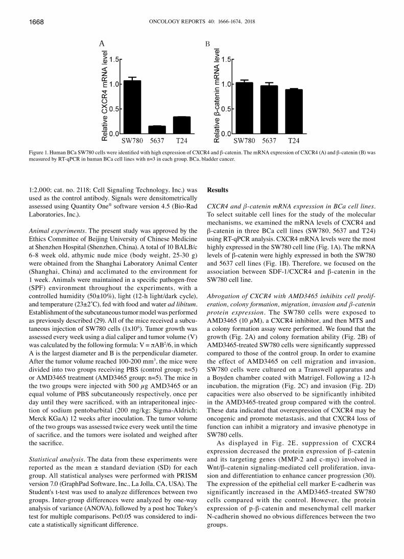
CXCR4 and β‑catenin mRNA expression in BCa cell lines. To select suitable cell lines for the study of the molecular mechanisms, we examined the mRNA levels of CXCR4 and β-catenin in three BCa cell lines (SW780, 5637 and T24) using RT-qPCR analysis. CXCR4 mRNA levels were the most highly expressed in the SW780 cell line (Fig. 1A). The mRNA levels of β-catenin were highly expressed in both the SW780 and 5637 cell lines (Fig. 1B). Therefore, we focused on the association between SDF-1/CXCR4 and β-catenin in the SW780 cell line.
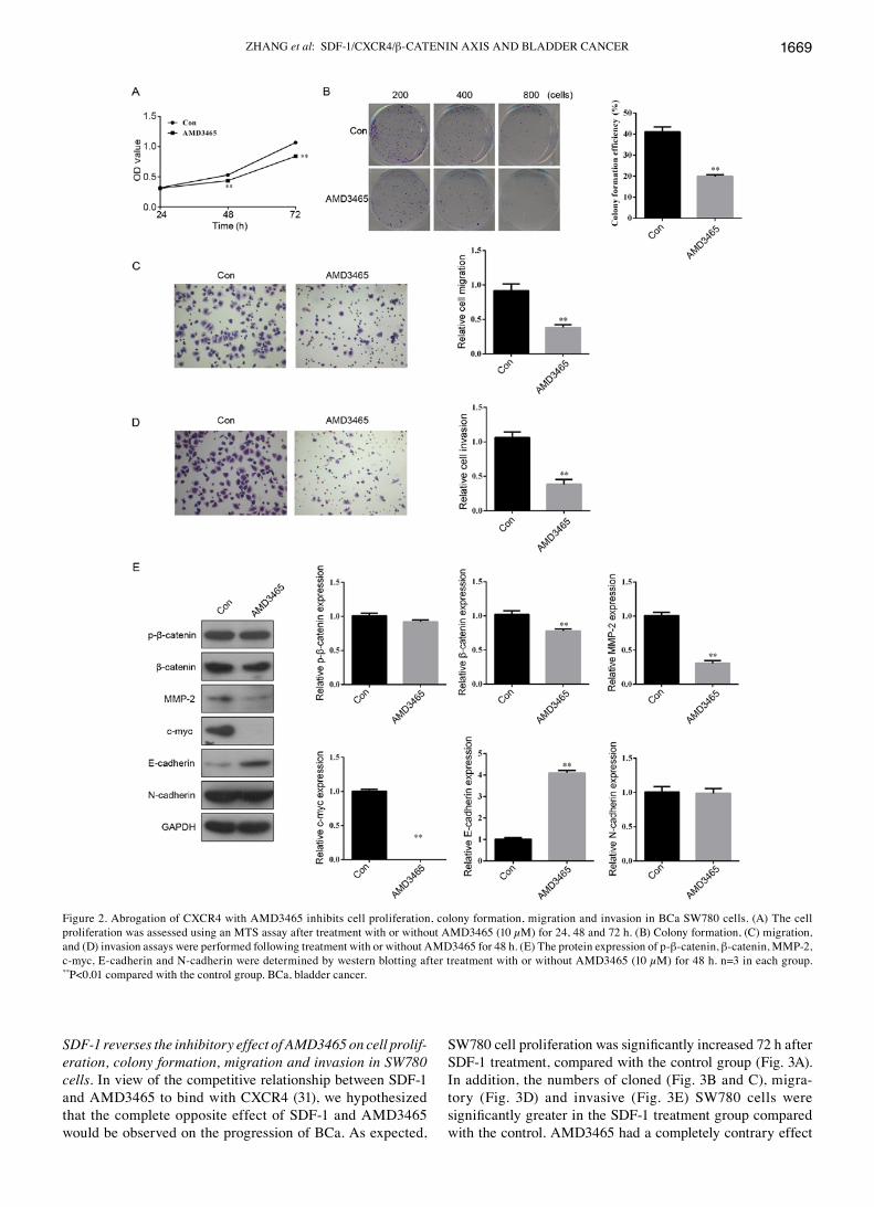
Abrogation of CXCR4 with AMD3465 inhibits cell prolif‑eration, colony formation, migration, invasion and β‑catenin protein expression. The SW780 cells were exposed to AMD3465 (10 µM), a CXCR4 inhibitor, and then MTS and a colony formation assay were performed. We found that the growth (Fig. 2A) and colony formation ability (Fig. 2B) of AMD3465‑treated SW780 cells were significantly suppressed compared to those of the control group. In order to examine the effect of AMD3465 on cell migration and invasion, SW780 cells were cultured on a Transwell apparatus and a Boyden chamber coated with Matrigel. Following a 12-h incubation, the migration (Fig. 2C) and invasion (Fig. 2D) capacities were also observed to be significantly inhibited in the AMD3465-treated group compared with the control. These data indicated that overexpression of CXCR4 may be oncogenic and promote metastasis, and that CXCR4 loss of function can inhibit a migratory and invasive phenotype in SW780 cells.
As displayed in Fig. 2E, suppression of CXCR4 expression decreased the protein expression of β-catenin and its targeting genes (MMP-2 and c-myc) involved in Wnt/β-catenin signaling-mediated cell proliferation, inva-sion and differentiation to enhance cancer progression (30). The expression of the epithelial cell marker E-cadherin was significantly increased in the AMD3465-treated SW780 cells compared with the control. However, the protein expression of p-β-catenin and mesenchymal cell marker N-cadherin showed no obvious differences between the two groups.
Figure 1. Human BCa SW780 cells were identified with high expression of CXCR4 and β-catenin. The mRNA expression of CXCR4 (A) and β-catenin (B) was measured by RT-qPCR in human BCa cell lines with n=3 in each group. BCa, bladder cancer.
ZHANG et al: SDF-1/CXCR4/β-CATENIN AXIS AND BLADDER CANCER 1669
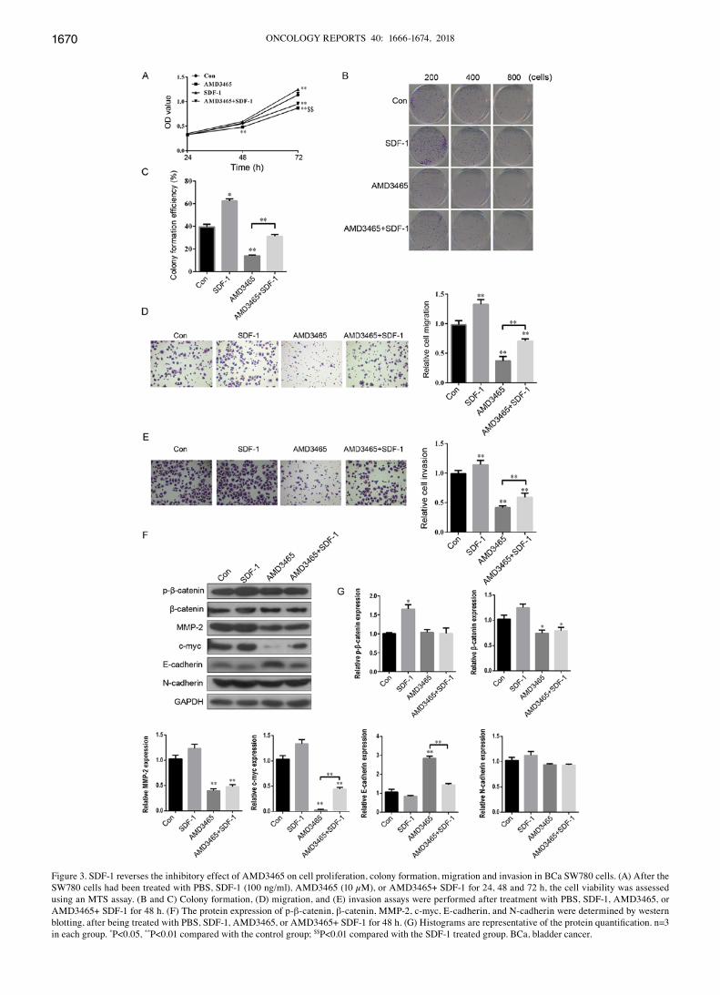
SDF‑1 reverses the inhibitory effect of AMD3465 on cell prolif‑eration, colony formation, migration and invasion in SW780 cells. In view of the competitive relationship between SDF-1 and AMD3465 to bind with CXCR4 (31), we hypothesized that the complete opposite effect of SDF-1 and AMD3465 would be observed on the progression of BCa. As expected,
SW780 cell proliferation was significantly increased 72 h after SDF-1 treatment, compared with the control group (Fig. 3A). In addition, the numbers of cloned (Fig. 3B and C), migra-tory (Fig. 3D) and invasive (Fig. 3E) SW780 cells were significantly greater in the SDF‑1 treatment group compared with the control. AMD3465 had a completely contrary effect
Figure 2. Abrogation of CXCR4 with AMD3465 inhibits cell proliferation, colony formation, migration and invasion in BCa SW780 cells. (A) The cell proliferation was assessed using an MTS assay after treatment with or without AMD3465 (10 µM) for 24, 48 and 72 h. (B) Colony formation, (C) migration, and (D) invasion assays were performed following treatment with or without AMD3465 for 48 h. (E) The protein expression of p-β-catenin, β-catenin, MMP-2, c-myc, E-cadherin and N-cadherin were determined by western blotting after treatment with or without AMD3465 (10 µM) for 48 h. n=3 in each group. **P<0.01 compared with the control group. BCa, bladder cancer.
ONCOLOGY REPORTS 40: 1666-1674, 20181670
Figure 3. SDF-1 reverses the inhibitory effect of AMD3465 on cell proliferation, colony formation, migration and invasion in BCa SW780 cells. (A) After the SW780 cells had been treated with PBS, SDF-1 (100 ng/ml), AMD3465 (10 µM), or AMD3465+ SDF-1 for 24, 48 and 72 h, the cell viability was assessed using an MTS assay. (B and C) Colony formation, (D) migration, and (E) invasion assays were performed after treatment with PBS, SDF-1, AMD3465, or AMD3465+ SDF-1 for 48 h. (F) The protein expression of p-β-catenin, β-catenin, MMP-2, c-myc, E-cadherin, and N-cadherin were determined by western blotting, after being treated with PBS, SDF‑1, AMD3465, or AMD3465+ SDF‑1 for 48 h. (G) Histograms are representative of the protein quantification. n=3 in each group. *P<0.05, **P<0.01 compared with the control group; $$P<0.01 compared with the SDF-1 treated group. BCa, bladder cancer.
ZHANG et al: SDF-1/CXCR4/β-CATENIN AXIS AND BLADDER CANCER 1671
on cell proliferation, colony formation, migration and inva-sion in SW780 cells (Fig. 3A‑E). Notably, SDF‑1 significantly reversed this inhibitory effect (Fig. 3A-E).
Furthermore, the effects of SDF-1 on the protein expres-sion of p-β-catenin, β-catenin, MMP-2, c-myc, E-cadherin
and N-cadherin in SW780 cells were evaluated. SDF-1 treatment had no significant effect on any of these factors, except for p-β‑catenin (Fig. 3F and G). c‑myc was significantly upregulated, and E-cadherin was downregulated in the group treated with SDF-1 combined with AMD3465 in SW780 cells
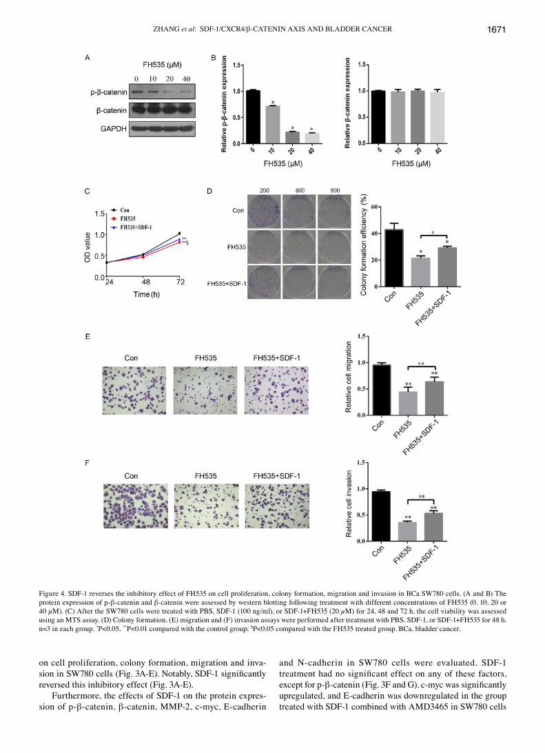
Figure 4. SDF-1 reverses the inhibitory effect of FH535 on cell proliferation, colony formation, migration and invasion in BCa SW780 cells. (A and B) The protein expression of p-β-catenin and β-catenin were assessed by western blotting following treatment with different concentrations of FH535 (0, 10, 20 or 40 µM). (C) After the SW780 cells were treated with PBS, SDF-1 (100 ng/ml), or SDF-1+FH535 (20 µM) for 24, 48 and 72 h, the cell viability was assessed using an MTS assay. (D) Colony formation, (E) migration and (F) invasion assays were performed after treatment with PBS, SDF-1, or SDF-1+FH535 for 48 h. n=3 in each group. *P<0.05, **P<0.01 compared with the control group; $P<0.05 compared with the FH535 treated group. BCa, bladder cancer.
ONCOLOGY REPORTS 40: 1666-1674, 20181672
compared with AMD3465 treatment alone. However, SDF-1 had no significant effect on p‑β-catenin, β-catenin, MMP-2, or N-cadherin in AMD3465-treated SW780 cells (Fig. 3F and G). These findings indicated an inverse relationship between SDF-1 and AMD3465 on cell proliferation, colony formation, migration and invasion in SW780 cells.
SDF‑1 reverses the inhibitory effect of FH535 on cell proliferation, colony formation, migration and invasion in SW780 cells. To investigate the role of β-catenin in the progres-sion of BCa, we treated cells with a β-catenin antagonist, FH535. First, we found that the protein expression of p-β-catenin was significantly inhibited in a concentration‑dependent manner by FH535 treatment of SW780 cells (Fig. 4A and B). In addition, the protein expression of β-catenin was not evidently different in SW780 cells with FH535 treatment (Fig. 4A and B). Compared with the control group, cell proliferation was significantly inhibited by FH535 treatment at 72 h (Fig. 4C). The down-regulation of β-catenin by FH535 treatment led to a reduction in the number of cloned (Fig. 4D), migratory (Fig. 4E), and invasive (Fig. 4F) SW780 cells. Interestingly, SDF-1 treatment significantly reversed the inhibitory effect of FH535 on cell proliferation, colony formation, migration and invasion in SW780 cells (Fig. 4C‑F). These findings indicated that SDF‑1 plays an important role in the regulation of the expression of β-catenin during the development of BCa.
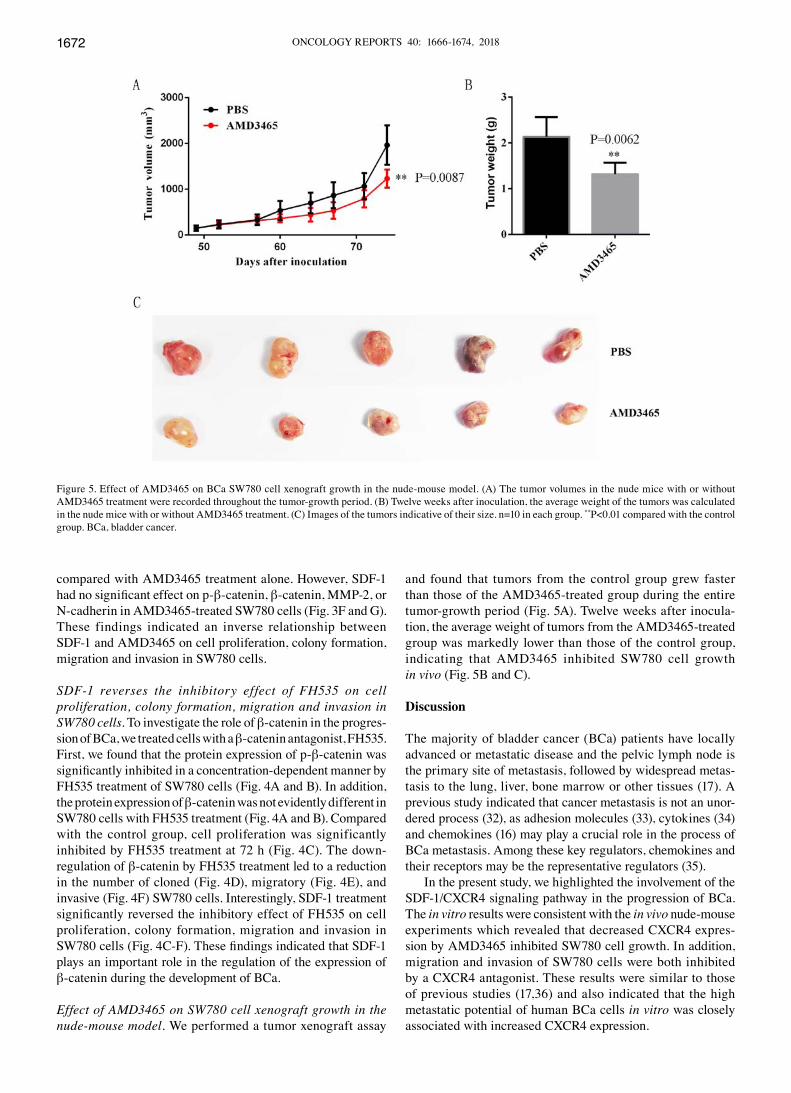
Effect of AMD3465 on SW780 cell xenograft growth in the nude‑mouse model. We performed a tumor xenograft assay
and found that tumors from the control group grew faster than those of the AMD3465-treated group during the entire tumor-growth period (Fig. 5A). Twelve weeks after inocula-tion, the average weight of tumors from the AMD3465-treated group was markedly lower than those of the control group, indicating that AMD3465 inhibited SW780 cell growth in vivo (Fig. 5B and C).
Discussion
The majority of bladder cancer (BCa) patients have locally advanced or metastatic disease and the pelvic lymph node is the primary site of metastasis, followed by widespread metas-tasis to the lung, liver, bone marrow or other tissues (17). A previous study indicated that cancer metastasis is not an unor-dered process (32), as adhesion molecules (33), cytokines (34) and chemokines (16) may play a crucial role in the process of BCa metastasis. Among these key regulators, chemokines and their receptors may be the representative regulators (35).
In the present study, we highlighted the involvement of the SDF-1/CXCR4 signaling pathway in the progression of BCa. The in vitro results were consistent with the in vivo nude-mouse experiments which revealed that decreased CXCR4 expres-sion by AMD3465 inhibited SW780 cell growth. In addition, migration and invasion of SW780 cells were both inhibited by a CXCR4 antagonist. These results were similar to those of previous studies (17,36) and also indicated that the high metastatic potential of human BCa cells in vitro was closely associated with increased CXCR4 expression.
Figure 5. Effect of AMD3465 on BCa SW780 cell xenograft growth in the nude-mouse model. (A) The tumor volumes in the nude mice with or without AMD3465 treatment were recorded throughout the tumor-growth period. (B) Twelve weeks after inoculation, the average weight of the tumors was calculated in the nude mice with or without AMD3465 treatment. (C) Images of the tumors indicative of their size. n=10 in each group. **P<0.01 compared with the control group. BCa, bladder cancer.
ZHANG et al: SDF-1/CXCR4/β-CATENIN AXIS AND BLADDER CANCER 1673
We further revealed that a CXCR4 antagonist can inhibit β-catenin protein expression. Previous studies indicated that β-catenin may be a biomarker for the metastatic progression of BCa patients and that it was associated with poor patient survival (37,38). Wnt/β-catenin signaling has been shown to be frequently involved in various signaling pathways related to cell proliferation and metastasis in BCa (39,40). For example, hepatocyte cell adhesion molecule (hepaCAM) specifically suppressed β-catenin expression during BCa cell proliferation (39). In addition, long non-coding RNA H19 increased BCa metastasis by activating the Wnt/β-catenin signaling pathway (40). Thus, our results showed that the CXCR4 antagonist induced the inhibition of cell proliferation, colony formation, migration and invasion, at least partially, by downregulating the expression of β-catenin.
β-catenin and its downstream target genes, c-myc and MMP-2, are associated with tumorigenesis and metastasis in multiple types of cancer (30,41). Numerous studies have shown that the suppression of the c-myc oncogene induced cellular senescence in BCa (42,43). MMP-2 is an enzyme that has been implicated in the malignant progression of BCa (44). Usually, β-catenin, c-myc, and MMP-2 have a synergistic effect on BCa tumorigenesis and progression (39,45). In the present study, the suppression of CXCR4 expression also decreased the protein expression of β-catenin targeting genes (MMP-2 and c‑myc). Our results confirmed the previously established role of the CXCR4/β-catenin axis in the malignant progression of cancers (18-20).
When investigating the interaction of chemokines and chemokine receptors, SDF-1 was proven to be a stimu-lator of migration and invasion in CXCR4-positive cancer cells (46), suggesting that SDF-1 may play a central role in the SDF-1/CXCR4 axis. Previous studies revealed that the addition of SDF-1 significantly induced the prolif-eration, migration and invasion of colon cancer cells (20,47). Song et al also found that β-catenin was recruited in the nuclei to stimulate cell proliferation in the presence of SDF-1, but SDF-1 had no obvious effect on β-catenin in a whole cell lysate (19). In this study, SDF-1 was revealed to induce the phosphorylation of β-catenin, which has been shown to promote β-catenin escape, disruption of E-cadherin associa-tion and the disassembly of adherent junctions, which all lead to cancer cell proliferation (48). Concurrently, SDF-1 could reverse the inhibitory effect of the β-catenin antagonist on cell proliferation, colony formation, migration and invasion in SW780 cells. All of these findings indicated that β-catenin was a promising key target gene in the SDF-1/CXCR4 axis in BCa metastasis.
To the best of our our knowledge, this is the first study that revealed the association between SDF-1/CXCR4 signaling and the activation of β-catenin in the malignant progression of BCa. Nevertheless, we also found that SDF-1/CXCR4 may induce epithelial-mesenchymal transition (EMT) by inhib-iting the E-cadherin expression involved in BCa metastasis; however, further investigation of this mechanism is required.
Acknowledgements
We sincerely thank Professor Xiangfu Zhou for his techno-logical guidance and support.
Funding
The present study was supported by the Scientific and Technology Planning Project of Shenzhen, Guangdong.
Availability of data and materials
The datasets used during the present study are available from the corresponding author upon reasonable request.
Authors' contributions
TZ conceived and designed the study, drafted and revised the manuscript. FY performed the experiments, analyzed the data and drafted the manuscript. WenbL, ZC, CW, BL and WendL helped in study design, study implementation and manuscript revision. All authors read and approved the manuscript and agree to be accountable for all aspects of the research in ensuring that the accuracy or integrity of any part of the work are appropriately investigated and resolved.
Ethics approval and consent to participate
The present study was approved by the Ethics Committee of Beijing University of Chinese Medicine at Shenzhen Hospital and approved the animal research carried out.
Patient consent for publication
Not applicable.
Competing interests
The authors declare that they have no competing interests.
References
1. Torre LA, Bray F, Siegel RL, Ferlay J, Lortet-Tieulent J and Jemal A: Global cancer statistics, 2012. CA Cancer J Clin 65: 87-108, 2015.
2. Chen W, Zheng R, Baade PD, Zhang S, Zeng H, Bray F, Jemal A, Yu XQ and He J: Cancer statistics in China, 2015. CA Cancer J Clin 66: 115-132, 2016.
3. Raj GV, Karavadia S, Schlomer B, Arriaga Y, Lotan Y, Sagalowsky A and Frenkel E: Contemporary use of perioperative cisplatin-based chemotherapy in patients with muscle-invasive bladder cancer. Cancer 117: 276-282, 2011.
4. Sternberg CN, Skoneczna IA, Castellano D, Theodore C, Blais N, Voog E, Bellmunt J, Peters F, Le-Guennec S, Cerbone L, et al: Larotaxel with Cisplatin in the first‑line treatment of locally advanced/metastatic urothelial tract or bladder cancer: A randomized, active-controlled, phase III trial (CILAB). Oncology 85: 208-215, 2013.
5. Duda DG, Kozin SV, Kirkpatrick ND, Xu L, Fukumura D and Jain RK: CXCL12 (SDF1alpha)-CXCR4/CXCR7 pathway inhibition: An emerging sensitizer for anticancer therapies? Clin Cancer Res 17: 2074-2080, 2011.
6. Teicher BA and Fricker SP: CXCL12 (SDF-1)/CXCR4 pathway in cancer. Clin Cancer Res 16: 2927-2931, 2010.
7. Meads MB, Hazlehurst LA and Dalton WS: The bone marrow microenvironment as a tumor sanctuary and contributor to drug resistance. Clin Cancer Res 14: 2519-2526, 2008.
8. Zheng N, Chen J, Liu W, Liu J, Li T, Chen H, Wang J and Jia L: Mifepristone inhibits ovarian cancer metastasis by intervening in SDF-1/CXCR4 chemokine axis. Oncotarget 8: 59123-59135, 2017.
ONCOLOGY REPORTS 40: 1666-1674, 20181674
9. De-Colle C, Menegakis A, Mönnich D, Welz S, Boeke S, Sipos B, Fend F, Mauz PS, Tinhofer I, Budach V, et al: SDF-1/CXCR4 expression is an independent negative prognostic biomarker in patients with head and neck cancer after primary radiochemo-therapy. Radiother Oncol 126: 125-131, 2018.
10. Saigusa S, Toiyama Y, Tanaka K, Yokoe T, Okugawa Y, Kawamoto A, Yasuda H, Inoue Y, Miki C and Kusunoki M: Stromal CXCR4 and CXCL12 expression is associated with distant recurrence and poor prognosis in rectal cancer after chemoradiotherapy. Ann Surg Oncol 17: 2051-2058, 2010.
11. Hinton CV, Avraham S and Avraham HK: Role of the CXCR4/CXCL12 signaling axis in breast cancer metastasis to the brain. Clin Exp Metastasis 27: 97-105, 2010.
12. Li X, Li P, Chang Y, Xu Q, Wu Z, Ma Q and Wang Z: The SDF-1/CXCR4 axis induces epithelial-mesenchymal transition in hepatocellular carcinoma. Mol Cell Biochem 392: 77-84, 2014.
13. Wang B, Wang W, Niu W, Liu E, Liu X, Wang J, Peng C, Liu S, Xu L, Wang L, et al: SDF-1/CXCR4 axis promotes directional migration of colorectal cancer cells through upregulation of integrin αvβ6. Carcinogenesis 35: 282-291, 2014.
14. Guo Q, Gao BL, Zhang XJ, Liu GC, Xu F, Fan QY, Zhang SJ, Yang B and Wu XH: CXCL12-CXCR4 axis promotes prolif-eration, migration, invasion, and metastasis of ovarian cancer. Oncol Res 22: 247-258, 2014.
15. Eisenhardt A, Frey U, Tack M, Rosskopf D, Lümmen G, Rübben H and Siffert W: Expression analysis and potential func-tional role of the CXCR4 chemokine receptor in bladder cancer. Eur Urol 47: 111-117, 2005.
16. Gosalbez M, Hupe MC, Lokeshwar SD, Yates TJ, Shields J, Veerapen MK, Merseburger AS, Rosser CJ, Soloway MS and Lokeshwar VB: Differential expression of SDF-1 isoforms in bladder cancer. J Urol 191: 1899-1905, 2014.
17. Retz MM, Sidhu SS, Blaveri E, Kerr SC, Dolganov GM, Lehmann J, Carroll P, Simko J, Waldman FM and Basbaum C: CXCR4 expression reflects tumor progression and regulates motility of bladder cancer cells. Int J Cancer 114: 182-189, 2005.
18. Lu Y, Hu B, Guan GF, Chen J, Wang CQ, Ma Q, Wen YH, Qiu XC, Zhang XP and Zhou Y: SDF-1/CXCR4 promotes F5M2 osteosarcoma cell migration by activating the Wnt/β-catenin signaling pathway. Med Oncol 32: 194, 2015.
19. Song ZY, Gao ZH, Chu JH, Han XZ and Qu XJ: Downregulation of the CXCR4/CXCL12 axis blocks the activation of the Wnt/β-catenin pathway in human colon cancer cells. Biomed Pharmacother 71: 46-52, 2015.
20. Hu TH, Yao Y, Yu S, Han LL, Wang WJ, Guo H, Tian T, Ruan ZP, Kang XM, Wang J, et al: SDF-1/CXCR4 promotes epithelial-mesenchymal transition and progression of colorectal cancer by activation of the Wnt/β-catenin signaling pathway. Cancer Lett 354: 417-426, 2014.
21. Moon RT, Kohn AD, De Ferrari GV and Kaykas A: WNT and beta-catenin signalling: Diseases and therapies. Nat Rev Genet 5: 691-701, 2004.
22. MacDonald BT, Tamai K and He X: Wnt/beta-catenin signaling: Components, mechanisms, and diseases. Dev Cell 17: 9-26, 2009.
23. Chen Z, Zhou L, Wang L, Kazobinka G, Zhang X, Han X, Li B and Hou T: HBO1 promotes cell proliferation in bladder cancer via activation of Wnt/β-catenin signaling. Mol Carcinog 57: 12-21, 2018.
24. Yuan H, Yu S, Cui Y, Men C, Yang D, Gao Z, Zhu Z and Wu J: Knockdown of mediator subunit Med19 suppresses bladder cancer cell proliferation and migration by downregulating Wnt/β-catenin signalling pathway. J Cell Mol Med 21: 3254-3263, 2017.
25. Chan GK, Kleinheinz TL, Peterson D and Moffat JG: A simple high-content cell cycle assay reveals frequent discrepancies between cell number and ATP and MTS proliferation assays. PLoS One 8: e63583, 2013.
26. Bodart V, Anastassov V, Darkes MC, Idzan SR, Labrecque J, Lau G, Mosi RM, Neff KS, Nelson KL, Ruzek MC, et al: Pharmacology of AMD3465: A small molecule antagonist of the chemokine receptor CXCR4. Biochem Pharmacol 78: 993-1000, 2009.
27. Chen Y, Rao X, Huang K, Jiang X, Wang H and Teng L: FH535 inhibits proliferation and motility of colon cancer cells by targeting Wnt/β-catenin signaling pathway. J Cancer 8: 3142-3153, 2017.
28. Livak KJ and Schmittgen TD: Analysis of relative gene expression data using real-time quantitative PCR and the 2-ΔΔCT method. Methods 25: 402-408, 2001.
29. Xu YC, Liang CJ, Zhang DX, Li GQ, Gao X, Fu JZ, Xia F, Ji JJ, Zhang LJ, Li GM, et al: LncSHRG promotes hepatocellular carcinoma progression by activating HES6. Oncotarget 8: 70630-70641, 2017.
30. Xie F, Xiang X, Huang Q, Ran P, Yuan Y, Li Q, Qi G, Guo X, Xiao C and Zheng S: Reciprocal control of lncRNA-BCAT1 and β-catenin pathway reveals lncRNA-BCAT1 long non-coding RNA acts as a tumor suppressor in colorectal cancer. Oncotarget 8: 23628-23637, 2017.
31. Van Hout A, D'Huys T, Oeyen M, Schols D and Van Loy T: Comparison of cell-based assays for the identification and evaluation of competitive CXCR4 inhibitors. PLoS One 12: e0176057, 2017.
32. Rossi D and Zlotnik A: The biology of chemokines and their receptors. Annu Rev Immunol 18: 217-242, 2000.
33. Muramaki M, Miyake H, Terakawa T, Kumano M, Sakai I and Fujisawa M: Expression profile of E‑cadherin and N‑cadherin in non-muscle-invasive bladder cancer as a novel predictor of intravesical recurrence following transurethral resection. Urol Oncol 30: 161-166, 2012.
34. Olbert PJ, Kesch C, Henrici M, Subtil FS, Honacker A, Hegele A, Hofmann R and Hänze J: TLR4- and TLR9-dependent effects on cytokines, cell viability, and invasion in human bladder cancer cells. Urol Oncol 33: 110.e19-e27, 2015.
35. Yates TJ, Knapp J, Gosalbez M, Lokeshwar SD, Gomez CS, Benitez A, Ekwenna OO, Young EE, Manoharan M and Lokeshwar VB: C-X-C chemokine receptor 7: A functionally associated molecular marker for bladder cancer. Cancer 119: 61-71, 2013.
36. Wang H, Yang D, Wang K and Wang J: Expression and potential role of chemokine receptor CXCR4 in human bladder carcinoma cell lines with different metastatic ability. Mol Med Rep 4: 525-528, 2011.
37. Shen CH, Wu JD, Jou YC, Cheng MC, Lin CT, Chen PC, Tseng YS, Shi CS, Chen SY, Chang DC, et al: The correlation between TWIST, E-cadherin, and beta-catenin in human bladder cancer. J BUON 16: 733-737, 2011.
38. Nakopoulou L, Zervas A, Gakiopoulou-Givalou H, Constantinides C, Doumanis G, Davaris P and Dimopoulos C: Prognostic value of E-cadherin, beta-catenin, P120ctn in patients with transitional cell bladder cancer. Anticancer Res 20: 4571-4578, 2000.
39. Du HF, Ou LP, Lv CK, Yang X, Song XD, Fan YR, Wu XH and Luo CL: Expression of hepaCAM inhibits bladder cancer cell proliferation via a Wnt/β-catenin-dependent pathway in vitro and in vivo. Cancer Biol Ther 16: 1502-1513, 2015.
40. Luo M, Li Z, Wang W, Zeng Y, Liu Z and Qiu J: Long non-coding RNA H19 increases bladder cancer metastasis by associating with EZH2 and inhibiting E-cadherin expression. Cancer Lett 333: 213-221, 2013.
41. Vaid M, Prasad R, Sun Q and Katiyar SK: Silymarin targets β-catenin signaling in blocking migration/invasion of human melanoma cells. PLoS One 6: e23000, 2011.
42. Ye W, Chen C, Gao Y, Zheng ZS, Xu Y, Yun M, Weng HW, Xie D, Ye S and Zhang JX: Overexpression of SLC34A2 is an inde-pendent prognostic indicator in bladder cancer and its depletion suppresses tumor growth via decreasing c-Myc expression and transcriptional activity. Cell Death Dis 8: e2581, 2017.
43. Tang Y, Simoneau AR, Liao WX, Yi G, Hope C, Liu F, Li S, Xie J, Holcombe RF, Jurnak FA, et al: WIF1, a Wnt pathway inhibitor, regulates SKP2 and c-myc expression leading to G1 arrest and growth inhibition of human invasive urinary bladder cancer cells. Mol Cancer Ther 8: 458-468, 2009.
44. Gao Y, Guan Z, Chen J, Xie H, Yang Z, Fan J, Wang X and Li L: CXCL5/CXCR2 axis promotes bladder cancer cell migration and invasion by activating PI3K/AKT-induced upregulation of MMP2/MMP9. Int J Oncol 47: 690-700, 2015.
45. Wu K, Ning Z, Zeng J, Fan J, Zhou J, Zhang T, Zhang L, Chen Y, Gao Y, Wang B, et al: Silibinin inhibits β-catenin/ZEB1 signaling and suppresses bladder cancer metastasis via dual-blocking epithelial-mesenchymal transition and stemness. Cell Signal 25: 2625-2633, 2013.
46. Wang Z, Ma Q, Liu Q, Yu H, Zhao L, Shen S and Yao J: Blockade of SDF-1/CXCR4 signalling inhibits pancreatic cancer progression in vitro via inactivation of canonical Wnt pathway. Br J Cancer 99: 1695-1703, 2008.
47. Liu Z, Hao C, Song D, Zhang N, Bao H and Qu Q: Androgen receptor coregulator CTBP1-AS is associated with polycystic ovary syndrome in Chinese Women: A preliminary study. Reprod Sci 22: 829-837, 2015.
48. Castellone MD, De Falco V, Rao DM, Bellelli R, Muthu M, Basolo F, Fusco A, Gutkind JS and Santoro M: The beta-catenin axis integrates multiple signals downstream from RET/papillary thyroid carcinoma leading to cell proliferation. Cancer Res 69: 1867-1876, 2009.