Supporting Information Bioevaluation as Potent ... · S1 Supporting Information An Insight into...
27
S1 Supporting Information An Insight into Tetrahydro-β-carboline-Tetrazole Hybrids: Synthesis and Bioevaluation as Potent Antileishmanial Agents Pooja Purohit, a Anand Kumar Pandey, a Deepti Singh Chauhan, a Pradeep Singh Chouhan, a Karthik Ramalingam, b Mahendra Shukla, c Neena Goyal, b Jawahar Lal, c Prem M. S. Chauhan a* a Medicinal and Process Chemistry Division, CSIR-Central Drug Research Institute, Lucknow-226031, U.P., India. b Division of Biochemistry, CSIR-Central Drug Research Institute, Lucknow-226031, U.P., India. c Pharmacokinetics & Metabolism Division, CSIR-Central Drug Research Institute, Lucknow, India. *Corresponding author mailing address: Medicinal and Process Chemistry Division, CSIR- Central Drug Research Institute, Sector 10, Jankipuram Extension, Sitapur Road, Lucknow- 226031, U.P, India, Phone: +91 522 2771940, 2771942; Fax: +91 522 2771941. Email: [email protected]; [email protected] Table of content Experimental section for Antileishmanial activity S2 – S4 Experimental section for Pharmacokinetics study S5 – S6 References S6 Spectra of compounds S7 – S27 Electronic Supplementary Material (ESI) for MedChemComm. This journal is © The Royal Society of Chemistry 2017
Transcript of Supporting Information Bioevaluation as Potent ... · S1 Supporting Information An Insight into...
S1
Supporting Information
An Insight into Tetrahydro-β-carboline-Tetrazole Hybrids: Synthesis and
Bioevaluation as Potent Antileishmanial Agents
Pooja Purohit,a Anand Kumar Pandey,a Deepti Singh Chauhan,a Pradeep Singh Chouhan,a Karthik
Ramalingam,b Mahendra Shukla,c Neena Goyal,b Jawahar Lal,c Prem M. S. Chauhana*
aMedicinal and Process Chemistry Division, CSIR-Central Drug Research Institute, Lucknow-226031, U.P., India.
bDivision of Biochemistry, CSIR-Central Drug Research Institute, Lucknow-226031, U.P., India.
cPharmacokinetics & Metabolism Division, CSIR-Central Drug Research Institute, Lucknow, India.
*Corresponding author mailing address: Medicinal and Process Chemistry Division, CSIR-
Central Drug Research Institute, Sector 10, Jankipuram Extension, Sitapur Road, Lucknow-
226031, U.P, India, Phone: +91 522 2771940, 2771942; Fax: +91 522 2771941. Email:
[email protected]; [email protected]
Table of content
Experimental section for Antileishmanial activity S2 – S4
Experimental section for Pharmacokinetics study S5 – S6
References S6
Spectra of compounds S7 – S27
Electronic Supplementary Material (ESI) for MedChemComm.This journal is © The Royal Society of Chemistry 2017
S2
Experimental section for antileishmanial activity: Each compound was evaluated for its in
vitro activity against WHO reference strain (MHOM/IN/80/Dd8) of extracellular promastigotes
and intramacrophagic amastigotes (expressing luciferase firefly reporter gene) of L. donovani.
The in vitro cytotoxicity assay was performed using murine macrophage J-774A.1 cell line.
In vitro antileishmanial assay: Leishmania donovani promastigotes (WHO designation
MHOM/IN/80/Dd8), originally obtained as a gift from (late) Prof. P. C. C. Garnham and
routinely maintained at the institute in golden hamsters, were used in the present study.
Promastigotes were grown in medium 199 medium (Sigma) supplemented with 10 % heat-
inactivated fetal calf serum (GBL) and 1% penicillin (50 U/ml) and streptomycin (50 mg/ml)
solution (Sigma) at 24 °C.1 Luciferase tagged with promastigotes maintained at 25 ± 1 0C in
medium 199 (Sigma Chemical) supplemented with 10 % Fetal Calf Serum (Gibco, Gaithersburg,
MD, USA) and G418 (20 g ⁄ ml) were used for in vitro evaluation of antileishmanial activity.2
Antipromastigote activity: The in vitro effect of test compounds on the growth of
promastigotes was assessed by monitoring the luciferase activity of viable cells after treatment.3
The transgenic promastigotes of late log phase were seeded at 5 X10⁵ cells ⁄ well in 96- well flat-
bottomed microtitre (MT) plates (CELLSTAR Greiner Bio-one Gmbh, Monroe, NC, USA) and
incubated for 72 h in medium, in absence (control) or the presence of test compounds (stock
prepared in 100 % DMSO, initial concentration, followed by serial dilution in media). The test
compounds were added at 2-fold dilutions in up to 7 points in fresh complete medium starting
from a 100 uM concentration. After incubation, an aliquot (50 L) of promastigote suspension
was aspirated from each well of a 96-well plate and mixed with an equal volume of Steady Glo
(R) reagent (Promega, Madison, WI, USA) and luminescence was measured in luminometer.
S3
Antiamastigote activity: For assessing the activity of compounds against amastigote stage of
the parasite, mouse macrophage cell line (J-774A.1), infected with promastigotes expressing
firefly luciferase reporter gene was used. Macrophage cells were seeded in a 96-well plate (5
X104 cells ⁄ 200 l ⁄ well) in RPMI-1640 containing 10 % fetal calf serum. The plates were
incubated at 37 0C in a CO2 incubator. After 24 h, the medium was replaced with fresh medium
containing stationary phase transgenic promastigotes (2.5 X10⁵ ⁄ 200 L ⁄ well). Promastigotes
invade the macrophage and are transformed into amastigotes. The test compounds were added at
2-fold dilutions in up to 7 points in fresh complete medium starting from a 100 µM
concentration, and the plates were incubated at 37 °C in a CO2 incubator for 72 h. After
incubation, the drug containing medium was decanted and 50 l phosphate-buffered saline (PBS)
was added in each well and mixed with an equal volume of Steady-Glo luciferase assay substrate
dissolved in Steady-Glo luciferase assay buffer. After gentle shaking for 1 to 2 min, the readings
were recorded in a luminometer. The values were expressed as relative luminescence
The IC50 value of each compound was calculated by non-linear regression analysis of the
concentration–response curve using the four-parameter Hill equations.
Cytotoxicity assay: The macrophage cell viability was determined using the MTT assay.4
Exponentially growing cells (J774) (1-2×10⁵ cells/100 µl/well) were incubated with test
compounds. The test compounds are added at three fold dilutions up to 7 points in complete
medium starting from 500 µM concentrations, and were incubated at 37 °C in a humidified
mixture of CO2 and 95 % air in an incubator. Control wells containing dimethyl sulfoxide
(DMSO) without compounds were also included in the experiment. After incubation, 25 µL of
MTT reagent (5 mg/mL) in PBS medium and incubated at 37 °C for 2 h. At the end of the
incubation period, the supernatant were removed by inverting the plate completely without
S4
disturbing cell layer and 150 µL of pure DMSO was added to each well. After 15 min. of
shaking the readings were recorded as absorbance at 544 nm on a micro plate reader. The
cytotoxic effect were expressed as 50 % lethal dose (i.e., as the concentration of a compound
which provoked a 50 % reduction in cell viability compared to cell in culture medium alone).
CC50 values were estimated as described by Huber et al.5 The selectivity index (SI) for each
compound was calculated as the ratio between, cytotoxicity (CC50) and activity (IC50) against
Leishmania amastigotes. Compounds with SI index > 5 were considered as safe in this cytotoxic
assay.
In vivo antileishmanial activity in hamster model: The modified method was used for in vivo
screening. Golden male hamsters (Inbred strain) weighing 40-45 gm were infected intra-cardially
with 1 x 10⁷ amastigotes per animal. Pre-treatment spleen biopsy of all the animals was carried
out to assess the degree of infection on day 20th of infection. The animals with +1 infection (5-15
amastigotes/100 spleen cell nuclei) were included in the experiment. The infected animals were
randomized into several groups of six animals each. Drug treatment by intraperitoneal route was
initiated after 2 days of biopsy and continued for 5 consecutive days. Negative control group
hamsters were administered 0.2 ml of saline solution. Post-treatment biopsies were done on day
7th and 28th day of the last drug administration and amastigote counts were assessed after Giemsa
staining. Intensity of infection in both, treated and untreated animals, and also the initial count in
treated animals was compared and the efficacy was expressed in terms of percent inhibition (PI)
using the following formula:
PI = 100- [ANAT x 100/ (INAT x TIUC)]
S5
Where PI is percent inhibition of amastigotes multiplication, ANAT is actual number of
amastigotes in treated animals, INAT is initial number of amastigotes in treated animals and
TIUC is times increase of parasites in untreated control animals.
Experimental Section for Pharmacokinetics:
Material and methods: The pharmacokinetics of 14t was studied in golden Syrian hamsters
weighing 100 10 gm. The hamsters were obtained from Laboratory Animal Division of the
Institute and were housed in plastic cages under standard laboratory conditions with a regular 12
h day-night cycle. Suspension formulation containing 12.5 mg/ml of compound was prepared by
triturating 14t, gum acacia (1%, w/v) and water (drop wise addition) in a mortar and pestle. A
single 50 mg/kg oral dose was given to conscious hamsters using feeding needle and blood
samples were withdrawn at various predefined times up to 24 h post dose. Serum samples were
harvested from blood. All samples were stored at -80 °C until analysis. The serum (50 µL; blank,
spiked or test) was extracted twice with 1 ml of extraction solvent (n-hexane: ethyl acetate,
60:40, % v/v) followed by vortex-mixing and centrifugation. The supernatant was evaporated to
dryness in Turbovap LX (Caliper, Massachusetts, USA). The residue was reconstituted in 100
μL mobile phase and centrifuged. Clear supernatant (80 µL) was transferred into HPLC vials and
50 μL was injected on to the LC-MS/MS system.
A Shimadzu UFLC pump (LC-20AD) with online degasser (DGU-20A3), an auto-sampler (SIL-
HTc) with a temperature-controlled peltier-tray and a API 4000 Q trap mass spectrometer
(Applied Biosystems, Toronto, Canada) was used for analysis on a Discovery HS C-18 column
(5 μm, 50 x 4.6 mm id) preceded with a guard column (5 μm, 20 x 4.0 mm, id) packed with the
same material under isocratic condition at a flow rate of 0.7 mL/min. The mobile phase [85 %
acetonitrile in aqueous ammonium acetate buffer (0.01 M)] was degassed by ultrasonication for
S6
15 min before use. LC-MS/MS system was equilibrated for approximately 20 min before
commencement of analysis. The column oven temperature was 40 °C. Total analysis time was 4
min per sample. Multiple reactions monitoring (MRM) was used to monitor the transitions
m/z503.2→ 331.1for 14t and m/z180.1 → 138.1 for phenacetin (Internal Standard) at 5500 V
spray voltage. Data acquisition and quantitation were performed using analyst software (version
1.4.2; AB Sciex, Toronto, Canada). The method was linear over the concentration range of 1-200
ng/mL with recovery of >70 % and acceptable accuracy and precision.
References:
1. A. Debrabant, M. Gottilieb, D. M. Dwyer, Mol Biochem Parasitol., 1995, 71, 51-63.
2. Ashutosh, S. Gupta, Ramesh, S. Sundar, N. Goyal, Antimicrob. Agents Chemother., 2005,
49, 3776-3783.
3. T. Mosmann, J. Immunol. Methods., 1983, 65, 55-63.
4. W. Huber, J. C. Koella, Acta Trop., 1993, 55, 257-261.
5. S. Bhatnagar, P. Y. Guru, J. C. Katiyar, R. Srivastava, A. Mukherjee, M. S. Akhtar, M.
Seth, A. P. Bhaduri, Indian J Med Res., 1989, 89, 439-44.
S7
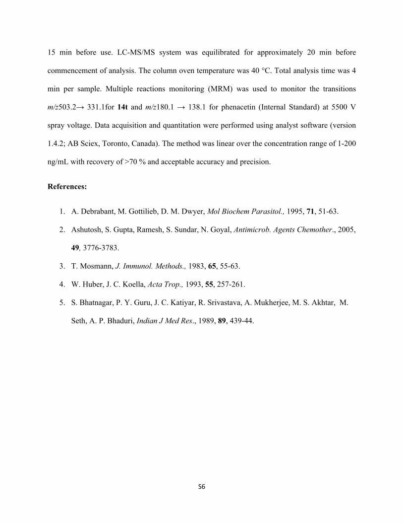
Supplementary Spectra of compounds:
1H and 13C NMR spectra of 14a
NH
N
NNNN
NH
N
NNNN
S8
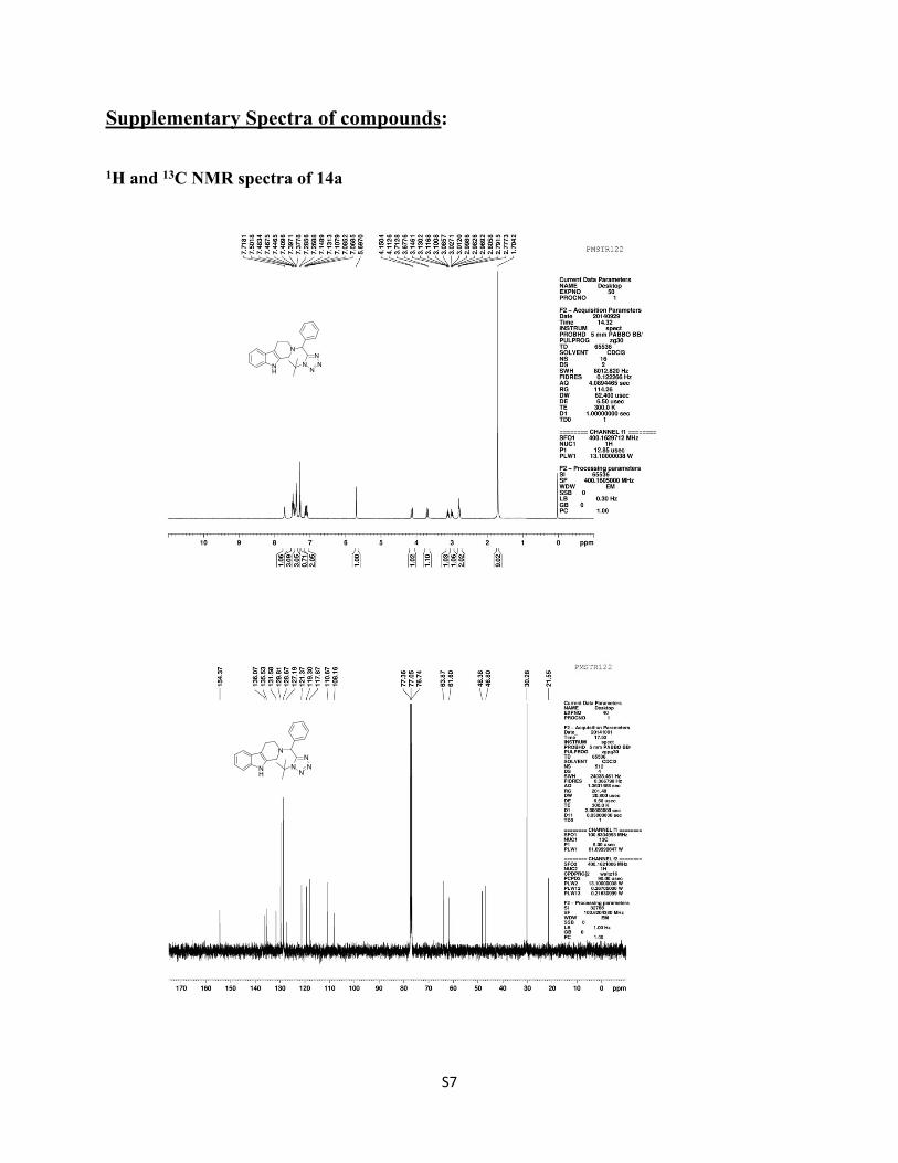
1H and 13C NMR spectra of 14b
NH
N
NNN
N
F
NH
N
NNN
N
F
S9
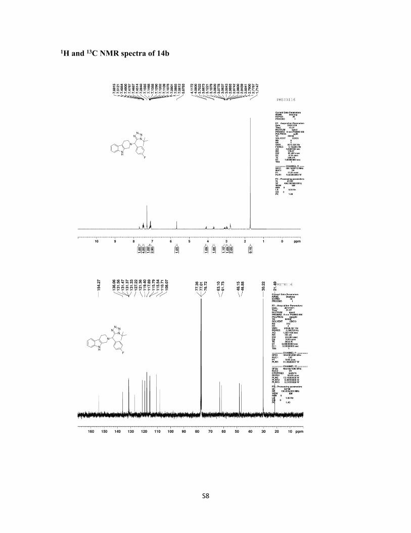
1H and 13C NMR spectra of 14c
NH
N
NN N
N
Cl
NH
N
NN N
N
Cl
S10
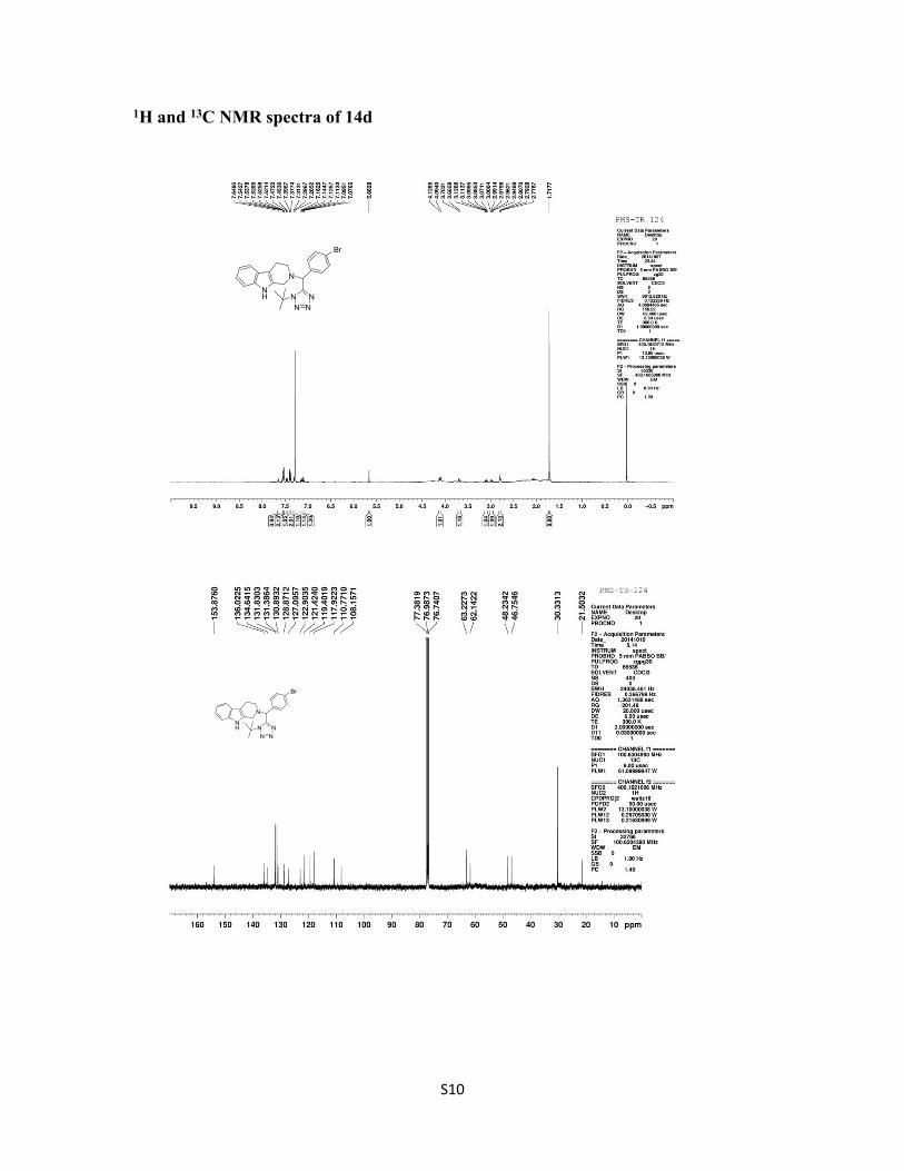
1H and 13C NMR spectra of 14d
NH
N
NN N
N
Br
NH
N
NN N
N
Br
S11
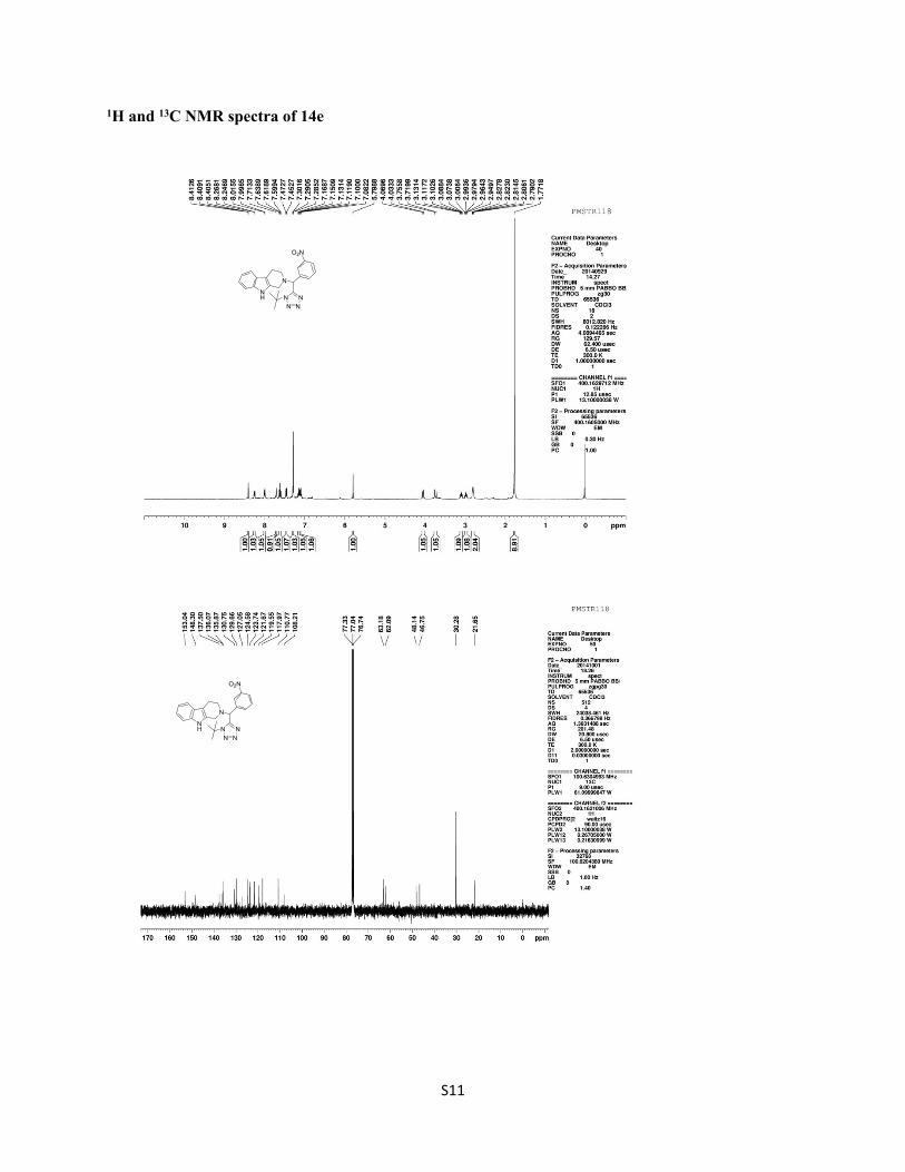
1H and 13C NMR spectra of 14e
NH
N
NN N
N
O2N
NH
N
NN N
N
O2N
S12
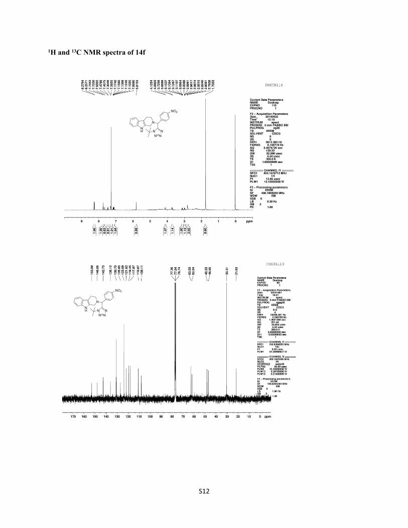
1H and 13C NMR spectra of 14f
NH
N
NN N
N
NO2
NH
N
NN N
N
NO2
S13
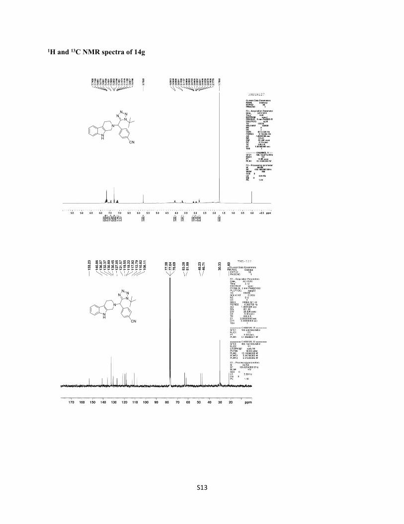
1H and 13C NMR spectra of 14g
NH
N
NNN
N
CN
NH
N
NNN
N
CN
S14
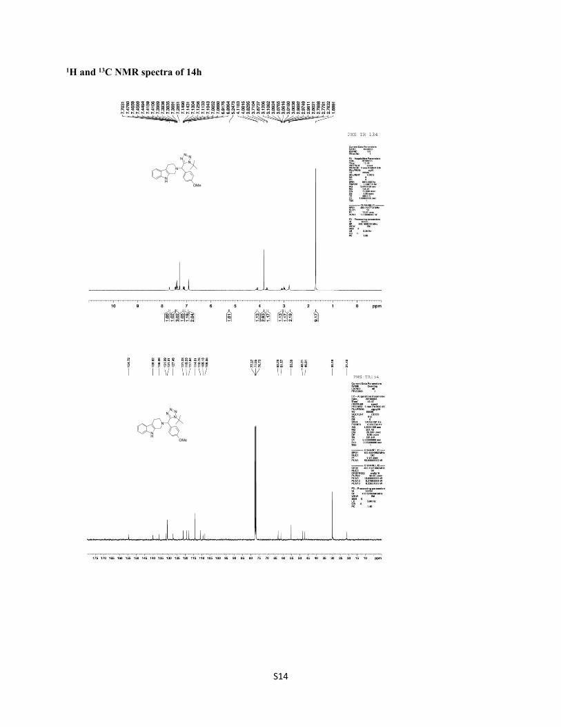
1H and 13C NMR spectra of 14h
NH
N
NNN
N
OMe
NH
N
NNN
N
OMe
S15
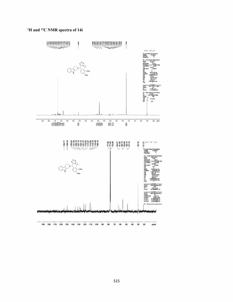
1H and 13C NMR spectra of 14i
NH
N
NNN
N
OMe
OMe
NH
N
NNN
N
OMe
OMe
S16
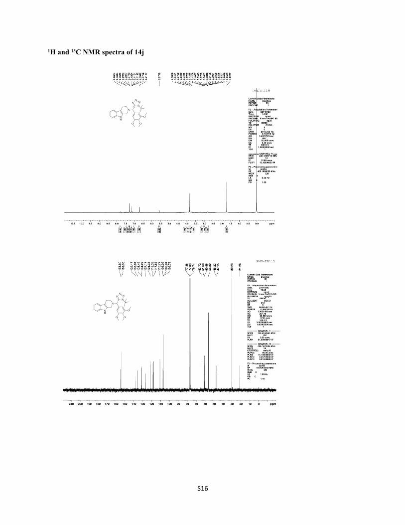
1H and 13C NMR spectra of 14j
NH
NC N
NN
N
O
OO
NH
NC N
NN
N
O
OO
S17
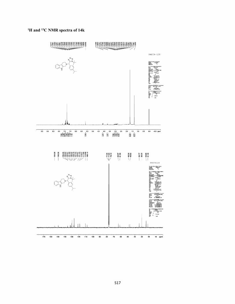
1H and 13C NMR spectra of 14k
NH
N
NNN
N
NH
N
NNN
N
S18
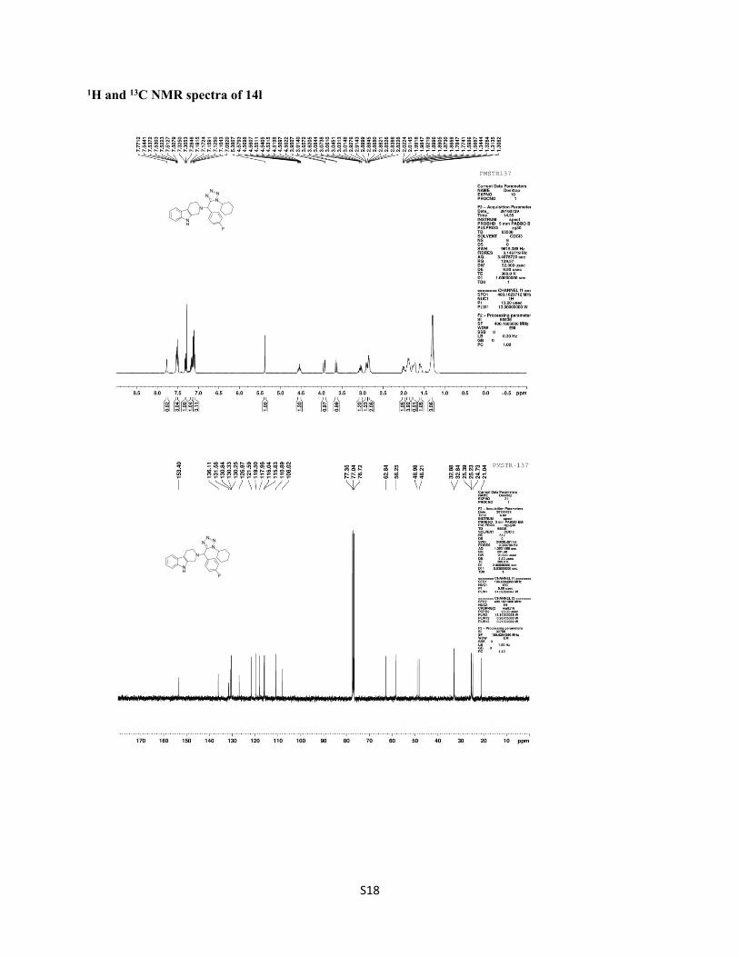
1H and 13C NMR spectra of 14l
NH
N
NNN
N
F
NH
N
NNN
N
F
S19
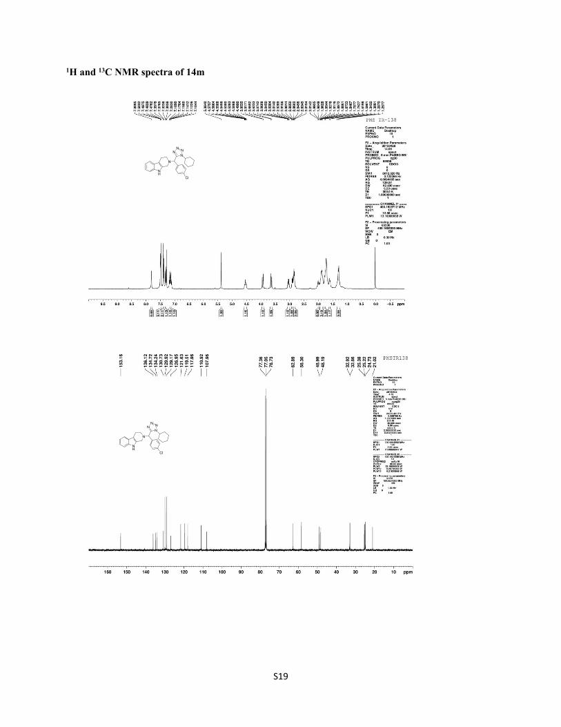
1H and 13C NMR spectra of 14m
NH
N
NNN
N
Cl
NH
N
NNN
N
Cl
S20
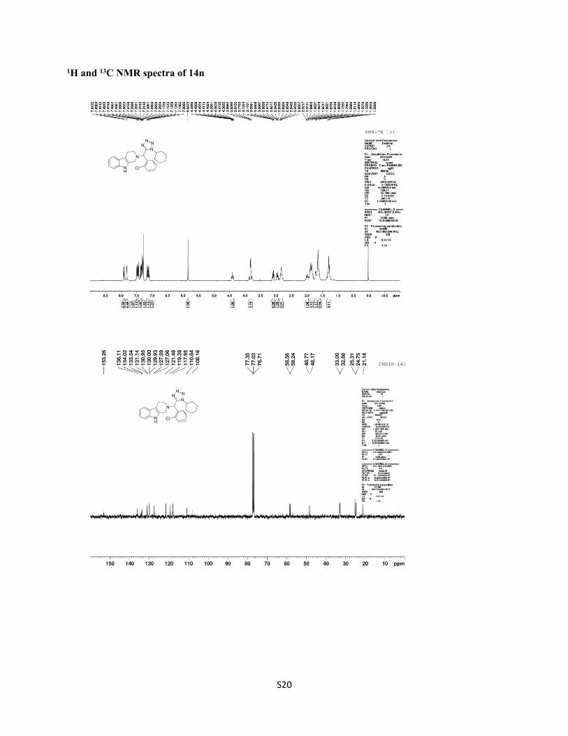
1H and 13C NMR spectra of 14n
NH
N
NNN
N
Cl
NH
N
NNN
N
Cl
S21
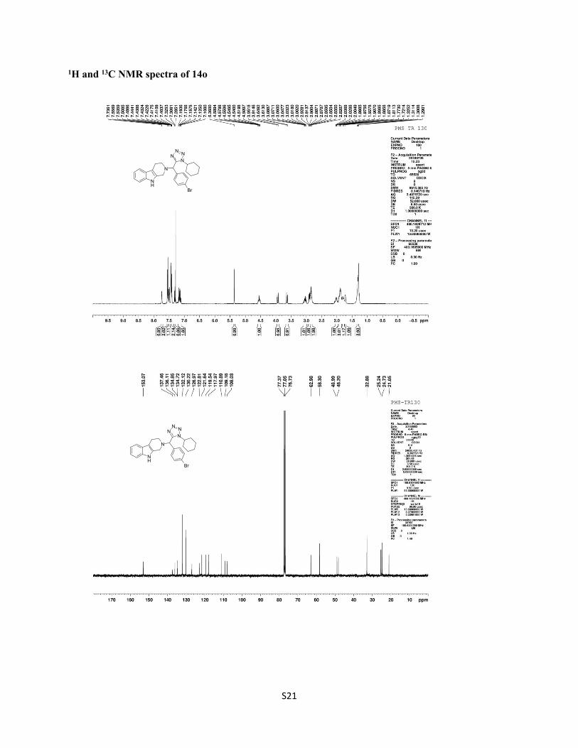
1H and 13C NMR spectra of 14o
NH
N
NNN
N
Br
NH
N
NNN
N
Br
S22
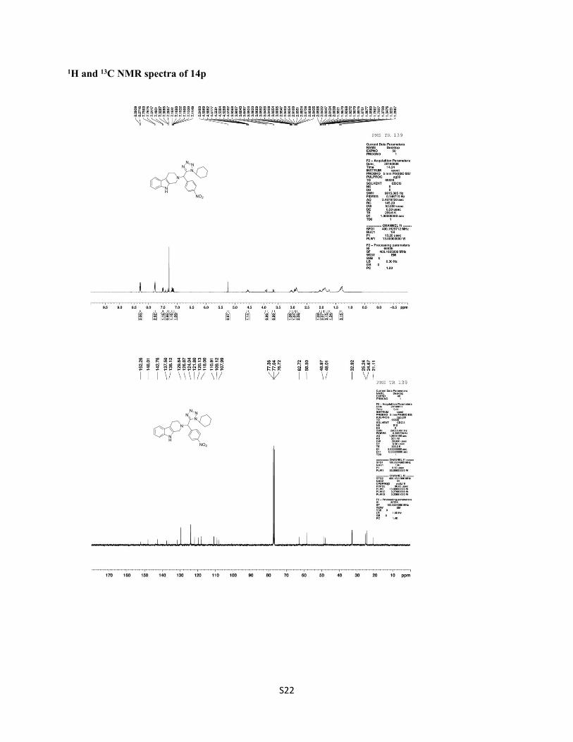
1H and 13C NMR spectra of 14p
NH
N
NNN
N
NO2
NH
N
NNN
N
NO2
S23
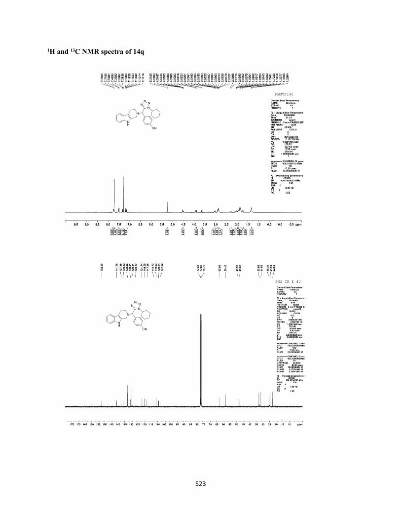
1H and 13C NMR spectra of 14q
NH
N
NNN
N
CN
NH
N
NNN
N
CN
S24
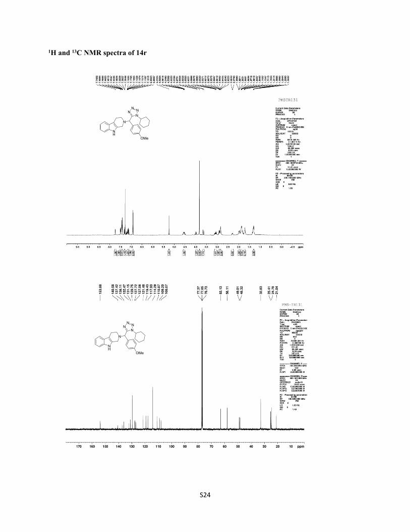
1H and 13C NMR spectra of 14r
NH
N
NNN
N
OMe
NH
N
NNN
N
OMe
S25
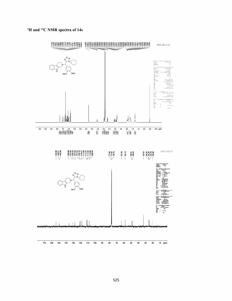
1H and 13C NMR spectra of 14s
NH
N
NNN
N
OMeMeO
NH
N
NNN
N
OMeMeO
S26
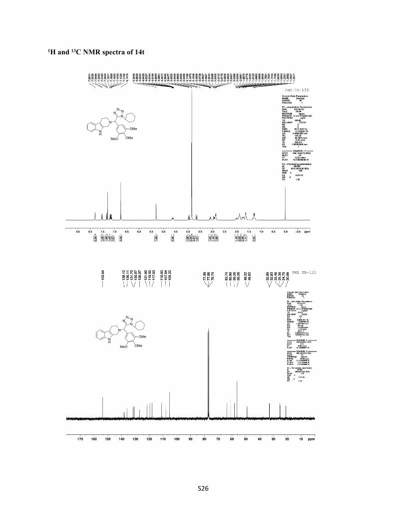
1H and 13C NMR spectra of 14t
NH
N
NNN
N
OMeMeO
OMe
NH
N
NNN
N
OMeMeO
OMe
S27
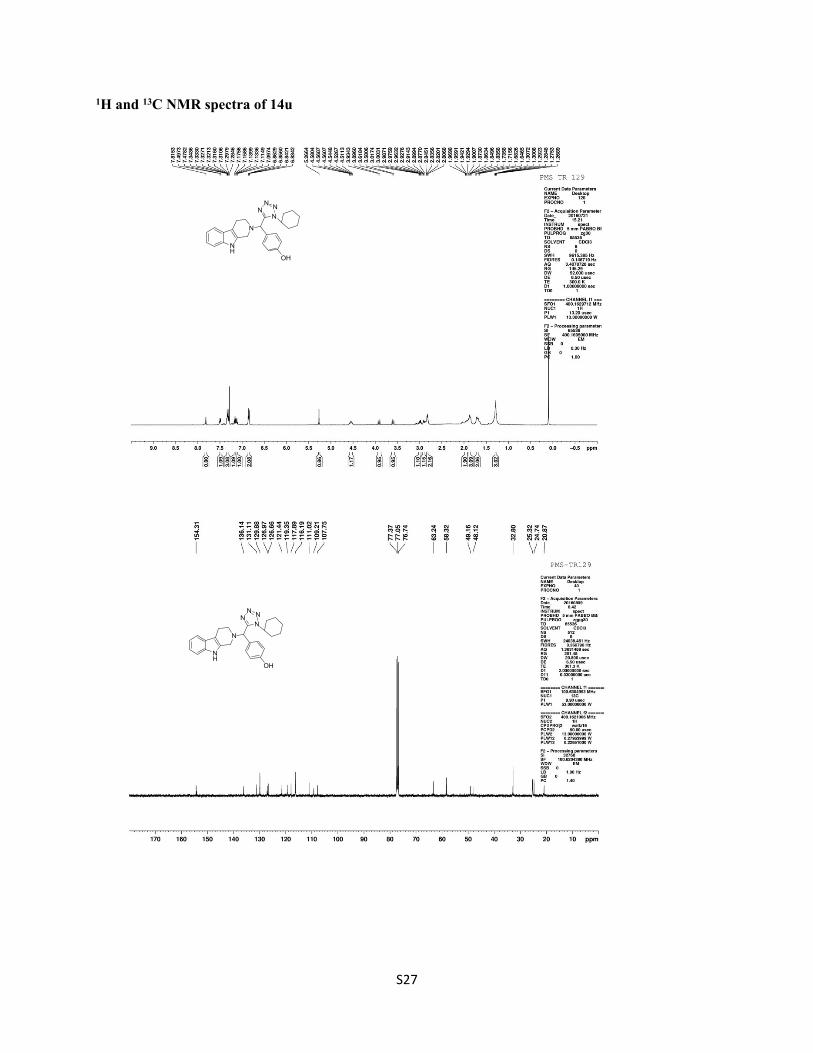
1H and 13C NMR spectra of 14u
NH
N
NNN
N
OH
NH
N
NNN
N
OH