Supporting Information - PNAS · After digestion with proteinase K (20 μg/mL; Tiangen) at 37 °C...
6
Supporting Information He et al. 10.1073/pnas.1521098113 SI Materials and Methods Insect Rearing and Embryonic Stage Statistics. All insects used in experiments were reared in the same locust colonies at the In- stitute of Zoology, Chinese Academy of Sciences, Beijing. Gre- garious locusts were reared at a density of about 400 insects per case. Solitarious locusts were cultured alone in small metal cages. Egg pods were collected three times a day, and each egg pod was put in one cup. The eggs were taken out of the sand after they had been incubated for 7 d at 30 °C and then washed in 0.08% sodium hypochlorite solution; next, they were detected under a micro- scope (Leica DFC490). The developmental stages were identified according to the criteria reported previously (50). Eggs in one cup were regarded as one biological repeat for the statistics of 60% hatching day, hatching peak duration, and embryonic stages. High-Throughput Sequencing of Small RNA. Ovary samples were from sexually mature female locusts. The abdomens of the females were vertically opened, and the ovaries were carefully separated from other tissues. Then the ovaries were washed in cold locust saline and quickly put into liquid nitrogen. Four ovaries were pooled together as one biological replicate for RNA extraction. Small RNA libraries from gregarious and solitarious locusts contained one biological replicate, respectively. Small RNAs (18–35 nt) were sequenced by an Illumina Genome Analyzer IIx sequencing system at the BGI–Shenzhen as described previously (21). The P values were calculated by using Bayesian algorithms (52). Antagomir, Agomir, and dsRNA Injection. Antagomir-276 is a chem- ically modified single-strand stable miR-276 inhibitor whose se- quence is reverse complementary to mature miR-276. Agomir-276 is a chemically modified double-strand stable miR-276 mimic. The sequence of a Caenorhabditis elegans miRNA, cel-miR-67-3p (5′ to 3′: UCACAACCUCCUAGAAAGAGUAGA), was used as the negative control of antagomir or agomir (antagomir-ck or agomir- ck). Double-strand RNA of brm (dsBrm) was used to knock down brm expression, and double-strand RNA of green fluorescent protein (dsGFP) was used as the negative control. dsRNAs were synthesized by using the T7 RiboMAX Express RNAi system (Promega). Female locusts were subjected to the first in- jection at the end of the fifth instar and then were injected every 5 d (0.1 nmol per injection for miRNA antagomir or agomir and 5 μg per injection for dsRNAs). After the third injection, the female locusts were either killed for RNA isolation or mated with male locusts. All injections were performed by using a nanoliter injector 2000 (World Precision Instruments) at the dorsal site near the lo- cust ovary. The primers for dsRNA synthesis are presented in Table S3. In Vitro Luciferase Reporter Gene Assays. The sequences around miR-276 binding sites (about 160 bp upstream and downstream flanks) of the putative target genes were inserted into the lu- ciferase reporter vector psiCHECK-2 plasmid (Promega). An ∼400-bp pre–miR-276 centered on the genome sequence was cloned into the pAc5.1/V5-HisA vector (Invitrogen) as the overexpression vector. Site mutation (Fig. S4) in the binding site of the miR-276 seed sequence in brm complementary sequences to the “seed” sites was performed by the Fast Mutagenesis Sys- tem (TransGen). A 20-ng portion of the luciferase reporter vector (WT or MT) was cotransfected with 80 ng of the miRNA expression vector into Drosophila S2 cells by Lipofect (Tiangen). The luciferase activities were detected at 45 h after transfection by using the Dual-Glo Luciferase Assay System (Promega) with a luminometer (Promega). RIP Experiments. The experiments were performed using a Magna RIP Quad Kit (Millipore). One biological duplication contained three to four ovaries. The ovaries were homogenized in ice-cold RIP lysis buffer and stored at −80 °C overnight for thorough tissue lysis. A 5-μg portion of Ago1/RL10a antibody or normal mouse IgG was incubated with magnetic beads for 30 min. Then, the lysate was thawed and centrifuged, and the supernatant was coincubated with the beads–antibody complex at 4 °C overnight. Meanwhile, 1/3 of the lysate was stored as “input” samples. Next, RNAs in the immunoprecipitates and input were extracted by TRIzol reagent (Invitrogen). A High Capacity RNA-to-cDNA Kit (ABI) was used for reverse-transcription. Then, qPCR was performed to analyze the expression levels of target genes. Input samples were used for normalization of the relative expression of mRNA and IgG controls were used for subtraction of the non- specific interactions of RNA-Ago1 or RNA-RL10a. Enrichment of brm RNA in Ago1 complex in nuclear and cyto- plasmic fractions was tested by RIP assay of nuclear and cytoplas- mic fractions separately as described (53), with slight modifications. The ovaries were homogenized in cold PBS containing 0.2% Nonidet P-40. The lysate was centrifuged at 30 × g for 2 min at 4 °C to remove the insoluble fragment of tissue. Then, the supernatant was centrifuged at 425 × g for 15 min at 4 °C. The nuclei were in the pellet whereas the cytoplasm remained in the supernatant. The cytoplasmic fraction was centrifuged at 2,000 × g for 10 min at 4 °C to remove the residual nuclei. The pellet was washed with buffer B [20 mM Tris·HCl, pH 8.0, 1.5 mM MgCl 2 , 0.2 mM EDTA, pH 8.0, 20 mM KCl, 25% (vol/vol) glycerol] several times and then was resuspended in five times the pellet volume of buffer B and 10 times the volume of buffer C [20 mM Tris·HCl, pH 8.0, 1.5 mM MgCl 2 , 0.2 mM EDTA, pH 8.0, 1.2 M KCl, 25% (vol/vol) glycerol]. The mixture was incubated for 45 min at 4 °C. Afterward, 10 times the volume of lysis buffer [150 mM NaCl, 6 mM MgCl 2 , 40 mM Hepes, pH 7.0, 2 mM DTT, 1 mM PMSF, 0.025% Nonidet P-40, 10% (vol/vol) glycerol] was added into the nuclear or cytoplasmic frac- tion. Subsequently, the two fractions were incubated with Ago1 antibody or IgG antibody overnight at 4 °C. RNA extraction and qPCR were performed as described. Western Blot. The sequences of BRM antibody epitopes are as follows: GVVTGPDLYRASGKFELLDRILPKLKATNHRVLL- FCQMTQLMTIMEDYLSWRGFTYLRLDGTTKAEDRGDL- LRKFNSPDSEFFLFLLSTRAGGLGLNLQAADTVIIFDSDW- NPHQDLQAQDRAHRIGQQNEVRVLRLMTVNSVEERILV- AARYKLNMDEKVIQAGMFDQKSTGSERQQFLQSILHQD- EAEEEEENEVPDDDSVNHMIARNADELALFHRMDLERR- REEAKLGPNRKSRLVEEAELPDWLVKDDDEVERWTFEE- EEEDRYLGRGSRQRKEVDYSDSLTEKEWLKAIDEGGEE- FEEEEEEEEEKLKKRTRKRRRKVEEEEEEESIPIQPKKRK- SSSMSCTVDPQLKRRMRKLMNIVIKYTDSDGRVLSDPFM- KLPSRRELPDYYEIIKKPLDIKKILQRIDENKFSDFDELEKE- FMTLCKNAQTY. Polyclonal antibody for BRM was produced from mouse. Total proteins were extracted by TRIzol reagent (Invitrogen). The proteins were subjected to polyacrylamide gel (8%) electrophoresis and then transferred to polyvinylidene di- fluoride (PVDF) membranes (Millipore). Blocking was per- formed in 5% (wt/vol) skimmed milk at room temperature for 1 h. The membranes were incubated with primary antibody (anti-BRM, 1:500; anti-LOK, OriGene, 1:500; anti-Histone H3, He et al. www.pnas.org/cgi/content/short/1521098113 1 of 6
Transcript of Supporting Information - PNAS · After digestion with proteinase K (20 μg/mL; Tiangen) at 37 °C...
Supporting InformationHe et al. 10.1073/pnas.1521098113SI Materials and MethodsInsect Rearing and Embryonic Stage Statistics. All insects used inexperiments were reared in the same locust colonies at the In-stitute of Zoology, Chinese Academy of Sciences, Beijing. Gre-garious locusts were reared at a density of about 400 insects percase. Solitarious locusts were cultured alone in small metal cages.Egg pods were collected three times a day, and each egg pod wasput in one cup. The eggs were taken out of the sand after they hadbeen incubated for 7 d at 30 °C and then washed in 0.08% sodiumhypochlorite solution; next, they were detected under a micro-scope (Leica DFC490). The developmental stages were identifiedaccording to the criteria reported previously (50). Eggs in one cupwere regarded as one biological repeat for the statistics of 60%hatching day, hatching peak duration, and embryonic stages.
High-Throughput Sequencing of Small RNA.Ovary samples were fromsexually mature female locusts. The abdomens of the females werevertically opened, and the ovaries were carefully separated fromother tissues. Then the ovaries were washed in cold locust salineand quickly put into liquid nitrogen. Four ovaries were pooledtogether as one biological replicate for RNA extraction. SmallRNA libraries from gregarious and solitarious locusts containedone biological replicate, respectively. Small RNAs (18–35 nt) weresequenced by an Illumina Genome Analyzer IIx sequencingsystem at the BGI–Shenzhen as described previously (21). TheP values were calculated by using Bayesian algorithms (52).
Antagomir, Agomir, and dsRNA Injection. Antagomir-276 is a chem-ically modified single-strand stable miR-276 inhibitor whose se-quence is reverse complementary to mature miR-276. Agomir-276is a chemically modified double-strand stable miR-276 mimic. Thesequence of a Caenorhabditis elegansmiRNA, cel-miR-67-3p (5′ to3′: UCACAACCUCCUAGAAAGAGUAGA), was used as thenegative control of antagomir or agomir (antagomir-ck or agomir-ck). Double-strand RNA of brm (dsBrm) was used to knock downbrm expression, and double-strand RNA of green fluorescentprotein (dsGFP) was used as the negative control. dsRNAswere synthesized by using the T7 RiboMAX Express RNAisystem (Promega). Female locusts were subjected to the first in-jection at the end of the fifth instar and then were injected every5 d (0.1 nmol per injection for miRNA antagomir or agomir and5 μg per injection for dsRNAs). After the third injection, the femalelocusts were either killed for RNA isolation or mated with malelocusts. All injections were performed by using a nanoliter injector2000 (World Precision Instruments) at the dorsal site near the lo-cust ovary. The primers for dsRNA synthesis are presented inTable S3.
In Vitro Luciferase Reporter Gene Assays. The sequences aroundmiR-276 binding sites (about 160 bp upstream and downstreamflanks) of the putative target genes were inserted into the lu-ciferase reporter vector psiCHECK-2 plasmid (Promega). An∼400-bp pre–miR-276 centered on the genome sequence wascloned into the pAc5.1/V5-HisA vector (Invitrogen) as theoverexpression vector. Site mutation (Fig. S4) in the binding siteof the miR-276 seed sequence in brm complementary sequencesto the “seed” sites was performed by the Fast Mutagenesis Sys-tem (TransGen). A 20-ng portion of the luciferase reportervector (WT or MT) was cotransfected with 80 ng of the miRNAexpression vector into Drosophila S2 cells by Lipofect (Tiangen).The luciferase activities were detected at 45 h after transfection
by using the Dual-Glo Luciferase Assay System (Promega) with aluminometer (Promega).
RIP Experiments. The experiments were performed using a MagnaRIP Quad Kit (Millipore). One biological duplication containedthree to four ovaries. The ovaries were homogenized in ice-coldRIP lysis buffer and stored at −80 °C overnight for thoroughtissue lysis. A 5-μg portion of Ago1/RL10a antibody or normalmouse IgG was incubated with magnetic beads for 30 min. Then,the lysate was thawed and centrifuged, and the supernatant wascoincubated with the beads–antibody complex at 4 °C overnight.Meanwhile, 1/3 of the lysate was stored as “input” samples. Next,RNAs in the immunoprecipitates and input were extracted byTRIzol reagent (Invitrogen). A High Capacity RNA-to-cDNAKit (ABI) was used for reverse-transcription. Then, qPCR wasperformed to analyze the expression levels of target genes. Inputsamples were used for normalization of the relative expression ofmRNA and IgG controls were used for subtraction of the non-specific interactions of RNA-Ago1 or RNA-RL10a.Enrichment of brm RNA in Ago1 complex in nuclear and cyto-
plasmic fractions was tested by RIP assay of nuclear and cytoplas-mic fractions separately as described (53), with slight modifications.The ovaries were homogenized in cold PBS containing 0.2%Nonidet P-40. The lysate was centrifuged at 30 × g for 2 min at 4 °Cto remove the insoluble fragment of tissue. Then, the supernatantwas centrifuged at 425 × g for 15 min at 4 °C. The nuclei were in thepellet whereas the cytoplasm remained in the supernatant. Thecytoplasmic fraction was centrifuged at 2,000 × g for 10 min at 4 °Cto remove the residual nuclei. The pellet was washed with buffer B[20 mM Tris·HCl, pH 8.0, 1.5 mM MgCl2, 0.2 mM EDTA, pH 8.0,20 mM KCl, 25% (vol/vol) glycerol] several times and then wasresuspended in five times the pellet volume of buffer B and 10 timesthe volume of buffer C [20 mM Tris·HCl, pH 8.0, 1.5 mM MgCl2,0.2 mM EDTA, pH 8.0, 1.2 M KCl, 25% (vol/vol) glycerol]. Themixture was incubated for 45 min at 4 °C. Afterward, 10 times thevolume of lysis buffer [150 mM NaCl, 6 mMMgCl2, 40 mMHepes,pH 7.0, 2 mM DTT, 1 mM PMSF, 0.025% Nonidet P-40, 10%(vol/vol) glycerol] was added into the nuclear or cytoplasmic frac-tion. Subsequently, the two fractions were incubated with Ago1antibody or IgG antibody overnight at 4 °C. RNA extraction andqPCR were performed as described.
Western Blot. The sequences of BRM antibody epitopes are asfollows: GVVTGPDLYRASGKFELLDRILPKLKATNHRVLL-FCQMTQLMTIMEDYLSWRGFTYLRLDGTTKAEDRGDL-LRKFNSPDSEFFLFLLSTRAGGLGLNLQAADTVIIFDSDW-NPHQDLQAQDRAHRIGQQNEVRVLRLMTVNSVEERILV-AARYKLNMDEKVIQAGMFDQKSTGSERQQFLQSILHQD-EAEEEEENEVPDDDSVNHMIARNADELALFHRMDLERR-REEAKLGPNRKSRLVEEAELPDWLVKDDDEVERWTFEE-EEEDRYLGRGSRQRKEVDYSDSLTEKEWLKAIDEGGEE-FEEEEEEEEEKLKKRTRKRRRKVEEEEEEESIPIQPKKRK-SSSMSCTVDPQLKRRMRKLMNIVIKYTDSDGRVLSDPFM-KLPSRRELPDYYEIIKKPLDIKKILQRIDENKFSDFDELEKE-FMTLCKNAQTY. Polyclonal antibody for BRM was producedfrom mouse. Total proteins were extracted by TRIzol reagent(Invitrogen). The proteins were subjected to polyacrylamide gel(8%) electrophoresis and then transferred to polyvinylidene di-fluoride (PVDF) membranes (Millipore). Blocking was per-formed in 5% (wt/vol) skimmed milk at room temperature for1 h. The membranes were incubated with primary antibody(anti-BRM, 1:500; anti-LOK, OriGene, 1:500; anti-Histone H3,
He et al. www.pnas.org/cgi/content/short/1521098113 1 of 6
Sigma, 1:2,000; anti-RL10a, Santa Cruz, 1:500; anti-V5, In-vitrogen, 1:5,000) in 5% (wt/vol) skimmed milk at 4 °C overnight.Secondary antibody (1:5,000) (CoWin) was incubated at roomtemperature for 1 h. Detection for the immunological blot wascarried out by an eECLWestern Blot Kit (CoWin). Densitometricanalysis of the band was performed by Quantity One software.
In Situ Fluorescence Hybridization. A double FISH experiment wasperformed according to a method that was described previously(54). The RNA probe for brm was synthetized by a T7/SP6 RNATranscription Kit (Roche) and was subsequently fragmented toabout 250 bp by carbonate buffer. The primers used for probesynthesis of brm are in Table S3. Ovarioles were separated fromovaries in locust saline and fixed in 4% (wt/vol) paraformaldehydeovernight. After digestion with proteinase K (20 μg/mL; Tiangen)at 37 °C for 15 min, these ovaries were hybridized with miRNAprobe (2 pmol/mL) and brm probe (5 ng/μL) at 37 °C overnight.Then, the ovarioles were successively washed in 2× SSC, 1× SSC,and 0.2× SSC at 37 °C. Anti-DIG alkaline phosphatase-conju-gated antibody (1:500) and anti-biotin antibody (1:100) wereused for probe detection. Then, the fluorescent signal of di-goxigenin (DIG) or biotin was obtained by HNPP/Fast Red orFluorescein-Tyramide (Perkin-Elmer). Images were captured on
an LSM 710 confocal fluorescence microscope (Zeiss) at amagnification of 20×. For detection of brm RNA distribution inthe S2 cells, the cells were cultured on coverslips (Citoglas) andthen transfected as described above. After 45 h, FISH experi-ments were carried out as previously described (55), with slightmodifications. The images of S2 cells were captured at a mag-nification of 63×.
Assays for the in Vitro Protein Expression. Full-length WT or mu-tated sequences were cloned into the PAC-5.1/V5-HisB plasmid(Invitrogen) using the KpnΙ and XhoΙ sites as protein expressionplasmids. Site mutations were gained by using a Phusion Site-Directed Mutagenesis Kit (ThermoFisher Scientific). The plas-mid was cotransfected with agomir-276/agomir-ck into the S2cells at 1:400 by Lipofectamine 3000 reagent (ThermoFisherScientific). The cells were sampled 45 h after transfection. Pro-tein was extracted by PIRA (CoWin), and 80 μg of total proteinwas used to perform Western blot. Anti-V5 antibody (MBL) wasused to detect to BRM level. β-Tubulin antibody (1:5,000;EASYBIO) was used as an internal control. Total mRNAs andnuclear and cytoplasmic RNAs were extracted as described andreverse transcribed using a FastQuant RT Kit (with gDNase)(Tiangen). The qPCR primers for S2 cells are included in Table S3.
antagomir-ck antagomir-27613.2
13.4
13.6
13.8
14.0
14.2
14.4
agomir-ck agomir-27613.0
13.2
13.4
13.6
13.8
14.0
14.2
14.4
dsGFP dsBRM
13.2
13.6
14.0
14.4
14.8
agomir-276+dsGFP
agomir-276+dsBRM13.0
13.2
13.4
13.6
13.8
14.0
14.2
60%
hat
chin
g tim
e (d
)
A B C
D
*
*
* *G S
13.0
13.2
13.4
13.6
13.8
14.0
14.2
**
E
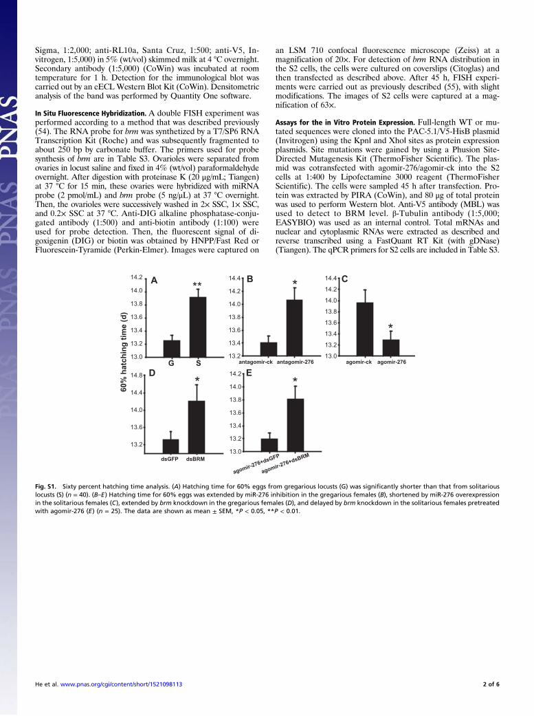
Fig. S1. Sixty percent hatching time analysis. (A) Hatching time for 60% eggs from gregarious locusts (G) was significantly shorter than that from solitariouslocusts (S) (n = 40). (B–E) Hatching time for 60% eggs was extended by miR-276 inhibition in the gregarious females (B), shortened by miR-276 overexpressionin the solitarious females (C), extended by brm knockdown in the gregarious females (D), and delayed by brm knockdown in the solitarious females pretreatedwith agomir-276 (E) (n = 25). The data are shown as mean ± SEM, *P < 0.05, **P < 0.01.
He et al. www.pnas.org/cgi/content/short/1521098113 2 of 6
agomir-ck agomir-276
Rel
ativ
e ex
pres
sion
leve
l
antagomir-ck antagomir-2760
1
2
3
4
5
6
7
**
miR-276
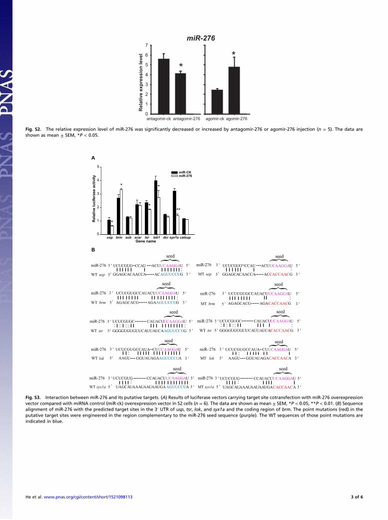
Fig. S2. The relative expression level of miR-276 was significantly decreased or increased by antagomir-276 or agomir-276 injection (n = 5). The data areshown as mean ± SEM, *P < 0.05.
lok GUAUAGAAGUUCCUA
miR-276 CCAUA UCAAGGAU 5
syx1a AUAAAUAAUAAUGA AGUUCCUA
ACAGUUCCUG
UGUGUUCAUUAUCAAGUUCCUG
WT brm AGAGCACG GA
3 '
{ seed
'3 '
5 '
{ seed
WT
miR-276 ACUUCAAGGAU 5
{
'3 '
seed
miR- 276
lokMT
3 'WT usp 5 '
{ seed
miR-276 3 '
5 '
WT
miR-276 UCUCGUG UCAAGGAU 5
{
'3 '
seed
5 ' 3 '
{ seed
miR-276 3 '
syx1aMT 5 '
miR-276 CAUACUUCAAGGAU 5
{
'3 '
seed
WT tsr 5 ' 3 '
MT usp
miR-276 UCUCGUGCCAUACUUCAAGGAU 5
{
'3 '
seed
5 ' 3 '
miR-276 UCUCGUGCCAUACUUCAAGGAU 5
{
'3 '
seed
MT brm 5 ' 3 '
CUUCUCGUG
AAGU G GUAUAGA A
CCAUA UCAAGGAU 5
3 '
'3 '
5 '
CUUCUCGUG
AAGU G CACCAAC·· ··
UACUCCA
··G CCAU
GGAGCACAACCA
UCUCGU
AC G
ACUUCAAGGAUG CCAU
GGAGCACAACCA
UCUCGU 5 '
3 'CACCAAC··
··UCUCGUGC
GGGG········
UGUGUUCAUUAUCA G
miR-276 CAUACUUCAAGGAU 5
{
'3 '
seed
WT tsr 5 ' 3 '··
UCUCGUGC
GGGG········
CACCAAC
GAGUUCCUA AGAGCACG GA GA·· ··
CACCAAC
GCUA··
AUAAAUAAUAAUGA A
UCUCGUG UCAAGGAU 5 '
3 '
UACUCCA
GCUA··
CACCAAC
usp brm aub scar tsr lok1 dcr syx1a catsup0
1
2
3
4
5
*
*
**
**
miR-CKmiR-276
Rel
ativ
e lu
cife
rase
act
ivity
Gene name
A
B
Fig. S3. Interaction between miR-276 and its putative targets. (A) Results of luciferase vectors carrying target site cotransfection with miR-276 overexpressionvector compared with miRNA control (miR-ck) overexpression vector in S2 cells (n = 6). The data are shown as mean ± SEM, *P < 0.05, **P < 0.01. (B) Sequencealignment of miR-276 with the predicted target sites in the 3′ UTR of usp, tsr, lok, and syx1a and the coding region of brm. The point mutations (red) in theputative target sites were engineered in the region complementary to the miR-276 seed sequence (purple). The WT sequences of those point mutations areindicated in blue.
He et al. www.pnas.org/cgi/content/short/1521098113 3 of 6
dsGFP dsBrm
Rel
ativ
e m
RN
A le
vel
0.000
0.002
0.004
0.006
0.008
0.010
0.012
** BRM
H3
dsGFP
dsBrm
dsGFP dsBrm
Rel
ativ
e ba
nd in
tens
ity
0.0
0.5
1.0
1.5
2.0
2.5
3.0
3.5
**
A B
300KD
250KD
180KD
130KD
Marke
r
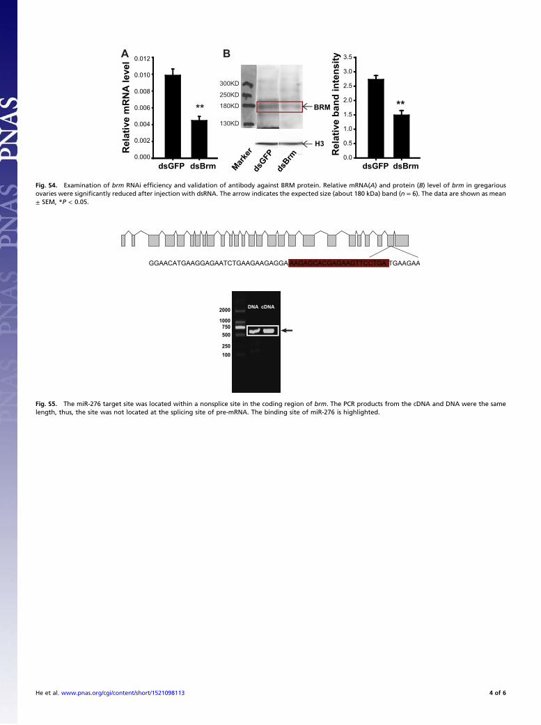
Fig. S4. Examination of brm RNAi efficiency and validation of antibody against BRM protein. Relative mRNA(A) and protein (B) level of brm in gregariousovaries were significantly reduced after injection with dsRNA. The arrow indicates the expected size (about 180 kDa) band (n = 6). The data are shown as mean± SEM, *P < 0.05.
GAGGA AAGAGCACGAGAAGTTCCTGA TGAAGAA
2000
1000750500
250100
GGAACATGAAGGAGAATCTGAAGAA
DNA cDNA
Fig. S5. The miR-276 target site was located within a nonsplice site in the coding region of brm. The PCR products from the cDNA and DNA were the samelength, thus, the site was not located at the splicing site of pre-mRNA. The binding site of miR-276 is highlighted.
He et al. www.pnas.org/cgi/content/short/1521098113 4 of 6
Hochest brm Merge Hochest brm Merge
agomir-ck agomir-276
vector
WT
MT1
MT2
MT3
A
B
C
D
E
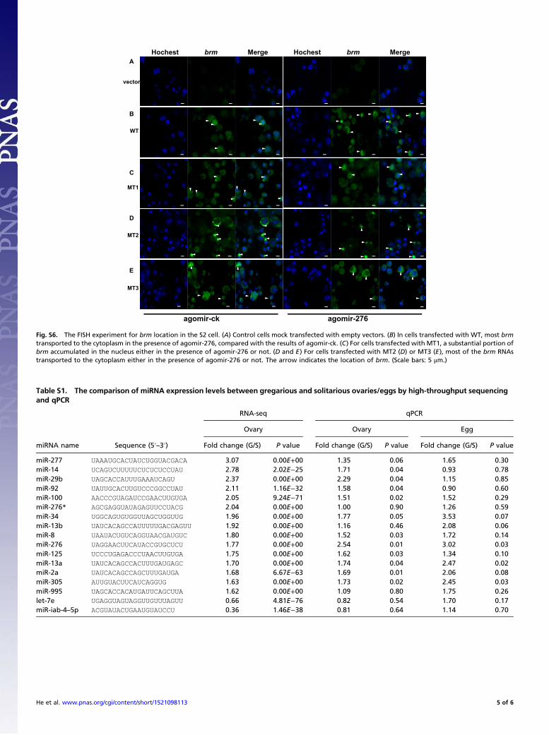
Fig. S6. The FISH experiment for brm location in the S2 cell. (A) Control cells mock transfected with empty vectors. (B) In cells transfected with WT, most brmtransported to the cytoplasm in the presence of agomir-276, compared with the results of agomir-ck. (C) For cells transfected with MT1, a substantial portion ofbrm accumulated in the nucleus either in the presence of agomir-276 or not. (D and E) For cells transfected with MT2 (D) or MT3 (E), most of the brm RNAstransported to the cytoplasm either in the presence of agomir-276 or not. The arrow indicates the location of brm. (Scale bars: 5 μm.)
Table S1. The comparison of miRNA expression levels between gregarious and solitarious ovaries/eggs by high-throughput sequencingand qPCR
miRNA name Sequence (5′–3′)
RNA-seq qPCR
Ovary Ovary Egg
Fold change (G/S) P value Fold change (G/S) P value Fold change (G/S) P value
miR-277 UAAAUGCACUAUCUGGUACGACA 3.07 0.00E+00 1.35 0.06 1.65 0.30miR-14 UCAGUCUUUUUCUCUCUCCUAU 2.78 2.02E−25 1.71 0.04 0.93 0.78miR-29b UAGCACCAUUUGAAAUCAGU 2.37 0.00E+00 2.29 0.04 1.15 0.85miR-92 UAUUGCACUUGUCCCGGCCUAU 2.11 1.16E−32 1.58 0.04 0.90 0.60miR-100 AACCCGUAGAUCCGAACUUGUGA 2.05 9.24E−71 1.51 0.02 1.52 0.29miR-276* AGCGAGGUAUAGAGUUCCUACG 2.04 0.00E+00 1.00 0.90 1.26 0.59miR-34 UGGCAGUGUGGUUAGCUGGUUG 1.96 0.00E+00 1.77 0.05 3.53 0.07miR-13b UAUCACAGCCAUUUUUGACGAGUU 1.92 0.00E+00 1.16 0.46 2.08 0.06miR-8 UAAUACUGUCAGGUAACGAUGUC 1.80 0.00E+00 1.52 0.03 1.72 0.14miR-276 UAGGAACUUCAUACCGUGCUCU 1.77 0.00E+00 2.54 0.01 3.02 0.03miR-125 UCCCUGAGACCCUAACUUGUGA 1.75 0.00E+00 1.62 0.03 1.34 0.10miR-13a UAUCACAGCCACUUUGAUGAGC 1.70 0.00E+00 1.74 0.04 2.47 0.02miR-2a UAUCACAGCCAGCUUUGAUGA 1.68 6.67E−63 1.69 0.01 2.06 0.08miR-305 AUUGUACUUCAUCAGGUG 1.63 0.00E+00 1.73 0.02 2.45 0.03miR-995 UAGCACCACAUGAUUCAGCUUA 1.62 0.00E+00 1.09 0.80 1.75 0.26let-7e UGAGGUAGUAGGUUGUUUAGUU 0.66 4.81E−76 0.82 0.54 1.70 0.17miR-iab-4–5p ACGUAUACUGAAUGUAUCCU 0.36 1.46E−38 0.81 0.64 1.14 0.70
He et al. www.pnas.org/cgi/content/short/1521098113 5 of 6
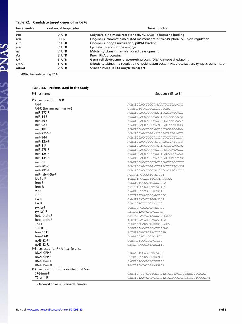
Table S2. Candidate target genes of miR-276
Gene symbol Location of target sites Gene function
usp 3′ UTR Ecdysteroid hormone receptor activity, juvenile hormone bindingbrm CDS Oogenesis, chromatin-mediated maintenance of transcription, cell cycle regulationaub 3′ UTR Oogenesis, oocyte maturation, piRNA bindingscar 3′ UTR Epithelial fusions in the embryotsr 3′ UTR Mitotic cytokinesis, female gonad developmentdcr 3′ UTR Pre-miRNA processinglok 3′ UTR Germ cell development, apoptotic process, DNA damage checkpointSyx1A 3′ UTR Mitotic cytokinesis, a regulation of pole, plasm oskar mRNA localization, synaptic transmissioncatsup 3′ UTR Ovarian nurse cell to oocyte transport
piRNA, Piwi-interacting RNA.
Table S3. Primers used in the study
Primer name Sequence (5′ to 3′)
Primers used for qPCRU6-F ACACTCCAGCTGGGTCAAAATCGTGAAGCG
U6-R (for nuclear marker) CTCAAGTGTCGTGGAGTCGGCAA
miR-277-F ACACTCCAGCTGGGTAAATGCACTATCTGG
miR-14-F ACACTCCAGCTGGGTCAGTCTTTTTCTCTC
miR-29-F ACACTCCAGCTGGGTAGCACCATTTGAAAT
miR-92-F ACACTCCAGCTGGGTATTGCACTTGTCCCG
miR-100-F ACACTCCAGCTGGGAACCCGTAGATCCGAA
miR-276*-F ACACTCCAGCTGGGAGCGAGGTATAGAGTT
miR-34-F ACACTCCAGCTGGGTGGCAGTGTGGTTAGC
miR-13b-F ACACTCCAGCTGGGTATCACAGCCATTTTT
miR-8-F ACACTCCAGCTGGGTTAATACTGTCAGGTA
miR-276-F ACACTCCAGCTGGGTAGGAACTTCATACCG
miR-125-F ACACTCCAGCTGGGTCCCTGAGACCCTAAC
miR-13a-F ACACTCCAGCTGGGTATCACAGCCACTTTGA
miR-2-F ACACTCCAGCTGGGTATCACAGCCAGCTTTG
miR-305-F ACACTCCAGCTGGGATTGTACTTCATCAGGT
miR-995-F ACACTCCAGCTGGGTAGCACCACATGATTCA
miR-iab-4–5p-F ACGTATACTGAATGTATCCT
let-7e-F TGAGGTAGTAGGTTGTTTAGTTAA
brm-F AGCGTCTTTGATTCACGAGGA
brm-R ACTTCTCGTGCTCTTTCCTCT
tsr-F AAACTGCTTTGCCCGTGATG
tsr-R AGTTTAATAACGCCAACAGGC
lok-F CAAGTTGATGTTTGGAGCCT
lok-R GTACCGTGTTGGGAAGGAG
syx1a-F CCAGGGAGAAATGATAGACC
syx1a1-R GATGACTACTACGAGGCAGA
beta-actin-F AATTACCATTGGTAACGAGCGATT
beta-actin-R TGCTTCCATACCCAGGAATGA
18S-F ATGCAAACAGAGTCCCGACCAGA
18S-R GCGCAGAACCTACCATCGACAG
brm-S2-F ACTGAAGAATACTACTCGCAA
brm-S2-R AGAATCGAGACCGAGGAGA
rp49-S2-F CCATAGTTGCCTGACTCCC
rp49-S2-R GATGGAGGCGGATAAAGTTG
Primers used for RNA interferenceRNAi-GFP-F CACAAGTTCAGCGTGTCCG
RNAi-GFP-R GTTCACCTTGATGCCGTTC
RNAi-Brm-F CACCACTCCCATAGTCCAAC
RNAi-Brm-R TGCTGAGATGCCGAAGGACA
Primers used for probe synthesis of brmSP6-brm-F GAATTGATTTAGGTGACACTATAGCTAGGTCCAAACCGCAAAT
T7-brm-R GAATTGTAATACGACTCACTATAGGGGGTGACATTCCTGCCATAT
F, forward primers; R, reverse primers.
He et al. www.pnas.org/cgi/content/short/1521098113 6 of 6