Supporting Information - PNAS · 2013-04-05 · Supporting Information Snyder et al....
8
Supporting Information Snyder et al. 10.1073/pnas.1215770110 SI Materials and Methods Protein Expression and Purification. The human MyD88 (myeloid differentiation factor 88) cDNA encoding residues M157 to P296 was cloned into a pET30a-derived bacterial expression vector containing an N-terminal protein G β1 domain (GB1) tag followed by a tobacco etch virus (TEV) protease cleavage site. The GB1 coding plasmid was a gift from Gerhard Wagner at Harvard Medical School (Boston, MA). The TEV coding plasmid was a gift from Dr. David Waugh at the National Cancer Institute (Freder- ick, MD). The MyD88 construct was transformed into BL21 (DE3) Codon Plus RIPL cells (Stratagene). Cells were grown and induced with 0.5 mM Isopropyl β-D-1-thiogalactopyranoside (IPTG). Harvested cells were lysed in a buffer containing 25 mM Tris·HCl (pH 8.0), 300 mM NaCl, 10 mM imidazole, and EDTA- free protease inhibitor (Roche Applied Science). The soluble protein was purified using Ni-NTA beads (Qiagen). Further pu- rification was carried out after TEV cleavage with a second im- mobilized metal ion affinity chromatography (IMAC) column and size exclusion chromatography. 15 N isotope labeled MyD88 Toll/ IL-1 receptor (TIR) domain was expressed using 15 NH 4 Cl (Cambridge Isotope Laboratories) as the sole nitrogen source in a minimal media (Molecular Dimensions) and purified as de- scribed above. Recombinant TcpC TIR domain (residues 170– 307) was expressed using the pASK-IBA3-TIR-TcpC construct and purified as reported previously (1). Crystallization and Structure Determination. Crystallization screen- ing was carried out at 4 °C using the hanging drop method. The MyD88 TIR domain was crystallized in the presence of the TcpC TIR domain using a well solution containing 100 mM Tris·HCl (pH 8.0) and 25% (vol/vol) PEG 350 MME. Although TcpC was present in a 1:1 molar ratio with MyD88, no electron density for TcpC was observed in the crystal lattice. The MyD88 TIR domain was also lysine-methylated (2) and crystallized similarly in the absence of TcpC. Essentially the same structures were observed for both the native (1.45 Å resolution) and lysine-methylated (1.8 Å resolution) MyD88 TIR domains. Initial diffraction studies were carried out using the X-ray diffraction facility at National Institute of Diabetes and Digestive and Kidney Diseases, National Institutes of Health. Subsequent data collection was carried out at the Advanced Photon Source, Argonne National Laboratory (beam line Southeast Re- gional Collaborative Access Team 22 ID) and the National Syn- chrotron Light Source, Brookhaven National Laboratory (beam line X29). Data were processed with the HKL2000 program suite (3). The MyD88 crystal structure was solved by molecular re- placement with the programs Amore and Phaser from the CCP4 Suite (4) using the deposited NMR structure of the MyD88 TIR domain (Protein Data Bank code 2JS7) as a search model. Model building was aided by Arp/Warp 7.0 (5) and carried out using the programs COOT (6) and O (7). Refinement was performed using the programs CNS (8) and PHENIX (9). The crystal structures were validated using Molprobity (10) in PHENIX and the Research Collaboratory for Structural Bioinformatics ADIT validation server (11). Solvent-accessible surface area was calculated with the program Areaimol from the CCP4 suite (4, 12). Figures were produced with Pymol (Schrödinger LLC). Molecular Dynamics Simulation. The program suite GROMACS 4.5.5 (13) and CHARMM27 force field (14) were used for mo- lecular dynamics (MD) simulations with the Biowulf Linux cluster at NIH. The MyD88 TIR domains were solvated with water molecules using the TIP3P water models (15). The structures were energy minimized using the steepest descent approach be- fore subjecting to MD simulations for 5 or 10 ns with 2 fs steps at 300 K. The Particle-Mesh Ewald method for long-range electro- statics and the Linear Constraint Solver algorithm for covalent bond constraints were applied. Analysis of the MD simulation trajectories was carried out using the programs g_rms and g_rmsf in the GROMACS package. Separately computed 5-ns and 10-ns trajectories yielded very similar root mean square fluctuation profiles as in Fig. 2B. Peptides. Peptides (Table S2) for the TcpC BB and DD loops were synthesized and HPLC purified by American Peptides, United Biosystems, or Thermo-Fisher Scientific. For the stimulation assays the peptides were synthesized as fusion with the TAT se- quence. A short linker (AS) plus the Strep-tag II (WSHPQFEK) was added to the peptide sequences for the pull-down assays. Pull-Down Assay. Purified TcpC TIR domain (TIR-TcpC) or the TcpC peptides carrying a C-terminal Strep-tag II were used as “bait.” Eighty micrograms of purified TIR-TcpC or TcpC-loop peptides were bound to 50% (vol/vol) suspension of Strep-Tactin MacroPrep Beads (IBA) in buffer PD1 [100 mM Tris·HCl (pH 8.0), 1 mM EDTA, and 150 mM NaCl] for 1.5–3 h at 4 °C under gentle agitation. After three wash steps and one blocking step with avidin (300 μg/mL in PD1) at room temperature for 12 min under occasional rocking, 50–200 mg cleared total cell lysates (“prey”) from HEK293 cells were added and incubated for 2 h at 4 °C followed by 30 min at 37 °C. The beads were then washed three times with an acetate buffer (product #1858605, Pierce/Thermo Scientific) containing 150 mM or 500 mM NaCl to remove non- specifically bound proteins. The bound “prey” proteins were then eluted in two consecutive steps with an elution buffer (100 mM sodium citrate, 100 mM glycine, and 3 mM EDTA) at pH 2.8 and neutralized with 2 M Tris·HCl (pH 8.0). Samples were then stored at -20 °C before analysis by SDS/PAGE or Western blot. To prepare the cell lysates, 5 × 10 6 HEK293 cells transfected using Lipofectamine 2000 (Invitrogen) with Myc-MyD88 or Flag- TLR4 (TLR, Toll-like receptor) plasmids were solubilized 24–48 h after transfection with a Nonidet P-40 lysis buffer containing 50 mM Hepes-Na (pH 7.6), 150 mM NaCl, 1 mM DTT, 1 mM EDTA, 1 mM EGTA, 0.5% (vol/vol) Nonident P-40, 0.125% (wt/ vol) n-octyl-β-D-glucopyranoside, 10% (vol/vol) glycerol, 20 mM β-glycerolphosphate, 1 mM Na 3 VO 4 , 0.4 mM PMSF, and 1 mM NaF, plus the Complete protease inhibitor mixture (EDTA free, Roche Diagnostics). Lysates were subsequently dounced using a tissue grinder and dialyzed against PBS plus the protease in- hibitor mixture. Western Blot. All samples were reduced with the Lämmli buffer plus DTT and subjected to heat denaturation before SDS/PAGE. Myc-tagged MyD88 and Flag-tagged TLR4 were detected with Anti-Flag monoclonal antibody from Sigma-Aldrich and Anti- Myc antibody from Invitrogen diluted 1:1,000 in TBST buffer [100 mM Tris·HCl (pH 7.6), 150 mM NaCl, and 0.05% Tween-20). The secondary anti-mouse antibody (Jackson ImmunoResearch Europe) was diluted 1:5,000 in TBST, and the Western blot films were developed using standard procedures. Cell Stimulation Assay. Bone marrow-derived macrophages (BMDMs) or J2 retrovirus-immortalized mouse BMDMs (kindly provided by Howard Young, National Cancer Institute, National Institutes of Health, Frederick, MD) were stimulated with the TLR ligands poly(I:C) (2.5 μg/mL), ultrapure LPS from Escherichia coli Snyder et al. www.pnas.org/cgi/content/short/1215770110 1 of 8
Transcript of Supporting Information - PNAS · 2013-04-05 · Supporting Information Snyder et al....
Supporting InformationSnyder et al. 10.1073/pnas.1215770110SI Materials and MethodsProtein Expression and Purification. The human MyD88 (myeloiddifferentiation factor 88) cDNA encoding residues M157 to P296was cloned into a pET30a-derived bacterial expression vectorcontaining anN-terminal protein G β1 domain (GB1) tag followedby a tobacco etch virus (TEV) protease cleavage site. The GB1coding plasmid was a gift from Gerhard Wagner at HarvardMedical School (Boston, MA). The TEV coding plasmid was a giftfrom Dr. David Waugh at the National Cancer Institute (Freder-ick, MD). The MyD88 construct was transformed into BL21(DE3) Codon Plus RIPL cells (Stratagene). Cells were grown andinduced with 0.5 mM Isopropyl β-D-1-thiogalactopyranoside(IPTG). Harvested cells were lysed in a buffer containing 25 mMTris·HCl (pH 8.0), 300 mM NaCl, 10 mM imidazole, and EDTA-free protease inhibitor (Roche Applied Science). The solubleprotein was purified using Ni-NTA beads (Qiagen). Further pu-rification was carried out after TEV cleavage with a second im-mobilized metal ion affinity chromatography (IMAC) column andsize exclusion chromatography. 15N isotope labeled MyD88 Toll/IL-1 receptor (TIR) domain was expressed using 15NH4Cl(Cambridge Isotope Laboratories) as the sole nitrogen source ina minimal media (Molecular Dimensions) and purified as de-scribed above. Recombinant TcpC TIR domain (residues 170–307) was expressed using the pASK-IBA3-TIR-TcpC constructand purified as reported previously (1).
Crystallization and Structure Determination. Crystallization screen-ing was carried out at 4 °C using the hanging drop method. TheMyD88 TIR domain was crystallized in the presence of the TcpCTIR domain using a well solution containing 100mMTris·HCl (pH8.0) and 25% (vol/vol) PEG350MME.AlthoughTcpCwas presentin a 1:1 molar ratio with MyD88, no electron density for TcpC wasobserved in the crystal lattice. The MyD88 TIR domain was alsolysine-methylated (2) and crystallized similarly in the absence ofTcpC. Essentially the same structures were observed for both thenative (1.45 Å resolution) and lysine-methylated (1.8 Å resolution)MyD88 TIR domains. Initial diffraction studies were carried outusing the X-ray diffraction facility at National Institute of Diabetesand Digestive and Kidney Diseases, National Institutes of Health.Subsequent data collection was carried out at theAdvanced PhotonSource, Argonne National Laboratory (beam line Southeast Re-gional Collaborative Access Team 22 ID) and the National Syn-chrotron Light Source, Brookhaven National Laboratory (beamline X29). Data were processed with the HKL2000 program suite(3). The MyD88 crystal structure was solved by molecular re-placement with the programs Amore and Phaser from the CCP4Suite (4) using the deposited NMR structure of the MyD88 TIRdomain (Protein Data Bank code 2JS7) as a search model. Modelbuilding was aided by Arp/Warp 7.0 (5) and carried out using theprograms COOT (6) and O (7). Refinement was performed usingthe programs CNS (8) and PHENIX (9). The crystal structureswere validated usingMolprobity (10) in PHENIXand theResearchCollaboratory for Structural Bioinformatics ADIT validationserver (11). Solvent-accessible surface area was calculated with theprogram Areaimol from the CCP4 suite (4, 12). Figures wereproduced with Pymol (Schrödinger LLC).
Molecular Dynamics Simulation. The program suite GROMACS4.5.5 (13) and CHARMM27 force field (14) were used for mo-lecular dynamics (MD) simulations with the Biowulf Linux clusterat NIH. The MyD88 TIR domains were solvated with watermolecules using the TIP3P water models (15). The structures
were energy minimized using the steepest descent approach be-fore subjecting to MD simulations for 5 or 10 ns with 2 fs steps at300 K. The Particle-Mesh Ewald method for long-range electro-statics and the Linear Constraint Solver algorithm for covalentbond constraints were applied. Analysis of the MD simulationtrajectories was carried out using the programs g_rms and g_rmsfin the GROMACS package. Separately computed 5-ns and 10-nstrajectories yielded very similar root mean square fluctuationprofiles as in Fig. 2B.
Peptides. Peptides (Table S2) for the TcpCBB andDD loops weresynthesized and HPLC purified by American Peptides, UnitedBiosystems, or Thermo-Fisher Scientific. For the stimulationassays the peptides were synthesized as fusion with the TAT se-quence. A short linker (AS) plus the Strep-tag II (WSHPQFEK)was added to the peptide sequences for the pull-down assays.
Pull-Down Assay. Purified TcpC TIR domain (TIR-TcpC) or theTcpC peptides carrying a C-terminal Strep-tag II were used as“bait.” Eighty micrograms of purified TIR-TcpC or TcpC-looppeptides were bound to 50% (vol/vol) suspension of Strep-TactinMacroPrep Beads (IBA) in buffer PD1 [100 mM Tris·HCl (pH8.0), 1 mM EDTA, and 150 mM NaCl] for 1.5–3 h at 4 °C undergentle agitation. After three wash steps and one blocking step withavidin (300 μg/mL in PD1) at room temperature for 12 min underoccasional rocking, 50–200 mg cleared total cell lysates (“prey”)from HEK293 cells were added and incubated for 2 h at 4 °Cfollowed by 30 min at 37 °C. The beads were then washed threetimes with an acetate buffer (product #1858605, Pierce/ThermoScientific) containing 150 mM or 500 mM NaCl to remove non-specifically bound proteins. The bound “prey” proteins were theneluted in two consecutive steps with an elution buffer (100 mMsodium citrate, 100 mM glycine, and 3 mM EDTA) at pH 2.8 andneutralized with 2MTris·HCl (pH 8.0). Samples were then storedat −20 °C before analysis by SDS/PAGE or Western blot.To prepare the cell lysates, 5 × 10⁶ HEK293 cells transfected
using Lipofectamine 2000 (Invitrogen) withMyc-MyD88 or Flag-TLR4 (TLR, Toll-like receptor) plasmids were solubilized 24–48h after transfection with a Nonidet P-40 lysis buffer containing50 mM Hepes-Na (pH 7.6), 150 mM NaCl, 1 mM DTT, 1 mMEDTA, 1 mMEGTA, 0.5% (vol/vol) Nonident P-40, 0.125% (wt/vol) n-octyl-β-D-glucopyranoside, 10% (vol/vol) glycerol, 20 mMβ-glycerolphosphate, 1 mM Na3VO4, 0.4 mM PMSF, and 1 mMNaF, plus the Complete protease inhibitor mixture (EDTA free,Roche Diagnostics). Lysates were subsequently dounced usinga tissue grinder and dialyzed against PBS plus the protease in-hibitor mixture.
Western Blot. All samples were reduced with the Lämmli bufferplus DTT and subjected to heat denaturation before SDS/PAGE.Myc-tagged MyD88 and Flag-tagged TLR4 were detected withAnti-Flag monoclonal antibody from Sigma-Aldrich and Anti-Myc antibody from Invitrogen diluted 1:1,000 in TBST buffer [100mMTris·HCl (pH 7.6), 150mMNaCl, and 0.05%Tween-20). Thesecondary anti-mouse antibody (Jackson ImmunoResearchEurope) was diluted 1:5,000 in TBST, and the Western blot filmswere developed using standard procedures.
Cell Stimulation Assay. Bone marrow-derived macrophages(BMDMs) or J2 retrovirus-immortalized mouse BMDMs (kindlyprovided by Howard Young, National Cancer Institute, NationalInstitutes of Health, Frederick,MD) were stimulated with the TLRligands poly(I:C) (2.5 μg/mL), ultrapure LPS from Escherichia coli
Snyder et al. www.pnas.org/cgi/content/short/1215770110 1 of 8
(100 ng/mL), CpG-DNA 1826 (2 μM), or R848 (1 μM) in theabsence or presence of titrated amounts of the TcpC peptides.Secreted TNF-α or keratinocyte chemoattractant (KC) wasquantitated by ELISA from the culture supernatants 3 h afterstimulation. All assays were performed in triplicate.
Mutagenesis of MyD88. Site-directed mutagenesis of MyD88 wasperformed with the plasmid pRK5-hMyD88-Myc using the fol-lowing PCR-primer pairs: hMyD88-C203S forward (fw): 5′-CTGCCTGGCACCTCTGTCTGGTCTATTG-3′ and hMyD88-C203S reverse (rev): 5′- CAATAGACCAGACAGAGGTGC-CAGGCAG-3′; hMyD88-C280S fw: 5′- ACTACACCAACCC-CTCCACCAAATCTTG-3′ and hMyD88-C280S rev: 5′-CAA-GATTTGGTGGAGGGGTTGGTGTAGT-3′. The underlinedbases indicate the mutation sites. After amplification, the parentalmethylated DNA templates were eliminated by enzymatic digestionwith Dpn1 (NEB), and the remaining products were transformed inTOP10 competent cells (Invitrogen). Mutagenesis was confirmedby DNA sequencing (GATC Biotec).
Luciferase Reporter Assay. HEK293 cells were seeded in 96-wellplates at a density of 3× 10⁴ cells per well inDMEMplus 10%FCS.The cells were transfected using Lipofectamine 2000 (Invitrogen)according to the manufacturer’s instructions with NF-κB fireflyluciferase (50 ng/mL) and Renilla luciferase reporter constructs(1 ng/mL), as well as plasmids encoding MyD88 wild type, C203S,C280S, or C203S plus C280Smutants. To test the inhibitory effectsof TcpC, the TIR-TcpC ΔTAT plasmid at 0.2, 2, 20, or 200 ng/mLwas cotransfected. The total amount of plasmid DNA was keptconstant by transfection with empty vectors. At 48 h after trans-fection, cells were harvested, and the firefly luciferase activity was
normalized to the activity of the Renilla luciferase. The luciferaseactivities were measured using the dual luciferase reporter assaysystem (Promega) and a microplate luminometer (Titertek Bert-hold). All assays were performed in triplicate.
NMR Titration of 15N-labeled MyD88 TIR Domain with the TcpC TIRDomain.A 0.35-mM 15N-labeled MyD88 TIR domain sample inPBS buffer (pH 6.5) was titrated with a 3-mM TcpC TIR do-main stock solution to 1:1 molar ratio. The 1H-15N hetero-nuclear single quantum coherence spectra of MyD88 wererecorded using a Brüker 800 MHz spectrometer, and the nor-malized 1H-15N chemical shift deviation (δHN) was calculated
asffiffiffiffiffiffiffiffiffiffiffiffiffiffiffiffiffiffiffiffiffiffiffiffiffiffiffiffiffiffiffiffiffiffiffiffiffiffiffiffi½ΔH2 + ðΔN=5Þ2�=2
q: The buffer effect was subtracted using
another blank buffer titration. The mean value of the chemicalshift difference is 4.2 ppb, and the SD is 2.1 ppb. The MyD88residues with chemical shift changes of more than 7.3 ppb(mean + 1.5 SD) are M157, S244, K250, R269, F270, T272,V273, C280, and W286, which are presumably involved in in-teraction with TcpC. To examine the interaction of the TcpCDD loop with the MyD88 TIR domain, 0.01 mM of 15N-labeledMyD88 TIR domain was mixed with a synthesized TcpC “DD-nostrand” peptide at 1:1 molar ratio. The MyD88-DD peptidecomplex sample was then concentrated to reach 0.1-mM pro-tein concentration. The chemical shift changes were comparedwith a MyD88-only sample in the same buffer. The mean valueof the chemical shift difference is 10.3 ppb and the SD is 8.79ppb. The MyD88 residues with chemical shift changes of morethan 23.5 ppb (mean + 1.5 SD) are V204, K250, L268, R269,C280, and W286, which are similar to those perturbed signifi-cantly by the TcpC TIR domain titration.
1. Cirl C, et al. (2008) Subversion of Toll-like receptor signaling by a unique family ofbacterial Toll/interleukin-1 receptor domain-containing proteins. Nat Med 14(4):399–406.
2. Walter TS, et al. (2006) Lysine methylation as a routine rescue strategy for proteincrystallization. Structure 14(11):1617–1622.
3. Otwinowski Z, Minor W (1997) Processing of X-ray diffraction data. Methods Enzymol276:307–326.
4. Potterton E, Briggs P, Turkenburg M, Dodson E (2003) A graphical user interface tothe CCP4 program suite. Acta Crystallogr D Biol Crystallogr 59(Pt 7):1131–1137.
5. Perrakis A, Harkiolaki M, Wilson KS, Lamzin VS (2001) ARP/wARP and molecularreplacement. Acta Crystallogr D Biol Crystallogr 57(Pt 10):1445–1450.
6. Emsley P, Cowtan K (2004) Coot: Model-building tools for molecular graphics. ActaCrystallogr D Biol Crystallogr 60(Pt 12 Pt 1):2126–2132.
7. Jones TA (2004) Interactive electron-density map interpretation: From INTER toO. Acta Crystallogr D Biol Crystallogr 60(Pt 12 Pt 1):2115–2125.
8. Brünger AT, et al. (1998) Crystallography & NMR system: A new software suite formacromolecular structure determination. Acta Crystallogr D Biol Crystallogr 54(Pt 5):905–921.
9. Adams PD, et al. (2010) PHENIX: A comprehensive Python-based system formacromolecular structure solution. Acta Crystallogr D Biol Crystallogr 66(Pt 2):213–221.
10. Chen VB, et al. (2010) MolProbity: All-atom structure validation for macromolecularcrystallography. Acta Crystallogr D Biol Crystallogr 66(Pt 1):12–21.
11. Yang H, et al. (2004) Automated and accurate deposition of structures solved by X-raydiffraction to the Protein Data Bank. Acta Crystallogr D Biol Crystallogr 60(Pt 10):1833–1839.
12. Lee B, Richards FM (1971) The interpretation of protein structures: Estimation of staticaccessibility. J Mol Biol 55(3):379–400.
13. Hess B, Kutzner C, van der Spoel D, Lindahl E (2008) GROMACS 4: Algorithms forhighly efficient, load-balanced, and scalable molecular simulation. J Chem TheoryComput 4:435–447.
14. MacKerell AD, et al. (1998) All-atom empirical potential for molecular modeling anddynamics studies of proteins. J Phys Chem B 102(18):3586–3616.
15. Jorgensen WL, Chandrasekhar J, Madura JD, Impey RW, Klein ML (1983)Comparison of simple potential functions for simulating liquid water. J Chem Phys79:926–935.
Snyder et al. www.pnas.org/cgi/content/short/1215770110 2 of 8
D
D
D
E E
C D
D
E
E F
P200
P681 P204
B
B
310
310
B P200
R196
V198
D197
L199
G201
T202
A
G
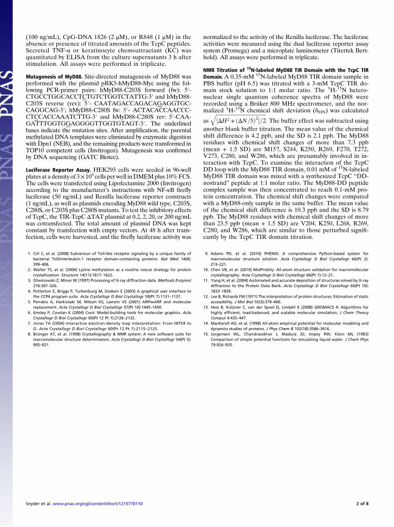
Fig. S1. Crystal structure of the MyD88 TIR domain. (A) 2Fo-Fc map (blue) calculated at 1.45-Å resolution and contoured at 1.2 sigma is shown for theMyD88 TIR domain BB loop. The significant structural differences among the MyD88 (cyan), TLR2 (green), and PdTLP (magenta) TIR domains are shown nearthe BB loop (B), CD loop (C), DD loop (D), DE loop (E), and EE loop (F). (G) Superposition of the crystal (cyan) and NMR (blue) structures of the MyD88 TIRdomains. The orientation is the same as that in Fig. 1A.
Snyder et al. www.pnas.org/cgi/content/short/1215770110 3 of 8
Box 1 Box 2
A AA A AB B BB B3
10
Human MyD88 159 E R F D A F I C Y C P S D I Q F V Q - E M I R Q L E Q T N Y R L K L C V S D R D V L P G - T C V W S I A S E L I E K R 215Mouse MyD88 159 E L F D A F I C Y C P N D I E F V Q - E M I R Q L E Q T D Y R L K L C V S D R D V L P G - T C V W S I A S E L I E K R 215Zebrafish MyD88 147 E T F D A F I C Y C Q S D I Q F V H - E M I K Q L E H T E Y N L K L C V F D R D V L P G - T C V W T I A S E L I E K R 203Human MAL 84 K D Y D V C V C H S E E D L V A A Q - D L V S Y L E G S T A S L R C F L Q L R D A T P G G A I V S E L C Q A L S S S H 141Mouse MAL 104 K D Y D V C V C H S E E D L E A A Q - E L V S Y L E G S Q A S L R C F L Q L R D A A P G G A I V S E L C Q A L S R S H 161Human TLR1 635 L Q F H A F I S Y S G H D S F W V K N E L L P N L E K E G - - M Q I C L H E R N F V P G K S I V E N I I T C I E K S Y 691Human TLR2 639 I C Y D A F V S Y S E R D A Y W V E N L M V Q E L E N F N P P F K L C L H K R D F I P G K W I I D N I I D S I E K S H 697Human TLR4 672 N I Y D A F V I Y S S Q D E D W V R N E L V K N L E E G V P P F Q L C L H Y R D F I P G V A I A A N I I H E G F H K S 730Human TLR10 632 V R F H A F I S Y S E H D S L W V K N E L I P N L E K E D G S I L I C L Y E S Y F D P G K S I S E N I V S F I E K S Y 690E. coli TcpC 169 T H Y D F F I S H A K E D K D T F V R P L V D E L N R L G - - V I I W Y D E Q T L E V G D S L R R N I D L G L R K A N 225Brucella TcpB 117 E E Y D F F I S H A S E D K E A F V Q D L V A A L R D L G - - A K I F Y D A Y T L K V G D S L R R K I D Q G L A N S K 173Parracoccus TIR 164 P P H D I F I S H A W E D K A D F V E A L A H T L R A A G - - A E V W Y D D F S L R P G D S L R R S I D K G L G S S R 220
BC C C C' CD D DD
Human MyD88 216 C R R M V V V V S D D Y L Q S K E C D F Q T K F A L S - L S P G A H Q K R L I P I K Y K A M K K E - - - - - - - F P S 266Mouse MyD88 216 C R R M V V V V S D D Y L Q S K E C D F Q T K F A L S - L S P G V Q Q K R L I P I K Y K A M K K D - - - - - - - F P S 266Zebrafish MyD88 204 C K R M V V V I S D D Y L D S D A C D F Q T K F A L S - L C P G A R T K R L I P V V Y K S M K R P - - - - - - - F P S 254Human MAL 142 C R - - V L L I T P G F L Q D P W C K Y Q M L Q A L T E A - P G A E G - C T I P L L S G L S R A A - - - - - - - Y P P 189Mouse MAL 162 C R - - A L L I T P G F L R D P W C K Y Q M L Q A L T E A - P A S E G - C T I P L L S G L S R A A - - - - - - - Y P P 209Human TLR1 692 K S - - I F V L S P N F V Q S E W C H Y E L Y F A H H N L F H E G S N S L I L I L L E P I P Q Y S I P S S Y H K L K S 748Human TLR2 698 K T - - V F V L S E N F V K S E W C K Y E L D F S H F R L F D E N N D A A I L I L L E P I E K K A I P Q R F C K L R K 754Human TLR4 731 R K - V I V V V S Q H F I Q S R W C I F E Y E I A Q T W Q F L S S R A G I I F I V L Q K V E K T L L R Q Q V E L Y - R 788Human TLR10 691 K S - - I F V L S P N F V Q N E W C H Y E F Y F A H H N L F H E N S D H I I L I L L E P I P F Y C I P T R Y H K L K A 747E. coli TcpC 226 Y G - - I V I L S H N F L N K K W T Q Y E L D S L I N R A - V Y D D N K I I L P I W H N I N A Q E V - - - - S K Y S H 277Brucella TcpB 174 F G - - I V V L S E H F F S K Q W P A R E L D G L T A - M E I G G Q T R - I L P I W H K V S Y D E V - - - - R R F S P 224Parracoccus TIR 221 F G - - I V V L S T H F F K K E W P Q K E L D G L F Q - L E S S G R S R - I L P I W H K V S K D E V - - - - A S F S P 271
D DE E EE Box 3 E
Human MyD88 267 I L R F I T V C D Y T N P C - T K S W F W T R L A K A L S L PMouse MyD88 267 I L R F I T I C D Y T N P C - T K S W F W T R L A K A L S L PZebrafish MyD88 255 I L R F L T I C D Y S K P C - T Q V W F W T R L A K A L S L PHuman MAL 190 E L R F M Y Y V D G R G P - - D G G F R Q V K E A V M R Y L Q T L SMouse MAL 210 E L R F M Y Y V D G R G K - - D G G F Y Q V K E A V I H Y L E T L SHuman TLR1 749 L M A R R T Y L E W P K E K S K R G L F W A N L R A A I N I K L T E Q A K KHuman TLR2 755 I M N T K T Y L E W P M D E A Q R E G F W V N L R A A I K SHuman TLR4 789 L L S R N T Y L E W E D S V L G R H I F W R R L R K A L L D G K S W N P E G THuman TLR10 748 L L E K K A Y L E W P K D R R K C G L F W A N L R A A I N V N V L A T R E M YE. coli TcpC 278 Y L A D K M A L Q T S L Y - - S V K E I A R E L A E I A Y R R RBrucella TcpB 225 S L A D K V A L N T S L K - - S V E E I A K E L H S L IParracoccus TIR 272 T M A D K L A F N T S T K - - S V D E I V A D L M A I I R D
296296
786826784
250299
786241
284221
307
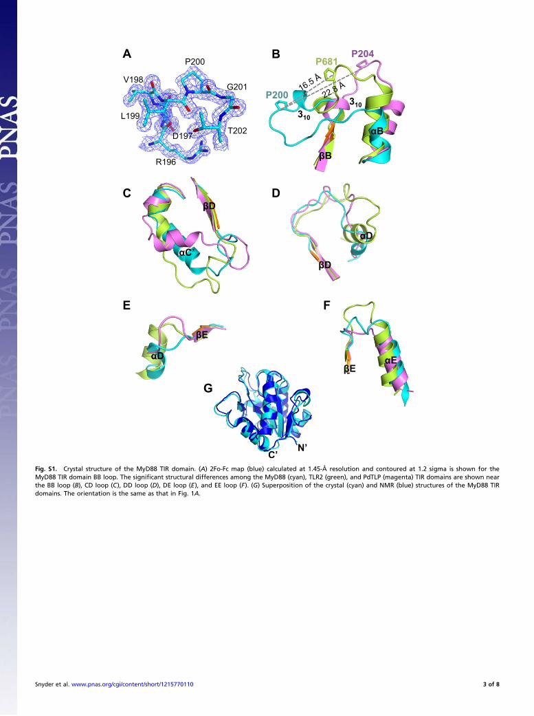
Fig. S2. Sequence alignment of selected TIR domains. Secondary structures of the MyD88 and Paracoccus PdTLP TIR domains are shown as arrows (β) andcylinders (α) above and below the sequences, respectively. The connecting loops and conserved boxes 1–3 are marked. The 310 or α-helices within the BB loopregions of the known structures are underlined. The functionally important residues identified in human or mouse are shaded in orange, and residues perturbedby TcpC titration in green. The Poc site residue is also marked with a red diamond. The conserved residues among all TIR domains are shaded in yellow, thoseconserved among MyD88 TIR domains are in blue boxes, among the receptor TIR domains in green boxes, among the microbial TIR domains in magenta boxes.
Snyder et al. www.pnas.org/cgi/content/short/1215770110 4 of 8
D D197 K256
R196
P200
C203
W205
N278
T277
T281
B
B
E
C280
K231
T277
S283
K282 K258
Y276 Y257 F174
E177 Q173
R180
Q176
K238
D234
L241 L243
H248
S266
F264
E263 R269
A E
D
E
C
B
A
310
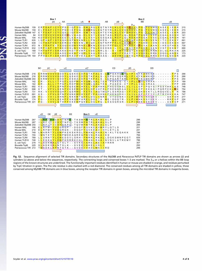
Fig. S3. MyD88 TIR domain crystal lattice interactions. (A) Overview of the two crystal lattice interfaces that bury significant amount of surface area. Theunique TIR domain in the crystal is colored cyan, and its symmetry mates are colored light blue. The five β-strands are colored orange to show the relativeorientations of the TIR domains. The detailed views of these interfaces are shown in B and C, with the hydrogen bonds indicated as gray dotted lines and watermolecules as red spheres.
WT Poc Mutant
Time (ps)
RM
SD
(nm
)
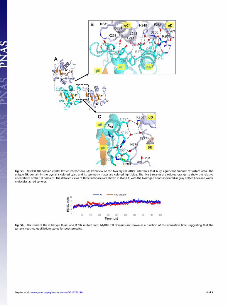
Fig. S4. The rmsd of the wild-type (blue) and I179N mutant (red) MyD88 TIR domains are shown as a function of the simulation time, suggesting that thesystems reached equilibrium states for both proteins.
Snyder et al. www.pnas.org/cgi/content/short/1215770110 5 of 8
A
C
B
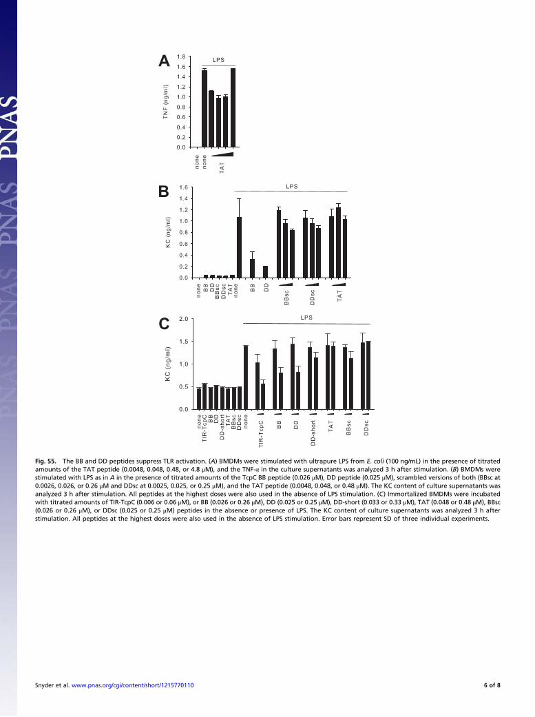
Fig. S5. The BB and DD peptides suppress TLR activation. (A) BMDMs were stimulated with ultrapure LPS from E. coli (100 ng/mL) in the presence of titratedamounts of the TAT peptide (0.0048, 0.048, 0.48, or 4.8 μM), and the TNF-α in the culture supernatants was analyzed 3 h after stimulation. (B) BMDMs werestimulated with LPS as in A in the presence of titrated amounts of the TcpC BB peptide (0.026 μM), DD peptide (0.025 μM), scrambled versions of both (BBsc at0.0026, 0.026, or 0.26 μM and DDsc at 0.0025, 0.025, or 0.25 μM), and the TAT peptide (0.0048, 0.048, or 0.48 μM). The KC content of culture supernatants wasanalyzed 3 h after stimulation. All peptides at the highest doses were also used in the absence of LPS stimulation. (C) Immortalized BMDMs were incubatedwith titrated amounts of TIR-TcpC (0.006 or 0.06 μM), or BB (0.026 or 0.26 μM), DD (0.025 or 0.25 μM), DD-short (0.033 or 0.33 μM), TAT (0.048 or 0.48 μM), BBsc(0.026 or 0.26 μM), or DDsc (0.025 or 0.25 μM) peptides in the absence or presence of LPS. The KC content of culture supernatants was analyzed 3 h afterstimulation. All peptides at the highest doses were also used in the absence of LPS stimulation. Error bars represent SD of three individual experiments.
Snyder et al. www.pnas.org/cgi/content/short/1215770110 6 of 8
A
B
160 180 200 220 240 260 280 296
Residue Number
0
0.01
0.02
0.03
0.04
0.05
H-
N C
hem
ical
sh
ift
chan
ge
(pp
m)
MyD88+DD peptide (1:1)
R269 C280
W286
L268
K250V204
115
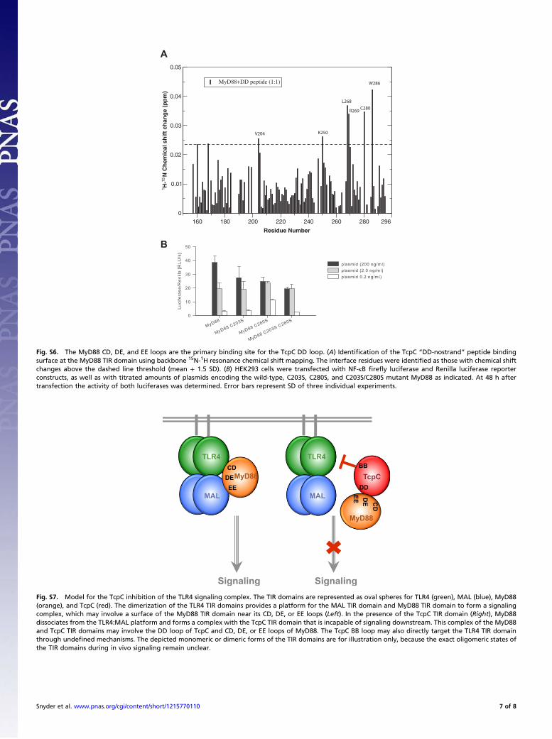
Fig. S6. The MyD88 CD, DE, and EE loops are the primary binding site for the TcpC DD loop. (A) Identification of the TcpC “DD-nostrand” peptide bindingsurface at the MyD88 TIR domain using backbone 15N-1H resonance chemical shift mapping. The interface residues were identified as those with chemical shiftchanges above the dashed line threshold (mean + 1.5 SD). (B) HEK293 cells were transfected with NF-κB firefly luciferase and Renilla luciferase reporterconstructs, as well as with titrated amounts of plasmids encoding the wild-type, C203S, C280S, and C203S/C280S mutant MyD88 as indicated. At 48 h aftertransfection the activity of both luciferases was determined. Error bars represent SD of three individual experiments.
CD
EE
BB
DD
CD
EE
MyD88
TLR4
MAL
MyD88
TLR4
MAL
TcpC
Signaling Signaling
DE
DE
Fig. S7. Model for the TcpC inhibition of the TLR4 signaling complex. The TIR domains are represented as oval spheres for TLR4 (green), MAL (blue), MyD88(orange), and TcpC (red). The dimerization of the TLR4 TIR domains provides a platform for the MAL TIR domain and MyD88 TIR domain to form a signalingcomplex, which may involve a surface of the MyD88 TIR domain near its CD, DE, or EE loops (Left). In the presence of the TcpC TIR domain (Right), MyD88dissociates from the TLR4:MAL platform and forms a complex with the TcpC TIR domain that is incapable of signaling downstream. This complex of the MyD88and TcpC TIR domains may involve the DD loop of TcpC and CD, DE, or EE loops of MyD88. The TcpC BB loop may also directly target the TLR4 TIR domainthrough undefined mechanisms. The depicted monomeric or dimeric forms of the TIR domains are for illustration only, because the exact oligomeric states ofthe TIR domains during in vivo signaling remain unclear.
Snyder et al. www.pnas.org/cgi/content/short/1215770110 7 of 8
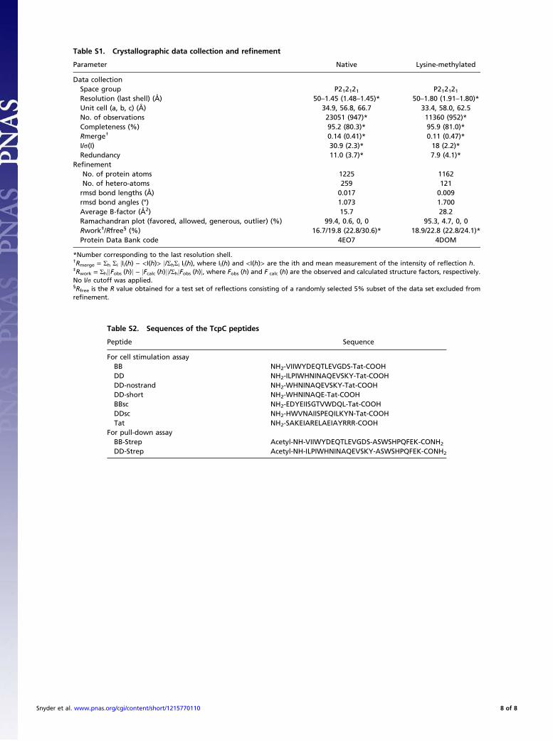
Table S1. Crystallographic data collection and refinement
Parameter Native Lysine-methylated
Data collectionSpace group P212121 P212121Resolution (last shell) (Å) 50–1.45 (1.48–1.45)* 50–1.80 (1.91–1.80)*Unit cell (a, b, c) (Å) 34.9, 56.8, 66.7 33.4, 58.0, 62.5No. of observations 23051 (947)* 11360 (952)*Completeness (%) 95.2 (80.3)* 95.9 (81.0)*Rmerge† 0.14 (0.41)* 0.11 (0.47)*I/σ(I) 30.9 (2.3)* 18 (2.2)*Redundancy 11.0 (3.7)* 7.9 (4.1)*
RefinementNo. of protein atoms 1225 1162No. of hetero-atoms 259 121rmsd bond lengths (Å) 0.017 0.009rmsd bond angles (°) 1.073 1.700Average B-factor (Å2) 15.7 28.2Ramachandran plot (favored, allowed, generous, outlier) (%) 99.4, 0.6, 0, 0 95.3, 4.7, 0, 0Rwork‡/Rfree§ (%) 16.7/19.8 (22.8/30.6)* 18.9/22.8 (22.8/24.1)*Protein Data Bank code 4EO7 4DOM
*Number corresponding to the last resolution shell.†Rmerge = Σh Σi jIi(h) − <I(h)> j/ΣhΣi Ii(h), where Ii(h) and <I(h)> are the ith and mean measurement of the intensity of reflection h.‡Rwork = ΣhjjFobs (h)j − jFcalc (h)jj/ΣhjFobs (h)j, where Fobs (h) and F calc (h) are the observed and calculated structure factors, respectively.No I/σ cutoff was applied.§Rfree is the R value obtained for a test set of reflections consisting of a randomly selected 5% subset of the data set excluded fromrefinement.
Table S2. Sequences of the TcpC peptides
Peptide Sequence
For cell stimulation assayBB NH2-VIIWYDEQTLEVGDS-Tat-COOHDD NH2-ILPIWHNINAQEVSKY-Tat-COOHDD-nostrand NH2-WHNINAQEVSKY-Tat-COOHDD-short NH2-WHNINAQE-Tat-COOHBBsc NH2-EDYEIISGTVWDQL-Tat-COOHDDsc NH2-HWVNAIISPEQILKYN-Tat-COOHTat NH2-SAKEIARELAEIAYRRR-COOH
For pull-down assayBB-Strep Acetyl-NH-VIIWYDEQTLEVGDS-ASWSHPQFEK-CONH2
DD-Strep Acetyl-NH-ILPIWHNINAQEVSKY-ASWSHPQFEK-CONH2
Snyder et al. www.pnas.org/cgi/content/short/1215770110 8 of 8