Supplementary Materials for -...
21
www.sciencemag.org/content/351/6269/180/suppl/DC1 Supplementary Materials for The structure of the β-barrel assembly machinery complex Jeremy Bakelar, Susan K. Buchanan, Nicholas Noinaj* *Corresponding author. E-mail: [email protected] Published 8 January 2016, Science 351, 180 (2016) DOI: 10.1126/science.aad3460 This PDF file includes Materials and Methods Figs. S1 to S6 Tables S1 to S6 References Movie legends S1 and S2 Other Supplementary Material for this manuscript includes the following: (available at www.sciencemag.org/content/351/6269/180/suppl/DC1) Movies S1 and S2 Model S1
-
Upload
trannguyet -
Category
Documents
-
view
217 -
download
1
Transcript of Supplementary Materials for -...
www.sciencemag.org/content/351/6269/180/suppl/DC1
Supplementary Materials for
The structure of the β-barrel assembly machinery complex
Jeremy Bakelar, Susan K. Buchanan, Nicholas Noinaj*
*Corresponding author. E-mail: [email protected]
Published 8 January 2016, Science 351, 180 (2016) DOI: 10.1126/science.aad3460
This PDF file includes
Materials and Methods Figs. S1 to S6 Tables S1 to S6 References Movie legends S1 and S2
Other Supplementary Material for this manuscript includes the following: (available at www.sciencemag.org/content/351/6269/180/suppl/DC1)
Movies S1 and S2 Model S1
Materials and Methods:
Expression of recombinant BamABCDE complex
A single plasmid (pJH114) containing all five Bam proteins (BamA, B, C, D, and E) was obtained from Harris Bernstein and used for expression and purification (11). The plasmid was transformed into BL21(DE3) cells (NEB), plated onto LB-carbenicillin agar plates (Teknova) and incubated overnight at 37°C. A single colony was used to inoculate a 5-mL LB-ampicillin culture and incubated overnight at 37°C. The overnight culture was then used to inoculate a 50 mL starter culture of LB-ampicillin which was allowed to grow to saturation. The cells were then centrifuged, washed three times with 1x PBS and then re-suspended in 12 mL 1x PBS. The resuspended cells (1 mL) were then added to twelve 2 L baffled flasks containing 1 L of 2xYT medium supplemented with ampicillin (50 μg/mL). These cultures were incubated at 37°C with shaking at 180 rpm, grown to an OD600 between 0.8-1.0, and then induced with 0.5 mM IPTG. Cultures were grown an additional 4 hours at 37°C before harvesting. Cell were either used immediately or flash frozen and stored at -20°C.
Purification and crystallization
Cells were resuspended in lysis buffer (1x PBS supplemented with DNase I (10 μg/ml) and PMSF (500 M)) and lysed with three passes through an Emusiflex C-3 high pressure homogenizer (Avestin) at 18,000 psi. The cell lysate was then centrifuged at 6000 x g for 10 min at 4°C to remove cell debris, and the resulting supernatant was centrifuged at 200,000 x g for 90 min at 4°C to isolate cell membranes. The membranes were then re-suspended in solubilization buffer (1x PBS, 1% DDM, and 37 mM imidazole) using a dounce homogenizer and stirred at medium speed overnight at 4°C. The solubilized sample was then centrifuged again at 200,000 x g for 60 min at 4°C and the supernatant collected.
Solubilized BamABCDE complex was purified by affinity chromatography using a 5 mL HiTrap Nickel column (Qiagen) and an ÄKTA Pure system (GE Healthcare). The column was equilibrated with Buffer A (1x PBS, 0.03% DDM, and 37 mM imidazole) and the sample automatically loaded using the sample pump with an in-line air sensor. Protein was eluted with a linear gradient of 37-500 mM imidazole using Buffer A and Buffer B (1x PBS, 0.03% DDM, and 1 M imidazole). Fractions containing BamABCDE were pooled, concentrated to ~2 mg/mL, and passed through a 16/60 Sephacryl S-300 HR column (GE Healthcare) at a flow rate of 0.5 mL/min using 25 mM Tris-HCl, pH 7.5, 150 mM NaCl, and 0.6% C8E4. All five Bam proteins (BamA, B, C, D, and E) eluted from the gel filtration column as a single monodisperse peak as verified by SDS-PAGE analysis. Fractions containing BamABCDE were pooled and concentrated to ~12 mg/mL.
Broad crystallization screening was performed using hanging drop method on a Mosquito LCP crystallization robot (TTP Labtech) with commercially available crystallization screens. An initial hit was improved by additive screening using the AdditiveHT screen (Hampton Research) with final crystals grown at 22°C in 100 mM Tris-HCl, pH 8.5, 200 mM MgCl2, 10 mM MnCl2, and 8% PEG 4000.
Data collection, structure determination, and modeling
Crystals were harvested by quick transfer directly into a cryoprotectant solution containing 20% glycerol and flash-cooled in liquid nitrogen. Diffraction data were collected to 3.4 Å resolution at the SER-CAT beamline (ID22) at the Advanced Photon Source at Argonne National Laboratory and processed using HKL2000 (35). The structure was solved by molecular replacement using Phaser (36) within PHENIX (37) using previously reported crystal structures of the Bam components. Search order was key here for success, first starting with the BamCD complex (PDB ID 3TGO) followed by the barrel domain of BamA (PDB ID 4C4V), POTRA5 and then POTRA4 of BamA (PDB ID 3Q6B). BamE (PDB ID 2KM7) and POTRA1 (PDB ID 3EFC) were then placed based on density within a difference (Fo-Fc) map. After several rounds of building and refinement, POTRA2 and 3 (PDB ID 3EFC) were then manually placed in weak density followed by rigid body refinement for all components for final placement. The structure was refined to R/Rfree values of 0.22/0.27. All model building and refinement were performed using COOT (38) and PHENIX (37), respectively. Final placement of side chains was based on evaluation of 2Fo-Fc, Fo-Fc, and feature-enhanced (FEM) density maps (39). RMSD analysis was performed within PyMOL (Schrödinger) for C- atoms using default settings. Surprisingly, BamB was not found within our crystal structure despite it being present in our purification. The absence of BamB was confirmed by analyzing crystals and our initial sample of the complex by SDS-PAGE analysis, which also were lacking BamB presumably due to proteolysis during storage/incubation.
To see how BamB interacts with the BamACDE complex, we modeled BamB into our complex using the previous reported BamAB crystal structure (PDB ID 4PK1) to produce the modeled structure of the fully assembled BamABCDE complex. Here, we were able to place BamB in our structure by performing a superposition of the two structures along POTRA3 of BamA. Analysis of interacting interfaces was performed using the PDBePISA (40). All figures were made with PyMOL (Schrödinger) and annotated and finalized with Adobe Illustrator.
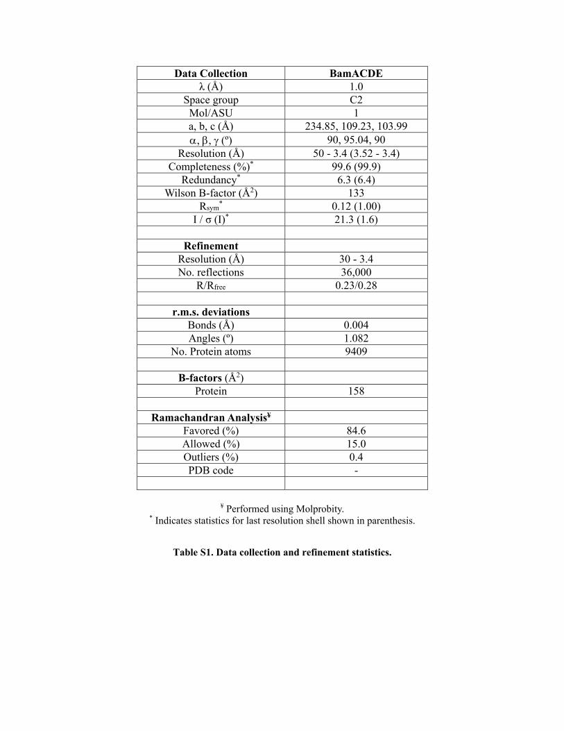
Data Collection BamACDE λ (Å) 1.0
Space group C2 Mol/ASU 1 a, b, c (Å) 234.85, 109.23, 103.99 , , (º) 90, 95.04, 90
Resolution (Å) 50 - 3.4 (3.52 - 3.4) Completeness (%)* 99.6 (99.9)
Redundancy* 6.3 (6.4) Wilson B-factor (Å2) 133
Rsym* 0.12 (1.00)
I / σ (I)* 21.3 (1.6)
Refinement Resolution (Å) 30 - 3.4 No. reflections 36,000
R/Rfree 0.23/0.28
r.m.s. deviations Bonds (Å) 0.004 Angles (º) 1.082
No. Protein atoms 9409
B-factors (Å2) Protein 158
Ramachandran Analysis¥
Favored (%) 84.6 Allowed (%) 15.0 Outliers (%) 0.4 PDB code -
¥ Performed using Molprobity. * Indicates statistics for last resolution shell shown in parenthesis.
Table S1. Data collection and refinement statistics.
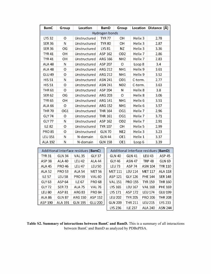
Table S2. Summary of interactions between BamC and BamD. This is a summary of all interactions between BamC and BamD as analyzed by PDBePISA.
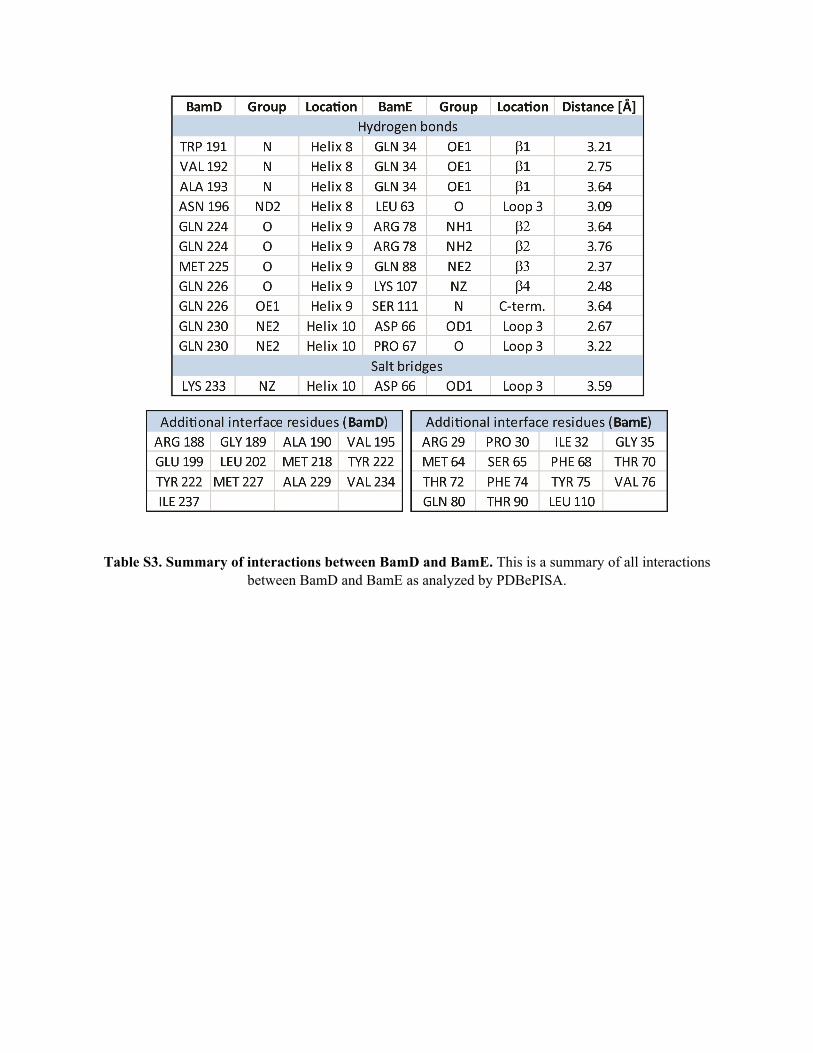
Table S3. Summary of interactions between BamD and BamE. This is a summary of all interactions between BamD and BamE as analyzed by PDBePISA.
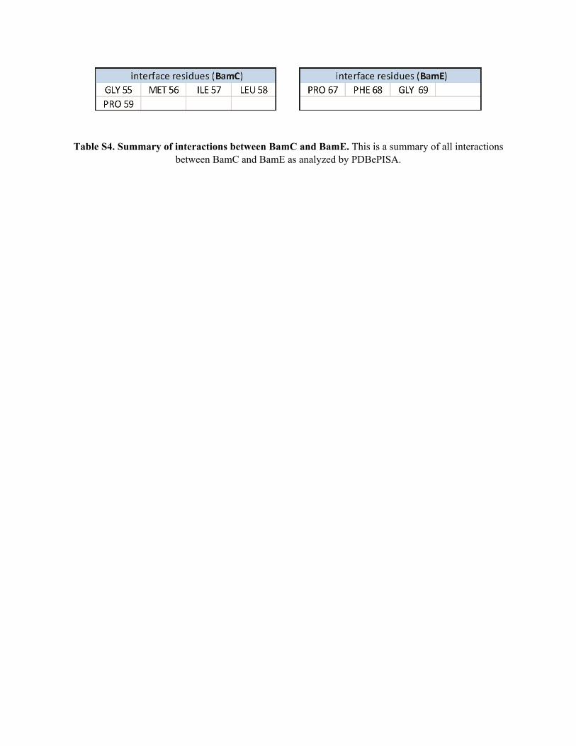
Table S4. Summary of interactions between BamC and BamE. This is a summary of all interactions between BamC and BamE as analyzed by PDBePISA.
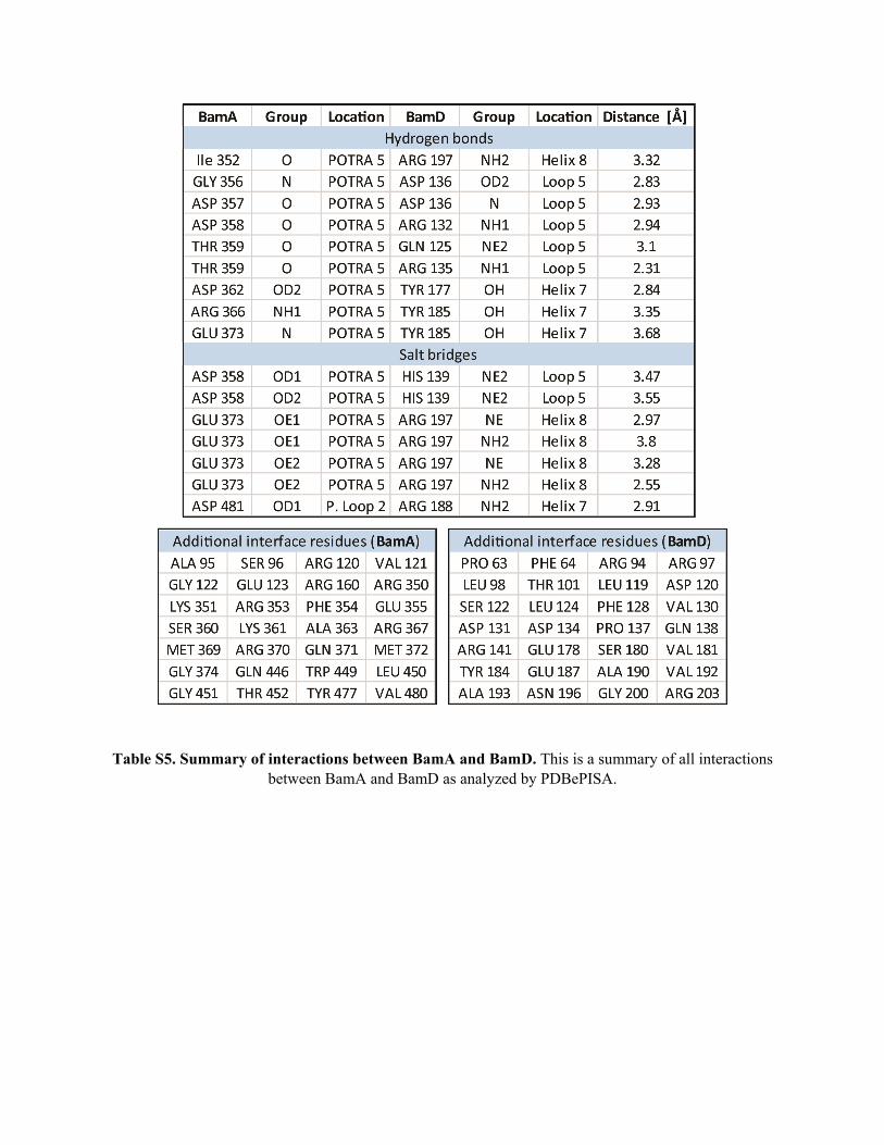
Table S5. Summary of interactions between BamA and BamD. This is a summary of all interactions between BamA and BamD as analyzed by PDBePISA.
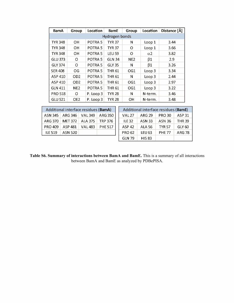
Table S6. Summary of interactions between BamA and BamE. This is a summary of all interactions between BamA and BamE as analyzed by PDBePISA.
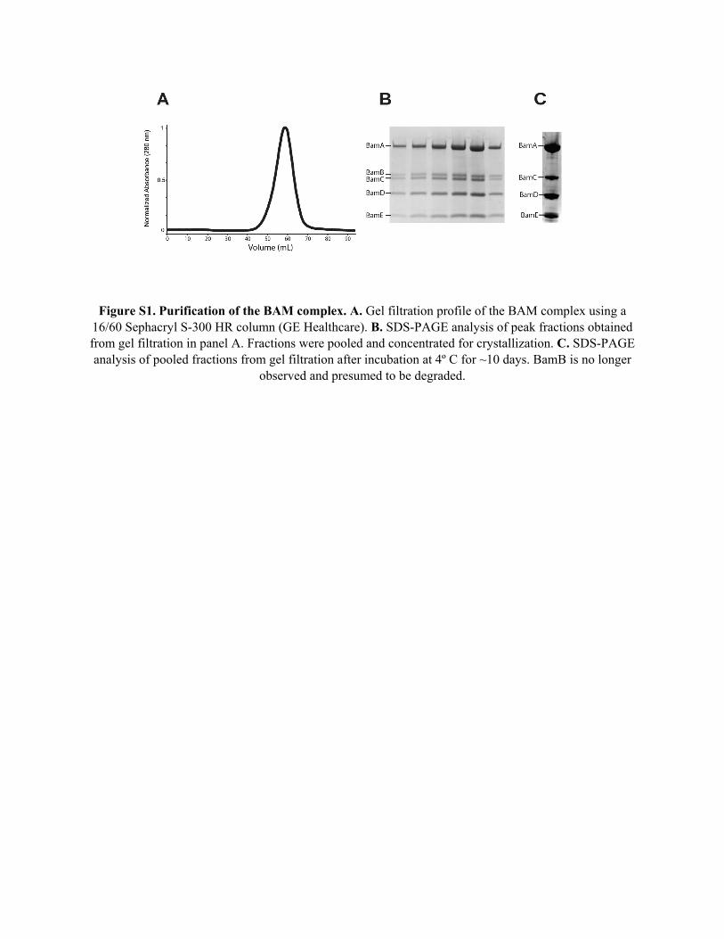
Figure S1. Purification of the BAM complex. A. Gel filtration profile of the BAM complex using a 16/60 Sephacryl S-300 HR column (GE Healthcare). B. SDS-PAGE analysis of peak fractions obtained from gel filtration in panel A. Fractions were pooled and concentrated for crystallization. C. SDS-PAGE analysis of pooled fractions from gel filtration after incubation at 4º C for ~10 days. BamB is no longer
observed and presumed to be degraded.
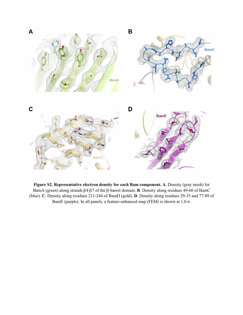
Figure S2. Representative electron density for each Bam component. A. Density (gray mesh) for BamA (green) along strands 4-7 of the -barrel domain. B. Density along residues 49-60 of BamC
(blue). C. Density along residues 211-244 of BamD (gold). D. Density along residues 29-35 and 77-89 of BamE (purple). In all panels, a feature-enhanced map (FEM) is shown at 1.0 .
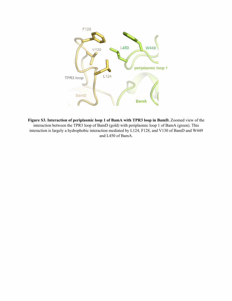
Figure S3. Interaction of periplasmic loop 1 of BamA with TPR3 loop in BamD. Zoomed view of the interaction between the TPR3 loop of BamD (gold) with periplasmic loop 1 of BamA (green). This
interaction is largely a hydrophobic interaction mediated by L124, F128, and V130 of BamD and W449 and L450 of BamA.
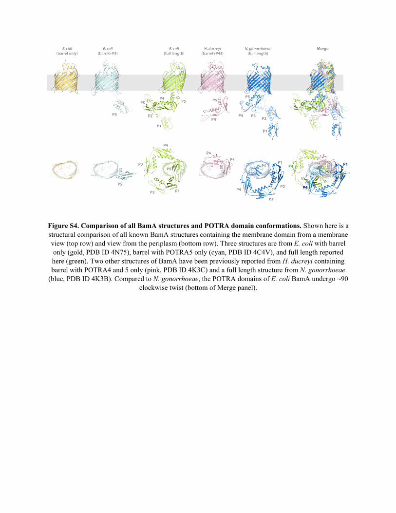
Figure S4. Comparison of all BamA structures and POTRA domain conformations. Shown here is a structural comparison of all known BamA structures containing the membrane domain from a membrane view (top row) and view from the periplasm (bottom row). Three structures are from E. coli with barrel only (gold, PDB ID 4N75), barrel with POTRA5 only (cyan, PDB ID 4C4V), and full length reported here (green). Two other structures of BamA have been previously reported from H. ducreyi containing barrel with POTRA4 and 5 only (pink, PDB ID 4K3C) and a full length structure from N. gonorrhoeae
(blue, PDB ID 4K3B). Compared to N. gonorrhoeae, the POTRA domains of E. coli BamA undergo ~90 clockwise twist (bottom of Merge panel).
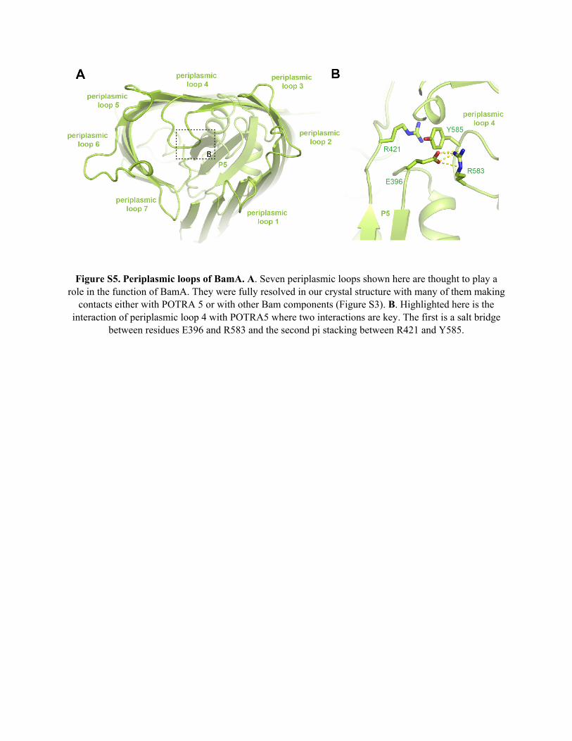
Figure S5. Periplasmic loops of BamA. A. Seven periplasmic loops shown here are thought to play a role in the function of BamA. They were fully resolved in our crystal structure with many of them making
contacts either with POTRA 5 or with other Bam components (Figure S3). B. Highlighted here is the interaction of periplasmic loop 4 with POTRA5 where two interactions are key. The first is a salt bridge
between residues E396 and R583 and the second pi stacking between R421 and Y585.
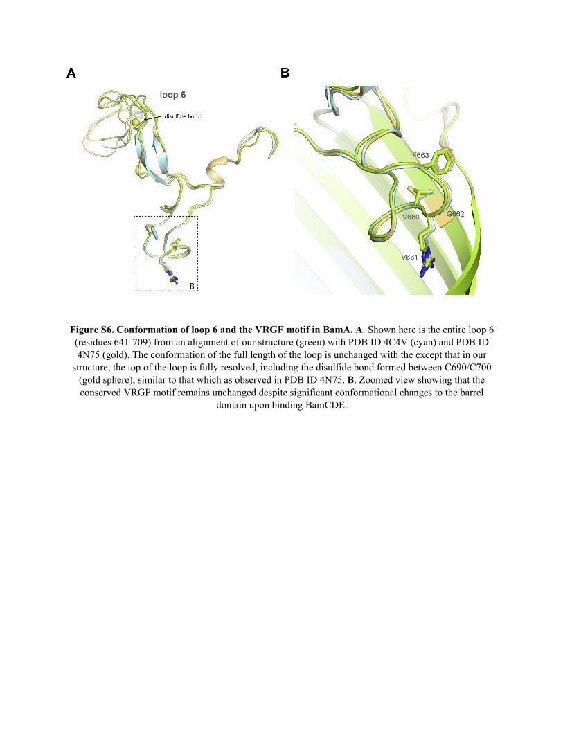
Figure S6. Conformation of loop 6 and the VRGF motif in BamA. A. Shown here is the entire loop 6 (residues 641-709) from an alignment of our structure (green) with PDB ID 4C4V (cyan) and PDB ID 4N75 (gold). The conformation of the full length of the loop is unchanged with the except that in our
structure, the top of the loop is fully resolved, including the disulfide bond formed between C690/C700 (gold sphere), similar to that which as observed in PDB ID 4N75. B. Zoomed view showing that the conserved VRGF motif remains unchanged despite significant conformational changes to the barrel
domain upon binding BamCDE.
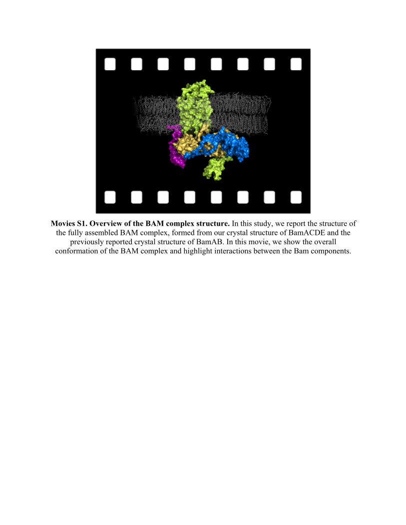
Movies S1. Overview of the BAM complex structure. In this study, we report the structure of the fully assembled BAM complex, formed from our crystal structure of BamACDE and the
previously reported crystal structure of BamAB. In this movie, we show the overall conformation of the BAM complex and highlight interactions between the Bam components.
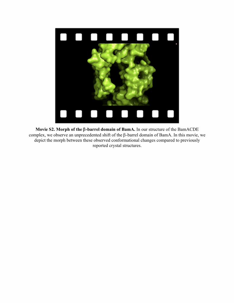
Movie S2. Morph of the -barrel domain of BamA. In our structure of the BamACDE complex, we observe an unprecedented shift of the -barrel domain of BamA. In this movie, we
depict the morph between these observed conformational changes compared to previously reported crystal structures.
REFERENCES AND NOTES 1. C. T. Webb, E. Heinz, T. Lithgow, Evolution of the β-barrel assembly machinery. Trends
Microbiol. 20, 612–620 (2012). Medline doi:10.1016/j.tim.2012.08.006
2. E. Schleiff, J. Soll, Membrane protein insertion: Mixing eukaryotic and prokaryotic concepts. EMBO Rep. 6, 1023–1027 (2005). Medline doi:10.1038/sj.embor.7400563
3. T. J. Knowles, A. Scott-Tucker, M. Overduin, I. R. Henderson, Membrane protein architects: The role of the BAM complex in outer membrane protein assembly. Nat. Rev. Microbiol. 7, 206–214 (2009). Medline doi:10.1038/nrmicro2069
4. C. L. Hagan, T. J. Silhavy, D. Kahne, β-Barrel membrane protein assembly by the Bam complex. Annu. Rev. Biochem. 80, 189–210 (2011). Medline doi:10.1146/annurev-biochem-061408-144611
5. R. Voulhoux, M. P. Bos, J. Geurtsen, M. Mols, J. Tommassen, Role of a highly conserved bacterial protein in outer membrane protein assembly. Science 299, 262–265 (2003). Medline doi:10.1126/science.1078973
6. N. Noinaj, S. E. Rollauer, S. K. Buchanan, The β-barrel membrane protein insertase machinery from Gram-negative bacteria. Curr. Opin. Struct. Biol. 31, 35–42 (2015). Medline doi:10.1016/j.sbi.2015.02.012
7. K. H. Kim, S. Aulakh, M. Paetzel, The bacterial outer membrane β-barrel assembly machinery. Protein Sci. 21, 751–768 (2012). Medline doi:10.1002/pro.2069
8. D. P. Ricci, T. J. Silhavy, The Bam machine: A molecular cooper. Biochim. Biophys. Acta 1818, 1067–1084 (2012). Medline doi:10.1016/j.bbamem.2011.08.020
9. J. C. Malinverni, J. Werner, S. Kim, J. G. Sklar, D. Kahne, R. Misra, T. J. Silhavy, YfiO stabilizes the YaeT complex and is essential for outer membrane protein assembly in Escherichia coli. Mol. Microbiol. 61, 151–164 (2006). Medline doi:10.1111/j.1365-2958.2006.05211.x
10. C. L. Hagan, S. Kim, D. Kahne, Reconstitution of Outer Membrane Protein Assembly from Purified Components, Reconstitution of outer membrane protein assembly from purified components. Science 328, 890–892 (2010). Medline doi:10.1126/science.1188919
11. G. Roman-Hernandez, J. H. Peterson, H. D. Bernstein, Reconstitution of bacterial autotransporter assembly using purified components. eLife 3, e04234 (2014). Medline doi:10.7554/eLife.04234
12. J. G. Sklar, T. Wu, L. S. Gronenberg, J. C. Malinverni, D. Kahne, T. J. Silhavy, Lipoprotein SmpA is a component of the YaeT complex that assembles outer membrane proteins in Escherichia coli. Proc. Natl. Acad. Sci. U.S.A. 104, 6400–6405 (2007). Medline doi:10.1073/pnas.0701579104
13. K. H. Kim, S. Aulakh, M. Paetzel, Crystal structure of β-barrel assembly machinery BamCD protein complex. J. Biol. Chem. 286, 39116–39121 (2011). Medline doi:10.1074/jbc.M111.298166
14. K. B. Jansen, S. L. Baker, M. C. Sousa, Crystal structure of BamB bound to a periplasmic domain fragment of BamA, the central component of the β-barrel assembly machine. J. Biol. Chem. 290, 2126–2136 (2015). Medline doi:10.1074/jbc.M114.584524
15. R. Albrecht, K. Zeth, Structural basis of outer membrane protein biogenesis in bacteria. J. Biol. Chem. 286, 27792–27803 (2011). Medline doi:10.1074/jbc.M111.238931
16. R. Albrecht, M. Schütz, P. Oberhettinger, M. Faulstich, I. Bermejo, T. Rudel, K. Diederichs, K. Zeth, Structure of BamA, an essential factor in outer membrane protein biogenesis. Acta Crystallogr. D Biol. Crystallogr. 70, 1779–1789 (2014). Medline doi:10.1107/S1399004714007482
17. N. Noinaj, J. W. Fairman, S. K. Buchanan, The crystal structure of BamB suggests interactions with BamA and its role within the BAM complex. J. Mol. Biol. 407, 248–260 (2011). Medline doi:10.1016/j.jmb.2011.01.042
18. N. Noinaj, A. J. Kuszak, J. C. Gumbart, P. Lukacik, H. Chang, N. C. Easley, T. Lithgow, S. K. Buchanan, Structural insight into the biogenesis of β-barrel membrane proteins. Nature 501, 385–390 (2013). Medline doi:10.1038/nature12521
19. A. Heuck, A. Schleiffer, T. Clausen, Augmenting β-augmentation: Structural basis of how BamB binds BamA and may support folding of outer membrane proteins. J. Mol. Biol. 406, 659–666 (2011). Medline doi:10.1016/j.jmb.2011.01.002
20. K. H. Kim, M. Paetzel, Crystal structure of Escherichia coli BamB, a lipoprotein component of the β-barrel assembly machinery complex. J. Mol. Biol. 406, 667–678 (2011). Medline doi:10.1016/j.jmb.2010.12.020
21. C. Dong, H. F. Hou, X. Yang, Y. Q. Shen, Y. H. Dong, Structure of Escherichia coli BamD and its functional implications in outer membrane protein assembly. Acta Crystallogr. D Biol. Crystallogr. 68, 95–101 (2012). Medline doi:10.1107/S0907444911051031
22. D. Ni, Y. Wang, X. Yang, H. Zhou, X. Hou, B. Cao, Z. Lu, X. Zhao, K. Yang, Y. Huang, Structural and functional analysis of the β-barrel domain of BamA from Escherichia coli. FASEB J. 28, 2677–2685 (2014). Medline doi:10.1096/fj.13-248450
23. T. J. Knowles, D. F. Browning, M. Jeeves, R. Maderbocus, S. Rajesh, P. Sridhar, E. Manoli, D. Emery, U. Sommer, A. Spencer, D. L. Leyton, D. Squire, R. R. Chaudhuri, M. R. Viant, A. F. Cunningham, I. R. Henderson, M. Overduin, Structure and function of BamE within the outer membrane and the β-barrel assembly machine. EMBO Rep. 12, 123–128 (2011). Medline doi:10.1038/embor.2010.202
24. N. Noinaj, A. J. Kuszak, C. Balusek, J. C. Gumbart, S. K. Buchanan, Lateral opening and exit pore formation are required for BamA function. Structure 22, 1055–1062 (2014). Medline doi:10.1016/j.str.2014.05.008
25. K. B. Jansen, S. L. Baker, M. C. Sousa, Crystal structure of BamB from Pseudomonas aeruginosa and functional evaluation of its conserved structural features. PLOS ONE 7, e49749 (2012). Medline doi:10.1371/journal.pone.0049749
26. C. T. Webb, J. Selkrig, A. J. Perry, N. Noinaj, S. K. Buchanan, T. Lithgow, Dynamic association of BAM complex modules includes surface exposure of the lipoprotein BamC. J. Mol. Biol. 422, 545–555 (2012). Medline doi:10.1016/j.jmb.2012.05.035
27. C. M. Sandoval, S. L. Baker, K. Jansen, S. I. Metzner, M. C. Sousa, Crystal structure of BamD: An essential component of the β-Barrel assembly machinery of gram-negative bacteria. J. Mol. Biol. 409, 348–357 (2011). Medline doi:10.1016/j.jmb.2011.03.035
28. S. Kim, J. C. Malinverni, P. Sliz, T. J. Silhavy, S. C. Harrison, D. Kahne, Structure and function of an essential component of the outer membrane protein assembly machine. Science 317, 961–964 (2007). Medline doi:10.1126/science.1143993
29. D. P. Ricci, C. L. Hagan, D. Kahne, T. J. Silhavy, Activation of the Escherichia coli β-barrel assembly machine (Bam) is required for essential components to interact properly with substrate. Proc. Natl. Acad. Sci. U.S.A. 109, 3487–3491 (2012). Medline doi:10.1073/pnas.1201362109
30. N. W. Rigel, D. P. Ricci, T. J. Silhavy, Conformation-specific labeling of BamA and suppressor analysis suggest a cyclic mechanism for β-barrel assembly in Escherichia coli. Proc. Natl. Acad. Sci. U.S.A. 110, 5151–5156 (2013). Medline doi:10.1073/pnas.1302662110
31. P. Z. Gatzeva-Topalova, T. A. Walton, M. C. Sousa, Crystal structure of YaeT: Conformational flexibility and substrate recognition. Structure 16, 1873–1881 (2008). Medline doi:10.1016/j.str.2008.09.014
32. P. Z. Gatzeva-Topalova, L. R. Warner, A. Pardi, M. C. Sousa, Structure and flexibility of the complete periplasmic domain of BamA: The protein insertion machine of the outer membrane. Structure 18, 1492–1501 (2010). Medline doi:10.1016/j.str.2010.08.012
33. P. K. O’Neil, S. E. Rollauer, N. Noinaj, S. K. Buchanan, Fitting the Pieces of the β-Barrel Assembly Machinery Complex. Biochemistry 54, 6303–6311 (2015). Medline doi:10.1021/acs.biochem.5b00852
34. D. Gessmann, Y. H. Chung, E. J. Danoff, A. M. Plummer, C. W. Sandlin, N. R. Zaccai, K. G. Fleming, Outer membrane β-barrel protein folding is physically controlled by periplasmic lipid head groups and BamA. Proc. Natl. Acad. Sci. U.S.A. 111, 5878–5883 (2014). Medline doi:10.1073/pnas.1322473111
35. Z. Otwinowski, W. Minor, in Methods Enzymol. (Academic Press, New York, 1997), vol. 276, pp. 307-326.
36. A. J. McCoy, R. W. Grosse-Kunstleve, P. D. Adams, M. D. Winn, L. C. Storoni, R. J. Read, Phaser crystallographic software. J. Appl. Crystallogr. 40, 658–674 (2007). Medline
37. P. D. Adams, P. V. Afonine, G. Bunkóczi, V. B. Chen, I. W. Davis, N. Echols, J. J. Headd, L. W. Hung, G. J. Kapral, R. W. Grosse-Kunstleve, A. J. McCoy, N. W. Moriarty, R. Oeffner, R. J. Read, D. C. Richardson, J. S. Richardson, T. C. Terwilliger, P. H. Zwart, PHENIX: A comprehensive Python-based system for macromolecular structure solution. Acta Crystallogr. D Biol. Crystallogr. 66, 213–221 (2010). Medline doi:10.1107/S0907444909052925
38. P. Emsley, B. Lohkamp, W. G. Scott, K. Cowtan, Features and development of Coot. Acta Crystallogr. D Biol. Crystallogr. 66, 486–501 (2010). Medline doi:10.1107/S0907444910007493
39. P. V. Afonine, N. W. Moriarty, M. Mustyakimov, O. V. Sobolev, T. C. Terwilliger, D. Turk, A. Urzhumtsev, P. D. Adams, FEM: Feature-enhanced map. Acta Crystallogr. D Biol. Crystallogr. 71, 646–666 (2015). Medline doi:10.1107/S1399004714028132
40. E. Krissinel, K. Henrick, Inference of macromolecular assemblies from crystalline state. J. Mol. Biol. 372, 774–797 (2007). Medline doi:10.1016/j.jmb.2007.05.022
![Supplementary Materials - Royal Society of Chemistry · Supplementary Materials Imidazo[1,5-a]pyridin-3-ylidenes as π-Accepting Carbene Ligands: Substituent Effects on Properties](https://static.fdocument.org/doc/165x107/5ec0ffb8f8271e7b336e6711/supplementary-materials-royal-society-of-supplementary-materials-imidazo15-apyridin-3-ylidenes.jpg)