Studies on the genetics of biotin-transducing, defective variants of bacteriophage λ
12
VIROLOGY 36, 30-41 (1968) Studies on the Genetics of Biotin-Transducing, Defective Variants of Bacteriophage A’ GARY KAYAJANIAN2 Department of Biology, University of Rochester, Rochester, New York l&%7, and Department of Human Genetics, University of Michigan, Ann Arbor, Michigan .@lOJ Accepted May 23, 1968 A collection of Xbio transducing particles were isolated and the phage and bac- terial extent of their genomes characterized. Xbio transducing phages transduce vari- able amounts of bacterial genome, and have densities that differ from one independent HFT to another. Some of the transducing particles are able to form plaques (kpb), others are not (xdb) ; the Xdbs have phage defects of various extents. The nature of the xdb particles conforms t.o the structural predictions of the Campbell model. Some of the properties of Xbio particles and (hdb) lysogens are described and discussed. INTRODUCTION Phage X acts as a transducing agent for host markers located very close to its attach- ment site: for a cluster of galactose genes (Morse et al., 1956a, b; Campbell, 1959), an aromatic marker (Wallace and Pittard, 1967), and a series of loci responsible for the synthesis of biotin (Rothman, 1965; de1 Campillo-Campbell et al., 1967). We undertook to isolate and character- ize a number of biotin transducing variants of X for a number of reasons. First, to test the validity of the Campbell model, which delimits in a very precise manner the ge- netic and physical quality of the genomes of tra.nsducing X variants. Some of these pre- dictions are explicit: namely, the DNA in transducing particles should represent a contiguous block of genetic and physical material drawn from a lysogenic bacterial chromosome. Implicit in the model is the requirement of the vegetative ends in all genomes able to go through a lysogenic cycle. Second, one might expect not to find ’ Supported by United States Public Health Service Grants E-2862 and PO 1 GM15419-01. 2 Present address: Department of Biochemis- try, Stanford University School of Medicine, Palo Alto, California 94304. certain classes of transducing genomes that could be formed & la Campbell but would be unable to survive as single defective lyso- gens for reasons of physiology. Genetic material in the arm of X that might be de- leted in defective biotin transducing genomes would be concerned with functions of X occurring early in the cellular development of phage X. Xc1 genomes are unable to es- tablish a stable relation with their host, but would hdb genomes missing cI and (according to Campbell) other early regions of X be able to form a stable lysogenic com- plex with its host? Third, we were interested in ordering a large number of biotin mutants of Escherichia coli (de1 Campillo-Campbell et al., 1967) and a large number of X mutants for which Campbellian Xbio genomes would be valuable. MATERIALS AND METHODS Media Synthetic medium with 1% glucose, 0.4 % 2,3,5-triphenyltetrazolium chloride, and supplements of leucine and thiamine served as our biotin transduction medium (Kaya- janian and Campbell, 1966). Cells not re- quiring exogenous biotin for growth (bio+) grow to form dark red colonies on a white 30
Transcript of Studies on the genetics of biotin-transducing, defective variants of bacteriophage λ
VIROLOGY 36, 30-41 (1968)
Studies on the Genetics of Biotin-Transducing, Defective
Variants of Bacteriophage A’
GARY KAYAJANIAN2
Department of Biology, University of Rochester, Rochester, New York l&%7, and Department of Human Genetics, University of Michigan, Ann Arbor, Michigan .@lOJ
Accepted May 23, 1968
A collection of Xbio transducing particles were isolated and the phage and bac- terial extent of their genomes characterized. Xbio transducing phages transduce vari- able amounts of bacterial genome, and have densities that differ from one independent HFT to another. Some of the transducing particles are able to form plaques (kpb), others are not (xdb) ; the Xdbs have phage defects of various extents. The nature of the xdb particles conforms t.o the structural predictions of the Campbell model. Some of the properties of Xbio particles and (hdb) lysogens are described and discussed.
INTRODUCTION
Phage X acts as a transducing agent for host markers located very close to its attach- ment site: for a cluster of galactose genes (Morse et al., 1956a, b; Campbell, 1959), an aromatic marker (Wallace and Pittard, 1967), and a series of loci responsible for the synthesis of biotin (Rothman, 1965; de1 Campillo-Campbell et al., 1967).
We undertook to isolate and character- ize a number of biotin transducing variants of X for a number of reasons. First, to test the validity of the Campbell model, which delimits in a very precise manner the ge- netic and physical quality of the genomes of tra.nsducing X variants. Some of these pre- dictions are explicit: namely, the DNA in transducing particles should represent a contiguous block of genetic and physical material drawn from a lysogenic bacterial chromosome. Implicit in the model is the requirement of the vegetative ends in all genomes able to go through a lysogenic cycle. Second, one might expect not to find
’ Supported by United States Public Health Service Grants E-2862 and PO 1 GM15419-01.
2 Present address: Department of Biochemis- try, Stanford University School of Medicine, Palo Alto, California 94304.
certain classes of transducing genomes that could be formed & la Campbell but would be unable to survive as single defective lyso- gens for reasons of physiology. Genetic material in the arm of X that might be de- leted in defective biotin transducing genomes would be concerned with functions of X occurring early in the cellular development of phage X. Xc1 genomes are unable to es- tablish a stable relation with their host, but would hdb genomes missing cI and (according to Campbell) other early regions of X be able to form a stable lysogenic com- plex with its host? Third, we were interested in ordering a large number of biotin mutants of Escherichia coli (de1 Campillo-Campbell et al., 1967) and a large number of X mutants for which Campbellian Xbio genomes would be valuable.
MATERIALS AND METHODS
Media
Synthetic medium with 1% glucose, 0.4 % 2,3,5-triphenyltetrazolium chloride, and supplements of leucine and thiamine served as our biotin transduction medium (Kaya- janian and Campbell, 1966). Cells not re- quiring exogenous biotin for growth (bio+) grow to form dark red colonies on a white
30
31 GEXETICS OF xdbs
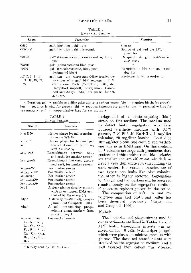
TABLE 1 BACTERIAL STRAINS
Strain ____-
C600 C600 (A)
W3330
W3805 W602
AC 2, 3, 4, 12, 17, 18, 19, 23, 2-l
Properties” F2l?UtiO?t
gal+, bio+, leu-, thi-, pm+ A assay gal+, bio+, leu-, thi-, lysogenic Source of gal and bio LFT
particles gal- (kinaseless and transferaseless) bio+, Recipient in gal transduction
pm- sus+ assa) gal- (epimeraseless) bio+, pm- “
gal- (transferaseless), bio-, pm-, Recipient in bio and gal trans- designated bio-0 duct ion
gal+, pm-, bio- nitrosoguanidine treated de- Recipient in bio transdltctioll rivatives of a gal+ bio+ segregant of E. coli strain X-68 (Campbell, 1965; de1 Campillo-Campbell, Kayajanian, Camp- bell and Adhya, 1967), designat,ed bio- 2, 3, 4, etc.
ct Notation: gal- = unable to utilize galactose as a carbon source; bio- = requires biotin for growth; leu- = requires leucine for growth; thi- = requires thiamine for growth; pm+ = permissive host for sus mutant,s; pm- = nonpermissive host for sus mutants.
TABLE 2 PHAGE STRAINS
Strains Function
A. w33.50
X.bio-0 AC71
XCII-68SUSS
XC~t2SuS29a XCIt”slSUS2Q” XCI&LS29” xc~t”468us29” xcItn~osus29” Xcb2
Xdgl-
Xdg,’
Helper phage for gal transduc- t,ions on W3350
Helper phage for bio and gal transductions on hio-0 Xc1 with X’s density
Recombinant between XCI-4~ and sus8, for marker rescue
Recombinant between xc11-6~~ and sus8, for marker rescue
For marker rescue For marker rescue For marker rescue For marker rescue For marker rescue A clear plaque density mutant
with an estimated DNA con- t,ent of 86.55 of our X’s
il density marker Xdg (Kaya- janian and Campbell, 1966)
rl gal+ transducing phage, missing phage markers from SILS A to .sus J
For marker rescue
(’ Kindly sent by Dr. fit. Lieb.
background of a biotin-requiring (bio-) strain on this medium. The medium used to detect biotin segregation was Tris- buffered synthetic medium with 0.4 ‘% glucose, 7 X low4 Al NaHC03, 1 mg/liter thiamine, 20 mg/liter leucine, about 5 X 10e2 pg/liter biotin, and eosin I’ and methyl- ene blue as in EMB agar. On this medium biof colonies are large with prominent dark centers and thick white rims; bio- colonies are smaller and are eit,her entirely dark or have a very thin white rim surrounding t,he dark center. Bio variable colonies are of two types: one looks like bio+ colonies; the obher is highly sectored. Segregation for the gal and bio markers can be observed simultaneously on the segregation medium if galactose replaces glucose in the recipe.
The composition of soft, 15% and 2 “; t’ryptone agar and brot’h and buffer has been described previously (I<nya,ianian and Campbell, 1966).
Methods
The bacterial and phage strains used in our experiments are listed in Tables 1 and 2. LFT biotin transducing activity was as- sayed on bio- 0 cells (with helper phage), which were plated on minimal medium with glucose. The dark red transductants were streaked on the segregation medium, and a well isolated bio+ colony was st,reaked
32 KAYAJANIAN
again. If this second plate contained at least one bio- or sectored colony from among 300 well isolated colonies, the transduced cell was classified as heterogenotic (het) ; if not, it was designated a stable bio+ transductant.
Xbio particles were characterized as plaque-forming (Xpb) if the lysogenic cen- ters of individual plaques made from an HFT lysate consisted of biof transductants. Induction of these lysogens produces par- ticles almost all of which transduce the lysogenic cells in the center of plaques they make on bio-O. In practice Xpb plaques are distinguished from X plaques by their pink color on a bio- host on transduction medium; X plaques do not have this hue. Isolation of defective lysogens among bio- cells transduced by other HFT lysates al- lowed the classification of non-plaque- forming, bio transducing particles (Xdb). We isolated Xdb lysogens after the proce- dures of Campbell (1959) for Xdg lysogens. These defective lysogens were picked as bio+ colonies which when induced and spotted with the mutant phage XsusJ6, on a nonpermissive (pm-) strain produced a region of lysis. Neither the phage XsusJ6 nor the induced lysogen spotted alone on a pm- background produced this lysis. The phage content of a Xdb genome was deter- mined by sus+ marker rescue from the de- fective lysogen. The presence of superin- fection immunity in the lysogen was as- sayed by plating dilute X lysates on back- grounds of the defective lysogens. When appropriate, the presence or absence of some cr+ and CII+ alleles in the Xdb genomes was assayed by infecting the induced Xdb lysogen with XcIsusO or XcIIsusO and plating the phage yield after 110 minutes on a pm- strain. Turbid plaques on the pm- host (at 43” for cIts mutants) indicate the presence in the Xdb genome of the c+ allele of the in- fecting phage mutant. The identity of bio+ alleles present in each Xbio isolate was de- termined by spotting about 0.05 ml of an HFT lysate on lawns of the different bio- mutants spread on the transduction medium fdel Campillo-Campbell et al., 1967).
Phage lysates were made by the induction of lysogens or by the infection of sensitive
cells in equal volumes of broth and buffer (Kayajanian and Campbell, 1966).
Density gradient experiments were per- formed as described in Kayajanian and Campbell (1966).
RESULTS
Of 420 biof transductants picked from an LFT lysate, 72 or 17.2 % were hets. The hets were the sole source of our HFT biotin transducing lysates. No (O/39) nonsegregat- ing, X lysogenic biotin transductants pro- duced a lysate with a biotin transducing activity greater than 5 X 10³/ml. Of nearly 100 hets examined, all but one had trans- ducing titers greater than 6 X 10⁴/ml. Most of the HFT titers were about lo6 to 10’ transductants/ml.
Our HFT lysates had disappointingly low transducing titers. We were concerned that somehow our assay for transducing particles was not very efficient. It is possible that the presence of some phage function(s) missing in Xbio might be provided by normal X. But the presence of X as helper does not markedly alter the efficiency of transduction by Xbio phage particles (Table 3).
Yet the measure of transduction fre- quency is not equivalent to the number of transducing particles. A lysate made by the induction of the single lysogen bio-0 (Xpb M72-3) yields a transducing activity on bio-0 of 1.3 X 10⁶/ml and a plaque-forming activity on the same host of 1.6 X lo* (the lysogenic centers of nearly all of the Xpb plaques contain bio+ cells). So the efficiency of transduction is 7.8 X 1O-3 of the plaque- forming activity of XpbM72-3.
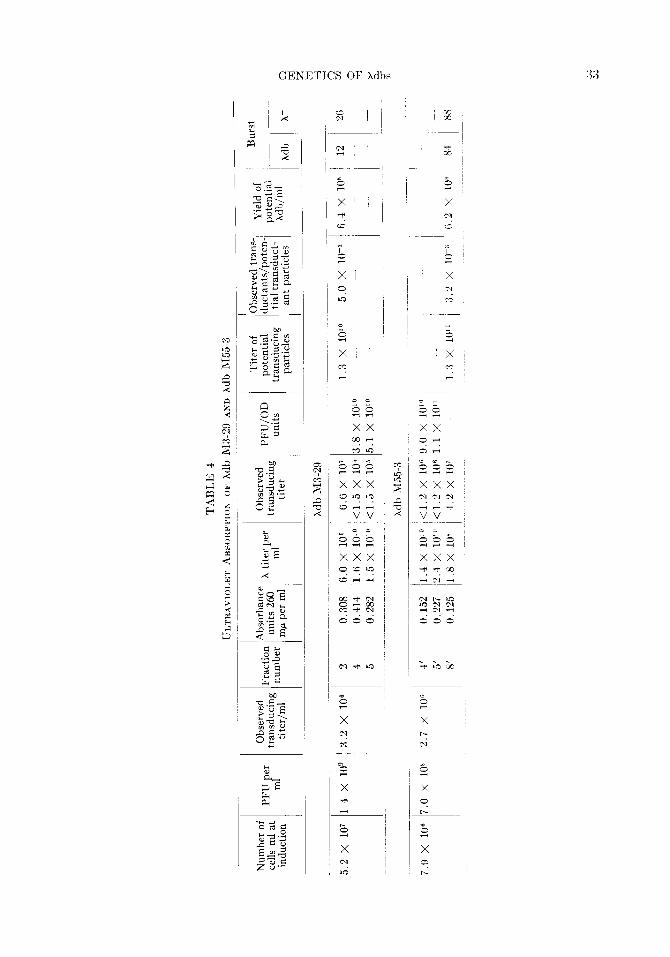
For hdb’s, ultraviolet (UV) absorption constitutes an independent assay of the
TABLE 3
EFFECT OF A HELPER ON TRANSDUCTION FREQUENCY OF THREF, xbio’s
Xbio Phage Frequency of transduction
end point No helper Moi of helper
- - - 2.3 M7-20 Xpb 3.8 x 10⁵ 1.2 x 10⁵ M3-29 N/Q 2.2 x 10⁵ 1.8 x lo5 M29-7 o/p 6.5 X lo4 2.3 x lo5
-i
X N ck
34 KAYAJANIAN
number of transducing particles (Smith, 1968). Phage induced from bio-O (Xdb M3- 29) (X) and from bio-0 (Xdb M55-3) (X) were concentrated by pelleting, treated with RNase and DNase, and fractionated in a CsCl density gradient. The UV absorption at 260 rnp of three fractions from these gradients, two primarily rich in h, the other predomniantly Xdb, was measured. These data (Table 4), corrected for the relative amounts of DNA in the transducing the plaque-forming phage [as judged by phage density measurements (Table 7)] and for the W-absorbing residue from the original lysates, reveal approximately the relative efficiency of transduction by Xdb RI3-29 and M55-3 as 5.0 X lop3 (6.6 X 10⁷/1.3 X lOlo) and 3.2 X 1O-3 (4.2 X 10’/1.3 X lOlo).
The plaque-forming and transducing ac- tivity in the lysate harvested from the induc- tion of bio-0 (Xdb M3-29) (at 5.2 X 10’ cells/ml) were, respectively, 1.4 X log/ml and 3.2 X lOe/ml. So the yield of hdb (M3-29) part#icles in this lysate is 6.4 X lo8 (3.2 X 10⁶/5.0 X 10-3). If the plaque- forming activity is a good measure of the number of X particles, the yield of transduc- ing particles is about one-half the number of X particles. The approximate burst size of X particles per induced cell is 26 (1.4 X log PFU/ml/5.2 X 10’ cells/ml); the Xdb burst is about 12. In a second experiment, the in- duction of bio-0 (Xdb M55-3) (x) (at 7.9 X lo6 cells/ml) yielded a lysate with a plaque- forming titer of 7.0 X 10⁸ and a transducing titer of 2.7 X 10⁶/ml. The X burst then is 88 (7.0 X lo* PFU/m1/7.9 X lo6 cells/ml). The Xdb burst is nearly at the X level (84). We do not find transducing particles in induction of Xdb defective lysogens. So it appears that Xdb genomes multiply with X help in the induced double lysogen.
Genetic Extent of Mb Genoms
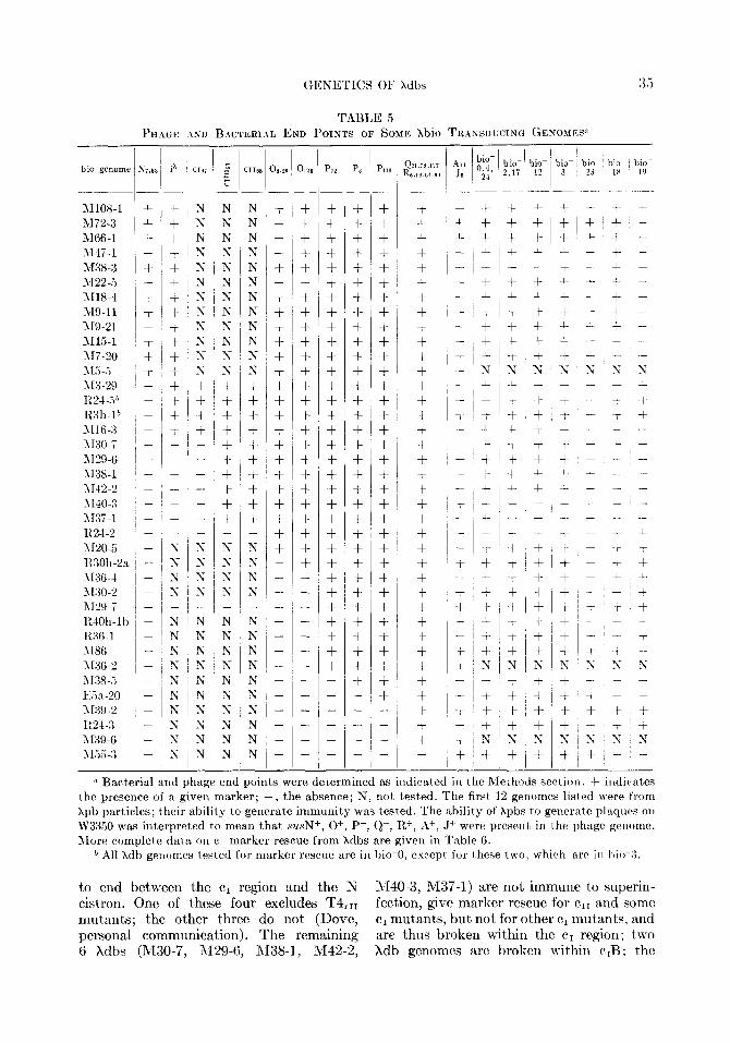
We assayed for the presence or absence of certain phage and bacterial markers in the genomes of a number of our Xbio transducing phages. For some of our assays, we made defective lysogens from bio transducing HFT lysates. The ability of defective lysogens to contribute certain sus+ or c+ alleles but not others, revealed which sus+ or c+ markers
were carried by each Xdb genome. The ability or inability of phage X to plate on defective lysogens indicated the absence or presence in the Xdb genome of an intact and functioning cr region. The ability or in- ability of Xbio lysates to transduce bio- mutants other than the one on which they were originally selected, revealed the extent of the bacterial chromosome carried inside the transducing phages. The extent of a number of Xbio genomes is described in Table 5. As in the case of Xdg, each Xbio genome represents a contiguous block of phage and bacterial genes from a X lyso- genie bacterial chromosome. Loss of the bio+ phenotype from Xdb lysogens, presumably by segregation, was nearly always accom- panied by loss of the phage markers asso- ciated with it in the transducing genome.
The variety of phage and bacterial markers in the Xbio genomes permits us to map the right arm of the X chromosome and to order our collection of bio- mutants. Three of the 26Xdb genomes (R30h-2a, M38-5, E5a-20) are clearly broken within the phage cistrons 0 and P and can be used to order the various sus 0 and P mutants:
N7,53 - Ow - 01~5 - P72 - p3 - PI16 - Q,R
The pattern of transduction of the Xbio lysates on different bio- mutants, orders the bacterial mutants into seven classes:
X - 0,4,24 - 2,17
- 12 - 3 - 23 - 18
(de1 Campillo-Campbell et al., 1967). Those Xdb genomes which gave
marker rescue for sus0, but not for
- 19
SUSf
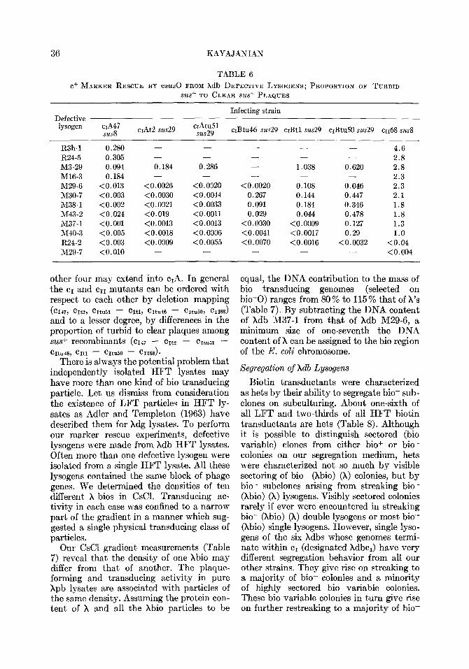
.S?L.SN were also tested for immunity against super- infection by X and for cIf and CII+ marker rescue with cIsusO and cIIsusO (Table 6). Of eleven Xdb genomes examined, one (R24-2) gives marker rescue for none of the clear mutants tested, is not immune to superinfection by phage X, and thus seems to be broken either within the cI1 cistron or between cII and the 0 cistron. Four (M3-29, R24-5, R3h-1, M16-3) give marker rescue for all of the cI and cII mutants tested, and are immune to superinfection, and thus seem
GENETICS OF Xdbs
TABLE ,5 PHAGF. ASD BACTERIAL END POINTS OF SOMI’. Xbio TR.\NSDUCING GENOMES”
I )io- bio- ,17 12
I.... __
+ +
+ +
+ +
+i+ + ~ +
+ +
(
I-
1372-3 1166-l l\r47-1 M38-3 1122-3 1\118-4 >I%11 >I%21 1115-l 117-20
IN N
IN IN iN I Ii
s IN
s s + + + + + + + + + +
N N N
1:
N N N N N + + + + + + + f + + -
+ + + + + + + + + +
+ + + +,+ + + + +l+‘+l+l + i+ +I+ + + + + -
+I+ +‘+ + + + + f + + + + + t’+ + + + +
+ +
+ + + + + + + + + + + + I -- +i+ + + + + - .- +I?; N ?; ?: T s 9 +(+ + + + A + -L
+:+ c +~+ +
+ + -i- + + + + f + +
+ + + + + - - - + + + + + + + +
+I + :+ +I+ + + + + -
11.X + + s 113-29 - +,+ 1~21..i* - +I+ R3h-lb - +‘+ 1116.3 - +l+ 1130.7 - - ~ - 1r29-6 - 1\138-1 ~ - - - 1142.2 - ~ - - 1\[40-3 - - - 1137-l ~ - - - R24-2 - - -
+ I+ ‘+
+ + +
+ + + + +
+! + I+ + +I + +,+ +~ + ~+I+ + + + +
!+I+ ~+ +
+I + I+ + + + + +
+ + + + + + -t + +
/
+ +
+ +
t -
- - - - - --
1 t
+ t-
i- +
+ +
- 1120.5 1130h-2a 3136.4 ~130-2 112%7 RlOh-lb 1~36.1 3186 1136-2 ;\I3%3
N t
* Bacterial and phage end points were determined as indicated in the Methods section. -i- indicat,es the presence of a given marker; -, the absence; N, not, tested. The first, 12 genomes listed were from Xpb particles; their ability to generate immunity was tested. The ability of Xpbs to generate plaqnos on W3350 was interpreted to mean that SUN+, 0+, Pf, Q+, R+, A+, J+ were present in the phnge penome. Afore complete data OII C+ marker rescue from xdbs are given in Table 6.
b All Xdh genomes tested for marker rescue are in bio-0, except, for these two, whirh are in bio-3.
to end between the cI region and the X 1140-3, 3,237-l) are not immune to superin- cistron. One of these four excludes T4,11 fection, give marker rescue for err and some mutants; the other three do not (Dove, cI mutants, but not for other c1 mutants, and personal communication). The remaining are thus broken within the cI region: t)wo 6 Xdbs (R430-7, M29-6, M38-1, M42-2, Xdb genomes are broken within c,B; the
36 KAYAJANIAN
TABLE 6
c+ MARKER RESCUE BY csus~ FROM xdb DEFECTIVE LYSOGENS; PROPORTION OF TURBID
sus+ TO CLEAR SW+ PLAQUES
Defective lysogen ~A47
susd cIAt2 sus29
Infecting strain
cIAtu51 sus29 crBtu46 sus29 cIBt1 sus29 cIBtu50 sus29 ~1168 susd
R3h-1 0.280 - - - - - 4.6 R24-5 0.305 - - - - - 2.8 M3-29 0.094 0.184 0.286 - 1.038 0.620 2.8 M16-3 0.184 - - - - - 2.3 M29-6 <0.013 <0.0026 <0.0020 <0.0020 0.108 0.046 2.3 M30-7 <0.003 <0.0030 <0.0044 0.267 0.144 0.447 2.1 M38-1 <0.002 <0.0021 <0.0033 0.091 0.184 0.346 1.8 M43-2 <0.024 <0.019 <O.OOll 0.029 0.044 0.478 1.8 h137-1 <O.OOl <0.0013 <0.0013 <0.0030 <0.0009 0.127 1.3 M40-3 <0.005 <0.0018 <0.0006 (0.0041 <0.0017 0.29 1.0 R24-2 <0.003 <0.0009 <0.0055 <0.0070 <0.0016 <0.0032 <0.04 M29-7 <O.OlO - - - - <0.004
other four may extend into c,A. In general the cI and cI1 mutants can be ordered with respect to each other by deletion mapping ( cI47, CItZ, CItu51 - CItl, CItu46 - CItu50, cII68)
and to a lesser degree, by differences in the proportion of turbid to clear plaques among .sus+ recombinants (~1~7 - crt2 - cItuE1 - hu46, CItl - hl50 - CII68 1.
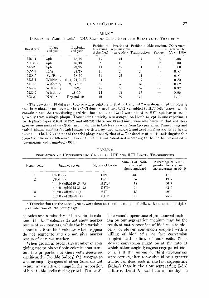
There is always the potential problem that independently isolated HFT lysates may have more than one kind of bio transducing particle. Let us dismiss from consideration the existence of LFT particles in HFT ly- sates as Adler and Templeton (1963) have described them for Xdg lysates. To perform our marker rescue experiments, defective lysogens were made from Xdb HFT lysates. Often more than one defective lysogen were isolated from a single HFT lysate. All these lysogens contained the same block of phage genes. We determined the densities of ten different X bios in CsCl. Transducing ac- tivity in each case was confined to a narrow part of the gradient in a manner which sug- gested a single physical transducing class of particles.
Our CsCl gradient measurements (Table 7) reveal that the density of one Xbio may differ from that of another. The plaque- forming and transducing activity in pure Xpb lysates are associated with particles of the same density. Assuming the protein con- tent of X and all the Xbio particles to be
equal, the DNA contribution to the mass of bio transducing genomes (selected on bio-0) ranges from 80 % to 115 % that of X’s (Table 7). By subtracting the DNA content of Xdb M37-1 from that of Xdb M29-6, a minimum size of one-seventh the DNA content of X can be assigned to the bio region of the E. coli chromosome.
Segregation of Xdb Lysogens
Biotin transductants were characterized as hets by their ability to segregate bio- sub- clones on subculturing. About one-sixth of all LFT and two-thirds of all HFT biotin transductants are hets (Table 8). Although it is possible to distinguish sectored (bio variable) clones from either biof or bio- colonies on our segregation medium, hets were characterized not so much by visible sectoring of bio- (hbio) (h) colonies, but by bio- subclones arising from streaking bio- (Xbio) (x) lysogens. Visibly sectored colonies rarely if ever were encountered in streaking bio- (Xbio) (X) double lysogens or most bio- (Xbio) single lysogens. However, single lyso- gens of the six Xdbs whose genomes termi- nate within cI (designated XdbcI) have very different segregation behavior from all our other strains. They give rise on streaking to a majority of bio- colonies and a minority of highly sectored bio variable colonies. These bio variable colonies in turn give rise on further restreaking to a majority of bio-
GENETICS OF Xdbs :37
Rio strain Phage Bacterial end point end point
1166-l W 18/19 hI108-1 Xpb 18/19 317-20 hpb 23/18 Ui3-Y K/A 23118 M38-.i Pi?/I’::.llS 18/19 1137-l Within CI 0. 4, 24/2, 17 M-IO-3 Within CI 2, li/12 hI42-2 Within CI 3/23 iU29-6 Withill c~ 18/19 RZR-29 S/i”, cd7 Beyond 19
Position of Position of Position of Xhio maxima DNh mass X maxima Xch2 maxima ~~--- _-- relative to (tube So.) (tube Ko.) Transduction Plaque h’s (= 1.00~
-~-~~ ~ ~ ~~~ .~~~ ~~~~~ .~~~~ ~ ~~ 12 21 i 8 1 .06 9 19 0 9 I .oo
11 22 11 11 1 .oo 10 20 20 0.87 1-I 27 :<1 0.81 4 1-l Ii 0 x2
42 Vi9 (i-l 0.82 42 .i!) .i2 0. !)z
1-l 24 17 0.96 42 59 20 1. 1.i
0 The density of 10 ditferent Xbio particles relative to that of X and Xcb2 was determined by plaving the three phage types together in a CsCl density gradient. Xcb2 was added to HFT kdb lysates, whirh rontain X and the transducing particles; both hn~-71 and kcb2 were added to HFT Xpb lysates made lyticallg from a single plaque. Transducing activity was assayed on bio-0, except in one experiment (n-ith phage types M40-3, M42-2, and M3-29) where bio-19 and bio3 were also hosts. Turbid and clear plaques were assayed on C600; turbid plaques in kpb lysates were from Xpb particles. Transducing and turbid plaque maxima for Xpb lysates are listed by tube number; X and hcb2 maxima are listed in t.he table too. The DNA content of the Xcb2 phage is 86.6% that of X. The density of ~171 is indistinguishable from X’S, The mass difference between xbio and X was calculated according to the method described in Kayajalrian and Campbell (1966).
TABLE 8
PILOPORTIOK OF HETEROGEKETIC CLONES IN LFT AND HFT BIOTIN TKINSDLXYI~ION
Experiment Induced strain
C600 (X, C600 (X) bio-0 (hdbM39-2) (X) bio-0 (XpbM72-3) (X) bio-0 (kdb20-5) (X) bio-0 (Xdb30-2) (X)
Sature of lysate
___~___ LFT LFTa HFT” HFTfl HFT IIFT
Numher of biotin transduced
clones analyzed
420 ‘2 ;‘8 16 1.5 12
Percentage of hetero- genetic clones among
transductants on bio-0 - __~-. ~.~ ~~~ -
17.6 19.2 66.7 62..i 60’ ; 7.5";
(1 Transduction for the three lysates were done on the same sample of cells with the same multiplic- ity of infection of “helper” phage.
colonies and a minority of bio variable colo- nies. The bio- colonies do not show marker rescue of sus markers, while the bio variable clones do. Rare bio+ colonies which appear do not segregate and do not give marker rescue of any sus markers.
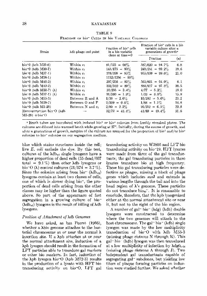
When grown in broth, the number of cells giving rise to bio variable colonies increases, but the proportion of these cells decreases significantly. Double (Xdbcl) (X) lysogens as well as single lysogens of other Xdbs do not exhibit any marked change in the proportion of bio+ t)o bio- cells during growth (Table 9).
The visual appearance of pronounced sector- ing on our segregation medium may be t’he result of fast conversion of biof cells to bio- cells, or slower conversion coupled with a killing of bio+ cells, or fast conversion coupled with killing of bio+ cells. (This slower conversion might be at the rate at which other single lysogens segregated bio- cells. ) If the second or third explanation were correct, then there should be a greater fract’ion of dead cells in the fast’ segregating (XdbcI) than in the slow segregat,ing (Xdb) cult,ures. Dead E. coli take up methylene
38 KAYAJANIAN
TABLE 9 FRACTION OF bio- CELLS IN bio VARIABLE COLONIES
Fraction of bio- cells in a bio Fraction of bio- cells variable culture after n
Strain Xdb phage end point in a bio variable generations of growth” clone at time=0
Fraction (4
bio-0 (Xcb M29-6) Within cr 81/123 = 66% 587/620 = 94.7% 6.8 bio-0 (xdb M30-7) Within cr 145/175 = 83% 249/251 = 99.2% 19.6 bio-0 (xdb M37-1) Within cr 219/259 = 8.5y0 955/959 = 99.6% 21.0 bio-0 (xdb M38-1) Within CI 1132/1256 = 90% bio-0 (xdb M40-3) Within cr 207/256 = 81% 565/601 = 94.07, 6.1 bio-0 (xdb M42-2) Within CI 303/340 = 89% 888/917 = 97.3y0 20.5 bio-0 (xdb M30-7) (X) Within CI 10/295 = 3.470 4177 = 5.2% 18.0 bio-0 (xdb M37-1) (X) Within cr 11/580 = 1.9% l/33 = 3.0% 15.9 bio-0 (xdb M55-3) Between R and A l/39 = 2.6% 10/202 = 5.0% 23.2 bio-0 (hdb M29-7) Between 0 and P 2/309 = 0.6% l/88 = 1.1% 14.8 bio-0 (xdb M3-29) Between N and CI 2/89 = 2.2y0 16/252 = 6.3yo 22.8 Reconstruction bio-0 (Xdb - 32/72 = 44.47o 44/89 = 49.4% 21.6 M3-29) + bio-0
a Broth tubes are inoculated with isolated biof or bio” colonies from freshly streaked plates. The cultures are diluted into warmed broth while growing at 37”. Initially, during the course of growth, and after n generations of growth, samples of the culture are assayed for the proportion of bio+ and/or bioT colonies to bio- colonies on our segregation medium.
blue which stains structures inside the cell; live E. coli exclude the dye. By this test, cultures of the Xdb,r single lysogens have a higher proportion of dead cells (55 dead/607 total = 9.1%) than other Xdb lysogens or bio-0 (X) control cultures (21/574 = 3.7 %). Since the colonies arising from bio- (Xdb,r) lysogens contain at least two classes of cells, one of which is slower dying bio-, the pro- portion of dead cells arising from the other classes may be higher than the figure quoted above. So part of the appearance of fast segregation in a growing culture of bio- (Xdb,,) lysogens is the result of killing of Xdb lysogens.
Position of Attachment of Xdb Genomes
We have asked, as has Fuerst (1966), whether a Xbio genome attaches to the bac- terial chromosome at or near the normal X insertion site. If a Xpb attaches at or near the normal attachment site, induction of a Xpb lysogen should result in the formation of LFT particles able to transduce gal markers or other bio markers. In fact, induction of the Xpb lysogen bio-0 (Xpb M72-3) results in the production of a lysate with HFT bio transducing activity on bio-0, LFT gal
transducing activity on W3805 and LFT bio transducing activity on bio-19. HFT lysates were made from three of the gal transduc- tants; the gal transducing particles in these lysates transduce bio at high frequency. These bio-gal transducing particles were de- fective as phages, missing a block of phage genes which includes SUSJ and extends in various lengths through the tail and into the head region of X’s genome. These particles do not transduce biorg-. It is reasonable to conclude, therefore, that the Xpb lysogenized either at the normal attachment site or near it, but not to the right of the bio region.
A number of gal- bio- (Xdg) (Xdb) double lysogens were constructed to determine where the two genomes will attach to the host chromosome. The gal- bio- (Xdb) single lysogen was made by the low multiplicity transduction of bio-0 with Xdb M55-3 (missing phage cistrons N through R). This gal- bio- (Xdb) lysogen was then transduced at a low multiplicity of infection by hdg01-26 (missing phage cistrons A through J). Two independent gal transductants capable of segregating gal- subclones, but yielding low titers of plaque forming particles on induc- tion were studied further. We asked whether
GFNETICS OF hdbs , L
the gal- segregants from the double lysogen were still bio+ and whether they retained cist,ron P and B markers (cistron P is asso- ciated with the gal+ in Xdg; B, with the bio+ in Xdb). A number of independently arising gal- segregants from each of the double lyso- gens were tested for the presence of B and I’ cistrons and the bio+ phenotype. We also assayed for the presence of B and P cistrons and the gal phenotype in bio- segregants of these double lysogens. These results are summarized in Table 10.
We have assumed that the formation of gal- segregants result’s from the lysogenic host chromosome recombining with itself in a region of gal+-gal- homology, and the subsequent loss of the small excised chromo- somal fragment,. Any marker bet’ween the two gal markers and t’herefore on this frag- ment would be lost’ too and not recoverable from the gal- segregnnt. The same assump- tions apply for the study of bio- segregant’s. Gal- segregants from double lysogen strain No. 5 have lost t’he P cistron, but neit,her the B cistron nor the bio+ phenotype. Bio- segregants from strain No. 5 are gal+ and have lost, cistron B but not P. Gal- segre- gants from strain Ko. 14 are bio- and have lost’ both B and 1’ markers. Bio- segregants from strain Xo. 14 are gal- and have lost cistron B and I’ genes. The only gene order possible for strain So. 5 is
gal- (Xdg) (Xdb) bio-
lvhere the prophages are in t’heir normal orientat8ion. The gene orders possible for strain So. 14 are:
gal- (Xdb) (Xdg) bio-
TABLE 10
AN.ILYSIS OF gal- .IND bio- SKGREGANTS FROM
Two gal- bio- (X db) (X dg) DOUBLE LYSOGENS
Residual genotype of gal- Residual genotypes of segregmts bio- segregants
Strain No.
Eumber 1 B- P- BtiOP; B+ P- Number B- P- B- P+ examined’ bio-
-I 1 i
bio- examined gal- gal+
5 -I-----
14 j ‘5 j f<L 1 ‘T , i P i 9
n A sister clone of one of these segregants was B+ PC bio-.
and
gal- (Xdb) bio- (Xdg)
where the prophages are in their normal orientat’ion. So, the Xdb genome resides be- tween the gal and bio markers of the Ii. coli host, and probably was inserted at, the X attachment site or in a region of homology with the host genome.
A subclone was isolated from strain No. 14 which would not, give marker rescue for SUB or s.usI’ markers, but still segregated gal- and bio- daught,er cells. For this segregant t,o arise from strain So. l-1, the prophage order in that double lysogen would haw to be
gal- (hiof ,Z .J) (S IX gal+) bio--
and the subclone from it is prohabl!
gal- (bio+ gal+) bio-.
In strains 5 nrltl 11 both Xdg and Xdh genomes reside between the gal and hio markers of the host’ chromosome. The pre- sumptive gal- (bio+ gal+) bio- strain was easily lysogenized by phage X, and the (X) (Xdbg) double lysogen produced a11 HFT lysate where gal and bio transducing ac- tivity were associated with particles of the same low density. *Joint transduction fre- quencies were no less than one-third of the individual transduction frequencies. The DYA content of t,hc Xdbg genome is less than the DX,4 mass of the Xdb by about as much as the Xdg’s is less than X’s (Kaya- janian, unpublished results).
I) IscusSIox
Xbio transducing particles derived from an HFT lysate are identical in t#heir content of phage markers and form a Gaussian distri- bution with a slight tail in a CsCl density gradient. HFT lysates are developed from independent transductants made from an LFT lysate. Xbio particles derived from HFT lysates may and generally do differ from one lysate to another in the content of phage and bacterial markers and in their density. As in the case of Xdg, the gene content in Xbio genomes represents a contiguous stretch of hereditary material lifted from the lysogenic bacterial chromosome. 111 general, t,he denser
40 KAYAJANIAN
a Xbio particle is, the larger its phage ge- nome and/or its bacterial component (Tables 5 and 7).
The variability in end points of Xdb ge- nomes suggests an independent origin for at least those Xdb genomes in LFT lysates that are capable of lysogenizing their bio- hosts. The difference in the ability of LFT and HFT lysates to transduce by lysogeny, rather than just transducing, suggests a dif- ference between Xbio particles in an LFT lysate and in all the HFT lysates so far studied. It is possible, therefore, that a sig- nificant fraction of biotin transducing parti- cles in LFT lysates may be structurally very different from those which we describe in HFT lysates and may be unable to transduce by lysogeny as a result of these structural differences. These structural differences may be, among other things, the absence of X’s cohesive ends. The variability that we ob- serve in the end points of Xdb genomes may not be as great as really exists for another reason. Some of the Xdb genomes capable of being maintained as reasonably stable lyso- gens in the presence of X as (X) (Xdb) lysogens (which give rise to HFT lysates) may be quite unable to be stably maintained as single (Xdb) lysogens. Some Xdb genomes may not be stable even as (X) (Xdb) lyso- gens. We were rather surprised when none of eleven Xdb genomes having phage end points between cistrons N and 0 were broken between cl and cII, but six were broken within cr, for we suspected the cl cistron to be smaller than the cI to cII inter- cistronic distance.
All of our Xdb genomes contain the phage markers from the J through A cistrons, though they need not contain cistron R markers. Symmetrically, all the Xdg ge- nomes we have encountered (Campbell, 1959,1962; Kayajanian and Campbell, 1966) contain the phage markers from the N through R cistrons, though they do not necessarily contain cistron A markers. In the absence of actually performing specific assays for the presence of the vegetative single-stranded ends of h (located between R and A) in all Xdb and Xdg particles in HFT lysates, we would like to argue that the method of selection and the measurable genetic end points of Xdb and Xdg genomes
themselves support the presence of X’s single-stranded ends in Xdbs missing cistron R markers and Xdg’s missing cistron A markers. Despite the fact t’hat Xdg defective lysogens are characterized by the presence of X immunity and the absence of plaque- forming ability, these genomes always con- tain the unselected markers in cistrons 0 through R. Similarly, Xdb defective lysogens are characterized by the presence of a J cistron marker and absence of plaque- forming ability, but always contain the un- selected markers from cistrons A through J. In view of the role in circularization and, therefore, in lysogenization attributed to the single-stranded ends of X, we consider the data above a strong indication that all Xdg and Xdb genomes capable of forming defective lysogens do contain the vegetative ends of X.
Assuming this, then Xdg%6 (missing A through J) and Xdb 1455-3 (missing N through R) both contain the vegetative ends of phage X. Both also contain an insertion region involved in the recombinational act in lysogenization. The Xdbg prophage re- combinant between these two, carrying gal and bio in place of both phage arms of X, should contain only the insertion and vege- tative regions that are of phage origin. The ability of Xdbg to undergo a lysogenic cycle with X’s help suggests that the structural requirement for undergoing such a circuit are satisfied by the presence of the prophage and vegetative ends of X.
Defective lysogens of the six Xdb genomes whose phage end points fall within the X im- munity region give rise to highly sectored colonies on our segregation medium. Lyso- gens of more defective and less defective Xdb genomes do not give this appearance. Neither do (x) (Xdb,r) double lysogens. We suppose that bacterial control maintains the more defective Xdbs as prophages. It seems reasonable to assume that the function of part of the X phage genome located between c1 and the 0 cistron is responsible for the altered phenotype of the (XdbQ1) lysogens. Lysogens of less complete Xdb genomes segregate more slowly; so these genomes would not have the wherewithal to manifest a fast segregation phenotype. Lysogens of more complete Xdb genomes would manage
GENETICS OF xdhs 41
to control the expression of the phenotype. We note that all t’he Xdb genomes more com- plete than (Xdb,,) contain an intact im- munity region which is recognized as such a cont8rol region.
The growth of bio- (Xdb,,) cells to form highly sectored colonies is the result, at least, of segregation of bio- subclones from pa- rental cells. That’ practically every colony arising from bio- (Xdb,,) lysogens is very highly sectored indicates that independent segregationnl events occur frequently-more frequently, than in other bio- (Xdb) or gal- (Xdg) lysogens. This greater rate of conver- sion of lysogenic to nonlysogenic cells may bv itself explain t’he very large fraction of bio- cells in a sect,ored colony and the pro- nounced increase in the proportion of bio- cells in a culture starting from a bio- (Xdb,,) colony. Segregation alone will account for highly sectored colonies, whereas differential growth rates will not,.
What about the killing? It may be an ir- relevance to our ot,her observations or a by- product of segregation which expresses i&elf at8 least one generation after the segrega- t,ional event. We have imagined the segrega- tional event to be the excision of the XdbC1 genome from the lysogenic host’ chromo- some. If the effect of t,he excision were to damage t,he host chromosome (in a poly- nucleate cell) so it would not function, then all the bio- segregants should be killed, and we would not see segregat)ion in bio- (Xdb,,) colonies. If the killing function were physio- logical, expressed some time after the segre- gat’ional event by a nonreplicating, non- repressible excision fragment, then the daughter cell t,hat would die would be the one containing the fragment. Sometimes Ohis cell would be bio+, and sometimes bio-. O-P killing (Packman and Sly, 1968), re- sulting from the inability of phage genomes t,o control 0 and P function could account for our dead cells. The absence of an X gene in our XdbCr genome could delay the killing
for more than a cell division so we would be able to see segregation.
The author is grat.efrd to the 111~. Campbell for looking after him.
REFERENCES
ADLER, J., and TEMPT,ETON, B. (1963). The amount of galactose genetic material in Adg hact,erio- phage with different densities. .I. Mol. Biol. 7, 710-720.
CAMPBELL, A. (1959). Ordering of genetic sites in bact,eriophage X by t.he use of galactose-trans- ducing defective phages. Virology 9, 293-305.
CAMPBELL, A. (1962). IIistrihution of genetic% types of transducing x phages. Genelics 48, 409-421.
DEL CAMPILLO-CAMPBELL, A., KAYAJ~NIAN, (i.. CAMPBELL, A., and ADHYA, S. (1967). Biotitt mutants of Escherichia coli K12. J. Hacleriol. 94, 2065-2066.
FUERST, C. It. (1966). Defective hiotirl-transdllc-. ing mutants of bacteriophage Lambda. T’irol- ogy 30, 581-583.
KAYAJANIAX, G., and CAMPBELL, A. (1966). The relationship hetween heritable physical and genetic properties of selected gal- and gal+.. transducing Xdg. Virology 30, 482-492.
MORSE, X. I,., LEDERBERG, E. hr., and LEDEW BERG, J. (1956a). Transduction in EscherichCz coli K12. Genelics 41, 142-156.
MORSE, M. L., LEDERBERG, E. M., and LEDEIL-- BERG, J. (1956h). Transductional heterogenotes in Escherichia coli. Genetics 41, 758-776.
PACKMAN, S., and SLY, W. S. (1968). Constitutive xDNA replication by Xc,;, a regulatory mutant. related t.o virulence. Virology 34, 778-789.
ROTHM~N, J. (1965). Transduction studies on the relation between prophage and host chromn- some. J. Mol. Biol. 12, 892-912.
SMWH, 1-I. 0. (1968). Dcfectlve phage formation by lysogens of integrat.inn deficient phage P’ZZ mutants. Virology 34, 203-223.
WALMCE, B., and PITI'ARI), J. (1967). (+enetic: and biochemical analysis of the isoenzymes concerned in the first reaction of aromatic: biosynt,hesis in Escherichia coli. .I. I~actwiol. 93, 237 213.

![Inhibition of γ-Secretase Leads to an Increase in Presenilin-1 · defective 1 (APH1), and presenilin enhancer 2 (PEN2) [7]. γ-Secretase acts an aspartyl protease, which catalytic](https://static.fdocument.org/doc/165x107/5fcf13aeec1c843f815764d3/inhibition-of-secretase-leads-to-an-increase-in-presenilin-1-defective-1-aph1.jpg)

![Technical Note - HPLC · Technical Note Vitamins are trace ... Excellent High Performance Liquid Chromatography (HPLC) ... Folic Acid (0.26@Ûg) 9; D-Biotin [Vitamin H] (2.02@Ûg)](https://static.fdocument.org/doc/165x107/5ad475c17f8b9a6d708ba707/technical-note-note-vitamins-are-trace-excellent-high-performance-liquid-chromatography.jpg)