Μ-Capture, Energy Rotation, Cooling and High-pressure Cavities David Neuffer Fermilab.
Structures of mammalian ER α-glucosidase II capturethe binding modes of broad-spectrumiminosugar antiviralsAlessandro T. Caputoa, Dominic S. Alonzia, Lucia Martib,1, Ida-Barbara Recab,1, J. L. Kiappesa, Weston B. Struwea,Alice Crossa, Souradeep Basua, Edward D. Lowea, Benoit Darlota,c, Angelo Santinob, Pietro Roversia,2,and Nicole Zitzmanna,2
aOxford Glycobiology Institute, Department of Biochemistry, University of Oxford, Oxford OX1 3QU, United Kingdom; bInstitute of Sciences of Food Production,Consiglio Nazionale delle Ricerche Unit of Lecce, 73100 Lecce, Italy; and cEcole Nationale Supérieure de Chimie de Montpellier, 34296 Montpellier Cedex 5, France
Edited by Peter Palese, Icahn School of Medicine at Mount Sinai, New York, NY, and approved June 21, 2016 (received for review March 17, 2016)
The biosynthesis of enveloped viruses depends heavily on the hostcell endoplasmic reticulum (ER) glycoprotein quality control (QC)machinery. This dependency exceeds the dependency of hostglycoproteins, offering a window for the targeting of ERQC for thedevelopment of broad-spectrum antivirals. We determined small-angle X-ray scattering (SAXS) and crystal structures of the main ERQCenzyme, ER α-glucosidase II (α-GluII; from mouse), alone and in com-plex with key ligands of its catalytic cycle and antiviral iminosugars,including two that are in clinical trials for the treatment of denguefever. The SAXS data capture the enzyme’s quaternary structure andsuggest a conformational rearrangement is needed for the simulta-neous binding of a monoglucosylated glycan to both subunits. TheX-ray structures with key catalytic cycle intermediates highlight thatan insertion between the +1 and +2 subsites contributes to theenzyme’s activity and substrate specificity, and reveal that the pres-ence of D-mannose at the +1 subsite renders the acid catalyst lessefficient during the cleavage of the monoglucosylated substrate. Thecomplexes with iminosugar antivirals suggest that inhibitors target-ing a conserved ring of aromatic residues between the α-GluII +1and +2 subsites would have increased potency and selectivity, thusproviding a template for further rational drug design.
broad-spectrum antiviral | ER α-glucosidase II | eukaryotic secretion |glycoprotein folding | iminosugar
Most antiviral drugs target viral proteins critical to the virallife cycle. In recent years, increasing levels of resistance to
direct-acting antivirals have become a major public health con-cern, highlighting the urgent need for the development of alter-native treatments (1). One appealing strategy to avoid antiviraldrug resistance is the targeting of virus–host interactions (2).Drugs that act by blocking specific host functions required bymany different viruses have the additional potential to treat a widerange of viral infections and coinfections (broad-spectrum antivi-rals) (3). Inhibitors of the endoplasmic reticulum (ER) glycopro-tein quality control (QC) machinery, used by most envelopedviruses for the correct folding of their surface glycoproteins (4),represent one class of promising host-targeting, broad-spectrumantivirals. ER α-glucosidase II (α-GluII) is the main ERQC gly-cosyl hydrolase, admitting folding client glycoproteins into ERQCand releasing them from it (4–9). Inhibition of the ERQC ma-chinery, including α-GluII inhibition, causes viral glycoproteins tomisfold and reduces virion secretion and/or infectivity (2). Thecentrality of α-GluII to viral glycoprotein folding and secretionmakes it an appealing target for host-targeting antivirals, withclinical relevance for broad-spectrum antiviral therapy. ERα-GluII is a heterodimer composed of a catalytic α-subunit (104–116 kDa) and an accessory β-subunit (58–80 kDa) (5, 8, 10, 11).The enzyme first removes a glucose residue from the conservedGlc2Man9GlcNAc2 N-linked glycan attached to a nascent glyco-protein (“first cleavage”), and the resulting Glc1Man9GlcNAc2glycan enables glycoprotein binding to the ER lectins calnexin and
calreticulin and associated chaperones and refolding isomerases(5, 12). After another α-GluII–mediated glycosidic bond hydrolysis(“second cleavage”), which removes the innermost Glc residue, theglycoprotein is left with a Man9GlcNAc2 glycan and loses bindingaffinity for the ER lectins and refolding machinery. At this stage, iffolded correctly, the α-GluII substrate glycoprotein is free to pro-ceed toward the Golgi apparatus and secretion. Iminosugars, gly-comimetics originally isolated from plants, represent a promisingclass of ERQC inhibitors that are well tolerated in mammals (13).Altering glycoprotein processing via inhibition of the ER α-GluIand α-GluII by iminosugars has demonstrated in vitro and in vivoefficacy against multiple virus families (14, 15), including Herpes-,Hepadna-, Toga-, Rhabdo-, Orthomyxo-, Paramyxo- and Retro-viridae, as well as those virus families that cause hemorrhagic fever(16, 17), including Arena-, Flavi- and Filoviridae. The deoxynojiri-mycin iminosugar derivative N-9′-methoxynonyl 1-deoxynojirimycin(MON-DNJ) has antiviral activity in vivo, against both dengue virus(18) and influenza A (H1N1) (19) and influenza B (17), with the
Significance
Most pathogenic enveloped viruses crucially depend on thequality control (QC) machinery in the endoplasmic reticulum (ER)of the host cell. ERQC inhibitors therefore have the double po-tential benefit of targeting a wide variety of viruses (“broad-spectrum antivirals”) without the risk of losing efficacy due toescape mutations in the viral genome. Our recent work hasproven that inhibition of the central enzyme of ERQC, α-glucosi-dase II (α-GluII), is sufficient for antiviral activity against denguefever in vitro and in vivo. Here, we show how antiviral inhibitorsbind to portions of α-GluII that are unique to this enzyme, and weopen the way to the development of potent and selective anti-virals against existing and emerging infectious disease.
Author contributions: A.T.C., L.M., A.S., P.R., andN.Z. designed research; A.T.C., D.S.A., L.M., I.-B.R.,J.L.K., W.B.S., A.C., S.B., B.D., and P.R. performed research; D.S.A. and J.L.K. contributed newreagents/analytic tools; A.T.C., D.S.A., L.M., I.-B.R., J.L.K., W.B.S., A.C., S.B., E.D.L., A.S., P.R., andN.Z. analyzed data; and A.T.C., L.M., P.R., and N.Z. wrote the paper.
Conflict of interest statement: N.Z. is a named inventor on the following patents, whichdescribe iminosugars and methods of treating viral diseases: WO/2001/010429(2001) andEP1210082(2002); WO/1999/029321(1999), EP1037636(2000) and KR1020010033028(2001);WO/2010/096764(2010) and EP2398321(2011); WO/2011/028775(2011) and EP2473482(2012);WO/2011/028779(2011) and EP2473046(2012); WO/2011/028781(2011) and EP2473493(2012);WO/2010/144759(2010) and EP2440205(2012); and WO/2010/099064(2010) andEP2400843(2012).
This article is a PNAS Direct Submission.
Data deposition: The atomic coordinates and structure factors have been deposited in theProtein Data Bank, www.pdb.org (PDB ID codes 5F0E, 5HJO, 5HJR, 5H9O, 5IED, 5IEE, 5IEF,and 5IEG).1L.M. and I.-B.R. contributed equally to this work.2To whom correspondence may be addressed. Email: [email protected] [email protected].
This article contains supporting information online at www.pnas.org/lookup/suppl/doi:10.1073/pnas.1604463113/-/DCSupplemental.
www.pnas.org/cgi/doi/10.1073/pnas.1604463113 PNAS Early Edition | 1 of 9
BIOCH
EMISTR
YPN
ASPL
US
latter being the first reported incidence of antiviral activity of imi-nosugars against these strains of influenza. Two iminosugars withknown inhibitory activity against α-GluII are in clinical trials againstdengue fever (https://clinicaltrials.gov/ct2/show/record/NCT02569827and https://clinicaltrials.gov/ct2/show/NCT02696291). The α-GluIIα-subunit belongs to the GH31 family of glycosyl hydrolases,which also comprises intestinal maltase-glucoamylase (MGAM)and sucrase-isomaltase (SI). Both glycosidic bonds specificallycleaved by α-GluII have α(1,3) linkages, unlike the α(1,4)-maltose–like or α(1,6) bonds hydrolyzed by intestinal GH31 α-glucosidases.However, iminosugars, which are glucose mimetics and inhibitα-GluII, also target intestinal α-glucosidases and the ceramide-specific glucosyltransferase involved in glycosphingolipid bio-synthesis (20). Toward an understanding of the molecular basisof iminosugar binding to their targets, and the development ofbetter inhibitors of α-GluII, we have successfully establishedand describe here the first, to our knowledge, expression systemcapable of producing milligram quantities of an intact recombinantmammalian α-GluII heterodimer. This expression system has en-abled us to carry out extensive biochemical and biophysical char-acterization of the enzyme. Eight crystal structures with a varietyof ligands, including the two iminosugars in clinical trials against
dengue fever, indicate that molecules targeting the +1 and +2subsites of the enzyme will improve both potency and selectivity.
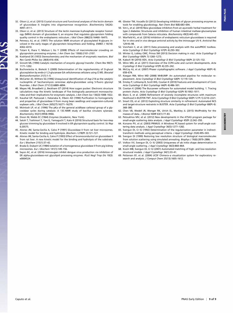
Results and DiscussionThe Full-Length Murine α-GluII α/β Holoenzyme Has an ElongatedShape. Both α- and β-subunit α-GluII sequences are highly con-served across eukaryotes. Initial tests screened expression of solubleα-GluII constructs from Saccharomyces cerevisiae, Caenorhabditiselegans, Mus musculus, and Homo sapiens. Wild-type (wt) recombi-nant mouse α-GluII (Mmα-GluII) gave the best preliminary resultsand was expressed and purified on a larger scale, together withits stable trypsinolytic fragment Mmα-GluIITryps (21) (SI Appendix,Fig. S1A). The murine and human α-subunit sequences share 92%identity, and the β-subunit sequences share 87% identity. Size exclusionchromatography multiangle laser light scattering (SEC-MALLS)and electrospray ionization mass spectrometry (ESI-MS) confirmedfolded α/β heterodimers of 166 kDa and 109 kDa, respectively,for Mmα-GluII and Mmα-GluIITryps (SI Appendix, Fig. S1B). TheMmα-GluIITryps small-angle X-ray scattering (SAXS) data (SI Ap-pendix, Fig. S1C and Table S1) reveal a globular molecule, withapproximate dimensions of 11 × 8 × 7 nm (green mesh in Fig. 1 Cand D and SI Appendix, Fig. S1D). The SAXS data for the wt
*
LDLRa
N
C
GH31 domain GH31 insert
N-terminal domain Proximal
C-terminal domain
Distal C-terminal
domain
B
A C
D
N C
N C
α-subunit
β-subunit
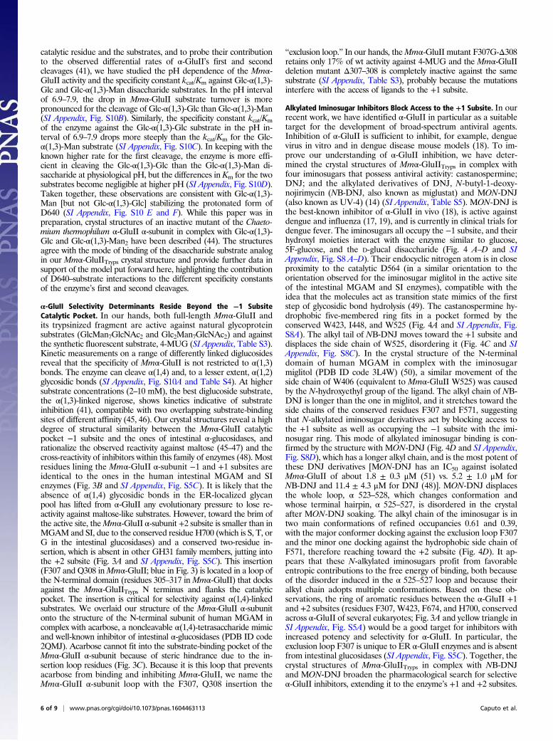
Fig. 1. SAXS and crystal structures of Mmα-GluII. (A) A 1-Dimensional representation of the α-GluII subunits. The trypsinolysis sites are symbolized by scissors,and portions of the α-subunit removed by trypsin are represented by magenta loops. (B) Crystal structure of Mmα-GluIITryps in cartoon representation. Theunique α-subunit N-terminal extension (residues 33–54) is shown in red, and the N-terminal domain (residues 55–392) is shown in purple. Dotted lines circlethe α-subunit portions removed by trypsinolysis. The α-subunit GH31 catalytic domain (residues 393–748) is shown in green, with its insertion subdomainshown in yellow (residues 492–531). The proximal and distal α-subunit C-terminal domains (residues 749–828 and 829–966, respectively) are shown in orangeand pink, respectively. After trypsinization, the α/βMmα-GluII dimer retains the N-terminal domain of the β-subunit, adopting two tandem LDLRa folds (blue),with the trypsinized β-subunit N and C termini marked by a letter. Two calcium ions in the β-subunit LDLRa subdomains are shown as green spheres. Theα-subunit active site residues, the N-linked glycan at N97, and the calcium-coordinating residues in the β-subunit are depicted in stick representation. (C and D)Crystal structure ofMmα-GluIITryps (in cartoon representation) overlaid on the SAXS-hydrated envelopes for Mmα-GluIITryps (green mesh) and Mmα-GluII (lightblue mesh). The green cartoon illustrates the α-subunit and the cyan cartoon illustrates the β-subunit (N-terminal LDLRa domains), the dashed line illustratesthe central part of the protein (unannotated), and the cyan shape illustrates the C-terminal M6PRH domain. The α-GluII–specific α-subunit N-terminal ex-tension is painted red. The green asterisk illustrates the volume taken up by the residues α 186–240, removed from Mmα-GluIITryps by trypsinolysis. Helices α427–441 and α 470–482, which the β-subunit protects from HDX, are shown in dark blue. A stereoregular, extended conformation of the Glc2Man9GlcNAc2glycan, with the terminal Glc residues inserted in the active site, is represented in sticks (yellow, carbon atoms; red, oxygen atoms). Two possible locations ofthe β-subunit M6PRH domain (cyan shape) are shown, either proximal to the α-subunit active site (C) or distal to the α-subunit active site (D), at the far end ofthe light blue holoenzyme SAXS envelope. The red arrows indicate hypothetical conformational changes in the β-subunit that would bring its M6PRH domaincloser to the enzyme’s α-subunit active site if simultaneous binding of both subunits to a single monoglucosylated glycan was necessary for cleavage.
2 of 9 | www.pnas.org/cgi/doi/10.1073/pnas.1604463113 Caputo et al.
Mmα-GluII full-length heterodimer yield an elongated shape thatextends off one end of the globular structure of the trypsinized frag-ment to an overall length of 22 nm (light blue mesh in Fig. 1 C and Dand SI Appendix, Fig. S1D). This shape and volume are in agreementwith the sedimentation velocity ultracentrifugation measurements onthe intact heterodimeric rat liver enzyme purified from tissue (21).
The α-GluII β-Subunit Associates with the Catalytic α-Subunit Via ItsN-Terminal Domains. Full-length Mmα-GluII did not yield crystals;however, Mmα-GluIITryps crystals enabled structure solution bymolecular replacement at a resolution of 1.74 Å (Fig. 1B and SIAppendix, Table S2). The Mmα-GluII catalytic α-subunit overallfold (green in Fig. 1 C–D) and catalytic pocket (Fig. 2A) confirm aGH31 family fold. Known α-subunit activity-impairing mutationscorresponding to Mmα-GluII E568Q (22), S569F, and S651F (23)localize to regions proximal to the active site or destabilize the foldof the α-subunit (SI Appendix, Fig. S2A). Unique to ER α-GluIIα-subunit is its N terminus (residues 33–54), which reaches thebrim of the catalytic pocket (red in Fig. 1 B–D) and supports aloop (residues 305–317 in Mmα-GluII) contributing to the +1 and+2 enzyme subsites (Fig. 3 A and B). In addition to the α-subunit,our Mmα-GluIITryps crystals contain two tandem low-densitylipoprotein receptor class A (LDLRa) subdomains, the N-terminaland only portion of the β-subunit still associated with the α-subunitafter trypsinolysis (light blue in Fig. 1 A–D and SI Appendix, Fig.S4A), in keeping with the predictions from previous biochemicalstudies (24–27). Each β-subunit LDLRa module folds around an
octahedrally coordinated calcium ion (SI Appendix, Figs. S2B andS3 A and B). The interface between the α-subunit and theβ-subunit LDLRa modules spans 700 Å2 of solvent-accessiblearea, and is organized around salt bridges between conservedaspartic acid residues in the β-subunit and positively chargedresidues in the α-subunit, clustered around residues α R837 and αR840 (SI Appendix, Figs. S4A and S5 A and B). Docking of theMmα-GluIITryps crystal structure onto the wt Mmα-GluII hetero-dimer SAXS envelope reveals that, contiguous to the β-subunitN-terminal LDLRa subdomains of Mmα-GluIITryps, the Mmα-GluII full-length heterodimer has a protuberance correspondingto the trypsin-sensitive portion of the β-subunit that is missingin the crystals (Fig. 1 C and D). Hydrogen deuterium exchange(HDX) MS conducted on the full-length Mmα-GluII heterodimersupports the observed α/β crystal interface (SI Appendix, Fig. S6).Additionally, helices α 427–441 and α 470–482, which contact eachother on the surface of the α-subunit between the active site andthe β-subunit, are protected from HDX (SI Appendix, Fig. S6 Aand C). In the context of the full-length Mmα-GluII heterodimer,these α-subunit helices (dark blue in Fig. 1 C and D) are buriedinside the SAXS envelope, indicating that they also form part ofthe α/β interface. The interaction between subunits at this spotmust be weak/transient, because the portion of β-subunit involvedin it does not survive trypsinolysis.
Arginine Residues on the α-GluII α-Subunit Mediate Its ER Retention.The α-GluII β-subunit carries a C-terminal ER-retention signal
D564
D640
A B
C D
-1 +1 R624
D305 M565
W525 I452
D451
W423
F673
H698
W637
1 2
3 4
5
6
6
5 4 3
2 1
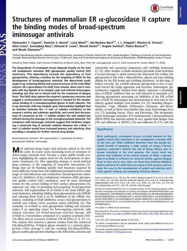
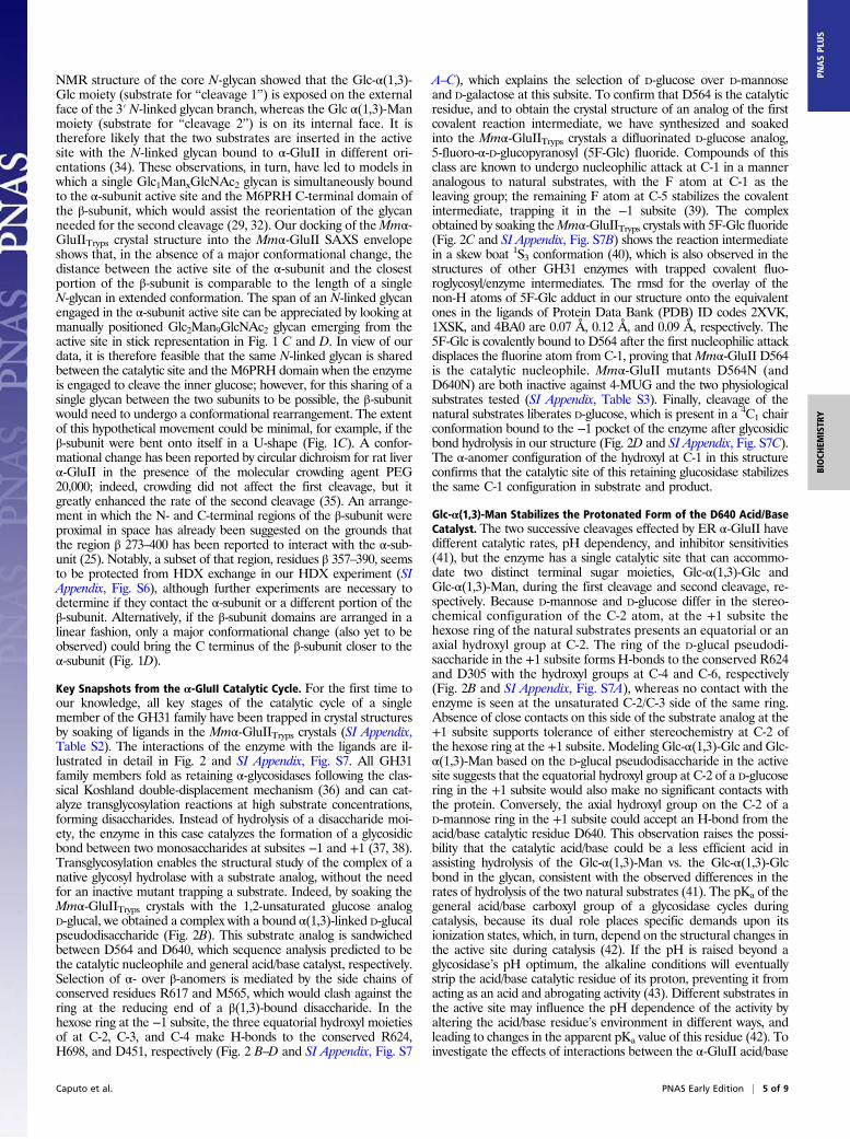
Fig. 2. Details of the Mmα-GluII α-subunit active site at key stages during its catalytic cycle (with corresponding chemical schemes shown in the upper rightcorners). The −1 and +1 subsites are marked in B. (A) Apo enzyme; the α-subunit pocket is filled with ordered water molecules (red spheres). (B) Complex with theD-glucal transglucosylation product, an analog of an α(1,3)-disaccharide substrate. (C) Covalent complex after nucleophilic attack from D564 to 5F-Glc fluoride (SIAppendix, Fig. S9; NMR spectra of 1), forming a reaction intermediate analog. (D) Complex with D-glucose, one of the products of the enzyme’s reactions. Proteinstick representation: green, carbon; red, oxygen; blue, nitrogen. Hydrogen atoms are omitted. The carbon atoms of the catalytic nucleophile D564 and the ligandsare colored purple. The carbon atoms of the catalytic acid/base D640 are colored orange. The fluorine atom in C is colored light green.
Caputo et al. PNAS Early Edition | 3 of 9
BIOCH
EMISTR
YPN
ASPL
US
that ensures ER retention of the GluII heterodimer (28). TheMmα-GluII α-subunit single mutant R840E, designed on thebasis of the crystal structure to disrupt α/β association, losesbinding capacity for the β-subunit in vitro. The latter no longercopurifies with the 6× His-tagged α-subunit R840E mutant asassayed by immobilized metal affinity chromatography (SI Ap-pendix, Fig. S4G). Similar results were obtained in vivo for theequivalent Arabidopsis thaliana (At) α-GluII α-subunit singlemutant R787E, transfected into tobacco leaves as a GFP fusion(SI Appendix, Fig. S4). The mutant shows weaker associationwith the β-subunit, as judged by fluorescence microscopy and(co)immunoprecipitation experiments (SI Appendix, Fig. S4 C, E,and F). The Atα-GluII α-subunit R784E/R787E double mutant(corresponding to theMmα-GluII α-subunit double-mutant R837E/R840E) completely abrogates the α/β association in vivo, again asshown by fluorescence microscopy and (co)immunoprecipitationexperiments (SI Appendix, Fig. S4 D–F). The murine α-GluIIα-subunit R840E mutant that abrogates the α/β interaction invitro and corresponds to the plant R787E mutant showing re-duced ER localization in planta, shows only 10% of the wt en-zyme activity in an in vitro assay (SI Appendix, Table S3),suggesting that ligands targeting the α-GluII α/β interface wouldconstitute selective α-GluII inhibitors.
The α-GluII β-Subunit LDLRa Subdomains Stabilize the α-Subunit ina Catalytically Competent Conformation. The differences in theactivity of the enzyme against glycoproteins carrying a variablenumber of N-linked glycans have led to the suggestion that morethan one glycan is needed for α-GluII to allow entry of a glyco-protein into the calnexin/calreticulin cycle (29). In this case, themannose 6-phosphate receptor homology (M6PRH) C-terminaldomain of the β-subunit would bind a terminal α(1,2)-linkedmannose residue on one N-linked glycan (30, 31), helping therecruitment of the other N-linked glycan to the active site (29, 32).The shape and dimensions of our Mmα-GluII SAXS envelope andMmα-GluIITryps crystal structure are compatible with a role forthe M6PRH C-terminal domain of the β-subunit assistingglucose hydrolysis by binding a second glycan on glycoproteinscarrying more than one glycan (29, 32). In the absence of theβ-subunit, α-GluII activity is reduced in vivo (27) and invitro (33) to about 5–10% of wt levels and the recombinantα-subunit is unstable unless associated with the β-subunit(4, 28). In our hands, Mmα-GluIITryps is still able to hydrolyzeGlc1Man7GlcNAc2 and Glc2Man7GlcNAc2, has identical bind-ing affinity for the 4-methylumbelliferyl α-D-glucopyranoside(4-MUG) synthetic substrate, and retains 70% of the wt catalyticefficiency against 4-MUG (SI Appendix, Table S3). Because thefirst cleavage seems to proceed even when the β-subunit is dis-rupted (26, 27, 29), it appears the C-terminal M6PHR domain ofthe β-subunit may simply increase the avidity of the α-subunit forthe substrate glycoprotein-linked Glc2ManxGlcNAc2 (32). Con-versely, the N-terminal portion of the β-subunit is necessary forα-GluII catalysis by the α-subunit in vitro and in vivo, and acts bystabilizing the α-subunit in a catalytically competent conformation.
A Conformational Change is Likely to Be Needed for SimultaneousBinding of a Glc1ManxGlcNAc2 Glycan to Both α-GluII Subunits. Inlight of the NMR structure of the core N-glycan (34), it has beenproposed that the monoglucosylated glycan must undergo a con-formational change for its hydrolysis to take place (26, 27, 29). The
A
N
-1 +1
H700 F307
Q306
+2 W423
B
C
F674
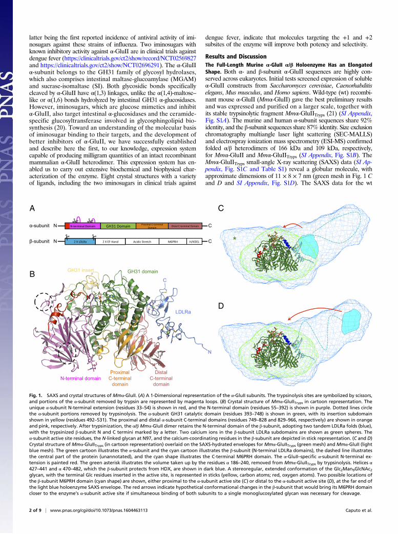
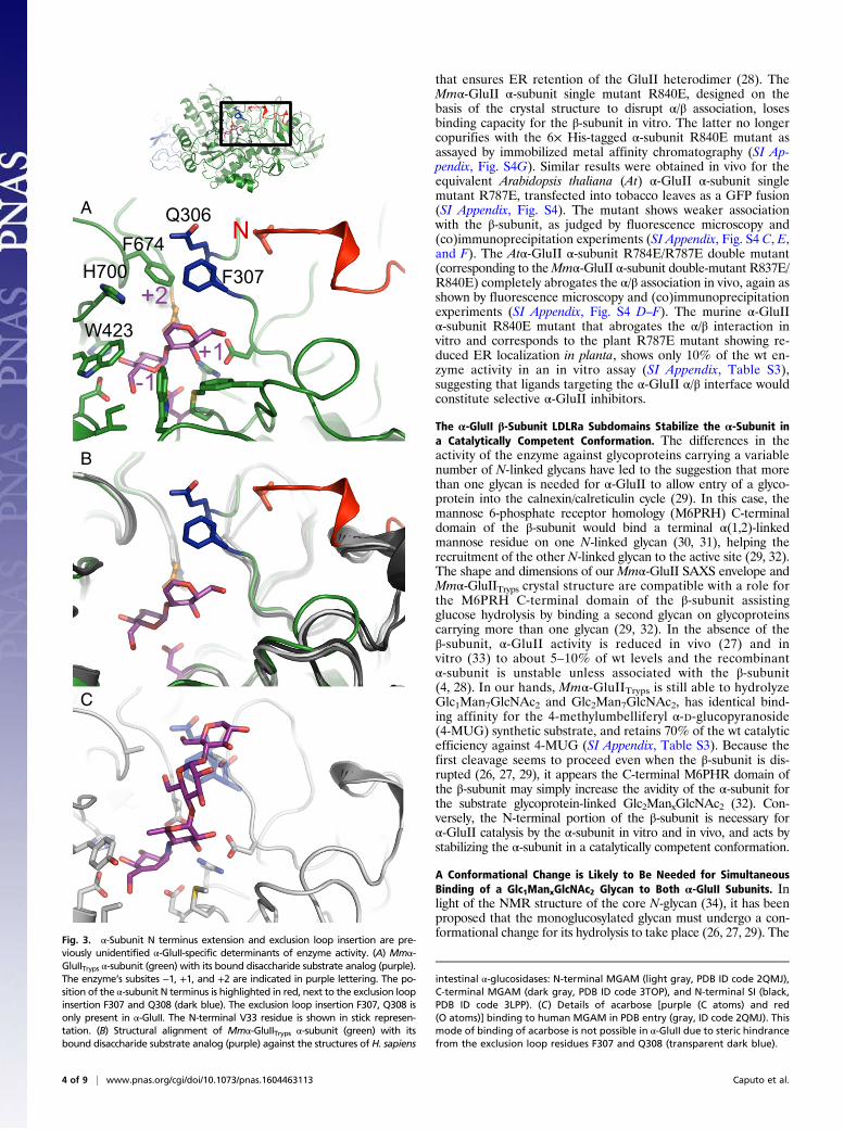
Fig. 3. α-Subunit N terminus extension and exclusion loop insertion are pre-viously unidentified α-GluII-specific determinants of enzyme activity. (A) Mmα-GluIITryps α-subunit (green) with its bound disaccharide substrate analog (purple).The enzyme’s subsites −1, +1, and +2 are indicated in purple lettering. The po-sition of the α-subunit N terminus is highlighted in red, next to the exclusion loopinsertion F307 and Q308 (dark blue). The exclusion loop insertion F307, Q308 isonly present in α-GluII. The N-terminal V33 residue is shown in stick represen-tation. (B) Structural alignment of Mmα-GluIITryps α-subunit (green) with itsbound disaccharide substrate analog (purple) against the structures of H. sapiens
intestinal α-glucosidases: N-terminal MGAM (light gray, PDB ID code 2QMJ),C-terminal MGAM (dark gray, PDB ID code 3TOP), and N-terminal SI (black,PDB ID code 3LPP). (C) Details of acarbose [purple (C atoms) and red(O atoms)] binding to human MGAM in PDB entry (gray, ID code 2QMJ). Thismode of binding of acarbose is not possible in α-GluII due to steric hindrancefrom the exclusion loop residues F307 and Q308 (transparent dark blue).
4 of 9 | www.pnas.org/cgi/doi/10.1073/pnas.1604463113 Caputo et al.
NMR structure of the core N-glycan showed that the Glc-α(1,3)-Glc moiety (substrate for “cleavage 1”) is exposed on the externalface of the 3′ N-linked glycan branch, whereas the Glc α(1,3)-Manmoiety (substrate for “cleavage 2”) is on its internal face. It istherefore likely that the two substrates are inserted in the activesite with the N-linked glycan bound to α-GluII in different ori-entations (34). These observations, in turn, have led to models inwhich a single Glc1ManxGlcNAc2 glycan is simultaneously boundto the α-subunit active site and the M6PRH C-terminal domain ofthe β-subunit, which would assist the reorientation of the glycanneeded for the second cleavage (29, 32). Our docking of theMmα-GluIITryps crystal structure into the Mmα-GluII SAXS envelopeshows that, in the absence of a major conformational change, thedistance between the active site of the α-subunit and the closestportion of the β-subunit is comparable to the length of a singleN-glycan in extended conformation. The span of an N-linked glycanengaged in the α-subunit active site can be appreciated by looking atmanually positioned Glc2Man9GlcNAc2 glycan emerging from theactive site in stick representation in Fig. 1 C and D. In view of ourdata, it is therefore feasible that the same N-linked glycan is sharedbetween the catalytic site and the M6PRH domain when the enzymeis engaged to cleave the inner glucose; however, for this sharing of asingle glycan between the two subunits to be possible, the β-subunitwould need to undergo a conformational rearrangement. The extentof this hypothetical movement could be minimal, for example, if theβ-subunit were bent onto itself in a U-shape (Fig. 1C). A confor-mational change has been reported by circular dichroism for rat liverα-GluII in the presence of the molecular crowding agent PEG20,000; indeed, crowding did not affect the first cleavage, but itgreatly enhanced the rate of the second cleavage (35). An arrange-ment in which the N- and C-terminal regions of the β-subunit wereproximal in space has already been suggested on the grounds thatthe region β 273–400 has been reported to interact with the α-sub-unit (25). Notably, a subset of that region, residues β 357–390, seemsto be protected from HDX exchange in our HDX experiment (SIAppendix, Fig. S6), although further experiments are necessary todetermine if they contact the α-subunit or a different portion of theβ-subunit. Alternatively, if the β-subunit domains are arranged in alinear fashion, only a major conformational change (also yet to beobserved) could bring the C terminus of the β-subunit closer to theα-subunit (Fig. 1D).
Key Snapshots from the α-GluII Catalytic Cycle. For the first time toour knowledge, all key stages of the catalytic cycle of a singlemember of the GH31 family have been trapped in crystal structuresby soaking of ligands in the Mmα-GluIITryps crystals (SI Appendix,Table S2). The interactions of the enzyme with the ligands are il-lustrated in detail in Fig. 2 and SI Appendix, Fig. S7. All GH31family members fold as retaining α-glycosidases following the clas-sical Koshland double-displacement mechanism (36) and can cat-alyze transglycosylation reactions at high substrate concentrations,forming disaccharides. Instead of hydrolysis of a disaccharide moi-ety, the enzyme in this case catalyzes the formation of a glycosidicbond between two monosaccharides at subsites −1 and +1 (37, 38).Transglycosylation enables the structural study of the complex of anative glycosyl hydrolase with a substrate analog, without the needfor an inactive mutant trapping a substrate. Indeed, by soaking theMmα-GluIITryps crystals with the 1,2-unsaturated glucose analogD-glucal, we obtained a complex with a bound α(1,3)-linked D-glucalpseudodisaccharide (Fig. 2B). This substrate analog is sandwichedbetween D564 and D640, which sequence analysis predicted to bethe catalytic nucleophile and general acid/base catalyst, respectively.Selection of α- over β-anomers is mediated by the side chains ofconserved residues R617 and M565, which would clash against thering at the reducing end of a β(1,3)-bound disaccharide. In thehexose ring at the −1 subsite, the three equatorial hydroxyl moietiesof at C-2, C-3, and C-4 make H-bonds to the conserved R624,H698, and D451, respectively (Fig. 2 B–D and SI Appendix, Fig. S7
A–C), which explains the selection of D-glucose over D-mannoseand D-galactose at this subsite. To confirm that D564 is the catalyticresidue, and to obtain the crystal structure of an analog of the firstcovalent reaction intermediate, we have synthesized and soakedinto the Mmα-GluIITryps crystals a difluorinated D-glucose analog,5-fluoro-α-D-glucopyranosyl (5F-Glc) fluoride. Compounds of thisclass are known to undergo nucleophilic attack at C-1 in a manneranalogous to natural substrates, with the F atom at C-1 as theleaving group; the remaining F atom at C-5 stabilizes the covalentintermediate, trapping it in the −1 subsite (39). The complexobtained by soaking theMmα-GluIITryps crystals with 5F-Glc fluoride(Fig. 2C and SI Appendix, Fig. S7B) shows the reaction intermediatein a skew boat 1S3 conformation (40), which is also observed in thestructures of other GH31 enzymes with trapped covalent fluo-roglycosyl/enzyme intermediates. The rmsd for the overlay of thenon-H atoms of 5F-Glc adduct in our structure onto the equivalentones in the ligands of Protein Data Bank (PDB) ID codes 2XVK,1XSK, and 4BA0 are 0.07 Å, 0.12 Å, and 0.09 Å, respectively. The5F-Glc is covalently bound to D564 after the first nucleophilic attackdisplaces the fluorine atom from C-1, proving thatMmα-GluII D564is the catalytic nucleophile. Mmα-GluII mutants D564N (andD640N) are both inactive against 4-MUG and the two physiologicalsubstrates tested (SI Appendix, Table S3). Finally, cleavage of thenatural substrates liberates D-glucose, which is present in a 4C1 chairconformation bound to the −1 pocket of the enzyme after glycosidicbond hydrolysis in our structure (Fig. 2D and SI Appendix, Fig. S7C).The α-anomer configuration of the hydroxyl at C-1 in this structureconfirms that the catalytic site of this retaining glucosidase stabilizesthe same C-1 configuration in substrate and product.
Glc-α(1,3)-Man Stabilizes the Protonated Form of the D640 Acid/BaseCatalyst. The two successive cleavages effected by ER α-GluII havedifferent catalytic rates, pH dependency, and inhibitor sensitivities(41), but the enzyme has a single catalytic site that can accommo-date two distinct terminal sugar moieties, Glc-α(1,3)-Glc andGlc-α(1,3)-Man, during the first cleavage and second cleavage, re-spectively. Because D-mannose and D-glucose differ in the stereo-chemical configuration of the C-2 atom, at the +1 subsite thehexose ring of the natural substrates presents an equatorial or anaxial hydroxyl group at C-2. The ring of the D-glucal pseudodi-saccharide in the +1 subsite forms H-bonds to the conserved R624and D305 with the hydroxyl groups at C-4 and C-6, respectively(Fig. 2B and SI Appendix, Fig. S7A), whereas no contact with theenzyme is seen at the unsaturated C-2/C-3 side of the same ring.Absence of close contacts on this side of the substrate analog at the+1 subsite supports tolerance of either stereochemistry at C-2 ofthe hexose ring at the +1 subsite. Modeling Glc-α(1,3)-Glc and Glc-α(1,3)-Man based on the D-glucal pseudodisaccharide in the activesite suggests that the equatorial hydroxyl group at C-2 of a D-glucosering in the +1 subsite would also make no significant contacts withthe protein. Conversely, the axial hydroxyl group on the C-2 of aD-mannose ring in the +1 subsite could accept an H-bond from theacid/base catalytic residue D640. This observation raises the possi-bility that the catalytic acid/base could be a less efficient acid inassisting hydrolysis of the Glc-α(1,3)-Man vs. the Glc-α(1,3)-Glcbond in the glycan, consistent with the observed differences in therates of hydrolysis of the two natural substrates (41). The pKa of thegeneral acid/base carboxyl group of a glycosidase cycles duringcatalysis, because its dual role places specific demands upon itsionization states, which, in turn, depend on the structural changes inthe active site during catalysis (42). If the pH is raised beyond aglycosidase’s pH optimum, the alkaline conditions will eventuallystrip the acid/base catalytic residue of its proton, preventing it fromacting as an acid and abrogating activity (43). Different substrates inthe active site may influence the pH dependence of the activity byaltering the acid/base residue’s environment in different ways, andleading to changes in the apparent pKa value of this residue (42). Toinvestigate the effects of interactions between the α-GluII acid/base
Caputo et al. PNAS Early Edition | 5 of 9
BIOCH
EMISTR
YPN
ASPL
US
catalytic residue and the substrates, and to probe their contributionto the observed differential rates of α-GluII’s first and secondcleavages (41), we have studied the pH dependence of the Mmα-GluII activity and the specificity constant kcat/Km against Glc-α(1,3)-Glc and Glc-α(1,3)-Man disaccharide substrates. In the pH intervalof 6.9–7.9, the drop in Mmα-GluII substrate turnover is morepronounced for the cleavage of Glc-α(1,3)-Glc than Glc-α(1,3)-Man(SI Appendix, Fig. S10B). Similarly, the specificity constant kcat/Kmof the enzyme against the Glc-α(1,3)-Glc substrate in the pH in-terval of 6.9–7.9 drops more steeply than the kcat/Km for the Glc-α(1,3)-Man substrate (SI Appendix, Fig. S10C). In keeping with theknown higher rate for the first cleavage, the enzyme is more effi-cient in cleaving the Glc-α(1,3)-Glc than the Glc-α(1,3)-Man di-saccharide at physiological pH, but the differences in Km for the twosubstrates become negligible at higher pH (SI Appendix, Fig. S10D).Taken together, these observations are consistent with Glc-α(1,3)-Man [but not Glc-α(1,3)-Glc] stabilizing the protonated form ofD640 (SI Appendix, Fig. S10 E and F). While this paper was inpreparation, crystal structures of an inactive mutant of the Chaeto-mium thermophilum α-GluII α-subunit in complex with Glc-α(1,3)-Glc and Glc-α(1,3)-Man2 have been described (44). The structuresagree with the mode of binding of the disaccharide substrate analogin our Mmα-GluIITryps crystal structure and provide further data insupport of the model put forward here, highlighting the contributionof D640–substrate interactions to the different specificity constantsof the enzyme’s first and second cleavages.
α-GluII Selectivity Determinants Reside Beyond the −1 SubsiteCatalytic Pocket. In our hands, both full-length Mmα-GluII andits trypsinized fragment are active against natural glycoproteinsubstrates (GlcMan7GlcNAc2 and Glc2Man7GlcNAc2) and againstthe synthetic fluorescent substrate, 4-MUG (SI Appendix, Table S3).Kinetic measurements on a range of differently linked diglucosidesreveal that the specificity of Mmα-GluII is not restricted to α(1,3)bonds. The enzyme can cleave α(1,4) and, to a lesser extent, α(1,2)glycosidic bonds (SI Appendix, Fig. S10A and Table S4). At highersubstrate concentrations (2–10 mM), the best diglucoside substrate,the α(1,3)-linked nigerose, shows kinetics indicative of substrateinhibition (41), compatible with two overlapping substrate-bindingsites of different affinity (45, 46). Our crystal structures reveal a highdegree of structural similarity between the Mmα-GluII catalyticpocket −1 subsite and the ones of intestinal α-glucosidases, andrationalize the observed reactivity against maltose (45–47) and thecross-reactivity of inhibitors within this family of enzymes (48). Mostresidues lining the Mmα-GluII α-subunit −1 and +1 subsites areidentical to the ones in the human intestinal MGAM and SIenzymes (Fig. 3B and SI Appendix, Fig. S5C). It is likely that theabsence of α(1,4) glycosidic bonds in the ER-localized glycanpool has lifted from α-GluII any evolutionary pressure to lose re-activity against maltose-like substrates. However, toward the brim ofthe active site, theMmα-GluII α-subunit +2 subsite is smaller than inMGAM and SI, due to the conserved residue H700 (which is S, T, orG in the intestinal glucosidases) and a conserved two-residue in-sertion, which is absent in other GH31 family members, jutting intothe +2 subsite (Fig. 3A and SI Appendix, Fig. S5C). This insertion(F307 and Q308 inMmα-GluII; blue in Fig. 3) is located in a loop ofthe N-terminal domain (residues 305–317 inMmα-GluII) that docksagainst the Mmα-GluIITryps N terminus and flanks the catalyticpocket. The insertion is critical for selectivity against α(1,4)-linkedsubstrates. We overlaid our structure of the Mmα-GluII α-subunitonto the structure of the N-terminal subunit of human MGAM incomplex with acarbose, a noncleavable α(1,4)-tetrasaccharide mimicand well-known inhibitor of intestinal α-glucosidases (PDB ID code2QMJ). Acarbose cannot fit into the substrate-binding pocket of theMmα-GluII α-subunit because of steric hindrance due to the in-sertion loop residues (Fig. 3C). Because it is this loop that preventsacarbose from binding and inhibiting Mmα-GluII, we name theMmα-GluII α-subunit loop with the F307, Q308 insertion the
“exclusion loop.” In our hands, theMmα-GluII mutant F307G-Δ308retains only 17% of wt activity against 4-MUG and the Mmα-GluIIdeletion mutant Δ307–308 is completely inactive against the samesubstrate (SI Appendix, Table S3), probably because the mutationsinterfere with the access of ligands to the +1 subsite.
Alkylated Iminosugar Inhibitors Block Access to the +1 Subsite. In ourrecent work, we have identified α-GluII in particular as a suitabletarget for the development of broad-spectrum antiviral agents.Inhibition of α-GluII is sufficient to inhibit, for example, denguevirus in vitro and in dengue disease mouse models (18). To im-prove our understanding of α-GluII inhibition, we have deter-mined the crystal structures of Mmα-GluIITryps in complex withfour iminosugars that possess antiviral activity: castanospermine;DNJ; and the alkylated derivatives of DNJ, N-butyl-1-deoxy-nojirimycin (NB-DNJ, also known as miglustat) and MON-DNJ(also known as UV-4) (14) (SI Appendix, Table S5). MON-DNJ isthe best-known inhibitor of α-GluII in vivo (18), is active againstdengue and influenza (17, 19), and is currently in clinical trials fordengue fever. The iminosugars all occupy the −1 subsite, and theirhydroxyl moieties interact with the enzyme similar to glucose,5F-glucose, and the D-glucal disaccharide (Fig. 4 A–D and SIAppendix, Fig. S8 A–D). Their endocyclic nitrogen atom is in closeproximity to the catalytic D564 (in a similar orientation to theorientation observed for the iminosugar miglitol in the active siteof the intestinal MGAM and SI enzymes), compatible with theidea that the molecules act as transition state mimics of the firststep of glycosidic bond hydrolysis (49). The castanospermine hy-drophobic five-membered ring fits in a pocket formed by theconserved W423, I448, and W525 (Fig. 4A and SI Appendix, Fig.S8A). The alkyl tail of NB-DNJ moves toward the +1 subsite anddisplaces the side chain of W525, disordering it (Fig. 4C and SIAppendix, Fig. S8C). In the crystal structure of the N-terminaldomain of human MGAM in complex with the iminosugarmiglitol (PDB ID code 3L4W) (50), a similar movement of theside chain of W406 (equivalent to Mmα-GluII W525) was causedby the N-hydroxyethyl group of the ligand. The alkyl chain of NB-DNJ is longer than the one in miglitol, and it stretches toward theside chains of the conserved residues F307 and F571, suggestingthat N-alkylated iminosugar derivatives act by blocking access tothe +1 subsite as well as occupying the −1 subsite with the imi-nosugar ring. This mode of alkylated iminosugar binding is con-firmed by the structure with MON-DNJ (Fig. 4D and SI Appendix,Fig. S8D), which has a longer alkyl chain, and is the most potent ofthese DNJ derivatives [MON-DNJ has an IC50 against isolatedMmα-GluII of about 1.8 ± 0.3 μM (51) vs. 5.2 ± 1.0 μM forNB-DNJ and 11.4 ± 4.3 μM for DNJ (48)]. MON-DNJ displacesthe whole loop, α 523–528, which changes conformation andwhose terminal hairpin, α 525–527, is disordered in the crystalafter MON-DNJ soaking. The alkyl chain of the iminosugar is intwo main conformations of refined occupancies 0.61 and 0.39,with the major conformer docking against the exclusion loop F307and the minor one docking against the hydrophobic side chain ofF571, therefore reaching toward the +2 subsite (Fig. 4D). It ap-pears that these N-alkylated iminosugars profit from favorableentropic contributions to the free energy of binding, both becauseof the disorder induced in the α 525–527 loop and because theiralkyl chain adopts multiple conformations. Based on these ob-servations, the ring of aromatic residues between the α-GluII +1and +2 subsites (residues F307, W423, F674, and H700, conservedacross α-GluII of several eukaryotes; Fig. 3A and yellow triangle inSI Appendix, Fig. S5A) would be a good target for inhibitors withincreased potency and selectivity for α-GluII. In particular, theexclusion loop F307 is unique to ER α-GluII enzymes and is absentfrom intestinal glucosidases (SI Appendix, Fig. S5C). Together, thecrystal structures of Mmα-GluIITryps in complex with NB-DNJand MON-DNJ broaden the pharmacological search for selectiveα-GluII inhibitors, extending it to the enzyme’s +1 and +2 subsites.
6 of 9 | www.pnas.org/cgi/doi/10.1073/pnas.1604463113 Caputo et al.
These subsites are shaped by the exclusion loop and the α-subunitN terminus (i.e., portions of α-GluII that this work has uncoveredas specific to α-GluII) and constitute valid druggable targets, in-creasing the size and scope of the α-GluII drug epitope space. OurMmα-GluIITryps crystal structures will assist in drug candidatediscovery against existing and emerging infectious disease.
Materials and MethodsCloning of Mmα-GluII. Amplification of the M. musculus ganab (α-GluIIα-subunit, isoform 2) and prkcsh (α-GluII β-subunit) genes (UniProt accessionnos. Q8BHN3-2 and O08795) was achieved by PCR using a standard PhusionFlash (ThermoFisher Scientific) protocol. A DpnI digestion was performed toprevent template DNA from contaminating the newly assembled product.Purification of the PCR products was achieved by AMPure XP magnetic beads(Beckman Coulter) following the manufacturer’s protocol. Assembly of theconstructs was carried out by mixing 1 μL of the linearized vector (pOPINGSand pOPING for ganab and prkcsh, respectively) with 5 μL of the purified PCRproduct to a total volume of 10 μL, which was added to lyophilized In-FusionHD EcoDry enzyme mix (Clontech). pOPINGS bears C-terminal Strep-II andhexahistidine tags, and pOPING bears a C-terminal hexahistidine tag.
Expression of Mmα-GluII. Cotransfection into the FreeStyle 293 ExpressionSystem (Life Technologies) was carried out according to the manufacturer’sprotocol. The transfection reagent was used at 0.125% (vol/vol) of the cul-ture volume. The plasmids were used in equimolar amounts at a total of0.1% (wt/vol) of the culture volume. Cells were maintained at 37 °C, 5% CO2,and shaking at 135 rpm.
Purification ofMmα-GluII.After 4 d, the cells were harvested by centrifugationat 3,000 × g for 15 min. The supernatant was adjusted to 1× PBS and 5 mMimidazole and a final pH above 7.5. The supernatant was flowed through a5-mL HisTrap excel (GE LifeSciences) column, washed, and finally eluted with10 column volume of 400 mM imidazole in PBS supplemented with 5% (wt/vol)glycerol. The imidazole was removed from the eluate by dialysis into StrepWash Buffer [100 mM Tris (pH 8.0), 150 mMNaCl, 1 mM EDTA]. TheMmα-GluIIwas bound to 10 mL of StrepTactin Superflow High Capacity resin (IBA). Theresin was washed and eluted following the manufacturer’s protocol. Theconcentrated enzyme was applied to a Superdex 200 16/600 column (GELifesciences) in 20 mM Hepes (pH 7.5) and 150 mM NaCl. Yield of the full-lengthMmα-GluII is 8 mg/L culture.
Trypsinolysis of Mmα-GluII. Mmα-GluII was treated with sequencing grademodified trypsin (Promega) at a 1:100 trypsin/Mmα-GluII mass ratio, sup-plemented with 2 mM CaCl2 for 4 h at room temperature. The trypsinizedmaterial was purified on a Superdex 200 column as above.
X-Ray Crystal Structures Determination.Crystal growth. All crystallization solutions were purchased from MolecularDimensions. Filtered Mmα-GluIITryps at 5.6 mg/mL was crystallized by vapordiffusion with 21% (vol/vol) ethylene glycol, 11% (wt/vol) PEG 8000 (fromMorpheus precipitant mix 2), 50 mM Morpheus carboxylic acids mix, and100 mM Morpheus buffer system 1 (pH 6.25) (all solutions from MolecularDimensions) in a 3:1 protein/precipitant ratio. Two hundred-micrometer-longrods formed after about 1 wk at 18 °C. Crystals were transferred into a solu-tion of 16% (wt/vol) PEG 8000, 50 mM Morpheus carboxylic acids mix, and100 mMMorpheus buffer system 2 (pH 7.2) and 20% (vol/vol) PEG 400, with orwithout ligands, before cooling in liquid nitrogen.
*
W525
F571
F307
A B
D C
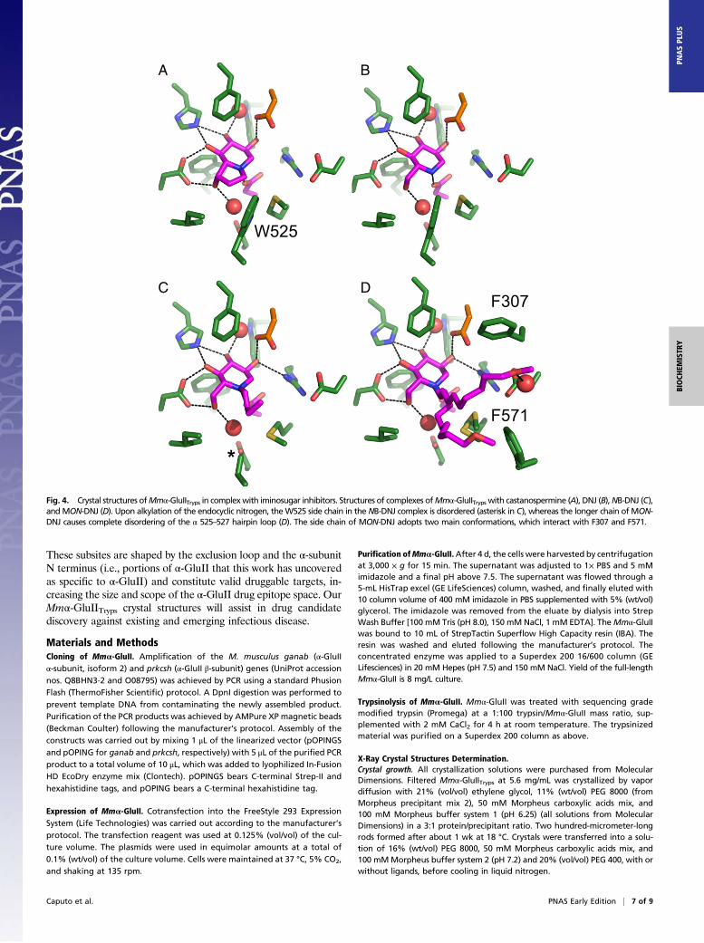
Fig. 4. Crystal structures ofMmα-GluIITryps in complexwith iminosugar inhibitors. Structures of complexes ofMmα-GluIITryps with castanospermine (A), DNJ (B),NB-DNJ (C),and MON-DNJ (D). Upon alkylation of the endocyclic nitrogen, theW525 side chain in the NB-DNJ complex is disordered (asterisk in C), whereas the longer chain of MON-DNJ causes complete disordering of the α 525–527 hairpin loop (D). The side chain of MON-DNJ adopts two main conformations, which interact with F307 and F571.
Caputo et al. PNAS Early Edition | 7 of 9
BIOCH
EMISTR
YPN
ASPL
US
X-ray diffraction. Diffraction from Mmα-GluIITryps crystals was measured at theDiamond Light Source, except for the apo and NB-DNJ structures, data forwhich were collected on beamlines ID30-1 and BM14, respectively, at theEuropean Synchrotron Radiation Facility (ESRF). All experiments were carriedout at a temperature of 90 K in a stream of cryogenic N2 gas.
Data Processing, Structure Determination, and Refinement. X-ray diffractionimages were processed using the autoPROC (52) or the xia2 (53) suite ofprograms, both of which index and integrate with XDS (54), and were scaledand merged using the CCP4 (55) suite of programs: Pointless, Aimless, andTruncate. Molecular replacement leading to structure determination of theApo form was performed with Phaser (56), which is also part of the CCP4suite, run with the automated MR pipeline MrBUMP (57), using chain A ofPDB ID code 3L4Z as a search model. Model building was performed withCoot and Buccaneer (58, 59), and refinement was performed with autoBUSTER,using local structure similarity restraints (LSSR) (60, 61). Model validationwas carried out with internal modules of Coot and through the MolProbityserver (62). Initial sets of phases for the 5F-glucosyl fluoride, glucal, glu-cose, and iminosugar soaks were obtained by molecular replacement fromthe apo structure. Idealized coordinates and stereochemical dictionariesfor ligands not present in the autoBUSTER libraries and nonstandard li-gands were generated using the GRADE server, starting from SMILESstrings (grade.globalphasing.org/). Each ligand was docked in the unbiasedFo-Fc difference electron density map calculated from the phases at theend of the iterative protein-only model building and refinement. A finalround of refinement of the protein and docked ligand used ligand ste-reochemical restraints from the GRADE-generated dictionary. All figureswere produced in PyMOL.
SAXS of Mmα-GluII and Mmα-GluIITryps. SAXS data for Mmα-GluII and Mmα-GluIITryps were collected at the BM29 beamline at the ESRF. The wavelengthwas set at 0.992 Å, and transmission was at 100%, with images recorded on aPilatus 1M detector set to a distance of 2.886 m. Calibration was conductedwith measurements on albumin or glucose oxidase to derive molecularweights from intensity at zero scattering angle (I0) values. All measurementswere carried out in 150 mM NaCl and 20 mM Hepes at pH 7.4. A twofolddilution series with six concentrations between 4.12 mg/mL and 0.13 mg/mLwas measured for Mmα-GluIITryps, and a twofold dilution series with fiveconcentrations between 2.97 mg/mL and 0.17 mg/mL was measured for Mm
α-GluII. Thirty microliters of each sample was flowed through a quartzcapillary taking 10 × 1-s images. Automated image processing followed bybuffer subtraction, as part of the processing pipeline at the beamline,allowed scattering curves to be used for further data processing. SAXS datawere processed using the ATSAS (63) software suite. Using PRIMUS (64), forboth Mmα-GluII and Mmα-GluIITryps samples, the low-angle regions of thelow-concentration scattering curves were merged with the high-angle re-gions of the high-concentration profile. This merging procedure was done tocompensate for interparticle effects at high concentration. The radius ofgyration was determined using PRIMUS (64), and the maximum particle sizeDmax was calculated from the pair distribution function calculated by GNOM(65). Ten bead models were created for each structure by DAMMIN (66), andthen aligned and averaged using DAMAVER (67). DAMMIN was then used tocompare the averaged model against raw data using reduced χ2 values. Allmodels possess 0.9 < χ2 < 1.1 against raw data. The Mmα-GluIITryps crystalstructure was initially fitted to the Mmα-GluIITryps SAXS envelope usingSUPCOMB (68). Chimera (69) was used to convert the SAXS envelopes tomaps (command MOLMAP, using a 2.5-nm filter) and to superpose theMmα-GluII SAXS map to the Mmα-GluIITryps SAXS map and model.
The full, detailed methods for cloning, expression, purification, enzy-mology, HDX-MS, in planta confocal microscopy and (co)immunoprecipita-tions, X-ray crystal structure determination, and SAXS used in this study canbe found in SI Appendix, SI Materials and Methods.
ACKNOWLEDGMENTS. We thank Raymond Dwek, Mark Wormald, MaxCrispin, David Harris, and Kathryn Scott for helpful discussions and com-ments on the manuscript, and the members of the Zitzmann laboratory forassistance with molecular biology and protein chemistry. Louise Bird,Heather Rada, and Ray Owens (Oxford Protein Production Facility at theResearch Complex) helped with cloning and initial expression trials. We alsothank Justin Benesch and Shane Chandler (Department of Chemistry,University of Oxford) for HDX support and access to the mass spectrometer.Snezana Vasiljevi�c assisted with mammalian cell expression. David Stauntontook the size exclusion chromatography multiangle laser light scatteringmeasurements. The staff at beamlines I02, I03, I04, and I04-1 (Diamond LightSource) and at beamlines BM14, ID30-1, and BM29 (ESRF) helped with X-raydata collection. Jesse Gayk helped with glycan 2AA labeling. A.T.C. wasfunded by a Wellcome Trust 4-Year Studentship (097300/Z/11/Z). J.L.K. is aLerner–Fink Fellow in Medicinal Chemistry. N.Z. is a Fellow of MertonCollege, Oxford.
1. Wisskirchen K, Lucifora J, Michler T, Protzer U (2014) New pharmacological strategies
to fight enveloped viruses. Trends Pharmacol Sci 35(9):470–478.2. Sayce AC, Miller JL, Zitzmann N (2010) Targeting a host process as an antiviral ap-
proach against dengue virus. Trends Microbiol 18(7):323–330.3. Martinez JP, Sasse F, Brönstrup M, Diez J, Meyerhans A (2015) Antiviral drug discov-
ery: Broad-spectrum drugs from nature. Nat Prod Rep 32(1):29–48.4. Pieren M, Galli C, Denzel A, Molinari M (2005) The use of calnexin and calreticulin by
cellular and viral glycoproteins. J Biol Chem 280(31):28265–28271.5. Hammond C, Braakman I, Helenius A (1994) Role of N-linked oligosaccharide recog-
nition, glucose trimming, and calnexin in glycoprotein folding and quality control.
Proc Natl Acad Sci USA 91(3):913–917.6. Khoury GA, Baliban RC, Floudas CA (2011) Proteome-wide post-translational modifica-
tion statistics: Frequency analysis and curation of the swiss-prot database. Sci Rep 1:90.7. Ohtsubo K, Marth JD (2006) Glycosylation in cellular mechanisms of health and dis-
ease. Cell 126(5):855–867.8. D’Alessio C, Caramelo JJ, Parodi AJ (2010) UDP-GlC:glycoprotein glucosyltransferase-
glucosidase II, the ying-yang of the ER quality control. Semin Cell Dev Biol 21(5):491–499.9. Lauc G, Pezer M, Rudan I, Campbell H (2016) Mechanisms of disease: The human N-
glycome. Biochim Biophys Acta 1860(8):1574–1582.10. Trombetta ES, Simons JF, Helenius A (1996) Endoplasmic reticulum glucosidase II is
composed of a catalytic subunit, conserved from yeast to mammals, and a tightly
bound noncatalytic HDEL-containing subunit. J Biol Chem 271(44):27509–27516.11. Roth J, Ziak M, Zuber C (2003) The role of glucosidase II and endomannosidase in
glucose trimming of asparagine-linked oligosaccharides. Biochimie 85(3-4):287–294.12. Lucocq JM, Brada D, Roth J (1986) Immunolocalization of the oligosaccharide trim-
ming enzyme glucosidase II. J Cell Biol 102(6):2137–2146.13. Jeyakumar M, Dwek RA, Butters TD, Platt FM (2005) Storage solutions: Treating ly-
sosomal disorders of the brain. Nat Rev Neurosci 6(9):713–725.14. Chang J, Block TM, Guo J-T (2013) Antiviral therapies targeting host ER alpha-glucosidases:
Current status and future directions. Antiviral Res 99(3):251–260.15. Dalziel M, Crispin M, Scanlan CN, Zitzmann N, Dwek RA (2014) Emerging principles for
the therapeutic exploitation of glycosylation. Science 343(6166):1235681.16. Chang J, et al. (2013) Small molecule inhibitors of ER α-glucosidases are active against
multiple hemorrhagic fever viruses. Antiviral Res 98(3):432–440.17. Warfield KL, et al. (2015) A novel iminosugar UV-12 with activity against the diverse
viruses influenza and dengue (novel iminosugar antiviral for influenza and dengue).
Viruses 7(5):2404–2427.
18. Perry ST, et al. (2013) An iminosugar with potent inhibition of dengue virus infection
in vivo. Antiviral Res 98(1):35–43.19. Stavale EJ, Vu H, Sampath A, Ramstedt U, Warfield KL (2015) In vivo therapeutic
protection against influenza A (H1N1) oseltamivir-sensitive and resistant viruses by
the iminosugar UV-4. PLoS One 10(3):e0121662.20. Butters TD, Van den Broek L, Fleet G, Krulle TM (2000) Molecular requirements of
imino sugars for the selective control of N-linked glycosylation and glycosphingolipid
biosynthesis. Tetrahedron 11(1):113–124.21. Trombetta ES, Fleming KG, Helenius A (2001) Quaternary and domain structure of
glycoprotein processing glucosidase II. Biochemistry 40(35):10717–10722.22. Feng J, Romaniouk AV, Samal SK, Vijay IK (2004) Processing enzyme glucosidase II:
Proposed catalytic residues and developmental regulation during the ontogeny of
the mouse mammary gland. Glycobiology 14(10):909–921.23. Lu X, et al. (2009) Uncoupling of sustained MAMP receptor signaling from early
outputs in an Arabidopsis endoplasmic reticulum glucosidase II allele. Proc Natl Acad
Sci USA 106(52):22522–22527.24. Pelletier MF, et al. (2000) The heterodimeric structure of glucosidase II is required for
its activity, solubility, and localization in vivo. Glycobiology 10(8):815–827.25. Arendt CW, Ostergaard HL (2000) Two distinct domains of the beta-subunit of glu-
cosidase II interact with the catalytic alpha-subunit. Glycobiology 10(5):487–492.26. Wilkinson BM, Purswani J, Stirling CJ (2006) Yeast GTB1 encodes a subunit of gluco-
sidase II required for glycoprotein processing in the endoplasmic reticulum. J Biol
Chem 281(10):6325–6333.27. Stigliano ID, Alculumbre SG, Labriola CA, Parodi AJ, D’Alessio C (2011) Glucosidase II
and N-glycan mannose content regulate the half-lives of monoglucosylated species in
vivo. Mol Biol Cell 22(11):1810–1823.28. Arendt CW, Ostergaard HL (1997) Identification of the CD45-associated 116-kDa and
80-kDa proteins as the alpha- and beta-subunits of alpha-glucosidase II. J Biol Chem
272(20):13117–13125.29. Deprez P, Gautschi M, Helenius A (2005) More than one glycan is needed for ER
glucosidase II to allow entry of glycoproteins into the calnexin/calreticulin cycle. Mol
Cell 19(2):183–195.30. Totani K, Ihara Y, Matsuo I, Ito Y (2006) Substrate specificity analysis of endoplasmic
reticulum glucosidase II using synthetic high mannose-type glycans. J Biol Chem
281(42):31502–31508.31. Ito Y, Takeda Y (2012) Analysis of glycoprotein processing in the endoplasmic re-
ticulum using synthetic oligosaccharides. Proc Jpn Acad Ser B Phys Bio Sci 88(2):31–40.
8 of 9 | www.pnas.org/cgi/doi/10.1073/pnas.1604463113 Caputo et al.
32. Olson LJ, et al. (2015) Crystal structure and functional analyses of the lectin domainof glucosidase II: Insights into oligomannose recognition. Biochemistry 54(26):4097–4111.
33. Olson LJ, et al. (2013) Structure of the lectin mannose 6-phosphate receptor homol-ogy (MRH) domain of glucosidase II, an enzyme that regulates glycoprotein foldingquality control in the endoplasmic reticulum. J Biol Chem 288(23):16460–16475.
34. Petrescu AJ, et al. (1997) The solution NMR structure of glucosylated N-glycans in-volved in the early stages of glycoprotein biosynthesis and folding. EMBO J 16(14):4302–4310.
35. Totani K, Ihara Y, Matsuo I, Ito Y (2008) Effects of macromolecular crowding onglycoprotein processing enzymes. J Am Chem Soc 130(6):2101–2107.
36. Koshland DE (1953) Stereochemistry and the mechanism of enzymatic reactions. BiolRev Camb Philos Soc 28(4):416–436.
37. Sinnott ML (1990) Catalytic mechanism of enzymic glycosyl transfer. Chem Rev 90(7):1171–1202.
38. Buchowiecka A, Bielecki S (2009) Determination of the regiochemistry of D-glucalglucosylation by endo-β-1,3-glucanase GA cellulomonas cellulans using CI MS. BiocatalBiotransformation 21(1):1–5.
39. McCarter JD, Withers SG (1996) Unequivocal identification of Asp-214 as the catalyticnucleophile of Saccharomyces cerevisiae alpha-glucosidase using 5-fluoro glycosylfluorides. J Biol Chem 271(12):6889–6894.
40. Mayes HB, Broadbelt LJ, Beckham GT (2014) How sugars pucker: Electronic structurecalculations map the kinetic landscape of five biologically paramount monosaccha-rides and their implications for enzymatic catalysis. J Am Chem Soc 136(3):1008–1022.
41. Kaushal GP, Pastuszak I, Hatanaka K, Elbein AD (1990) Purification to homogeneityand properties of glucosidase II from mung bean seedlings and suspension-culturedsoybean cells. J Biol Chem 265(27):16271–16279.
42. McIntosh LP, et al. (1996) The pKa of the general acid/base carboxyl group of a gly-cosidase cycles during catalysis: A 13C-NMR study of bacillus circulans xylanase.Biochemistry 35(31):9958–9966.
43. Dixon M, Webb EC (1964) Enzymes (Academic, New York).44. Satoh T, Toshimori T, Yan G, Yamaguchi T, Kato K (2016) Structural basis for two-step
glucose trimming by glucosidase II involved in ER glycoprotein quality control. Sci Rep6:20575.
45. Alonso JM, Santa-Cecilia A, Calvo P (1991) Glucosidase II from rat liver microsomes.Kinetic model for binding and hydrolysis. Biochem J 278(Pt 3):721–727.
46. Alonso JM, Santa-Cecilia A, Calvo P (1993) Effect of bromoconduritol on glucosidase IIfrom rat liver. A new kinetic model for the binding and hydrolysis of the substrate.Eur J Biochem 215(1):37–42.
47. Brada D, Dubach UC (1984) Isolation of a homogeneous glucosidase II from pig kidneymicrosomes. Eur J Biochem 141(1):149–156.
48. Sayce AC, et al. (2016) Iminosugars inhibit dengue virus production via inhibition ofER alpha-glucosidases-not glycolipid processing enzymes. PLoS Negl Trop Dis 10(3):e0004524.
49. Gloster TM, Vocadlo DJ (2012) Developing inhibitors of glycan processing enzymes astools for enabling glycobiology. Nat Chem Biol 8(8):683–694.
50. Sim L, et al. (2010) New glucosidase inhibitors from an ayurvedic herbal treatment fortype 2 diabetes: Structures and inhibition of human intestinal maltase-glucoamylasewith compounds from Salacia reticulata. Biochemistry 49(3):443–451.
51. Warfield KL, et al. (2016) Inhibition of endoplasmic reticulum glucosidases is requiredfor in vitro and in vivo dengue antiviral activity by the iminosugar UV-4. Antiviral Res129:93–98.
52. Vonrhein C, et al. (2011) Data processing and analysis with the autoPROC toolbox.Acta Crystallogr D Biol Crystallogr 67(Pt 4):293–302.
53. Winter G, Lobley CMC, Prince SM (2013) Decision making in xia2. Acta Crystallogr DBiol Crystallogr 69(Pt 7):1260–1273.
54. Kabsch W (2010) XDS. Acta Crystallogr D Biol Crystallogr 66(Pt 2):125–132.55. Winn MD, et al. (2011) Overview of the CCP4 suite and current developments. Acta
Crystallogr D Biol Crystallogr 67(Pt 4):235–242.56. McCoy AJ, et al. (2007) Phaser crystallographic software. J Appl Crystallogr 40(Pt 4):
658–674.57. Keegan RM, Winn MD (2008) MrBUMP: An automated pipeline for molecular re-
placement. Acta Crystallogr D Biol Crystallogr 64(Pt 1):119–124.58. Emsley P, Lohkamp B, Scott WG, Cowtan K (2010) Features and development of Coot.
Acta Crystallogr D Biol Crystallogr 66(Pt 4):486–501.59. Cowtan K (2006) The Buccaneer software for automated model building. 1. Tracing
protein chains. Acta Crystallogr D Biol Crystallogr 62(Pt 9):1002–1011.60. Blanc E, et al. (2004) Refinement of severely incomplete structures with maximum
likelihood in BUSTER-TNT. Acta Crystallogr D Biol Crystallogr 60(Pt 12 Pt 1):2210–2221.61. Smart OS, et al. (2012) Exploiting structure similarity in refinement: Automated NCS
and target-structure restraints in BUSTER. Acta Crystallogr D Biol Crystallogr 68(Pt 4):368–380.
62. Chen VB, Wedell JR, Wenger RK, Ulrich EL, Markley JL (2015) MolProbity for themasses-of data. J Biomol NMR 63(1):77–83.
63. Petoukhov MV, et al. (2012) New developments in the ATSAS program package forsmall-angle scattering data analysis. J Appl Crystallogr 45(Pt 2):342–350.
64. Konarev PV, et al. (2003) PRIMUS: A Windows PC-based system for small-angle scat-tering data analysis. J Appl Crystallogr 36(5):1277–1282.
65. Svergun DI, Cr IU (1992) Determination of the regularization parameter in indirect-transform methods using perceptual criteria. J Appl Crystallogr 25(4):495–503.
66. Svergun DI (1999) Restoring low resolution structure of biological macromoleculesfrom solution scattering using simulated annealing. Biophys J 76(6):2879–2886.
67. Volkov VV, Svergun DI, Cr IU (2003) Uniqueness of ab initio shape determination insmall-angle scattering. J Appl Crystallogr 36(3):860–864.
68. Kozin MB, Svergun DI, Cr IU (2001) Automated matching of high- and low-resolutionstructural models. J Appl Crystallogr 34(1):33–41.
69. Pettersen EF, et al. (2004) UCSF Chimera–a visualization system for exploratory re-search and analysis. J Comput Chem 25(13):1605–1612.
Caputo et al. PNAS Early Edition | 9 of 9
BIOCH
EMISTR
YPN
ASPL
US


![The glucose-lowering effects of α-glucosidase inhibitor ...The glucose-lowering effectsof α-glucosidase inhibitor require a bile ... transport and reab-sorption [14, 15]. Recent](https://static.fdocument.org/doc/165x107/5f0a34737e708231d42a84ec/the-glucose-lowering-effects-of-glucosidase-inhibitor-the-glucose-lowering.jpg)