![surpass all possibilities - Waters Corporation · surpass all possibilities [ CORTECS 2.7 µm COLUMNS ] A SOLID-CORE PARTICLE COLUMN THAT LIVES UP TO ITS POTENTIAL. 2.7 m SLIDCORE](https://static.fdocument.org/doc/165x107/5e87c3eaed583a7aec5a497b/surpass-all-possibilities-waters-corporation-surpass-all-possibilities-cortecs.jpg)
Structure and Function of the Nine Formed Elements · 7.5 µm 2.0 µm Sectional view Surfaceview...
49
Chapter 19 Structure and Function of the Nine Formed Elements
Transcript of Structure and Function of the Nine Formed Elements · 7.5 µm 2.0 µm Sectional view Surfaceview...
Chapter 19
Structure and Functionof the Nine Formed Elements
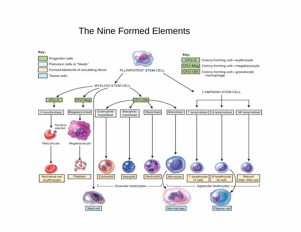
The Nine Formed Elements
7.5 µm
2.0 µm
Sectional view
Surfaceview
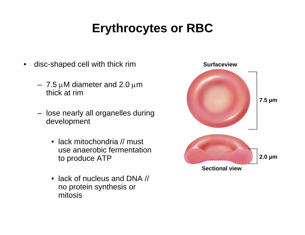
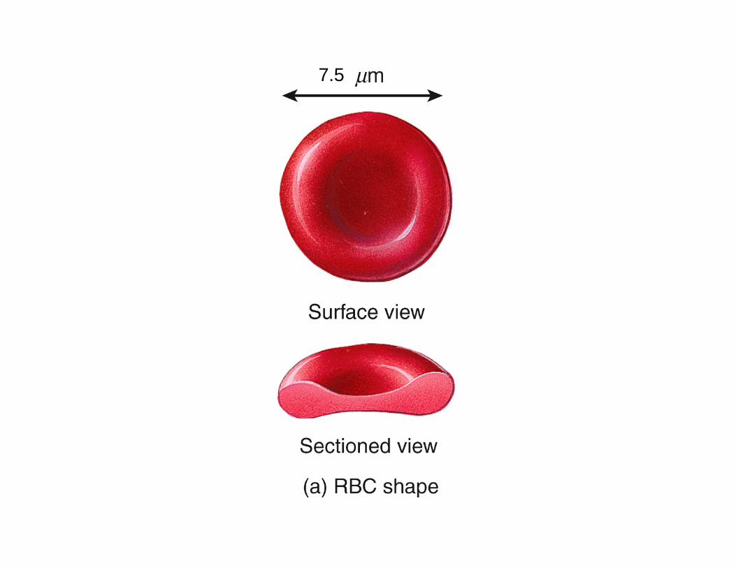
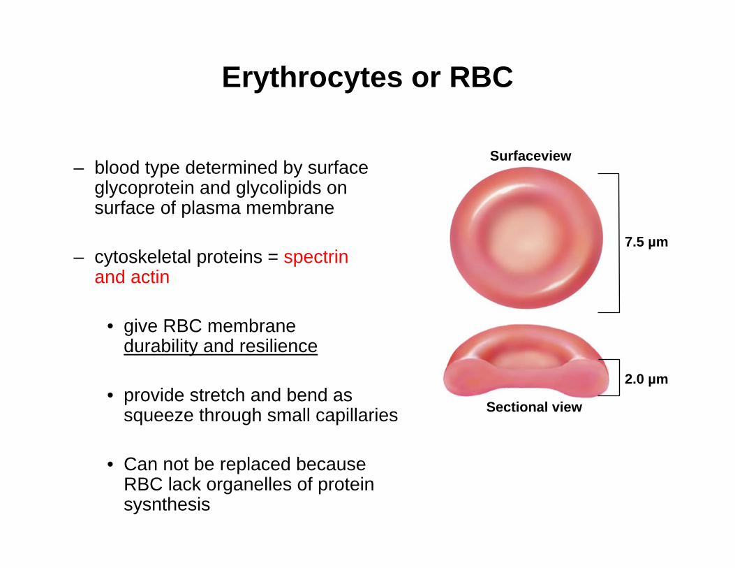
Erythrocytes or RBC
• disc-shaped cell with thick rim
– 7.5 μM diameter and 2.0 μm thick at rim
– lose nearly all organelles during development
• lack mitochondria // must use anaerobic fermentation to produce ATP
• lack of nucleus and DNA // no protein synthesis or mitosis
7.5
7.5 µm
2.0 µm
Sectional view
Surfaceview
Erythrocytes or RBC
– blood type determined by surface glycoprotein and glycolipids on surface of plasma membrane
– cytoskeletal proteins = spectrinand actin
• give RBC membranedurability and resilience
• provide stretch and bend as squeeze through small capillaries
• Can not be replaced because RBC lack organelles of protein sysnthesis
• 2.5 million RBCs are produced per second
• average lifespan of about 120 days
• development takes 3-5 days /// reduction in cell size, increase in cell number, synthesis of hemoglobin and loss of nucleus
• first committed cell - erythrocyte colony forming unit // has receptors for erythropoietin (EPO) from kidneys
• erythroblasts (normoblast) multiply and synthesize hemoglobin
• As erythroblast mature they discard their nucleus and become a reticulocyte
– named for fine network of endoplasmic reticulum still in cytoplasm // 0.5 to 1.5% of circulating RBCs are reticulocytes
Erythrocytes or RBC

Erythrocytes
• principal function /// carry oxygen from lungs to cellular tissues of body
– Note most of the carbon dioxide transported as bicarbonate in plasma from tissue to lungs
• insufficient RBC function may result in necrosis within 4 – 5 minutes due to lack of oxygen and too much CO2
Erythrocytes
7 µm
Capillarywall
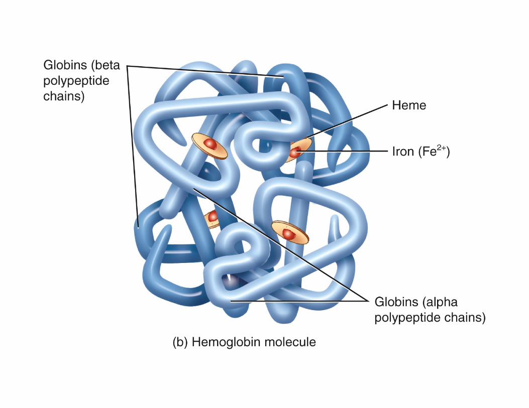
Hemoglobin (Hb) Structure
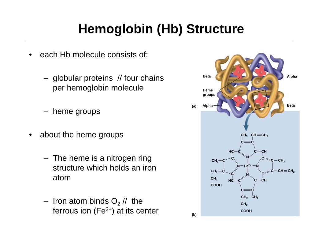
• each Hb molecule consists of:
– globular proteins // four chains per hemoglobin molecule
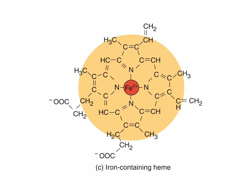
– heme groups
• about the heme groups
– The heme is a nitrogen ring structure which holds an iron atom
– Iron atom binds O2 // the ferrous ion (Fe2+) at its center
(a)
(b)
C
CH3
C
C
C
C C
C
C
CC
CC
N
N
NN
CH
CH
CH
CH
CH2
COOH
CH3 CH3
CH2
CH2
CH2
COOH
CH2 CH3
HC
C
C C
CHC
Fe2+
CH2
Beta
Alpha
Alpha
Beta
Hemegroups
Hemoglobin (Hb) Structure
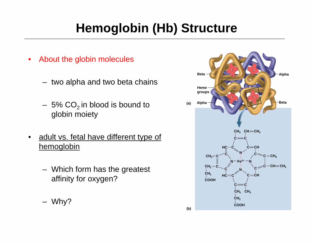
• About the globin molecules
– two alpha and two beta chains
– 5% CO2 in blood is bound to globin moiety
• adult vs. fetal have different type of hemoglobin
– Which form has the greatest affinity for oxygen?
– Why?
(a)
(b)
C
CH3
C
C
C
C C
C
C
CC
CC
N
N
NN
CH
CH
CH
CH
CH2
COOH
CH3 CH3
CH2
CH2
CH2
COOH
CH2 CH3
HC
C
C C
CHC
Fe2+
CH2
Beta
Alpha
Alpha
Beta
Hemegroups
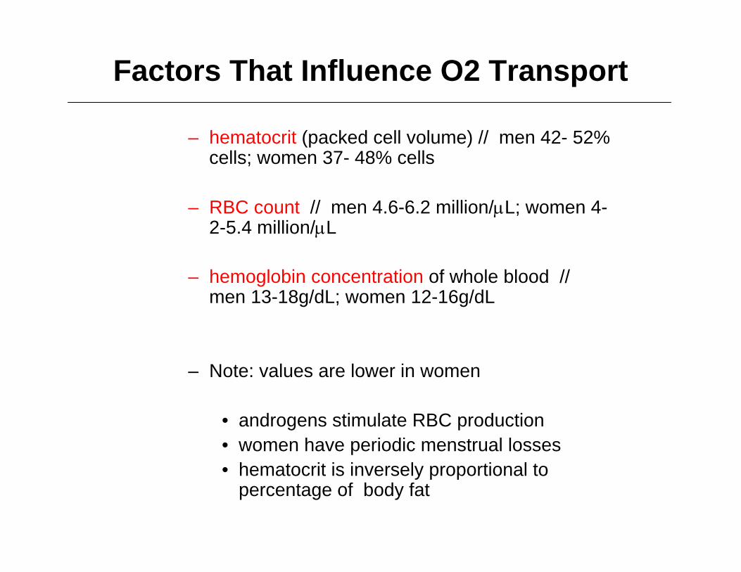
Factors That Influence O2 Transport
– hematocrit (packed cell volume) // men 42- 52% cells; women 37- 48% cells
– RBC count // men 4.6-6.2 million/μL; women 4-2-5.4 million/μL
– hemoglobin concentration of whole blood // men 13-18g/dL; women 12-16g/dL
– Note: values are lower in women
• androgens stimulate RBC production • women have periodic menstrual losses• hematocrit is inversely proportional to
percentage of body fat
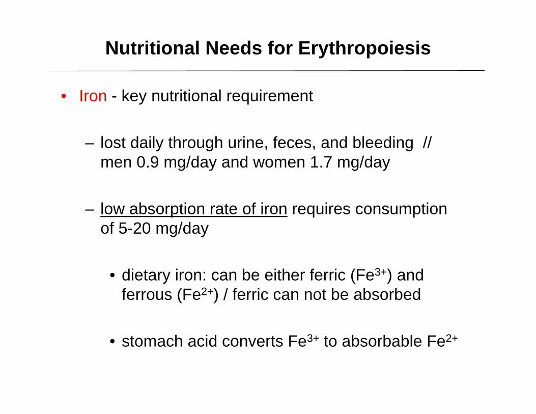
Nutritional Needs for Erythropoiesis
• Iron - key nutritional requirement
– lost daily through urine, feces, and bleeding // men 0.9 mg/day and women 1.7 mg/day
– low absorption rate of iron requires consumption of 5-20 mg/day
• dietary iron: can be either ferric (Fe3+) and ferrous (Fe2+) / ferric can not be absorbed
• stomach acid converts Fe3+ to absorbable Fe2+
Nutritional Needs for Erythropoiesis
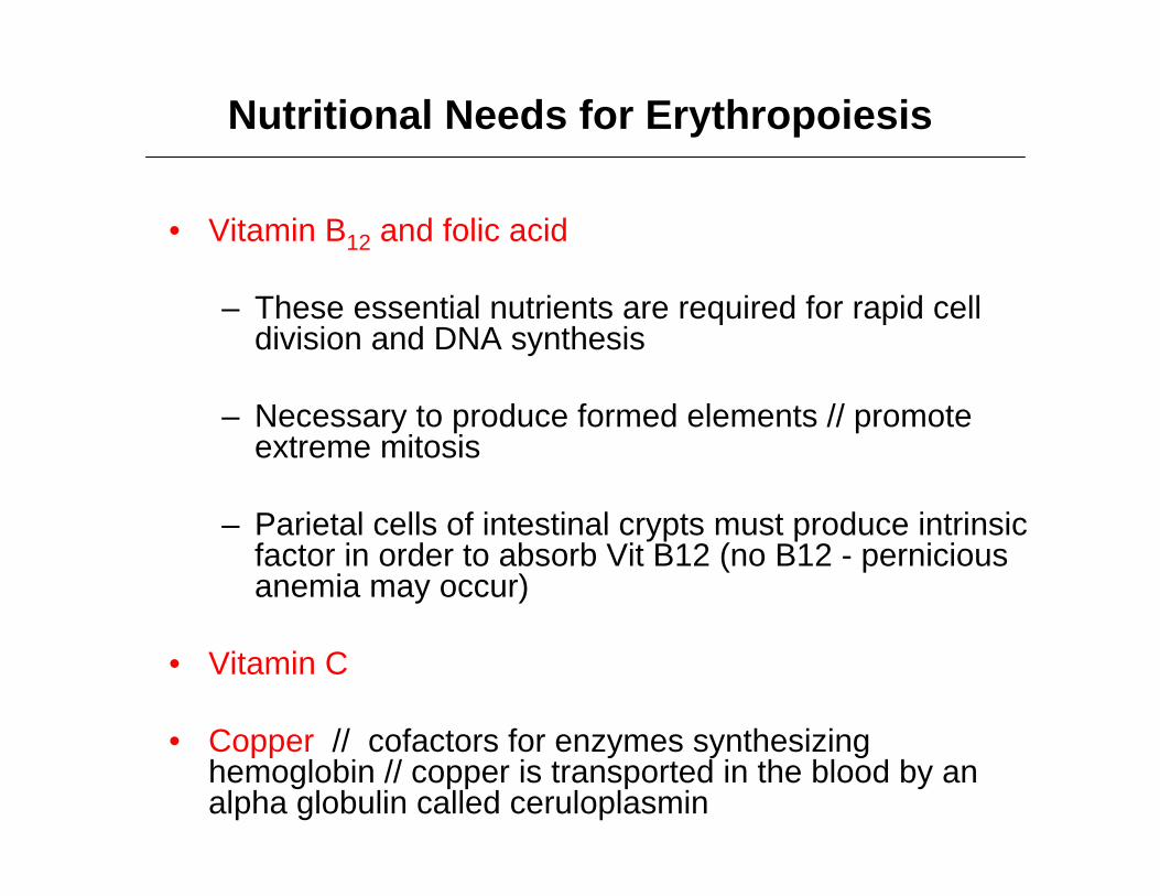
• Vitamin B12 and folic acid
– These essential nutrients are required for rapid cell division and DNA synthesis
– Necessary to produce formed elements // promote extreme mitosis
– Parietal cells of intestinal crypts must produce intrinsic factor in order to absorb Vit B12 (no B12 - pernicious anemia may occur)
• Vitamin C
• Copper // cofactors for enzymes synthesizing hemoglobin // copper is transported in the blood by an alpha globulin called ceruloplasmin
Regulating Erythrocyte Homeostasis
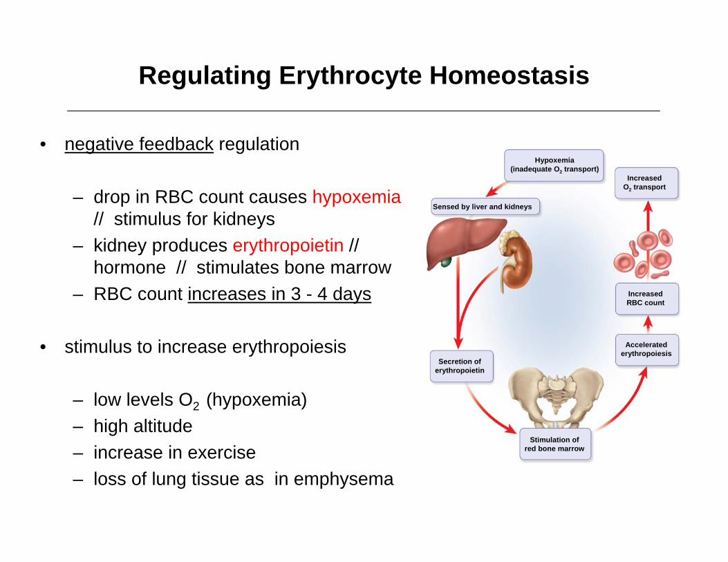
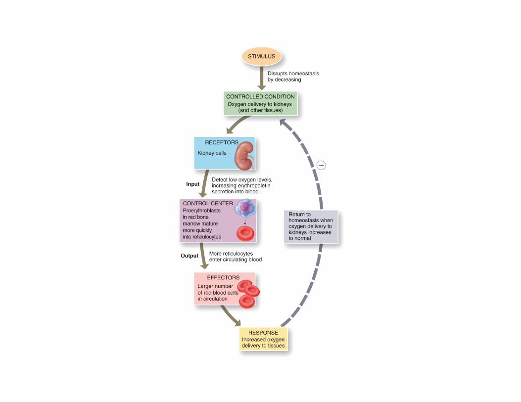
• negative feedback regulation
– drop in RBC count causes hypoxemia // stimulus for kidneys
– kidney produces erythropoietin // hormone // stimulates bone marrow
– RBC count increases in 3 - 4 days
• stimulus to increase erythropoiesis
– low levels O2 (hypoxemia)– high altitude– increase in exercise– loss of lung tissue as in emphysema
leaves
Hypoxemia(inadequate O2 transport)
Sensed by liver and kidneys
Secretion oferythropoietin
Acceleratederythropoiesis
IncreasedRBC count
IncreasedO2 transport
Stimulation ofred bone marrow
Erythrocytes Death and Disposal
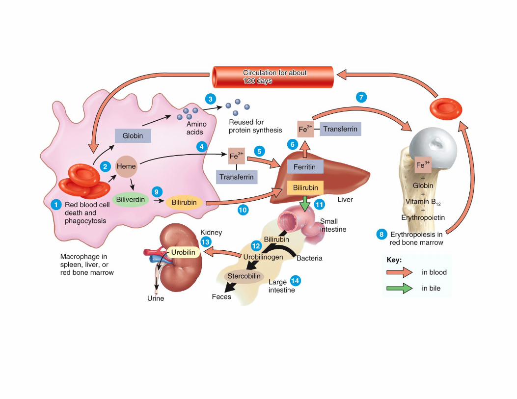
• RBCs lyse in the narrow (2 micrometer) capillaries found in the spleen = graveyard for old RBC
• High concentration of macrophages in spleen // resident phagocytes
– digest and recycle membranes
– separate heme from globin
• globins hydrolyzed into amino acids
• iron removed from heme
Erythrocytes Death and Disposal
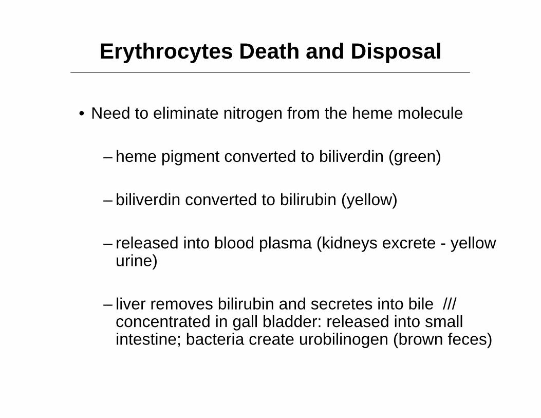
• Need to eliminate nitrogen from the heme molecule
– heme pigment converted to biliverdin (green)
– biliverdin converted to bilirubin (yellow)
– released into blood plasma (kidneys excrete - yellow urine)
– liver removes bilirubin and secretes into bile /// concentrated in gall bladder: released into small intestine; bacteria create urobilinogen (brown feces)
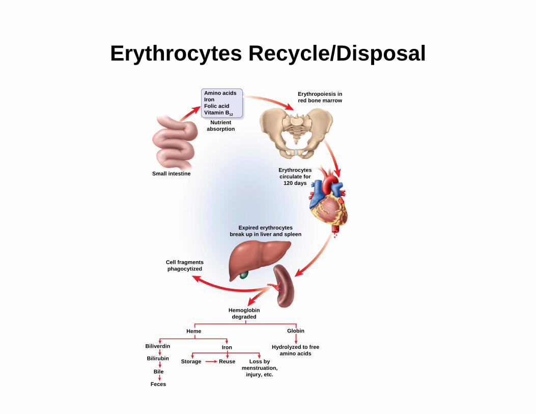
Erythrocytes Recycle/Disposal
Small intestine
GlobinHeme
IronBiliverdin
Bilirubin
Bile
Feces
Storage Reuse Loss bymenstruation,
injury, etc.
Amino acidsIronFolic acidVitamin B12
Nutrientabsorption
Erythrocytescirculate for
120 days
Expired erythrocytesbreak up in liver and spleen
Cell fragmentsphagocytized
Erythropoiesis inred bone marrow
Hemoglobindegraded
Hydrolyzed to freeamino acids
Erythrocyte Disorder
• Polycythemia = an excess of RBCs
– primary polycythemia (polycythemia vera) // cancer of erythropoietic cell line in red bone marrow // RBC count as high as 11 million/μL; hematocrit 80% // note – erythropoietin low concentration in blood
– secondary polycythemia // from dehydration, emphysema, high altitude, or physical conditioning // RBC count up to 8 million/μL // note – erythropoietin high concentration in blood
Dangers Associated with Polycythemia
– increased blood volume
– pressure, viscosity
– can lead to embolism
– stroke
– increase in blood viscosity
– heart failure
Anemia
• Not able to supply oxygen to tissue
• Three main categories
– Inadequate erythropoiesis (or failure to produce functional hemoglobin –e.g. sickle cell anemia)
– Hemorrhagic anemia
– Hemolytic anemia
Anemia Types
• Iron-deficiency anemia
• Pernicious anemia
– Problem often lack of intrinsic factor // required to carry B12across mucosa / stomach glands fail to produce intrinsic factor
– Vitamin B12 deficiency (vitamin usually present in diet / meat)
• Sickle cell anemia
• Hypoplastic anemia
• Aplastic anemia
Anemia
• Three potential consequences:
– Hypoxia // oxygen deprivation / low energy / if severe result in necrosis
– Reduced blood osmolarity // causes edema
– Reduced blood viscosity
• little blood resistance• heart beats faster • may lead to low blood volume and viscosity
to lead to low blood pressure• leads to cardiac failure
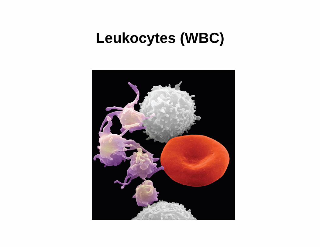
Leukocytes (WBC)
Leukocytes (WBCs)
• least abundant of all the formed elements // 5,000 to 10,000 WBCs/μL
• Primary function = protect against infectious microorganisms and other pathogens
• WBCs have conspicuous nucleus
• spend only a few hours in the blood stream before migrating out of blood and into connective tissue (i.e. reticuloendothelial system)
• retain their organelles for protein synthesis
• All WBC have granules but some cells don’t stain!

Leukocytes (WBCs)
• All WBC have granules in their cytoplasm but some cell’s granules don’t stain!
– all WBCs have lysosomes called nonspecific (azurophilic) granules /// these don’t stain so called inconspicuous (cytoplasm looks clear) known as the agranulocytes
– the granulocytes have specific granules that stain // contain enzymes and other chemicals employed in defense against pathogens
Types of Leukocytes
• Granulocytes // these cells stain // known as the “NEBs”
– neutrophils (60-70– eosinophils (2-4%) – basophils (<1%)
• Agranulocytes // these don’t stain
– lymphocytes (25-33%) – monocytes (3-8%)
• How to remember WBC ranking = Never let monkeys eat bananas
The “NEB”
The “LM”
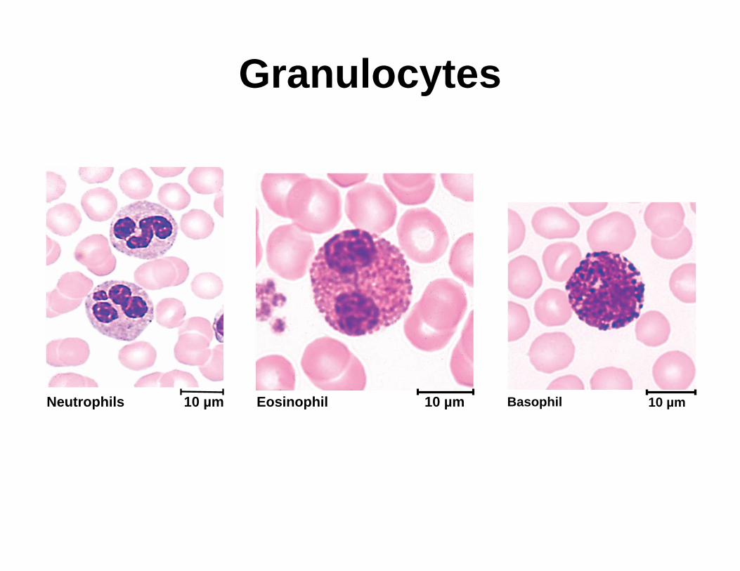
Granulocytes
Neutrophils 10 µm Eosinophil 10 µm Basophil 10 µm
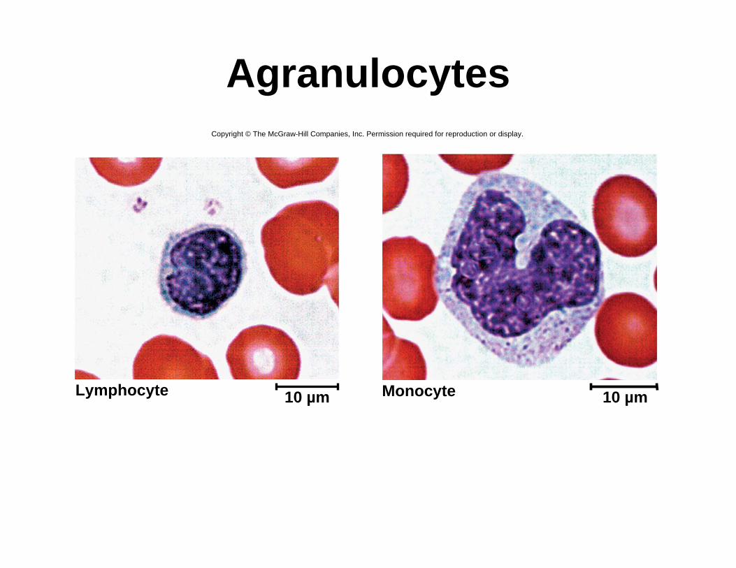
Agranulocytes
Lymphocyte 10 µm Monocyte 10 µm
Copyright © The McGraw-Hill Companies, Inc. Permission required for reproduction or display.
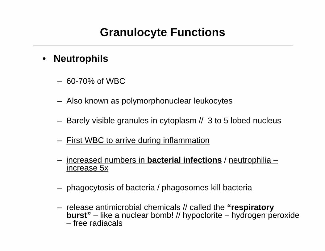
Granulocyte Functions
• Neutrophils
– 60-70% of WBC
– Also known as polymorphonuclear leukocytes
– Barely visible granules in cytoplasm // 3 to 5 lobed nucleus
– First WBC to arrive during inflammation
– increased numbers in bacterial infections / neutrophilia –increase 5x
– phagocytosis of bacteria / phagosomes kill bacteria
– release antimicrobial chemicals // called the “respiratory burst” – like a nuclear bomb! // hypoclorite – hydrogen peroxide – free radiacals
Insert art from Clinical Case onp. 463
If possible on this slide, include title:Oxidative Burst
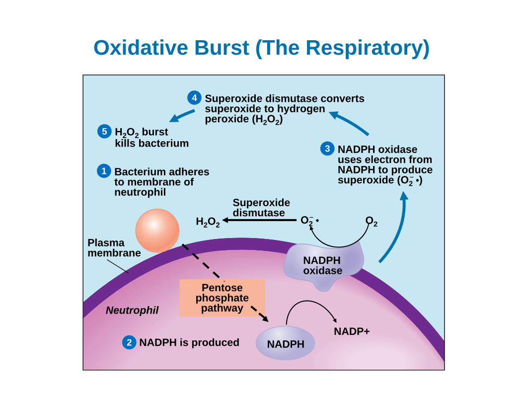
Oxidative Burst (The Respiratory)
H2O2 burstkills bacterium
Superoxide dismutase convertssuperoxide to hydrogenperoxide (H2O2)
NADPH oxidaseuses electron from NADPH to produce superoxide (O2 •)
Bacterium adheres to membrane of neutrophil
Superoxidedismutase O2 • O2H2O2
Neutrophil
NADPH is produced
NADPH oxidase
NADPHNADP+
Plasma membrane
Pentose phosphate pathway
1
2
3
4
5
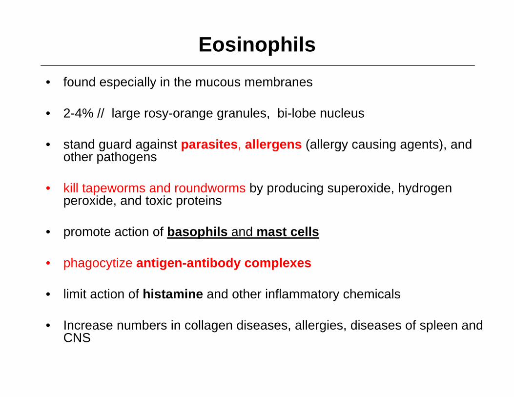
Eosinophils• found especially in the mucous membranes
• 2-4% // large rosy-orange granules, bi-lobe nucleus
• stand guard against parasites, allergens (allergy causing agents), and other pathogens
• kill tapeworms and roundworms by producing superoxide, hydrogen peroxide, and toxic proteins
• promote action of basophils and mast cells
• phagocytize antigen-antibody complexes
• limit action of histamine and other inflammatory chemicals
• Increase numbers in collagen diseases, allergies, diseases of spleen and CNS
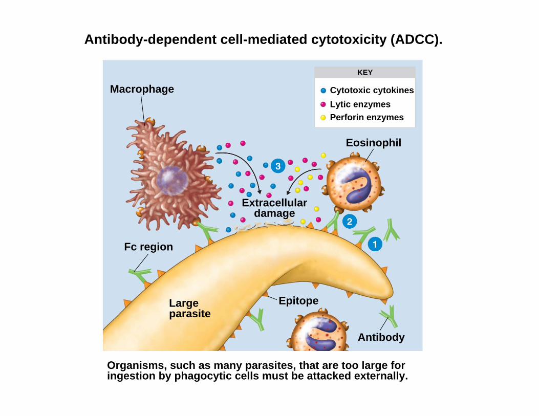
Antibody-dependent cell-mediated cytotoxicity (ADCC).
Organisms, such as many parasites, that are too large for ingestion by phagocytic cells must be attacked externally.
Cytotoxic cytokinesLytic enzymesPerforin enzymes
KEY
Macrophage
Fc region
Large parasite
Eosinophil
Extracellulardamage
Epitope
Antibody
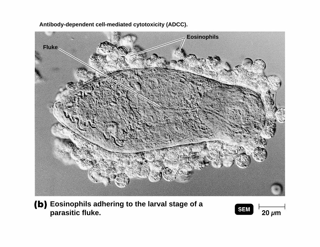
Antibody-dependent cell-mediated cytotoxicity (ADCC).
Eosinophils adhering to the larval stage of a parasitic fluke.
Fluke
Eosinophils
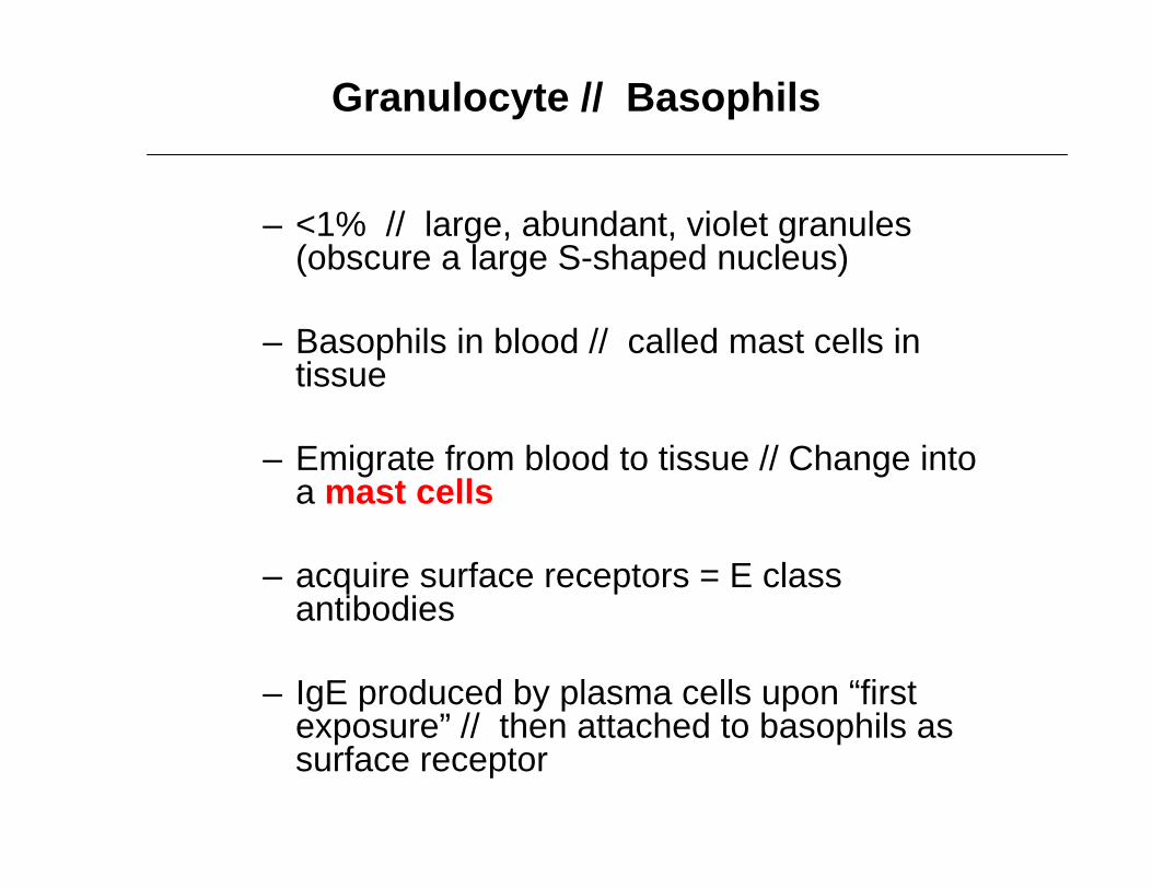
Granulocyte // Basophils
– <1% // large, abundant, violet granules (obscure a large S-shaped nucleus)
– Basophils in blood // called mast cells in tissue
– Emigrate from blood to tissue // Change into a mast cells
– acquire surface receptors = E class antibodies
– IgE produced by plasma cells upon “first exposure” // then attached to basophils as surface receptor
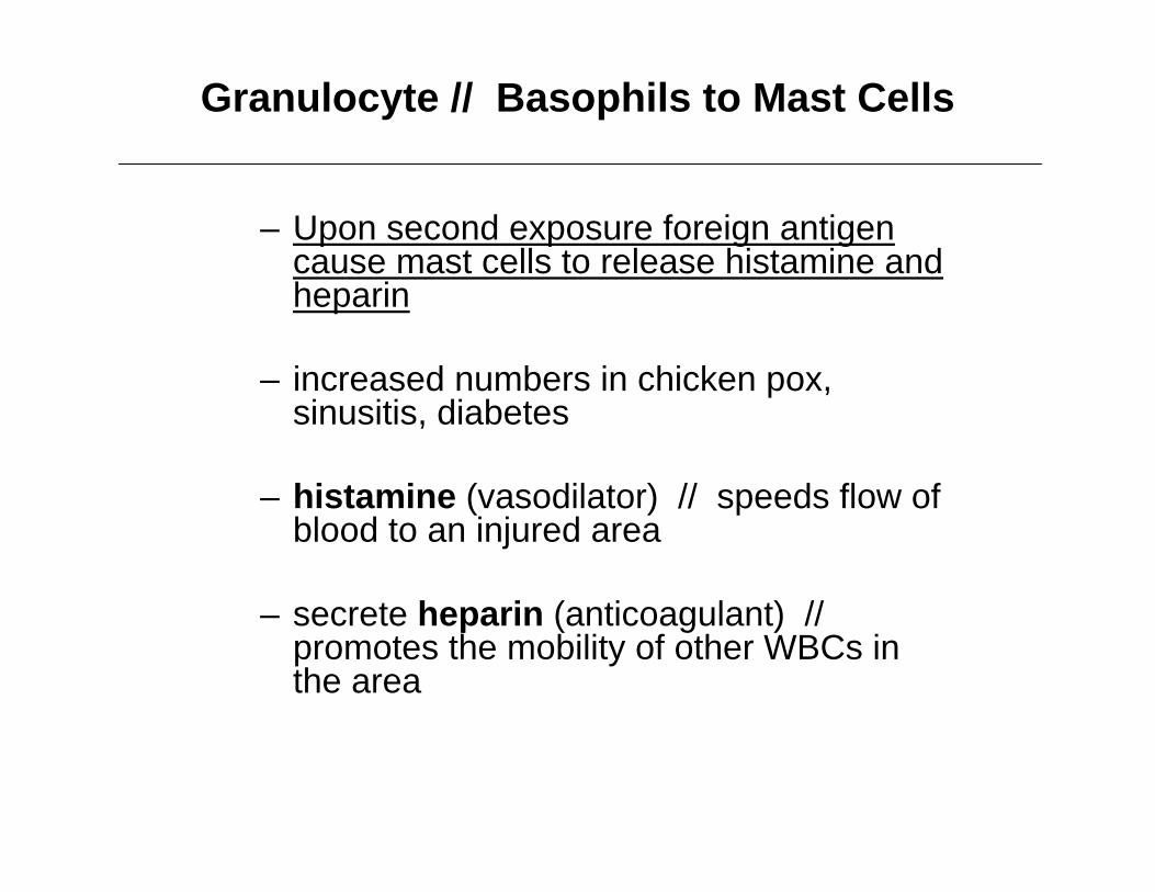
Granulocyte // Basophils to Mast Cells
– Upon second exposure foreign antigen cause mast cells to release histamine and heparin
– increased numbers in chicken pox, sinusitis, diabetes
– histamine (vasodilator) // speeds flow of blood to an injured area
– secrete heparin (anticoagulant) // promotes the mobility of other WBCs in the area
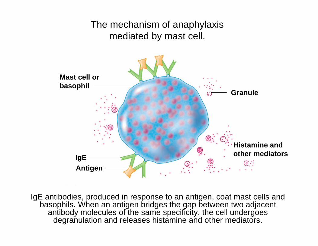
The mechanism of anaphylaxismediated by mast cell.
Granule
Histamine andother mediators
Mast cell orbasophil
AntigenIgE
IgE antibodies, produced in response to an antigen, coat mast cells and basophils. When an antigen bridges the gap between two adjacent
antibody molecules of the same specificity, the cell undergoes degranulation and releases histamine and other mediators.
Agranulocyte Function
Lymphocytes (T cells / B cells / NK cells)
– 25-33% // variable amounts of bluish cytoplasm (scanty to abundant); ovoid/round, uniform dark violet nucleus
– increased numbers in diverse infections and immune responses
– T lymphocytes able to destroy cells (cancer, foreign, and virally infected cells) // cellular immunity
– “present” antigens to activate other immune cells
– coordinate actions of other immune cells // cytokines = messenger molecules
– B lymphocytes secrete antibodies and provide immune memory // humeral immunity
Agranulocyte Function
Monocytes
– 3-8% // largest WBC; ovoid, kidney or horseshoe shaped nucleus
– increased numbers in viral infections and inflammation
– produce and secrete cytokines = group of molecules which regulate immune response
– leave bloodstream and transform into macrophages (i.e. big eater)
– phagocytize pathogens and debris // the “garbage collector”
– “present” antigens to activate other immune cells // antigen presenting cells (APCs)
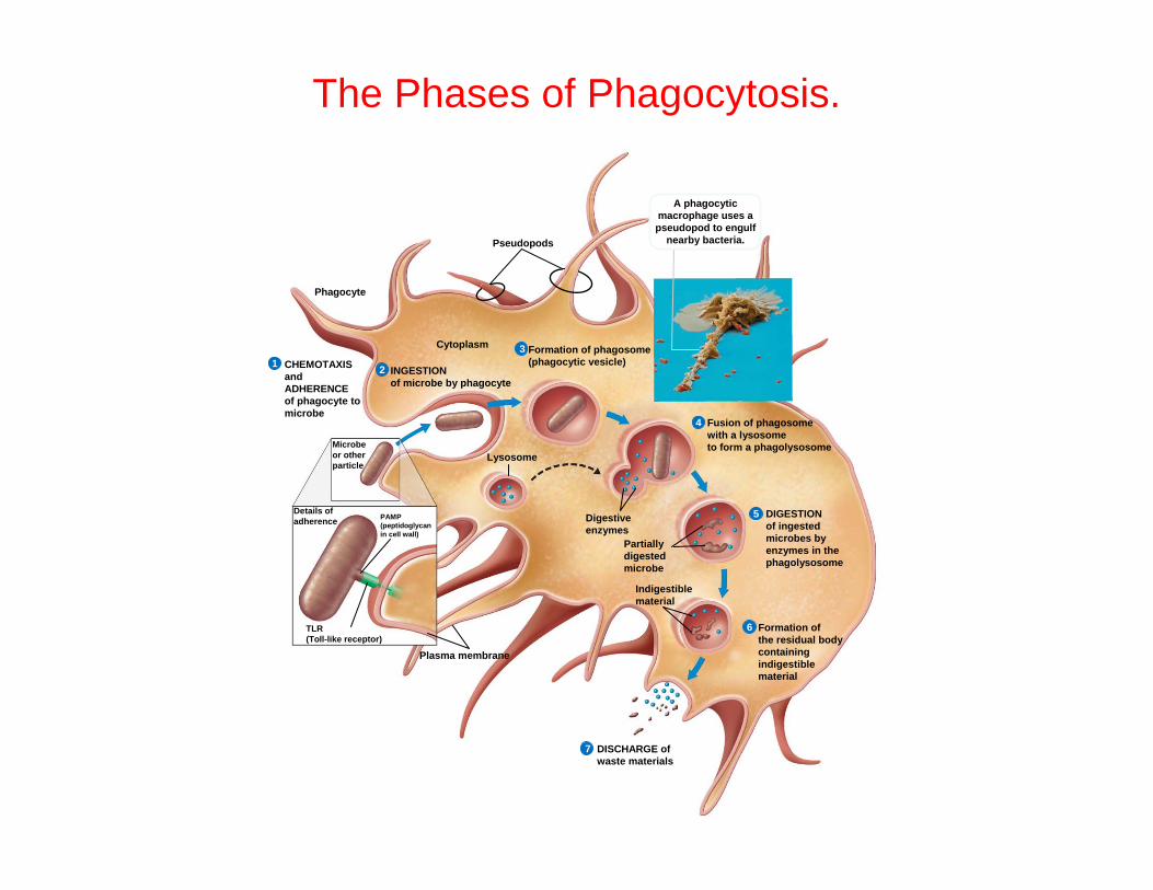
Pseudopods
Phagocyte
Cytoplasm
Microbeor otherparticle
Details ofadherence PAMP
(peptidoglycanin cell wall)
TLR(Toll-like receptor)
Lysosome
Digestiveenzymes
Indigestiblematerial
Plasma membrane
Partiallydigestedmicrobe
CHEMOTAXISandADHERENCEof phagocyte tomicrobe
1INGESTIONof microbe by phagocyte
2
Formation of phagosome(phagocytic vesicle)
3
Fusion of phagosomewith a lysosometo form a phagolysosome
4
DIGESTIONof ingestedmicrobes byenzymes in thephagolysosome
5
Formation ofthe residual bodycontainingindigestiblematerial
6
DISCHARGE ofwaste materials
7
A phagocyticmacrophage uses a pseudopod to engulf
nearby bacteria.
The Phases of Phagocytosis.

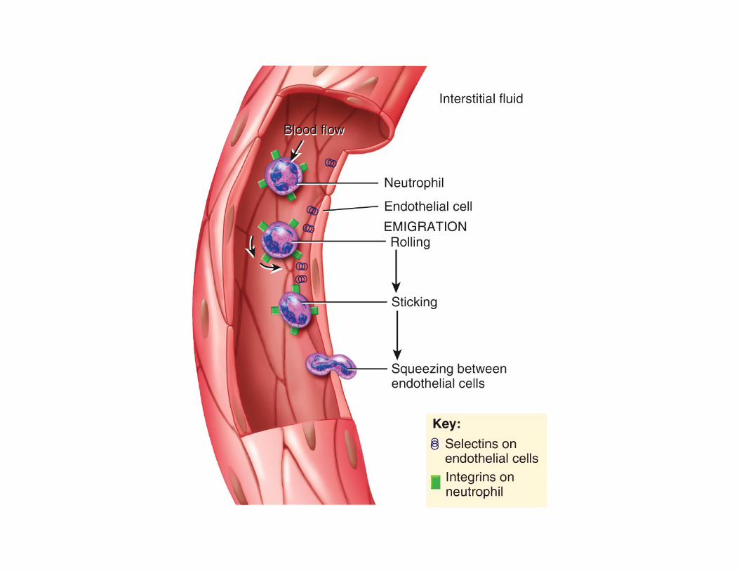
Diapedis: How Leukocytes Emigrate into Tissue Spaces
• Circulating WBCs do not stay in bloodstream
– Area of inflammation causes endothelial cells outer face to become “sticky” – results in margination
– Granulocytes (NEB) leave in 8 hours and live 5 days longer
– Monocytes leave in 20 hours, transform into macrophages and live for several years
– Lymphocytes provide long-term immunity // live for decades // continuously recycled from blood to tissue fluid to lymphatic system and back into the blood
Leukocyte Disorders
• leukopenia - low WBC count below 5000/μL
– causes: radiation, poisons, infectious disease
– effects: elevated risk of infection
• leukocytosis - high WBC count above 10,000/μL
– causes: infection, allergy and disease
– differential WBC count – identifies what percentage of the total WBC count consist of each type of leukocyte
Leukocyte Disorders
• Leukemia - cancer of hemopoietic tissue that usually produces an extraordinary high number of circulating leukocytes and their precursors
– myeloid leukemia – uncontrolled granulocyte production
– lymphoid leukemia - uncontrolled lymphocyte or monocyte production
– acute leukemia – appears suddenly, progresses rapidly, death within months
– chronic leukemia –undetected for months, survival time three years
– effects - normal cell percentages disrupted; impaired clotting; opportunistic infections
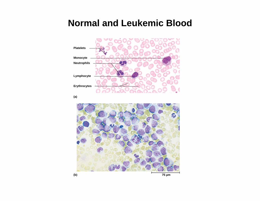
Normal and Leukemic Blood
Platelets
Neutrophils
Lymphocyte
Erythrocytes
(a)
Monocyte
(b) 75 µm
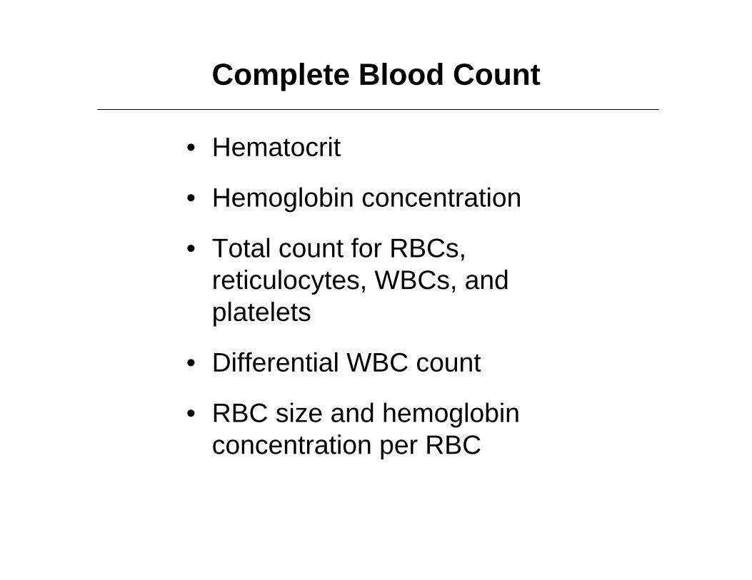
Complete Blood Count
• Hematocrit
• Hemoglobin concentration
• Total count for RBCs, reticulocytes, WBCs, and platelets
• Differential WBC count
• RBC size and hemoglobin concentration per RBC

![diap-12.ppt [Mode de compatibilité] · Rayonnement thermique: de 0.1 à 100 µm domaine qui inclut le rayonnement visible: de 0.4 à 0.7 µm Système idéal: le corps noir Propriétés:-il](https://static.fdocument.org/doc/165x107/5fd1fded5e423b7b434036dc/diap-12ppt-mode-de-compatibilit-rayonnement-thermique-de-01-100-m-domaine.jpg)