Structure and assembly of a paramyxovirus matrix protein · β-sandwich folds, suggesting gene...
5
Structure and assembly of a paramyxovirus matrix protein Anthony J. Battisti a,1 , Geng Meng a,1 , Dennis C. Winkler b , Lori W. McGinnes c , Pavel Plevka a , Alasdair C. Steven b , Trudy G. Morrison c , and Michael G. Rossmann a,2 a Department of Biological Sciences, Purdue University, West Lafayette, IN 47907-2032; b Laboratory of Structural Biology Research, National Institute of Arthritis and Musculoskeletal and Skin Diseases, National Institutes of Health, Bethesda, MD 20892-8025; and c Department of Microbiology and Physiological Systems/Program in Immunology and Virology, University of Massachusetts Medical School, Worcester, MA 01655-0122 Edited by Peter Palese, Mount Sinai School of Medicine, New York, NY, and approved July 26, 2012 (received for review June 15, 2012) Many pleomorphic, lipid-enveloped viruses encode matrix proteins that direct their assembly and budding, but the mechanism of this process is unclear. We have combined X-ray crystallography and cryoelectron tomography to show that the matrix protein of New- castle disease virus, a paramyxovirus and relative of measles virus, forms dimers that assemble into pseudotetrameric arrays that generate the membrane curvature necessary for virus budding. We show that the glycoproteins are anchored in the gaps between the matrix proteins and that the helical nucleocapsids are associated in register with the matrix arrays. About 90% of virions lack matrix arrays, suggesting that, in agreement with previous biological observations, the matrix protein needs to dissociate from the viral membrane during maturation, as is required for fusion and release of the nucleocapsid into the host’ s cytoplasm. Structure and sequence conservation imply that other paramyxovirus matrix pro- teins function similarly. T he structure of icosahedral viruses has been extensively stu- died by X-ray crystallography and by a combination of X-ray crystallography and cryoelectron microscopy, but less is known about the atomic structure of viral proteins in the context of pleomorphic viruses. Paramyxoviridae, Rhabdoviridae, Filoviridae, and Bornaviridae are pleomorphic viruses belonging to the order Mononegavirales. These enveloped viruses all have a linear, non- segmented, negative-sense, 10- to 20-kb–long RNA genome that is encapsidated by a nucleocapsid protein into a helical assembly (1). Paramyxoviruses include human pathogens that infect the re- spiratory system. Newcastle disease virus (NDV) is an avian para- myxovirus that is the prototypical species of the genus Avulavirus, belonging to the Paramyxovirinae subfamily. Poultry are especially susceptible to NDV infections, and some virulent strains have been classified by the US Department of Agriculture as select agents (2, 3). Paramyxoviruses (Fig. 1) are pleomorphic and encode at least six proteins (1). They all have two transmembrane glyco- proteins—the attachment protein termed HN (approximately 75 kDa), G, or H (depending upon the virus) and the F protein (approximately 60 kDa)—that form spikes protruding from the lipid bilayer. HN is a dual-function hemagglutinin/neuramini- dase, capable of binding to cell surface sialic acids (4). F is re- quired for fusion with the host cell plasma membrane (5–7). The nucleocapsid protein, NP (approximately 50 kDa), together with the genomic RNA forms a helical structure that encapsidates and protects the viral RNA genome. The helical parameters of the nucleocapsid structure exhibit variability among paramyxoviruses (8, 9). The matrix (M) protein of many pleomorphic, membrane- enveloped viruses directs assembly and budding (10–14). The structures of three matrix proteins of viruses belonging to the order Mononegavirales [respiratory syncytial virus (RSV) (14), Ebola virus (12), and Borna virus (13)] have been determined. These structures are built of one or two domains that have similar β-sandwich folds, suggesting gene duplication during evolution. The dimers of paramyxovirus M protein (approximately 40 kDa) can form a grid-like array on the inner surface of the viral mem- brane (15–18), and probably interact with both the cytoplasmic tails of the HN and F glycoproteins (19, 20) as well as the nucleo- capsid (21–24) to initiate virus assembly and budding (20). The NDV M protein has greater than 20% sequence identity with other paramyxovirus M proteins including measles, mumps, and the parainfluenza viruses. However, the NDV M protein has no sequence homology to any proteins with known structure. Using X-ray crystallography in combination with electron tomo- graphy we determined the pseudoatomic structure of assembled M protein arrays in NDV and showed how the matrix protein arrays organize the glycoproteins and nucleocapsid and suggest the sequence of events during budding and fusion. Here, we report one of the few structural investigations of whole pleomorphic virions and a unique description of a matrix array in atomic detail. Results and Discussion Morphology of Newcastle Disease Virions. Consistent with previous observations on paramyxoviruses (1), the morphology and size of NDV, as seen here in tomographic reconstructions (Fig. 1A–C, Left), varied from approximately spherical to ellipsoidal. The diameters of the spherical virions ranged from about 100 to 250 nm, whereas the ellipsoidal virions were sometimes as long as 350 nm and as narrow as 125 nm. On most virions, a layer of glycoprotein spikes extended about 12 to 18 nm beyond the sur- face of the 4–5 nm thick membrane. The electron tomographic results reported here show more detail of the paramyxovirus matrix proteins and nucleocapsid proteins in the virus than re- ported previously (17). Within the virions, the nucleocapsids have both linear and bend segments. When viewed end-on, they have a ring-like cross- section with an outer diameter of about 20 nm and an inner diameter of 4–5 nm (Fig. 1A and C). For straight segments, the nucleocapsid has an approximately 7 nm repeating structure along its long axis, representing a helical arrangement of the nucleocapsid (Fig. 1B and Fig. S1). Some virions have an additional layer of density on the inner surface of the viral membrane (Fig. 1A and B, Left). This layer is about 4 to 5 nm thick and represents the matrix protein. Although this layer was visible in only about 10% of the virions, SDS-PAGE Author contributions: A.J.B., G.M., and M.G.R. designed research; A.J.B., G.M., D.C.W., and L.W.M. performed research; L.W.M. and T.G.M. contributed new reagents/analytic tools; A.J.B., G.M., D.C.W., L.W.M, P.P., A.C.S., T.G.M., and M.G.R. analyzed data; and A.J.B., G.M., and M.G.R. wrote the paper. The authors declare no conflict of interest. This article is a PNAS Direct Submission. Data deposition: The atomic coordinates of the Newcastle disease virus M crystal structures have been deposited with the Protein Data Bank, www.pdb.org (PDB ID codes 4G1G, 4G1L, and 4G1O); the averaged cryoelectron tomography map of the matrix layer has been deposited with the Electron Microscopy Data Bank, www.emdatabank.org (EMDB ID code 5448). 1 A.J.B. and G.M. contributed equally to this work. 2 To whom correspondence should be addressed. E-mail: [email protected]. This article contains supporting information online at www.pnas.org/lookup/suppl/ doi:10.1073/pnas.1210275109/-/DCSupplemental. 13996–14000 ∣ PNAS ∣ August 28, 2012 ∣ vol. 109 ∣ no. 35 www.pnas.org/cgi/doi/10.1073/pnas.1210275109
Transcript of Structure and assembly of a paramyxovirus matrix protein · β-sandwich folds, suggesting gene...
Structure and assembly of a paramyxovirusmatrix proteinAnthony J. Battistia,1, Geng Menga,1, Dennis C. Winklerb, Lori W. McGinnesc, Pavel Plevkaa, Alasdair C. Stevenb,Trudy G. Morrisonc, and Michael G. Rossmanna,2
aDepartment of Biological Sciences, Purdue University, West Lafayette, IN 47907-2032; bLaboratory of Structural Biology Research, National Institute ofArthritis and Musculoskeletal and Skin Diseases, National Institutes of Health, Bethesda, MD 20892-8025; and cDepartment of Microbiology andPhysiological Systems/Program in Immunology and Virology, University of Massachusetts Medical School, Worcester, MA 01655-0122
Edited by Peter Palese, Mount Sinai School of Medicine, New York, NY, and approved July 26, 2012 (received for review June 15, 2012)
Many pleomorphic, lipid-enveloped viruses encode matrix proteinsthat direct their assembly and budding, but the mechanism of thisprocess is unclear. We have combined X-ray crystallography andcryoelectron tomography to show that the matrix protein of New-castle disease virus, a paramyxovirus and relative of measles virus,forms dimers that assemble into pseudotetrameric arrays thatgenerate the membrane curvature necessary for virus budding.Weshow that the glycoproteins are anchored in the gaps between thematrix proteins and that the helical nucleocapsids are associated inregister with the matrix arrays. About 90% of virions lack matrixarrays, suggesting that, in agreement with previous biologicalobservations, the matrix protein needs to dissociate from the viralmembrane during maturation, as is required for fusion and releaseof the nucleocapsid into the host’s cytoplasm. Structure andsequence conservation imply that other paramyxovirus matrix pro-teins function similarly.
The structure of icosahedral viruses has been extensively stu-died by X-ray crystallography and by a combination of X-ray
crystallography and cryoelectron microscopy, but less is knownabout the atomic structure of viral proteins in the context ofpleomorphic viruses. Paramyxoviridae, Rhabdoviridae, Filoviridae,and Bornaviridae are pleomorphic viruses belonging to the orderMononegavirales. These enveloped viruses all have a linear, non-segmented, negative-sense, 10- to 20-kb–long RNA genome thatis encapsidated by a nucleocapsid protein into a helical assembly(1). Paramyxoviruses include human pathogens that infect the re-spiratory system. Newcastle disease virus (NDV) is an avian para-myxovirus that is the prototypical species of the genus Avulavirus,belonging to the Paramyxovirinae subfamily. Poultry are especiallysusceptible to NDV infections, and some virulent strains havebeen classified by the US Department of Agriculture as selectagents (2, 3).
Paramyxoviruses (Fig. 1) are pleomorphic and encode atleast six proteins (1). They all have two transmembrane glyco-proteins—the attachment protein termed HN (approximately75 kDa), G, or H (depending upon the virus) and the F protein(approximately 60 kDa)—that form spikes protruding from thelipid bilayer. HN is a dual-function hemagglutinin/neuramini-dase, capable of binding to cell surface sialic acids (4). F is re-quired for fusion with the host cell plasma membrane (5–7). Thenucleocapsid protein, NP (approximately 50 kDa), together withthe genomic RNA forms a helical structure that encapsidates andprotects the viral RNA genome. The helical parameters of thenucleocapsid structure exhibit variability among paramyxoviruses(8, 9). The matrix (M) protein of many pleomorphic, membrane-enveloped viruses directs assembly and budding (10–14). Thestructures of three matrix proteins of viruses belonging to theorder Mononegavirales [respiratory syncytial virus (RSV) (14),Ebola virus (12), and Borna virus (13)] have been determined.These structures are built of one or two domains that have similarβ-sandwich folds, suggesting gene duplication during evolution.The dimers of paramyxovirus M protein (approximately 40 kDa)can form a grid-like array on the inner surface of the viral mem-
brane (15–18), and probably interact with both the cytoplasmictails of the HN and F glycoproteins (19, 20) as well as the nucleo-capsid (21–24) to initiate virus assembly and budding (20). TheNDV M protein has greater than 20% sequence identity withother paramyxovirus M proteins including measles, mumps, andthe parainfluenza viruses. However, the NDV M protein has nosequence homology to any proteins with known structure.
Using X-ray crystallography in combination with electron tomo-graphy we determined the pseudoatomic structure of assembledM protein arrays in NDV and showed how the matrix proteinarrays organize the glycoproteins and nucleocapsid and suggest thesequence of events during budding and fusion. Here, we report oneof the few structural investigations of whole pleomorphic virionsand a unique description of a matrix array in atomic detail.
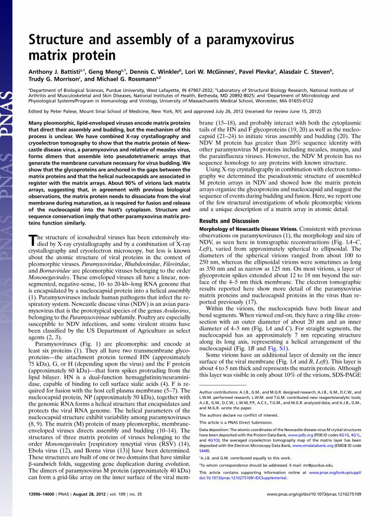
Results and DiscussionMorphology of Newcastle Disease Virions. Consistent with previousobservations on paramyxoviruses (1), the morphology and size ofNDV, as seen here in tomographic reconstructions (Fig. 1A–C,Left), varied from approximately spherical to ellipsoidal. Thediameters of the spherical virions ranged from about 100 to250 nm, whereas the ellipsoidal virions were sometimes as longas 350 nm and as narrow as 125 nm. On most virions, a layer ofglycoprotein spikes extended about 12 to 18 nm beyond the sur-face of the 4–5 nm thick membrane. The electron tomographicresults reported here show more detail of the paramyxovirusmatrix proteins and nucleocapsid proteins in the virus than re-ported previously (17).
Within the virions, the nucleocapsids have both linear andbend segments. When viewed end-on, they have a ring-like cross-section with an outer diameter of about 20 nm and an innerdiameter of 4–5 nm (Fig. 1A and C). For straight segments, thenucleocapsid has an approximately 7 nm repeating structurealong its long axis, representing a helical arrangement of thenucleocapsid (Fig. 1B and Fig. S1).
Some virions have an additional layer of density on the innersurface of the viral membrane (Fig. 1A and B, Left). This layer isabout 4 to 5 nm thick and represents the matrix protein. Althoughthis layer was visible in only about 10% of the virions, SDS-PAGE
Author contributions: A.J.B., G.M., and M.G.R. designed research; A.J.B., G.M., D.C.W., andL.W.M. performed research; L.W.M. and T.G.M. contributed new reagents/analytic tools;A.J.B., G.M., D.C.W., L.W.M, P.P., A.C.S., T.G.M., and M.G.R. analyzed data; and A.J.B., G.M.,and M.G.R. wrote the paper.
The authors declare no conflict of interest.
This article is a PNAS Direct Submission.
Data deposition: The atomic coordinates of the Newcastle disease virus M crystal structureshave been depositedwith the Protein Data Bank, www.pdb.org (PDB ID codes 4G1G, 4G1L,and 4G1O); the averaged cryoelectron tomography map of the matrix layer has beendeposited with the Electron Microscopy Data Bank, www.emdatabank.org (EMDB ID code5448).1A.J.B. and G.M. contributed equally to this work.2To whom correspondence should be addressed. E-mail: [email protected].
This article contains supporting information online at www.pnas.org/lookup/suppl/doi:10.1073/pnas.1210275109/-/DCSupplemental.
13996–14000 ∣ PNAS ∣ August 28, 2012 ∣ vol. 109 ∣ no. 35 www.pnas.org/cgi/doi/10.1073/pnas.1210275109
showed that the matrix protein was in approximately the sameabundance as the other structural proteins (Fig. S2). In thoseNDV particles where there is at least a partial matrix layer, thesurface glycoproteins and internal nucleocapsids associate withthe matrix array (Fig. 1A and B).
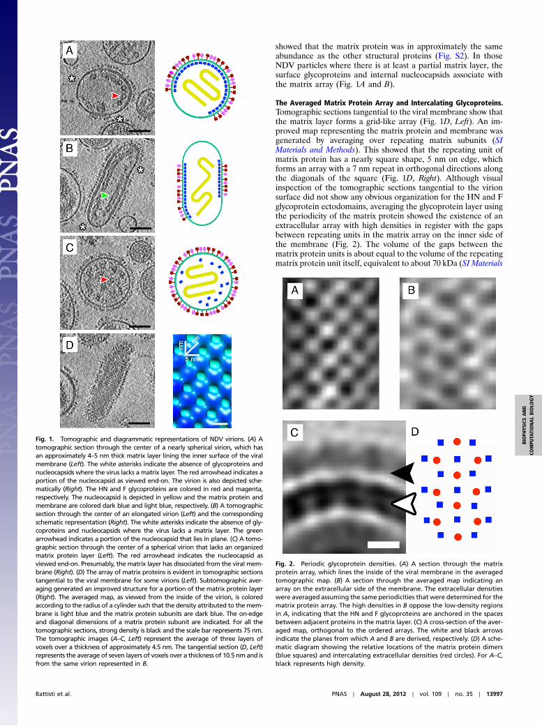
The Averaged Matrix Protein Array and Intercalating Glycoproteins.Tomographic sections tangential to the viral membrane show thatthe matrix layer forms a grid-like array (Fig. 1D, Left). An im-proved map representing the matrix protein and membrane wasgenerated by averaging over repeating matrix subunits (SIMaterials and Methods). This showed that the repeating unit ofmatrix protein has a nearly square shape, 5 nm on edge, whichforms an array with a 7 nm repeat in orthogonal directions alongthe diagonals of the square (Fig. 1D, Right). Although visualinspection of the tomographic sections tangential to the virionsurface did not show any obvious organization for the HN and Fglycoprotein ectodomains, averaging the glycoprotein layer usingthe periodicity of the matrix protein showed the existence of anextracellular array with high densities in register with the gapsbetween repeating units in the matrix array on the inner side ofthe membrane (Fig. 2). The volume of the gaps between thematrix protein units is about equal to the volume of the repeatingmatrix protein unit itself, equivalent to about 70 kDa (SI Materials
Fig. 1. Tomographic and diagrammatic representations of NDV virions. (A) Atomographic section through the center of a nearly spherical virion, which hasan approximately 4–5 nm thick matrix layer lining the inner surface of the viralmembrane (Left). The white asterisks indicate the absence of glycoproteins andnucleocapsids where the virus lacks a matrix layer. The red arrowhead indicates aportion of the nucleocapsid as viewed end-on. The virion is also depicted sche-matically (Right). The HN and F glycoproteins are colored in red and magenta,respectively. The nucleocapsid is depicted in yellow and the matrix protein andmembrane are colored dark blue and light blue, respectively. (B) A tomographicsection through the center of an elongated virion (Left) and the correspondingschematic representation (Right). The white asterisks indicate the absence of gly-coproteins and nucleocapsids where the virus lacks a matrix layer. The greenarrowhead indicates a portion of the nucleocapsid that lies in plane. (C) A tomo-graphic section through the center of a spherical virion that lacks an organizedmatrix protein layer (Left). The red arrowhead indicates the nucleocapsid asviewed end-on. Presumably, the matrix layer has dissociated from the viral mem-brane (Right). (D) The array of matrix proteins is evident in tomographic sectionstangential to the viral membrane for some virions (Left). Subtomographic aver-aging generated an improved structure for a portion of the matrix protein layer(Right). The averaged map, as viewed from the inside of the virion, is coloredaccording to the radius of a cylinder such that the density attributed to themem-brane is light blue and the matrix protein subunits are dark blue. The on-edgeand diagonal dimensions of a matrix protein subunit are indicated. For all thetomographic sections, strong density is black and the scale bar represents 75 nm.The tomographic images (A–C, Left) represent the average of three layers ofvoxels over a thickness of approximately 4.5 nm. The tangential section (D, Left)represents the average of seven layers of voxels over a thickness of 10.5 nm and isfrom the same virion represented in B.
Fig. 2. Periodic glycoprotein densities. (A) A section through the matrixprotein array, which lines the inside of the viral membrane in the averagedtomographic map. (B) A section through the averaged map indicating anarray on the extracellular side of the membrane. The extracellular densitieswere averaged assuming the same periodicities that were determined for thematrix protein array. The high densities in B oppose the low-density regionsin A, indicating that the HN and F glycoproteins are anchored in the spacesbetween adjacent proteins in the matrix layer. (C) A cross-section of the aver-aged map, orthogonal to the ordered arrays. The white and black arrowsindicate the planes from which A and B are derived, respectively. (D) A sche-matic diagram showing the relative locations of the matrix protein dimers(blue squares) and intercalating extracellular densities (red circles). For A–C,black represents high density.
Battisti et al. PNAS ∣ August 28, 2012 ∣ vol. 109 ∣ no. 35 ∣ 13997
BIOPH
YSICSAND
COMPU
TATIONALBIOLO
GY
and Methods). The volume of this gap would be ample to accom-modate the cytoplasmic tails of the tetrameric HN or trimeric Fglycoproteins. Modeling of the NDV HN (25) or parainfluenzavirus F (6) structures into the glycoprotein array shows that theglycoprotein ectodomains have the correct size to fit into neigh-boring sites consistent with the matrix protein periodicity.Although the head of the HN protein can be accommodated inarrays along one direction, the spaces in the orthogonal directionwould only be able to accommodate the F protein (Fig. 3).
The Tomographic Nucleocapsid Density. The tomograms of NDVshowed long regions of nucleocapsids that have a repeating pat-tern (Fig. S1). This pattern was shown to correspond to a helixwith a pitch (the axial translation for one turn of the helix) of7 nm, consistent with that of other paramyxovirus nucleocapsids(8, 9, 26). The twist (or number of NP repeats per turn of thehelix) could not be determined because of the limited resolutionof the tomographic maps (SI Materials and Methods and Fig. S1).The nucleocapsids observed in the tomograms were often in reg-ister with the matrix protein dimers, such that the 7 nm pitchalong the long axis of the nucleocapsid was placed along the sameaxis as the 7 nm repeat along the diagonal of the matrix array(Fig. S3). However, a helically organized matrix protein envelop-
ing the nucleocapsid structure as described for measles virus (24)was not observed.
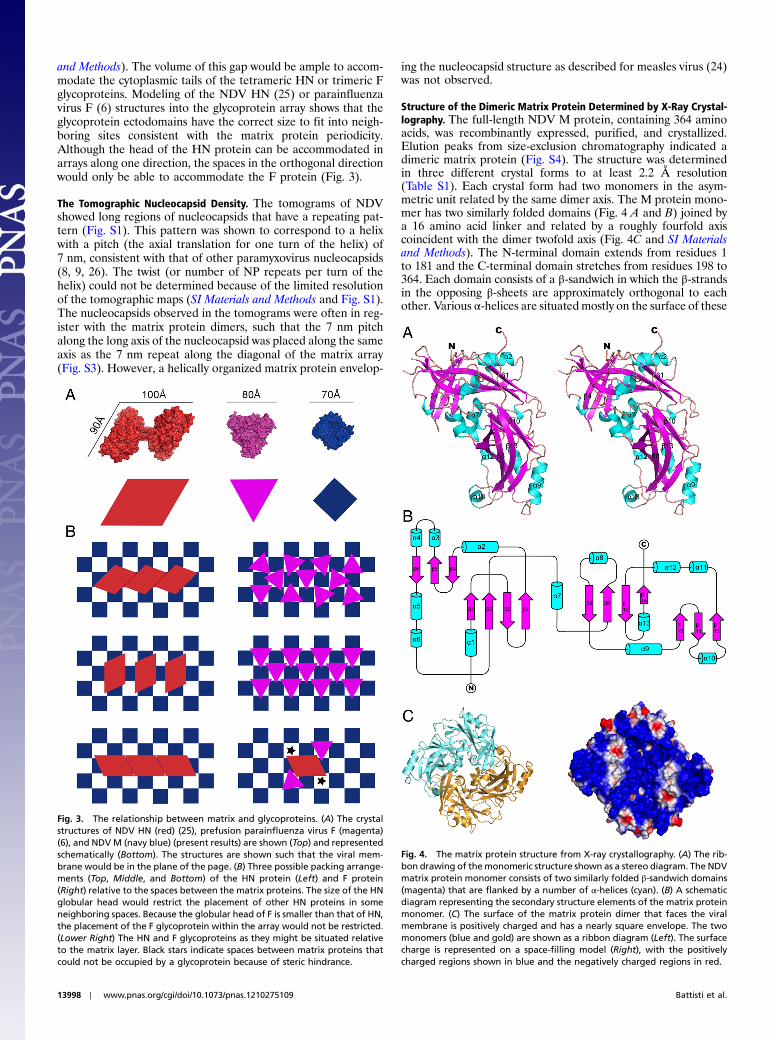
Structure of the Dimeric Matrix Protein Determined by X-Ray Crystal-lography. The full-length NDV M protein, containing 364 aminoacids, was recombinantly expressed, purified, and crystallized.Elution peaks from size-exclusion chromatography indicated adimeric matrix protein (Fig. S4). The structure was determinedin three different crystal forms to at least 2.2 Å resolution(Table S1). Each crystal form had two monomers in the asym-metric unit related by the same dimer axis. The M protein mono-mer has two similarly folded domains (Fig. 4 A and B) joined bya 16 amino acid linker and related by a roughly fourfold axiscoincident with the dimer twofold axis (Fig. 4C and SI Materialsand Methods). The N-terminal domain extends from residues 1to 181 and the C-terminal domain stretches from residues 198 to364. Each domain consists of a β-sandwich in which the β-strandsin the opposing β-sheets are approximately orthogonal to eachother. Various α-helices are situated mostly on the surface of these
Fig. 3. The relationship between matrix and glycoproteins. (A) The crystalstructures of NDV HN (red) (25), prefusion parainfluenza virus F (magenta)(6), and NDVM (navy blue) (present results) are shown (Top) and representedschematically (Bottom). The structures are shown such that the viral mem-brane would be in the plane of the page. (B) Three possible packing arrange-ments (Top, Middle, and Bottom) of the HN protein (Left) and F protein(Right) relative to the spaces between the matrix proteins. The size of the HNglobular head would restrict the placement of other HN proteins in someneighboring spaces. Because the globular head of F is smaller than that of HN,the placement of the F glycoprotein within the array would not be restricted.(Lower Right) The HN and F glycoproteins as they might be situated relativeto the matrix layer. Black stars indicate spaces between matrix proteins thatcould not be occupied by a glycoprotein because of steric hindrance.
Fig. 4. The matrix protein structure from X-ray crystallography. (A) The rib-bon drawing of themonomeric structure shown as a stereo diagram. The NDVmatrix protein monomer consists of two similarly folded β-sandwich domains(magenta) that are flanked by a number of α-helices (cyan). (B) A schematicdiagram representing the secondary structure elements of the matrix proteinmonomer. (C) The surface of the matrix protein dimer that faces the viralmembrane is positively charged and has a nearly square envelope. The twomonomers (blue and gold) are shown as a ribbon diagram (Left). The surfacecharge is represented on a space-filling model (Right), with the positivelycharged regions shown in blue and the negatively charged regions in red.
13998 ∣ www.pnas.org/cgi/doi/10.1073/pnas.1210275109 Battisti et al.
β-sandwich domains (Fig. 4A). The domains can be superimposedwith a rmsd of 3.8 Å between 74 equivalent Cα atoms of 167 re-sidues.
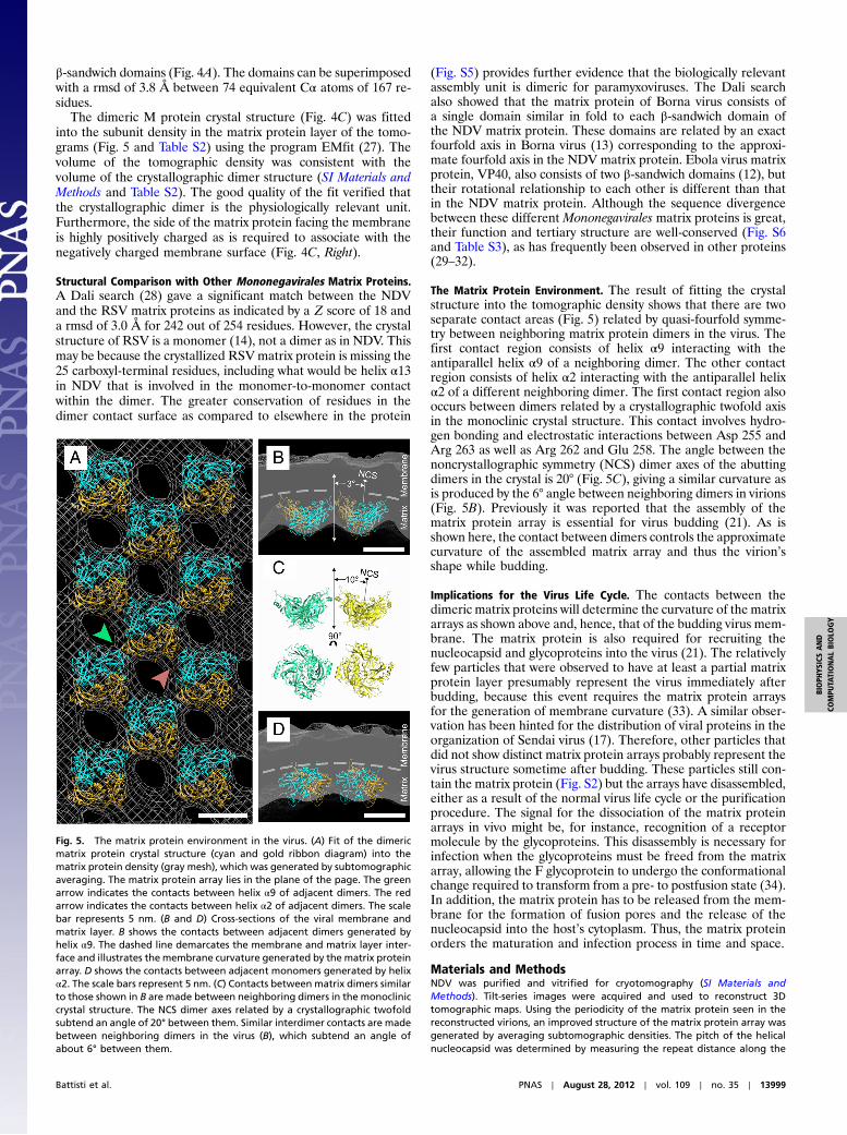
The dimeric M protein crystal structure (Fig. 4C) was fittedinto the subunit density in the matrix protein layer of the tomo-grams (Fig. 5 and Table S2) using the program EMfit (27). Thevolume of the tomographic density was consistent with thevolume of the crystallographic dimer structure (SI Materials andMethods and Table S2). The good quality of the fit verified thatthe crystallographic dimer is the physiologically relevant unit.Furthermore, the side of the matrix protein facing the membraneis highly positively charged as is required to associate with thenegatively charged membrane surface (Fig. 4C, Right).
Structural Comparison with Other Mononegavirales Matrix Proteins.A Dali search (28) gave a significant match between the NDVand the RSV matrix proteins as indicated by a Z score of 18 anda rmsd of 3.0 Å for 242 out of 254 residues. However, the crystalstructure of RSV is a monomer (14), not a dimer as in NDV. Thismay be because the crystallized RSVmatrix protein is missing the25 carboxyl-terminal residues, including what would be helix α13in NDV that is involved in the monomer-to-monomer contactwithin the dimer. The greater conservation of residues in thedimer contact surface as compared to elsewhere in the protein
(Fig. S5) provides further evidence that the biologically relevantassembly unit is dimeric for paramyxoviruses. The Dali searchalso showed that the matrix protein of Borna virus consists ofa single domain similar in fold to each β-sandwich domain ofthe NDV matrix protein. These domains are related by an exactfourfold axis in Borna virus (13) corresponding to the approxi-mate fourfold axis in the NDV matrix protein. Ebola virus matrixprotein, VP40, also consists of two β-sandwich domains (12), buttheir rotational relationship to each other is different than thatin the NDV matrix protein. Although the sequence divergencebetween these different Mononegavirales matrix proteins is great,their function and tertiary structure are well-conserved (Fig. S6and Table S3), as has frequently been observed in other proteins(29–32).
The Matrix Protein Environment. The result of fitting the crystalstructure into the tomographic density shows that there are twoseparate contact areas (Fig. 5) related by quasi-fourfold symme-try between neighboring matrix protein dimers in the virus. Thefirst contact region consists of helix α9 interacting with theantiparallel helix α9 of a neighboring dimer. The other contactregion consists of helix α2 interacting with the antiparallel helixα2 of a different neighboring dimer. The first contact region alsooccurs between dimers related by a crystallographic twofold axisin the monoclinic crystal structure. This contact involves hydro-gen bonding and electrostatic interactions between Asp 255 andArg 263 as well as Arg 262 and Glu 258. The angle between thenoncrystallographic symmetry (NCS) dimer axes of the abuttingdimers in the crystal is 20° (Fig. 5C), giving a similar curvature asis produced by the 6° angle between neighboring dimers in virions(Fig. 5B). Previously it was reported that the assembly of thematrix protein array is essential for virus budding (21). As isshown here, the contact between dimers controls the approximatecurvature of the assembled matrix array and thus the virion’sshape while budding.
Implications for the Virus Life Cycle. The contacts between thedimeric matrix proteins will determine the curvature of the matrixarrays as shown above and, hence, that of the budding virus mem-brane. The matrix protein is also required for recruiting thenucleocapsid and glycoproteins into the virus (21). The relativelyfew particles that were observed to have at least a partial matrixprotein layer presumably represent the virus immediately afterbudding, because this event requires the matrix protein arraysfor the generation of membrane curvature (33). A similar obser-vation has been hinted for the distribution of viral proteins in theorganization of Sendai virus (17). Therefore, other particles thatdid not show distinct matrix protein arrays probably represent thevirus structure sometime after budding. These particles still con-tain the matrix protein (Fig. S2) but the arrays have disassembled,either as a result of the normal virus life cycle or the purificationprocedure. The signal for the dissociation of the matrix proteinarrays in vivo might be, for instance, recognition of a receptormolecule by the glycoproteins. This disassembly is necessary forinfection when the glycoproteins must be freed from the matrixarray, allowing the F glycoprotein to undergo the conformationalchange required to transform from a pre- to postfusion state (34).In addition, the matrix protein has to be released from the mem-brane for the formation of fusion pores and the release of thenucleocapsid into the host’s cytoplasm. Thus, the matrix proteinorders the maturation and infection process in time and space.
Materials and MethodsNDV was purified and vitrified for cryotomography (SI Materials andMethods). Tilt-series images were acquired and used to reconstruct 3Dtomographic maps. Using the periodicity of the matrix protein seen in thereconstructed virions, an improved structure of the matrix protein array wasgenerated by averaging subtomographic densities. The pitch of the helicalnucleocapsid was determined by measuring the repeat distance along the
Fig. 5. The matrix protein environment in the virus. (A) Fit of the dimericmatrix protein crystal structure (cyan and gold ribbon diagram) into thematrix protein density (gray mesh), which was generated by subtomographicaveraging. The matrix protein array lies in the plane of the page. The greenarrow indicates the contacts between helix α9 of adjacent dimers. The redarrow indicates the contacts between helix α2 of adjacent dimers. The scalebar represents 5 nm. (B and D) Cross-sections of the viral membrane andmatrix layer. B shows the contacts between adjacent dimers generated byhelix α9. The dashed line demarcates the membrane and matrix layer inter-face and illustrates the membrane curvature generated by the matrix proteinarray. D shows the contacts between adjacent monomers generated by helixα2. The scale bars represent 5 nm. (C) Contacts between matrix dimers similarto those shown in B are made between neighboring dimers in the monocliniccrystal structure. The NCS dimer axes related by a crystallographic twofoldsubtend an angle of 20° between them. Similar interdimer contacts are madebetween neighboring dimers in the virus (B), which subtend an angle ofabout 6° between them.
Battisti et al. PNAS ∣ August 28, 2012 ∣ vol. 109 ∣ no. 35 ∣ 13999
BIOPH
YSICSAND
COMPU
TATIONALBIOLO
GY
length of the helix in the tomographic data. In addition, the full-length NDVmatrix protein was recombinantly expressed, purified, and crystallized (SIMaterials and Methods). The structure was determined by X-ray crystallogra-phy using single-wavelength anomalous dispersion data. The dimeric crystal-lographic structure was then fitted into the tomographic density andcompared to the previously determined structures of homologous matrixproteins.
ACKNOWLEDGMENTS. We thank Anastasia Aksyuk and Giovanni Cardone forhelpful comments and suggestions regarding image processing; Ken Hiblerand Zhiheng Yu for assistance with the FEI Titan Krios Microscope; Paul Chip-
man, Siyang Sun, and Ye Xiang for helpful comments and suggestionsthroughout the course of this project; Xinzheng Zhang for assistance withelectron microscopy; and Sheryl Kelly for help in the preparation of thismanuscript. Use of the Advanced Photon Source was supported by the USDepartment of Energy, Office of Science, Office of Basic Energy Sciencesunder Contract DE-AC02-06CH11357. We thank National Institutes of Healthfor both an individual research award (AI11219 to M.G.R.) and support viathe intramural research program of National Institute of Arthritis and Mus-culoskeletal and Skin Diseases (to A.C.S.). Support for A.J.B. was provided by aBiophysics Training Grant (T32 GM008296-20) from the National Institutes ofHealth/General Medicine (C.V. Stauffacher, principal investigator) and a Bils-land Dissertation Fellowship from Purdue University Graduate School.
1. Lamb RA, Parks GD (2007) Paramyxoviridae: The viruses and their replication. FieldsVirology, eds DM Knipe and PM Howley (Lippincott Williams and Wilkins, Philadel-phia), 5th Ed,, pp 1449–1496.
2. McGinnes LW, Pantua H, Reitter J, Morrison TG (2006) Newcastle disease virus: Propa-gation, quantification, and storage. Curr Protoc Microbiol Chapter 15:Unit 15F.2.
3. National Select Agent Registry, http://www.selectagents.gov. Accessed August 2012.4. Scheid A, Caliguiri LA, Compans RW, Choppin PW (1972) Isolation of paramyxovirus
glycoproteins. Association of both hemagglutinating and neuraminidase activitieswith the larger SV5 glycoprotein. Virology 50:640–652.
5. Yin HS, Paterson RG, Wen X, Lamb RA, Jardetzky TS (2005) Structure of the uncleavedectodomain of the paramyxovirus (hPIV3) fusion protein. Proc Natl Acad Sci USA102:9288–9293.
6. Yin HS, Wen X, Paterson RG, Lamb RA, Jardetzky TS (2006) Structure of the parain-fluenza virus 5 F protein in its metastable, prefusion conformation. Nature 439:38–44.
7. Nagai Y, Klenk H-D, Rott R (1976) Proteolytic cleavage of the viral glycoproteins and itssignificance for the virulence of Newcastle disease virus. Virology 72:494–508.
8. Bhella D, Ralph A, Yeo RP (2004) Conformational flexibility in recombinant measlesvirus nucleocapsids visualised by cryo-negative stain electron microscopy and real-space helical reconstruction. J Mol Biol 340:319–331.
9. Egelman EH, Wu SS, Amrein M, Portner A, Murti G (1989) The Sendai virus nucleocap-sid exists in at least four different helical states. J Virol 63:2233–2243.
10. Harris A, Forouhar F, Qiu S, Sha B, Luo M (2001) The crystal structure of the influenzamatrix protein M1 at neutral pH: M1–M1 protein interfaces can rotate in the oligo-meric structures of M1. Virology 289:34–44.
11. Rao Z, et al. (1995) Crystal structure of SIV matrix antigen and implications for virusassembly. Nature 378:743–747.
12. Dessen A, Volchkov V, Dolnik O, Klenk H-D, Weissenhorn W (2000) Crystal structure ofthe matrix protein VP40 from Ebola virus. EMBO J 19:4228–4236.
13. Neumann P, et al. (2009) Crystal structure of the Borna disease virus matrix protein(BDV-M) reveals ssRNA binding properties. Proc Natl Acad Sci USA 106:3710–3715.
14. Money VA, McPhee HK, Mosely JA, Sanderson JM, Yeo RP (2009) Surface features of aMononegavirales matrix protein indicate sites of membrane interaction. Proc NatlAcad Sci USA 106:4441–4446.
15. Russell PH, Almeida JD (1984) A regular subunit pattern seen on non-infectious New-castle disease virus particles. J Gen Virol 65:1023–1031.
16. Heggeness MH, Smith PR, Choppin PW (1982) In vitro assembly of the nonglycosylatedmembrane protein (M) of Sendai virus. Proc Natl Acad Sci USA 79:6232–6236.
17. Bachi T (1980) Intramembrane structural differentiation in Sendai virus maturation.Virology 106:41–49.
18. Hewitt JA (1977) Studies on the subunit composition of the M-protein of Sendai virus.FEBS Lett 81:395–397.
19. Waning DL, Schmitt AP, Leser GP, Lamb RA (2002) Roles for the cytoplasmic tails of thefusion and hemagglutinin-neuraminidase proteins in budding of the paramyxovirussimian virus 5. J Virol 76:9284–9297.
20. Pantua HD, McGinnes LW, Peeples ME, Morrison TG (2006) Requirements for theassembly and release of Newcastle disease virus-like particles. J Virol 80:11062–11073.
21. Harrison MS, Sakaguchi T, Schmitt AP (2010) Paramyxovirus assembly and budding:Building particles that transmit infections. Int J Biochem Cell Biol 42:1416–1429.
22. Ghildyal R, Mills J, Murray M, Vardaxis N, Meanger J (2002) Respiratory syncytial virusmatrix protein associates with nucleocapsids in infection cells. J Gen Virol 83:753–757.
23. Schmitt PT, Ray G, Schmitt AP (2010) The C-terminal end of parainfluence virus 5 NPprotein is important for virus-like particle production and M–NP protein interaction.J Virol 84:12810–12823.
24. Liljeroos L, Huiskonen JT, Ora A, Susi P, Butcher SJ (2011) Electron cryotomography ofmeasles virus reveals how matrix protein coats the ribonucleocapsid within intactvirions. Proc Natl Acad Sci USA 108:18085–18090.
25. Yuan P, et al. (2011) Structure of the Newcastle disease virus hemagglutinin–neurami-nidase (HN) ectodomain reveals a four-helix bundle stalk. Proc Natl Acad Sci USA108:14920–14925.
26. Tawar RG, et al. (2009) Crystal structure of a nucleocapsid-like nucleoprotein-RNAcomplex of respiratory syncytial virus. Science 326:1279–1283.
27. Rossmann MG, Bernal R, Pletnev SV (2001) Combining electron microscopic with x-raycrystallographic structures. J Struct Biol 136:190–200.
28. Holm L, Rosenström P (2010) Dali server: Conservation mapping in 3D. Nucleic AcidsRes 38:W545–W549.
29. Perutz MF, et al. (1960) Structure of haemoglobin. A three-dimensional Fourier synth-esis at 5.5-Å resolution, obtained by X-ray analysis. Nature 185:416–422.
30. RossmannMG,Moras D, Olsen KW (1974) Chemical and biological evolution of nucleo-tide-binding protein. Nature 250:194–199.
31. Rossmann MG, et al. (1985) Structure of a human common cold virus and functionalrelationship to other picornaviruses. Nature 317:145–153.
32. Nandhagopal N, et al. (2002) The structure and evolution of the major capsid proteinof a large, lipid-containing DNA virus. Proc Natl Acad Sci USA 99:14758–14763.
33. Shnyrova AV, et al. (2007) Vesicle formation by self-assembly of membrane-boundmatrix proteins into a fluidlike budding domain. J Cell Biol 179:627–633.
34. Lamb RA, Jardetzky TS (2007) Structural basis of viral invasion: Lessons from paramyx-ovirus F. Curr Opin Struct Biol 17:427–436.
14000 ∣ www.pnas.org/cgi/doi/10.1073/pnas.1210275109 Battisti et al.


![TRACING THE SOLUTION SURFACE WITH FOLDS OF A …faculty.stust.edu.tw/~slchang/paper/paper20040616.pdf · TRACING THE SOLUTION SURFACE WITH FOLDS OF A ... 2000]. Perhaps AUTO ... Here](https://static.fdocument.org/doc/165x107/5aa145fc7f8b9a1f6d8ba003/tracing-the-solution-surface-with-folds-of-a-slchangpaperpaper20040616pdftracing.jpg)