Structural study of fluorescence-quenching tryptophan residues in...
9
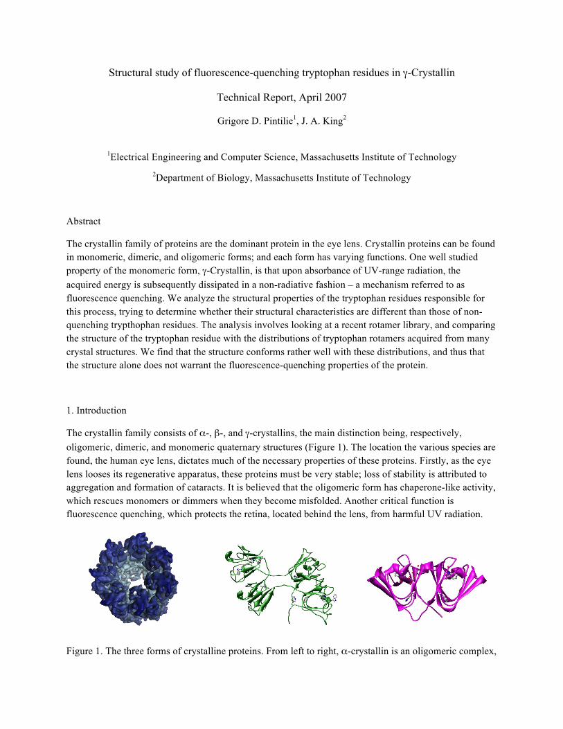
Structural study of fluorescence-quenching tryptophan residues in γ-Crystallin Technical Report, April 2007 Grigore D. Pintilie 1 , J. A. King 2 1 Electrical Engineering and Computer Science, Massachusetts Institute of Technology 2 Department of Biology, Massachusetts Institute of Technology Abstract The crystallin family of proteins are the dominant protein in the eye lens. Crystallin proteins can be found in monomeric, dimeric, and oligomeric forms; and each form has varying functions. One well studied property of the monomeric form, γ-Crystallin, is that upon absorbance of UV-range radiation, the acquired energy is subsequently dissipated in a non-radiative fashion – a mechanism referred to as fluorescence quenching. We analyze the structural properties of the tryptophan residues responsible for this process, trying to determine whether their structural characteristics are different than those of non- quenching trypthophan residues. The analysis involves looking at a recent rotamer library, and comparing the structure of the tryptophan residue with the distributions of tryptophan rotamers acquired from many crystal structures. We find that the structure conforms rather well with these distributions, and thus that the structure alone does not warrant the fluorescence-quenching properties of the protein. 1. Introduction The crystallin family consists of α-, β-, and γ-crystallins, the main distinction being, respectively, oligomeric, dimeric, and monomeric quaternary structures (Figure 1). The location the various species are found, the human eye lens, dictates much of the necessary properties of these proteins. Firstly, as the eye lens looses its regenerative apparatus, these proteins must be very stable; loss of stability is attributed to aggregation and formation of cataracts. It is believed that the oligomeric form has chaperone-like activity, which rescues monomers or dimmers when they become misfolded. Another critical function is fluorescence quenching, which protects the retina, located behind the lens, from harmful UV radiation. Figure 1. The three forms of crystalline proteins. From left to right, α-crystallin is an oligomeric complex,
Transcript of Structural study of fluorescence-quenching tryptophan residues in...
Structural study of fluorescence-quenching tryptophan residues in γ-Crystallin
Technical Report, April 2007
Grigore D. Pintilie1, J. A. King2
1Electrical Engineering and Computer Science, Massachusetts Institute of Technology 2Department of Biology, Massachusetts Institute of Technology
Abstract
The crystallin family of proteins are the dominant protein in the eye lens. Crystallin proteins can be found in monomeric, dimeric, and oligomeric forms; and each form has varying functions. One well studied property of the monomeric form, γ-Crystallin, is that upon absorbance of UV-range radiation, the acquired energy is subsequently dissipated in a non-radiative fashion – a mechanism referred to as fluorescence quenching. We analyze the structural properties of the tryptophan residues responsible for this process, trying to determine whether their structural characteristics are different than those of non-quenching trypthophan residues. The analysis involves looking at a recent rotamer library, and comparing the structure of the tryptophan residue with the distributions of tryptophan rotamers acquired from many crystal structures. We find that the structure conforms rather well with these distributions, and thus that the structure alone does not warrant the fluorescence-quenching properties of the protein.
1. Introduction
The crystallin family consists of α-, β-, and γ-crystallins, the main distinction being, respectively, oligomeric, dimeric, and monomeric quaternary structures (Figure 1). The location the various species are found, the human eye lens, dictates much of the necessary properties of these proteins. Firstly, as the eye lens looses its regenerative apparatus, these proteins must be very stable; loss of stability is attributed to aggregation and formation of cataracts. It is believed that the oligomeric form has chaperone-like activity, which rescues monomers or dimmers when they become misfolded. Another critical function is fluorescence quenching, which protects the retina, located behind the lens, from harmful UV radiation.
Figure 1. The three forms of crystalline proteins. From left to right, α-crystallin is an oligomeric complex,
β-crystallin is a dimmer, and γ-crystallin is a monomer.
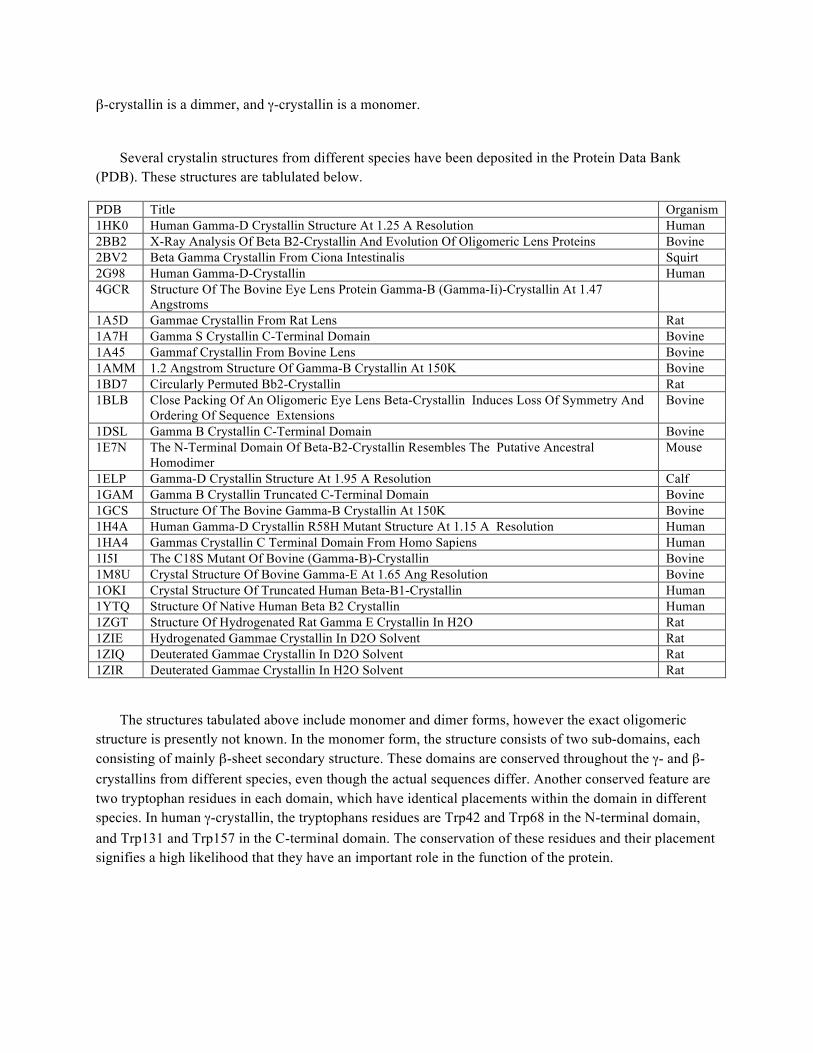
Several crystalin structures from different species have been deposited in the Protein Data Bank (PDB). These structures are tablulated below.
PDB Title Organism 1HK0 Human Gamma-D Crystallin Structure At 1.25 A Resolution Human 2BB2 X-Ray Analysis Of Beta B2-Crystallin And Evolution Of Oligomeric Lens Proteins Bovine 2BV2 Beta Gamma Crystallin From Ciona Intestinalis Squirt 2G98 Human Gamma-D-Crystallin Human 4GCR Structure Of The Bovine Eye Lens Protein Gamma-B (Gamma-Ii)-Crystallin At 1.47
Angstroms
1A5D Gammae Crystallin From Rat Lens Rat 1A7H Gamma S Crystallin C-Terminal Domain Bovine 1A45 Gammaf Crystallin From Bovine Lens Bovine 1AMM 1.2 Angstrom Structure Of Gamma-B Crystallin At 150K Bovine 1BD7 Circularly Permuted Bb2-Crystallin Rat 1BLB Close Packing Of An Oligomeric Eye Lens Beta-Crystallin Induces Loss Of Symmetry And
Ordering Of Sequence Extensions Bovine
1DSL Gamma B Crystallin C-Terminal Domain Bovine 1E7N The N-Terminal Domain Of Beta-B2-Crystallin Resembles The Putative Ancestral
Homodimer Mouse
1ELP Gamma-D Crystallin Structure At 1.95 A Resolution Calf 1GAM Gamma B Crystallin Truncated C-Terminal Domain Bovine 1GCS Structure Of The Bovine Gamma-B Crystallin At 150K Bovine 1H4A Human Gamma-D Crystallin R58H Mutant Structure At 1.15 A Resolution Human 1HA4 Gammas Crystallin C Terminal Domain From Homo Sapiens Human 1I5I The C18S Mutant Of Bovine (Gamma-B)-Crystallin Bovine 1M8U Crystal Structure Of Bovine Gamma-E At 1.65 Ang Resolution Bovine 1OKI Crystal Structure Of Truncated Human Beta-B1-Crystallin Human 1YTQ Structure Of Native Human Beta B2 Crystallin Human 1ZGT Structure Of Hydrogenated Rat Gamma E Crystallin In H2O Rat 1ZIE Hydrogenated Gammae Crystallin In D2O Solvent Rat 1ZIQ Deuterated Gammae Crystallin In D2O Solvent Rat 1ZIR Deuterated Gammae Crystallin In H2O Solvent Rat
The structures tabulated above include monomer and dimer forms, however the exact oligomeric structure is presently not known. In the monomer form, the structure consists of two sub-domains, each consisting of mainly β-sheet secondary structure. These domains are conserved throughout the γ- and β-crystallins from different species, even though the actual sequences differ. Another conserved feature are two tryptophan residues in each domain, which have identical placements within the domain in different species. In human γ-crystallin, the tryptophans residues are Trp42 and Trp68 in the N-terminal domain, and Trp131 and Trp157 in the C-terminal domain. The conservation of these residues and their placement signifies a high likelihood that they have an important role in the function of the protein.
Mutation experiments have shown that, in human γD-crystallin, Trp68 and Trp157 have very large quenching characteristics, while Trp42 and Trp131 show lower quenching1. Hybrid QM-MD computations have revealed important factors that make the quenching process possible2. Fluorescence quenching has also been observed in other proteins, and taking into account these various factors makes it possible to predict the degree of quenching of particular tryptophan residues3. In the remainder of this report, the molecular conformations of the quenching and non-quenching tryptophan residues will be studied. The conformation of the residues will be characterized based on the known rotameric forms.
2. Molecular Conformations
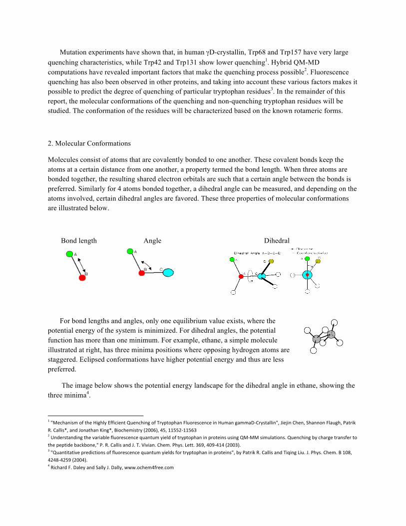
Molecules consist of atoms that are covalently bonded to one another. These covalent bonds keep the atoms at a certain distance from one another, a property termed the bond length. When three atoms are bonded together, the resulting shared electron orbitals are such that a certain angle between the bonds is preferred. Similarly for 4 atoms bonded together, a dihedral angle can be measured, and depending on the atoms involved, certain dihedral angles are favored. These three properties of molecular conformations are illustrated below.
Bond length Angle Dihedral
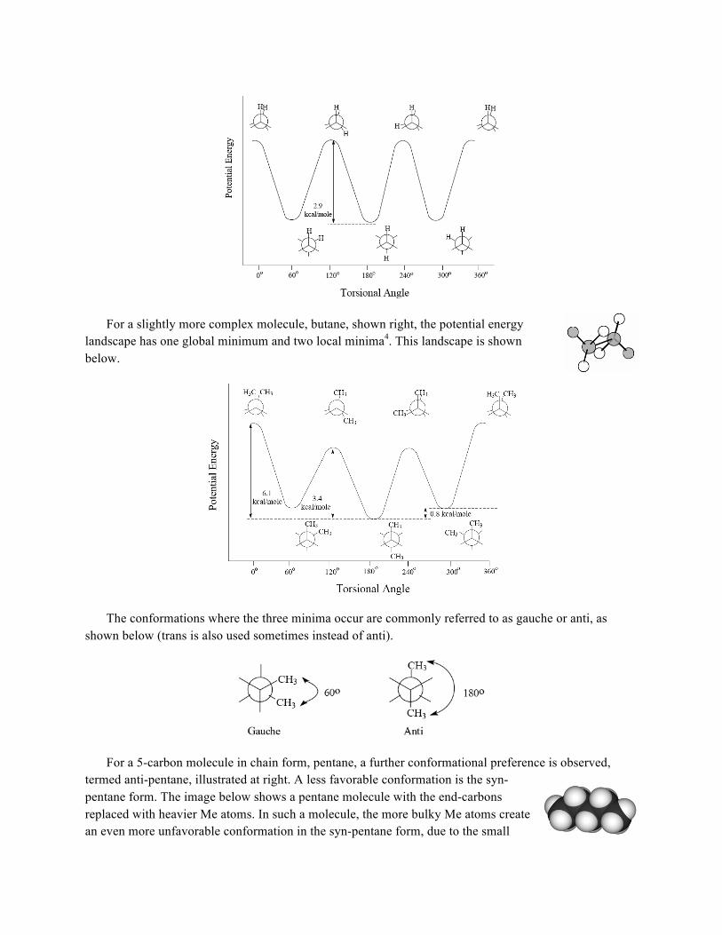
For bond lengths and angles, only one equilibrium value exists, where the potential energy of the system is minimized. For dihedral angles, the potential function has more than one minimum. For example, ethane, a simple molecule illustrated at right, has three minima positions where opposing hydrogen atoms are staggered. Eclipsed conformations have higher potential energy and thus are less preferred.
The image below shows the potential energy landscape for the dihedral angle in ethane, showing the three minima4.
1 "Mechanism of the Highly Efficient Quenching of Tryptophan Fluorescence in Human gammaD-‐Crystallin", Jiejin Chen, Shannon Flaugh, Patrik R. Callis*, and Jonathan King*, Biochemistry (2006), 45, 11552-‐11563 2 Understanding the variable fluorescence quantum yield of tryptophan in proteins using QM-‐MM simulations. Quenching by charge transfer to the peptide backbone," P. R. Callis and J. T. Vivian. Chem. Phys. Lett. 369, 409-‐414 (2003). 3 "Quantitative predictions of fluorescence quantum yields for tryptophan in proteins", by Patrik R. Callis and Tiqing Liu. J. Phys. Chem. B 108, 4248-‐4259 (2004). 4 Richard F. Daley and Sally J. Dally, www.ochem4free.com
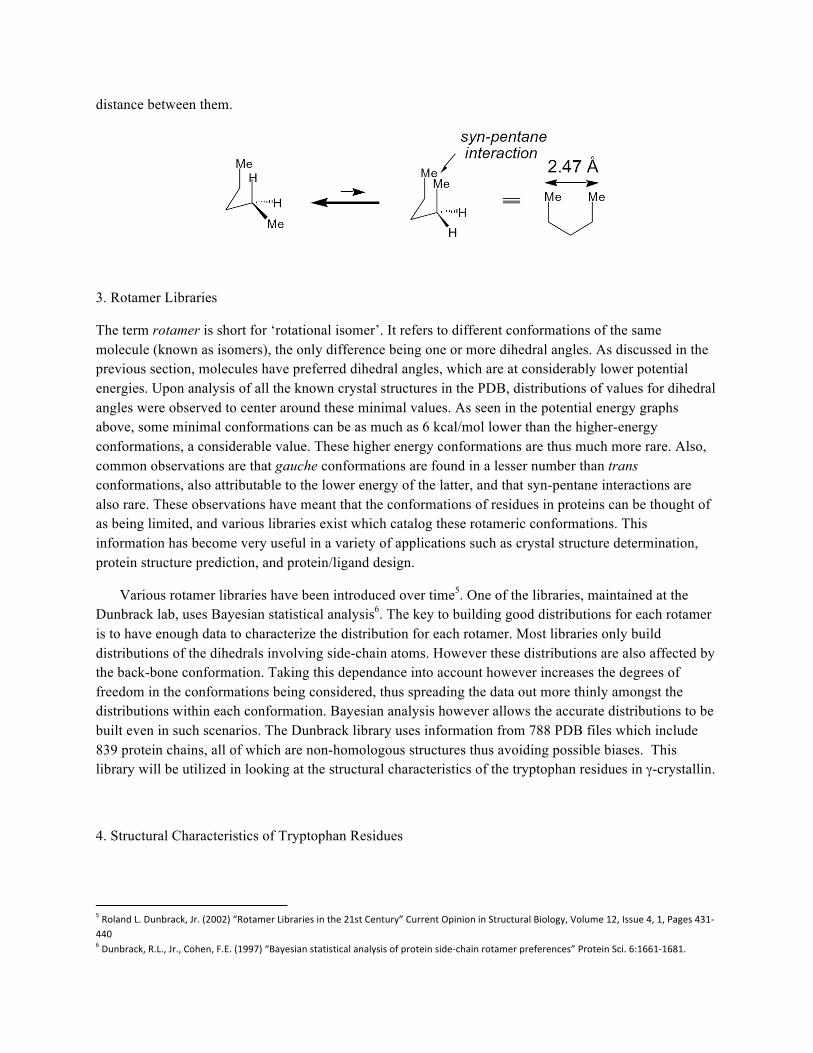
For a slightly more complex molecule, butane, shown right, the potential energy landscape has one global minimum and two local minima4. This landscape is shown below.
The conformations where the three minima occur are commonly referred to as gauche or anti, as shown below (trans is also used sometimes instead of anti).
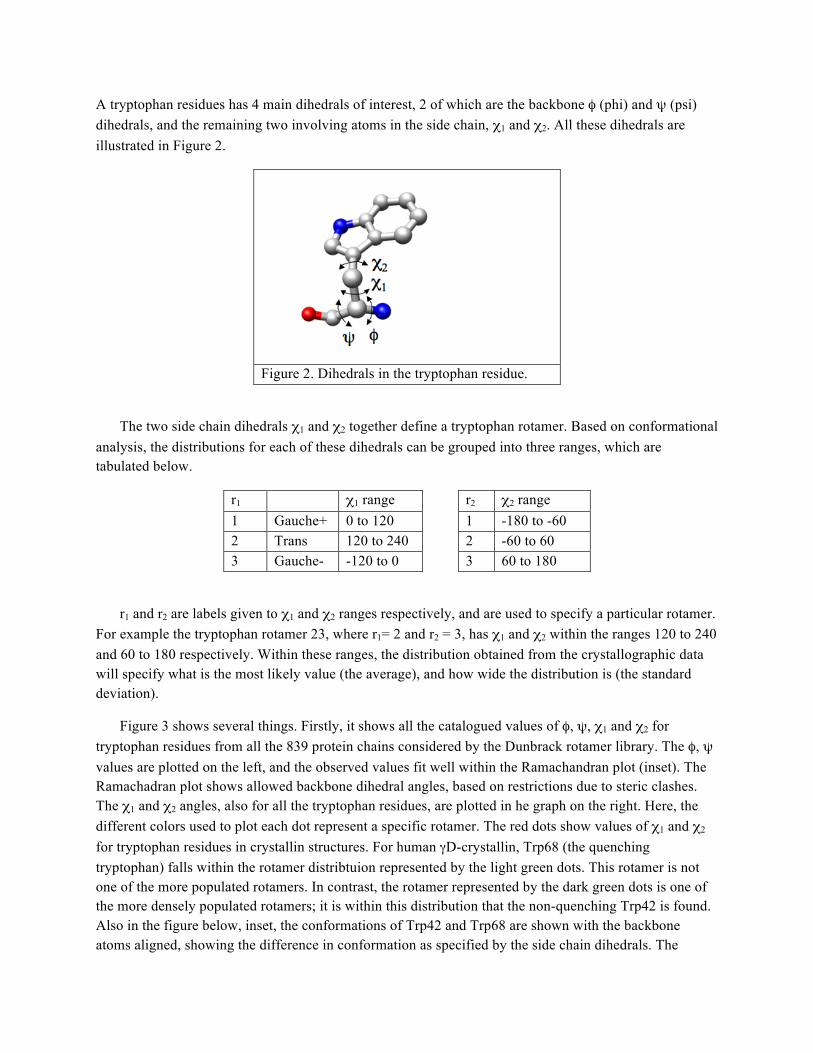
For a 5-carbon molecule in chain form, pentane, a further conformational preference is observed, termed anti-pentane, illustrated at right. A less favorable conformation is the syn-pentane form. The image below shows a pentane molecule with the end-carbons replaced with heavier Me atoms. In such a molecule, the more bulky Me atoms create an even more unfavorable conformation in the syn-pentane form, due to the small
distance between them.
3. Rotamer Libraries
The term rotamer is short for ‘rotational isomer’. It refers to different conformations of the same molecule (known as isomers), the only difference being one or more dihedral angles. As discussed in the previous section, molecules have preferred dihedral angles, which are at considerably lower potential energies. Upon analysis of all the known crystal structures in the PDB, distributions of values for dihedral angles were observed to center around these minimal values. As seen in the potential energy graphs above, some minimal conformations can be as much as 6 kcal/mol lower than the higher-energy conformations, a considerable value. These higher energy conformations are thus much more rare. Also, common observations are that gauche conformations are found in a lesser number than trans conformations, also attributable to the lower energy of the latter, and that syn-pentane interactions are also rare. These observations have meant that the conformations of residues in proteins can be thought of as being limited, and various libraries exist which catalog these rotameric conformations. This information has become very useful in a variety of applications such as crystal structure determination, protein structure prediction, and protein/ligand design.
Various rotamer libraries have been introduced over time5. One of the libraries, maintained at the Dunbrack lab, uses Bayesian statistical analysis6. The key to building good distributions for each rotamer is to have enough data to characterize the distribution for each rotamer. Most libraries only build distributions of the dihedrals involving side-chain atoms. However these distributions are also affected by the back-bone conformation. Taking this dependance into account however increases the degrees of freedom in the conformations being considered, thus spreading the data out more thinly amongst the distributions within each conformation. Bayesian analysis however allows the accurate distributions to be built even in such scenarios. The Dunbrack library uses information from 788 PDB files which include 839 protein chains, all of which are non-homologous structures thus avoiding possible biases. This library will be utilized in looking at the structural characteristics of the tryptophan residues in γ-crystallin.
4. Structural Characteristics of Tryptophan Residues
5 Roland L. Dunbrack, Jr. (2002) “Rotamer Libraries in the 21st Century” Current Opinion in Structural Biology, Volume 12, Issue 4, 1, Pages 431-‐440 6 Dunbrack, R.L., Jr., Cohen, F.E. (1997) “Bayesian statistical analysis of protein side-‐chain rotamer preferences” Protein Sci. 6:1661-‐1681.
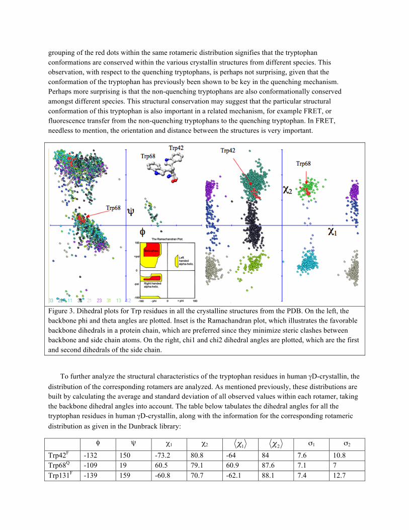
A tryptophan residues has 4 main dihedrals of interest, 2 of which are the backbone φ (phi) and ψ (psi) dihedrals, and the remaining two involving atoms in the side chain, χ1 and χ2. All these dihedrals are illustrated in Figure 2.
Figure 2. Dihedrals in the tryptophan residue.
The two side chain dihedrals χ1 and χ2 together define a tryptophan rotamer. Based on conformational analysis, the distributions for each of these dihedrals can be grouped into three ranges, which are tabulated below.
r1 χ1 range r2 χ2 range 1 Gauche+ 0 to 120 1 -180 to -60 2 Trans 120 to 240 2 -60 to 60 3 Gauche- -120 to 0 3 60 to 180
r1 and r2 are labels given to χ1 and χ2 ranges respectively, and are used to specify a particular rotamer. For example the tryptophan rotamer 23, where r1= 2 and r2 = 3, has χ1 and χ2 within the ranges 120 to 240 and 60 to 180 respectively. Within these ranges, the distribution obtained from the crystallographic data will specify what is the most likely value (the average), and how wide the distribution is (the standard deviation).
Figure 3 shows several things. Firstly, it shows all the catalogued values of φ, ψ, χ1 and χ2 for tryptophan residues from all the 839 protein chains considered by the Dunbrack rotamer library. The φ, ψ values are plotted on the left, and the observed values fit well within the Ramachandran plot (inset). The Ramachadran plot shows allowed backbone dihedral angles, based on restrictions due to steric clashes. The χ1 and χ2 angles, also for all the tryptophan residues, are plotted in he graph on the right. Here, the different colors used to plot each dot represent a specific rotamer. The red dots show values of χ1 and χ2 for tryptophan residues in crystallin structures. For human γD-crystallin, Trp68 (the quenching tryptophan) falls within the rotamer distribtuion represented by the light green dots. This rotamer is not one of the more populated rotamers. In contrast, the rotamer represented by the dark green dots is one of the more densely populated rotamers; it is within this distribution that the non-quenching Trp42 is found. Also in the figure below, inset, the conformations of Trp42 and Trp68 are shown with the backbone atoms aligned, showing the difference in conformation as specified by the side chain dihedrals. The
grouping of the red dots within the same rotameric distribution signifies that the tryptophan conformations are conserved within the various crystallin structures from different species. This observation, with respect to the quenching tryptophans, is perhaps not surprising, given that the conformation of the tryptophan has previously been shown to be key in the quenching mechanism. Perhaps more surprising is that the non-quenching tryptophans are also conformationally conserved amongst different species. This structural conservation may suggest that the particular structural conformation of this tryptophan is also important in a related mechanism, for example FRET, or fluorescence transfer from the non-quenching tryptophans to the quenching tryptophan. In FRET, needless to mention, the orientation and distance between the structures is very important.
Figure 3. Dihedral plots for Trp residues in all the crystalline structures from the PDB. On the left, the backbone phi and theta angles are plotted. Inset is the Ramachandran plot, which illustrates the favorable backbone dihedrals in a protein chain, which are preferred since they minimize steric clashes between backbone and side chain atoms. On the right, chi1 and chi2 dihedral angles are plotted, which are the first and second dihedrals of the side chain.
To further analyze the structural characteristics of the tryptophan residues in human γD-crystallin, the distribution of the corresponding rotamers are analyzed. As mentioned previously, these distributions are built by calculating the average and standard deviation of all observed values within each rotamer, taking the backbone dihedral angles into account. The table below tabulates the dihedral angles for all the tryptophan residues in human γD-crystallin, along with the information for the corresponding rotameric distribution as given in the Dunbrack library:
φ ψ χ1 χ2
€
χ1
€
χ2 σ1 σ2
Trp42F -132 150 -73.2 80.8 -64 84 7.6 10.8 Trp68Q -109 19 60.5 79.1 60.9 87.6 7.1 7 Trp131F -139 159 -60.8 70.7 -62.1 88.1 7.4 12.7

Trp157Q -101 12 58.4 86.3 54.9 87.3 8.7 7.9
In the table
€
χ1 and σ1 refer to the average and standard deviation, respectively, of the distributions of χ1, and similarly for χ2. Each distribution can be approximated by a Gaussian function. The superscripts Q and F are added to signify which of the tryptophans are quenching or fluorescing, respectively. The Gaussian distributions for the Trp42 rotamers are plotted below as blue curves, with green bars showing one standard deviation.
In the plot above the actual Trp42 χ1 and χ2 values are shown as red circles on the corresponding Gaussian distribution. From this superposition, we can see that χ1 is just over one standard deviation away from the mean, while χ2 is within one standard deviation away from the corresponding average. This in effect tells us that the Trp42 conformation is not a significant outlier with respect to the distribution observed amongst other tryptophan residues corresponding to the same rotamer. Similar plots are shown below for Trp68, Trp131, and Trp157.
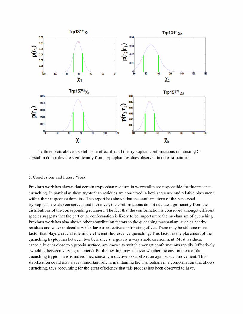
The three plots above also tell us in effect that all the tryptophan conformations in human γD-crystallin do not deviate significantly from tryptophan residues observed in other structures.
5. Conclusions and Future Work
Previous work has shown that certain tryptophan residues in γ-crystallin are responsible for fluorescence quenching. In particular, these tryptophan residues are conserved in both sequence and relative placement within their respective domains. This report has shown that the conformations of the conserved tryptophans are also conserved, and moreover, the conformations do not deviate significantly from the distributions of the corresponding rotamers. The fact that the conformation is conserved amongst different species suggests that the particular conformation is likely to be important to the mechanism of quenching. Previous work has also shown other contribution factors to the quenching mechanism, such as nearby residues and water molecules which have a collective contributing effect. There may be still one more factor that plays a crucial role in the efficient fluorescence quenching. This factor is the placement of the quenching tryptophan between two beta sheets, arguably a very stable environment. Most residues, especially ones close to a protein surface, are known to switch amongst conformations rapidly (effectively switching between varying rotamers). Further testing may uncover whether the environment of the quenching tryptophans is indeed mechanically inductive to stabilization against such movement. This stabilization could play a very important role in maintaining the tryptophans in a conformation that allows quenching, thus accounting for the great efficiency that this process has been observed to have.