Soluble α-Klotho serum levels in Chronic Kidney … α-Klotho serum levels in Chronic Kidney...
16
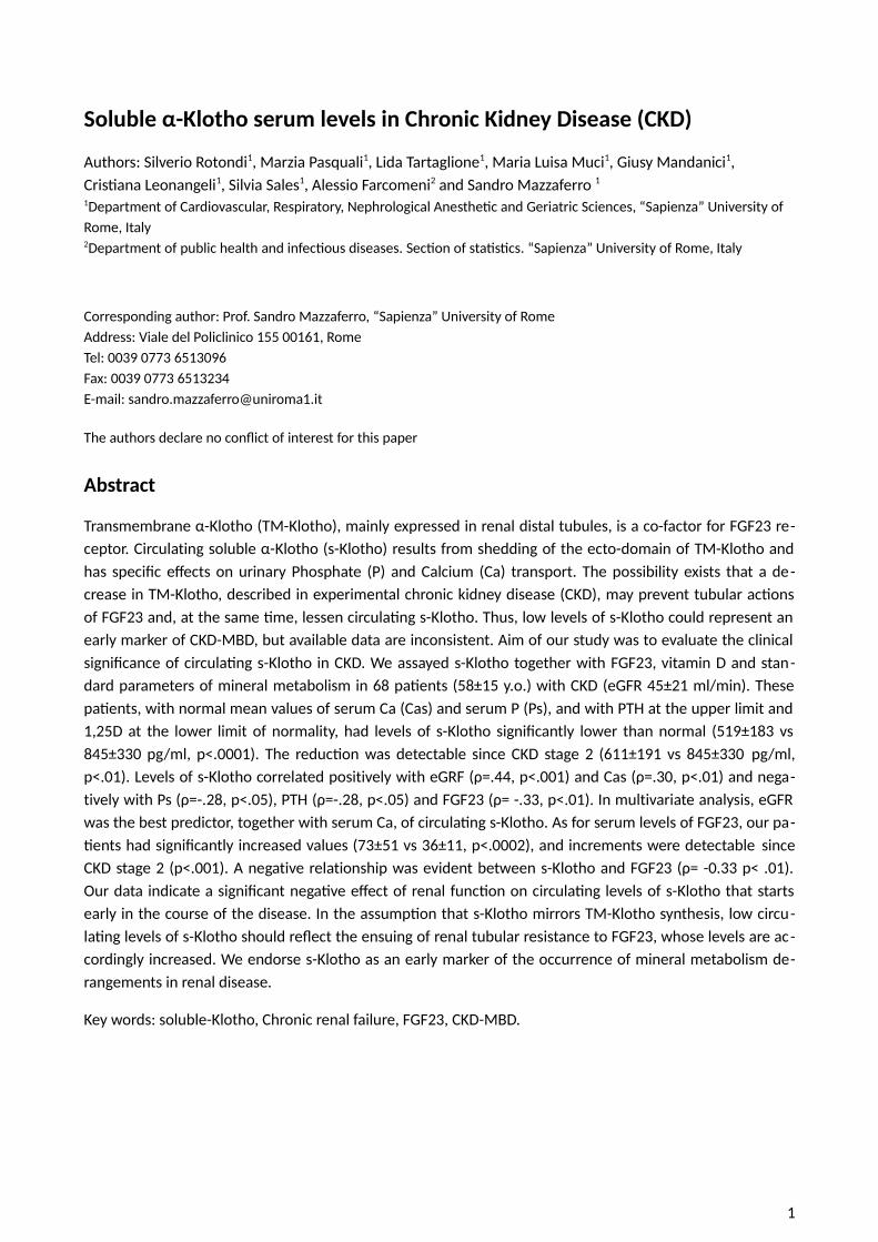
Soluble α-Klotho serum levels in Chronic Kidney Disease (CKD) Authors: Silverio Rotondi 1 , Marzia Pasquali 1 , Lida Tartaglione 1 , Maria Luisa Muci 1 , Giusy Mandanici 1 , Crisana Leonangeli 1 , Silvia Sales 1 , Alessio Farcomeni 2 and Sandro Mazzaferro 1 1 Department of Cardiovascular, Respiratory, Nephrological Anesthec and Geriatric Sciences, “Sapienza” University of Rome, Italy 2 Department of public health and infecous diseases. Secon of stascs. “Sapienza” University of Rome, Italy Corresponding author: Prof. Sandro Mazzaferro, “Sapienza” University of Rome Address: Viale del Policlinico 155 00161, Rome Tel: 0039 0773 6513096 Fax: 0039 0773 6513234 E-mail: [email protected] The authors declare no conflict of interest for this paper Abstract Transmembrane α-Klotho (TM-Klotho), mainly expressed in renal distal tubules, is a co-factor for FGF23 re- ceptor. Circulating soluble α-Klotho (s-Klotho) results from shedding of the ecto-domain of TM-Klotho and has specific effects on urinary Phosphate (P) and Calcium (Ca) transport. The possibility exists that a de- crease in TM-Klotho, described in experimental chronic kidney disease (CKD), may prevent tubular acons of FGF23 and, at the same me, lessen circulang s-Klotho. Thus, low levels of s-Klotho could represent an early marker of CKD-MBD, but available data are inconsistent. Aim of our study was to evaluate the clinical significance of circulang s-Klotho in CKD. We assayed s-Klotho together with FGF23, vitamin D and stan- dard parameters of mineral metabolism in 68 paents (58±15 y.o.) with CKD (eGFR 45±21 ml/min). These paents, with normal mean values of serum Ca (Cas) and serum P (Ps), and with PTH at the upper limit and 1,25D at the lower limit of normality, had levels of s-Klotho significantly lower than normal (519±183 vs 845±330 pg/ml, p<.0001). The reducon was detectable since CKD stage 2 (611±191 vs 845±330 pg/ml, p<.01). Levels of s-Klotho correlated positively with eGRF (ρ=.44, p<.001) and Cas (ρ=.30, p<.01) and nega- tively with Ps (ρ=-.28, p<.05), PTH (ρ=-.28, p<.05) and FGF23 (ρ= -.33, p<.01). In multivariate analysis, eGFR was the best predictor, together with serum Ca, of circulang s-Klotho. As for serum levels of FGF23, our pa- ents had significantly increased values (73±51 vs 36±11, p<.0002), and increments were detectable since CKD stage 2 (p<.001). A negative relationship was evident between s-Klotho and FGF23 (ρ= -0.33 p< .01). Our data indicate a significant negave effect of renal funcon on circulang levels of s-Klotho that starts early in the course of the disease. In the assumpon that s-Klotho mirrors TM-Klotho synthesis, low circu- lang levels of s-Klotho should reflect the ensuing of renal tubular resistance to FGF23, whose levels are ac- cordingly increased. We endorse s-Klotho as an early marker of the occurrence of mineral metabolism de- rangements in renal disease. Key words: soluble-Klotho, Chronic renal failure, FGF23, CKD-MBD. 1
-
Upload
truongxuyen -
Category
Documents
-
view
218 -
download
0
Transcript of Soluble α-Klotho serum levels in Chronic Kidney … α-Klotho serum levels in Chronic Kidney...
Soluble α-Klotho serum levels in Chronic Kidney Disease (CKD)
Authors: Silverio Rotondi1, Marzia Pasquali1, Lida Tartaglione1, Maria Luisa Muci1, Giusy Mandanici1, Cristiana Leonangeli1, Silvia Sales1, Alessio Farcomeni2 and Sandro Mazzaferro 1
1Department of Cardiovascular, Respiratory, Nephrological Anesthetic and Geriatric Sciences, “Sapienza” University of Rome, Italy2Department of public health and infectious diseases. Section of statistics. “Sapienza” University of Rome, Italy
Corresponding author: Prof. Sandro Mazzaferro, “Sapienza” University of Rome Address: Viale del Policlinico 155 00161, Rome Tel: 0039 0773 6513096 Fax: 0039 0773 6513234 E-mail: [email protected]
The authors declare no conflict of interest for this paper
Abstract
Transmembrane α-Klotho (TM-Klotho), mainly expressed in renal distal tubules, is a co-factor for FGF23 re-ceptor. Circulating soluble α-Klotho (s-Klotho) results from shedding of the ecto-domain of TM-Klotho andhas specific effects on urinary Phosphate (P) and Calcium (Ca) transport. The possibility exists that a de-crease in TM-Klotho, described in experimental chronic kidney disease (CKD), may prevent tubular actionsof FGF23 and, at the same time, lessen circulating s-Klotho. Thus, low levels of s-Klotho could represent anearly marker of CKD-MBD, but available data are inconsistent. Aim of our study was to evaluate the clinicalsignificance of circulating s-Klotho in CKD. We assayed s-Klotho together with FGF23, vitamin D and stan-dard parameters of mineral metabolism in 68 patients (58±15 y.o.) with CKD (eGFR 45±21 ml/min). Thesepatients, with normal mean values of serum Ca (Cas) and serum P (Ps), and with PTH at the upper limit and1,25D at the lower limit of normality, had levels of s-Klotho significantly lower than normal (519±183 vs845±330 pg/ml, p<.0001). The reduction was detectable since CKD stage 2 (611±191 vs 845±330 pg/ml,p<.01). Levels of s-Klotho correlated positively with eGRF (ρ=.44, p<.001) and Cas (ρ=.30, p<.01) and nega-tively with Ps (ρ=-.28, p<.05), PTH (ρ=-.28, p<.05) and FGF23 (ρ= -.33, p<.01). In multivariate analysis, eGFRwas the best predictor, together with serum Ca, of circulating s-Klotho. As for serum levels of FGF23, our pa-tients had significantly increased values (73±51 vs 36±11, p<.0002), and increments were detectable sinceCKD stage 2 (p<.001). A negative relationship was evident between s-Klotho and FGF23 (ρ= -0.33 p< .01).Our data indicate a significant negative effect of renal function on circulating levels of s-Klotho that startsearly in the course of the disease. In the assumption that s-Klotho mirrors TM-Klotho synthesis, low circu-lating levels of s-Klotho should reflect the ensuing of renal tubular resistance to FGF23, whose levels are ac-cordingly increased. We endorse s-Klotho as an early marker of the occurrence of mineral metabolism de-rangements in renal disease.
Key words: soluble-Klotho, Chronic renal failure, FGF23, CKD-MBD.
1
Introduction
Alpha-Klotho (Klotho) is a transmembrane (TM) protein primarily expressed in the kidney distaltubular cells [1] where it acts as an obligate co-receptor for the bone derived protein fibroblast growthfactor-23 (FGF23) [2,3]. In fact, TM-Klotho is required for FGF23 regulation of both renal handling ofphosphate and renal synthesis of calcitriol [4]. At variance with TM-Klotho, soluble α-Klotho (s-Klotho) is thecirculating protein resulting from the shedding of the extracellular domain of TM-Klotho operated by twometalloproteinases of the ADAM (A Disintegrin and Metalloproteinase domain-containing protein) family:ADAM 10 and 17 [5]. Importantly, s-Klotho also acts as a paracrine substance (with no receptor identified sofar) with specific and FGF23 independent renal and extra-renal effects [6, 7]. In particular, s-Klotho inhibitsthe sodium-phosphate co-transporter NaPi2a expression in the proximal tubules thus generating aphosphaturic effect additive to and independent of FGF23 [7, 8], and activates the ion channel TRPV5 in thedistal tubules, thus increasing tubular reabsorption of calcium [9]. In summary, α-Klotho, with itstransmembrane and soluble forms, is deeply involved with the physiologic regulation of mineral metabolism[10].
Experimental models of CKD evidence early reduction of renal Klotho mRNA expression and of bothTM-Klotho and s-Klotho [11, 12], and this is considered responsible for the development of kidney tubularcell resistance to FGF23. In agreement, in a model of selective Klotho deletion in the distal tubule, bonesynthesis of FGF23 increased possibly aimed at overcoming tubular resistance; as a result fractionalexcretion of phosphate increased and calcitriol synthesis declined. These animal models have been invokedto describe the early changes that in renal patients anticipate the development of the newly identified CKD-MBD syndrome [13] or, as pointed out in a recent paper, disorder [14].
Available data on serum levels of s-Klotho in CRF patients almost invariably describe reduced values,a finding referred to a primary proportional reduction of TM-Klotho, as described in animal studies [15, 16,17]. Moreover, circulating serum levels of s-Klotho are not always correlated with estimated glomerularfiltration rate (eGFR) [18, 19, 20, 21]. Excessive variability of some of the commercial kits available for s-Klotho assay in humans, as described by Heijboer et al [22] could contribute, at least partially, to theseconflicting results. Actually, Klotho assays have not been largely validated and various forms of circulatingalpha-klotho could be responsible for this variability. Nonetheless, a very recent paper based on renalbiopsies of patients with glomerulonephritis and variable renal damage, for the first time showed reducedtubular TM-Klotho expression associated with reduced serum levels of s-Klotho. Importantly, these patientshad almost undetectable changes in the standard markers of mineral metabolism [23]. As a whole, thediagnostic role of the assay of s-Klotho as a useful biomarker of early mineral metabolism derangements inCKD patients may be relevant but warrants further research [18, 19, 20, 21].
In the present study, we evaluated the diagnostic performance of s-Klotho in our CKD population ofpatients by using the most accredited commercial s-Klotho assay [22]. We aimed at confirming thereduction of circulating levels of s-Klotho and at verifying the links with FGF23 and with other markers ofmineral metabolism derangements.
Methods
SubjectsWe enrolled in the study, from our outpatient unit, eligible patients who gave informed consent.
Inclusion criteria were white race, age 18-80 years, on conservative therapy with no evidence of acuteunderlying illness and naïve to treatment with any active or precursor metabolite of vitamin D.
2
Fasting blood samples were drawn from all participants to measure creatinine (Crs), albumin,calcium (Ca), phosphate (P), parathyroid hormone (PTH), 25(OH)-vitamin D (25D), 1,25(OH)2-vitaminD(1,25D), fibroblast growth factor-23 (FGF23) and soluble-α-Klotho (s-Klotho). We also collected fasting spoturine samples from all participants at the time of blood sampling to measure creatinine, phosphate andcalcium. In each patient, we recorded clinical parameters and prescribed therapies.
We classified CKD stage according to the National Kidney Foundation Disease Outcomes QualityInitiative clinical practice guidelines (KDOQI) [24].
Thirty normal subjects, recruited among the employees and fellows attending our Unit, served ascontrol to obtain our reference values in particular for s-Klotho and FGF23. They were 14 males and 16females, aged 35.0±12.4 years with normal renal function (eGFR: 105.8±15.4 ml/min/1,73m2) negativeurinary dipstick and no evidence of acute or chronic underlying illness.
Assays Serum creatinine (kinetic alkaline picrate method), albumin (bromcresol purple method), Ca
(cresolphthalein-complexone method) and P (ammonium molybdate method) were assayed by routine,standard colorimetric techniques with a Technicon RA-500 analyzer (Bayer Corporation Inc, Tarrytown, NY).
Serum PTH was assayed by an immunoradiometric technique (DiaSorin, Stillwater, MN, USA) basedon a double antibody against the intact molecule; our normal values are within 10-55 pg/mL, with intra-and inter-assay variations of 6.5% and 9.8%, respectively.
Serum 25D determination was done with a commercial kit (Dia-Sorin, Stillwater, MN, USA) thatincluded sample purification with acetonitrile followed by a 125I-based radioimmunoassay. Intra- and inter-assay coefficients of variation were 10.8% and 9.4%, respectively.
Serum levels of 1,25D were measured with a radioimmunoassay according to the manufacturer’sprotocol (IDS Ltd, Boldon, UK) including a monoclonal immunoextraction, followed by quantitation with astandard 125I-based radioimmunoassay. Intra- and inter-assay coefficients of variation were <12% and <14%,respectively. The normal range observed in our laboratory was between 19.5 and 67.0 pg/mL.
Serum levels of FGF23 were assayed with a commercially available kit (Kainos Lab Inc., Tokyo, Japan)that utilizes a 2-site ELISA for the full-length molecule. Two specific murine monoclonal antibodiesrecognized the biologically active FGF23, with a lower limit of detection of 3 pg/mL, and inter- and intra-assay coefficients of variation of <5%. This assay has been demonstrated to be the most sensitive among the3 different methods available [25]. In 30 normal subjects, we obtained a mean value of 29.8 ± 10.9 pg/mL(range 18-52 pg/mL), in line with reported data [25].
Serum levels of soluble alpha- Klotho were assayed with a novel enzyme-linked immunosorbentassay (ELISA) method detecting human s-Klotho developed first by establishing a monoclonal antibody withstrong affinity for human Klotho protein, recognizing with high selectivity the tertiary protein structure of itsextracellular domain (Immuno-Biological Laboratories Co., Ltd.). This ELISA system can specifically detectand measure the circulating serum s-Klotho levels in humans [26]. It was recently tested by Heijboer [22]and the within- and between-run variation of the α-Klotho IBL was <5 and <8%, respectively.Measurements in serum and EDTA plasma samples were in agreement (R2 = 0.99; n = 20) and linearity wastested by dilution in two samples with a concentration of 1929 and 2864 pg/ml. In one sample, 2-, 4- and 8-time dilutions gave results as expected (100–117% of expected values). In our experience, in 30 normalsubjects we obtained a mean value of 845 ± 330 pg/mL (range 2048-481 pg/ml).
We estimated Glomerular Filtration Rate (eGFR) according to the abbreviated Modification of Dietin Renal Disease (MDRD) equation (eGFR= 186 x serum creatinine-1.154 x age-0.203 x 0.742 if female) [27]
3
Statistical analysis
Data are expressed as means ± SD for Gaussian variables or median and IQR when normality wasnot tenable. We used Kolmogorov-Smirnov test to evaluate normality of continuous measurements.Spearman correlation was used to assess monotonic co-variation of measurements. Tests on Spearmancorrelation were Bonferroni adjusted for multiplicity. Non-parametric ANOVA (Kruskall-Wallis test) was usedto compare measurements among groups and post-hoc comparisons in pairs were conducted by Bonferroniadjusted Mann-Whitney tests. Log-measurements were confirmed to be normally distributed and were used as outcomes in multivariateregression models. The final multivariate model was obtained by minimizing the Akaike InformationCriterion via a forward stepwise regression. All tests are two tailed and (adjusted) p-values <0.05 wereconsidered as statistically significant. Analyses were performed using the open source software package R version 3.0.2.
ResultsFor this study we recruited sixty-eight CKD patients, and their clinical and biochemical parameters
are reported in Tab. 1 and 2. Renal function, as reflected by eGFR, averaged 45±21 ml/min (median ± SD)and included CKD stages from 2 to 4 [stage 2, n = 22 (32%); stage 3, n = 28 (41%); stage 4, n = 18 (27%)].
s-Klotho, FGF23 and eGFRs-Klotho levels in our CKD patients were significantly lower (519±183 vs 845 ± 330 pg/ml, p<.0001,
Tab.3) and FGF23 levels were significantly higher (73±51 vs 36±11 pg/ml, p<0002, Tab.3) than our referencevalues.
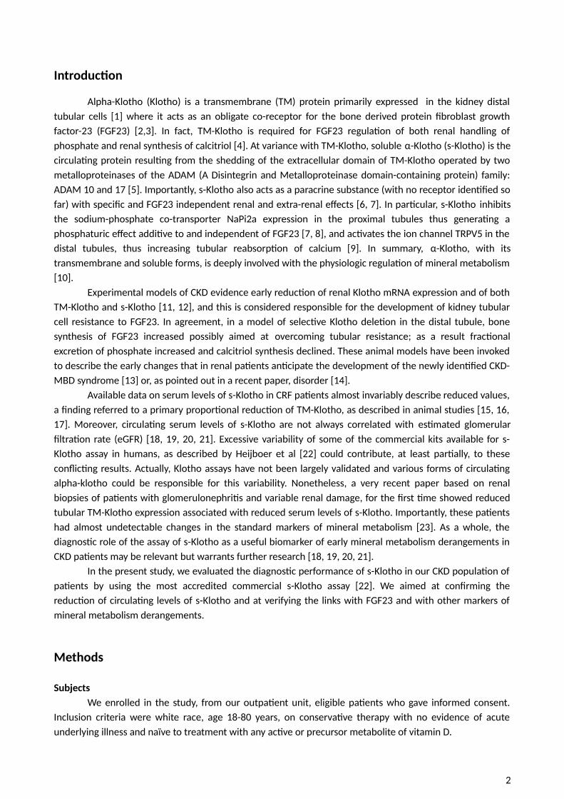
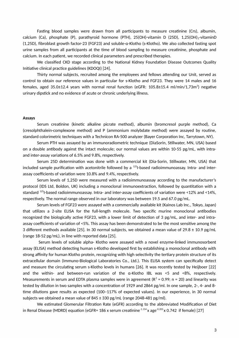
In patients s-Klotho and FGF23 were negatively correlated (ρ= -0.33 p< .01, Fig.1). In addition, therewas a positive correlation between eGFR and s-Klotho (ρ=0.43 p<.001, Fig. 2a) and a negative correlationbetween eGFR and FGF23 (ρ= -0.66 p<.0001, Fig. 2b).
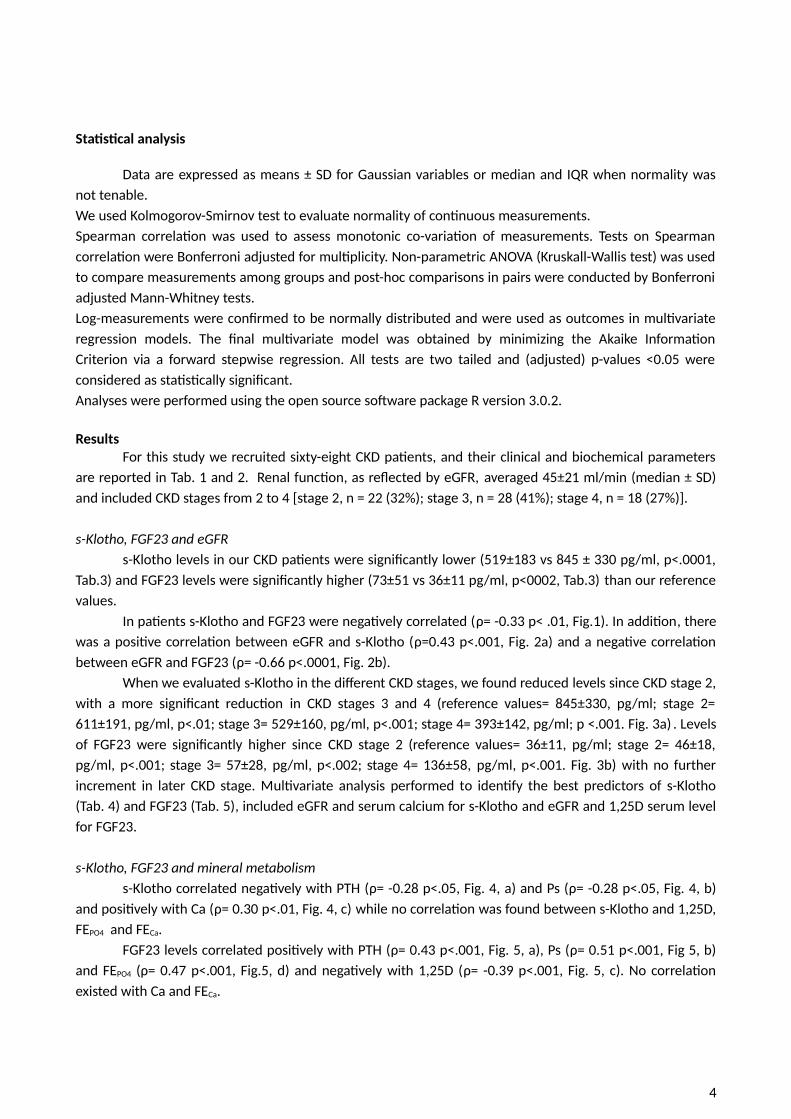
When we evaluated s-Klotho in the different CKD stages, we found reduced levels since CKD stage 2,with a more significant reduction in CKD stages 3 and 4 (reference values= 845±330, pg/ml; stage 2=611±191, pg/ml, p<.01; stage 3= 529±160, pg/ml, p<.001; stage 4= 393±142, pg/ml; p <.001. Fig. 3a) . Levelsof FGF23 were significantly higher since CKD stage 2 (reference values= 36±11, pg/ml; stage 2= 46±18,pg/ml, p<.001; stage 3= 57±28, pg/ml, p<.002; stage 4= 136±58, pg/ml, p<.001. Fig. 3b) with no furtherincrement in later CKD stage. Multivariate analysis performed to identify the best predictors of s-Klotho(Tab. 4) and FGF23 (Tab. 5), included eGFR and serum calcium for s-Klotho and eGFR and 1,25D serum levelfor FGF23.
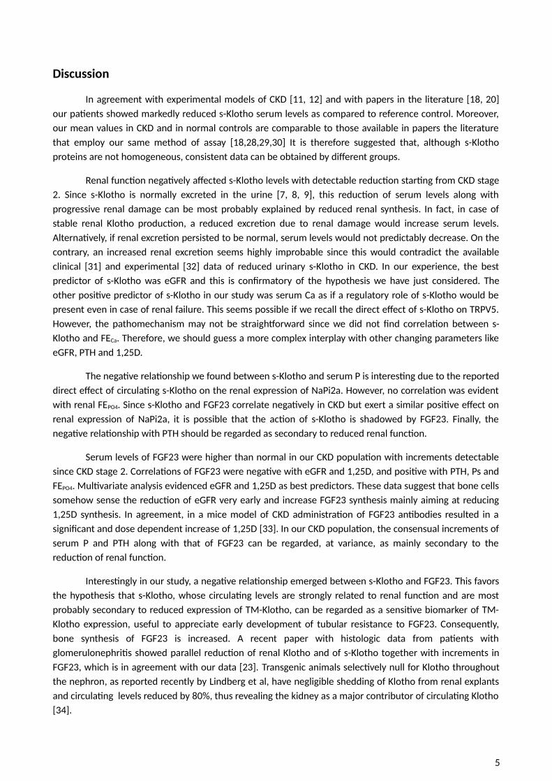
s-Klotho, FGF23 and mineral metabolisms-Klotho correlated negatively with PTH (ρ= -0.28 p<.05, Fig. 4, a) and Ps (ρ= -0.28 p<.05, Fig. 4, b)
and positively with Ca (ρ= 0.30 p<.01, Fig. 4, c) while no correlation was found between s-Klotho and 1,25D,FEPO4 and FECa.
FGF23 levels correlated positively with PTH (ρ= 0.43 p<.001, Fig. 5, a), Ps (ρ= 0.51 p<.001, Fig 5, b)and FEPO4 (ρ= 0.47 p<.001, Fig.5, d) and negatively with 1,25D (ρ= -0.39 p<.001, Fig. 5, c). No correlationexisted with Ca and FECa.
4
Discussion
In agreement with experimental models of CKD [11, 12] and with papers in the literature [18, 20]our patients showed markedly reduced s-Klotho serum levels as compared to reference control. Moreover,our mean values in CKD and in normal controls are comparable to those available in papers the literaturethat employ our same method of assay [18,28,29,30] It is therefore suggested that, although s-Klothoproteins are not homogeneous, consistent data can be obtained by different groups.
Renal function negatively affected s-Klotho levels with detectable reduction starting from CKD stage2. Since s-Klotho is normally excreted in the urine [7, 8, 9], this reduction of serum levels along withprogressive renal damage can be most probably explained by reduced renal synthesis. In fact, in case ofstable renal Klotho production, a reduced excretion due to renal damage would increase serum levels.Alternatively, if renal excretion persisted to be normal, serum levels would not predictably decrease. On thecontrary, an increased renal excretion seems highly improbable since this would contradict the availableclinical [31] and experimental [32] data of reduced urinary s-Klotho in CKD. In our experience, the bestpredictor of s-Klotho was eGFR and this is confirmatory of the hypothesis we have just considered. Theother positive predictor of s-Klotho in our study was serum Ca as if a regulatory role of s-Klotho would bepresent even in case of renal failure. This seems possible if we recall the direct effect of s-Klotho on TRPV5.However, the pathomechanism may not be straightforward since we did not find correlation between s-Klotho and FECa. Therefore, we should guess a more complex interplay with other changing parameters likeeGFR, PTH and 1,25D.
The negative relationship we found between s-Klotho and serum P is interesting due to the reporteddirect effect of circulating s-Klotho on the renal expression of NaPi2a. However, no correlation was evidentwith renal FEPO4. Since s-Klotho and FGF23 correlate negatively in CKD but exert a similar positive effect onrenal expression of NaPi2a, it is possible that the action of s-Klotho is shadowed by FGF23. Finally, thenegative relationship with PTH should be regarded as secondary to reduced renal function.
Serum levels of FGF23 were higher than normal in our CKD population with increments detectablesince CKD stage 2. Correlations of FGF23 were negative with eGFR and 1,25D, and positive with PTH, Ps andFEPO4. Multivariate analysis evidenced eGFR and 1,25D as best predictors. These data suggest that bone cellssomehow sense the reduction of eGFR very early and increase FGF23 synthesis mainly aiming at reducing1,25D synthesis. In agreement, in a mice model of CKD administration of FGF23 antibodies resulted in asignificant and dose dependent increase of 1,25D [33]. In our CKD population, the consensual increments ofserum P and PTH along with that of FGF23 can be regarded, at variance, as mainly secondary to thereduction of renal function.
Interestingly in our study, a negative relationship emerged between s-Klotho and FGF23. This favorsthe hypothesis that s-Klotho, whose circulating levels are strongly related to renal function and are mostprobably secondary to reduced expression of TM-Klotho, can be regarded as a sensitive biomarker of TM-Klotho expression, useful to appreciate early development of tubular resistance to FGF23. Consequently,bone synthesis of FGF23 is increased. A recent paper with histologic data from patients withglomerulonephritis showed parallel reduction of renal Klotho and of s-Klotho together with increments inFGF23, which is in agreement with our data [23]. Transgenic animals selectively null for Klotho throughoutthe nephron, as reported recently by Lindberg et al, have negligible shedding of Klotho from renal explantsand circulating levels reduced by 80%, thus revealing the kidney as a major contributor of circulating Klotho[34].
5
Significantly in our study, levels of s-Klotho and of FGF23 are roughly halved and respectivelydoubled in a population with an average eGFR of 45 ml/min/1,73, but with substantially normal values of Caand P and with mean values of 1,25D and of PTH respectively at the lower and higher upper limits ofnormality. This confirms that changes in serum Ca and P are not reliable to detect early CKD-MBD, while s-Klotho seems more sensitive than PTH and 1,25D.
Limitations of our study are several. First, the number of evaluated patients is rather low. In fact, anincreased number of cases would have reinforced the reliability of our results that, however, are in line withpublished data in the literature. Second, we did not include CKD stage 1 patients and this does not allow usto verify if s-Klotho diminish even in case of “normal” renal function. Certainly, this important issuewarrants further investigations. Third, we did not measure urinary s-Klotho. Effectively, evidence of reducedurinary Klotho in our experience would have reinforced the hypothesis of reduced renal synthesis. However,although scanty, available papers report reduced urinary Klotho in patients with renal failure [31] and inanimals selectively null for nephron Klotho [34], both results confirming our hypothesis.
In conclusion, our results favor the hypothesis that renal Klotho synthesis diminishes early in renaldisease and that s-Klotho proportionally lessens; bone detects these changes somehow and increasesFGF23 production. Accordingly, s-Klotho represents an early marker of renal damage and of ensuing CKD-MBD, indicative of the cross-talk between bone and kidney.
6
Reference List
1. Mian IS. Sequence, structural, functional, and phylogenetic analyses of three glycosidase families. Blood Cells Mol Dis 1998; 24: 83-100
2. Kurosu H, Ogawa Y, Miyoshi M et al. Regulation of fibroblast growth factor-23 signaling by Klotho. J Biol Chem 2006; 281: 6120-6123
3. Urakawa I, Yamazaki Y, Shimada T et al. Klotho converts canonical FGF receptor into a specific receptor for FGF23. Nature 2006; 444: 770-774
4. Liu S, Tang W, Zhou J et al. Fibroblast growth factor 23 is a counter-regulatory phosphaturic hormone for vitamin D. J Am Soc Nephrol 2006; 17: 1305-1315
5. Chen CD, Podvin S, Gillespie E et al. Insulin stimulates the cleavage and release of the extracellular domain of Klotho by ADAM10 and ADAM17. Proc Natl Acad Sci U S A 2007; 104: 19796-19801
6. Hu MC, Kuro O, Moe OW. Klotho and chronic kidney disease. Contrib Nephrol 2013; 180: 47-63
7. Hu MC, Kuro-o M, Moe OW. Renal and extrarenal actions of Klotho. Semin Nephrol 2013; 33: 118-129
8. Hu MC, Shi M, Zhang J et al. Klotho: a novel phosphaturic substance acting as an autocrine enzyme in the renal proximal tubule. FASEB J 2010; 24: 3438-3450
9. Cha SK, Ortega B, Kurosu H et al. Removal of sialic acid involving Klotho causes cell-surface retention of TRPV5 channel via binding to galectin-1. Proc Natl Acad Sci U S A 2008; 105: 9805-9810
10. Kuro O. Phosphate and Klotho. Kidney Int Suppl 2011; S20-S23
11. Aizawa H, Saito Y, Nakamura Tet al. Downregulation of the Klotho gene in the kidney under sustained circulatory stress in rats. Biochem Biophys Res Commun. 1998 Aug 28; 249(3):865-71
12. Yu J, Deng M, Zhao J, Huang L. Decreased expression of Klotho gene in uremic atherosclerosis in apolipoprotein E-deficient mice.Biochem Biophys Res Commun. 2010 Jan 1;391(1):261-6
13. Olauson H, Lindberg K, Amin R et al. Targeted deletion of Klotho in kidney distal tubule disrupts mineral metabolism. J Am Soc Nephrol 2012; 23: 1641-1651
14. Cozzolino M, Ureña-Torres P, Vervloet MG et al. Is chronic kidney disease-mineral bone disorder (CKD-MBD) really a syndrome? Nephrol Dial Transplant. 2014 Feb 24
15. Hu MC, Shi M, Zhang J et al. Klotho deficiency causes vascular calcification in chronic kidney disease. J Am Soc Nephrol 2011; 22: 124-136
7
16. Koh N, Fujimori T, Nishiguchi S et al. Severely reduced production of Klotho in human chronic renal failure kidney. Biochem Biophys Res Commun 2001; 280: 1015-1020
17. Akimoto T, Kimura T, Watanabe Y et al. The impact of nephrectomy and renal transplantation on serum levels of soluble Klotho protein. Transplant Proc 2013; 45: 134-136
18. Pavik I, Jaeger P, Ebner L et al. Secreted Klotho and FGF23 in chronic kidney disease Stage 1 to 5: a sequence suggested from a cross-sectional study. Nephrol Dial Transplant 2013; 28: 352-359
19. Seiler S, Wen M, Roth HJ et al. Plasma Klotho is not related to kidney function and does not predict adverse outcome in patients with chronic kidney disease. Kidney Int 2013; 83: 121-128
20. Wan M, Smith C, Shah V et al. Fibroblast growth factor 23 and soluble Klotho in children with chronic kidney disease. Nephrol Dial Transplant 2013; 28: 153-161
21. Devaraj S, Syed B, Chien A et al. Validation of an immunoassay for soluble Klotho protein: decreased levels in diabetes and increased levels in chronic kidney disease. Am J Clin Pathol 2012; 137: 479–485
22. Heijboer C, Blankenstein A, Hoenderop j et al. Laboratory aspects of circulating α-Klotho. Nephrol Dial Transplant (2013) 28: 2287–2294
23. Sakan H, Nakatani K, Asai O et al. Reduced renal α-Klotho expression in CKD patients and its effect on renal phosphate handling and vitamin D metabolism. PLoS One. 2014 Jan 23
24. National Kidney Foundation (2002) K/DOQI clinical practice guidelines for chronic kidney disease: evaluation, classification, and stratification. Am J Kidney Dis 39: S1–266
25. Imel EA, Peacock M, Pitukcheewanont P, et al. Sensitivity of fibroblast growth factor 23 measurements in tumor-induced osteomalacia. J Clin Endocrinol Metab. 2006; 91(6):2055-2061
26. Yamazaki Y, Imura A, Urakawa I et al. Establishment of sandwich ELISA for soluble alpha-Klotho measurement: age-dependent change of soluble alpha-Klotho levels in healthy subjects. Biochem Biophys Res Commun 2010; 398: 513–518
27. Levey AS, Bosch JP, Lewis JB et al: A more accurate method to estimate glomerular filtrationrate from serum creatinine: a new prediction equation. Modification of Diet in RenalDisease Study Group. Ann Intern Med 1999, 130: 461–470
28. Yokoyama K, Imura A, Ohkido I et al. Serum soluble alpha-klotho in haemodialysis patients.Clin Nephrol 2012, 77: 347-351
29. Kitagawa M, Sugiyama H, Morinaga H et al. A decreased level of serum soluble Klotho is anindependent biomarker associated with arterial stiffness in patients with chronic kidneydisease. PLos One 2013, 8: e56695
8
30. Nowak A, Friedrich B, Artunc F et al, Prognostic Value and Link to Atrial Fibrillation ofSoluble Klotho and FGF23 in Hemodialysis Patients. PlosOne 2014, 9:e100688
31. Akimoto T, Yoshizawa H, Watanabe Y et al. Characteristics of urinary and serum soluble Klotho protein in patients with different degrees of chronic kidney disease. BMC Nephrol. 2012 Nov 23;13:155
32. Hu MC, Shi M, Zhang J et al. Klotho deficiency is an early biomarker of renal ischemia-reperfusion injury and its replacement is protective. Kidney Int 2010, 78:1240–1251.
33. Hasegawa H, Nagano N, Urakawa I et al. Direct evidence for a causative role of FGF23 in the abnormal renal phosphate handling and vitamin D metabolism in rats with early-stage chronic kidney disease. Kidney Int. 2010 Nov;78(10):975-80
34. Lindberg K, Amin R, Moe OW at al. The kidney is the principal organ mediating klotho effects. J Am Soc Nephrol 2014; 25: 2169–2175
9
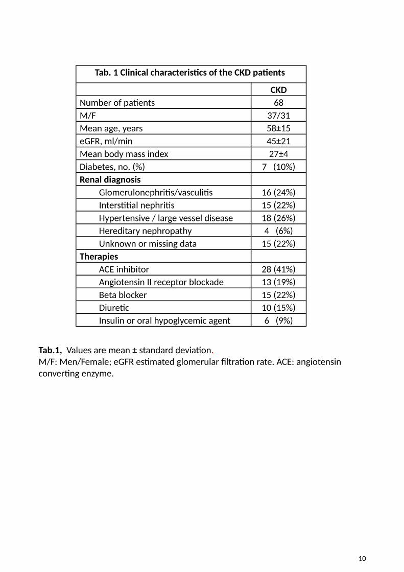
Tab. 1 Clinical characteristics of the CKD patients
CKDNumber of patients 68M/F 37/31Mean age, years 58±15eGFR, ml/min 45±21Mean body mass index 27±4Diabetes, no. (%) 7 (10%)Renal diagnosis Glomerulonephritis/vasculitis 16 (24%) Interstitial nephritis 15 (22%) Hypertensive / large vessel disease 18 (26%) Hereditary nephropathy 4 (6%) Unknown or missing data 15 (22%)Therapies ACE inhibitor 28 (41%) Angiotensin II receptor blockade 13 (19%) Beta blocker 15 (22%) Diuretic 10 (15%) Insulin or oral hypoglycemic agent 6 (9%)
Tab.1, Values are mean ± standard deviation.M/F: Men/Female; eGFR estimated glomerular filtration rate. ACE: angiotensin converting enzyme.
10
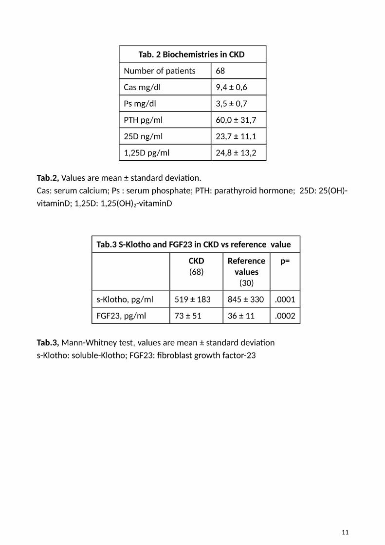
Tab. 2 Biochemistries in CKD
Number of patients 68
Cas mg/dl 9,4 ± 0,6
Ps mg/dl 3,5 ± 0,7
PTH pg/ml 60,0 ± 31,7
25D ng/ml 23,7 ± 11,1
1,25D pg/ml 24,8 ± 13,2
Tab.2, Values are mean ± standard deviation.Cas: serum calcium; Ps : serum phosphate; PTH: parathyroid hormone; 25D: 25(OH)-vitaminD; 1,25D: 1,25(OH)2-vitaminD
Tab.3 S-Klotho and FGF23 in CKD vs reference value
CKD(68)
Referencevalues
(30)
p=
s-Klotho, pg/ml 519 ± 183 845 ± 330 .0001
FGF23, pg/ml 73 ± 51 36 ± 11 .0002
Tab.3, Mann-Whitney test, values are mean ± standard deviation s-Klotho: soluble-Klotho; FGF23: fibroblast growth factor-23
11
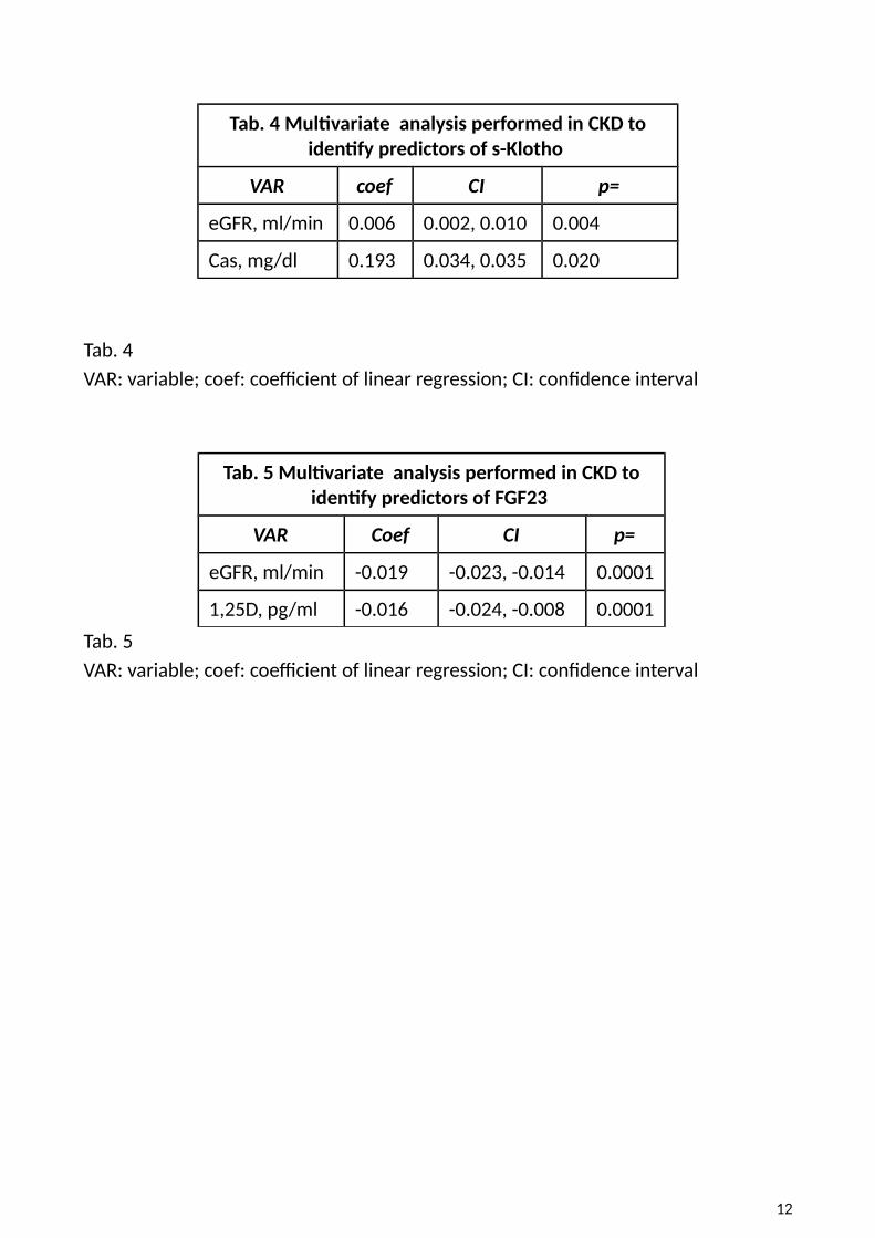
Tab. 4 Multivariate analysis performed in CKD toidentify predictors of s-Klotho
VAR coef CI p=
eGFR, ml/min 0.006 0.002, 0.010 0.004
Cas, mg/dl 0.193 0.034, 0.035 0.020
Tab. 4 VAR: variable; coef: coefficient of linear regression; CI: confidence interval
Tab. 5 Multivariate analysis performed in CKD toidentify predictors of FGF23
VAR Coef CI p=
eGFR, ml/min -0.019 -0.023, -0.014 0.0001
1,25D, pg/ml -0.016 -0.024, -0.008 0.0001
Tab. 5 VAR: variable; coef: coefficient of linear regression; CI: confidence interval
12
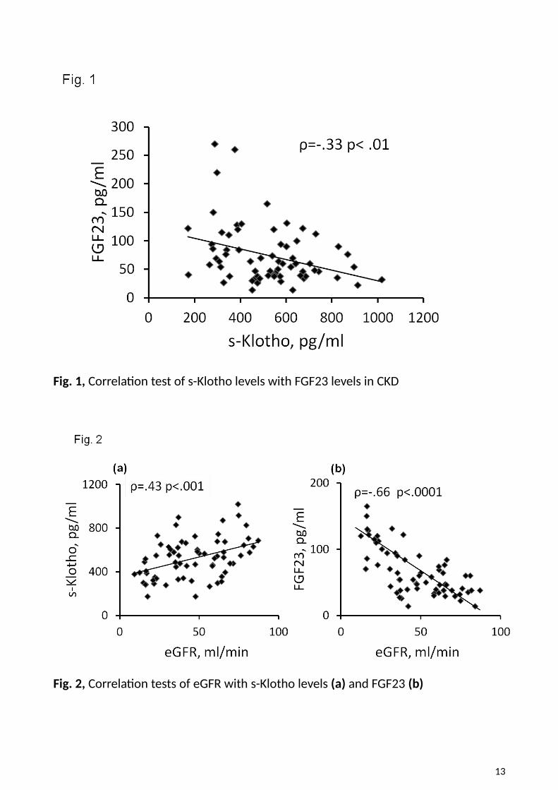
Fig. 1, Correlation test of s-Klotho levels with FGF23 levels in CKD
Fig. 2, Correlation tests of eGFR with s-Klotho levels (a) and FGF23 (b)
13
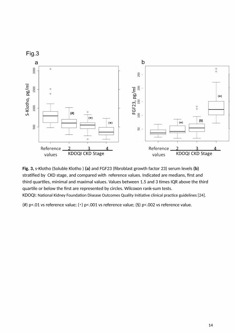
Fig. 3, s-Klotho (Soluble Klotho ) (a) and FGF23 (fibroblast growth factor 23) serum levels (b) stratified by CKD stage, and compared with reference values. Indicated are medians, first and third quartiles, minimal and maximal values. Values between 1.5 and 3 times IQR above the third quartile or below the first are represented by circles. Wilcoxon rank-sum tests.KDOQI: National Kidney Foundation Disease Outcomes Quality Initiative clinical practice guidelines [24].
(#) p<.01 vs reference value; (*) p<.001 vs reference value; (§) p<.002 vs reference value.
14
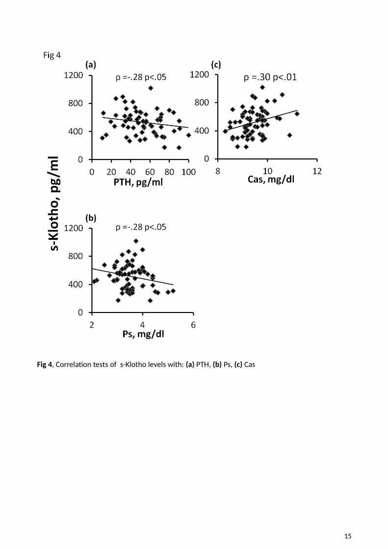
Fig 4, Correlation tests of s-Klotho levels with: (a) PTH, (b) Ps, (c) Cas
15
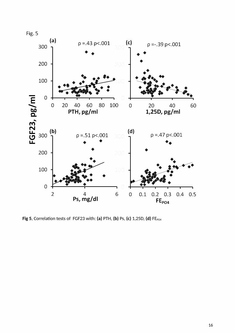
Fig 5, Correlation tests of FGF23 with: (a) PTH, (b) Ps, (c) 1,25D, (d) FEPO4
16