
SOLICITAÇÃO DE DEFESA DE MONOGRAFIA
47
FACULDADE DE BIOCIÊNCIAS PROGRAMA DE PÓS-GRADUAÇÃO EM BIOLOGIA CELULAR E MOLECULAR MESTRADO EM BIOLOGIA CELULAR E MOLECULAR NATÁLIA ELTZ SILVA EFEITO DA INJEÇÃO DA PROTEÍNA Β-AMILOIDE 1-42 EM DIFERENTES FORMAS NO VENTRÍCULO ENCEFÁLICO: UM MODELO DE ASPECTOS CELULARES DA DOENÇA DE ALZHEIMER EM ZEBRAFISH Porto Alegre 2017
Transcript of SOLICITAÇÃO DE DEFESA DE MONOGRAFIA
FACULDADE DE BIOCIÊNCIAS PROGRAMA DE PÓS-GRADUAÇÃO EM BIOLOGIA CELULAR E MOLECULAR
MESTRADO EM BIOLOGIA CELULAR E MOLECULAR
NATÁLIA ELTZ SILVA
EFEITO DA INJEÇÃO DA PROTEÍNA Β-AMILOIDE1-42 EM DIFERENTES FORMAS NO VENTRÍCULO ENCEFÁLICO: UM MODELO DE ASPECTOS CELULARES DA DOENÇA DE
ALZHEIMER EM ZEBRAFISH
Porto Alegre 2017
Pontifícia Universidade Católica do Rio Grande do Sul
Faculdade de Biociências Programa de Pós-Graduação em Biologia Celular e Molecular
EFEITO DA INJEÇÃO DE DIFERENTES FORMAS DA PROTEÍNA Β-AMILOIDE1-42 NO VENTRÍCULO ENCEFÁLICO DE ZEBRAFISH: UM MODELO DE ASPECTOS CELULARES DA DOENÇA DE
ALZHEIMER
NATÁLIA ELTZ SILVA
Orientadora: Profª Drª Monica Ryff Moreira Roca Vianna
Dissertação apresentada como requisito para a obtenção do grau de Mestre pelo Programa de Pós-Graduação em Biologia Celular e Molecular da Faculdade de Biociências da Pontifícia Universidade Católica do Rio Grande do Sul.
PORTO ALEGRE
2017

AGRADECIMENTOS
A minha orientadora Monica Vianna por ter me acolhido desde o meu 3º semestre de
faculdade e por todos os ensinamentos, auxílio e carinho ao longo destes anos.
A Laura Nery que foi como minha coorientadora durante esses anos e por todos os
ensinamentos, paciência e amizade. Ambas foram essenciais para o meu crescimento
pessoal e como pesquisadora.
Ao Fabiano Peres pelas inúmeras explicações, apoio, por ouvir minhas reclamações e dividir
o lanche conosco.
Às alunas Raphaela, Gabriela e Christiane por me ajudarem nos experimentos e pela
companhia nos finais de semana e feriados.
A Laura, Luiza, Stefani, Débora, Camila e Paula por fazerem meus dias no laboratório mais
felizes.
A Ângela, Marina, Daniela, Mariana e Ana pelos anos de amizade.
A Karina, Daiani, Gabriela, Rafael e Raphaela pela nossa união nos tempos de Iniciação
Científica que dura até hoje.
Aos demais membros do ZebLab foi um prazer conviver e aprender com vocês.
Ao Cristiano por acreditar em mim e por todo o apoio, companheirismo, amor e
compreensão.
A minha madrinha Zeneida e meu padrinho Anderson, meus avós Maria, Maria Helena e
Ernesto por estarem ao meu lado.
Aos meus irmãos Vinicius e Nicole que são parte de mim e que amo com todo o meu
coração.
A minha pequena e doce afilhada Marina, que com sua chegada me trouxe luz e amor.
Principalmente ao meu pai Gilberto que foi quem mais me apoiou, acreditou e incentivou os
meus estudos e a minha mãe Maureen que esteve ao meu lado durante todos os momentos,
inclusive nos mais difíceis e estressantes. A pessoa que sou hoje é fruto de todo e esforço,
dedicação, amor e carinho de vocês. Serei eternamente grata por ter vocês em minha vida.
Resumo
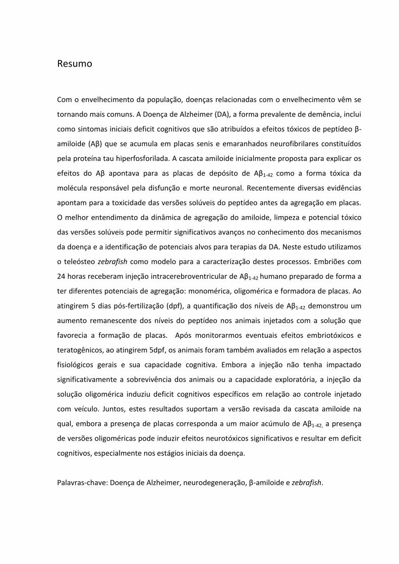
Com o envelhecimento da população, doenças relacionadas com o envelhecimento vêm se
tornando mais comuns. A Doença de Alzheimer (DA), a forma prevalente de demência, inclui
como sintomas iniciais deficit cognitivos que são atribuídos a efeitos tóxicos de peptídeo β-
amiloide (Aβ) que se acumula em placas senis e emaranhados neurofibrilares constituídos
pela proteína tau hiperfosforilada. A cascata amiloide inicialmente proposta para explicar os
efeitos do Aβ apontava para as placas de depósito de Aβ1-42 como a forma tóxica da
molécula responsável pela disfunção e morte neuronal. Recentemente diversas evidências
apontam para a toxicidade das versões solúveis do peptídeo antes da agregação em placas.
O melhor entendimento da dinâmica de agregação do amiloide, limpeza e potencial tóxico
das versões solúveis pode permitir significativos avanços no conhecimento dos mecanismos
da doença e a identificação de potenciais alvos para terapias da DA. Neste estudo utilizamos
o teleósteo zebrafish como modelo para a caracterização destes processos. Embriões com
24 horas receberam injeção intracerebroventricular de Aβ1-42 humano preparado de forma a
ter diferentes potenciais de agregação: monomérica, oligomérica e formadora de placas. Ao
atingirem 5 dias pós-fertilização (dpf), a quantificação dos níveis de Aβ1-42 demonstrou um
aumento remanescente dos níveis do peptídeo nos animais injetados com a solução que
favorecia a formação de placas. Após monitorarmos eventuais efeitos embriotóxicos e
teratogênicos, ao atingirem 5dpf, os animais foram também avaliados em relação a aspectos
fisiológicos gerais e sua capacidade cognitiva. Embora a injeção não tenha impactado
significativamente a sobrevivência dos animais ou a capacidade exploratória, a injeção da
solução oligomérica induziu deficit cognitivos específicos em relação ao controle injetado
com veículo. Juntos, estes resultados suportam a versão revisada da cascata amiloide na
qual, embora a presença de placas corresponda a um maior acúmulo de Aβ1-42, a presença
de versões oligoméricas pode induzir efeitos neurotóxicos significativos e resultar em deficit
cognitivos, especialmente nos estágios iniciais da doença.
Palavras-chave: Doença de Alzheimer, neurodegeneração, β-amiloide e zebrafish.
Abstract
Aging-related diseases are becoming more common. Alzheimer's disease (AD), the most
prevalent form of dementia, includes as initial symptoms cognitive deficits that are
attributed to the toxic effects of amyloidβ peptide (Aβ) that accumulates in senile plaques
and neurofibrillary tangles composed of hyperphosphorylated tau protein. The amyloid
cascade initially proposed to explain the effects of Aβ pointed to the plaques as the most
toxic form of the Aβ molecule responsible for neuronal dysfunction and death. Recently,
several evidences point to the increased toxicity of the soluble forms of the peptide. A better
understanding of the dynamics of amyloid aggregation, clearance and toxic potential of the
soluble versions may foster significant advances in the understanding AD mechanisms and
the identification of potential targets for AD therapies. In this study we used the zebrafish as
a model. 24-hour embryos received intracerebroventricular injection of human Aβ1-42
prepared to have different aggregation potentials: monomeric, oligomeric and plaque-
forming. At 5 days post-fertilization (dpf), quantification of Aβ1-42 levels demonstrated a
remnant increase in peptide levels in the animals injected with the solution that favored
plaque formation. After monitoring for embryotoxic and teratogenic effects, 5dpf the
animals were also evaluated in relation to general physiological aspects and their cognitive
ability. Although the injection did not significantly impact animal survival or exploratory
ability, the oligomeric solution induced specific cognitive deficits in relation to the vehicle-
injected control. Together these results support the revised version of the amyloid cascade
in which, although the presence of plaques corresponds to a greater accumulation of Aβ1-42,
oligomeric forms may induce significant neurotoxic effects and result in cognitive deficits
specially at disease’s early stages.
Key words: Alzheimer's disease, neurodegeneration, β-amyloid and zebrafish.
LISTA DE ABREVIAÇÕES
Aβ β-Amiloide APP Proteína Precursora Amiloide BOD Demanda Bioquímica de Oxigênio CEUA Comitê de Ética para Uso de Animais DA Doença de Alzheimer DMSO Dimetilsulfóxido DPF Dias Pós-Fertilização HPF Horas Pós-Fertilização MS222 Metanosulfato (Tricaína) PET Positron Emission Tomography

Sumário CAPÍTULO 1 ............................................................................................................................ 6
1 Introdução ........................................................................................................................... 6 1.1 Doença de Alzheimer ...............................................................................................................6
1.2 Zebrafish ................................................................................................................................ 11
1.3 Fármacos Neuroprotetores ................................................................................................... 12
1.3.1 Rapamicina .................................................................................................................... 13
2 Justificativa ........................................................................................................................ 14
3 Objetivos ........................................................................................................................... 15 3.1 OBJETIVO GERAL ................................................................................................................... 15
3.2 OBJETIVOS ESPECÍFICOS ........................................................................................................ 15
CAPÍTULO 2 ............................................................................................................................... 16 3.1 ARTIGO CIENTÍFICO ............................................................................................................... 16
CAPÍTULO 3 ............................................................................................................................... 33
1 RESULTADOS ADICIONAIS ................................................................................................. 33 1.1 Resultados do teste inicial com fármaco neuroprotetor ...................................................... 33
1.2 Aβ e Rapamicina .................................................................................................................... 34
1.3 Ensaio para avaliação de Morte Celular ................................................................................ 35
1.4 Western Blotting ................................................................................................................... 36
CAPÍTULO 4 ............................................................................................................................... 37
CONSIDERAÇÕES FINAIS ........................................................................................................... 37
REFERÊNCIAS ............................................................................................................................ 39
APÊNDICE .................................................................................................................................. 44
6
CAPÍTULO 1
1 Introdução
1.1 Doença de Alzheimer
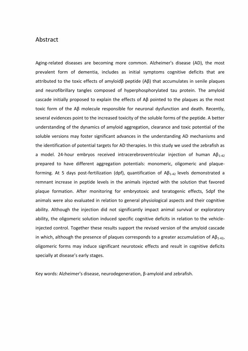
O aumento da expectativa de vida é um fenômeno mundial que a partir da década de
70 tem feito a população de idosos com mais de 65 anos crescer significativamente. Estima-
-se que em 2050 haverá cerca de 1,5 bilhão de pessoas acima dos 65 anos de idade [1] (Figura
1), sendo que em 2014 eram 841 milhões de idosos [2]. Este aumento se deve, em grande
parte, a um aumento na longevidade média relacionada ao declínio de mortes por doenças
anteriormente consideradas intratáveis e à eficácia nas intervenções de saúde.
Com o envelhecimento populacional, torna-se cada vez mais crítico o estudo,
entendimento e busca de terapias para doenças associadas ao envelhecimento, cuja
prevalência também aumenta. Dentre as doenças associadas ao envelhecimento, as
demências e doenças neurodegenerativas correspondem a mais de 100 diferentes
patologias que têm como característica comum o comprometimento das funções cerebrais,
como memória, percepção e habilidades cognitivas [3]. Entre estas, a Doença de Alzheimer
(DA) é a mais prevalente doença degenerativa e foi descrita há 110 anos pelo médico Alois
Alzheimer.
O envelhecimento populacional, além de afetar pacientes e familiares, onera o
sistema de saúde e a economia. Os custos financeiros com tratamentos e cuidados ficaram
em 604 bilhões de dólares por ano em 2012, sendo a DA a causa mais comum,
possivelmente, contribuindo para até 70% dos casos, segundo a Organização Mundial da
Saúde [5].
Apesar de ser uma doença descrita há mais de um século, as características gerais
diagnósticas da DA se assemelham a outras demências, sendo a doença identificada
normalmente de forma tardia, dificultando o tratamento. O diagnóstico inclui teste de
estado mental, exames físicos e neurológicos, exames genéticos (para identificação de
Alzheimer Familiar) e de imagens cerebrais [6]. Apesar de ser acompanhada por conhecidas
7
alterações moleculares, apenas recentemente ficaram disponíveis marcadores moleculares
para diagnóstico de placas senis, alteração molecular típica da doença, por tomografia por
emissão de pósitrons (positron emission tomography (PET) [6, 7], embora o acesso a estes
recursos seja ainda restrito: Em 2012 a Food and Drug Administration dos EUA aprovou o
primeiro marcador para imagem molecular a ser usado para a DA, o Amyvid (Florbetapir F-
18) que se liga ao peptídeo β-amiloide (Aβ); Em 2013 foi aprovado o Vizamyl (Flutametamol
F18) e em 2014 o Neuraceq (Florbetaben F18), os três marcadores possuem a característica
de serem visualizados durante o PET, mostrando a presença de placas senis [6, 7].
Figura 1: Distribuição geográfica dos casos demência no mundo e estimativa de aumento em
2050[4].
A DA possui características moleculares típicas como o acúmulo do peptídeo β-
amiloide (Aβ) em placas senis no meio extracelular e emaranhados de proteína tau
hiperfosforilada intracelular. A tau é uma proteína associada ao citoesqueleto de
microtúbulos que confere estabilidade ao citoesqueleto de neuritos[8], em especial do
axônio, podendo ser fosforilada por diferentes quinases. A hiperfosforilação da tau acarreta
seu desprendimento dos microtúbulos e agregação, acumulando no citoplasma e assumindo
8
conformações patogênicas que comprometem o fluxo de informações e processos celulares
neuronais [8, 9].
A dinâmica de agregação dos peptídeos vem sendo estudada, enquanto as placas
senis provavelmente começam como agregados não filamentosos de peptídeos Aβ entre 40
e 42/43 resíduos [10], formando inicialmente as fibrilas amiloides na DA, são polímeros
produzidos a partir de um péptido de aproximadamente 4,5 kDa β-amiloide [11]. Sabe-se que
a Doença de Alzheimer possui duas ‘formas’ caracterizadas por sintomas comuns: a forma
principal, considerada esporádica, responsável pela maioria dos casos, tem como fatores de
risco aspectos relacionados com o estilo de vida, nível de instrução e comorbidades, entre
outros [12], e a Doença de Alzheimer Familiar (FAD), determinada geneticamente, responsável
pela minoria dos casos.
Estudos da FAD tiveram grande importância no descobrimento dos genes
relacionados com a origem da doença, interação entre o mau processamento da Proteína
Precursora do Amiloide (APP) e o desenvolvimento da patologia [13, 14,15], assim como a
participação crítica dos genes da presenilina 1(PSEN1) e presenilina 2 (PSEN2), que codificam
proteínas dos complexos transmembrana das secretases. Quando em processo de mutação,
estas proteínas geram peptídeos Aβ de tamanhos diversos, com efeitos tóxicos e maior
tendência a formar agregados [16,17,18].
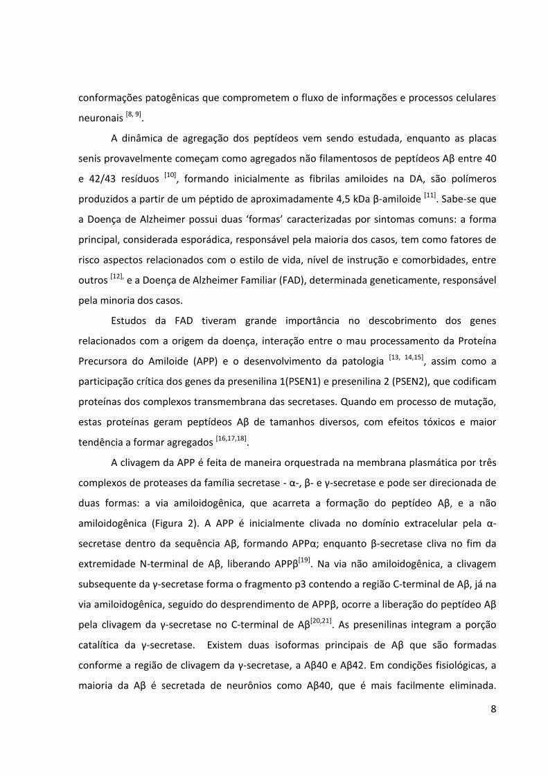
A clivagem da APP é feita de maneira orquestrada na membrana plasmática por três
complexos de proteases da família secretase - α-, β- e γ-secretase e pode ser direcionada de
duas formas: a via amiloidogênica, que acarreta a formação do peptídeo Aβ, e a não
amiloidogênica (Figura 2). A APP é inicialmente clivada no domínio extracelular pela α-
secretase dentro da sequência Aβ, formando APPα; enquanto β-secretase cliva no fim da
extremidade N-terminal de Aβ, liberando APPβ[19]. Na via não amiloidogênica, a clivagem
subsequente da γ-secretase forma o fragmento p3 contendo a região C-terminal de Aβ, já na
via amiloidogênica, seguido do desprendimento de APPβ, ocorre a liberação do peptídeo Aβ
pela clivagem da γ-secretase no C-terminal de Aβ[20,21]. As presenilinas integram a porção
catalítica da γ-secretase. Existem duas isoformas principais de Aβ que são formadas
conforme a região de clivagem da γ-secretase, a Aβ40 e Aβ42. Em condições fisiológicas, a
maioria da Aβ é secretada de neurônios como Aβ40, que é mais facilmente eliminada.
9
Porém, formas mais longas de Aβ agregam-se mais facilmente devido à natureza hidrofóbica
e podem levar à formação de fibrilas de Aβ que por sua vez poderão servir como núcleos
para a formação de placas [22].
Figura 2: Esquema da clivagem da APP nas vias amiloidogênica e não amiloidogênica
(Strooper DB, Vassar R & Golde T, 2010) [20].
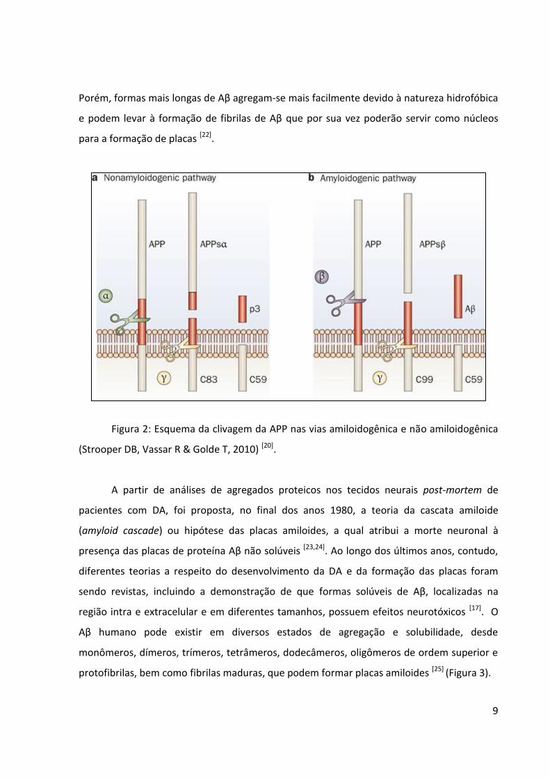
A partir de análises de agregados proteicos nos tecidos neurais post-mortem de
pacientes com DA, foi proposta, no final dos anos 1980, a teoria da cascata amiloide
(amyloid cascade) ou hipótese das placas amiloides, a qual atribui a morte neuronal à
presença das placas de proteína Aβ não solúveis [23,24]. Ao longo dos últimos anos, contudo,
diferentes teorias a respeito do desenvolvimento da DA e da formação das placas foram
sendo revistas, incluindo a demonstração de que formas solúveis de Aβ, localizadas na
região intra e extracelular e em diferentes tamanhos, possuem efeitos neurotóxicos [17]. O
Aβ humano pode existir em diversos estados de agregação e solubilidade, desde
monômeros, dímeros, trímeros, tetrâmeros, dodecâmeros, oligômeros de ordem superior e
protofibrilas, bem como fibrilas maduras, que podem formar placas amiloides [25] (Figura 3).
10
A versão atualizada da cascata amiloide atribui efeitos tóxicos a oligômeros e fibrilas
através de diversos mecanismos ainda em estudo [17], embora o debate acerca da real
contribuição das diferentes formas de amiloide seja polêmico e esteja longe de um consenso
[26,27]. Huang e Mucke propuseram que as fibrilas Aβ insolúveis encontradas nas placas de
amiloides e a forma monomérica são menos patogênicas do que as fibrilas não solúveis que
são os dímeros Aβ, trimeros e oligômeros maiores de Aβ [28] (Figura 3).
Figura 3: Esquema da agregação da proteínas Aβ. (a) proteínas nativas se dobram e sofrem
alteração conformacional para formar protofibrilas e fibrilas maduras, (b) monômeros
dobrados formam oligômeros, (c) fibrilas maduras sofrem fragmentação para formar
oligômeros fibrilares que novamente se agregam para formar fibrilas maduras, (d) as fibrilas
maduras atuam como um modelo para oligomerização e catalisam a reação de nucleação
secundária formando oligômeros tóxicos a partir dos monômeros (Verma et al. 2015) [29].
Apesar dos altos índices de incidência da DA, atualmente ainda não há nenhuma
terapia eficaz que previna, retarde ou reverta a doença [6]. Atualmente as estratégias
farmacológicas disponíveis se restringem a tratar sintomas da DA, como déficits de memória,
alterações no sono ou tentar retardar o processo de degeneração e incluem inibidores da
11
enzima colinesterase que potencializam a transmissão colinérgica e antagonistas de
receptores glutamatérgicos NMDA que previnem a excitotoxicidade induzida por glutamato.
Neste cenário, o entendimento das dinâmicas moleculares dos agentes tóxicos em suas
diferentes apresentações é crítico para o entendimento dos mecanismos da doença e para a
identificação de possíveis alvos terapêuticos.
1.2 Zebrafish
O teleósteo Danio rerio, conhecido mundialmente como zebrafish, foi descrito
primeiramente por Francis Hamilton, em 1822, pertencente à família Cyprinidae, uma das
mais ricas em número de representantes entre os vertebrados. Esta espécie tem distribuição
geográfica no sul e sudeste da Ásia. George Streissinger (1983) propôs primeiramente o uso
do zebrafish como animal modelo para pesquisas em biologia do desenvolvimento, genética,
neurofisiologia e biomedicina [30].
Este animal modelo é um pequeno peixe de água doce com fertilização e
desenvolvimento externos e ovos relativamente grandes (cerca de 0,7 milímetros de
diâmetro) e transparentes, o que facilita a observação e manipulação dos embriões quando
comparado com os demais animais modelo de vertebrados. O seu desenvolvimento
embrionário é muito rápido, sendo completo em até 72 horas pós-fertilização (hpf), quando
eclode do córion dando origem a uma larva [30] que só então inicia o processo de
pigmentação, embora isto também possa ser prevenido farmacologicamente, o que facilita o
acompanhamento dos processos em tempo real. Por ser um organismo aquático, possui a
capacidade de absorver substâncias adicionadas ao seu meio, sendo usado para screening
farmacológico e testes de novos fármacos. Atualmente muitos procedimentos já estão
validados para avaliar parâmetros comportamentais e cognitivos, com poder translacional
para mamíferos e humanos [31]. Portanto, é um ótimo modelo animal para estudos de
desenvolvimento e alterações estruturais e funcionais do sistema nervoso.
Pela complexidade das doenças neurodegenerativas e os diversos processos nela
envolvidos, cada vez mais se torna necessário modelar aspectos das doenças em organismos
menos complexos para conseguirmos entendimento de dinâmicas e seleção de alvos
moleculares para estudo.
12
Considerando que a DA atinge exclusivamente humanos, apesar de manifestações
similares em canídeos [32,33] , é importante garantir que os animais modelo usados no seu
estudo possuam toda a maquinaria molecular associada à doença [34]. Diversas estratégias já
foram estabelecidas em animais modelos para o desenvolvimento de patologias com
aspectos neurodegenerativos, diversos animais transgênicos para o estudo da DA foram
criados utilizando roedores e, mais recentemente, também zebrafish [35].
Estudos utilizando o zebrafish para investigações da Doença de Alzheimer ainda são
escassos, mas Cunvong e colaboradores aplicaram injeção intraocular de Aβ em zebrafish
adulto, o que resultou em ramificações anormais dos vasos sanguíneos da retina [36]. Em
abordagem similar, foi observado o mesmo efeito da Aβ no cérebro de embriões e larvas e a
senescência de células endoteliais no desenvolvimento vascular de zebrafish [37]. Nosso
grupo de pesquisa estabeleceu recentemente uma metodologia para injeção
intracerebroventricular de peptídeo humano Aβ1-42 em larvas de zebrafish [38], estudos
utilizando estratégias similares em roedores, e observou déficits cognitivos e aumento na
fosforilação de tau [39].
1.3 Fármacos Neuroprotetores
Considerando a complexidade da DA e o desconhecimento atual de sua etiologia, a
busca por agentes neuroprotetores capazes de prevenir o seu desenvolvimento é bastante
atrativa. Apesar da grande gama de estudos utilizando animais modelo para o entendimento
da doença, é difícil encontrar um animal que combine características da DA como acúmulo
de Aβ e efeitos comportamentais associados à doença que permita ao mesmo tempo o
screening de drogas neuroprotetoras. Assim o zebrafish demonstra uma vantagem e vem
sendo utilizado para screening farmacológico por ter a capacidade de absorver substâncias
do meio, além de ser um modelo para estudo de neurodegeneração.
13
1.3.1 Rapamicina
A Rapamicina é um composto antifúngico produzido pela bactéria Streptomyces
hygroscopicus, isolado na década de 70 em forma pura, cristalina, com baixa toxicidade
aguda, inibe crescimento e proliferação celular e atua por inibição da sinalização de mTOR
(alvo da rapamicina em mamíferos) [40,41]. A via de sinalização mTOR desempenha papel
importante na rede de sinalização que controla o crescimento e metabolismo em resposta a
estímulos ambientais [42], e forma dois complexos: mTORC1 e mTORC2, estes complexos
possuem diferenças quanto à sensibilidade à rapamicina. A mTOR é regulada por vários
estímulos fisiológicos e patológicos através de vias de sinalização e proteínas intermediárias.
A mTOR ativa várias proteínas envolvidas na regulação da síntese proteica, crescimento e
proliferação celular, plasticidade sináptica e expressão de canais iônicos [43].
A rapamicina já foi utilizada para tratar crises convulsivas e esclerose lateral em
zebrafish, porém, em um curto período de tempo, viram que os animais tratados tiveram
uma melhora no nado comparado com os animais controle [44], foi mostrado que a
rapamicina pode prolongar a longevidade de células de levedura quiescentes [45], a inibição
da atividade de sinalização da via mTOR pode ser uma abordagem terapêutica futura para
prevenir ou reverter a DA [46], podendo ser usada para distúrbios neurológicos como para a
doença de Parkinson [35] e doença de Alzheimer [46,47]. A rapamicina também mostrou possuir
um efeito na longevidade de mamíferos, retardando o envelhecimento [48], além de
apresentar uma melhora na aprendizagem e memória, além de reduzir níveis de Aβ e tau [49].
14
2 Justificativa
Com o aumento da expectativa de vida populacional e com isso a incidência de
doenças neurodegenerativas, cada vez se torna mais necessário o entendimento dos
mecanismos celulares e moleculares subjacentes às doenças e a busca por novas terapias.
O desenvolvimento de modelos animais para o estudo e entendimento dos
diferentes fatores envolvidos na DA se torna crucial para a busca de novos fármacos e
diagnóstico precoce.
15
3 Objetivos
3.1 OBJETIVO GERAL
Caracterizar os efeitos da injeção intracerebral de Aβ1-42 em condições monoméricas,
oligoméricas e formadoras de placas sobre parâmetros embrionários, comportamentais, vias
de sinalização associadas à Doença de Alzheimer e potencial agregador. Adicionalmente
avaliar os possíveis efeitos do fármaco rapamicina.
3.2 OBJETIVOS ESPECÍFICOS
Avaliar os efeitos embriotoxicológicos, incluindo alterações morfológicas dos
animais injetados com os tipos de β-amiloide a partir das 2dpf até os 5dpf.
Avaliar a comportamento locomotor e respostas cognitivas aversivas aos 5dpf.
Avaliar os níveis de apoptose no tecido cerebral aos 5dpf.
Quantificar as proteínas β-amiloide, tau, tau-p por western blot aos 5dpf.
Padronizar técnica de ELISA e quantificar os níveis encefálicos de Aβ nos
indivíduos aos 5dpf.
Avaliar os possíveis efeitos neuroprotetores do fármaco rapamicina.
16
CAPÍTULO 2
3.1 ARTIGO CIENTÍFICO Title: Cognitive deficits induced by Aβ42 oligomers but not monomers and fibrillar forms of the
peptide in zebrafish independently of brain accumulation
Authors: Natália Eltz1,2, Laura R. Nery1,2, Raphaela S. Fonseca1,2, Gabriela Urbanski1,2,
Christiane Pizzato1,2, Monica R. Vianna1,2
Affiliation: 1ZebLab & 2Laboratório de Biologia e Desenvolvimento do Sistema Nervoso,
Faculdade de Biociências, Pontifícia Universidade Católica do Rio Grande do Sul, Porto
Alegre, Rio Grande do Sul, Brazil
Corresponding author: Monica Vianna, Ph.D.: [email protected]
Abstract
Plaques composed of insoluble fibrils of Amyloid-β peptides (Aβ) mainly of 42 (Aβ42) and 40
(Aβ40) amino acids have long been considered a key pathological marker and a prime target
for Alzheimer’s disease prevention and treatment. Aβ42 is more hydrophobic than Aβ40,
aggregates more rapidly forming stable oligomers and is predominant in plaques. Recent
data supports soluble Aβ oligomers as toxic mediators of synaptic dysfunction, redefining
the traditional “amyloid cascade” theory. In this scenario, additional platforms for in vivo
studies of Aβ42 dynamics and evaluation of specific effects and of its transitional monomeric,
oligomeric and fibrillar forms are essential to much needed advances in the field. We tested
the toxicity, cognitive effects and brain accumulation after hindbrain ventricle injection of
human Aβ42 monomers, oligomers and fibrils in the zebrafish embryo. We demonstrate that
soluble oligomers are associated with specific cognitive deficits at 5dpf independent of the
brain accumulation of Aβ42 detected by ELISA only in animals injected with a plaque-forming
fibrillar preparation. Our data reinforces the updated version of the amyloid cascade

17
suggesting the synaptotoxic effects of soluble oligomers preceding accumulation and
supports zebrafish as an additional model for studying its underlying mechanisms.
Key words: Zebrafish; Alzheimer’s Disease, Amyloid-β, Oligomers, Cognition, ELISA
18
Introduction
Alzheimer's disease (AD) is the most common cause of dementia in the elderly with
increasing 4.6 million new patients per year [1] and estimated 66 million cases in 2030 [2]. AD
is a progressive neurological disorder initially characterized by synaptic dysfunction and
cognitive deficits such as memory loss, speech difficulties, depression, hallucinations and
evolving to a costly dependent care before death. Disease-modifying therapies are still
unavailable mostly due to our lack of understanding of the underlying molecular
mechanisms of this devastating disease progression. The presence of intraneuronal
neurofibrillary tangles of hyperphosphorylated tau and extracellular deposits of amyloid-β
(Aβ) peptides in senile plaques have long been considered the molecular hallmarks of the
disease.
The extracellular release of Aβ peptides of varying sizes, among which the major
forms are of 42 (Aβ42) and 40 (Aβ40) amino acids, is a result of the amyloid precursor protein
(AβAPP) proteolytic processing by β- and γ-secretases in the amyloidogenic pathway. Aβ
soluble forms tend to aggregate in oligomers of different masses and progressively form
fibrils that integrate large deposits known as senile plaques. Aβ brain levels can be elevated
because of enhanced production of aggregative forms or reduced clearance. Aβ42 is more
hydrophobic than Aβ40, aggregates more rapidly forming stable oligomers and is
predominant form in plaques [3].
The Amyloid Cascade Hypothesis postulated that the progressive deposition of
insoluble fibrils in plaques is the central event of AD pathology [4]. This scenario encouraged
drug development efforts targeting Aβ large aggregates and clearance despite the lack of
correspondence between the amount of plaques and the degree of synaptic loss or cognitive
deficits in patients and murine models of AD [5,6,7]. More than a decade ago, several lines of
evidence began to support a paradigm shift and soluble oligomers (but not fibrils or plaques)
are currently seen as the major toxic agents causing synaptic loss and cognitive decline in AD
[3]. The toxic effects of natural and synthetic oligomers on synaptic plasticity include
cognitive deficits in murine and inhibition of LTP (long-term potentiation, an
19
electrophysiological correlate of long-term memory) maintenance [8,9,10,11,12]. Despite a
significant correlation between the amount of soluble oligomers, synapses and cognitive
dysfunction [3,13,14,15] our current understanding of the different oligomeric species and its
dynamics remain elusive and new study platforms are needed [16,17].
Zebrafish is a model organism advantageous for translational experiments due to its
significant genetic similarity with humans associated to valuable high-throughput screening
potential that can contribute to studies of neurological human diseases mechanisms [18,19,20].
Our lab previously established a protocol for intraventricular injection of human Aβ42 in
zebrafish and demonstrated that it induces cognitive deficits, increases tau phosphorylation
in residues that resemble AD early stages and diminishes synaptic markers [21]. In this study
we aimed to compare the behavioral and molecular effects of Aβ42 with different
aggregation potentials including soluble monomers, oligomers and fibrils and test if this
strategy could aid in future studies dedicated to investigate the oligomers dynamics and
cellular targets.
Methodology
Animals
Adult AB strain wild type zebrafish from our facility colony were kept and bred
according to standard procedures in an automated re-circulating system (Tecniplast, Italy) at
a density of 1.5 fish per liter with a constant light-dark cycle (14–10h) [22]. For breeding,
females and males (2:1) were housed in a breeding tank overnight separated by a
transparent barrier that was removed when lights went on in the following morning.
Embryos were collected, cleaned and placed in sterile petry dishes (30 embryos per dish)
with the system’s clean water (reverse osmosis filtered and salt equilibrated), stored in an
incubator with controlled temperature of 27.5 °C and a light-dark cycle (14-10h). All
procedures were approved by the Institutional Animal Care Committee from Pontifícia
Universidade Católica do Rio Grande do Sul (CEUA-PUCRS, 15/00490), followed the Brazilian
legislation (no.11.794/08) and conducted according to the Canadian Council on Animal Care
guidelines for the use of fish in research [23].
20
Amyloid-β solutions for injection
The human Aβ42 peptide (Sigma-Aldrich) solutions were diluted following
manufacturer instructions in PBS at an initial concentration of 1000 µM and diluted to the
final 10 µM. Solutions with different aggregation potentials were prepared according to the
literature, as follows: the monomeric Aβ solution was prepared as described by Cameron et
al. [24]; a preferentially oligomeric solution was prepared by incubating the monomeric
solution for 5 days at 37 ° C, according to Pontrello et al. [25] while the fibrillar solution was
produced by the incubation of the monomeric solution under 800rpm shaking for 36 hours
at 23 ° C as detailed in O'Hare et al. [26].
Brain intra-ventricular Injection
Embryos at 1 day post-fertilization (dpf) had their chorion removed and were
prepared for brain intra-ventricular injection according to Gutzman and Sive (2009) [27] as
described in Nery et al. (2014) [21]. Embryos were anesthetized with tricaine (MS-222, Sigma-
Aldrich) and placed in 0.75% agar-coated dishes so that the hindbrain ventricle was
accessible for injection. The microinjection needle attached to a micromanipulator
(Narishige) and a Picoliter injection pump (Warner Instruments) was used under a
stereomicroscope (Nikon, SMZ 1500). The needle was carefully placed on the roof plate of
the hindbrain and 5–10 nl of the Aβ42 solutions in 1% DMSO 0.5% Phenol Red Phosphate
Buffered Saline (PBS) solution was injected while the control group received the equivalent
1% DMSO 0.5% Phenol Red in PBS vehicle. The procedure took no more than 10 minutes per
animal and all groups were run and analyzed simultaneously throughout the study. After the
injections the animals were transferred to 6-well plates (15 animals per well) and maintained
in the incubator for 4 days.
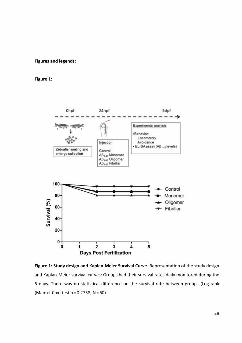
Animals were assigned to the following groups and subsequent analyses were
performed blind throughout the study: vehicle injected controls (Control group), animals
injected with Aβ42 solutions prepared as previously validated to contain Monomers
21
(Monomer group), Oligomers (Oligomer group) and fibrillar forms of Aβ42 (Fibrillar group)
(Figure 1).
Survival and morphological evaluation
Survival and morphological defects were monitored daily under a stereomicroscope
(Nikon, SMZ 1500) from day 1 to 5 during the daily media change. The morphological
phenotypes analyzed included pericardial and brain edema, aberrations in yolk extension,
eyes and tail malformations and blood accumulation. Survival was analyzed using Kaplan-
Meier test [21].
Behavioral Analysis
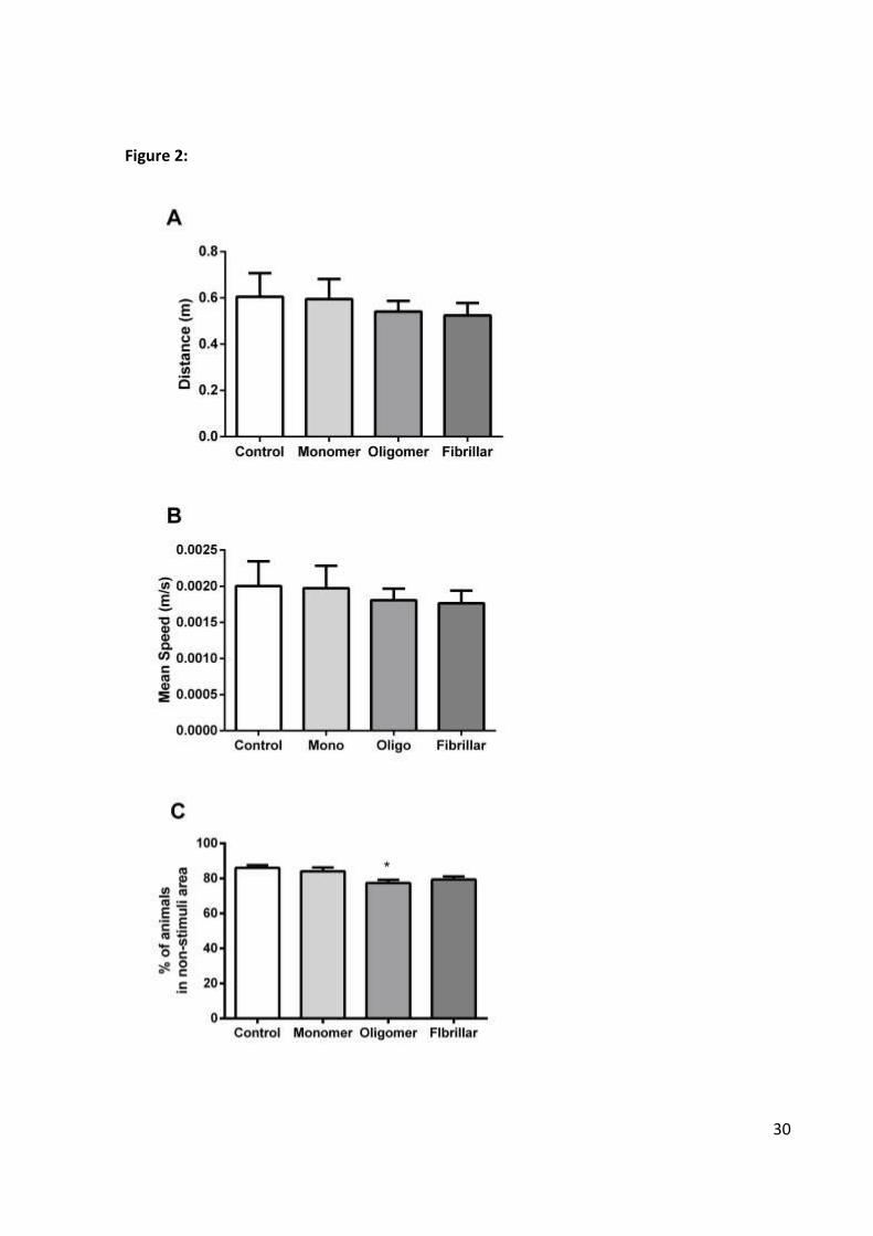
Exploratory test
At 5 dpf, animals were individually exposed to a new environment to test their
exploration and locomotion abilities [28] (n = 20 animals in triplicate). The new environment
consisted of a 24-well plate well with 3 ml of water in which behavioral parameters were
analyzed for 5 minutes after 1min of habituation. Animal’s performances in the round arena
were recorded with an HD digital webcam (Logitech) and blindly analyzed using an
automated protocol (ANYmaze, Stoelting). Among the evaluated parameters were the total
distance traveled, speed when active, periods of activity and thigmotaxis (preference for the
outer portions of the arena [29]).
Avoidance task
For assessing animals’ cognitive capacity, their scape response to an aversive
stimulus (a 1.35 cm diameter red bouncing ball) was tested in 6-well cell culture plates in
which they were placed 5 animals per well (n = 10 in triplicate) according to Nery et al.
(2014) [21] in a protocol adapted from Pelkowski et al. (2011) [30]. The plate was adjusted on
the surface of an LCD screen in which an animation was presented for 5 minutes after 2
minutes of habituation.
The red bouncing ball travelled from left to right over a straight 2 cm trajectory on
half of the well area (stimuli area) and animal’s scape efficiency was measure according to
their distribution on the opposite hemisphere (non-stimulus area) after successive intervals.
22
Animals’ performance was recorded with an HD digital webcam (Logitech) and analyzed by
"blind observers". Both behavioral experiments were performed in a controlled temperature
environment (27 ± 2 °C).
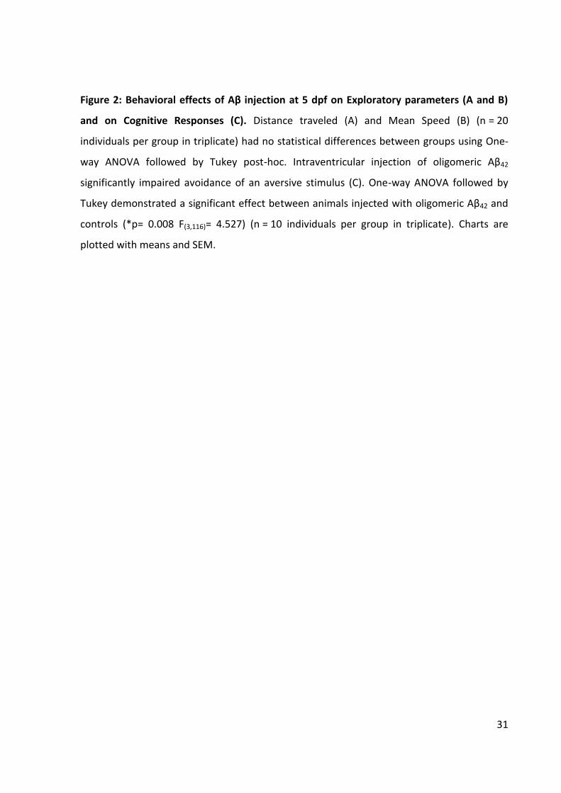
Enzyme-linked immunosorbent assay of Aβ42 detection
Aβ42 brain levels were quantified using an Aβ42 sandwich enzyme-linked
immunosorbent assay (ELISA) according to the manufacturer's instructions (Human Aβ42
Ultrasensitive ELISA Kit, Life). At 5 dpf the larvae were crioanesthetized and euthanized by,
decapitation. Dissected brains (pool of 20 animals per group in triplicate) were stored in a
freezer -80°C in a protease inhibitor (Sigma-Aldrich) and homogenized with RIPA (Sigma-
Aldrich). Samples were incubated in the 96-well plate containing human Aβ42 antibody
overnight at 4°C. After the incubation period the wells were washed and the secondary
antibody HRP IgG was added for 30 min before a final wash. The reading was performed
with absorbance of 450nm at Spectramax M2 reader (Molecular Devices, USA).
Statistical analysis
Survival throughout the 5 experimental days was analyzed by Kaplan-Meier test. Data
from the other approaches were parametrically analyzed after normality confirmation using
one-way ANOVA followed by Tukey’s post-hoc test. The level of significance was considered
p<0.05.
Results and Discussion
During the 5 days of evaluation no significant morphological effect was attributed to
the injection regimens nor any delay in hatching rates among groups was observed. Kaplan-
Meier survival curves are presented in Figure 1 and show no significant effect when all
groups were compared (Log-rank (Mantel-Cox) test p = 0.2738, n = 60).
These results are in accordance to Gutzman and Sive, 2009 [27] and our previous
study[21] showing no significant deleterious effects of the injection procedure. Despite being
an invasive process in a young animal, intraventricular injections in 1dpf zebrafish embryos is
a viable process and may be further explored. Importantly, the process is also very fast and
23
precise and confirmation of correct injection site occurs in real time due to animals
transparency and use of phenol red, different from equivalent approaches in rodents in
which a post-mortem histological evaluation is necessary to confirm cannula placement and
injection site.
The evaluation of 5dpf larvae exploratory behavior when individually exposed to a
new environment was used to assess general locomotion, orientation and anxiety
parameters and showed no differences between groups (Figure 2). As shown in Figure 1, no
significant differences were observed in the total travelled distance during the 5 minutes
exploration period (p= 0.830 F(3,143)= 0.293) (Figure 2A) and the mean speed when active (p=
0.881 F(3,143)= 0.221) (Figure 2B), among other testes parameters not shown. These results
support the lack of other unspecific effects of injection and ensured that animals could be
further compared in the subsequent cognitive test.
When animals from different groups with equivalent exploratory performances were
cognitively tested regarding their ability to avoid an aversive stimulus, a significant deficit in
the oligomer-injected group was observed when compared to controls (p= 0.008 F(3,116)=
4.527) (Figure 2C). Our findings corroborate other demonstrations of deleterious cognitive
effects of soluble Aβ injection into the brain ventricle of rodents [8,10,31,32,33]. We have also
previously demonstrated a deleterious effect of Aβ oligomeric injection in the hindbrain
ventricle of zebrafish, corroborating data pointing to neurotoxic effects of soluble oligomeric
Aβ forms. Our data contrasts the results from O’hare and collaborators [26] that deleterious
cognitive effects of fibrillar Aβ42 intrahippoampal injection in rats. The observed effect,
however, was only evident after several weeks, what could be attributed to other cellular
and molecular effects related to inflammation and microglial activation. Oligomeric
assemblies of Aβ vary significantly in their size and tissue distribution and future studies
should detail forms associated with this effect and its potential targets [8,10].
Next we tested the potential accumulation of Aβ in the brain of injected animals by
ELISA. Despite the proliferative and clearance prone cellular scenario of zebrafish brain at
this early developmental stage, a significant increase in Aβ42 levels was observed in animals
injected with the preferential fibrillar solution (p = 0.03 F(3.18)= 3.676) (Figure 3). The method
used is capable of selectively detecting peptides of 42 aminoacids with a very high
24
sensitivity, supporting the efficacy of the protocol used to prepare the fibrillar forms of Aβ42
[26]. To our knowledge, this is the first quantification of Aβ brain levels in zebrafish using
ELISA. This finding reinforces the aggregation and clearance resistant nature of Aβ42 fibrils
and this model’s potential contribution for studies of the molecular events associated to
Aβ42 deposits. It remains to be investigated the specific tissue and cellular distribution of the
increase observed in the fibrillar-injected group, as well as the deposits characteristics.
Importantly, this data supports evidences of memory impairment effects of oligomeric forms
of Aβ independently of plaques and neuronal loss [10].
The escalation in elderly population makes increasingly important to study the
neurodegenerative diseases associated with aging such as AD, which is usually diagnosed
late and barely treatable for symptoms. Understanding the disease underlying mechanisms
at early stages is of paramount importance for mechanistic and therapeutic purposes. The
updated amyloid cascade [34] suggests that Aβ has toxic effects from early stages, before
plaques deposition, and reinforce the need to understand the molecular alterations that
occurs in the early stages of the disease [35,36]. Future studies using ventricular injections in
zebrafish could contribute to current efforts to elucidade the differential toxicity of
oligomeric forms, its target membrane receptors and underlying signaling pathways
connecting to tau hyperphosphotilation and other cellular events [37,38].
Acknowledgements: This work was supported by Fundação de Amparo à pesquisa do Rio
Grande do Sul (FAPERGS) (2352-2551/14-8) and Conselho Nacional de Desenvolvimento
Científico e Tecnológico (CNPq) (484124/2013-7 and 467572/2014-3). N.S.E. received a
graduate fellowship from Coordenação de Aperfeiçoamento de Pessoal de Nível Superior
(CAPES). G.U. and C.P. were recipients of FAPERGS fellowships. L.R.N. and M.R.M.V. are
CNPq fellows.
25
References:
[1] Tjin PV, Kamphuis W, Marlatt MW, Hol EM, Lucassen PJ (2011). Presenilin mouse and Zebrafish models for dementia: Focus on neurogenesis. Progress in Neurobiology. 93,149-164. *2+ Alzheimer’s Disease International. Policy Brief for Heads of Government The Global Impact of Dementia 2013–2050, https://www.alz.co.uk/research/G8-policy-brief, Last updated June 20, 2017, Accessed on May 29, 2017. [3] Haass C, Selkoe DJ (2007). Soluble protein oligomers in neurodegeneration: lessons from the Alzheimer's amyloid β-peptide. Nat. Rev. Mol. Cell Biol. 8, 101–112. [4] Hardy JA, Higgins GA (1992). Alzheimer’s disease: the amyloid cascade hypothesis. Science. 256, 184–185. [5] Hsia AY, Masliah E, McConlogue L, Yu GQ, Tatsuno G, Hu K, Kholodenko D, Malenka RC, Nicoll RA, Mucke L (1999). Plaque-independent disruption of neural circuits in Alzheimer’s disease mouse models. Proc. Natl. Acad. Sci. U S A. 96, 3228–3233. [6] Masliah E, Terry RD, Mallory M, Alford M, Hansen LA (1990). Diffuse plaques do not accentuate synapse loss in Alzheimer’s disease. Am. J. Pathol. 137, 1293–1297. [7] Mucke L, Masliah E, Yu GQ, Mallory M, Rockenstein EM, Tatsuno G, Hu K, Kholodenko D, Johnson-Wood K, McConlogue L (2000). High-level neuronal expression of abeta 1–42 in wild-type human amyloid protein precursor transgenic mice: synaptotoxicity without plaque formation. J. Neurosci. 20, 4050–4058. [8] Cleary JP, Walsh DM, Hofmeister JJ, Shankar GM, Kuskowski MA, Selkoe DJ, Ashe KH (2005). Natural oligomers of the amyloid-beta protein specifically disrupt cognitive function. Nat Neurosci. 8, 79-84. [9] Reed MN, Hofmeister JJ, Jungbauer L, Welzel AT, Yu C, Sherman MA, Lesné S, LaDu MJ, Walsh DM, Ashe KH, Cleary JP (2011). Cognitive effects of cell-derived and synthetically derived Aβ oligomers. Neurobiol Aging. 32, 1784-1794. [10] Lesne S, Koh MT, Kotilinek L, Kayed R, Glabe CG, Yang A, Gallagher M, Ashe KH (2006). A specific amyloid-beta protein assembly in the brain impairs memory. Nature. 440, 352-357. [11] Shankar GM, Li S, Mehta TH, Garcia-Munoz A, Shepardson NE, Smith I, Brett FM, Farrell MA, Rowan MJ, Lemere CA, Regan CM, Walsh DM, Sabatini BL, Selkoe DJ (2008). Amyloid-beta protein dimers isolated directly from Alzheimer's brains impair synaptic plasticity and memory. Nat Med. 14, 837-842.
26
[12] Walsh DM, Klyubin I, Fadeeva JV, Cullen WK, Anwyl R, Wolfe MS, Rowan MJ, Selkoe DJ (2002). Naturally secreted oligomers of amyloid beta protein potently inhibit hippocampal long-term potentiation in vivo. Nature. 416, 535–539. [13] Wang ZH, Zagzag D, Zeng B, Kolodny EH (1999). In vivo and in vitro glioma cell killing induced by an adenovirus expressing both cytosine deaminase and thymidine kinase and its association with interferon-alpha. J Neuropathol Exp Neurol. 58, 847-858. [14] Naslund J, Haroutunian V, Mohs R, Davis KL, Davies P, Greengard P, Buxbaum JD (2000). Correlation between elevated levels of amyloid beta-peptide in the brain and cognitive decline. JAMA. 283, 1571-1577. [15] Lue LF, Kuo YM, Roher AE, Brachova L, Shen Y, Sue L, Beach T, Kurth JH, Rydel RE, Rogers J (1999). Soluble amyloid beta peptide concentration as a predictor of synaptic change in Alzheimer's disease. Am J Pathol. 155, 853-862. *16+ Benilova I, Karran E, De Strooper B (2012). The toxic Aβ oligomer and Alzheimer’s disease: an emperor in need of clothes. Nat Neurosci. 15, 349–357. *17+ Larson ME, Lesné SE (2012). Soluble Aβ oligomer production and toxicity. J Neurochem. 1, 125-39. [18] Phillips JB, Westerfield M (2014). Zebrafish models in translational research: tipping the scales toward advancements in human health. Dis Model Mech. 7, 739-743. [19] Santoriello C, Zon LI (2012). Hooked! Modeling human disease in zebrafish. J Clin Invest. 122, 2337-2343. [20] Santana S, Rico EP, Burgos JS (2012). Can zebrafish be used as animal model to study Alzheimer’s disease?. Am J Neurodegener Dis. 1, 32–48. [21] Nery LR, Eltz NS, Hackman C, Fonseca R, Altenhonfen S, Guerra HN, Freitas VM, Bonan CD, Vianna MR (2014) . Brain Intraventricular Injection of Amyloid-β in Zebrafish Embryo Impairs Cognition and Increases Tau Phosphorylation, Effects Reversed by Lithium. PLoS One. 9, DOI: 10.1371/journal.pone.0105862. [22] Westerfield, Monte. The zebrafish book. A guide for the laboratory use of zebrafish (Danio rerio). Eugene: University of Oregon Press, 2000. http://zfin.org/zf_info/zfbook/zfbk.html. Last updated May 29, 2007, Accessed on January 29, 2008. [23] CCAC guidelines on: the care and use of fish in research, teaching and testing. Ottawa: Canadian Council on Animal Care. 2005:94.
27
[24] Cameron DJ, Galvin C, Alkam T, Sidhu H, Ellison J, Luna S, Ethell DW (2012). Alzheimer’s-Related Peptide Amyloid-β Plays a Conserved Role in Angiogenesis. PLoS One. 7, DOI: e39598. doi:10.1371/journal.pone.0039598 [25] Pontrello CG, Sun MY, Lin A, Fiacco TA, DeFea KA, Ethell IM (2012). Cofilin under control of β-arrestin-2 in NMDA-dependent dendritic spine plasticity, long-term depression (LTD), and learning. Proc Natl Acad Sci U S A. 109, 442–451. *26+ O’Hare E, Weldon DT, Mantyh PW, Ghilardi JR, Finke MP, Kuskowski MA, Maggio JE, Shephard RA, Cleary J (1999). Delayed behavioral effects following intrahippocampal injection of aggregated A beta (1–42). Brain Res. 815, 1-10. [27] Gutzman JH, Sive H (2009) Zebrafish Brain Ventricle Injection. J Vis Exp. DOI: 10.3791/1218. [28] Creton R (2009). Automated analysis of behavior in zebrafish larvae. Behav Brain Res. 203, 127-136. [29] Schnörr SJ, Steenbergen PJ, Richardson MK, Champagne DL (2012). Measuring thigmotaxis in larval zebrafish. Behav Brain Res. 228, 367-74. [30] Pelkowski SD, Kapoor M, Richendrfer HA, Wang X, Colwill RM, Creton R (2011). A novel high-throughput imaging system for automated analyses of avoidance behavior in Zebrafish larvae. Behav Brain Res. 223, 135-144. [31] Balducci C, Beeg M, Stravalaci M, Bastone A, Sclip A, Biasini E, Tapella L, Colombo L, Manzoni C, Borsello T, Chiesa R, Gobbi M, Salmona M, Forloni G (2010). Synthetic amyloid-beta oligomers impair long-term memory independently of cellular prion protein. Proc. Natl. Acad. Sci. U. S. A., 107, 2295–2300. [32] Balducci C, Forloni G (2014). In vivo application of beta amyloid oligomers: a simple tool to evaluate mechanisms of action and new therapeutic approaches. Curr Pharm Des. 20, 2491-505. [33] Epelbaum S, Youssef I, Lacor PN, Chaurand P, Duplus E, Brugg B, Duyckaerts C, Delatour B (2015). Acute amnestic encephalopathy in amyloid-β oligomer-injected mice is due to their widespread diffusion in vivo. Neurobiol. Aging. 36, 2043–2052. [34] Hardy J (2017). The Discovery of Alzheimer causing Mutations in the APP Gene and the Formulation of the "Amyloid Cascade Hypothesis". FEBS J. DOI: 10.1111/febs.14004. [35] Dumurgier J, Gabelle A, Vercruysse O, Bombois S, Laplanche JL, Peoc'h K, Schraen S, Sablonnière B, Pasquier F, Touchon J, Lehmann S, Hugon J, Paquet C (2013). Exacerbated CSF abnormalities in younger patients with Alzheimer’s disease. Neurobiol Dis. 54, 486–491.
28
[36] Arriagada PV, Growdon JH, Hedley-Whyte ET, Hyman BT (1992). Neurofibrillary tangles but not senile plaques parallel duration and severity of Alzheimer's disease. Neurology. 42, 631–639. [37] Brito-Moreira J, Lourenco MV, Oliveira MM, Ribeiro FC, Ledo JH, Diniz LP, Vital JFS, Magdesian MH, Melo HM, Barros-Aragão F, de Souza JM, Alves-Leon SV, Gomes FCA, Clarke JR, Figueiredo CP, De Felice FG, Ferreira ST (2017). Interaction of amyloid-β (Aβ) oligomers with neurexin 2α and neuroligin 1 mediates synapse damage and memory loss in mice. J Biol Chem., 292(18):7327-7337. [38] Amar F, Sherman MA, Rush T, Larson M, Boyle G, Chang L, Götz J, Buisson A, Lesné SE (2017). The amyloid-β oligomer Aβ*56 induces specific alterations in neuronal signaling that lead to tau phosphorylation and aggregation. Sci Signal., 10 (478). pii: eaal2021.
29
Figures and legends:
Figure 1:
Figure 1: Study design and Kaplan-Meier Survival Curve. Representation of the study design
and Kaplan-Meier survival curves: Groups had their survival rates daily monitored during the
5 days. There was no statistical difference on the survival rate between groups (Log-rank
(Mantel-Cox) test p = 0.2738, N = 60).
30
Figure 2:
31
Figure 2: Behavioral effects of Aβ injection at 5 dpf on Exploratory parameters (A and B)
and on Cognitive Responses (C). Distance traveled (A) and Mean Speed (B) (n = 20
individuals per group in triplicate) had no statistical differences between groups using One-
way ANOVA followed by Tukey post-hoc. Intraventricular injection of oligomeric Aβ42
significantly impaired avoidance of an aversive stimulus (C). One-way ANOVA followed by
Tukey demonstrated a significant effect between animals injected with oligomeric Aβ42 and
controls (*p= 0.008 F(3,116)= 4.527) (n = 10 individuals per group in triplicate). Charts are
plotted with means and SEM.
32
Figure 3:
Figure 3: Relative Aβ42 levels measured by ELISA. Aβ42 levels in the 5dpf larvae brains were
measured using a sandwich ELISA assay. Animals injected with the fibrillar form showed a
significant increase in Aβ42 levels in the brain compared to controls using a One-way ANOVA
followed by Tukey (p= 0.03 F(3.18)= 3.676). Columns represent means and SEM.
33
CAPÍTULO 3
1 RESULTADOS ADICIONAIS
Além dos resultados compilados no manuscrito apresentado no capítulo 2, outros
resultados obtidos são apresentados neste capítulo. Eles não foram incluídos no artigo por
terem sido insuficientemente avaliados ou terem enfrentados problemas técnicos.
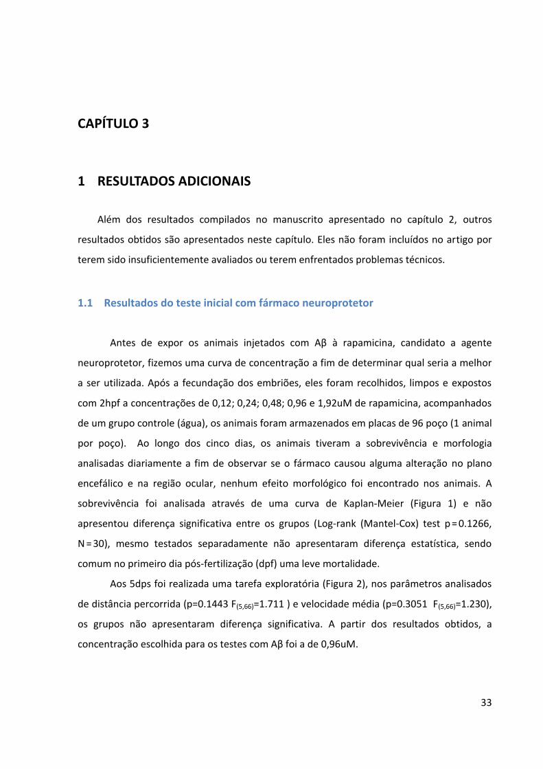
1.1 Resultados do teste inicial com fármaco neuroprotetor
Antes de expor os animais injetados com Aβ à rapamicina, candidato a agente
neuroprotetor, fizemos uma curva de concentração a fim de determinar qual seria a melhor
a ser utilizada. Após a fecundação dos embriões, eles foram recolhidos, limpos e expostos
com 2hpf a concentrações de 0,12; 0,24; 0,48; 0,96 e 1,92uM de rapamicina, acompanhados
de um grupo controle (água), os animais foram armazenados em placas de 96 poço (1 animal
por poço). Ao longo dos cinco dias, os animais tiveram a sobrevivência e morfologia
analisadas diariamente a fim de observar se o fármaco causou alguma alteração no plano
encefálico e na região ocular, nenhum efeito morfológico foi encontrado nos animais. A
sobrevivência foi analisada através de uma curva de Kaplan-Meier (Figura 1) e não
apresentou diferença significativa entre os grupos (Log-rank (Mantel-Cox) test p = 0.1266,
N = 30), mesmo testados separadamente não apresentaram diferença estatística, sendo
comum no primeiro dia pós-fertilização (dpf) uma leve mortalidade.
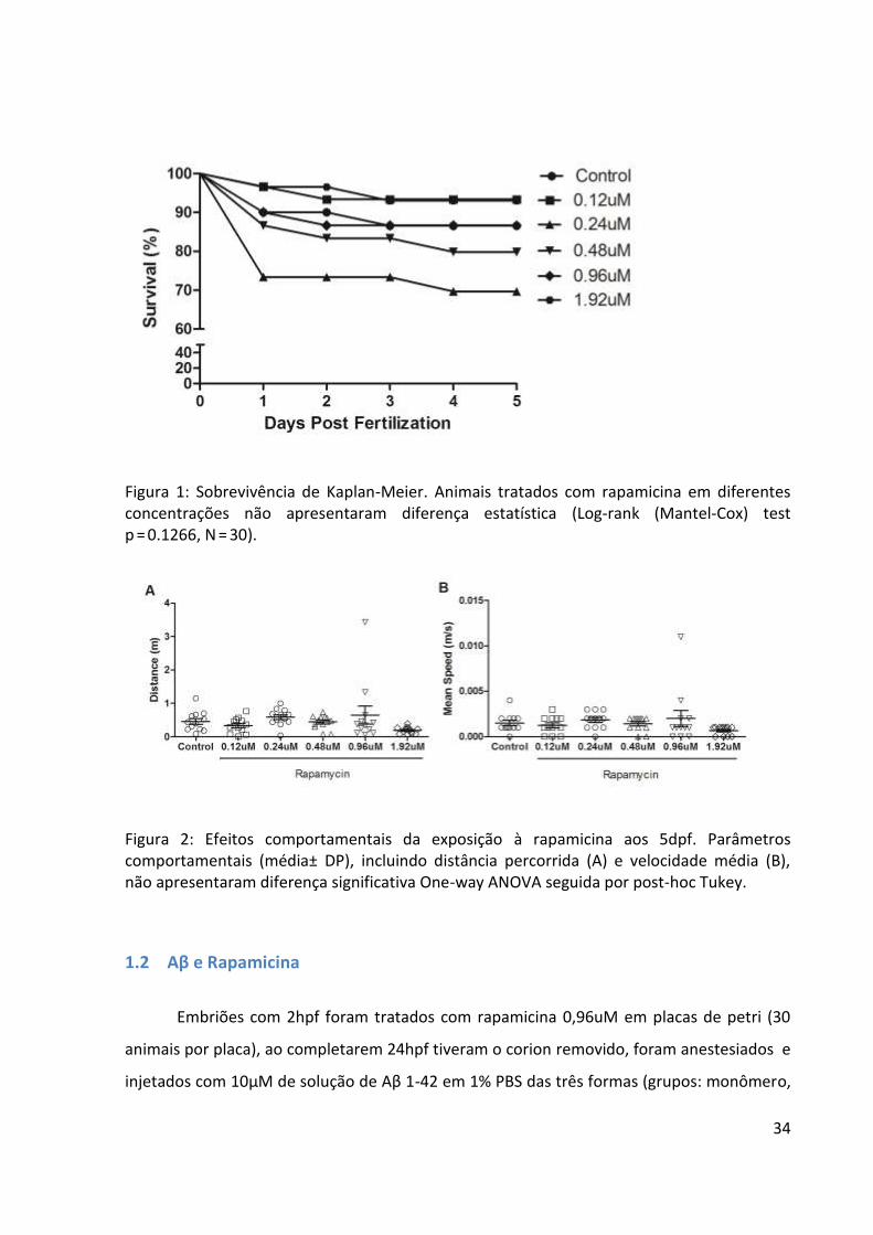
Aos 5dps foi realizada uma tarefa exploratória (Figura 2), nos parâmetros analisados
de distância percorrida (p=0.1443 F(5,66)=1.711 ) e velocidade média (p=0.3051 F(5,66)=1.230),
os grupos não apresentaram diferença significativa. A partir dos resultados obtidos, a
concentração escolhida para os testes com Aβ foi a de 0,96uM.
34
Figura 1: Sobrevivência de Kaplan-Meier. Animais tratados com rapamicina em diferentes concentrações não apresentaram diferença estatística (Log-rank (Mantel-Cox) test p = 0.1266, N = 30).
Figura 2: Efeitos comportamentais da exposição à rapamicina aos 5dpf. Parâmetros comportamentais (média± DP), incluindo distância percorrida (A) e velocidade média (B), não apresentaram diferença significativa One-way ANOVA seguida por post-hoc Tukey.
1.2 Aβ e Rapamicina
Embriões com 2hpf foram tratados com rapamicina 0,96uM em placas de petri (30
animais por placa), ao completarem 24hpf tiveram o corion removido, foram anestesiados e
injetados com 10μM de solução de Aβ 1-42 em 1% PBS das três formas (grupos: monômero,
35
oligômero e placa) e solução veiculo 1% PBS em corante vermelho (controle) sempre em
tratamento. Ao completarem 2dpf, tivemos uma mortalidade de 70% nos grupos monômero
e placa, nos demais grupos controle e oligômero foi 100%. Na segunda tentativa, ao
completarem 24hpf, os animais que sobreviveram não estavam viáveis para o procedimento
de injeção. Com isso, testei as diferentes concentrações apenas na placa a fim de ver se o
meio tornaria a ficar esbranquiçado como nos experimentos anteriores, testando também
animais injetados apenas com o corante vermelho e animais sem tratamento. Animais
injetados e tratados morreram ao completarem 2dpf, já os animais que não receberam
tratamento sobreviveram até os 5dpf.
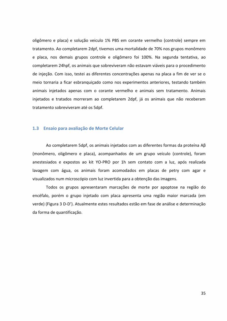
1.3 Ensaio para avaliação de Morte Celular Ao completarem 5dpf, os animais injetados com as diferentes formas da proteína Aβ
(monômero, oligômero e placa), acompanhados de um grupo veículo (controle), foram
anestesiados e expostos ao kit YO-PRO por 1h sem contato com a luz, após realizada
lavagem com água, os animais foram acomodados em placas de petry com agar e
visualizados num microscópio com luz invertida para a obtenção das imagens.
Todos os grupos apresentaram marcações de morte por apoptose na região do
encéfalo, porém o grupo injetado com placa apresenta uma região maior marcada (em
verde) (Figura 3 D-D’). Atualmente estes resultados estão em fase de análise e determinação
da forma de quantificação.
36
Figura 3: Detecção de apoptose por YO-PRO. A) Controle B) Monômero C) Oligômero D) Placa. A’) Controle B’) Monômero C’) Oligômero D’) Placa marcados para apoptose.
1.4 Western Blotting
Ao completarem 5dpf, os animais foram anestesiados e passaram por eutanásia por
decapitação, depois tiveram o encéfalo dissecado e armazenado em inibidor de protease a -
80ºC [50]. Após a homogeneização e quantificação proteica (amostras apresentaram cerca de
20mg/ml de proteína), as amostras foram aplicadas em um gel de acrilamida, corridas e
transferidas para uma membrana de nitrocelulose. Utilizamos Ponceu red para verificação
da presença das proteínas na membrana e este se mostrou positivo. Após isso, incubamos
com os anticorpos primários e secundários específicos e não foi possível observar nenhuma
banda através da revelação com o kit ECL. Realizamos 3 tentativas com amostras diferentes,
renovando os reagentes. Não foi possível adquirir uma especificidade dos anticorpos
suficiente para a quantificação através desta técnica. Supomos que os anticorpos não
tenham afinidade com essas amostras, visto que, em outras preparações de encéfalo de
zebrafish, foi possível realizar esta técnica.
37
CAPÍTULO 4
CONSIDERAÇÕES FINAIS
Com o aumento da expectativa de vida e consequentemente aumento dos casos de
demência e em especial a DA[5], a busca pela compreensão dos mecanismos moleculares que
estão associados à doença e a identificação de processos moleculares relevantes cujos alvos
possam ser a base no desenvolvimento de fármacos eficazes é crítica [51]. Esta motivação deu
origem ao trabalho aqui apresentado que pretendeu estudar aspectos moleculares e
comportamentais da DA num promissor modelo animal.
De forma molecular, a DA é caracterizada pela presença de β-amiloide, sendo
encontrada em diferentes formas e tamanhos, e também pelo acumulo da proteína tau
hiperfosforilada[14]. Nossa principal motivação foi caracterizar eventuais diferenças em
soluções com diferentes potenciais agregadores de peptídeo Aβ1-42. Para isso, realizamos
injeções no ventrículo cerebral de embriões com 24hpf de soluções que favoreciam
diferentes formas de Aβ (monômeros, oligômeros e a formação de placas). Aos 5dpf foram
observados aspectos moleculares e comportamentais, incluindo o desempenho cognitivo
dos animais em uma tarefa aversiva. Os animais que receberam a injeção de oligômero
apresentaram um pior desempenho na tarefa cognitiva sem deficit em aspectos gerais de
locomoção e motivação que pudessem potencialmente comprometer os resultados
observados, corroborando com estudos nos quais o oligômero altera a cognição e a
memória [52] e aqueles que sugerem que esta seja uma forma tóxica em adição às placas
[26,38]. Ao analisar o nível de Aβ no encéfalo dos animais, vimos que o grupo injetado com a
forma placa apresentou um nível significativamente maior, o que era esperado, uma vez que
as placas se depositam [53] e possuem maior dificuldade de clearance [54]. Juntos, estes
resultados reforçam a visão moderna de que a forma mais tóxica de Aβ1-42 não são as placas
e sim as versões solúveis que podem atuar sobre vias de sinalização e induzir efeitos
neurotóxicos [17,25].
Neste contexto, além da solubilidade e capacidade de interagir com receptores de
membrana celular, a eliminação da proteína ou clearance é outro fator importante quando
se pretende entender o real papel do peptídeo Aβ na DA. Nos últimos anos, autores têm

38
focado não apenas no acúmulo, mas no controle dos níveis de peptídeo pelas taxas de
eliminação do clearance como principal fator para o desenvolvimento da DA [55]. Sabe-se que
a microglia possui um papel importante da regulação dos níveis de amiloide cerebral e a
deposição [56], além disso. a eliminação no fluido intersticial e vasos sanguíneos e a atividade
de proteases contribuem para o processo. Tendo em vista o estudo da limpeza da Aβ
associado à facilidade de manipulação do zebrafish, futuramente serão realizadas injeções
com a Aβ fluorescente a fim de observar o processo de clearance e larvas de zebrafish
injetadas com soluções com perfil agregador de Aβ.
39
REFERÊNCIAS
1 WHO. Global Health and Aging. 2011
2 WHO.“Ageing well” must be a global priority. 2014.( http://www.who.int), acessado
em janeiro de 2017
3 Authoritative information and statistics to promote better health and wellbeing
(http://www.aihw.gov.au), acesso em janeiro de 2017
4 Alzheimer’s Disease International. The Global Impact of Dementia 2013-2050’. 2013
5 WHO. Dementia cases set to triple by 2050 but still largely ignored. 2012
6 Alzheimer’s Disease International. Alzheimer Disease Diagnostic
(http://www.alz.org), acessado em janeiro de 2017
7 Johnson KA, Minoshima S, Bohnen NI, Donohoe KJ, Foster NL, Herscovitch P,
Karlawish JH, et al. Appropriate use criteria for amyloid PET: A report of the Amyloid Imaging
Task Force, the Society of Nuclear Medicine and Molecular Imaging, and the Alzheimer’s
Association. Alzheimer’s & Dementia. (2013) 9: 1-16
8 Mandelkow EM & Mandelkow E. Biochemistry and Cell Biology of Tau Protein in
Neurofibrillary Degeneration. Cold Spring Harb Perspect Med. (2012)
9 Newman M, Verdile G, Martins RN, Lardelli M. Zebrafish as a tool in Alzheimer’s
disease research. Biochimica et Biophysica Acta. (2100) 1812: 346-352
10 Basic Neurochemistry: Molecular, Cellular and Medical Aspects. Alzheimer's Disease
Is the Most Common Neurodegenerative Disorder. 6th edition
11 Glenner GG, Wong CW. Alzheimer's disease: initial report of the purification and
characterization of a novel cerebrovascular amyloid protein. Biochem Biophys Res
Commun. (1984) 3: 885-90
12 Tschanz JT, Norton MC, Zandi PP, Lyketsos CG. The Cache County Study on Memory in
Aging: Factors Affecting Risk of Alzheimer's disease and its Progression after Onset. Int Rev
Psychiatry. (2013) 25: 673–685
13 Tjin PV, Kamphuis W, Marlatt MW, Hol EM, Lucassen PJ. Presenilin mouse and
Zebrafish models for dementia: Focus on neurogenesis. Progress in Neurobiology. (2011) 93:
149-164
40
14 Roberson ED, Muck L. 100 years and counting: prospects for defeating Alzheimer’s
disease. Science. (2006) 314:781-784
2 15 Guo Q, Wang Z, Li H, Wiese M, Zheng H. APP physiological and pathophysiological functions: insights from animal models. Cell Research. (2012) 22: 78-89
16 Goate A, Chartier-Harlin MC, Mullan M, Brown J, Crawford F, Fidani L, Giuffra L et al.
Segregation of a missense mutation in the amyloid precursor protein gene with familial
Alzheimer's disease. Nature. (1991) 349: 704-706
17 Ferreira, S.T., Klein, W.L. The Aß oligomer hypotesis for synapse failure and memory
loss in Alzheimer’s disease. Neurobiology of Learning and Memory. (2011) 96: 529-543
18 Levy-Lahad E, Wijsman EM, Nemens E, Anderson L, Goddard KA, Weber JL, Bird
TD, Schellenberg GD. A familial Alzheimer's disease locus on chromosome 1.
Science. (1995) 269: 970-3
19 Newman M, Musgrave IF, Lardelli M. Alzheimer disease: amyloidogenesis, the
presenilins and animal models. Biochim Biophys Acta. (2007) 3: 285–97
20 De Strooper B, Vassar R, Golde T. The secretases: enzymes with therapeutic potential
in Alzheimer disease. Nat Rev Neurol. (2010) 2: 99-107
21 Nunan J, Small DH. Regulation of APP cleavage by α-, β- and γ-secretases. FEBS
Lett. (2000) 483: 6-10
22 Tseng BP, Esler WP, Clish CB, Stimson ER, Ghilardi JR, Vinters HV, et al. Deposition of
monomeric, not oligomeric, Abeta mediates growth of Alzheimer’s disease amyloid plaques
in human brain preparations. Biochemistry. (1999) 38:10424–10431
23 Selkoe, D.J. Amyloid beta protein precursor and the pathogenesis of Alzheimer’s
disease. Cell. (1989) 58: 611-612
24 Allsop D, Wong CW, Ikeda S, Landon M, Kidd M, Glenner GG. Immunohistochemical
evidence for the derivation of a peptide ligand from the amyloid beta-protein precursor of
Alzheimer disease. Porceeding of the National Academy of Science of the United States of
America. (1998) 85: 270-2794
25 Glabe CG. Structural Classification of Toxic Amyloid Oligomers. J Biol Chem. (2008)
44: 29639–29643
https://www.ncbi.nlm.nih.gov/pubmed/?term=Stimson%20ER%5BAuthor%5D&cauthor=true&cauthor_uid=10441137
41
26 Hardy J. The Discovery of Alzheimer causing Mutations in the APP Gene and the
Formulation of the "Amyloid Cascade Hypothesis". FEBS J. (2017) 284:1040-1044
27 Herrup K. The case for rejecting the amyloid cascade hypothesis. Nature Neuroscience.
(2015) 6:794–799
28 Huang Y, Mucke L. Alzheimer mechanisms and therapeutic strategies. Cell. (2012)
148: 1204-1222
29 Verma M, Vats A, Taneja V. Toxic species in amyloid disorders: Oligomers or mature
fibrils. Ann Indian Acad Neurol. (2015) 2:138–145
30 Spence R, Gerlach G, Lawrence C, Smith C. The behavior and ecology of the zebrafish,
Danio rerio. Biological Reviews of the Cambridge Philosophical Society. (2008) 83: 13-34
31 Götz J, Ittner LM. Animal Models Of Alzheimer's Disease And Frontotemporal
Dementia. Nature Reviews Neuroscience. (2008) 9: 532-544
32 Head E, N.W. Milgram and C.W. Cotman. Neurobiological models of aging in the dog
and other vertebrate species, in: Functional Neurobiology of Aging., P.R. Hof and C.V.
Mobbs. Academic Press. (2001) 457– 468
33 Cotman CW, Head E. The Canine (Dog) Model of Human Aging and Disease: Dietary,
Environmental and Immunotherapy Approaches. Journal of Alzheimer’s Disease. (2008) 15:
685–707
34 Newman M, Ebrahimie E, Lardelli M. Using the zebrafish model for Alzheimer's
disease research. Front Genet. (2014) 30: 185-189
35 Vallée A, Lecarpentier Y. Alzheimer Disease: Crosstalk between the Canonical
Wnt/Beta-Catenin Pathway and PPARs Alpha and Gamma. Front Neurosci. (2016) 10: 459-
471
36 Cunvong K, Huffmire D, Etthel DW, Cameron DJ. Amyloid-β increases capillary bed
density in the adult zebrafish retina. Invest Ophthalmol Vis Sci. (2013) 54:1516
37 Donnini S, Giachetti A, Ziche M. Assessing vascular senescence in zebrafish. Methods
Mol Biol. (2013) 965:517-31
38 Nery LR, Eltz NS, Hackman C, Fonseca R, Altenhonfen S, Guerra HN et al. Brain
Intraventricular Injection of Amyloid-β in Zebrafish Embryo Impairs Cognition and Increases
Tau Phosphorylation, Effects Reversed by Lithium. PLoS One. (2014) 9:e105862
42
39 Kim HY, Lee DK, Chung BR, Kim HV, Kim Y. Intracerebroventricular Injection of
Amyloid-β Peptides in Normal Mice to Acutely Induce Alzheimer-like Cognitive
Deficits. Jove. (2016) doi:10.3791/53308
40 Vézina C, Kudelski A, Sehgal SN. Rapamycin (AY-22,989), a new antifungal antibiotic. I.
Taxonomy of the producing streptomycete and isolation of the active principle. J. Antibiot.
(Tokyo). (1975) 28:721–726
41 Baker H, Sidorowicz A, Sehgal SN, Vézina C. Rapamycin (AY-22,989), a new antifungal
antibiotic. III. In vitro and in vivo evaluation. J Antibiot (Tokyo). (1978) 31: 539-45.
42 Mathieu l and David MS. mTOR Signaling. Cold Spring Harb Perspect Biol. (2012) 4:
a011593.
43 Tese Anna MS. O papel do sistema purinérgico e da via de sinalização TOR em crises
convulsivas e estresse oxidativo. (2013)
44 Siebel AM, Menezes FP, da Costa Schaefer I, Petersen BD, Bonan CD.Rapamycin
suppresses PTZ-induced seizures at different developmental stages of zebrafish. Pharmacol
Biochem Behav. (2015)
45 Powers RW3rd, Kaeberlein M, Caldwell SD, Kennedy BK, Fields S. Extension of
chronological life span in yeast by decreased TOR pathway signaling. Genes Dev. (2006) 20:
174-84.
46 Tsang CK, Qi H, Liu LF, Zheng XF. Targeting mammalian target of rapamycin (mTOR)
for health and diseases. Drug Discov Today. (2007) 12: 112-24
47 Cai Z, Zhao B, Li K, Zhang L, Li C, Quazi SH, Tan Y. Mammalian target of rapamycin: a
valid therapeutic target through the autophagy pathway for Alzheimer'sdisease? J Neurosci
Res. (2012) 90: 1105-18
48 Harrison DE, Strong R, Sharp ZD, Nelson JF, Astle CM, Flurkey K, et al. Rapamycin fed
late in life extends life-span in genetically heterogeneous mice. Nature. (2009) 460: 392–395
49 Caccamo A, Majumder S, Richardson A, Strong R, Oddo S. Molecular interplay
between mammalian target of rapamycin (mTOR), amyloid-beta, tau: effects on cognitive
impairments. J Biol Chem. (2010) 285: 13107–13120
http://www.ncbi.nlm.nih.gov/pubmed/?term=Petersen%20BD%5BAuthor%5D&cauthor=true&cauthor_uid=26051026
43
50 Nornes S, Groth C, Camp E, Ey P, Lardelli M. Developmental control of Presenilin1
expression, endoproteolysis, and interaction in zebrafish embryos. Exp Cell Res. (2003)
289:124–132
51 Abbott A, Dolgin E. Failed Alzheimer's trial does not kill leading theory of disease.
Nature. (2016) 540:15-16
52 Cleary JP, Walsh DM, Hofmeister JJ, Shankar GM, Kuskowski MA, Selkoe DJ, Ashe KH.
Natural oligomers of the amyloid-β protein specifically disrupt cognitive function. Nature
Neuroscience. (2005) 8: 79–84
53 Gouras GK, Relkin NR, Sweeney D, Munoz DG, Mackenzie IR, Gandy S. Increased
apolipoprotein E epsilon 4 in epilepsy with senile plaques. Ann Neurol. (1997) 3: 402-4
54 Oddo, S., Billings, L., Kesslak, J. P., Cribbs, D. H. & LaFerla, F. M. Aβ immunotherapy
leads to clearance of early, but not late, hyperphosphorylated τ aggregates via the
proteasome. Neuron. (2004) 43:321–332
55 Sang-Sun Yoon and Sangmee Ahn Jo. Mechanisms of Amyloid-β Peptide Clearance:
Potential Therapeutic Targets for Alzheimer’s Disease Biomol Ther (Seoul). (2012) 20: 245–
255
56 Zhiqiang L, Carlo C, Aaron S, Roa Harb, Jaime G. CX3CR1 in Microglia Regulates Brain
Amyloid Deposition through Selective Protofibrillar Amyloid-β Phagocytosis The Journal of
Neuroscience. (2010) 30: 17091-17101.
44
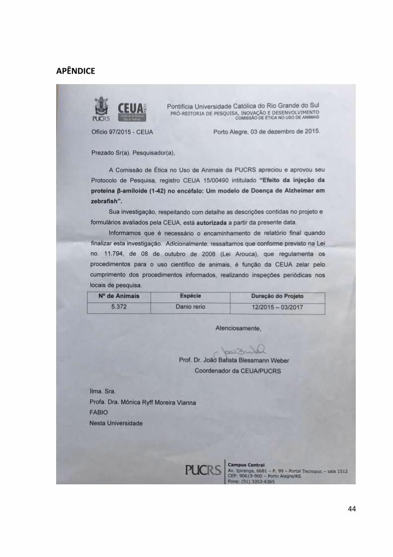
APÊNDICE


![[Apresentação de Defesa] Análise comparativa entre os métodos HMM e GMM-UBM na busca pelo α-ótimo dos locutores crianças para utilização da técnica VTLN](https://static.fdocument.org/doc/165x107/559ebccc1a28ab832a8b4719/apresentacao-de-defesa-analise-comparativa-entre-os-metodos-hmm-e-gmm-ubm-na-busca-pelo-otimo-dos-locutores-criancas-para-utilizacao-da-tecnica-vtln.jpg)