
Skelemin Association with αIIbβ Integrin: A … Association with α IIbβ 3 Integrin: A Structural...
10
Skelemin Association with α IIb β 3 Integrin: A Structural Model Vitaliy Gorbatyuk, † Khiem Nguyen, † Nataly P. Podolnikova, ‡ Lalit Deshmukh, †,⊥ Xiaochen Lin, † Tatiana P. Ugarova, ‡ and Olga Vinogradova* ,† † Department of Pharmaceutical Sciences, School of Pharmacy, University of Connecticut at Storrs, Storrs, Connecticut 06269, United States ‡ Center for Metabolic and Vascular Biology, School of Life Sciences, Arizona State University, Tempe, Arizona 85287, United States * S Supporting Information ABSTRACT: Over the last two decades, our knowledge concerning intracellular events that regulate integrin’saffinity to their soluble ligands has significantly improved. However, the mechanism of adhesion-induced integrin clustering and development of focal complexes, which could further mature to form focal adhesions, still remains under-investigated. Here we present a structural model of tandem IgC2 domains of skelemin in complex with the cytoplasmic tails of integrin α IIb β 3 . The model of tertiary assembly is generated based upon NMR data and illuminates a potential link between the essential cell adhesion receptors and myosin filaments. This connection may serve as a basis for generating the mechanical forces necessary for cell migration and remodeling. I n order for multicellular organisms to survive, individual cells must adhere to each other and to their extracellular surrounding. This adhesion is primarily mediated by integrins, 1 a family of transmembrane glycoprotein heterodimers. Integrins connect the extracellular matrix (ECM) and the cytoskeleton within the cells through numerous interactions with their cytoplasmic targets. Integrins also function as bidirectional signal transducers 2 and may serve as sensors of ever-changing mechanical forces. 3 It has been shown that integrins bind to ECM proteins via their extracellular domains, which triggers conformational changes and clustering of integrins. This clustering initially forms a small network called motility- inducing focal complexes (FXs), which could be ultimately replaced in fully spread cells by large intracellular complexes of variable content known as focal adhesions (FAs). 4 Skelemin, also known as myomesin-1.1 and originally identified as a muscle M-line cytoskeletal protein of 185 kDa, is expressed primarily in embryonic heart 5 and has been shown to play a critical role in mediating the connection between ECM and cytoskeleton during the early stages of cell spreading. 6 It belongs to a family of cytoskeletal proteins, all associated with myosin thick filaments in skeletal and cardiac muscles, and contains a unique N-terminal myosin-binding domain, five fibronectin (FN) type III-like domains, six immunoglobulin C2-like (IgC2) domains, and a C-terminal immunoglobulin domain involved in homodimerization. 7 Its major isoform (myomesin-1.2) is shorter by about a hundred residues, which are spliced out between FN domains 2 and 3. Skelemin is localized to FXs, but not FAs, through the direct interaction of its IgC2 domains 4 and 5 with β 3 -integrin cytoplasmic tail. 8 The second major member of this family, myomesin-2, is a product of myomesin 2 gene with a shorter N- terminus, resulting in a molecular weight of about 165 kDa, and has about 71% homology with skelemin. It is expressed in diverse nonmuscle tissues including CHO cells, platelets, and endothelial cells. 8,9 We and the others have shown that skelemin is one of the rare proteins that can bind both β and α subunit of integrin receptors. 10,11 Although skelemin cannot activate integrins, it has been suggested that skelemin exerts contractile force and modulates the attachment of cytoskeletal proteins and Src to integrin clusters during early stages of cell spreading. 12 A recent study 13 has unveiled the ability of skelemin filaments to be stretched to about 2.5-fold its original length by reversible unfolding of the linkers connecting Ig domains. Pinotsis and co- workers have employed a combination of four complementary structural biology methods to investigate how the repetitive structure of skelemin contributes to muscle elasticity. This work explained, for the first time, skelemin’s capability to act as a highly elastic ribbon for maintaining the overall structural organization of the sarcomeric M-band of skeletal muscle. In the present work, we investigated how two skelemin repeats are organized and may contribute to its unique elastic properties in nonmuscle cells. Knowledge of these details is particularly important considering the role of skelemin as a connector between cell surface receptors and the cytoskeleton. We previously determined the solution structure of skelemin immoglobulin domain 4 (Sk4), modeled domain 5 (Sk5), and Received: June 3, 2014 Revised: August 11, 2014 Published: September 15, 2014 Article pubs.acs.org/biochemistry © 2014 American Chemical Society 6766 dx.doi.org/10.1021/bi500680s | Biochemistry 2014, 53, 6766−6775
Transcript of Skelemin Association with αIIbβ Integrin: A … Association with α IIbβ 3 Integrin: A Structural...
Skelemin Association with αIIbβ3 Integrin: A Structural ModelVitaliy Gorbatyuk,† Khiem Nguyen,† Nataly P. Podolnikova,‡ Lalit Deshmukh,†,⊥ Xiaochen Lin,†
Tatiana P. Ugarova,‡ and Olga Vinogradova*,†
†Department of Pharmaceutical Sciences, School of Pharmacy, University of Connecticut at Storrs, Storrs, Connecticut 06269, UnitedStates‡Center for Metabolic and Vascular Biology, School of Life Sciences, Arizona State University, Tempe, Arizona 85287, United States
*S Supporting Information
ABSTRACT: Over the last two decades, our knowledge concerning intracellular events thatregulate integrin’s affinity to their soluble ligands has significantly improved. However, themechanism of adhesion-induced integrin clustering and development of focal complexes,which could further mature to form focal adhesions, still remains under-investigated. Herewe present a structural model of tandem IgC2 domains of skelemin in complex with thecytoplasmic tails of integrin αIIbβ3. The model of tertiary assembly is generated based uponNMR data and illuminates a potential link between the essential cell adhesion receptors andmyosin filaments. This connection may serve as a basis for generating the mechanical forcesnecessary for cell migration and remodeling.
In order for multicellular organisms to survive, individual cellsmust adhere to each other and to their extracellular
surrounding. This adhesion is primarily mediated by integrins,1
a family of transmembrane glycoprotein heterodimers. Integrinsconnect the extracellular matrix (ECM) and the cytoskeletonwithin the cells through numerous interactions with theircytoplasmic targets. Integrins also function as bidirectionalsignal transducers2 and may serve as sensors of ever-changingmechanical forces.3 It has been shown that integrins bind toECM proteins via their extracellular domains, which triggersconformational changes and clustering of integrins. Thisclustering initially forms a small network called motility-inducing focal complexes (FXs), which could be ultimatelyreplaced in fully spread cells by large intracellular complexes ofvariable content known as focal adhesions (FAs).4
Skelemin, also known as myomesin-1.1 and originallyidentified as a muscle M-line cytoskeletal protein of 185 kDa,is expressed primarily in embryonic heart5 and has been shownto play a critical role in mediating the connection betweenECM and cytoskeleton during the early stages of cellspreading.6 It belongs to a family of cytoskeletal proteins, allassociated with myosin thick filaments in skeletal and cardiacmuscles, and contains a unique N-terminal myosin-bindingdomain, five fibronectin (FN) type III-like domains, siximmunoglobulin C2-like (IgC2) domains, and a C-terminalimmunoglobulin domain involved in homodimerization.7 Itsmajor isoform (myomesin-1.2) is shorter by about a hundredresidues, which are spliced out between FN domains 2 and 3.Skelemin is localized to FXs, but not FAs, through the directinteraction of its IgC2 domains 4 and 5 with β3-integrincytoplasmic tail.8 The second major member of this family,
myomesin-2, is a product of myomesin 2 gene with a shorter N-terminus, resulting in a molecular weight of about 165 kDa, andhas about 71% homology with skelemin. It is expressed indiverse nonmuscle tissues including CHO cells, platelets, andendothelial cells.8,9
We and the others have shown that skelemin is one of therare proteins that can bind both β and α subunit of integrinreceptors.10,11 Although skelemin cannot activate integrins, ithas been suggested that skelemin exerts contractile force andmodulates the attachment of cytoskeletal proteins and Src tointegrin clusters during early stages of cell spreading.12 A recentstudy13 has unveiled the ability of skelemin filaments to bestretched to about 2.5-fold its original length by reversibleunfolding of the linkers connecting Ig domains. Pinotsis and co-workers have employed a combination of four complementarystructural biology methods to investigate how the repetitivestructure of skelemin contributes to muscle elasticity. This workexplained, for the first time, skelemin’s capability to act as ahighly elastic ribbon for maintaining the overall structuralorganization of the sarcomeric M-band of skeletal muscle.In the present work, we investigated how two skelemin
repeats are organized and may contribute to its unique elasticproperties in nonmuscle cells. Knowledge of these details isparticularly important considering the role of skelemin as aconnector between cell surface receptors and the cytoskeleton.We previously determined the solution structure of skeleminimmoglobulin domain 4 (Sk4), modeled domain 5 (Sk5), and
Received: June 3, 2014Revised: August 11, 2014Published: September 15, 2014
Article
pubs.acs.org/biochemistry
© 2014 American Chemical Society 6766 dx.doi.org/10.1021/bi500680s | Biochemistry 2014, 53, 6766−6775
investigated how major platelet integrin αIIbβ3 binds to Sk45.11
Here, we examined skelemin tandem IgC2 domains 4 and 5(hereafter addressed as Sk45) together with their interconnect-ing linker using solution nuclear magnetic resonance (NMR)spectroscopy. We present the structure of Sk45 and the dockingmodel of its tertiary complex with αIIbβ3 integrin cytoplasmictails. We also investigated thermodynamic profiles of skelemininteractions with integrin cytoplasmic tails by isothermaltitration calorimetry (ITC). Overall, the docking modelsupports the role of skelemin in stabilizing integrin activated,clustered state through the simultaneous binding to its twoseparated cytoplasmic tails.
■ EXPERIMENTAL PROCEDURESExpression and Purification, Peptides, and Cells. The
cloning of mouse Sk4 and Sk45 has been describedpreviously.11 Single-site mutagenesis of Sk45, convertingsolvent-exposed C1354 to S (to improve solubility ofrecombinant construct) and C-terminal K1424 to C (tointroduce paramagnetic spin label), was performed using theQuikChange kit (Agilent Technologies). The mutant plasmidswere transformed into Rosetta (DE3) competent cells (EMDMillipore). Protein expression was carried out using LB or M9minimal media with 15NH4Cl and/or
13C-glucose as the solenitrogen and carbon sources, at 37 °C. Cultures were inducedwith 1 mM ITPG at an OD600 of ∼0.6. The cells were harvested4 h after induction. For Sk4, Sk45 (construct with C1354Smutation), and Sk45m (construct with both C1354S and K1424Cmutations), cells were resuspended in a buffer containing 20mM Tris, pH 8, 300 mM NaCl, 10 mM imidazole, 1 mM DTT,1 mM PMSF, 1 tablet of complete protease inhibitor cocktail(Roche Applied Science). The suspension was lysed by passagethrough a French press (Thermo Electron). The supernatantfractions were loaded onto Ni-NTA agarose resin (Qiagen),and proteins were eluted with a buffer containing 20 mM Tris,pH 7.5, 300 mM NaCl, and 500 mM imidazole. To cleave theHis-tag, thrombin was added directly to the Ni-NTA column.Cleavage was done in a 37 °C incubator for 4 h with periodicmixing (50 mM Tris, 150 mM NaCl, 1 mM EDTA, pH 7.5).Thrombin was inhibited by the addition of equivalent amountsof PMSF. The eluate was further purified by size exclusionchromatography (16/60 Superdex 75 column, GE Healthcare)in 50 mM NaCl, 20 mM KPO4, pH 6.8.Peptides, corresponding to the integrin cytoplasmic tails,
were synthesized chemically (NEOpeptides): αIIb (startingfrom W988), N-terminus of β3 (K
716−W739), and C-terminus ofβ3 (K738-T762). Myristoylated peptides, corresponding toskelemin’s sequence, were synthesized by Peptide 2.0(Chantilly, VA): 1369THIVWYKDEREISVDEKHD1387 and1377EREISAAAKHD1387, which is a triple mutant (VDE →AAA).Human fibrinogen was obtained from Enzyme Research
Laboratories (South Bend, IN). CHO cells expressing αIIbβ3were described previously14 and were maintained in Dulbecco’smodified Eagle’s medium/F-12 medium supplemented with10% fetal bovine serum and 25 mM HEPES.NMR Spectroscopy. NMR experiments were performed on
uniformly 15N/13C-labeled samples (unless stated otherwise)prepared in buffer containing 20 mM KPO4, pH 6.8, and 7%D2O. Spectra were recorded at 25 °C on a Varian INOVA 600MHz spectrometer equipped with a cold probe. The standardBioPack pulse sequences were used. Spectra were processedusing NMRPipe15 and analyzed with CCPN Analysis.16
The backbone resonance assignments for Sk45 wereobtained from the set of triple resonance experiments,HNCO, HN(CA)CO, HNCA, HNCACB, CBCA(CO)NH,and HBHA(CO)NH. Because of a very high degeneracy of thepeaks in HCCH−COSY and HCCH-TOCSY spectra and veryweak C(CO)NH and H(CCO)NH spectra, only a partialassignment of the side chain resonances was achieved fromthese data. The Sk45 tandem is very rich in methyl-containingresidues, which constitute 32% of the protein sequence. Sincemethyl groups form the core of a protein and, thus, couldprovide very valuable distance restrains for the structurecalculation, we collected a methyl-optimized (H)CCmHm-TOCSY experiment from the BioPack library. From thisspectrum, we were able to obtain the chemical shift assignmentfor all methyl groups, and this information turned to be crucialfor the convergence of the structure calculation. In total,chemical shift assignment was obtained for 80% of protons,41% of carbons, and 71% of nitrogens.
1DHN residual dipolar couplings (RDC) were derived fromthe difference between peaks positions in 1H−15N HSQC andTROSY spectra recorded in isotropic and anisotropicconditions. Three different alignment media were used: Pf1phage (ASLA Biotech), mixture of C12E5/hexanol, or C12E5/hexanol/sodium octyl sulfate (Sigma-Aldrich, C12E15 concen-tration was 4.1%, C12E5:SOS = 30:1 molar ratio). In all media,the His-tag of Sk45 was cleaved prior to collecting RDCs. Adata set for Sk45 with the His-tag intact was also collected inPf1 phage solution.Paramagnetic relaxation enhancement (PRE) experiments
were performed with Sk45m, where the solvent-exposed K1424
was mutated to cysteine. The 1H−15N HSQC spectra werecollected with and without 3-maleimido-PROXYL (m-PROX-YL) spin label. The effect of the spin label on the signalintensities was estimated from the normalized intensitydifferences in the spectra with and without the spin label.Transferred NOEs (trNOE) for αIIb cytoplasmic tail in
complex with Sk45 were obtained at the peptide to proteinratio of 100:1 with the mixing time of 400 ms as previouslymentioned.11 The 1H assignments for αIIb were performedpreviously.17
The titrations of αIIb or β3 into the Sk45 solution weremonitored with 1H−15N HSQC spectra at the peptide toprotein ratios varied from 1:1 to 5:1.
Structures Calculation. The Sk45 structure calculationand NOE assignment was carried out with ARIA 2.318 andXplor-NIH.19 The unassigned NOEs from 3D 15N-NOESY and13C-NOESY experiments along with dihedral angle constraintswere used as an input for ARIA. The dihedral angles werepredicted with DANGLE in CCPN Analysis.16 In thesubsequent calculations, hydrogen bond constraints wereintroduced. The characteristic NOE pattern, the CSI secondarystructure prediction, and H-D exchange data gave rise to 156constraints (two constraints per H-bond). In some cases,hydrogen bond constraints were set to have multiple partners.Later, the obtained ARIA NOE assignments were manuallyverified in CCPN Analysis. Stereospecific assignment ofprochiral groups was achieved using a floating assignmentapproach as implemented in ARIA.The structure was further refined with RDCs using Xplor-
NIH. Initially, the two domains were refined with RDCsindependently. For this, the sets of 1DHN RDCs for the threemedia were divided in the two parts: one for each skelemindomain. In addition to RDC restraints, the calculation was
Biochemistry Article
dx.doi.org/10.1021/bi500680s | Biochemistry 2014, 53, 6766−67756767
supplied with ARIA-derived distance constraints, dihedral andH-bond restraints. In the next step, the two domains weretreated as rigid bodies connected by a flexible linker and RDCswere used to orient the domains. This time, every set of RDCrestraints for the three different media was used as a whole,containing RDCs for the both domains. A total of 1000structures were calculated in the “rigid body” run and thelowest energy 20 structures were further refined in water withXplor-NIH. In the water refinement step, all experimentalconstraints were used, including NOEs, dihedral, H-bond, andRDC restraints. These 20 refined structures were chosen as arepresentative ensemble.The αIIb peptide structure in the complex with Sk45 was
obtained based upon trNOE restraints using ARIA, and theensemble of 20 structures with minimal overall energy wasrefined in explicit water.During the course of the calculations, the quality of the
molecular structures was assessed with ARIA/CNS built-inscripts and PROCHECK-NMR.20
Modeling. The HADDOCK Web server21 was used fordocking of αIIb and β3 integrin tails to Sk45. For αIIb/Sk45binary complex determination, the best NMR structures of αIIband Sk45 from the NMR ensembles were chosen. During thedocking, the following residues were set as active: 990, 992−997 of αIIb and 1361, 1363, 1394 of Sk45. For β3 docking, wehave used the first representative of the ensemble with PDB ID1M80. The active residues for this docking were 716, 722, 724,and 725 of β3 and 1368, 1370, 1372, 1374, 1382, 1383, and1411 of Sk45. The flexible unstructured N-terminal residues(1207 to 1225) of Sk45 were removed before docking.Electrostatic Potential. The electrostatic potential of Sk45
was calculated using Adaptive Poisson−Boltzmann Solver(APBS).22 The pdb file of Sk45 was uploaded to the pdb2pqr Web server23 using PARSE as the force field andPROPKA to assign protonation states. The output files wereused to create the electrostatic potential map by APBS. Theelectrostatic potential map was visualized by UCSF Chimera24
using the built-in electrostatic surface coloring module.ITC. Isothermal titration calorimetry was performed on a low
volume Nano ITC (TA Instruments). Peptides correspondingto integrin cytoplasmic tails were solubilized in the buffer 50mM NaCl, 20 mM KPO4, and pH 6.8. All ITC experimentswere performed at 25 °C, 300 rpm mixing, 300 s time intervalsbetween injections, and 3 μL injection volumes. Theconcentrations used are as follows: 2.5 mM C-terminal β3and 0.175 mM Sk4, 1.3 mM N-terminal β3 and 0.177 mMSk45, and 1.9 mM αIIb and 0.177 mM Sk45. The analysis of thedata was done in NanoAnalyze Software (TA Instruments)suite using “Independent” model.Adhesion Assays. The wells of 96-well tissue culture plates
(Immulon 4B) were coated with the 2.5 μg/mL of fibrinogenovernight at 4 °C. The wells were postcoated with 1% BSA.Cells were labeled with 10 μM calcein AM (Molecular Probes,Eugene, OR) for 30 min at 37 °C, washed and resuspended inDMEM/F-12 medium at 1 × 10⁵ cells/mL. Cells were mixedwith different concentrations of peptides for 20 min at 22 °Cbefore they were added to the wells coated with adhesivesubstrates. Aliquots (100 μL) of cells were added to the wellsand incubated at 37 °C for 30 min. The nonadherent cells wereremoved by two washes with phosphate-buffered saline (PBS),and fluorescence was measured in a fluorescence plate reader(Applied Biosystems, Framingham, MA). The number of
adherent cells was determined using the fluorescence ofaliquots with a known number of labeled cells.
■ RESULTSStructure of Skelemin Tandem Sk45. Skelemin IgC
domains 4 and 5 have been shown to interact with β3cytoplasmic tail.9 We have determined, by solution NMR,that a single domain Sk4 adopts a very well-known IgC2-foldcontaining seven β-strands in two β-sheets forming a β-sandwich.11 However, poor behavior of skelemin domain 5 inthe solution has precluded its detailed structural character-ization by NMR. To avoid this problem, and to improve thesolubility of the tandem Sk45, we have mutated the surface-exposed C1354 of domain 5 to serine in order to obtain a 27 kDaprotein construct suitable for solution NMR studies.The complete backbone and partial side chain chemical shifts
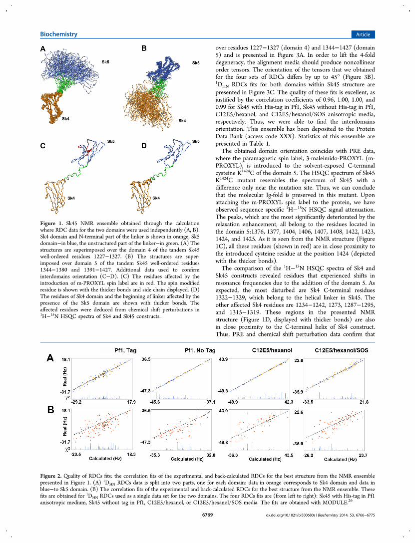
assignments were found sufficient for ab initio structurecalculation of the domains fold via the classical NOE-basedapproach supported by RDC data. A total of 2647 NOEconstraints, 351 RDCs, 156 H-bonds, and 311 backbonedihedral restraints, allowed us to obtain the Sk45 folds withRMSD 0.97 Å for Sk4 domain (the well-ordered residues1227−1327 are used for superimposition) and 1.37 Å for Sk5domain (the well-ordered residues 1344−1380 and 1391−1427). As expected, the tandem domains 4 and 5 adopted awell-known IgC2-fold and contained seven β-strands in two β-sheets forming two β-sandwiches connected through a partiallyhelical linker. Figure 1A,B presents 20 structures with thelowest target energy functions. These structures were obtainedfrom the calculations where RDCs were divided in the twoseparate sets (one for each domain), effectively treating the twodomains as independent entities. The RDC fits for thiscalculation show an impeccable correlation within theindividual domains (Figure 2A). However, the correlation ispoor for the tandem (Figure 2B), reflecting the fact that themutual orientation of the domains in these structures is notoptimized.Thus, the observed NOEs alone were insufficient for
restricting the domains connected in the tandem through aloosely structured helical linker. Therefore, in order to definethe mutual orientation of domains 4 and 5, we obtained andutilized long-range data, such as RDC and PRE. As a rule, theRDC approach is based on the notion that when a multidomainmolecule with no or little relative interdomain motion is settledwithin an anisotropic environment, the environment does notchange the mutual domain orientation. In turn, this implies thatthe alignment tensors for each domain and for the entiremolecule in the anisotropic environment are equal. Thus, fororienting the domains within the multidomain molecule bymeans of RDCs, the order tensors of the domains have to bemade collinear. However, an inherent 4-fold degeneracy existssince residual dipolar coupling constants can be satisfied bytensors which differ by 180° around the tensor axes. To resolvethis ambiguity, data from several types of alignment media areused simultaneously during structure calculations.25
We have used the “rigid body” approach for the domainorientation in the Sk45 tandem. The starting structures had thedomains refined with all experimental constraints as describedabove. In the “rigid body” Xplor-NIH run, we calculated 1000structures in order to sample the entire conformational space,and we used RDC constraints as a single set for every alignmentmedium, which would treat the tandem as a whole. The finalensemble of the 20 lowest energy structures has RMSD 3.1 Å
Biochemistry Article
dx.doi.org/10.1021/bi500680s | Biochemistry 2014, 53, 6766−67756768
over residues 1227−1327 (domain 4) and 1344−1427 (domain5) and is presented in Figure 3A. In order to lift the 4-folddegeneracy, the alignment media should produce noncollinearorder tensors. The orientation of the tensors that we obtainedfor the four sets of RDCs differs by up to 45° (Figure 3B).1DHN RDCs fits for both domains within Sk45 structure arepresented in Figure 3C. The quality of these fits is excellent, asjustified by the correlation coefficients of 0.96, 1.00, 1.00, and0.99 for Sk45 with His-tag in Pf1, Sk45 without His-tag in Pf1,C12E5/hexanol, and C12E5/hexanol/SOS anisotropic media,respectively. Thus, we were able to find the interdomainsorientation. This ensemble has been deposited to the ProteinData Bank (access code XXX). Statistics of this ensemble arepresented in Table 1.The obtained domain orientation coincides with PRE data,
where the paramagnetic spin label, 3-maleimido-PROXYL (m-PROXYL), is introduced to the solvent-exposed C-terminalcysteine K1424C of the domain 5. The HSQC spectrum of Sk45K1424C mutant resembles the spectrum of Sk45 with adifference only near the mutation site. Thus, we can concludethat the molecular Ig-fold is preserved in this mutant. Uponattaching the m-PROXYL spin label to the protein, we haveobserved sequence specific 1H−15N HSQC signal attenuation.The peaks, which are the most significantly deteriorated by therelaxation enhancement, all belong to the residues located inthe domain 5:1376, 1377, 1404, 1406, 1407, 1408, 1422, 1423,1424, and 1425. As it is seen from the NMR structure (Figure1C), all these residues (shown in red) are in close proximity tothe introduced cysteine residue at the position 1424 (depictedwith the thicker bonds).The comparison of the 1H−15N HSQC spectra of Sk4 and
Sk45 constructs revealed residues that experienced shifts inresonance frequencies due to the addition of the domain 5. Asexpected, the most disturbed are Sk4 C-terminal residues1322−1329, which belong to the helical linker in Sk45. Theother affected Sk4 residues are 1234−1242, 1273, 1287−1295,and 1315−1319. These regions in the presented NMRstructure (Figure 1D, displayed with thicker bonds) are alsoin close proximity to the C-terminal helix of Sk4 construct.Thus, PRE and chemical shift perturbation data confirm that
Figure 1. Sk45 NMR ensemble obtained through the calculationwhere RDC data for the two domains were used independently (A, B).Sk4 domain and N-terminal part of the linker is shown in orange, Sk5domain−in blue, the unstructured part of the linker−in green. (A) Thestructures are superimposed over the domain 4 of the tandem Sk45well-ordered residues 1227−1327. (B) The structures are super-imposed over domain 5 of the tandem Sk45 well-ordered residues1344−1380 and 1391−1427. Additional data used to confirminterdomains orientation (C−D). (C) The residues affected by theintroduction of m-PROXYL spin label are in red. The spin modifiedresidue is shown with the thicker bonds and side chain displayed. (D)The residues of Sk4 domain and the beginning of linker affected by thepresence of the Sk5 domain are shown with thicker bonds. Theaffected residues were deduced from chemical shift perturbations in1H−15N HSQC spectra of Sk4 and Sk45 constructs.
Figure 2. Quality of RDCs fits: the correlation fits of the experimental and back-calculated RDCs for the best structure from the NMR ensemblepresented in Figure 1. (A) 1DHN RDCs data is split into two parts, one for each domain: data in orange corresponds to Sk4 domain and data inblue−to Sk5 domain. (B) The correlation fits of the experimental and back-calculated RDCs for the best structure from the NMR ensemble. Thesefits are obtained for 1DHN RDCs used as a single data set for the two domains. The four RDCs fits are (from left to right): Sk45 with His-tag in Pf1anisotropic medium, Sk45 without tag in Pf1, C12E5/hexanol, or C12E5/hexanol/SOS media. The fits are obtained with MODULE.26
Biochemistry Article
dx.doi.org/10.1021/bi500680s | Biochemistry 2014, 53, 6766−67756769
the two domains are rather distant in space, and nointerdomain interaction in the Sk45 tandem is observed.Interaction of Sk45 with Cytoplasmic Tails of αIIbβ3
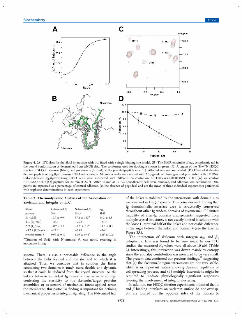
Integrin. Previously, we defined skelemin binding surface onplatelet integrin αIIbβ3 and demonstrated that this interaction isconsistent with an attenuated intersubunit clasp.11 To addressthe mechanism of this interaction, and to define thethermodynamic forces driving the process, we have employedITC. We used Sk4 and Sk45 constructs titrated with either full-length αIIb cytoplasmic tail (Figure 4A, Figure S1.B) or shortsynthetic peptides corresponding to β3 N- or C-termini (FigureS1.A), as previously described.11 The results, summarized inTable 2, revealed very weak interactions which were in the tensof micromolar range. These reactions were predominantlydriven by entropy. For all cases, the stoichiometry ofinteractions was found to be one.
Because the interaction of αIIb with skelemin is weak with fastoff-rate, we were able to perform the transferred NOEexperiments on αIIb/Sk45 solution. With trNOE constraintssupplied to ARIA, we calculated the structure of the boundpeptide. Upon binding, the peptide adopts a U-shaped structure(ensemble depicted in Figure 4B). From the HSQC titrationexperiments, we found that residues 1361, 1363, and 1394 formthe binding site for αIIb on the Sk45 surface. The best NMRstructures of αIIb and Sk45 were used for in silica docking withHADDOCK software. After the docking of 1000 structures, thebest 200 models were refined in water, and among them thefive clusters were identified by HADDOCK. The largest clusterwith the lowest score had 114 models with RMSD 4.1 Å fromthe overall lowest energy model.We performed trNOE experiments for the short synthetic β3
peptides earlier and found a very limited number of additionalpeaks, preventing us from determining the structure of the
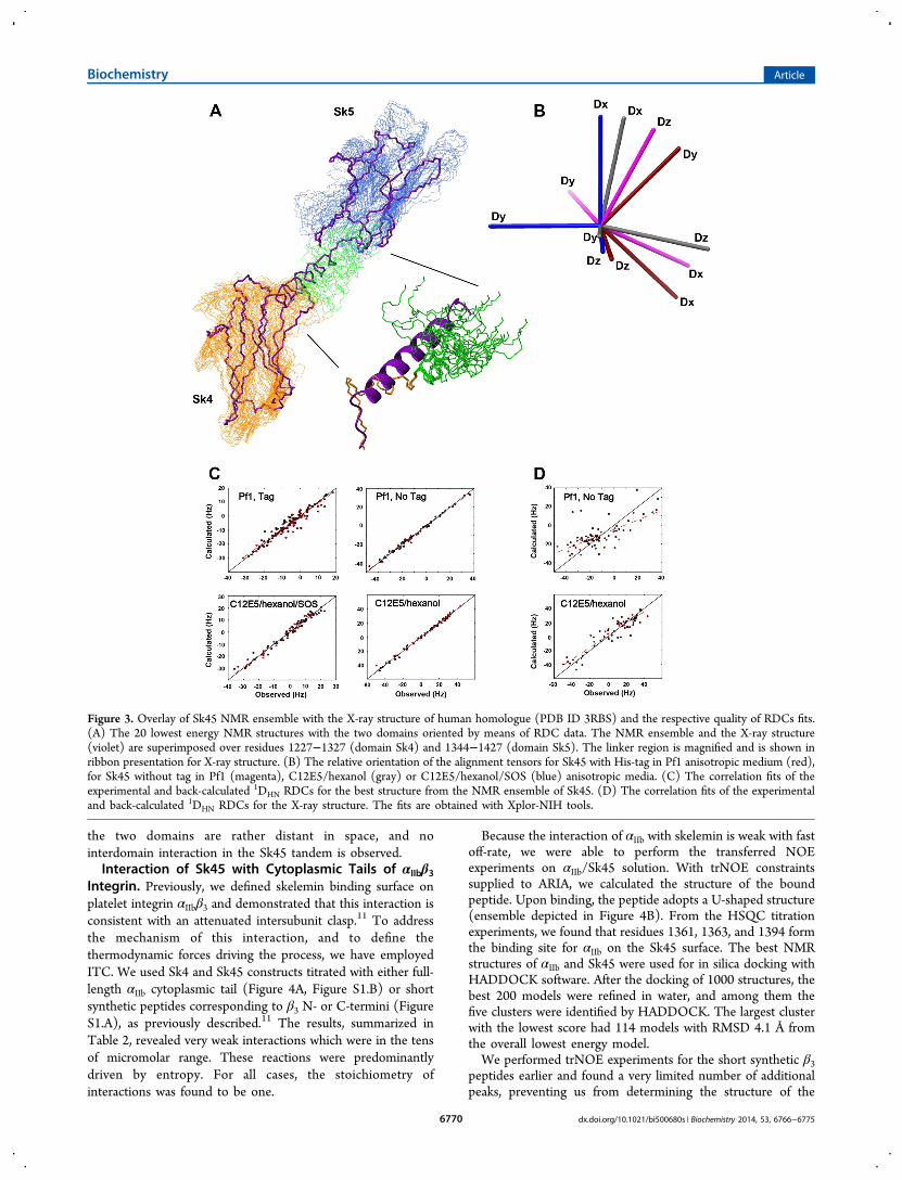
Figure 3. Overlay of Sk45 NMR ensemble with the X-ray structure of human homologue (PDB ID 3RBS) and the respective quality of RDCs fits.(A) The 20 lowest energy NMR structures with the two domains oriented by means of RDC data. The NMR ensemble and the X-ray structure(violet) are superimposed over residues 1227−1327 (domain Sk4) and 1344−1427 (domain Sk5). The linker region is magnified and is shown inribbon presentation for X-ray structure. (B) The relative orientation of the alignment tensors for Sk45 with His-tag in Pf1 anisotropic medium (red),for Sk45 without tag in Pf1 (magenta), C12E5/hexanol (gray) or C12E5/hexanol/SOS (blue) anisotropic media. (C) The correlation fits of theexperimental and back-calculated 1DHN RDCs for the best structure from the NMR ensemble of Sk45. (D) The correlation fits of the experimentaland back-calculated 1DHN RDCs for the X-ray structure. The fits are obtained with Xplor-NIH tools.
Biochemistry Article
dx.doi.org/10.1021/bi500680s | Biochemistry 2014, 53, 6766−67756770
bound β3 peptides.11 Here, we studied β3 interaction with Sk45
by HSQC titration experiments performed on the 15N-labeledSk45 sample. In these experiments, we were unable to observeany reliable chemical shift perturbations in titration with C-terminal β3 peptide, most probably due to the repelling effect ofthe flexible unstructured N-terminus of Sk45. However, weobserved concentration-dependent chemical shifts (Figure 4C,Figure S2, supporting Information) and found that residues1368, 1370, 1372, 1374, 1382−1384, and 1411 of Sk45 areaffected by N-terminal β3 peptide binding. These residues wereused for in silica docking of the β3 conformer (PDB ID 1M80)into the NMR structure of Sk45 presented here. The bestHADDOCK cluster contained 17 models with RMSD 0.9 Åfrom the overall lowest energy model.The combined docking model of Sk45 tertiary complex with
integrin cytoplasmic tails is presented in Figure 5 and discussedbelow. The data used to generate the model are summarized inTable S1.To prove the relevance of the generated model interface in
vivo, and to further assess the role of the identified binding sitefor the integrin αIIbβ3, we synthesized the peptide THIVW-YKDEREISVDEKHD, and tested the effect of this peptide onthe adhesion of CHO cells expressing αIIbβ3 to immobilizedfibrinogen. This peptide represents the two full-length β-strandsof the domain 5 β-sandwich of skelemin that come into contactwith the membrane-proximal region of β3 integrin, according toour model. This peptide was considered to be stable enough tomaintain a hairpin sort of structure through the internalhydrogen bonds in the absence of the rest β-strands. Increasingthe concentration of the peptide progressively blocked celladhesion in dose-dependent manner, where the IC50 wasdetermined to be 33 μM as shown in Figure 4D. The inhibitionwas specific as the control peptide, in which VDE residues weremutated to AAA, did not appear to have any effect on celladhesion.
■ DISCUSSION
As a family of major cell adhesion receptors, integrins uniquelycombine bidirectional signaling capabilities with the structuralfunctions of linking extracellular matrix proteins to thecytoskeleton. With a myriad of potential intracellular targets,all these functions could be accomplished only through spatiallyand temporally controlled interactions. We have investigatedhow major platelet integrin, αIIbβ3, binds to skelemin, acytoskeletal protein found in nonmuscle cell focal complexesduring the earlier phases of cell spreading.We solved the NMR structure of the skelemin tandem
domains with the help of RDC data from the four alignmentmedia for orienting the Sk45 two domains in a solution withthe “rigid body” approach. Figure 3A depicts the superpositionof the representative ensemble of Sk45 NMR structures withthe X-ray structure of human myomesin-1 for comparison(PDB ID 3RBS, shown in violet with thicker bonds).13 Wehave back-calculated the RDCs for the crystal structure of themyomesin-1 domains 10 and 11 and found no correlation withRDCs set for the Pf1 medium (with 0.69 correlation coefficientof this fit) as well as a rather poor correlation (correlationcoefficient of 0.93 in comparison to 1.00 of NMR ensemble)for the C12E5/hexanol alignment medium (Figure 3D),suggesting a notable deviation from the ensemble in thesolution. Not surprisingly, the major differences were foundwithin the linker connecting domains 4 and 5 (shown zoomedin the inset of Figure 3A with Sk4 domain used forsuperimposition). While the X-ray structure suggests thepresence of well-defined straight helix through-out this region,this is not the case in this solution. We observed that only theN-terminal half of the linker, connected to domain 4, forms aregular α-helix. The second C-terminal half of the linker wasquite loose, with a number of NMR resonances broadened outdue to conformational heterogeneity and no evidence forregular α-helical characteristic (i, i+3) connections in NOESY
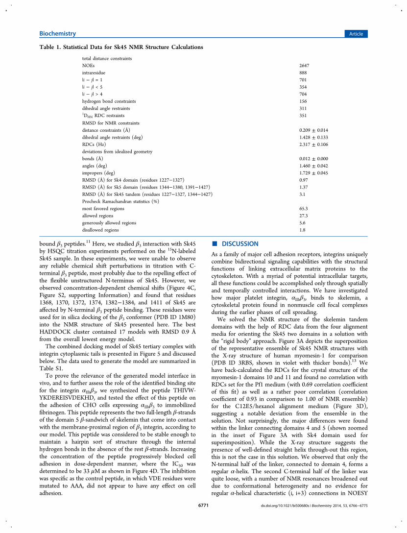
Table 1. Statistical Data for Sk45 NMR Structure Calculations
total distance constraintsNOEs 2647intraresidue 888|i − j| = 1 701|i − j| < 5 354|i − j| > 4 704hydrogen bond constraints 156dihedral angle restraints 3111DHN RDC restraints 351
RMSD for NMR constraintsdistance constraints (Å) 0.209 ± 0.014dihedral angle restraints (deg) 1.428 ± 0.133RDCs (Hz) 2.317 ± 0.106deviations from idealized geometrybonds (Å) 0.012 ± 0.000angles (deg) 1.460 ± 0.042impropers (deg) 1.729 ± 0.045RMSD (Å) for Sk4 domain (residues 1227−1327) 0.97RMSD (Å) for Sk5 domain (residues 1344−1380, 1391−1427) 1.37RMSD (Å) for Sk45 tandem (residues 1227−1327, 1344−1427) 3.1Procheck Ramachandran statistics (%)most favored regions 65.3allowed regions 27.3generously allowed regions 5.6disallowed regions 1.8
Biochemistry Article
dx.doi.org/10.1021/bi500680s | Biochemistry 2014, 53, 6766−67756771
spectra. There is also a noticeable difference in the anglebetween the helix formed and the β-strand to which it isattached. Thus, we conclude that in solution the linkerconnecting two domains is much more flexible and dynamicso that it could be deduced from the crystal structure. As thelinkers between individual Ig domains may serve as springs,conferring the elasticity to the skelemin/target proteinsassemblies, or as sensors of mechanical forces applied acrossthe membrane, this particular finding is important for definingmechanical properties in integrin signaling. The N-terminal half
of the linker is stabilized by the interactions with domain 4 aswe observed in HSQC spectra. This coincides with finding thatIg domain/helix interface area is structurally conservedthroughout other Ig tandem domains of myomesin-1.13 Limitedflexibility of inter-Ig domains arrangements, suggested frommultiple crystal structures, is not exactly limited in solution withthe loose C-terminal half of the linker and noticeable differencein the angle between the linker and domain 4 (see the inset inFigure 3A).The interaction of skelemin with integrin αIIb and β3
cytoplasmic tails was found to be very weak. In our ITCstudies, the measured Kd values were all above 10 μM (Table2). Interestingly, this interaction was driven mainly by entropysince the enthalpy contribution was measured to be very small.The present data confirmed our previous findings,11 suggestingthat (i) the skelemin/integrin interactions are not very stable,which is an important feature allowing dynamic regulation ofcell spreading process, and (ii) multiple interactions might berequired to mediate physiologically significant responsesfavoring the involvement of integrin clustering.In addition, our HSQC titration experiments indicated that α
and β binding interfaces on skelemin surface do not overlap,but are located on the opposite sides of the domain 5,
Figure 4. (A) ITC data for the Sk45 interaction with αIIb fitted with a single binding site model. (B) The NMR ensemble of αIIb cytoplasmic tail inthe bound conformation as determined from trNOE data. The conformer used for docking is shown in green. (C) A region of the 1H−15N HSQCspectra of Sk45 in absence (black) and presence of β3 (red) at the protein/peptide ratio 1:3. Affected residues are labeled. (D) Effect of skelemin-derived peptide on αIIbβ3-expressing CHO cell adhesion. Microtiter wells were coated with 2.5 μg/mL of fibrinogen and postcoated with 1% BSA.Calcein-labeled αIIbβ3-expressing CHO cells were incubated with different concentration of THIVWYKDEREISVDEKHD (•) or controlEREISAAAKHD (○) peptides for 20 min at 22 °C. After 30 min at 37 °C, nonadherent cells were removed, and adhesion was determined. Datapoints are expressed as a percentage of control adhesion (in the absence of peptides) and are the mean of three individual experiments performedwith triplicate determinations in each experiment.
Table 2. Thermodynamic Analysis of the Association ofSkelemin and Integrin by ITC
titrant C-terminal β3 N-terminal β3 αIIbprotein Sk4 Sk45 Sk45Kd (μM) 10.7 ± 9.9 37.2 ± 180a 14.2 ± 4.3ΔG (kJ/mol) −28.3 −25.3 −27.7ΔH (kJ/mol) −0.7 ± 0.1 −1.7 ± 0.9a −1.6 ± 0.1−TΔS (kJ/mol) −27.6 −23.6 −26.1stoichiometry, n 0.99 ± 0.10 1.30 ± 0.41a 1.26 ± 0.05
aTitration of Sk45 with N-terminal β3 was noisy, resulting ininaccurate fitting.
Biochemistry Article
dx.doi.org/10.1021/bi500680s | Biochemistry 2014, 53, 6766−67756772
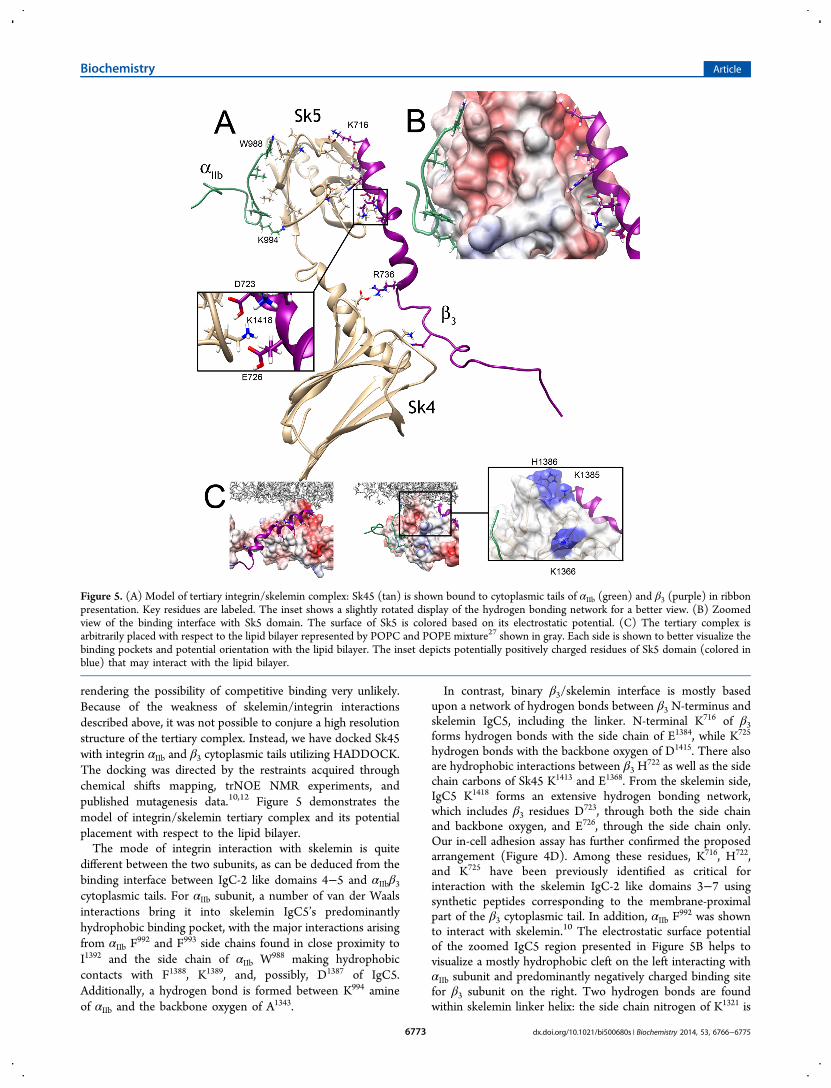
rendering the possibility of competitive binding very unlikely.Because of the weakness of skelemin/integrin interactionsdescribed above, it was not possible to conjure a high resolutionstructure of the tertiary complex. Instead, we have docked Sk45with integrin αIIb and β3 cytoplasmic tails utilizing HADDOCK.The docking was directed by the restraints acquired throughchemical shifts mapping, trNOE NMR experiments, andpublished mutagenesis data.10,12 Figure 5 demonstrates themodel of integrin/skelemin tertiary complex and its potentialplacement with respect to the lipid bilayer.The mode of integrin interaction with skelemin is quite
different between the two subunits, as can be deduced from thebinding interface between IgC-2 like domains 4−5 and αIIbβ3cytoplasmic tails. For αIIb subunit, a number of van der Waalsinteractions bring it into skelemin IgC5’s predominantlyhydrophobic binding pocket, with the major interactions arisingfrom αIIb F
992 and F993 side chains found in close proximity toI1392 and the side chain of αIIb W988 making hydrophobiccontacts with F1388, K1389, and, possibly, D1387 of IgC5.Additionally, a hydrogen bond is formed between K994 amineof αIIb and the backbone oxygen of A1343.
In contrast, binary β3/skelemin interface is mostly basedupon a network of hydrogen bonds between β3 N-terminus andskelemin IgC5, including the linker. N-terminal K716 of β3forms hydrogen bonds with the side chain of E1384, while K725
hydrogen bonds with the backbone oxygen of D1415. There alsoare hydrophobic interactions between β3 H
722 as well as the sidechain carbons of Sk45 K1413 and E1368. From the skelemin side,IgC5 K1418 forms an extensive hydrogen bonding network,which includes β3 residues D
723, through both the side chainand backbone oxygen, and E726, through the side chain only.Our in-cell adhesion assay has further confirmed the proposedarrangement (Figure 4D). Among these residues, K716, H722,and K725 have been previously identified as critical forinteraction with the skelemin IgC-2 like domains 3−7 usingsynthetic peptides corresponding to the membrane-proximalpart of the β3 cytoplasmic tail. In addition, αIIb F
992 was shownto interact with skelemin.10 The electrostatic surface potentialof the zoomed IgC5 region presented in Figure 5B helps tovisualize a mostly hydrophobic cleft on the left interacting withαIIb subunit and predominantly negatively charged binding sitefor β3 subunit on the right. Two hydrogen bonds are foundwithin skelemin linker helix: the side chain nitrogen of K1321 is
Figure 5. (A) Model of tertiary integrin/skelemin complex: Sk45 (tan) is shown bound to cytoplasmic tails of αIIb (green) and β3 (purple) in ribbonpresentation. Key residues are labeled. The inset shows a slightly rotated display of the hydrogen bonding network for a better view. (B) Zoomedview of the binding interface with Sk5 domain. The surface of Sk5 is colored based on its electrostatic potential. (C) The tertiary complex isarbitrarily placed with respect to the lipid bilayer represented by POPC and POPE mixture27 shown in gray. Each side is shown to better visualize thebinding pockets and potential orientation with the lipid bilayer. The inset depicts potentially positively charged residues of Sk5 domain (colored inblue) that may interact with the lipid bilayer.
Biochemistry Article
dx.doi.org/10.1021/bi500680s | Biochemistry 2014, 53, 6766−67756773
connected to β3 N744 while E1328 oxygen interacts with the side
chain amine of R736. Because we have not observed thechemical shift perturbations for domain 4 in our titrationexperiments, no restraints linking β3 C-terminus to IgC4 wereintroduced during the HADDOCK docking. Not surprisingly,the model demonstrates no major interactions between IgC4and β3, with only a couple of hydrophobic contacts foundbetween side chains of IgC4 residues E1293 and N1294 and β3A750.Previously, we structurally characterized αIIbβ3 cytoplasmic
heterodimer, which plays an important role in maintainingintegrin in its latent state.17 Superimposition of our tertiarymodel with αIIbβ3 heterodimer reveals steric clashes, making itextremely difficult, if not impossible, for both the subunits tointeract with skelemin in the presence of intersubunit clasp.Since some of the αIIb and β3 residues critical for skeleminbinding (αIIbF
922 and β3H722) are also involved in the formation
of the αIIbβ3 clasp,17 it appears that unclasping of the tails is
prerequisite for skelemin binding. Although we have previouslydemonstrated that αIIbβ3 does not interact with skelemin inresting platelets, it is recruited to the receptor during plateletadhesion and upon activation with agonists. Considering thefact that cytoplasmic tails of αIIbβ3 have a higher mutual affinity(of 5.7 μM as measured by ITC, Figure S1.C) than any singlesubunit to skelemin (Table 2), this observation supports thenotion that skelemin is not an integrin activator, but ratherserves as a modulator of integrin attachment to Src orcytoskeleton.In Figure 5C, we have positioned the tertiary complex with
respect to the lipid bilayer. As presented, skelemin Ig domains 4and 5 may be placed parallel to the membrane surface. Withthis arrangement, αIIb subunit comes out of the membranealmost perpendicularly with its W988 side chain making contactswith lipid head-groups at the membrane-cytoplasm interface.Integrin β3 subunit, on other hand, is arranged at a sharp acuteangle with its K716 side chain making contacts with negativelycharged patch on the surface of IgC5 which sticks out as beingrepelled by negatively charged inner leaflet of the lipid bilayer.In the inset of Figure 5C, we have also marked IgC5 positivelycharged residues, which potentially may interact with negativelycharged lipids. The rest of IgC5 negative patch is arranged tointeract with either the sixth IgC-2 like domain of skelemin orwith any other potential target having positively chargedsolvent exposed surface.To conclude, we have determined a three-dimensional
structure of tandem IgC2-like domains 4 and 5 of skelemin,connected through a stretchable helix-containing linker, byNMR. This linker region was found to be restrained enough todefine the mutual orientation of the two domains tumbling as asingle unit in solution, although it is less more dynamic thansuggested by earlier X-ray studies of the human homologuemyomesin-1. We have shown that interaction between skeleminand integrin cytoplasmic domain is weak and predominantlyentropy driven. This is consistent with the findings thatskelemin is unable to activate the receptors. We have built atertiary model of αIIbβ3 integrin cytoplasmic tails complexedwith the tandem domains of skelemin and validated this modelwith cellular adhesion assays. While some of the interactionsbetween K716 side chain of β3 tail and skelemin may occur inthe presence of αIIb subunit still bound to β3, disruption ofmembrane-proximal α/β clasp and release of αIIb F
992 and β3H722/D723/E726 side chains is necessary for αIIb to bind toskelemin and for β3 to stabilize its interface with skelemin.
Thus, our model favors skelemin role as a stabilizer for theactivated integrins and integrin clusters by connecting α and βsubunits from adjacent receptors.
■ ASSOCIATED CONTENT*S Supporting InformationTable S1 with the summary of residues from both, skeleminand integrin sites, used for docking, and two figures: Figure S1,presenting the examples of ITC thermograms and respectivebinding curves, and Figure S2, showing the graph of chemicalshift perturbations and the affected surface area of Sk45 upontitration with N-terminal β3 peptide. This material is availablefree of charge via the Internet at http://pubs.acs.org.
■ AUTHOR INFORMATIONCorresponding Author*Address: Department of Pharmaceutical Sciences, 69 NorthEagleville Rd, Unit 3092, University of Connecticut at Storrs,Storrs, CT 06269-3092, Phone: (860) 486-2972, Fax: (860)486-6857, E-mail: [email protected] Address⊥(L.D.) Laboratory of Chemical Physics, National Institute ofDiabetes and Digestive and Kidney Diseases, NIH, Bethesda,MD 20892−0520FundingThis work was supported in part by AHA 0855768D to O.V.,AHA 0835257N to N.P.P, and NIH HL 63199 to T.P.U.NotesThe authors declare no competing financial interest.
■ ACKNOWLEDGMENTSWe would like to thank Drs. Sergiy Tyukhtenko from NorthEastern University, Mark Maciejewski from University ofConnecticut Health Center, and G.T.V. Swapna from RutgersUniversity for their help with data acquisition at high fieldNMR spectrometers. The WeNMR project (European FP7 e-Infrastructure grant, Contract No. 261572, www.wenmr.eu),supported by the European Grid Initiative (EGI) through thenational GRID Initiatives of Belgium, France, Italy, Germany,The Netherlands, Poland, Portugal, Spain, UK, South Africa,Malaysia, Taiwan, the Latin America GRID infrastructure viathe Gisela project and the US Open Science Grid (OSG) areacknowledged for the use of web portals, computing, andstorage facilities.
■ ABBREVIATIONSC12E5, pentaethylene glycol monododecyl ether; DTT,dithiothreitol; ECM, extracellular matrix; FA, focal adhesions;FN, fibronectin; FX, focal complex; IgC2, immunoglobulin C2-like domain; ITC, isothermal titration calorimetry; m-PROXYL, 3-maleimido-PROXYL; NMR, nuclear magneticresonance spectroscopy; NOE, nuclear Overhauser effect;PMSF, phenylmethanesufonyl fluoride; PRE, paramagneticrelaxation enhancement; RDC, residual dipolar coupling;SOS, sodium octyl sulfate; Tris, Tris(hydroxymethyl)-aminomenthane; trNOE, transferred nuclear Overhauser effect
■ REFERENCES(1) Hynes, R. O. (1992) Integrins: versatility, modulation, andsignaling in cell adhesion. Cell 69, 11−25.(2) Hynes, R. O. (2002) Integrins: bidirectional, allosteric signalingmachines. Cell 110, 673−687.
Biochemistry Article
dx.doi.org/10.1021/bi500680s | Biochemistry 2014, 53, 6766−67756774
(3) Geiger, B., and Bershadsky, A. (2001) Assembly andmechanosensory function of focal contacts. Curr. Opin. Cell Biol. 13,584−592.(4) Webb, D. J., Parsons, J. T., and Horwitz, A. F. (2002) Adhesionassembly, disassembly and turnover in migrating cells – over and overand over again. Nat. Cell Biol. 4, E97−100.(5) Steiner, F., Weber, K., and Furst, D. O. (1999) M band proteinsmyomesin and skelemin are encoded by the same gene: analysis of itsorganization and expression. Genomics 56, 78−89.(6) Price, M. G., and Gomer, R. H. (1993) Skelemin, a cytoskeletalM-disc periphery protein, contains motifs of adhesion/recognition andintermediate filament proteins. J. Biol. Chem. 268, 21800−21810.(7) Pinotsis, N., Lange, S., Perriard, J. C., Svergun, D. I., andWilmanns, M. (2008) Molecular basis of the C-terminal tail-to-tailassembly of the sarcomeric filament protein myomesin. EMBO J. 27,253−264.(8) Reddy, K. B., Bialkowska, K., and Fox, J. E. (2001) Dynamicmodulation of cytoskeletal proteins linking integrins to signalingcomplexes in spreading cells. Role of skelemin in initial integrin-induced spreading. J. Biol. Chem. 276, 28300−28308.(9) Reddy, K. B., Gascard, P., Price, M. G., Negrescu, E. V., and Fox,J. E. (1998) Identification of an interaction between the m-bandprotein skelemin and beta-integrin subunits. Colocalization of askelemin-like protein with beta1- and beta3-integrins in non-musclecells. J. Biol. Chem. 273, 35039−35047.(10) Podolnikova, N. P., O’Toole, T. E., Haas, T. A., Lam, S. C., Fox,J. E., and Ugarova, T. P. (2009) Adhesion-induced unclasping ofcytoplasmic tails of integrin alpha(IIb)beta3. Biochemistry 48, 617−629.(11) Deshmukh, L., Tyukhtenko, S., Liu, J., Fox, J. E., Qin, J., andVinogradova, O. (2007) Structural insight into the interaction betweenplatelet integrin alphaIIbbeta3 and cytoskeletal protein skelemin. J.Biol. Chem. 282, 32349−32356.(12) Li, X., Liu, Y., and Haas, T. A. (2013) Skelemin in integrinalpha(IIb)beta(3) mediated cell spreading. Biochemistry 52, 681−689.(13) Pinotsis, N., Chatziefthimiou, S. D., Berkemeier, F., Beuron, F.,Mavridis, I. M., Konarev, P. V., Svergun, D. I., Morris, E., Rief, M., andWilmanns, M. (2012) Superhelical architecture of the myosin filament-linking protein myomesin with unusual elastic properties. PLoS Biol.10, e1001261.(14) Podolnikova, N. P., Yakubenko, V. P., Volkov, G. L., Plow, E. F.,and Ugarova, T. P. (2003) Identification of a novel binding site forplatelet integrins alpha IIb beta 3 (GPIIbIIIa) and alpha 5 beta 1 in thegamma C-domain of fibrinogen. J. Biol. Chem. 278, 32251−32258.(15) Delaglio, F., Grzesiek, S., Vuister, G. W., Zhu, G., Pfeifer, J., andBax, A. (1995) NMRPipe: a multidimensional spectral processingsystem based on UNIX pipes. J. Biomol. NMR 6, 277−293.(16) Vranken, W. F., Boucher, W., Stevens, T. J., Fogh, R. H., Pajon,A., Llinas, M., Ulrich, E. L., Markley, J. L., Ionides, J., and Laue, E. D.(2005) The CCPN data model for NMR Spectroscopy. Proteins 59,687−696.(17) Vinogradova, O., Velyvis, A., Velyviene, A., Hu, B., Haas, T.,Plow, E., and Qin, J. (2002) A structural mechanism of integrinalpha(IIb)beta(3) “inside-out” activation as regulated by itscytoplasmic face. Cell 110, 587−597.(18) Rieping, W., Habeck, M., Bardiaux, B., Bernard, A., Malliavin, T.E., and Nilges, M. (2007) ARIA2: automated NOE assignment anddata integration in NMR structure calculation. Bioinformatics 23, 381−382.(19) Schwieters, C. D., Kuszewski, J. J., Tjandra, N., and Clore, G. M.(2003) The Xplor-NIH NMR molecular structure determinationpackage. J. Magn. Reson. 160, 65−73.(20) Laskowski, R. A., Rullmannn, J. A., MacArthur, M. W., Kaptein,R., and Thornton, J. M. (1996) AQUA and PROCHECK-NMR:programs for checking the quality of protein structures solved byNMR. J. Biomol NMR 8, 477−486.(21) de Vries, S. J., van Dijk, M., and Bonvin, A. M. (2010) TheHADDOCK web server for data-driven biomolecular docking. Nat.Protoc. 5, 883−897.
(22) Baker, N. A., Sept, D., Joseph, S., Holst, M. J., and McCammon,J. A. (2001) Electrostatics of nanosystems: application to microtubulesand the ribosome. Proc. Natl. Acad. Sci. U. S. A. 98, 10037−10041.(23) Dolinsky, T. J., Czodrowski, P., Li, H., Nielsen, J. E., Jensen, J.H., Klebe, G., and Baker, N. A. (2007) PDB2PQR: expanding andupgrading automated preparation of biomolecular structures formolecular simulations. Nucleic Acids Res. 35, W522−525.(24) Pettersen, E. F., Goddard, T. D., Huang, C. C., Couch, G. S.,Greenblatt, D. M., Meng, E. C., and Ferrin, T. E. (2004) UCSFChimera–a visualization system for exploratory research and analysis. J.Comput. Chem. 25, 1605−1612.(25) Al-Hashimi, H. M., Valafar, H., Terrell, M., Zartler, E. R.,Eidsness, M. K., and Prestegard, J. H. (2000) Variation of molecularalignment as a means of resolving orientational ambiguities in proteinstructures from dipolar couplings. J. Magn. Reson. 143, 402−406.(26) Dosset, P., Hus, J. C., Marion, D., and Blackledge, M. (2001) Anovel interactive tool for rigid-body modeling of multi-domainmacromolecules using residual dipolar couplings. J. Biomol NMR 20,223−231.(27) Gurtovenko, A. A., and Vattulainen, I. (2007) Lipid trans-membrane asymmetry and intrinsic membrane potential: two sides ofthe same coin. J. Am. Chem. Soc. 129, 5358−5359.
Biochemistry Article
dx.doi.org/10.1021/bi500680s | Biochemistry 2014, 53, 6766−67756775