Sintering effects on the hardness of β-tricalcium...
5
Ceramic Processing Research Sintering effects on the hardness of β-tricalcium phosphate Introduction
Transcript of Sintering effects on the hardness of β-tricalcium...
Journal of Ceramic Processing Research. Vol. 13, No. 4, pp. 486~490 (2012)
486
J O U R N A L O F
CeramicProcessing Research
Sintering effects on the hardness of β-tricalcium phosphate
Behzad Mehdikhania,*, Bahman Mirhadib and Nayereh Askaria
aStandard Research Institute, Ceramic Department, Karaj, IranbImam Khomeini International University, Engineering Department, Qavin, Iran
Nano-size β-tricalcium phosphate (ß-TCP) powders with an average grain size of 70-100 nm were prepared by the wetchemical precipitation method with calcium nitrate and di-ammonium hydrogen phosphate as calcium and phosphorus
precursors, respectively. The precipitation process employed was also found to be suitable for the production of sub-micrometre β-TCP powder in situ. The sinterability of the nano-size powders, and the microstructure, mechanical strength of
the β-TCP bioceramics prepared were investigated. Bioceramic sample characterization was achieved by powder X-raydiffraction (XRD), scanning electron microscopy (SEM), Fourier transform infrared spectroscopy (FTIR) and density
measurements. Powders compacted and sintered at 800, 900, 1000 and 1100 οC showed an increase in relative density from68% to 93%. The results revealed that the maximum hardness of 240 HV was obtained for β-TCP sintered at 1100 οC.
Key words: Beta tricalcium phosphate, Bioceramic, Sintering, Mechanical properties.
Introduction
Calcium phosphate bioceramics are materials of
choice for bone tissue repair because of their
similarity of composition with bone mineral; excellent
bioactivity; ability to promote cellular expressions;
and osteoconductivity [1-3]. The calcination constitutes
a necessary step of the preparation when wet chemical
routes are used for the synthesis of calcium phosphate,
i.e. precipitation from the neutralization of Ca(OH)2
with H3PO4 or from the decomposition of Ca(NO3)2
and (NH4)2HPO4. Moreover, an analysis of particle
growth during calcination is required to understanding
of grain growth phenomena that occur during the final
stages of the densification [4]. β-TCP ceramic is
known as β-whitlockite and is a slow degrading
resorbable phase [5] and is thus a promising material
in biomedical applications. β-TCP is known to have
significant biological affinity, activity and hence
responds well to physiological environments [6].
Because of these positive characteristics, porous β-
TCP is regarded as an ideal bone substitute, which
would degrade in vivo with time allowing bone tissue
to grow inside the scaffold [7]. TCP has three
polymorphs, ß-TCP is stable below 1180 οC, α-TCP
between 1180 οC and 1400 οC, and α -TCP above
1470 οC. Among the three allotropic forms, β-TCP is
preferred as a bioceramic on account of its chemical
stability, mechanical strength, and proper bioresorption
rate. To use β-TCP ceramics as surgical implants, the
mechanical strength of β-TCP ceramics must be as
high as possible. The density of β-TCP ceramics is also
an important factor. Generally, it is difficult to sinter β-
TCP ceramics fully because β-TCP ceramics should be
sintered at a lower temperature than that of the phase
transition to α-TCP [8]. TCP is a resorbable temporary
bone space filler material. When implanted, TCP will
interact with body fluids and form HA in accordance to
the following equation:
4Ca3(PO4)2+2H2O → Ca10(PO4)6(OH)2(surface) + 2Ca2 + 2HPO42-
(1)
Theoretically, resorbable TCP is an ideal implant
material. After implantation, TCP will degrade with
time and be replaced with natural tissues. This leads
to the regeneration of tissues instead of their
replacement and so solves the problem of interfacial
stability [9, 10]. β-TCP powders are reportedly
prepared by liquid-solution methods, such as sol-gel,
hydrothermal, micro emulsion and precipitation, as
well as gas phase reactions [11-13]. β-TCP is
routinely used as a bone replacement, especially in the
field of oral and craniofacial surgery, in the form of
granules and rods [14, 15] or as filler in polymeric
scaffolds [16]. In the bulk, β-TCP bioceramics have
mechanical properties too poor to be used in load-
bearing clinical applications [17-19], which has been
attributed to the difficulties in fully densifying β-TCP
powders [18, 20-22]. These difficulties are associated
with the presumption that the sintering temperature
should be kept below 1125 οC to avoid the ß → α
phase transformation that is considered deleterious to
*Corresponding author: Tel : 00989126417516Fax: 00982413230496E-mail: [email protected]
Sintering effects on the hardness of β-tricalcium phosphate 487
mechanical properties. Material properties deemed
particularly important when designing calcium phosphates
with improved resorption are surface characteristics,
such as: roughness, grain size and porosity, and
chemical properties, in particular phase composition. It
is possible to use different sintering regimes to change
the surface roughness, grain size and density for
example by using high and low temperatures [23],
however the use of high sintering temperatures causes
unwanted phase changes to occur (e.g. β-TCP to α-
TCP) [24].One of the critical controlling parameter that
requires attention during the processing of β-TCP is the
selection of a suitable sintering method to obtain a
solid and high density β-TCP body that is characterized
by having a fine-grained microstructure [25]. Dense
nano structured bioceramic materials are usually
obtained by pressing and conventional sintering of
nanopowders using pressure assisted methods, such as
hot pressing, hot isostatic pressing, sinter forging, etc
[26-28]. The high sintering temperatures and long
sintering times required for the consolidation of β-TCP
powders often result in extreme grain coarsening and
decomposition of the β-TCP, which is a characteristic
for the conventional sintering methods and results in
the deterioration of the mechanical properties of β-TCP
ceramics [29, 30]. The aim of this study was to
determine the effects of the sintering temperature on
the microstructure and mechanical properties of β-TCP
scaffolds by means of nanoindentation testing. In this
study, β-TCP scaffolds were manufactured through a
wet chemical precipitation process and subsequently
sintered at four different temperatures. Scaffolds were
characterized in terms of their crystallographic phases,
microstructural morphology, grain size, density and
hardness.
Experimental Procedure
Powder synthesisThe precipitation procedures that were used in the
synthesis of β-TCP pure bioceramic powders are
described in detail in the process flowchart given in
Fig. 1. β-TCP nanopowders were synthesized by the
reaction of calcium nitrate tetra-hydrate (Ca(NO3)2 × 4H2O,
98%, Merck) with diammonium hydrogen phosphate
((NH4)2HPO4, 99%, Merck). Briefly, 500 ml of 0.4 mol
(NH4)2HPO4 solution with a pH = 4 was vigorously
stirred at room temperature, and 500 ml of 0.6 mol
Ca(NO3)2 with a pH = 7.3 was added drop wise over
150-200 minute to produce a white precipitate. Throughout
the mixing process the pH of the system was
maintained at pH = 8 by adding of 0.1 M sodium
hydroxide (NaOH, 99%, Merck). The white suspension
obtained was then stirred for 12 h. The synthesized
precipitate was washed with distilled water and then
with 100% ethanol to improve the dispersion
characteristics. The suspension was filtered in a filter
glass with application of mild suction. After filtration
the compact, sticky filter cake, was dried at 80 οC for
24 h. The as-dried powders were crushed by using a
mortar and pestle and calcined in an alumina crucible
at 700 οC for 2 h. The synthesized β-TCP powder was
isostatically pressed at 50 MPa, for 1 minute, resulting
in uniform green compacts, which were sintered at
temperatures from 800, 900, 1000 and 1100 οC in an
air atmosphere for 6 h. The initial heating rate was
20 ο K.minute-1. The β-TCP compact density was
measured using Archimedes’ method. The sintered β-TCP
compacts were tested for microhardness. Microhardness
was measured with a Vicker’s indenter. Samples were
embedded in epoxy and surfaces were ground flat and
polished to a 1 µm finish with diamond paste. Each
sample was indented 10 times with 200 g and 300 g
loads. No effect of load on hardness values was noticed
at these loads.
Characterization of β-TCP bioceramic powdersEvaluation of crystalline phases was investigated by
X-ray diffractometer (Siemens, model D-500) using
CuKα radiation. Silicon powder was used as the
standard material for semi-quantitative analysis of the
precipitated phases. The mean crystallite size (D) was
calculated from XRD line broadening measurements
from the Scherrer equation [31]:
D = 0.89λ / β cosθ (2)
where λ is the wavelength of the Cu Kα radiation
used, β is the full width at the half maximum of the β-
TCP line and θ is the diffraction angle. Infrared spectra
were performed by FTIR Bomem (model MB100,
Quebec, Canada) in the 400-4000 cm-1 wavenumber region.
For infrared spectroscopy, samples were pulverized and
mixed with a given amount of potassium bromide (KBr)
Fig. 1. Chemical precipitation flowchart used in the synthesis of β-TCP pure nanobioceramic powders.
488 Behzad Mehdikhani, Bahman Mirhadi and Nayereh Askari
and pressed into very thin tablets. The Ca/P ratio of the
dried powder was measured by inductively coupled
plasma (ICP) atomic emission spectroscopy (model
Varian). Powder morphology and particle size were
evaluated using a scanning electron microscope (SEM,
VEGA-TESCAN).
Results and Discussion
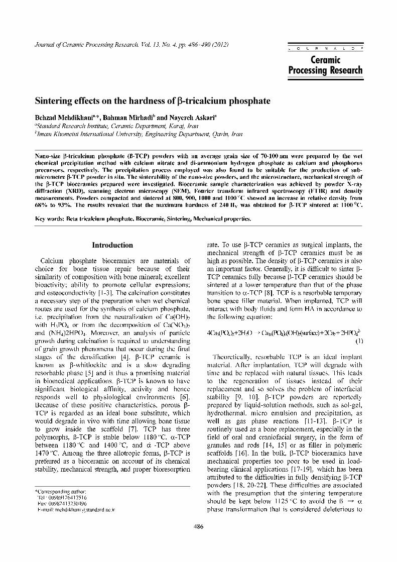
The morphology of the TCP powder precipitated is
shown in Fig. 2. It can be seen that the β-TCP powder
is highly agglomerated with almost spherical particles
having an average size of 70-100 nm.The Ca/P ratio of
the β-TCP powder, as determined by ICPanalyses, was
1.51 ± 0.01. The XRD analysis was performed using
the X-ray diffractometer. The straight base line and
sharp peaks of the diffractogram in Fig. 3 confirmed
that the products were well crystallized. The results
revealed that the as-prepared powder was highly
crystalline ß-TCP will no second phase. The average
crystallite size was determined by the Scherrer
equation, of this sample was 80 nm. In order to identify
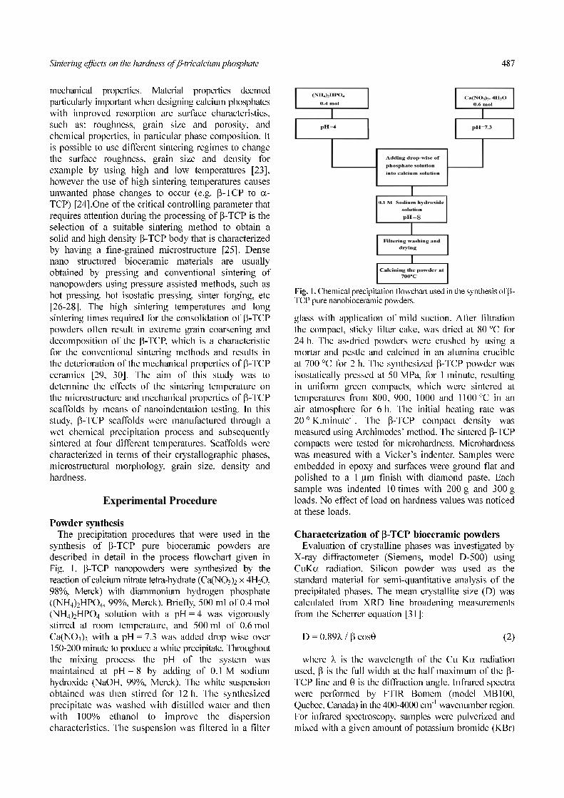
the molecular arrangement of the precipitated powders,
FT-IR analysis was performed. The FTIR spectrum of
the powder is shown in Fig. 4. The characteristic
absorption bands at 3433 and 1631 cm-1 are attributed
to adsorbed water. The bands at 900-1200 cm-1 were
the stretching mode of the PO43- group. The sharp
peaks at 561 and 607 cm-1 represent the vibration peaks
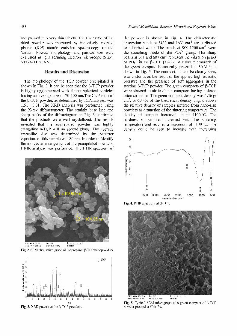
of PO43- in the β-TCP [32-33]. A SEM micrograph of
the green compact isostatically pressed at 50 MPa is
shown in Fig. 5. The compact, as can be clearly seen,
was uniform, as the result of the applied high isostatic
pressure and the presence of soft aggregates in the
starting β-TCP powder. The green compacts of β-TCP
were sintered in air to obtain compacts having a dense
microstructure. The green compact density was 1.36 g/
cm3, or 60.4% of the theoretical density. Fig. 6 shows
the relative density of samples sintered from nano-size
powders as a function of the sintering temperature. The
density of samples increased up to 1100 οC. The
hardness of samples increased with the sintering
temperature and reached a maximum at 1100 οC. The
density could be seen to increase with increasing
Fig. 2. SEM photomicrograph of the prepared β-TCP nanopowders.
Fig. 3. XRD pattern of the β-TCP powders.
Fig. 4. FTIR spectrum of β-TCP.
Fig. 5. Typical SEM micrograph of a green compact of β-TCPpowder pressed at 50 MPa.
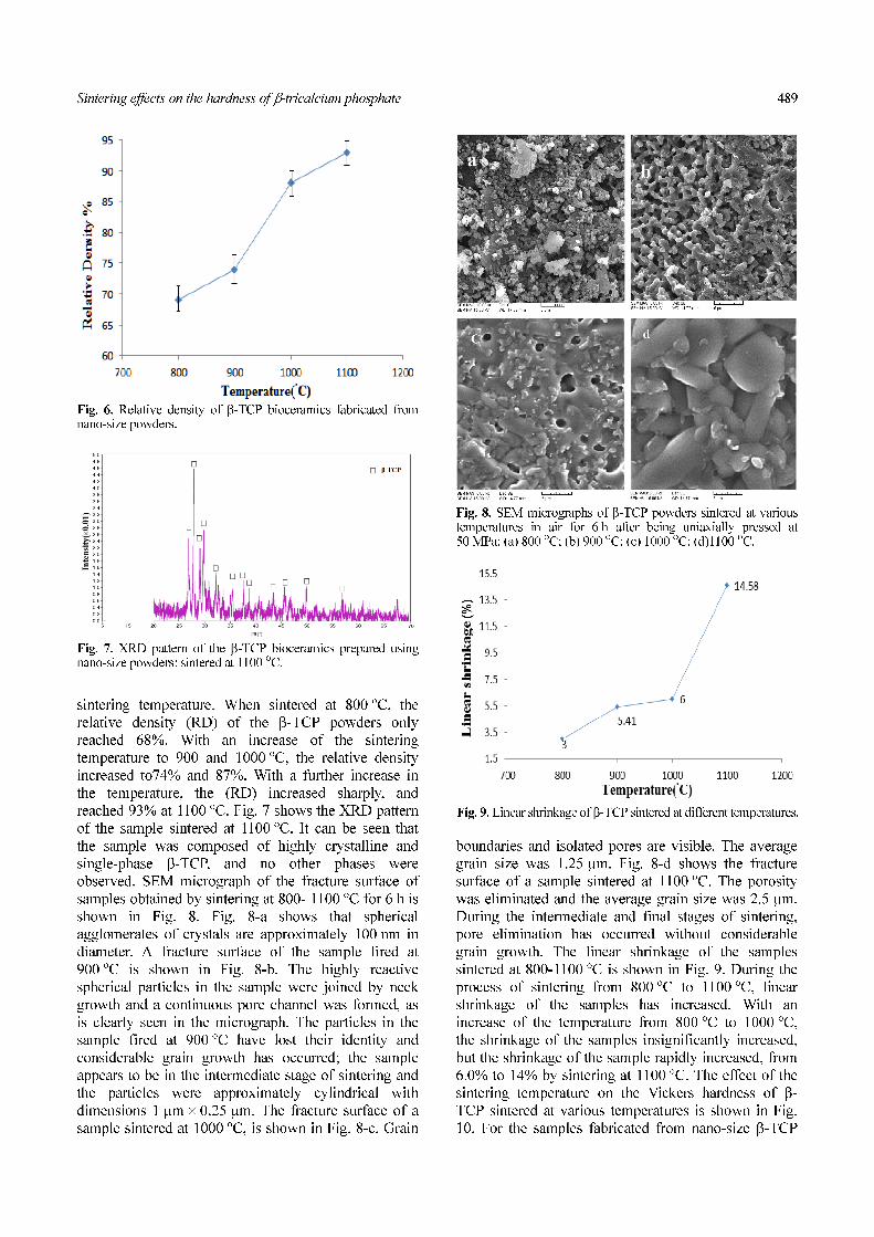
Sintering effects on the hardness of β-tricalcium phosphate 489
sintering temperature. When sintered at 800 οC, the
relative density (RD) of the β-TCP powders only
reached 68%. With an increase of the sintering
temperature to 900 and 1000 οC, the relative density
increased to74% and 87%. With a further increase in
the temperature, the (RD) increased sharply, and
reached 93% at 1100 οC. Fig. 7 shows the XRD pattern
of the sample sintered at 1100 οC. It can be seen that
the sample was composed of highly crystalline and
single-phase β-TCP, and no other phases were
observed. SEM micrograph of the fracture surface of
samples obtained by sintering at 800- 1100 οC for 6 h is
shown in Fig. 8. Fig. 8-a shows that spherical
agglomerates of crystals are approximately 100 nm in
diameter. A fracture surface of the sample fired at
900 οC is shown in Fig. 8-b. The highly reactive
spherical particles in the sample were joined by neck
growth and a continuous pore channel was formed, as
is clearly seen in the micrograph. The particles in the
sample fired at 900 οC have lost their identity and
considerable grain growth has occurred; the sample
appears to be in the intermediate stage of sintering and
the particles were approximately cylindrical with
dimensions 1 µm × 0.25 µm. The fracture surface of a
sample sintered at 1000 οC, is shown in Fig. 8-c. Grain
boundaries and isolated pores are visible. The average
grain size was 1.25 µm. Fig. 8-d shows the fracture
surface of a sample sintered at 1100 οC. The porosity
was eliminated and the average grain size was 2.5 µm.
During the intermediate and final stages of sintering,
pore elimination has occurred without considerable
grain growth. The linear shrinkage of the samples
sintered at 800-1100 οC is shown in Fig. 9. During the
process of sintering from 800 οC to 1100 οC, linear
shrinkage of the samples has increased. With an
increase of the temperature from 800 οC to 1000 οC,
the shrinkage of the samples insignificantly increased,
but the shrinkage of the sample rapidly increased, from
6.0% to 14% by sintering at 1100 οC. The effect of the
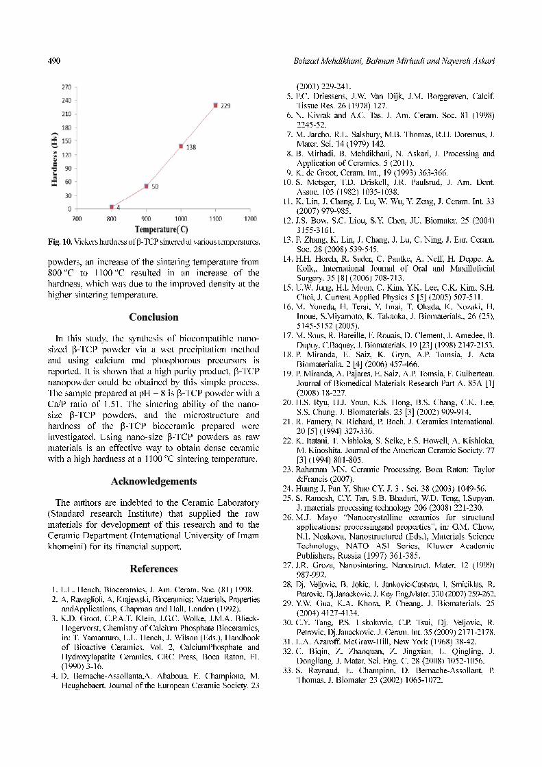
sintering temperature on the Vickers hardness of β-
TCP sintered at various temperatures is shown in Fig.
10. For the samples fabricated from nano-size β-TCP
Fig. 6. Relative density of β-TCP bioceramics fabricated fromnano-size powders.
Fig. 7. XRD pattern of the β-TCP bioceramics prepared usingnano-size powders: sintered at 1100 οC.
Fig. 9. Linear shrinkage of β-TCP sintered at different temperatures.
Fig. 8. SEM micrographs of β-TCP powders sintered at varioustemperatures in air for 6 h after being uniaxially pressed at50 MPa: (a) 800 οC; (b) 900 οC; (c) 1000 οC; (d)1100 οC.
490 Behzad Mehdikhani, Bahman Mirhadi and Nayereh Askari
powders, an increase of the sintering temperature from
800 οC to 1100 οC resulted in an increase of the
hardness, which was due to the improved density at the
higher sintering temperature.
Conclusion
In this study, the synthesis of biocompatible nano-
sized β-TCP powder via a wet precipitation method
and using calcium and phosphorous precursors is
reported. It is shown that a high purity product, β-TCP
nanopowder could be obtained by this simple process.
The sample prepared at pH = 8 is β-TCP powder with a
Ca/P ratio of 1.51. The sintering ability of the nano-
size β-TCP powders, and the microstructure and
hardness of the β-TCP bioceramic prepared were
investigated. Using nano-size β-TCP powders as raw
materials is an effective way to obtain dense ceramic
with a high hardness at a 1100 οC sintering temperature.
Acknowledgements
The authors are indebted to the Ceramic Laboratory
(Standard research Institute) that supplied the raw
materials for development of this research and to the
Ceramic Department (International University of Imam
khomeini) for its financial support.
References
1. L.L. Hench, Bioceramics, J. Am. Ceram. Soc. (81) 1998.2. A. Ravaglioli, A. Krajewski, Bioceramics: Materials, Properties
andApplications, Chapman and Hall, London (1992).3. K.D. Groot, C.P.A.T. Klein, J.G.C. Wolke, J.M.A. Blieck-
Hogervorst, Chemistry of Calcium Phosphate Bioceramics,in: T. Yamamuro, L.L. Hench, J. Wilson (Eds.), Handbookof Bioactive Ceramics, Vol. 2, CalciumPhosphate andHydroxylapatite Ceramics, CRC Press, Boca Raton, FL(1990) 3-16.
4. D. Bernache-Assollanta,A. Ababoua, E. Championa, M.Heughebaert, Journal of the European Ceramic Society. 23
(2003) 229-241.5. F.C. Driessens, J.W. Van Dijk, J.M. Borggreven, Calcif.
Tissue Res. 26 (1978) 127.6. N. Kivrak and A.C. Tas. J. Am. Ceram. Soc. 81 (1998)
2245-52.7. M. Jarcho, R.L. Salsbury, M.B. Thomas, R.H. Doremus, J.
Mater. Sci. 14 (1979) 142.8. B. Mirhadi, B, Mehdikhani, N. Askari, J. Processing and
Application of Ceramics. 5 (2011).9. K. de Groot, Ceram. Int., 19 (1993) 363-366.
10. S. Metsger, T.D. Driskell, J.R. Paulsrud, J. Am. Dent.Assoc. 105 (1982) 1035-1038.
11. K. Lin, J. Chang, J. Lu, W. Wu, Y. Zeng, J. Ceram. Int. 33(2007) 979-985.
12. J.S. Bow, S.C. Liou, S.Y. Chen, JU. Biomater. 25 (2004)3155-3161.
13. F. Zhang, K. Lin, J. Chang, J. Lu, C. Ning, J. Eur. Ceram.Soc. 28 (2008) 539-545.
14. H.H. Horch, R. Sader, C. Pautke, A. Neff, H. Deppe, A.Kolk,, International Journal of Oral and MaxillofacialSurgery. 35 [8] (2006) 708-713.
15. U.W. Jung, H.I. Moon, C. Kim, Y.K. Lee, C.K. Kim, S.H.Choi, J. Current Applied Physics 5 [5] (2005) 507-511.
16. M. Yoneda, H. Terai, Y. Imai, T. Okada, K. Nozaki, H.Inoue, S.Miyamoto, K. Takaoka, J. Biomaterials., 26 (25),5145-5152 (2005).
17. M. Sous, R. Bareille, F. Rouais, D. Clement, J. Amedee, B.Dupuy, C.Baquey, J. Biomaterials. 19 [23] (1998) 2147-2153.
18. P. Miranda, E. Saiz, K. Gryn, A.P. Tomsia, J. ActaBiomaterialia. 2 [4] (2006) 457-466.
19. P. Miranda, A. Pajares, E. Saiz, A.P. Tomsia, F. Guiberteau.Journal of Biomedical Materials Research Part A. 85A [1](2008) 18-227.
20. H.S. Ryu, H.J. Youn, K.S. Hong, B.S. Chang, C.K. Lee,S.S. Chung. J. Biomaterials. 23 [3] (2002) 909-914.
21. R. Famery, N. Richard, P. Boch. J. Ceramics International.20 [5] (1994) 327-336.
22. K. Itatani, T. Nishioka, S. Seike, F.S. Howell, A. Kishioka,M. Kinoshita. Journal of the American Ceramic Society. 77[3] (1994) 801-805.
23. Rahaman MN. Ceramic Processing. Boca Raton: Taylor&Francis (2007).
24. Huang J, Pan Y, Shao CY. J. 3 . Sci. 38 (2003) 1049-56.25. S. Ramesh, C.Y. Tan, S.B. Bhaduri, W.D. Teng, I.Sopyan.
J. materials processing technology 206 (2008) 221-230.26. M.J. Mayo “Nanocrystalline ceramics for structural
applications: processingand properties”, in: G.M. Chow,N.I. Noskova, Nanostructured (Eds.), Materials ScienceTechnology, NATO ASI Series, Kluwer AcademicPublishers, Russia (1997) 361-385.
27. J.R. Groza, Nanosintering, Nanostruct. Mater. 12 (1999)987-992.
28. Dj. Veljovic, B. Jokic, I. Jankovic-Castvan, I. Smiciklas, R.Petrovic, Dj.Janackovic. J. Key Eng.Mater. 330 (2007) 259-262.
29. Y.W. Gua, K.A. Khora, P. Cheang. J. Biomaterials. 25(2004) 4127-4134.
30. C.Y. Tang, P.S. Uskokovic, C.P. Tsui, Dj. Veljovic, R.Petrovic, Dj.Janackovic. J. Ceram. Int. 35 (2009) 2171-2178.
31. L.A. Azaroff. McGraw-Hill, New York (1968) 38-42.32. C. Biqin, Z. Zhaoquan, Z. Jingxian, L. Qingling, J.
Dongliang. J. Mater. Sci. Eng. C. 28 (2008) 1052-1056.33. S. Raynaud, E. Champion, D. Bernache-Assollant, P.
Thomas. J. Biomater 23 (2002) 1065-1072.
Fig. 10. Vickers hardness of β-TCP sintered at various temperatures.