Significance of Blood Cellular lxr- Gene Aberration in...
4
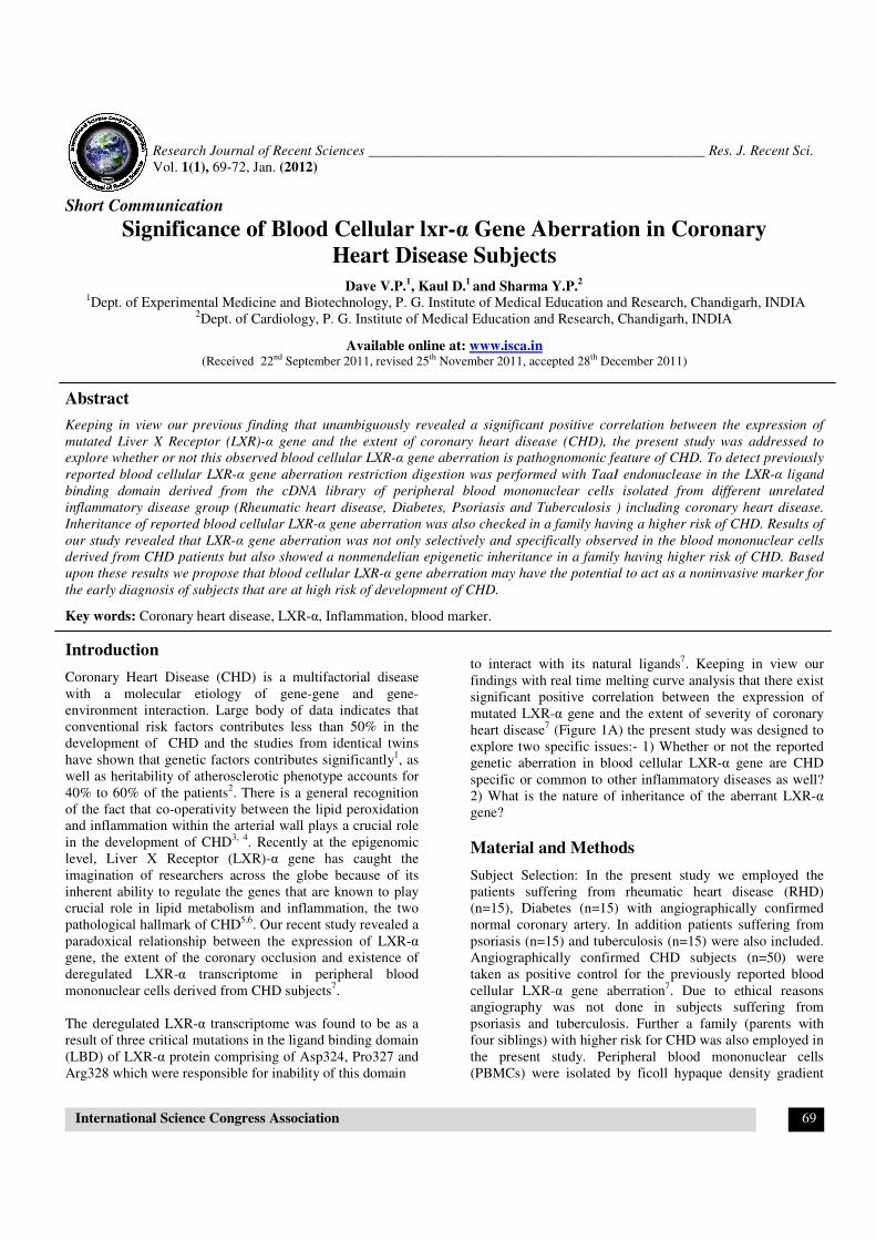
Research Journal of Recent Sciences _______________________________________________ Res. J. Recent Sci. Vol. 1(1), 69-72, Jan. (2012) International Science Congress Association 69 Short Communication Significance of Blood Cellular lxr-α Gene Aberration in Coronary Heart Disease Subjects Dave V.P. 1 , Kaul D. 1 and Sharma Y.P. 2 1 Dept. of Experimental Medicine and Biotechnology, P. G. Institute of Medical Education and Research, Chandigarh, INDIA 2 Dept. of Cardiology, P. G. Institute of Medical Education and Research, Chandigarh, INDIA Available online at: www.isca.in (Received 22 nd September 2011, revised 25 th November 2011, accepted 28 th December 2011) Abstract Keeping in view our previous finding that unambiguously revealed a significant positive correlation between the expression of mutated Liver X Receptor (LXR)-α gene and the extent of coronary heart disease (CHD), the present study was addressed to explore whether or not this observed blood cellular LXR-α gene aberration is pathognomonic feature of CHD. To detect previously reported blood cellular LXR-α gene aberration restriction digestion was performed with TaaI endonuclease in the LXR-α ligand binding domain derived from the cDNA library of peripheral blood mononuclear cells isolated from different unrelated inflammatory disease group (Rheumatic heart disease, Diabetes, Psoriasis and Tuberculosis ) including coronary heart disease. Inheritance of reported blood cellular LXR-α gene aberration was also checked in a family having a higher risk of CHD. Results of our study revealed that LXR-α gene aberration was not only selectively and specifically observed in the blood mononuclear cells derived from CHD patients but also showed a nonmendelian epigenetic inheritance in a family having higher risk of CHD. Based upon these results we propose that blood cellular LXR-α gene aberration may have the potential to act as a noninvasive marker for the early diagnosis of subjects that are at high risk of development of CHD. Key words: Coronary heart disease, LXR-α, Inflammation, blood marker. Introduction Coronary Heart Disease (CHD) is a multifactorial disease with a molecular etiology of gene-gene and gene- environment interaction. Large body of data indicates that conventional risk factors contributes less than 50% in the development of CHD and the studies from identical twins have shown that genetic factors contributes significantly 1 , as well as heritability of atherosclerotic phenotype accounts for 40% to 60% of the patients 2 . There is a general recognition of the fact that co-operativity between the lipid peroxidation and inflammation within the arterial wall plays a crucial role in the development of CHD 3, 4 . Recently at the epigenomic level, Liver X Receptor (LXR)-α gene has caught the imagination of researchers across the globe because of its inherent ability to regulate the genes that are known to play crucial role in lipid metabolism and inflammation, the two pathological hallmark of CHD 5,6 . Our recent study revealed a paradoxical relationship between the expression of LXR-α gene, the extent of the coronary occlusion and existence of deregulated LXR-α transcriptome in peripheral blood mononuclear cells derived from CHD subjects 7 . The deregulated LXR-α transcriptome was found to be as a result of three critical mutations in the ligand binding domain (LBD) of LXR-α protein comprising of Asp324, Pro327 and Arg328 which were responsible for inability of this domain to interact with its natural ligands 7 . Keeping in view our findings with real time melting curve analysis that there exist significant positive correlation between the expression of mutated LXR-α gene and the extent of severity of coronary heart disease 7 (Figure 1A) the present study was designed to explore two specific issues:- 1) Whether or not the reported genetic aberration in blood cellular LXR-α gene are CHD specific or common to other inflammatory diseases as well? 2) What is the nature of inheritance of the aberrant LXR-α gene? Material and Methods Subject Selection: In the present study we employed the patients suffering from rheumatic heart disease (RHD) (n=15), Diabetes (n=15) with angiographically confirmed normal coronary artery. In addition patients suffering from psoriasis (n=15) and tuberculosis (n=15) were also included. Angiographically confirmed CHD subjects (n=50) were taken as positive control for the previously reported blood cellular LXR-α gene aberration 7 . Due to ethical reasons angiography was not done in subjects suffering from psoriasis and tuberculosis. Further a family (parents with four siblings) with higher risk for CHD was also employed in the present study. Peripheral blood mononuclear cells (PBMCs) were isolated by ficoll hypaque density gradient
Transcript of Significance of Blood Cellular lxr- Gene Aberration in...
Research Journal of Recent Sciences _______________________________________________ Res. J. Recent Sci.
Vol. 1(1), 69-72, Jan. (2012)
International Science Congress Association 69
Short Communication
Significance of Blood Cellular lxr-α Gene Aberration in Coronary
Heart Disease Subjects
Dave V.P.1, Kaul D.
1 and Sharma Y.P.
2
1Dept. of Experimental Medicine and Biotechnology, P. G. Institute of Medical Education and Research, Chandigarh, INDIA
2Dept. of Cardiology, P. G. Institute of Medical Education and Research, Chandigarh, INDIA
Available online at: www.isca.in (Received 22nd September 2011, revised 25th November 2011, accepted 28th December 2011)
Abstract
Keeping in view our previous finding that unambiguously revealed a significant positive correlation between the expression of
mutated Liver X Receptor (LXR)-α gene and the extent of coronary heart disease (CHD), the present study was addressed to
explore whether or not this observed blood cellular LXR-α gene aberration is pathognomonic feature of CHD. To detect previously
reported blood cellular LXR-α gene aberration restriction digestion was performed with TaaI endonuclease in the LXR-α ligand
binding domain derived from the cDNA library of peripheral blood mononuclear cells isolated from different unrelated
inflammatory disease group (Rheumatic heart disease, Diabetes, Psoriasis and Tuberculosis ) including coronary heart disease.
Inheritance of reported blood cellular LXR-α gene aberration was also checked in a family having a higher risk of CHD. Results of
our study revealed that LXR-α gene aberration was not only selectively and specifically observed in the blood mononuclear cells
derived from CHD patients but also showed a nonmendelian epigenetic inheritance in a family having higher risk of CHD. Based
upon these results we propose that blood cellular LXR-α gene aberration may have the potential to act as a noninvasive marker for
the early diagnosis of subjects that are at high risk of development of CHD.
Key words: Coronary heart disease, LXR-α, Inflammation, blood marker.
Introduction
Coronary Heart Disease (CHD) is a multifactorial disease
with a molecular etiology of gene-gene and gene-
environment interaction. Large body of data indicates that
conventional risk factors contributes less than 50% in the
development of CHD and the studies from identical twins
have shown that genetic factors contributes significantly1, as
well as heritability of atherosclerotic phenotype accounts for
40% to 60% of the patients2. There is a general recognition
of the fact that co-operativity between the lipid peroxidation
and inflammation within the arterial wall plays a crucial role
in the development of CHD3, 4
. Recently at the epigenomic
level, Liver X Receptor (LXR)-α gene has caught the
imagination of researchers across the globe because of its
inherent ability to regulate the genes that are known to play
crucial role in lipid metabolism and inflammation, the two
pathological hallmark of CHD5,6
. Our recent study revealed a
paradoxical relationship between the expression of LXR-α
gene, the extent of the coronary occlusion and existence of
deregulated LXR-α transcriptome in peripheral blood
mononuclear cells derived from CHD subjects7.
The deregulated LXR-α transcriptome was found to be as a
result of three critical mutations in the ligand binding domain
(LBD) of LXR-α protein comprising of Asp324, Pro327 and
Arg328 which were responsible for inability of this domain
to interact with its natural ligands7. Keeping in view our
findings with real time melting curve analysis that there exist
significant positive correlation between the expression of
mutated LXR-α gene and the extent of severity of coronary
heart disease7 (Figure 1A) the present study was designed to
explore two specific issues:- 1) Whether or not the reported
genetic aberration in blood cellular LXR-α gene are CHD
specific or common to other inflammatory diseases as well?
2) What is the nature of inheritance of the aberrant LXR-α
gene?
Material and Methods
Subject Selection: In the present study we employed the
patients suffering from rheumatic heart disease (RHD)
(n=15), Diabetes (n=15) with angiographically confirmed
normal coronary artery. In addition patients suffering from
psoriasis (n=15) and tuberculosis (n=15) were also included.
Angiographically confirmed CHD subjects (n=50) were
taken as positive control for the previously reported blood
cellular LXR-α gene aberration7. Due to ethical reasons
angiography was not done in subjects suffering from
psoriasis and tuberculosis. Further a family (parents with
four siblings) with higher risk for CHD was also employed in
the present study. Peripheral blood mononuclear cells
(PBMCs) were isolated by ficoll hypaque density gradient
Research Journal of Recent Sciences ____________________________________________________________ Res. J. Recent Sci. Vol. 1(1), 69-72, Jan. (2012)
International Science Congress Association 70
method from 5ml of blood drawn from each subject
employed in the study with their prior informed consent8.The
demographic and clinical findings of all diseases group have
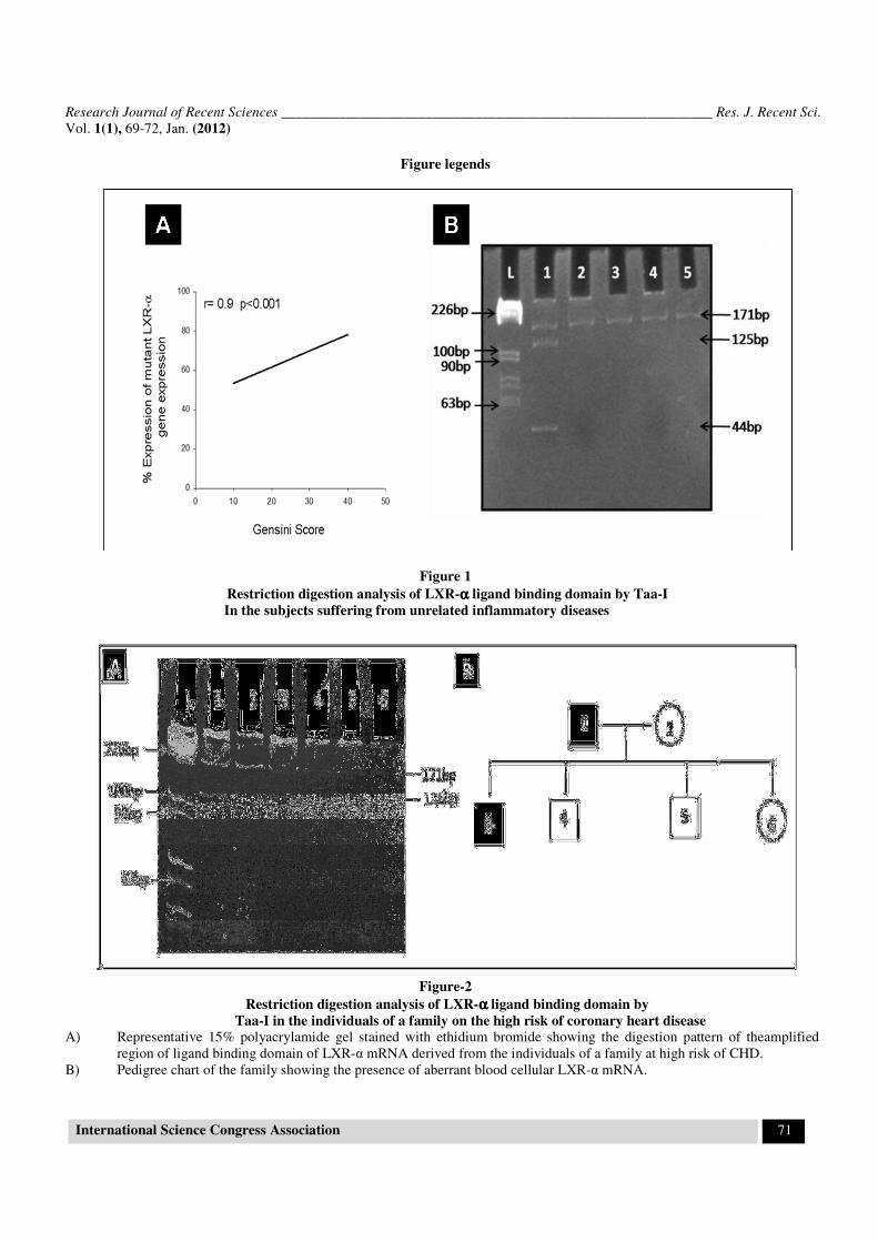
been shown in Tables. A) Based upon melting curve analysis
(Dave et al., 2009), correlation between the percent
expression of mutated LXR-α mRNA with respect to severity
of CHD. Values of “r” show Spearman rank correlation
coefficient.
B) Representative 15% polyacrylamide gel stained with
ethidium bromide showing the digestion pattern of the
amplified region of ligand binding domain of LXR-α derived
from different diseased group. L= Ladder, 1= Coronary
Artery disease, 2= Rheumatic heart disease, 3= Diabetes, 4=
Psoriosis and 5= Tuberculosis.
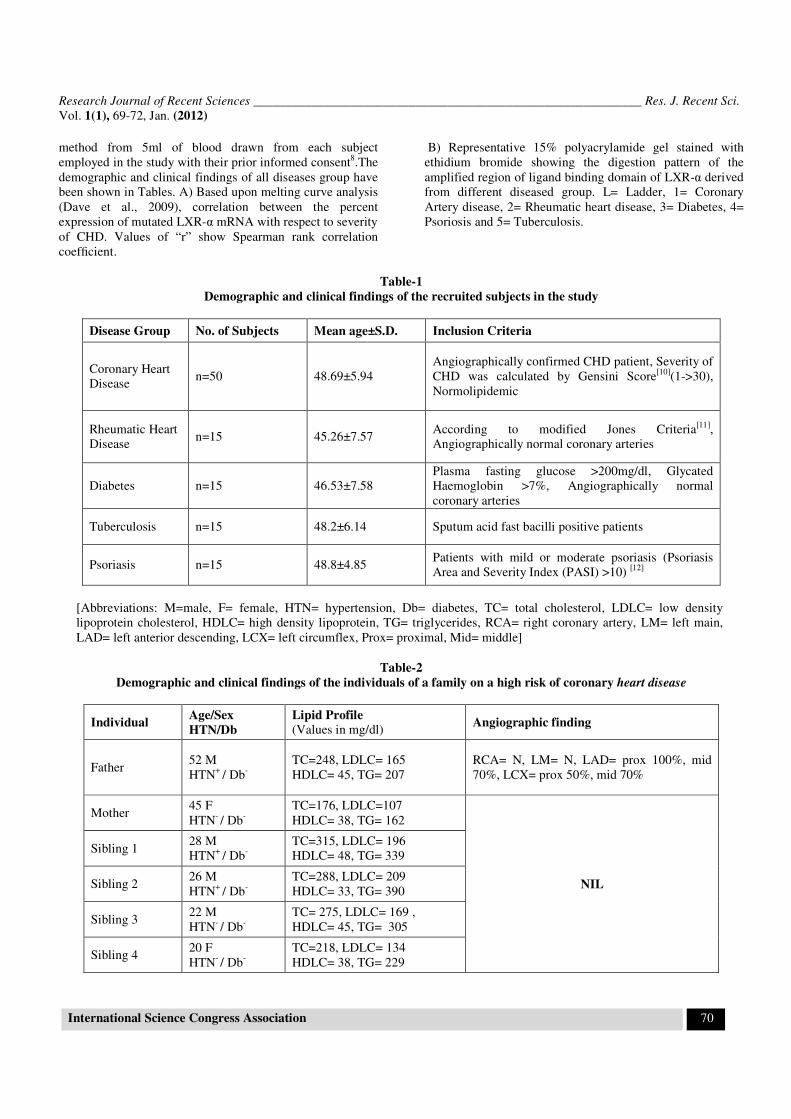
Table-1
Demographic and clinical findings of the recruited subjects in the study
Disease Group No. of Subjects Mean age±S.D. Inclusion Criteria
Coronary Heart
Disease n=50 48.69±5.94
Angiographically confirmed CHD patient, Severity of
CHD was calculated by Gensini Score[10]
(1->30),
Normolipidemic
Rheumatic Heart
Disease n=15 45.26±7.57
According to modified Jones Criteria[11]
,
Angiographically normal coronary arteries
Diabetes n=15 46.53±7.58
Plasma fasting glucose >200mg/dl, Glycated
Haemoglobin >7%, Angiographically normal
coronary arteries
Tuberculosis n=15 48.2±6.14 Sputum acid fast bacilli positive patients
Psoriasis n=15 48.8±4.85 Patients with mild or moderate psoriasis (Psoriasis
Area and Severity Index (PASI) >10) [12]
[Abbreviations: M=male, F= female, HTN= hypertension, Db= diabetes, TC= total cholesterol, LDLC= low density
lipoprotein cholesterol, HDLC= high density lipoprotein, TG= triglycerides, RCA= right coronary artery, LM= left main,
LAD= left anterior descending, LCX= left circumflex, Prox= proximal, Mid= middle]
Table-2
Demographic and clinical findings of the individuals of a family on a high risk of coronary heart disease
Individual Age/Sex
HTN/Db
Lipid Profile
(Values in mg/dl) Angiographic finding
Father 52 M
HTN+
/ Db-
TC=248, LDLC= 165
HDLC= 45, TG= 207
RCA= N, LM= N, LAD= prox 100%, mid
70%, LCX= prox 50%, mid 70%
Mother 45 F
HTN- / Db
-
TC=176, LDLC=107
HDLC= 38, TG= 162
NIL
Sibling 1 28 M
HTN+
/ Db-
TC=315, LDLC= 196
HDLC= 48, TG= 339
Sibling 2 26 M
HTN+
/ Db-
TC=288, LDLC= 209
HDLC= 33, TG= 390
Sibling 3 22 M
HTN- / Db
-
TC= 275, LDLC= 169 ,
HDLC= 45, TG= 305
Sibling 4 20 F
HTN- / Db
-
TC=218, LDLC= 134
HDLC= 38, TG= 229
Research Journal of Recent Sciences ____________________________________________________________ Res. J. Recent Sci. Vol. 1(1), 69-72, Jan. (2012)
International Science Congress Association 71
Figure legends
Figure 1
Restriction digestion analysis of LXR-αααα ligand binding domain by Taa-I
In the subjects suffering from unrelated inflammatory diseases
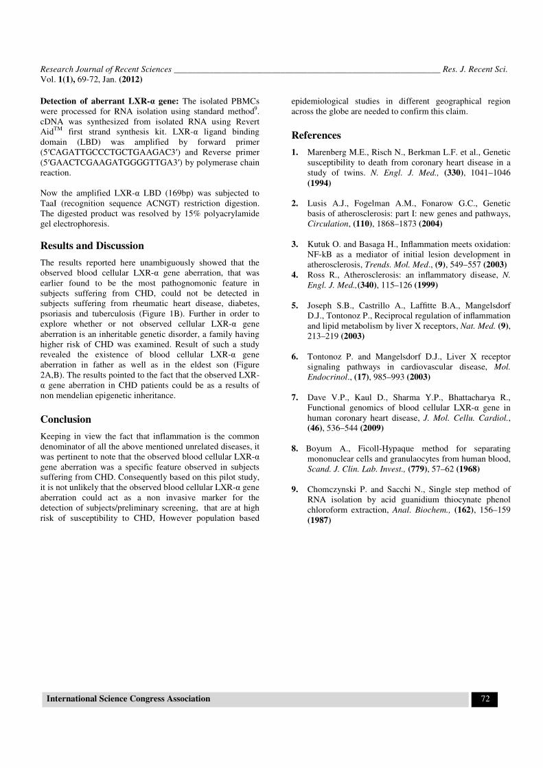
Figure-2
Restriction digestion analysis of LXR-αααα ligand binding domain by
Taa-I in the individuals of a family on the high risk of coronary heart disease
A) Representative 15% polyacrylamide gel stained with ethidium bromide showing the digestion pattern of theamplified
region of ligand binding domain of LXR-α mRNA derived from the individuals of a family at high risk of CHD.
B) Pedigree chart of the family showing the presence of aberrant blood cellular LXR-α mRNA.
Research Journal of Recent Sciences ____________________________________________________________ Res. J. Recent Sci. Vol. 1(1), 69-72, Jan. (2012)
International Science Congress Association 72
Detection of aberrant LXR-α gene: The isolated PBMCs
were processed for RNA isolation using standard method9.
cDNA was synthesized from isolated RNA using Revert
AidTM
first strand synthesis kit. LXR-α ligand binding
domain (LBD) was amplified by forward primer
(5′CAGATTGCCCTGCTGAAGAC3′) and Reverse primer
(5′GAACTCGAAGATGGGGTTGA3′) by polymerase chain
reaction.
Now the amplified LXR-α LBD (169bp) was subjected to
TaaI (recognition sequence ACNGT) restriction digestion.
The digested product was resolved by 15% polyacrylamide
gel electrophoresis.
Results and Discussion
The results reported here unambiguously showed that the
observed blood cellular LXR-α gene aberration, that was
earlier found to be the most pathognomonic feature in
subjects suffering from CHD, could not be detected in
subjects suffering from rheumatic heart disease, diabetes,
psoriasis and tuberculosis (Figure 1B). Further in order to
explore whether or not observed cellular LXR-α gene
aberration is an inheritable genetic disorder, a family having
higher risk of CHD was examined. Result of such a study
revealed the existence of blood cellular LXR-α gene
aberration in father as well as in the eldest son (Figure
2A,B). The results pointed to the fact that the observed LXR-
α gene aberration in CHD patients could be as a results of
non mendelian epigenetic inheritance.
Conclusion
Keeping in view the fact that inflammation is the common
denominator of all the above mentioned unrelated diseases, it
was pertinent to note that the observed blood cellular LXR-α
gene aberration was a specific feature observed in subjects
suffering from CHD. Consequently based on this pilot study,
it is not unlikely that the observed blood cellular LXR-α gene
aberration could act as a non invasive marker for the
detection of subjects/preliminary screening, that are at high
risk of susceptibility to CHD, However population based
epidemiological studies in different geographical region
across the globe are needed to confirm this claim.
References
1. Marenberg M.E., Risch N., Berkman L.F. et al., Genetic
susceptibility to death from coronary heart disease in a
study of twins. N. Engl. J. Med., (330), 1041–1046
(1994)
2. Lusis A.J., Fogelman A.M., Fonarow G.C., Genetic
basis of atherosclerosis: part I: new genes and pathways,
Circulation, (110), 1868–1873 (2004)
3. Kutuk O. and Basaga H., Inflammation meets oxidation:
NF-kB as a mediator of initial lesion development in
atherosclerosis, Trends. Mol. Med., (9), 549–557 (2003)
4. Ross R., Atherosclerosis: an inflammatory disease, N.
Engl. J. Med.,(340), 115–126 (1999)
5. Joseph S.B., Castrillo A., Laffitte B.A., Mangelsdorf
D.J., Tontonoz P., Reciprocal regulation of inflammation
and lipid metabolism by liver X receptors, Nat. Med. (9),
213–219 (2003)
6. Tontonoz P. and Mangelsdorf D.J., Liver X receptor
signaling pathways in cardiovascular disease, Mol.
Endocrinol., (17), 985–993 (2003)
7. Dave V.P., Kaul D., Sharma Y.P., Bhattacharya R.,
Functional genomics of blood cellular LXR-α gene in
human coronary heart disease, J. Mol. Cellu. Cardiol.,
(46), 536–544 (2009)
8. Boyum A., Ficoll-Hypaque method for separating
mononuclear cells and granulaocytes from human blood,
Scand. J. Clin. Lab. Invest., (779), 57–62 (1968)
9. Chomczynski P. and Sacchi N., Single step method of
RNA isolation by acid guanidium thiocynate phenol
chloroform extraction, Anal. Biochem., (162), 156–159
(1987)