
Signalling regulates activity of DHCR24, the final … · catalysed by the enzyme,...
36
1 Signalling regulates activity of DHCR24, the final enzyme in cholesterol synthesis Winnie Luu 1 *, Eser J. Zerenturk 1 *, Ika Kristiana 1 , Martin P. Bucknall 2 , Laura J. Sharpe 1 , and Andrew J. Brown 1 1 School of Biotechnology and Biomolecular Sciences, The University of New South Wales, Sydney, NSW 2052, Australia; 2 Bioanalytical Mass Spectrometry Facility, The University of New South Wales, Sydney, NSW 2052, Australia Abbreviated title: Signalling regulates DHCR24 activity * These authors contributed equally to this work. To whom correspondence should be addressed: Prof. Andrew J. Brown, BABS, School of Biotechnology and Biomolecular Sciences, Biological Sciences Building D26, The University of New South Wales, Sydney, NSW 2052, Australia. Phone: +61-2-9385-2005; Fax: +61-2-9385-1483; E-mail: [email protected] Abbreviations: 24,25EC, 24(S),25-epoxycholesterol; Arg-TLC, argentation thin layer chromatography; BIM, bisindolylmaleimide I; CD, methyl-β-cyclodextrin; CHO, Chinese hamster ovary; DHCR24, 3β-hydroxysterol Δ24-reductase; FAD, flavin adenine dinucleotide; FCS, fetal calf serum; GC-MS, gas chromatography-mass spectrometry; LPDS, lipoprotein-deficient serum; NADPH, nicotinamide adenine dinucleotide phosphate; PBGD, porphobilinogen deaminase; PKA, protein kinase A; PKB, protein kinase B; PKC, protein kinase C; SREBP, sterol-regulatory element-binding protein. by guest, on September 7, 2018 www.jlr.org Downloaded from
-
Upload
truongcong -
Category
Documents
-
view
222 -
download
0
Transcript of Signalling regulates activity of DHCR24, the final … · catalysed by the enzyme,...
1
Signalling regulates activity of DHCR24, the final enzyme in
cholesterol synthesis
Winnie Luu1*, Eser J. Zerenturk
1*, Ika Kristiana1, Martin P. Bucknall2, Laura J. Sharpe
1, and
Andrew J. Brown1
1 School of Biotechnology and Biomolecular Sciences, The University of New South Wales,
Sydney, NSW 2052, Australia; 2 Bioanalytical Mass Spectrometry Facility, The University of
New South Wales, Sydney, NSW 2052, Australia
Abbreviated title: Signalling regulates DHCR24 activity
* These authors contributed equally to this work.
To whom correspondence should be addressed:
Prof. Andrew J. Brown, BABS, School of Biotechnology and Biomolecular Sciences,
Biological Sciences Building D26, The University of New South Wales, Sydney, NSW 2052,
Australia.
Phone: +61-2-9385-2005;
Fax: +61-2-9385-1483;
E-mail: [email protected]
Abbreviations:
24,25EC, 24(S),25-epoxycholesterol; Arg-TLC, argentation thin layer chromatography; BIM,
bisindolylmaleimide I; CD, methyl-β-cyclodextrin; CHO, Chinese hamster ovary; DHCR24,
3β-hydroxysterol Δ24-reductase; FAD, flavin adenine dinucleotide; FCS, fetal calf serum; GC-MS, gas
chromatography-mass spectrometry; LPDS, lipoprotein-deficient serum; NADPH, nicotinamide
adenine dinucleotide phosphate; PBGD, porphobilinogen deaminase; PKA, protein kinase A; PKB,
protein kinase B; PKC, protein kinase C; SREBP, sterol-regulatory element-binding protein.
by guest, on Septem
ber 7, 2018w
ww
.jlr.orgD
ownloaded from
2
Abstract
The role of signalling in regulating cholesterol homeostasis is gradually becoming more
widely recognised. Here, we explored how kinases and phosphorylation sites regulate the
activity of the enzyme involved in the final step of cholesterol synthesis (3β-hydroxysterol
Δ24-reductase; DHCR24). Many factors are known to regulate DHCR24 transcriptionally,
but little is known about its post-translational regulation. We developed a system to
specifically test human ectopic DHCR24 activity in a model cell-line (Chinese hamster ovary-
7) using siRNA targeted only to hamster DHCR24, thus ensuring that all activity could be
attributed to the human enzyme. We determined the effect of known phosphorylation sites
and found that mutating certain residues (T110, Y299, and Y507) inhibited DHCR24 activity.
In addition, inhibitors of protein kinase C ablated DHCR24 activity, although not through a
known phosphorylation site. Our data indicate a novel mechanism whereby DHCR24
activity is regulated by signalling.
Keywords: DHCR24, cholesterol, phosphorylation, bisindolylmaleimide I, protein kinase C,
regulation, signalling, desmosterol, GC-MS
by guest, on Septem
ber 7, 2018w
ww
.jlr.orgD
ownloaded from
3
1. Introduction
Cholesterol is a vital raw material for the cell, which is toxic in excess and leads to a number
of diseases. Thus, cellular cholesterol levels are tightly regulated through a balance of
synthesis, uptake, and efflux. The final step in the Bloch cholesterol synthetic pathway is
catalysed by the enzyme, 3β-hydroxysterol Δ24-reductase (DHCR24). DHCR24 is a
60.1 kDa endoplasmic reticulum (ER) membrane-bound oxidoreductase that converts
desmosterol to cholesterol by saturating the C-24,25 double bond in the side-chain (1).
DHCR24 activity is strictly dependent on nicotinamide adenine dinucleotide phosphate
(NADPH) as the reducing agent (1), although no consensus sequence for NADPH binding is
predicted. However, DHCR24 does contain a highly conserved flavin adenine dinucleotide
(FAD) binding domain (aa 111-203, (2)), characteristic of the well-defined family of FAD-
dependent oxidoreductases (3); with reduction of desmosterol dependent on FAD,
suggesting functionality of the conserved domain (1). Additionally, a substrate binding
domain was predicted in DHCR24 (aa 236-491) by homology modelling on an FAD-
dependent oxidoreductase, plant cytokinin dehydrogenase (4).
DHCR24 is involved in multiple cellular functions. Related to its role in cholesterol
biosynthesis, DHCR24 modulates lipid raft formation, thus facilitating signal transduction and
trafficking (5-7). It is involved in regulating oxidative stress (8, 9), and is neuroprotective (5,
10, 11), anti-apoptotic (8, 10), and anti-inflammatory (12, 13). DHCR24 is also implicated in
many diseases, such as cardiovascular diseases (12, 14), cancers (e.g. prostate cancer
(15)), and hepatitis C (16). Moreover, mutations in DHCR24 underlie the rare autosomal
recessive disease, desmosterolosis, whereby patients have elevated desmosterol and
lowered cholesterol, resulting in multiple congenital anomalies (17). Specifically, seven
missense mutations have been described in desmosterolosis: N294T, K306N, Y471S,
E191K, R94H, E480K, and R103C (1, 18-21).
by guest, on Septem
ber 7, 2018w
ww
.jlr.orgD
ownloaded from
4
Like many cholesterol synthetic genes, DHCR24 is transcriptionally regulated by
sterols via the sterol-regulatory element-binding protein (SREBP)-2 transcription factor (22),
and we recently identified two sterol-regulatory elements (SREs) and nuclear factor Y (NF-Y)
sites in the human DHCR24 promoter that mediate this regulation (23). Moreover, DHCR24
is regulated at the transcriptional level by sex steroids (24, 25), adrenocorticotropic hormone
(26), thyroid hormone (27), and xenobiotics (28). Epigenetic factors such as methylation and
acetylation also regulate DHCR24 expression (29). In contrast, relatively little is known
about the post-translational regulation of DHCR24 activity. We recently found that the
oxysterol regulator, 24(S),25-epoxycholesterol (24,25EC), inhibits DHCR24 activity with
potent effects on cellular cholesterol levels (30). Plant sterols (31), progesterone and similar
progestins (32, 33) similarly inhibit DHCR24 activity. However, the role of post-translational
modifications in DHCR24 regulation, such as phosphorylation, has yet to be investigated.
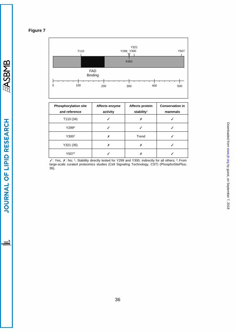
Large-scale proteomic studies have identified phosphorylation sites on DHCR24; T110 (34)
and Y321 (35), and there is evidence for additional phosphorylation sites identified by large-
scale unpublished proteomics studies (PhosphoSitePlus; 36). The function of these
phosphorylation sites and the kinases/phosphatases involved are currently unknown due to
a lack of mechanistic studies.
Cell signalling and phosphorylation is a major mode of regulating cellular processes,
and is implicated in various facets of cholesterol homeostasis (reviewed in 37). Since
DHCR24 catalyses the ultimate step in cholesterol synthesis, we hypothesised that it is a
likely target for feedback regulation via signalling notably phosphorylation, as this would
provide a rapid means of switching cholesterol synthesis on or off. In this study, we
examined the effect of known phosphorylation sites on DHCR24 expression and activity.
Our data demonstrate that particular putative phosphorylated residues (T110, Y299, and
Y507) have significant effects on DHCR24 activity. In addition, inhibiting a major
serine/threonine kinase, protein kinase C (PKC), ablated DHCR24 activity, although this
effect was independent of T110, the only published serine/threonine phosphorylation site.
by guest, on Septem
ber 7, 2018w
ww
.jlr.orgD
ownloaded from

5
Together, these results indicate a novel regulatory mechanism for DHCR24 by kinases and
phosphorylation.
by guest, on Septem
ber 7, 2018w
ww
.jlr.orgD
ownloaded from
6
2. Materials and Methods
2.1 Materials
Chinese hamster ovary (CHO)-7 cells (38) were a generous gift from Drs. Michael S. Brown
and Joseph L. Goldstein (UT Southwestern Medical Center, Dallas, TX, USA). The
Flp-In-CHO cell-line (for stable-cell generation) was generated from CHO-7 cells by Gorjana
Mitic. HeLaT cells were generously donated by Dr Noel Whitaker (UNSW, Sydney, NSW,
Australia). SensiMix SYBR No-Rox was from Bioline (London, UK). Fetal calf serum (FCS)
was from Bovogen (East Keilor, Vic, AU). Amersham Hyperfilm ECL was from GE
Healthcare (Buckinghamshire, England). Horseradish peroxidase-conjugated AffiniPure
donkey anti-mouse IgG antibody was from Jackson ImmunoResearch Laboratories, Inc.
(West Grove, PA, USA). Anti-V5 antibody, Dulbecco’s Modified Eagle’s Medium: Ham’s
Nutrient Mixture F12 (1:1) (DMEM/F12), Lipofectamine RNAiMAX transfection reagent,
newborn calf serum, Roswell Park Memorial Institute 1640 medium (RPMI), and Superscript
III Reverse Transcriptase were from Life Technologies (Carlsbad, CA, USA). Akt inhibitor
VIII was from Merck (Darmstadt, Germany). Immobilon Western Chemiluminescent
horseradish peroxidase substrate enhanced chemiluminescent detection system was from
Millipore (Billerica, MA, USA). Phusion High-Fidelity buffer, Phusion Hot Start II High-Fidelity
Polymerase, and purified bovine serum albumin (BSA) were from New England Biolabs
(Ipswich, MA, USA). [14C]-acetate sodium salt (specific radioactivity: 55.3 mCi/mmol, 2.046
GBq/mmol) was from Perkin Elmer (Waltham, MA, USA). α-tubulin antibody, Bis-Tris,
bisindolylmaleimide I (BIM), cycloheximide, compactin (mevastatin), desmosterol, H89,
primers, mevalonate, MG132 (Z-Leu-Leu-Leu-al), MOPS, N-O-bis-(trimethylsilyl)
trifluoroacetamide containing 1% trimethylchlorosilane (BSTFA), protease inhibitor cocktail,
Ro-318220, siRNA, sodium bisulfite, zinc (II) nitrate, and Tri Reagent were from Sigma-
Aldrich (St. Louis, MO, USA). [2H6]-desmosterol was from Avanti Polar Lipids (Alabaster,
AL, USA). Phos-tag acrylamide pendant [10 mg supplied in 100 µL 3% (v/v) methanol] was
by guest, on Septem
ber 7, 2018w
ww
.jlr.orgD
ownloaded from
7
from Wako Pure Chemical Industries (Osaka, Japan). Lipoprotein-deficient serum (LPDS)
was prepared from heat-inactivated newborn calf serum as previously described (39).
2.2 Cell culture and treatments
CHO-7 cells were maintained in 5% (v/v) LPDS/DMEM/F12. CHO-7 stable cell-lines were
generated and maintained in 5% (v/v) LPDS/DMEM/F12 supplemented with 150 µg/mL
hygromycin B. HeLaT cells were maintained in 10% (v/v) FCS/RPMI. Where there were
treatments, cells were treated in fresh media with various test agents [added in dimethyl
sulfoxide or ethanol; desmosterol was complexed to methyl-β-cyclodextrin (CD) according to
(40)], as indicated in the figure legends. Within an experiment, the final concentrations of
solvent were kept constant between conditions and did not exceed 0.2% (v/v).
2.3 Generation of CHO overexpressing cell-lines
Primers were designed to perform megaprimed site-directed mutagenesis (41) on a plasmid
encoding the human DHCR24 sequence (accession number NM_014762.3) containing a
FRT recombination site (pcDNA5-FRT-DHCR24-V5) (30) to create plasmids encoding
DHCR24 phosphorylation mutants; T110A, Y299F, Y300F, Y321F, Y507F; and a T110
phosphomimetic (T110E). These plasmids were then used to generate stable cell lines
using the CHO-7 Flp-In system, as described in (30). DHCR24 protein expression was
screened by Western blotting with the anti-V5 antibody, and stable cells expressing similar
levels of DHCR24 were selected and characterised.
The empty vector stable cell-line (CHO-EV) was prepared as described (42), and the
human wild-type DHCR24 overexpressing cell-line (CHO-DHCR24-WT) was prepared as
described (30).
by guest, on Septem
ber 7, 2018w
ww
.jlr.orgD
ownloaded from
8
2.4 siRNA transfection
Cells were transfected with 25 nM control or hamster-specific DHCR24 siRNA (target
sequence: GAGAGCCACGTGTGAAGCA; designed by Sigma-Aldrich) for 24 h using
Lipofectamine RNAiMAX transfection reagent, according to the manufacturer’s instructions.
The cells were washed with phosphate-buffered saline (PBS), and then refed fresh media
with [14C]-acetate labelling for argentation thin layer chromatography (Arg-TLC).
2.5 Quantitative real-time reverse transcription PCR (qRT-PCR)
Cells were seeded in triplicate wells per condition as described. Total RNA (1 µg) was
reverse transcribed into cDNA using the SuperScript III First Strand cDNA Synthesis Kit.
mRNA levels were determined by qRT-PCR using the Corbett Rotorgene 3000 (Corbett Life
Sciences, Sydney, Australia) and analysed using Rotor Gene Version 6.0 (Build 27)
(Qiagen, Doncaster, VIC, Australia). Primers were used to amplify the cDNA of hamster or
human DHCR24 (43) and the housekeeping control, porphobilinogen deaminase (PBGD)
(44, 45). Changes in DHCR24 gene expression levels were normalised to PBGD for each
sample by the ΔΔCt method, and made relative to the CHO-DHCR24-WT cell-line, which
was set to 1.
2.6 Western blotting
After treatment, cells were harvested in 10% (w/v) SDS supplemented with 5% (v/v)
protease inhibitor cocktail. Equal amounts of protein were mixed with loading buffer (final
conc.: 50 mM Tris-HCl (pH 6.8), 2% (w/v) SDS, 5% (v/v) glycerol, 0.04% (w/v) bromophenol
blue, and 1% (v/v) β-mercaptoethanol), boiled for 5 min, and subjected to SDS-PAGE. After
electrophoresis, the proteins were transferred to a nitrocellulose membrane, blocked for 1 h,
incubated with primary anti-V5 antibody (1:5,000) or anti-α-tubulin (1:200,000), and then
further incubated with secondary antibody (1:20,000). The antibodies were visualised by the
enhanced chemiluminescent detection system, and membranes were exposed to Hyperfilm.
by guest, on Septem
ber 7, 2018w
ww
.jlr.orgD
ownloaded from
9
Proteins were identified by their predicted molecular weight (α-tubulin, 50 kDa; DHCR24, 60
kDa). Protein band intensities from Western blots were quantified by densitometry using
ImageJ (Version 1.47t).
2.7 Cholesterol synthesis assay
Cholesterol and desmosterol synthesis were measured as an indicator of DHCR24 activity
using Arg-TLC as described previously (30). The relative intensities of bands were
quantified using Sciencelab ImageGauge 4.0 software (Fujifilm).
2.8 Phos-tag SDS-PAGE
Phosphorylated proteins were visualised using the phos-tag SDS-PAGE method described
in (46), with modifications. Cells were seeded in 60 mm or 100 mm dishes, and treated with
various test agents, as indicated in the figure legends. After treatments, cells were washed
twice with ice cold PBS. The cells were scraped in PBS, then pelleted and lysed in 100 µL
modified RIPA buffer [50 mM Tris-HCl (pH 7.3), 150 mM NaCl, 1% sodium deoxycholate, 1%
Triton X-100; (47)]. The lysates were passed through a 21 gauge needle 20 times, and
centrifuged at 20,000 g at 4°C for 15 min. Equal amounts of protein were mixed with 0.25
volume 5× loading buffer and boiled for 5 min before subjecting to phos-tag SDS-PAGE
(7.5% separation gel containing Zn2+-phos-tag complex and a 4% stacking gel), transferred
to nitrocellulose membranes, and Western blotted as indicated in the figure legends.
Purified BSA (0.5 µg) was prepared in the same volume of modified RIPA buffer containing
loading buffer, and served as a molecular weight marker (66 kDa).
2.9 Gas chromatography-mass spectrometry
Cells were treated as indicated in the figure legends with or without 1 μg/mL
[2H6]-desmosterol/CD for 4 h. Cells were harvested and lipid extracts were prepared as
described in (30).
by guest, on Septem
ber 7, 2018w
ww
.jlr.orgD
ownloaded from
10
Lipids were derivatised with BSTFA for 1 h at 60°C. Derivatised samples were
analysed using a Thermo Trace gas chromatograph (GC) coupled with a Thermo DSQII
mass spectrometer (MS) and Thermo Triplus Autosampler (Thermo Fisher Scientific;
Waltham, MA, USA). Samples (1 μL) were injected via a heated (290°C) splitless inlet into
an Rxi-5Sil MS w/Integra-Guard, 30 m x 0.25 mm, df 0.25 μm film thickness, capillary GC
column (Restek; Waltham, MA, USA). The column oven was initially held at 80°C for 1 min,
then heated to 260°C at 80°C min-1, then to 280°C at 10°C min-1 and then to 295°C at 2°C
min-1. Finally, the oven was increased to 305°C at 10°C min-1 and held for 1 min. Helium
was used as a carrier gas at a constant flow (1.3 mL min-1, with vacuum compensation on).
Mass spectrometry conditions: electron energy 70 eV, ion source temperature 200°C,
transfer line temperature 305°C. The emission current was set to 130 μA and the detector
gain to 3.0 x 10⁵. Samples were analysed either in scan mode (35-520 Da, 2.5 scans s-1) to
obtain mass spectra for peak identification, or in single ion monitoring (SIM) mode to
measure DHCR24 activity. Method details and the m/z values of SIM ions monitored for 5α-
cholestane, [2H6]-cholesterol, cholesterol, [2H6]-desmosterol and desmosterol are listed in
Supplementary Table 1. Thermo Xcalibur Software (Version 2.1.0.1140) was used to
acquire and process the data. 5α-cholestane (0.1 μg added during cell harvesting) was
used as an internal standard. Peak identification was based on comparison of retention
times with those of standards and comparison of mass spectra with the Wiley 9/NIST 2011
combined mass spectral library.
2.10 Statistical analysis
Statistical differences were determined by the student’s paired t-test (two-tailed), where
p-values of <0.01 (**) were considered statistically significant. Statistical analysis was
performed in Figure 5D using a two way ANOVA with repeated measures (stability over time
of WT versus Y299F or Y300F).
by guest, on Septem
ber 7, 2018w
ww
.jlr.orgD
ownloaded from
11
3. Results
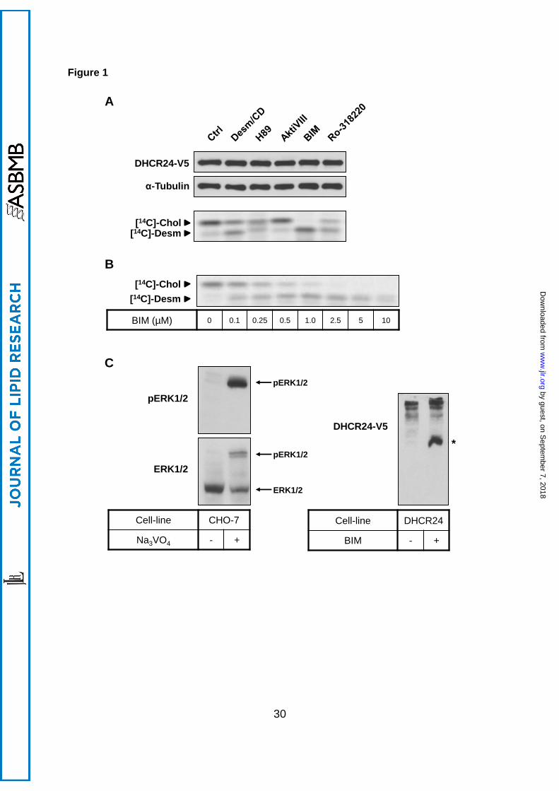
3.1 PKC inhibition decreases DHCR24 activity
To examine the effect of common protein kinases on DHCR24 activity, we used inhibitors of
protein kinases A [PKA; H89 (48)], B [PKB, also called Akt; AktiVIII (49)], and C [PKC; BIM
(50) and Ro-318220 (51)]. CHO-7 cells stably transfected with epitope-tagged human
DHCR24 (30) were radiolabelled with [14C]-acetate, which feeds into the beginning of the
cholesterol synthetic pathway. Cholesterol and desmosterol were resolved by Arg-TLC, and
radioactive bands were visualised by phosphorimaging. The cholesterol to desmosterol ratio
was used as an indicator of DHCR24 activity (30).
As shown previously (30), treatment with unlabelled desmosterol/CD decreased the
cholesterol to desmosterol ratio, demonstrating competitive inhibition of DHCR24 enzyme.
Inhibition of PKA (H89) or PKB (AktiVIII) did not have a selective effect on DHCR24 activity
(Figure 1A), although H89 affected cholesterol synthesis on a global level (Supplementary
Figure 1). In contrast, PKC inhibitors BIM and Ro-318220 reduced cholesterol levels and
accumulated desmosterol within 4 h, indicating decreased DHCR24 activity. BIM had the
most robust effect, and ablated DHCR24 activity in a dose-dependent manner (Figure 1B).
Conversely, PKC stimulation with phorbol myristate acetate increased cholesterol synthesis
by ~30% (± 5%, p<0.01, the student’s t-test). Since inhibition did not affect DHCR24 protein
levels (Figure 1A), these results suggest PKC may be influencing DHCR24 activity through
modulating its phosphorylation state.
To confirm that DHCR24 is phosphorylated in our cell system, we employed the
phos-tag SDS-PAGE method (46). This system utilises gels supplemented with phos-tag
acrylamide-bound metal complexes, which have affinity for phosphate groups on proteins.
This therefore slows the migration of phosphorylated species during electrophoresis,
producing a mobility shift. As a positive control, we treated cells with a phosphatase inhibitor
by guest, on Septem
ber 7, 2018w
ww
.jlr.orgD
ownloaded from
12
(Na3VO4) and determined ERK1/2 phosphorylation. Na3VO4 produced two distinct bands
when probed with total ERK1/2 antibody, with the bottom band being the non-
phosphorylated ERK1/2, as confirmed with phosphorylated ERK1/2 (pERK1/2) antibody
(Figure 1C, left panel). In CHO-DHCR24-WT cells, non-treated cells resulted in multiple,
slower migrating DHCR24 bands (Figure 1C, right panel). Interestingly, BIM treatment
produced a faster migrating band, suggesting that DHCR24 is constitutively phosphorylated
at more than one site.
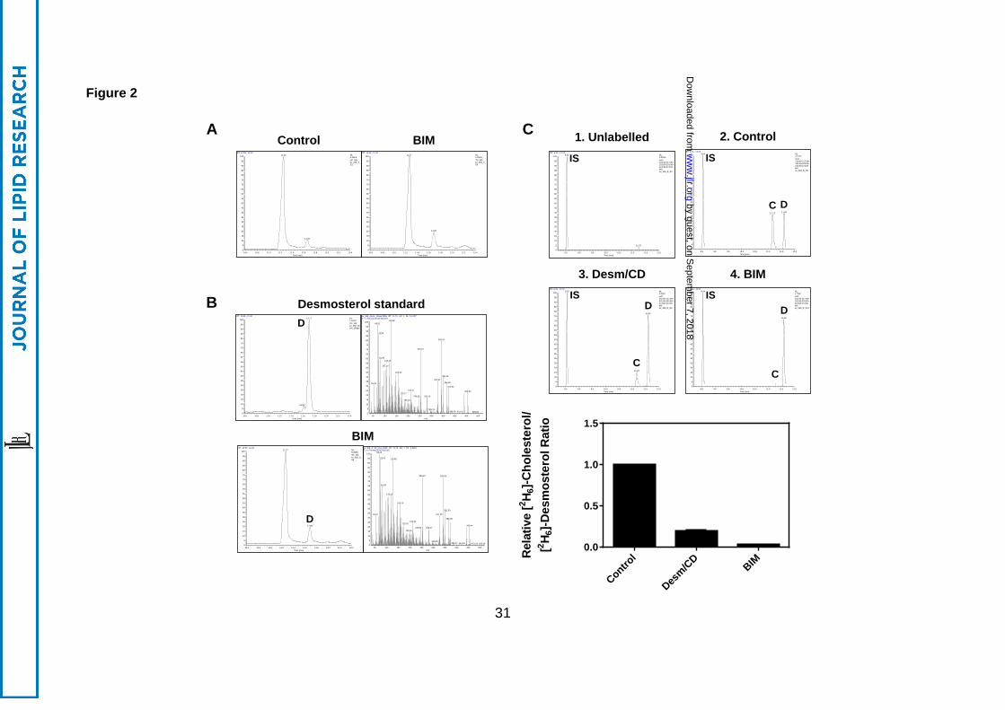
3.2 PKC inhibition results in desmosterol accumulation
To confirm the identity of the desmosterol band accumulating with BIM treatment using
Arg-TLC, samples were re-analysed using gas chromatography-mass spectrometry (GC-
MS; Figure 2). Two peaks were identified, where the major peak was cholesterol and a
second minor peak, which increased with BIM treatment, was identified as desmosterol
based on the mass spectra (Figure 2A). To measure DHCR24 activity, samples were
treated with BIM and [2H6]-desmosterol. As observed with Arg-TLC (Figure 1A), competition
from unlabelled desmosterol inhibited the conversion of [2H6]-desmosterol to [2H6]-
cholesterol, resulting in an increase in [2H6]-desmosterol (Figure 2B). BIM treatment had
similar effects, strongly inhibiting DHCR24 activity, as evidenced by the [2H6]-cholesterol to
desmosterol ratio (Figure 2B).
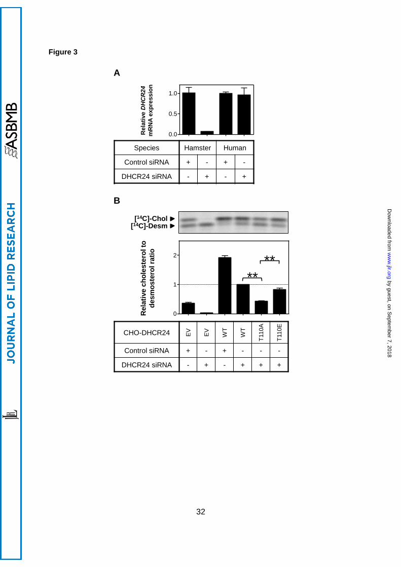
3.3 DHCR24-T110 phosphorylation mutant decreases DHCR24 activity
From the panel of kinase inhibitors tested, only PKC inhibitors affected DHCR24 activity.
Since PKC phosphorylates serine and threonine residues, it is possible that BIM inhibits
DHCR24 activity through the only known serine/threonine phosphorylation site, T110 (34).
Thus, we mutated this to alanine (T110A; mutant) or glutamic acid (T110E;
phosphomimetic), and stably overexpressed these constructs in CHO-7 cells, as we did
by guest, on Septem
ber 7, 2018w
ww
.jlr.orgD
ownloaded from
13
previously with the generation of the CHO-DHCR24-WT cells (30). DHCR24 mRNA and
protein levels between the CHO-DHCR24 cell-lines were comparable (Supplementary Figure
2).
To measure the specific effects of the mutants on human DHCR24, endogenous
hamster DHCR24 expression was reduced using RNA interference. Transfection of
hamster-specific DHCR24 siRNA ablated endogenous hamster DHCR24 mRNA without
affecting human DHCR24 expression (Figure 3A). Thus, transfecting CHO-DHCR24 cells
with hamster siRNA reduced background activity in our stable cells, with measured DHCR24
activity attributed to the stably expressed human DHCR24 only.
Using this approach, we examined whether DHCR24-T110 mutants affect DHCR24
activity. As indicated in Figure 3B, transfecting DHCR24 siRNA into EV cells ablated
endogenous DHCR24 activity. Mutating T110 so that it cannot be phosphorylated (T110A),
significantly decreased DHCR24 activity by ~60% (Figure 3B). Furthermore, DHCR24
activity of the phosphomimetic (T110E) mutant was increased relative to T110A, with activity
not statistically different from WT cells. These results suggest that phosphorylation at T110
modulates DHCR24 activity.
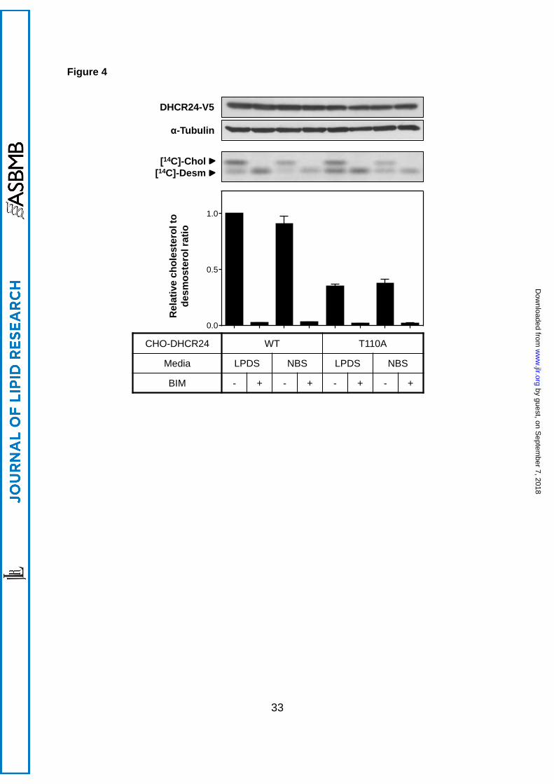
3.4 PKC inhibition decreases DHCR24 activity independently of T110
Next, we determined whether BIM inhibition of DHCR24 activity occurs at T110. Cells were
treated in low (LPDS) or high (newborn calf serum) cholesterol conditions to exclude any
potential effects of cholesterol status. BIM had similar effects in DHCR24-T110A as WT
cells: a decrease in DHCR24 activity, with no effect on DHCR24 protein levels (Figure 4).
This indicates that BIM targets a different residue in DHCR24, or that the effect is indirect.
by guest, on Septem
ber 7, 2018w
ww
.jlr.orgD
ownloaded from
14
3.5 Other DHCR24 phosphorylation sites
Another published phosphorylation site is Y321 (35), and PhosphoSitePlus (36) gives
evidence for three other phosphorylated tyrosine residues; Y299, Y300, and Y507. Thus, we
similarly generated DHCR24 stable cell-lines containing these mutants, and characterised
mRNA and protein expression. DHCR24 protein correlated with mRNA expression
(Supplementary Figure 3), with the exception of the DHCR24-Y299F and DHCR24-Y300F
mutants. Compared to the DHCR24-WT, the DHCR24-Y299F stable cells contained lower
mRNA expression whilst having comparable protein levels. Conversely, the DHCR24-
Y300F stable cells have similar levels of mRNA to the DHCR24-WT stable cells, while
containing less protein.
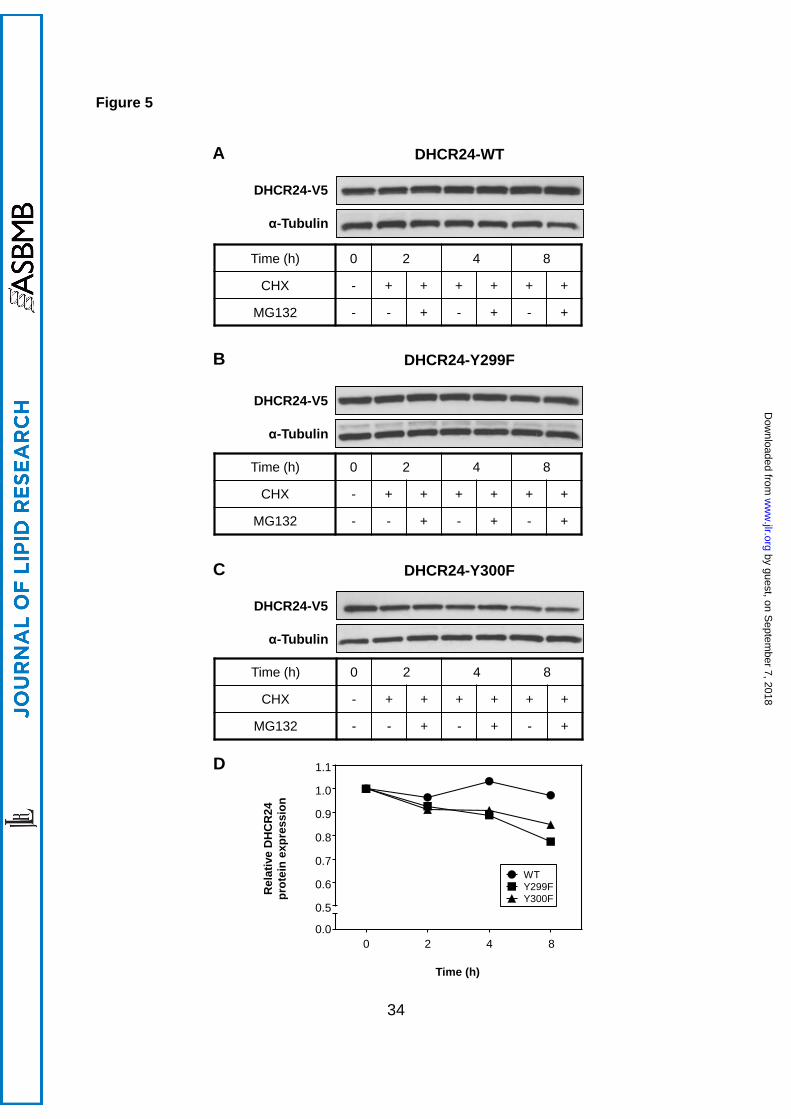
3.6 Examining the protein stability of DHCR24-Y299F and DHCR24-Y300F
Y299 and Y300 are neighbouring residues, yet phosphorylation mutants at these sites have
vastly different effects on DHCR24 mRNA and protein expression, suggesting that mutating
DHCR24 at these residues may alter protein stability. In addition, there is evidence for
DHCR24 ubiquitination (36), including at K301 (52), which is adjacent to Y299 and Y300.
Therefore, we compared DHCR24 protein stability in DHCR24-WT, -Y299F, and -Y300F
stable cells by inhibiting protein synthesis (using cycloheximide) and proteasomal
degradation (using MG132). Both DHCR24-Y299F and DHCR24-Y300F were destabilised
during the 8 h treatments, though only DHCR24-Y299F was statistically different compared
to DHCR24-WT (p<0.01); DHCR24-Y300F displayed a non-significant trend (p=0.12) (Figure
5). Moreover, proteasomal inhibition did not rescue this degradation. Importantly, DHCR24-
WT was found to be stable throughout the treatments, suggesting that DHCR24 has a
relatively long half-life under these culturing conditions.
by guest, on Septem
ber 7, 2018w
ww
.jlr.orgD
ownloaded from
15
3.7 Effect of tyrosine mutants on DHCR24 activity
Next, we determined whether DHCR24 stable cell-lines containing mutant tyrosine residues
affected DHCR24 activity. Mutating Y299 and Y507 significantly decreased DHCR24 activity
by ~40% and ~60% respectively, whilst mutating Y300 and the published site, Y321, had no
effect (Figure 6). Thus, Y299 and Y507, but not Y300 and Y321, may be phosphorylation
sites that regulate DHCR24 activity.
by guest, on Septem
ber 7, 2018w
ww
.jlr.orgD
ownloaded from
16
4. Discussion
We discovered a novel mode of regulation of a key protein in cholesterol synthesis,
DHCR24. Recent studies on DHCR24 have identified a number of regulatory mechanisms,
primarily at the transcriptional level, particularly via feedback regulation by sterol/steroid
molecules, with only a few at the post-translational level. Regulation by post-translational
modifications such as phosphorylation has yet to be fully investigated, despite evidence that
DHCR24 is phosphorylated at multiple sites (36), which is in line with our phos-tag SDS-
PAGE results (Figure 1C).
One aim of this work was to identify protein kinase(s) involved in DHCR24
phosphorylation by looking at common protein kinases, using a panel of kinase inhibitors.
Whilst inhibiting serine/threonine kinases did not affect DHCR24 protein expression,
inhibition of PKC (using BIM and Ro-318220) markedly decreased desmosterol to
cholesterol conversion. A potential caveat with studying PKC with pharmacological inhibitors
is the issue of specificity (53). For example, BIM and Ro-318220 have been shown to
exhibit comparable inhibition of 90 kDa and 70 kDa ribosomal S6 kinase and PKC, in vitro,
but in cell culture, at least ten times higher concentrations are required to inhibit the S6
kinases (54). BIM and Ro-318220 are therefore relatively specific inhibitors of the PKC
family, with BIM reducing DHCR24 activity in a dose-dependent manner, from
concentrations as low as 0.1 µM. Thus, our results are consistent with the involvement of
PKC in moderating DHCR24 activity. However, further experiments should confirm this, and
determine which of the 10 distinct PKC isoforms is involved.
We then tested if BIM inhibition of DHCR24 activity could be negated by mutating the
only published serine/threonine phosphoresidue in DHCR24, T110. T110 is also predicted
as a low stringency PKC substrate by a matrix-based prediction program, Scansite [(55) and
see Supplementary Table 2 for a list of sites examined here]. However, the BIM inhibitory
effect was still observed in the DHCR24 T110 mutant cell-line (CHO-DHCR24-T110A),
by guest, on Septem
ber 7, 2018w
ww
.jlr.orgD
ownloaded from
17
indicating that PKC does not phosphorylate DHCR24 at T110. This suggests that BIM may
be affecting DHCR24 directly through an as yet unidentified phosphorylation site(s), or
indirectly. Future work should involve testing other putative sites as predicted by Scansite,
for PKC phosphorylation and effect on DHCR24 activity.
Another aim of this study was to characterise the known phosphorylation sites in
DHCR24, and examine their effects on DHCR24 expression and activity. We developed a
novel method for measuring DHCR24 activity in cell culture; by stably expressing human
DHCR24 in hamster (CHO-7) cells, combined with metabolic labelling. To selectively
examine the activity of stably expressed human DHCR24, hamster DHCR24 was knocked
down by hamster-specific DHCR24 siRNA (Figure 3A). Hence, this assay measured
cholesterol conversion from desmosterol by ectopic human DHCR24 only.
We first examined T110, a published phosphoresidue ((34) and Supplementary Table
2), and created CHO cell-lines stably expressing DHCR24 T110 mutant (T110A) or a
phosphomimetic (T110E). DHCR24-T110A significantly decreased DHCR24 activity relative
to DHCR24-WT (by ~40%). Mutating T110 to a phosphomimetic (T110E) restored most of
this activity (Figure 3B), indicating that this is indeed a functional phosphorylation site.
Although the physiological relevance of T110 phosphorylation needs to be elucidated, this
study introduces a new mode of regulation for DHCR24 activity, and may explain settings
where desmosterol accumulates, such as in cholesterol-laden macrophage foam cells (14),
or in HCV infection (56).
As T110 lies within the predicted FAD binding domain (55-235) (4) and just outside of
the highly conserved region for FAD binding (111-203) [(2, 3) and Figure 7], it is possible
that phosphorylation of T110 affects DHCR24 enzyme activity by influencing cofactor
interaction. Amino acids located near the highly conserved region of the FAD binding
domain have been shown to be crucial for DHCR24 function; mutation of arginine 94
(R94H), resulted in an almost complete loss of activity, causing desmosterolosis (20). R94
by guest, on Septem
ber 7, 2018w
ww
.jlr.orgD
ownloaded from
18
has also been predicted to be involved in specific interactions between FAD phosphate
groups and DHCR24 (4). T110 is preceded by three positively charged residues, which are
all conserved in higher organisms, and may also be involved in electrostatic interactions with
the negatively-charged phosphate groups in FAD. Further work testing FAD binding affinity
in DHCR24-T110A is required to consolidate this hypothesis.
The other published phosphorylation site was a tyrosine residue (Y321), with three
other tyrosine residues implicated. We created phosphorylation mutants by substituting the
tyrosine with phenylalanine (Y to F), and stably expressed these in CHO cells. Similar to
T110A, the Y299F and Y507F mutants significantly decreased DHCR24 activity, with a
~60% reduction in activity observed for Y507F relative to DHCR24-WT (Figure 6). Y507 is a
highly conserved residue at the extreme C-terminal end of DHCR24 (Figure 7), which lies
within a small region predicted to interact with the cofactor FAD (4).
The kinases/phosphatases responsible for T110, Y299, and Y507 phosphorylation
remain elusive and require further investigation. In this study, we have excluded the effects
of PKA, PKB, and PKC (Figure 1A and Figure 4), as well as ERK1/2, PI3K, mTOR, JAK2,
SHP1/2, PTP1B (data not shown). We have also attempted to confirm these
phosphorylation sites in our cell system by liquid chromatography-tandem mass
spectrometry. While we had reasonable sequence coverage for DHCR24 (up to 32%), we
were unable to detect the required peptides, as determining phosphorylation with this
approach remains challenging in our hands (57). Nevertheless, the phosphorylation sites
examined in this study were originally identified in large-scale phosphoproteomics studies,
with T110 being a published phosphorylation site (34), as well as being identified in Cell
Signaling Technology MS/MS studies. As discussed, T110 is very likely physiologically
relevant as the phosphorylation mutant decreased, and phosphomimetic restored, DHCR24
activity. Whilst Y507 is not a published phosphorylation site, PhosphoSitePlus indicated this
site to be a phosphorylated residue in 12 data sets (36), increasing confidence that it is a
by guest, on Septem
ber 7, 2018w
ww
.jlr.orgD
ownloaded from
19
bona fide phosphorylation site. Furthermore, our phos-tag result suggested that DHCR24 is
phosphorylated at more than one site in our cell system (Figure 1C).
Although this study focused on potential post-translational modification by
phosphorylation, there is a vast amount of evidence that DHCR24 is ubiquitinated. At
present, there are 11 identified ubiquitination sites [9 published (52, 58-60), and 2 from
PhosphoSitePlus (36)], suggesting that DHCR24 may be regulated by proteasomal
degradation. One such site, K301 (52), is a highly conserved residue that neighbours
putative phosphoresidues, Y299 and Y300 (Figure 7). Although the mutants are in close
proximity, they have differing effects on DHCR24 mRNA and protein expression, with
DHCR24-Y300F levels exhibiting a striking decrease from mRNA to protein (Supplementary
Figure 3). Therefore, we suspected that this mutant may be subject to proteasomal
degradation. However, both DHCR24-Y299F and DHCR24-Y300F caused only minor
destabilisation that cannot explain the observed mRNA and protein levels, which are not due
to differences in cell-lines, as we used a Flp-In system which introduces only one copy of a
gene into each cell, and different clones gave the same effect (data not shown). Notably,
DHCR24-WT was very stable under conditions tested (Figure 5).
There is emerging evidence that other enzymes in the cholesterol synthetic pathway
besides 3-hydroxy-3-methylglutaryl coenzyme A reductase (the classic rate-limiting step) are
potential control points for rapid regulation of cholesterol synthesis (61), such as squalene
monooxygenase (62). DHCR24 is a prime candidate (61) due to its position in the Bloch
pathway as the final enzyme. Thus, regulating DHCR24 by phosphorylation would allow for
a rapid means for attenuating cholesterol synthesis, with previous studies already
demonstrating its ability for rapid inhibition by multiple feedback mechanisms (30, 32).
Furthermore, modulating DHCR24 activity alters levels of desmosterol, a strong liver X
receptor ligand (14, 63), further reducing cellular cholesterol status. Together, this study has
demonstrated that signalling and phosphorylation have functional effects on DHCR24.
by guest, on Septem
ber 7, 2018w
ww
.jlr.orgD
ownloaded from
20
Further work is required to investigate the precise kinase(s) responsible for the
phosphorylation sites investigated in this study, as well as the phosphorylation site(s)
required for PKC’s action.
Acknowledgements
We thank members of the Brown Lab for critically reviewing this manuscript. The Brown Lab
is supported by grants from the National Health and Medical Research Council (1008081)
and the National Heart Foundation of Australia (G11S5757).
by guest, on Septem
ber 7, 2018w
ww
.jlr.orgD
ownloaded from
21
References
1. Waterham, H. R., J. Koster, G. J. Romeijn, R. C. Hennekam, P. Vreken, H. C. Andersson, D. R. FitzPatrick, R. I. Kelley, and R. J. Wanders. 2001. Mutations in the 3beta-hydroxysterol Delta24-reductase gene cause desmosterolosis, an autosomal recessive disorder of cholesterol biosynthesis. American journal of human genetics 69: 685-694. 2. Finn, R. D., J. Mistry, J. Tate, P. Coggill, A. Heger, J. E. Pollington, O. L. Gavin, P. Gunasekaran, G. Ceric, K. Forslund, L. Holm, E. L. Sonnhammer, S. R. Eddy, and A. Bateman. 2010. The Pfam protein families database. Nucleic Acids Res 38: D211-222. 3. Mushegian, A. R., and E. V. Koonin. 1995. A putative FAD-binding domain in a distinct group of oxidases including a protein involved in plant development. Protein Sci 4: 1243-1244. 4. Pedretti, A., E. Bocci, R. Maggi, and G. Vistoli. 2008. Homology modelling of human DHCR24 (seladin-1) and analysis of its binding properties through molecular docking and dynamics simulations. Steroids 73: 708-719. 5. Crameri, A., E. Biondi, K. Kuehnle, D. Lutjohann, K. M. Thelen, S. Perga, C. G. Dotti, R. M. Nitsch, M. D. Ledesma, and M. H. Mohajeri. 2006. The role of seladin-1/DHCR24 in cholesterol biosynthesis, APP processing and Abeta generation in vivo. The EMBO journal 25: 432-443. 6. Gilk, S. D., D. C. Cockrell, C. Luterbach, B. Hansen, L. A. Knodler, J. A. Ibarra, O. Steele-Mortimer, and R. A. Heinzen. 2013. Bacterial colonization of host cells in the absence of cholesterol. PLoS Pathog 9: e1003107. 7. Jansen, M., V. M. Pietiaïnen, H. Pölönen, L. Rasilainen, M. Koivusalo, U. Ruotsalainen, E. Jokitalo, and E. Ikonen. 2008. Cholesterol substitution increases the structural heterogeneity of caveolae. Journal of Biological Chemistry 283: 14610-14618. 8. Kuehnle, K., A. Crameri, R. E. Kalin, P. Luciani, S. Benvenuti, A. Peri, F. Ratti, M. Rodolfo, L. Kulic, F. L. Heppner, R. M. Nitsch, and M. H. Mohajeri. 2008. Prosurvival effect of DHCR24/Seladin-1 in acute and chronic responses to oxidative stress. Mol Cell Biol 28: 539-550. 9. Wu, C., I. Miloslavskaya, S. Demontis, R. Maestro, and K. Galaktionov. 2004. Regulation of cellular response to oncogenic and oxidative stress by Seladin-1. Nature 432: 640-645. 10. Greeve, I., I. Hermans-Borgmeyer, C. Brellinger, D. Kasper, T. Gomez-Isla, C. Behl, B. Levkau, and R. M. Nitsch. 2000. The human DIMINUTO/DWARF1 homolog seladin-1 confers resistance to Alzheimer's disease-associated neurodegeneration and oxidative stress. J Neurosci 20: 7345-7352. 11. Sarajärvi, T., A. Haapasalo, J. Viswanathan, P. Mäkinen, M. Laitinen, H. Soininen, and M. Hiltunen. 2009. Down-regulation of seladin-1 increases BACE1 levels and activity through enhanced GGA3 depletion during apoptosis. Journal of Biological Chemistry 284: 34433-34443. 12. Wu, B. J., K. Chen, S. Shrestha, K. L. Ong, P. J. Barter, and K. A. Rye. 2013. High-density lipoproteins inhibit vascular endothelial inflammation by increasing 3beta-hydroxysteroid-delta24 reductase expression and inducing heme oxygenase-1. Circ Res 112: 278-288. 13. McGrath, K. C., X. H. Li, R. Puranik, E. C. Liong, J. T. Tan, V. M. Dy, B. A. DiBartolo, P. J. Barter, K. A. Rye, and A. K. Heather. 2009. Role of 3beta-hydroxysteroid-delta 24 reductase in mediating antiinflammatory effects of high-density lipoproteins in endothelial cells. Arteriosclerosis, thrombosis, and vascular biology 29: 877-882. 14. Spann, N. J., L. X. Garmire, J. G. McDonald, D. S. Myers, S. B. Milne, N. Shibata, D. Reichart, J. N. Fox, I. Shaked, D. Heudobler, C. R. Raetz, E. W. Wang, S. L. Kelly, M. C. Sullards, R. C. Murphy, A. H. Merrill, Jr., H. A. Brown, E. A. Dennis, A. C. Li, K. Ley, S. Tsimikas, E. Fahy, S. Subramaniam, O. Quehenberger, D. W. Russell, and C. K. Glass.
by guest, on Septem
ber 7, 2018w
ww
.jlr.orgD
ownloaded from
22
2012. Regulated accumulation of desmosterol integrates macrophage lipid metabolism and inflammatory responses. Cell 151: 138-152. 15. Battista, M. C., M. O. Guimond, C. Roberge, A. A. Doueik, L. Fazli, M. Gleave, R. Sabbagh, and N. Gallo-Payet. 2010. Inhibition of DHCR24/seladin-1 impairs cellular homeostasis in prostate cancer. Prostate 70: 921-933. 16. Takano, T., K. Tsukiyama-Kohara, M. Hayashi, Y. Hirata, M. Satoh, Y. Tokunaga, C. Tateno, Y. Hayashi, T. Hishima, N. Funata, M. Sudoh, and M. Kohara. 2011. Augmentation of DHCR24 expression by hepatitis C virus infection facilitates viral replication in hepatocytes. J Hepatol 55: 512-521. 17. Horvat, S., J. McWhir, and D. Rozman. 2011. Defects in cholesterol synthesis genes in mouse and in humans: lessons for drug development and safer treatments. Drug metabolism reviews 43: 69-90. 18. FitzPatrick, D. R., J. W. Keeling, M. J. Evans, A. E. Kan, J. E. Bell, M. E. Porteous, K. Mills, R. M. Winter, and P. T. Clayton. 1998. Clinical phenotype of desmosterolosis. Am J Med Genet 75: 145-152. 19. Andersson, H. C., L. Kratz, and R. Kelley. 2002. Desmosterolosis presenting with multiple congenital anomalies and profound developmental delay. Am J Med Genet 113: 315-319. 20. Schaaf, C. P., J. Koster, P. Katsonis, L. Kratz, O. A. Shchelochkov, F. Scaglia, R. I. Kelley, O. Lichtarge, H. R. Waterham, and M. Shinawi. 2011. Desmosterolosis-phenotypic and molecular characterization of a third case and review of the literature. Am J Med Genet A 155A: 1597-1604. 21. Zolotushko, J., H. Flusser, B. Markus, I. Shelef, Y. Langer, M. Heverin, I. Bjorkhem, S. Sivan, and O. S. Birk. 2011. The desmosterolosis phenotype: spasticity, microcephaly and micrognathia with agenesis of corpus callosum and loss of white matter. Eur J Hum Genet 19: 942-946. 22. Horton, J. D., N. A. Shah, J. A. Warrington, N. N. Anderson, S. W. Park, M. S. Brown, and J. L. Goldstein. 2003. Combined analysis of oligonucleotide microarray data from transgenic and knockout mice identifies direct SREBP target genes. Proceedings of the National Academy of Sciences of the United States of America 100: 12027-12032. 23. Zerenturk, E. J., L. J. Sharpe, and A. J. Brown. 2012. Sterols regulate 3beta-hydroxysterol Delta24-reductase (DHCR24) via dual sterol regulatory elements: cooperative induction of key enzymes in lipid synthesis by Sterol Regulatory Element Binding Proteins. Biochim Biophys Acta 1821: 1350-1360. 24. Bonaccorsi, L., P. Luciani, G. Nesi, E. Mannucci, C. Deledda, F. Dichiara, M. Paglierani, F. Rosati, L. Masieri, S. Serni, M. Carini, L. Proietti-Pannunzi, S. Monti, G. Forti, G. Danza, M. Serio, and A. Peri. 2008. Androgen receptor regulation of the seladin-1/DHCR24 gene: altered expression in prostate cancer. Lab Invest 88: 1049-1056. 25. Luciani, P., C. Deledda, F. Rosati, S. Benvenuti, I. Cellai, F. Dichiara, M. Morello, G. B. Vannelli, G. Danza, M. Serio, and A. Peri. 2008. Seladin-1 is a fundamental mediator of the neuroprotective effects of estrogen in human neuroblast long-term cell cultures. Endocrinology 149: 4256-4266. 26. Battista, M. C., C. Roberge, A. Martinez, and N. Gallo-Payet. 2009. 24-dehydrocholesterol reductase/seladin-1: a key protein differentially involved in adrenocorticotropin effects observed in human and rat adrenal cortex. Endocrinology 150: 4180-4190. 27. Ishida, E., K. Hashimoto, S. Okada, T. Satoh, M. Yamada, and M. Mori. 2013. Thyroid hormone receptor and liver X receptor competitively up-regulate human selective Alzheimer's disease indicator-1 gene expression at the transcriptional levels. Biochemical and biophysical research communications 432: 513-518. 28. Yoshinari, K., H. Ohno, S. Benoki, and Y. Yamazoe. 2012. Constitutive androstane receptor transactivates the hepatic expression of mouse Dhcr24 and human DHCR24
by guest, on Septem
ber 7, 2018w
ww
.jlr.orgD
ownloaded from
23
encoding a cholesterogenic enzyme 24-dehydrocholesterol reductase. Toxicol Lett 208: 185-191. 29. Drzewinska, J., A. Walczak-Drzewiecka, and M. Ratajewski. 2011. Identification and analysis of the promoter region of the human DHCR24 gene: involvement of DNA methylation and histone acetylation. Mol Biol Rep 38: 1091-1101. 30. Zerenturk, E. J., I. Kristiana, S. Gill, and A. J. Brown. 2012. The endogenous regulator 24(S),25-epoxycholesterol inhibits cholesterol synthesis at DHCR24 (Seladin-1). Biochim Biophys Acta 1821: 1269-1277. 31. Fernandez, C., Y. Suarez, A. J. Ferruelo, D. Gomez-Coronado, and M. A. Lasuncion. 2002. Inhibition of cholesterol biosynthesis by Delta22-unsaturated phytosterols via competitive inhibition of sterol Delta24-reductase in mammalian cells. Biochem J 366: 109-119. 32. Jansen, M., W. Wang, D. Greco, G. C. Bellenchi, U. di Porzio, A. J. Brown, and E. Ikonen. 2013. What dictates the accumulation of desmosterol in the developing brain? FASEB J 27: 865-870. 33. Lindenthal, B., A. L. Holleran, T. A. Aldaghlas, B. Ruan, G. J. Schroepfer, Jr., W. K. Wilson, and J. K. Kelleher. 2001. Progestins block cholesterol synthesis to produce meiosis-activating sterols. FASEB journal : official publication of the Federation of American Societies for Experimental Biology 15: 775-784. 34. Molina, H., D. M. Horn, N. Tang, S. Mathivanan, and A. Pandey. 2007. Global proteomic profiling of phosphopeptides using electron transfer dissociation tandem mass spectrometry. Proceedings of the National Academy of Sciences of the United States of America 104: 2199-2204. 35. Rigbolt, K. T., T. A. Prokhorova, V. Akimov, J. Henningsen, P. T. Johansen, I. Kratchmarova, M. Kassem, M. Mann, J. V. Olsen, and B. Blagoev. 2011. System-wide temporal characterization of the proteome and phosphoproteome of human embryonic stem cell differentiation. Sci Signal 4: rs3. 36. Hornbeck, P. V., J. M. Kornhauser, S. Tkachev, B. Zhang, E. Skrzypek, B. Murray, V. Latham, and M. Sullivan. 2012. PhosphoSitePlus: a comprehensive resource for investigating the structure and function of experimentally determined post-translational modifications in man and mouse. Nucleic acids research 40: D261-270. 37. Luu, W., L. J. Sharpe, I. C. Gelissen, and A. J. Brown. 2013. The role of signalling in cellular cholesterol homeostasis. IUBMB Life. 38. Metherall, J. E., J. L. Goldstein, K. L. Luskey, and M. S. Brown. 1989. Loss of transcriptional repression of three sterol-regulated genes in mutant hamster cells. J Biol Chem 264: 15634-15641. 39. Goldstein, J. L., S. K. Basu, and M. S. Brown. 1983. Receptor-mediated endocytosis of low-density lipoprotein in cultured cells. Methods Enzymol 98: 241-260. 40. Brown, A. J., L. Sun, J. D. Feramisco, M. S. Brown, and J. L. Goldstein. 2002. Cholesterol addition to ER membranes alters conformation of SCAP, the SREBP escort protein that regulates cholesterol metabolism. Molecular cell 10: 237-245. 41. Tseng, W. C., J. W. Lin, T. Y. Wei, and T. Y. Fang. 2008. A novel megaprimed and ligase-free, PCR-based, site-directed mutagenesis method. Analytical biochemistry 375: 376-378. 42. Luu, W., L. J. Sharpe, J. Stevenson, and A. J. Brown. 2012. Akt acutely activates the cholesterogenic transcription factor SREBP-2. Biochim Biophys Acta 1823: 458-464. 43. Sharpe, L. J., J. Wong, B. Garner, G. M. Halliday, and A. J. Brown. 2012. Is seladin-1 really a selective Alzheimer's disease indicator? J Alzheimers Dis 30: 35-39. 44. Du, X., I. Kristiana, J. Wong, and A. J. Brown. 2006. Involvement of Akt in ER-to-Golgi transport of SCAP/SREBP: a link between a key cell proliferative pathway and membrane synthesis. Mol Biol Cell 17: 2735-2745.
by guest, on Septem
ber 7, 2018w
ww
.jlr.orgD
ownloaded from
24
45. Kielar, D., W. Dietmaier, T. Langmann, C. Aslanidis, M. Probst, M. Naruszewicz, and G. Schmitz. 2001. Rapid quantification of human ABCA1 mRNA in various cell types and tissues by real-time reverse transcription-PCR. Clinical chemistry 47: 2089-2097. 46. Kinoshita, E., and E. Kinoshita-Kikuta. 2011. Improved Phos-tag SDS-PAGE under neutral pH conditions for advanced protein phosphorylation profiling. Proteomics 11: 319-323. 47. Bogoyevitch, M. A., Y. Y. Yeap, Z. Qu, K. R. Ngoei, Y. Y. Yip, T. T. Zhao, J. I. Heng, and D. C. Ng. 2012. WD40-repeat protein 62 is a JNK-phosphorylated spindle pole protein required for spindle maintenance and timely mitotic progression. J Cell Sci 125: 5096-5109. 48. Chijiwa, T., A. Mishima, M. Hagiwara, M. Sano, K. Hayashi, T. Inoue, K. Naito, T. Toshioka, and H. Hidaka. 1990. Inhibition of forskolin-induced neurite outgrowth and protein phosphorylation by a newly synthesized selective inhibitor of cyclic AMP-dependent protein kinase, N-[2-(p-bromocinnamylamino)ethyl]-5-isoquinolinesulfonamide (H-89), of PC12D pheochromocytoma cells. J Biol Chem 265: 5267-5272. 49. Barnett, S. F., D. Defeo-Jones, S. Fu, P. J. Hancock, K. M. Haskell, R. E. Jones, J. A. Kahana, A. M. Kral, K. Leander, L. L. Lee, J. Malinowski, E. M. McAvoy, D. D. Nahas, R. G. Robinson, and H. E. Huber. 2005. Identification and characterization of pleckstrin-homology-domain-dependent and isoenzyme-specific Akt inhibitors. Biochem J 385: 399-408. 50. Toullec, D., P. Pianetti, H. Coste, P. Bellevergue, T. Grand-Perret, M. Ajakane, V. Baudet, P. Boissin, E. Boursier, F. Loriolle, and et al. 1991. The bisindolylmaleimide GF 109203X is a potent and selective inhibitor of protein kinase C. J Biol Chem 266: 15771-15781. 51. Davis, P. D., C. H. Hill, E. Keech, G. Lawton, J. S. Nixon, A. D. Sedgwick, J. Wadsworth, D. Westmacott, and S. E. Wilkinson. 1989. Potent selective inhibitors of protein kinase C. FEBS Lett 259: 61-63. 52. Wagner, S. A., P. Beli, B. T. Weinert, M. L. Nielsen, J. Cox, M. Mann, and C. Choudhary. 2011. A proteome-wide, quantitative survey of in vivo ubiquitylation sites reveals widespread regulatory roles. Mol Cell Proteomics 10: M111 013284. 53. Wu-Zhang, A. X., and A. C. Newton. 2013. Protein kinase C pharmacology: refining the toolbox. The Biochemical journal 452: 195-209. 54. Roberts, N. A., M. S. Marber, and M. Avkiran. 2004. Specificity of action of bisindolylmaleimide protein kinase C inhibitors: do they inhibit the 70kDa ribosomal S6 kinase in cardiac myocytes? Biochem Pharmacol 68: 1923-1928. 55. Obenauer, J. C., L. C. Cantley, and M. B. Yaffe. 2003. Scansite 2.0: Proteome-wide prediction of cell signaling interactions using short sequence motifs. Nucleic acids research 31: 3635-3641. 56. Rodgers, M. A., V. A. Villareal, E. A. Schaefer, L. F. Peng, K. E. Corey, R. T. Chung, and P. L. Yang. 2012. Lipid metabolite profiling identifies desmosterol metabolism as a new antiviral target for hepatitis C virus. J Am Chem Soc 134: 6896-6899. 57. Sharpe, L. J., W. Luu, and A. J. Brown. 2011. Akt phosphorylates Sec24: new clues into the regulation of ER-to-Golgi trafficking. Traffic 12: 19-27. 58. Danielsen, J. M., K. B. Sylvestersen, S. Bekker-Jensen, D. Szklarczyk, J. W. Poulsen, H. Horn, L. J. Jensen, N. Mailand, and M. L. Nielsen. 2011. Mass spectrometric analysis of lysine ubiquitylation reveals promiscuity at site level. Molecular & Cellular Proteomics 10. 59. Kim, W., E. J. Bennett, E. L. Huttlin, A. Guo, J. Li, A. Possemato, M. E. Sowa, R. Rad, J. Rush, M. J. Comb, J. W. Harper, and S. P. Gygi. 2011. Systematic and quantitative assessment of the ubiquitin-modified proteome. Mol Cell 44: 325-340. 60. Wagner, S. A., P. Beli, B. T. Weinert, C. Scholz, C. D. Kelstrup, C. Young, M. L. Nielsen, J. V. Olsen, C. Brakebusch, and C. Choudhary. 2012. Proteomic analyses reveal divergent ubiquitylation site patterns in murine tissues. Mol Cell Proteomics 11: 1578-1585.
by guest, on Septem
ber 7, 2018w
ww
.jlr.orgD
ownloaded from
25
61. Sharpe, L. J., and A. J. Brown. 2013. Controlling cholesterol synthesis beyond 3-hydroxy-3-methylglutaryl-CoA reductase (HMGCR). J Biol Chem 288: 18707-18715. 62. Gill, S., J. Stevenson, I. Kristiana, and A. J. Brown. 2011. Cholesterol-dependent degradation of squalene monooxygenase, a control point in cholesterol synthesis beyond HMG-CoA reductase. Cell Metab 13: 260-273. 63. Yang, C., J. G. McDonald, A. Patel, Y. Zhang, M. Umetani, F. Xu, E. J. Westover, D. F. Covey, D. J. Mangelsdorf, J. C. Cohen, and H. H. Hobbs. 2006. Sterol intermediates from cholesterol biosynthetic pathway as liver X receptor ligands. The Journal of biological chemistry 281: 27816-27826. 64. Flicek, P., I. Ahmed, M. R. Amode, D. Barrell, K. Beal, S. Brent, D. Carvalho-Silva, P. Clapham, G. Coates, S. Fairley, S. Fitzgerald, L. Gil, C. Garcia-Giron, L. Gordon, T. Hourlier, S. Hunt, T. Juettemann, A. K. Kahari, S. Keenan, M. Komorowska, E. Kulesha, I. Longden, T. Maurel, W. M. McLaren, M. Muffato, R. Nag, B. Overduin, M. Pignatelli, B. Pritchard, E. Pritchard, H. S. Riat, G. R. Ritchie, M. Ruffier, M. Schuster, D. Sheppard, D. Sobral, K. Taylor, A. Thormann, S. Trevanion, S. White, S. P. Wilder, B. L. Aken, E. Birney, F. Cunningham, I. Dunham, J. Harrow, J. Herrero, T. J. Hubbard, N. Johnson, R. Kinsella, A. Parker, G. Spudich, A. Yates, A. Zadissa, and S. M. Searle. 2013. Ensembl 2013. Nucleic Acids Res 41: D48-55.
by guest, on Septem
ber 7, 2018w
ww
.jlr.orgD
ownloaded from
26
Figure legends
Figure 1. Effect of kinase inhibitors on DHCR24 activity. (A) CHO-7 cells stably
overexpressing DHCR24-V5 were seeded and treated for 4 h with desmosterol/CD
(Desm/CD, 2 µg/mL), H89 (10 µM), Akt inhibitor VIII (AktiVIII, 5 µM), bisindolylmaleimide I
(BIM, 5 µM), or Ro-318220 (10 µM). (B) CHO-7 cells stably overexpressing DHCR24-V5
were seeded, pretreated in NBS, and then treated for 4 h with BIM at the indicated
concentrations. (A, B) Cells were either harvested for Western blotting, or radiolabelled with
[14C]-acetate during the treatment for Arg-TLC. For Western blotting: Whole cell lysates
were subjected to SDS-PAGE and Western blotting for DHCR24 (V5) and α-tubulin.
Western blots are representative of at least two separate experiments. For Arg-TLC: Lipid
extracts were separated by Arg-TLC, and bands corresponding to cholesterol and
desmosterol were visualised by phosphorimaging. Arg-TLC is representative of at least two
separate experiments. (C) Left panel: CHO-7 cells were seeded and then treated with or
without Na3VO4 for 4 h. Right panel: CHO-7 cells stably overexpressing DHCR24-WT were
seeded and treated with or without BIM (5 µM) for 4 h. Whole cell lysates were subjected to
7.5% SDS-PAGE supplemented with Zn2+ phos-tag (50 µM), and Western blotted for
pERK1/2 and total ERK1/2 (left panel) or DHCR24 (V5; right panel). * denotes ~66 kDa
where BSA migrates, and close to the calculated molecular weight of DHCR24, 60 kDa.
These results were observed at least twice.
Figure 2. BIM inhibits DHCR24 activity. CHO-7 cells stably overexpressing DHCR24-V5
were treated with desmosterol/CD (Desm/CD, 2 µg/mL) or bisindolylmaleimide I (BIM, 5 µM)
for 4 h. Lipid extracts and, for (C), a desmosterol standard, were analysed by GC-MS. (A,
B) show total ion chromatograms, and (B) also shows mass spectra obtained for the peak
labelled ‘D’ and used to verify its identity as desmosterol. (C) Cells were labelled with
by guest, on Septem
ber 7, 2018w
ww
.jlr.orgD
ownloaded from
27
[2H6]-desmosterol/CD (1 μg/mL) during the treatment, then SIM ion traces were used to
quantify [2H6]-cholesterol and [2H6]-desmosterol relative to the internal standard, (IS) 5α-
cholestane. Data are mean + SEM (C) from at least three separate experiments.
Figure 3. Effect of T110 mutants on DHCR24 activity. (A) CHO-7 (hamster) and HeLaT
(human) cells were transfected with control or hamster-specific DHCR24 siRNA (25 nM) for
24 h. The cells were washed with PBS and refed fresh media overnight. Total RNA was
harvested and reverse transcribed to cDNA, and gene expression levels of DHCR24 were
quantified using qRT-PCR, and normalised to the housekeeping gene, PBGD. Data are
presented relative to the hamster control siRNA condition which has been set to 1, and are
mean + SD performed in triplicate cultures per condition. (B) CHO-7 cells stably
overexpressing EV or DHCR24 (WT, T110A, T110E) were seeded and transfected with
control or hamster-specific DHCR24 siRNA (25 nM) for 24 h. The cells were washed with
PBS and refed fresh media overnight, and then radiolabelled with [14C]-acetate in fresh
media. Lipid extracts were separated by Arg-TLC, and bands corresponding to cholesterol
and desmosterol were visualised by phosphorimaging. Arg-TLC is representative of 4
separate experiments, and the relative cholesterol to desmosterol ratio was quantified by
densitometry, normalised to protein expression in Supplementary Figure 2B, and then
normalised to the DHCR24 siRNA transfected CHO-DHCR24-WT condition, which has been
set to 1. **, p<0.01. Data are mean + SEM (n=4).
Figure 4. BIM does not affect DHCR24-T110A activity. CHO-7 cells stably
overexpressing DHCR24 (WT, T110A) were pretreated in either 5% (v/v) LPDS/DMEM/F12
or 5% (v/v) NBS/DMEM/F12 overnight, and treated in fresh media, with or without BIM (5
µM) for 4 h. Cells were either harvested for Western blotting, or radiolabelled with [14C]-
acetate during the treatment for Arg-TLC. Whole cell lysates were subjected to SDS-PAGE
and Western blotting for DHCR24 (V5) and α-tubulin. Western blots are representative of 2
by guest, on Septem
ber 7, 2018w
ww
.jlr.orgD
ownloaded from
28
separate experiments. Lipid extracts were separated by Arg-TLC, and bands corresponding
to cholesterol and desmosterol were visualised by phosphorimaging. Arg-TLC is
representative of 4 separate experiments, and the relative cholesterol to desmosterol ratio
was quantified by densitometry, and normalised to the non-treated control CHO-DHCR24-
WT condition (in LPDS), which has been set to 1. Data are mean + SEM (n=4).
Figure 5. The effects of mutating DHCR24 at Y299 or Y300 on protein stability. (A-C)
CHO-7 cells stably overexpressing DHCR24 (WT, Y299F, and Y300F) were treated with
cycloheximide (CHX; 10 µg/mL) in the presence or absence of MG132 (10 µM) for 0-8 h.
Whole cell lysates were subjected to SDS-PAGE and Western blotting for DHCR24 (V5) and
α-tubulin. Western blots are representative of 4 separate experiments. (D) Line graphs
represent the mean of the –MG132 conditions from (A-C), and indicate stability of DHCR24
over an 8 h period.
Figure 6. Effect of mutating tyrosine residues on DHCR24 activity. CHO-7 cells stably
overexpressing EV or DHCR24 (WT, Y299F, Y300F, Y321F, Y507F) were seeded and
transfected with control or hamster-specific DHCR24 siRNA (25 nM) for 24 h. The cells
were washed with PBS and refed fresh media overnight, and then radiolabelled with
[14C]-acetate in fresh media. Lipid extracts were separated by Arg-TLC, and bands
corresponding to cholesterol and desmosterol were visualised by phosphorimaging. Arg-
TLC is representative of 4 separate experiments, and the relative cholesterol to desmosterol
ratio was quantified by densitometry, normalised to protein expression in Supplementary
Figure 3B, and then normalised to the DHCR24 siRNA transfected CHO-DHCR24-WT
condition, which has been set to 1. **, p<0.01 compared to the DHCR24 siRNA transfected
CHO-DHCR24-WT condition. Data are mean + SEM (n=4).
by guest, on Septem
ber 7, 2018w
ww
.jlr.orgD
ownloaded from
29
Figure 7. DHCR24 schematic with phosphoresidues marked. Human DHCR24 protein
sequence with putative FAD binding domain (region matching the Pfam (2) entry for an FAD-
binding domain) and tested phosphoresidues, as well as a putative ubiquitination site, K301.
T110, Y299, Y300, K301, Y321, and Y507 are conserved in mammalian DHCR24, including
chimpanzee (ENSP00000360316), mouse (ENSMUSP00000038063) and rabbit
(ENSOCUP00000004499). Sequences from Ensembl (64).
by guest, on Septem
ber 7, 2018w
ww
.jlr.orgD
ownloaded from
30
Figure 1
A
B
[14C]-Desm[14C]-Chol
BIM (µM) 0 0.1 0.25 0.5 1.0 2.5 5 10
[14C]-Desm
[14C]-Chol
DHCR24-V5
α-Tubulin
Cell-line CHO-7
Na3VO4 - +
pERK1/2
ERK1/2
ERK1/2
pERK1/2
pERK1/2
Cell-line DHCR24
BIM - +
DHCR24-V5
*
C
by guest, on Septem
ber 7, 2018w
ww
.jlr.orgD
ownloaded from
31
Figure 2
C
Contr
ol
Des
m/C
DBIM
0.0
0.5
1.0
1.5
Rela
tive [
2H
6]-
Ch
ole
ste
rol/
[2H
6]-
Desm
oste
rol R
ati
o
Control BIMA
ex_369_A_S6_rerun #859 RT: 11.73 AV: 1 NL: 3.54E7T: + c Full ms [35.00-520.00]
50 100 150 200 250 300 350 400 450 500
m/z
0
5
10
15
20
25
30
35
40
45
50
55
60
65
70
75
80
85
90
95
100
Re
lative
Ab
un
da
nce
69.05
73.03 129.08
253.23 343.29
81.09
119.10
159.15
351.35
327.3555.07
366.38
213.20173.15
456.44282.27245.25
199.16
309.26386.37 414.44 475.16 505.34
BIM
ex_369_desm_10ngul #856 RT: 11.70 AV: 1 NL: 5.14E7T: + c Full ms [35.00-520.00]
50 100 150 200 250 300 350 400 450 500
m/z
0
5
10
15
20
25
30
35
40
45
50
55
60
65
70
75
80
85
90
95
100
Re
lative
Ab
un
da
nce
129.08
69.01
73.06
343.31
253.24
81.08
119.10
107.10
159.15
351.36
327.37
366.3855.06
372.35
213.21456.45
173.17
245.26 282.29
199.19
295.29386.39 423.41 489.28
Desmosterol standard
D
B
3. Desm/CD
ISD
C
4. BIM
IS
2. Control
IS
DC
1. Unlabelled
ISRT: 8.20 - 12.02
8.5 9.0 9.5 10.0 10.5 11.0 11.5 12.0
Time (min)
0
5
10
15
20
25
30
35
40
45
50
55
60
65
70
75
80
85
90
95
100
Re
lative
Ab
un
da
nce
8.53
11.23
NL:9.92E6
m/z= 216.90-217.90+332.90-333.90+373.90-374.90 MS ex_369_B_S2
RT: 8.20 - 12.02
8.5 9.0 9.5 10.0 10.5 11.0 11.5 12.0
Time (min)
0
5
10
15
20
25
30
35
40
45
50
55
60
65
70
75
80
85
90
95
100
Re
lative
Ab
un
da
nce
8.55
11.6011.17
NL:2.21E7
m/z= 216.90-217.90+332.90-333.90+373.90-374.90 MS ex_369_B_S5
RT: 8.20 - 12.02
8.5 9.0 9.5 10.0 10.5 11.0 11.5 12.0
Time (min)
0
5
10
15
20
25
30
35
40
45
50
55
60
65
70
75
80
85
90
95
100
Re
lative
Ab
un
da
nce
8.55
11.61
11.18
NL:2.02E7
m/z= 216.90-217.90+332.90-333.90+373.90-374.90 MS ex_369_B_S8
RT: 8.20 - 12.02
8.5 9.0 9.5 10.0 10.5 11.0 11.5 12.0
Time (min)
0
5
10
15
20
25
30
35
40
45
50
55
60
65
70
75
80
85
90
95
100
Re
lative
Ab
un
da
nce
8.54
11.60
NL:1.70E7
m/z= 216.90-217.90+332.90-333.90+373.90-374.90 MS ex_369_B_S10
D
C
RT: 10.59 - 12.40
10.6 10.8 11.0 11.2 11.4 11.6 11.8 12.0 12.2 12.4
Time (min)
0
5
10
15
20
25
30
35
40
45
50
55
60
65
70
75
80
85
90
95
100
Re
lative
Ab
un
da
nce
11.26
11.66
NL:5.45E9
TIC MS ex_369_A_S1
RT: 10.59 - 12.40
10.6 10.8 11.0 11.2 11.4 11.6 11.8 12.0 12.2 12.4
Time (min)
0
5
10
15
20
25
30
35
40
45
50
55
60
65
70
75
80
85
90
95
100
Re
lative
Ab
un
da
nce
11.27
11.69
NL:5.05E9
TIC MS ex_369_A_S6
RT: 10.59 - 12.40
10.6 10.8 11.0 11.2 11.4 11.6 11.8 12.0 12.2 12.4
Time (min)
0
5
10
15
20
25
30
35
40
45
50
55
60
65
70
75
80
85
90
95
100
Re
lative
Ab
un
da
nce
11.27
11.69
NL:5.05E9
TIC MS ex_369_A_S6
RT: 10.59 - 12.40
10.6 10.8 11.0 11.2 11.4 11.6 11.8 12.0 12.2 12.4
Time (min)
0
5
10
15
20
25
30
35
40
45
50
55
60
65
70
75
80
85
90
95
100
Re
lative
Ab
un
da
nce
11.71
11.62
NL:1.52E9
TIC MS ex_369_desm_10ngul
D by guest, on S
eptember 7, 2018
ww
w.jlr.org
Dow
nloaded from
32
Figure 3
0.0
0.5
1.0
Species Hamster Human
Control siRNA + - + -
DHCR24 siRNA - + - +
Re
lati
ve
DH
CR
24
mR
NA
ex
pre
ss
ion
A
CHO-DHCR24 EV
EV
WT
WT
T11
0A
T11
0E
Control siRNA + - + - - -
DHCR24 siRNA - + - + + +
[14C]-Desm[14C]-Chol
B
0
1
2
Rela
tive c
ho
leste
rol to
desm
oste
rol ra
tio
**
**
by guest, on Septem
ber 7, 2018w
ww
.jlr.orgD
ownloaded from
33
Figure 4
CHO-DHCR24 WT T110A
Media LPDS NBS LPDS NBS
BIM - + - + - + - +
[14C]-Desm
[14C]-Chol
DHCR24-V5
α-Tubulin
Rela
tive c
ho
leste
rol to
desm
oste
rol ra
tio
0.0
0.5
1.0
by guest, on Septem
ber 7, 2018w
ww
.jlr.orgD
ownloaded from
34
Figure 5
Time (h) 0 2 4 8
CHX - + + + + + +
MG132 - - + - + - +
Time (h) 0 2 4 8
CHX - + + + + + +
MG132 - - + - + - +
Time (h) 0 2 4 8
CHX - + + + + + +
MG132 - - + - + - +
A
B
C
DHCR24-V5
α-Tubulin
DHCR24-V5
α-Tubulin
DHCR24-V5
α-Tubulin
DHCR24-WT
DHCR24-Y299F
DHCR24-Y300F
D
Rela
tive
DH
CR
24
pro
tein
ex
pre
ssio
n
0 2 4 8
Time (h)
0.0
0.5
0.6
0.7
0.8
0.9
1.0
1.1
WT
Y299F
Y300F
0.0
0.5
0.6
0.7
0.8
0.9
1.0
1.1
WT
Y299F
Y300F
by guest, on Septem
ber 7, 2018w
ww
.jlr.orgD
ownloaded from
35
Figure 6
CHO-DHCR24 EV
EV
WT
WT
Y2
99
F
Y3
00
F
Y3
21
F
Y5
07
F
Control siRNA + - + - - - - -
DHCR24 siRNA - + - + + + + +
[14C]-Desm
[14C]-Chol
0
1
2
3
Rela
tive c
ho
leste
rol to
desm
oste
rol ra
tio
****
by guest, on Septem
ber 7, 2018w
ww
.jlr.orgD
ownloaded from
36
Figure 7
0 100 200 300 400 500
FAD
Binding
T110 Y299 Y300
Y321
Y507
K301
Phosphorylation site
and reference
Affects enzyme
activity
Affects protein
stability1
Conservation in
mammals
T110 (34) ✓ ✗ ✓
Y2992 ✓ ✓ ✓
Y3002 ✗ Trend ✓
Y321 (35) ✗ ✗ ✓
Y5072 ✓ ✗ ✓
: Yes, : No; 1, Stability directly tested for Y299 and Y300, indirectly for all others; 2, From
large-scale curated proteomics studies (Cell Signaling Technology, CST) (PhosphoSitePlus;
36).
by guest, on Septem
ber 7, 2018w
ww
.jlr.orgD
ownloaded from