SCUOLA DI DOTTORATO IN SCIENZE E TECNOLOGIE CHIMICHE … · TESI DI DOTTORATO DI RICERCA STUDIES ON...
179
SCUOLA DI DOTTORATO IN SCIENZE E TECNOLOGIE CHIMICHE DIPARTIMENTO DI CHIMICA CORSO DI DOTTORATO DI RICERCA IN SCIENZE CHIMICHE TESI DI DOTTORATO DI RICERCA STUDIES ON NATURAL AND EDIBLE BIOPOLYMERS. ISOLATION, CHARACTERIZATION AND CHEMICAL MODIFICATIONS OF POLY(Γ-GLUTAMIC ACID) (γ-PGA) FROM BACILLUS SUBTILIS CHIM/06 CANDIDATE Giovanni Borghese Matricola n. R08785 TUTOR: Prof.ssa Giovanna Speranza COORDINATOR: Prof.ssa Emanuela Licandro ANNO ACCADEMICO 2011/2012
Transcript of SCUOLA DI DOTTORATO IN SCIENZE E TECNOLOGIE CHIMICHE … · TESI DI DOTTORATO DI RICERCA STUDIES ON...

SCUOLA DI DOTTORATO IN SCIENZE E TECNOLOGIE CHIMICHE
DIPARTIMENTO DI CHIMICA
CORSO DI DOTTORATO DI RICERCA IN SCIENZE CHIMICHE
TESI DI DOTTORATO DI RICERCA
STUDIES ON NATURAL AND EDIBLE BIOPOLYMERS. ISOLATION, CHARACTERIZATION AND CHEMICAL MODIFICATIONS OF POLY(Γ-GLUTAMIC ACID) (γ-PGA) FROM BACILLUS SUBTILIS
CHIM/06
CANDIDATE
Giovanni Borghese
Matricola n. R08785
TUTOR: Prof.ssa Giovanna Speranza COORDINATOR: Prof.ssa Emanuela Licandro
ANNO ACCADEMICO 2011/2012

INTRODUCTION .......................................................................................................................................
BIOPOLYMERS .............................................................................................................................................. 2
Polysaccharides ................................................................................................................................... 5
Polyesters ............................................................................................................................................ 7
Polyanhydrides .................................................................................................................................... 8
Polyamides .......................................................................................................................................... 9 Cyanophycin ...................................................................................................................................................11 Poly(lysine) .....................................................................................................................................................12 Poly-γ-glutamic-acid .......................................................................................................................................13
Physiological function of γ-PGA ......................................................................................................... 15
Chemical properties of γ-PGA ............................................................................................................ 17
Biosynthesis of γ-PGA ........................................................................................................................ 22
Degradation of -PGA ........................................................................................................................ 26
Genetic aspects of -PGA synthesis ................................................................................................... 29
Applications of Γ-PGA ........................................................................................................................ 32 Biopolymer flocculant ....................................................................................................................................33 Metal and radionuclide binding .....................................................................................................................34 Bioremediation ..............................................................................................................................................35 Drug carrier ....................................................................................................................................................35 Biological adhesives .......................................................................................................................................37 Vaccine ...........................................................................................................................................................38 Thermoplastics and hydrogels .......................................................................................................................38 Food preservation and enrichment ................................................................................................................39 Other applications ..........................................................................................................................................40
Chemical modification of γ-PGA ........................................................................................................ 41
AIM OF THE THESIS ..............................................................................................................................44
RESULTS AND DISCUSSION ...................................................................................................................52
PRODUCTION ............................................................................................................................................. 53
Isolation ............................................................................................................................................. 58
Stereochemical L/D composition ....................................................................................................... 60
Molecular weight determination ....................................................................................................... 63
Sonication .......................................................................................................................................... 70
Rheology ............................................................................................................................................ 71
Conformational aspects ..................................................................................................................... 73
Chemical synthesis ............................................................................................................................. 76 Synthesis in solution.......................................................................................................................................77 Solid phase synthesis .....................................................................................................................................80 Derivatisation .................................................................................................................................................84 Nucleophilic substitution on carboxylates .....................................................................................................86 Transterification .............................................................................................................................................89 Direct esterification ........................................................................................................................................91 Use of condensing agents ..............................................................................................................................95 Novel methodologies in course of development ...........................................................................................95
Γ-GLUTAMYL TRANSPEPTIDASE (Γ-GGT) OF B. SUBTILIS........................................................................96
ENZYMATIC ACTIVITY OF B. SUBTILIS GGT ...................................................................................................... 106
Hydrolase/transpeptidase ratio of B. subtilis GGT .......................................................................... 107
Hydrolase activity of B. subtilis GGT towards γ-PGA ....................................................................... 108
pH-dependence of Glutaminase activity of B. subtilis GGT ............................................................. 109
EXPERIMENTAL PART ......................................................................................................................... 114
MATERIALS AND METHODS ........................................................................................................................ 115
Abbreviations used .......................................................................................................................... 116
BIOPRODUCTION ...................................................................................................................................... 117
Cells growth and sporulation ........................................................................................................... 117
γ-PGA culture ................................................................................................................................... 117
γ-PGA Separation on SDS-PAGE ....................................................................................................... 118
ISOLATION ............................................................................................................................................... 118
STEREOCHEMICAL L/D COMPOSITION DETERMINATION .................................................................................... 118
Derivatisation method ..................................................................................................................... 118
HPLC protocol .................................................................................................................................. 119
Commercial sample analysis ........................................................................................................... 120
Non commercial sample analysis .................................................................................................... 121
DETERMINATION OF MOLECULAR WEIGHT ...................................................................................................... 122
Chromatographic method ............................................................................................................... 122 Chromatographic system .............................................................................................................................122 Multi-Angle Laser Light Scattering (MALS) ...................................................................................................123 dn/dc ............................................................................................................................................................123 Analysis conditions .......................................................................................................................................123
γ-PGA molecular weight assay by derivatisation with Sanger’s reagent and hydrolysis................. 124 Preparation of DNP-Glu ................................................................................................................................124 Preparation of DNP-γ-PGA ...........................................................................................................................125 Hydrolysis of DNP-γ-PGA ..............................................................................................................................126 UV measurements ........................................................................................................................................126
RHEOLOGICAL MEASUREMENTS ................................................................................................................... 127
Natto Bioscience sample ................................................................................................................. 128
Biological Tech sample .................................................................................................................... 128
University of Pavia sample .............................................................................................................. 129
SYNTHETIC PROCEDURES ............................................................................................................................ 130
Synthesis of γ-Benzyl Glutamic Acid (1) ........................................................................................... 130
Synthesis of N-Boc γ-Benzyl Glutamic Acid (2) ................................................................................ 131
Synthesis of N-Boc-α-Methyl-γ-Benzyl Glutamic Acid (3) ................................................................ 132
Synthesis of α-Methyl-γ-Benzyl Glutamate·CF3CO2H (4) ................................................................. 133
Synthesis of N-Boc-α-Methyl-Glutamic acid (5) ............................................................................... 134
Synthesis of N-Boc-α-α’–Dimethyl-γ-Benzyl-Glutamic Acid Dimer (6) ............................................. 135
Synthesis of N-Boc-α-α’–Dimethyl-Glutamic Acid Dimer (7) ........................................................... 136
Synthesis of α-α’–Dimethyl-Glutamic Acid Dimer·HCl (8) ................................................................ 137
Polycondensation (9) ....................................................................................................................... 138
Synthesis of N-Cbz-Glutamic Acid (10) ............................................................................................. 139
Synthesis of α-Methyl-γ-Benzyl Glutamic Acid (11) ......................................................................... 140
Synthesis of N-Cbz-α-Methyl-Glutamic Acid (12)............................................................................. 141
Synthesis of N-Cbz-α-α’–Dimethyl-γ-Benzyl-Glutamic Acid Dimer (13) ........................................... 142
Synthesis of α-α’–Dimethyl-Glutamic Acid Dimer (14) .................................................................... 143
Functionalisation of 2-Chloro-Trytil-Chloride Resin (15) .................................................................. 144
Synthesis of α-tert-butyl-glutamic acid γ-decamer (16) .................................................................. 145
Cleavage of the Resin (17) ............................................................................................................... 146
Coupling of the decamers (18) ......................................................................................................... 147
Sinthesis of N,N,N’,N’-Tetramethyl-fluoro-formamidinium hexafluorophosphate (TFFH) .............. 148
α-ethyl ester of γ-PGA ...................................................................................................................... 149
α-benzyl ester of γ-PGA ................................................................................................................... 150
α-methyl ester of γ-PGA .................................................................................................................. 151
α-ethyl ester of γ-PGA ...................................................................................................................... 152

α-isopropyl ester of γ-PGA ............................................................................................................... 153
α-n-butyl ester of γ-PGA .................................................................................................................. 154
α-tetradecanoyl ester of γ-PGA ....................................................................................................... 155
α-ethyl-α’-poly(ethylene glycol)-yl ester of γ-PGA ........................................................................... 156
α-benzyl-α’-poly(ethylene glycol)-yl ester of γ-PGA ......................................................................... 157
REFERENCES ....................................................................................................................................... 158

INTRODUCTION

2
Biopolymers
Recently, naturally occurring polymers have been attracting the interest of material
scientists as well as of biologists, in a quest for the production of environmentally
friendly novel materials, possibly offering an alternative to petroleum-based, non
biodegradable polymers, whose supply is limited in time.
Biopolymers, in fact, offer several advantages in this regard: their production, based on
renewable sources, is sustainable in environmental terms. First of all, they are obviously
biodegradable, being fully decomposed and mineralized to CO2 and water when
exposed to microbial flora present in soil and water. Moreover, they are often also
biocompatible (i.e. they are non- toxic and therefore they don’t cause an immune
response), therefore they are suitable for several medical applications, in which they are
used as scaffold, or matrices, for tissue engineering, wound healing and drug delivery.
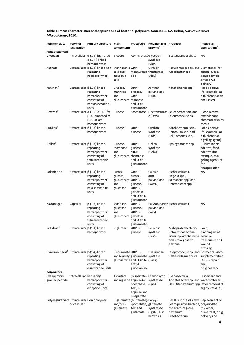
Bacteria can produce four major classes of polymers: polysaccharides, polyesters,
inorganic polyanhydrides (such as polyphosphates) and polyamides (Figure 1), with
different chemical and material properties, from various carbon sources (Rehm, 2010).
A few of them is intracellular, while a vast amount of extracellular ones is known.
These macromolecules cover a broad range of biological functions, such as reserve
material, or constituent of a protective structure, and help bacteria survive under
stressing environmental conditions. Their biosynthesis, as well as their properties, is
controlled by quite complex regulatory paths, acting in response of external stimuli.

3
Figure 1: some examples of polymers of bacterial origin. Intracellular polymers are represented by Glycogen (a), a polysaccharide, Cyanophycin (f), a polyamide, polyhydroxyalkanoate (h) and Polyphosphate, a polyanhydride (i); among extracellular ones we find the polysaccharides Xanthan (b), Alginate (d), which is secreted and/or is part of cyst cell wall, Dextran (e) and the secreted polyamide poly-γ-glutamate (g). A capsular polymer is the K30 antigen (c), a polysaccharide. The most common polymerization degree found for each polymer is indicated by numbers below the chemical structure brackets. Source: B.H.A. Rehm, Nature Reviews Microbiology, 2010.
In the last years, the biosynthesis of bacterial polymer has been extensively
investigated, particularly dealing with its molecular mechanism and regulatory aspects;
discoveries in the field represent a strong advance for biotechnology, and are the first,
necessary step to the production of tailor-made biopolymers, suitable for specific, high-
value industrial as well as medical application, by means of rational engineering of
bacteria. Table 1 summarizes current knowledge about bacterial polymers and their
possible field of application.
4
Table 1: main characteristics and applications of bacterial polymers. Source: B.H.A. Rehm, Nature Reviews Microbiology, 2010.
Polymer class Polymer localization
Primary structure Main components
Precursors Polymerizing enzyme*
Producer Industrial applications‡
Polysaccharides Glycogen Intracellular α-(1,6)-branched
α-(1,4 )-linked homopolymer
Glucose ADP–glucose Glycogen synthase (GlgA)
Bacteria and archaea NA
Alginate Extracellular β-(1,4)-linked non-repeating heteropolymer
Mannuronic acid and guluronic acid
GDP–mannuronic acid
Glycosyl transferase (Alg8)
Pseudomonas spp. and Azotobacter spp.
Biomaterial (for example, as a tissue scaffold or for drug delivery)
Xanthan§ Extracellular β-(1,4)-linked repeating heteropolymer consisting of pentasaccharide units
Glucose, mannose and glucuronate
UDP–glucose, GDP–mannose and UDP–glucuronate
Xanthan polymerase (GumE)
Xanthomonas spp. Food additive (for example, as a thickener or an emulsifier)
Dextran§ Extracellular α-(1,2)/α-(1,3)/α-(1,4)-branched α-(1,6)-linked homopolymer
Glucose Saccharose Dextransucrase (DsrS)
Leuconostoc spp. and Streptococcus spp.
Blood plasma extender and chromatograp-hy media
Curdlan§ Extracellular β-(1,3)-linked homopolymer
Glucose UDP–glucose
Curdlan synthase (CrdS)
Agrobacterium spp., Rhizobium spp. and Cellulomonas spp.
Food additive (for example, as a thickener or a gelling agent)
Gellan§ Extracellular β-(1,3)-linked repeating heteropolymer consisting of tetrasaccharide units
Glucose, rhamnose and glucuronate
UDP–glucose, dTDP–rhamnose and UDP–glucuronate
Gellan synthase (GelG)
Sphingomonas spp. Culture media additive, food additive (for example, as a gelling agent) or for encapsulation
Colanic acid Extracellular β-(1,4)-linked repeating heteropolymer consisting of hexasaccharide units
Fucose, glucose, glucuronate and galactose
GDP–L-fucose, UDP–D-glucose, UDP–D-galactose and UDP–D-glucuronate
Colanic acid polymerase (WcaD)
Escherichia coli, Shigella spp., Salmonella spp. and Enterobacter spp.
NA
K30 antigen Capsular β-(1,2)-linked repeating heteropolymer consisting of tetrasaccharide units
Mannose, galactose and glucuronate
UDP–D-glucose, UDP–D-galactose and UDP–D-glucuronate
Polysaccharide polymerase (Wzy)
Escherichia coli NA
Cellulose§ Extracellular β-(1,4)-linked homopolymer
D-glucose UDP–D-glucose
Cellulose synthase (BcsA)
Alphaproteobacteria, Betaproteobacteria, Gammaproteobacteria and Gram-positive bacteria
Food, diaphragms of acoustic transducers and wound dressing
Hyaluronic acid§ Extracellular β-(1,4)-linked repeating heteropolymer consisting of disaccharide units
Glucuronate and N-acetyl glucosamine
UDP–D-glucuronate and UDP–N-acetyl glucosamine
Hyaluronan synthase (HasA)
Streptococcus spp. and Pasteurella multocida
Cosmetics, visco-supplementation, tissue repair and drug delivery
Polyamides Cyanophycin granule peptide
Intracellular Repeating heteropolymer consisting of dipeptide units
Aspartate and arginine
(β-spartate-arginine)3-phosphate, ATP, L-arginine and L-aspartate
Cyanophycin synthetase (CphA)
Cyanobacteria, Acinetobacter spp. and Desulfitobacterium spp.
Dispersant and water softener (after removal of arginyl residues)
Poly-γ-glutamate Extracellular or capsular
Homopolymer D-glutamate and/or L-glutamate
(Glutamate)n
-phosphate, ATP and glutamate
Poly-γ-glutamate synthetase (PgsBC; also known as
Bacillus spp. and a few Gram-positive bacteria, the Gram-negative bacterium Fusobacterium
Replacement of polyacrylate, thickener, humectant, drug delivery and
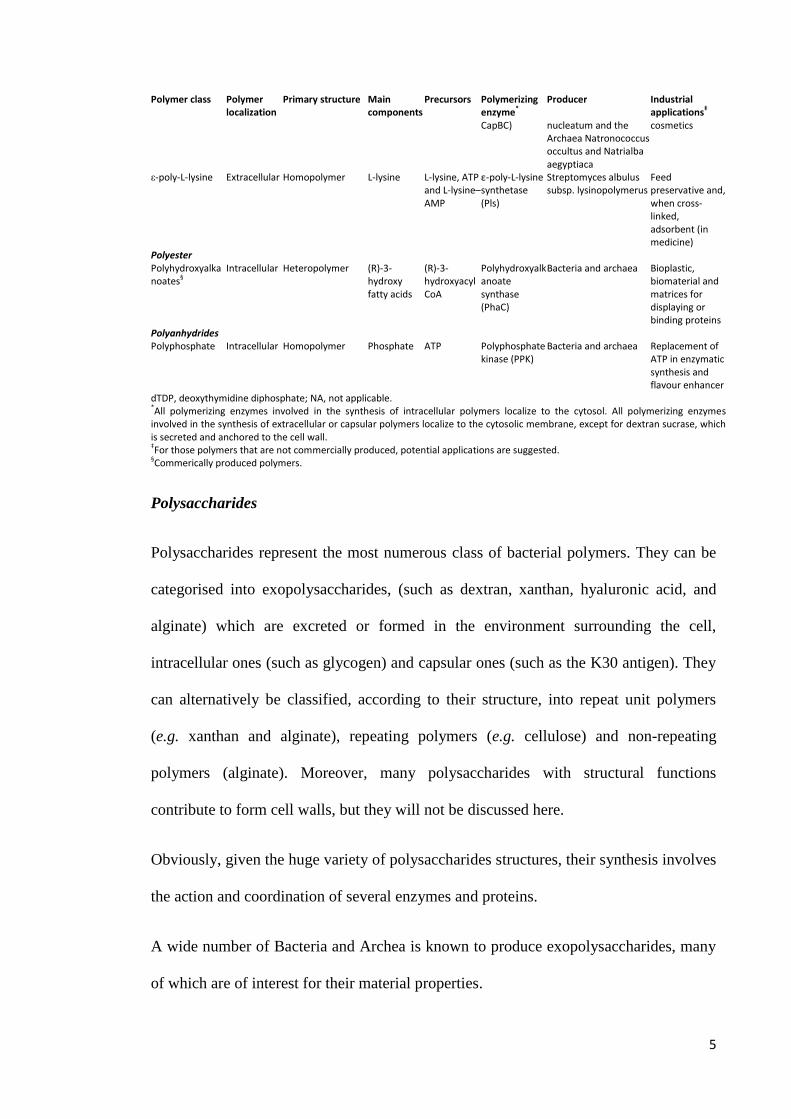
5
Polymer class Polymer localization
Primary structure Main components
Precursors Polymerizing enzyme*
Producer Industrial applications‡
CapBC) nucleatum and the Archaea Natronococcus occultus and Natrialba aegyptiaca
cosmetics
ε-poly-L-lysine Extracellular Homopolymer L-lysine L-lysine, ATP and L-lysine–AMP
ε-poly-L-lysine synthetase (Pls)
Streptomyces albulus subsp. lysinopolymerus
Feed preservative and, when cross-linked, adsorbent (in medicine)
Polyester Polyhydroxyalkanoates§
Intracellular Heteropolymer (R)-3-hydroxy fatty acids
(R)-3-hydroxyacyl CoA
Polyhydroxyalkanoate synthase (PhaC)
Bacteria and archaea Bioplastic, biomaterial and matrices for displaying or binding proteins
Polyanhydrides Polyphosphate Intracellular Homopolymer Phosphate ATP Polyphosphate
kinase (PPK) Bacteria and archaea Replacement of
ATP in enzymatic synthesis and flavour enhancer
dTDP, deoxythymidine diphosphate; NA, not applicable. *All polymerizing enzymes involved in the synthesis of intracellular polymers localize to the cytosol. All polymerizing enzymes involved in the synthesis of extracellular or capsular polymers localize to the cytosolic membrane, except for dextran sucrase, which is secreted and anchored to the cell wall. ‡For those polymers that are not commercially produced, potential applications are suggested. §Commerically produced polymers.
Polysaccharides
Polysaccharides represent the most numerous class of bacterial polymers. They can be
categorised into exopolysaccharides, (such as dextran, xanthan, hyaluronic acid, and
alginate) which are excreted or formed in the environment surrounding the cell,
intracellular ones (such as glycogen) and capsular ones (such as the K30 antigen). They
can alternatively be classified, according to their structure, into repeat unit polymers
(e.g. xanthan and alginate), repeating polymers (e.g. cellulose) and non-repeating
polymers (alginate). Moreover, many polysaccharides with structural functions
contribute to form cell walls, but they will not be discussed here.
Obviously, given the huge variety of polysaccharides structures, their synthesis involves
the action and coordination of several enzymes and proteins.
A wide number of Bacteria and Archea is known to produce exopolysaccharides, many
of which are of interest for their material properties.
6
Dextrans are synthesized thanks to a key enzyme called dextransucrase, with an average
molecular weight around 160 kDa, which is anchored to the cell wall; it belongs to the
family of glycoside hydrolase. The glycosidic bond in sucrose is hydrolyzed by
dextransucrase, and then glucose is transferred to the reducing end of the growing
glucan chain which is covalently bond. This reaction happens via an insertion
mechanism granted by two separate catalytic sites present in the same enzymatic active
site. Driving force of the process is the hydrolysis of the glycosidic bond (Robyt, 2008).
The resulting polymer is quite polydisperse (molecular mass is between 106 and 10
9 Da)
and soluble in water, where it has a Newtonian behavior, with a viscosity that depends
on concentration, molecular mass and temperature (Carrasco, 1989). Moreover, on the
biological point of view, it has a very low immunogenicity, and thus it has been used for
many pharmaceutical applications (Leathers, 2005).
Alginate is produced by a multiprotein complex extending between the periplasm, the
cytoplasmic membrane and the outer membrane (Kim, 1994) that, although extensively
studied, has still not disclosed all the aspects of the polymerization molecular
mechanism. Still, it is possible to genetically engineer bacteria constitutively able to
produce alginate (such as Pseudomonas fluorescens and Azobacter vinelandii) to get
polymers with controlled characteristics and material properties, which depend on
factors such as the molecular mass, degree of acetylation, sequence and molar ratio of
glucoronic and mannuronic acid constituents (Remminghorst, 2006; Steigedal, 2008).
The interest of material scientists for alginates is justified by the ability as viscosifying,
stabilizing, and water retaining agents.
Xanthan is another polymer of bacterial origin whose technological properties have
attracted much interest; it is an exopolysaccharide, and its structure is that of a repeating
unit heteropolymer, formed by pentasaccharides. Its biosynthesis has been elucidated
7
taking as a paradigm capsular polysaccharides (such as the K30 antigen); that don’t
have commercial interest themselves, since they often have the functions of virulence
factors. However, the study of their biosynthesis for medical and pharmaceutical
reasons provided a good model for the class of repeating unit polymers, which xanthane
belongs to. The process is initiated by glycosyl transferase, WbaP, a polyisoprenyl sugar
phosphate transferase anchored to the cell wall, that transfers the sugar phosphate from
the corresponding nucleotide sugar to undecaprenyl phosphate, and another
monofunctional glycosyl transferase enzyme called GumK. Polymerization reaction
happens at the cytoplasmic membrane, on the periplasmic side, thanks to a transport
protein called Wzx, specific for polysaccharides, which transfers the undecaprenyl
phosphate bond repeat unit across the membrane. It is believed that this enzyme has also
a role in controlling the polymer’s length (Wang, 1994; Wang, 1996; Drummelsmith,
1999; Tocilj, 2008).
Polyesters
When growth is limited by lack of nutrients like nitrogen or phosphorus, but at the same
time a rich carbon source is available, bacteria can accumulate a carbon reserve in the
form of polyhydroxyalkanoate (PHA) (Anderson, 1990; Kessler, 2001). The polymer is
kept inside the cell as an inclusion in which a hydrophobic core of PHA is surrounded
by the proteins which regulate its metabolism (Grage, 2009; Jendrossek, 2009). PHAs
are usually classified according to their chain length, into medium- chain- length (C6-
C14) and short-chain-length (C3-C5) ones; the former are produced by Pseudomonads,
the latter by numerous Bacteria and Archea. The biosynthesis of polyhydroxyalkanoate
is made possible by the PHA synthase enzyme (PhaC); almost any organic molecule
endowed with a hydroxyl and a carbonyl group can theoretically be polymerized to
form PHA, via the transformation in the corresponding CoA thioesther. This is possible

8
thanks to the broad substrate specificity of PhaC (Rehm, 1999). Its composition is
extremely variable, since more than 150 constituents have been identified so far,
therefore also its physical-chemical properties can dramatically differ: for instance,
melting temperatures range from 50 to 180°C, and crystallinity between 30 and 70%.
This fact is of great interest with regard to the possible technological use of PHA as
biocompatible and biodegradable plastic, replacing oil-based products, particularly in
the field of thermoplastic resins, considering that some two-thirds of the current oil-
deriving commodities belong to this category (Andrade, 2003).
Polyanhydrides
Polyphosphate is the only polyanhydride present in living cells. Bacteria use it to
perform a variety of tasks, such as creating storage particles in the cell, or helping the
uptake of metal ions and DNA thanks to the formation of a membrane complex with
polyhydroxybutyrate (Reusch, 1988). Polyphosphate is also involved in bacterial
motility, response to stress, pathogenicity, and many other physiological activities (Rao,
2009). Its biosynthesis is performed by the key enzyme polyphosphate kinase (PPK),
which can produce it from ATP. Since it is also able to perform the inverse reaction,
namely the phosphorilation of ADP to give ATP, this enzyme is also used in enzymatic
synthetic processes that are ATP dependent to recycle expensive ATP using cheap
polyphosphate (Kameda, 2001). The polymer itself has many applications, from flavor
enhancer in food industry to flame retardant, with a performance comparable to that of
asbestos. Its production by bacteria is not economically affordable since it is easily
obtained by inorganic resources, but it can be exploited as a means to remove phosphate
from industrial waste water, thus contributing to the removal of pollutants.
9
Polyamides
Among other biopolymers which bacteria are able to synthesize, three different
polyamides are known, namely poly-γ-glutamic acid (PGA), poly-ε-lysine, and multi-L-
arginyl-poly (L-aspartic acid), also known as cyanophycin. Their main producers belong
to the genus Bacillus, as well as to Cnidaria and cyanobacteria. Their possible use in
material science makes them a remarkable subject of research.
Polyamides are defined as polymeric compounds whose constituents are linked by
amide bonds. They can be further divided in homopolyamides, which are composed by
just one type of amino acid monomers, and copolyamides, consisting of different amino
acids. Obviously, proteins represent the most numerous category of known
copolyamides; a small number of compounds are indeed known as poly (amino acids),
since their biosynthesis is significantly different from proteins’ one. In particular, four
capital features distinguish proteins from poly(amino acids): in poly(amino acids), only
one amino acid monomer is present, at least in its backbone, while proteins incorporate
usually up to 21 amino acids; the biosynthesis of poly(amino acid) does not involve
ribosomial transcription, but is effected by enzymes, whose characteristics and
coordination is relatively simple; thus, poly(amino acids), differently from proteins, are
highly polydisperse and show a great size distribution, while proteins present
monodispersity and a well defined and precise length. Finally, in poly (amino acids) the
amide bonds which keep the structure together involve side chain functions and not just
α- amino and α- carboxylic groups as it happens in proteins.
In nature, three poly(amino acids) have been discovered to date, i.e. cyanophicycin,
composed by α-aspartic acid monomers bond to arginine residues at the β-carboxylic
group position, poly(lysine), formed by lysine monomers connected by linkages
between the α-carboxylic group and the ε- amino group, and poly-γ-glutamic acid

10
(PGA), in which the polymer backbone is formed by glutamic acid residues whose α-
amino group is bond to the γ-carboxylic one (Table 2).
Table 2: poly (amino acid)s in Nature and their main features. Adapted from Oppermann- Sanio, 2002.
Poly(amino acid) Distribution in organisms Applications
Γ-poly (glutamic acid)
Bacillus anthracis
B. licheniformis
B. megaterium
B. subtilis
Sporosarcina halophila
Planococcus halophila
Natrialba aegyptiaca
Nematocysts of Cnidaria
Dispersant
Water softener
Waste water treatment
Thickener in food and cosmetics
Superabsorber
Drug delivery devices
Humectant in cosmetics
Treatment of leather
Poly (lysine)
Streptomyces albulus
lysinopolymerus
Superabsorber
Drug delivery devices
Additive to animal feeding stuff
Cyanophycin
Most cyanobacterial species
Dispersant
Water softener (after removal of arginyl
residues)
Although evidence so far indicates the existence of these very three poly (amino acids),
it is indeed possible that many others are still waiting to be discovered and investigated.

11
Cyanophycin
Cyanophycin granule polypeptide (CGP), also called cyanophycin, is a compound
occurring exclusively (Simon 1987) in all cyanobacterial groups, of all kinds, nitrogen
fixing and non-nitrogen fixing, unicellular and filamentous (Lawry, 1982; Allen, 1988;
Golecki, 1991). The discovery of it by Borzi, who detected the material microscopically
as cell inclusions with highly refractive features, dates back to 1887. The physiological
role of cyanophycin is that of intracellular temporary nitrogen storage. In fact, every
polymer’s building block contains five nitrogen atoms, and the polymer itself, at cell
internal pH and ionic strength, is insoluble. Cyanophycin is accumulated during
stationary growth phase of the cell, while it is present at very low levels during
exponential and balanced growth phase. Moreover, cyanobacteria are able to detect the
nitrogen level in the medium surrounding them, and therefore to trigger the production
of cyanophycin to protect themselves from nitrogen depletion (Liotenber, 1996). The
polymerization of cyanophycin is catalysed by an enzyme called CGP synthetase or
CphA; polymer synthesis requires the presence of ATP, ions such as K+ and Mg
2+, a
thiol reagent and a CPG primer (Aboumalgd,. 2001; Simon, 1976; Ziegler, 1998) ; and
probably resembles an amide-ligase dependent process (Berg, 2000). Experimental data
suggests that CphA synthetase should form a homodimer endowed with two binding
sites for ATP and substrate, respectively in order to include both arginine and aspartate
in the polymer (Krehenbrink, 2004). Degradation of the polymer to allow bacteria to
exploit the nitrogen reserve is granted by the enzyme cyanophycinase, or CphB.
Isolation of cyanophycin is possible by repeated centrifugation of disrupted cells from
culture broth (Simon 1971); polymer purification is achieved, taking advantage of its
solubility properties, by repeated dissolution in acidic solution, followed by
centrifugation and precipitation in neutral conditions. In fact, cyanphycin is soluble in

12
acidic conditions, under pH 2, as well as in basic ones, over pH 9, but insoluble in
physiological conditions and at low ionic strength. It can, however, be dissolved in
concentrated urea solution. It is also pretty insoluble in the most common organic
solvents, such as methanol, DMSO, DMF. Quantification of the polymer in cells is
possible by means of HPLC of the amino acid components after isolation (Simon 1973),
or by NMR of the crude product (Aboulgmad, 2000). On the structural point of view,
cyanophycin is formed by aspartic acid and arginine in almost equimolar amounts;
aspartic acid forms the polymer backbone, while arginine is bond to the aspartic β-
carboxylic group. Its molecular weight may vary between (as was estimated by SDS
polyacrilamide electrophoresis) 25 and 100 kDa, thus it is quite polydisperse (Simon
1973, 1976). Circular dichroism and Raman studies allowed to make hypothesis about
the polymer’s secondary structure, and it is commonly believed to assume a β-sheet
conformation under acidic solution and likely, it was inferred, in the insoluble form
(Simon 1980). Structural studies were also confirmed by 1H,
13C, and
15N NMR
spectroscopy (Suarez 1999). Cyanophycin may be used, after chemical modification to
reduce its arginine content (Joentgen, 1998) as a good biodegradable substitute of
polyacrilate (Schwamborn, 1998); however, it is still not a practical material on the
industrial point of view, since its biosynthesis, both from native cyanobacteria of from
recombinant E.coli strains optimized for production, is too expensive to make it cost-
effective (Hai, 2000; Ziegler, 1998; Oppermann, 1999; Aboumalgd, 2000).
Poly(lysine)
The only organism, among bacteria and eukaryotes, so far known to be able to produce
poly (lysine) (PL), is the Gram-positive bacterium Streptomyces albulus ssp.
lysinopolymerus strain 346. It was isolated from Japanese soil at the end of the 1970s.

13
This bacterium is constituently able to excrete the biopolymer in the medium at
concentrations of up to 4-5 g/L (Shima, 1977, 1981).
Isolation of poly (lysine) is possible by means of ionic exchange chromatography of the
culture filtrate, since it is a cationic polymer and neutral pH, and precipitation with
ethanol/diethylether (Shima, Sakai, 1981). Polymer filaments are typically composed by
25-30 residues; therefore its molecular weight is not high.
Its physiological function is mainly that of an antibiotic; in fact, poly (lysine) can inhibit
the growth of both Gram positive and Gram negative bacteria already at concentration
around 1-8 mg/L (Shima, 1982, 1984) and it can also inactivate bacteriophages.
In view of these properties, poly (lysine) has been proposed as a preservative for animal
feed. It is also able to form hydrogels highly capable of water adsorption (Choi, 1995,
Kunioka, 1995) and, if crosslinked it can be used in pharmaceutical applications as a
cationic adsorbent (Hyrayama, 1999). Poly (lysine) is nowadays produced
biotechnologically by means of mutant strains optimized for the polymer yield (Hiraki,
1999).
Poly-γ-glutamic-acid
Figure 2: Poly-γ-glutamic- acid
Poly-γ-glutamic- acid, also known as γ-PGA, is an anionic homopolyamide formed by
D- and L- glutamic acid units, unusually connected by amide bonds between α-amino
and γ-carboxylic acid groups. It was first isolated by Ivanovics and coworkers
(Ivanovics, 1937a; 1937b) in 1937 as the constituent of the capsule of Bacillus

14
anthracis; it is also known to be an important part of the mucilage of several soybean-
fermented traditional Eastern foods, such as the Japanase natto (Sawamura, 1913, Fujii,
1963) and the Korean seasoning chungkookjang (Ashiuchi, 2005) (Figure 3).
Figure 3: Natto, a traditional Japanese dish naturally containing γ-PGA.
To date, it is known to be produced by some Archea, such as Natronococcus occultus
(Niemitz, 1997) and Natrialba aegypytiaca (Hezayen, 2001), and several Gram positive
bacteria strains, all belonging to the genus Bacillus, and so phylogenetically related.
Recently, also the Gram negative bacterium Fusobacterium nucleatum was found to be
a γ-PGA producer (Candela, 2009). The only eukaryote organisms in which it is known
to be present are some Cnidaria, animals armed with stringing cells called nematocysts
(Weber, 1990).
Γ-PGA possesses many peculiar properties; among these, to be water-soluble (in its
anionic, salt-form), biodegradable, non-toxic, non immunogenic, and even edible. As a
polyamide, it is also quite robust with regard to hydrolytic degradation, both in acid and
basic conditions. Therefore, the potential applications of this material cover a broad
15
range in industrial fields such as food, cosmetics, medicine, waste-water treatment, and
will later be discussed.
Physiological function of γ-PGA
Poly-γ-glutamic- acid has several physiological functions, which may differ a lot
according to the specific needs of the producer species; what is constant in all cases, as
it will be illustrated, is the role of the biopolymer as an adaptation agent in hostile
environments (Ashiuchi, 2002).
B. anthracis (a Gram-positive, sporulating bacterium) is notoriously the causal agent of
the lethal and infectious human disease called anthrax. Its virulence has been found to
be correlated with the presence of an anchored capsule composed by poly-γ-D-PGA
(Makino, 1989); this capsule (which is, on the stereochemical point of view, composed
by only D-glutamic acid monomers) enables the bacteria to avoid phagocytosis
(Zwartouw, 1956), and makes it particularly non-immunogenic. Moreover, it prevent
antibodies from accessing the bacterium (Mesnage, 1998), and, acting as a passive
barrier, protect B. anthracis against phage infections (Mc Cloy, 1951).Poly-γ-D-PGA is,
in this case, an essential virulence factor as well as a structural component.
Other bacterial strains release poly-γ-glutamic- acid in the environment, with different
purposes.
Some B. subtilis and B. licheniformis strains, in fact, take advantage of high-
cryoprotectant properties of the polymer to survive in the cold (Mitsuiki, 1998); other
Bacillus soil-strains release the polymer to sequestrate toxic metal ions, to increase their
resistance (Mc Lean 1990, 1992). Poly-γ-glutamic- acid is also used as a protective
agent against high-saline conditions, thanks to its prominent water binding capacity, by
halophilic Archea such as Natrialba aegyptica (Hezayen, 2001). Finally, the polymer

16
can be used as a source of glutamate in starvation conditions (Kimura, 2004), or as a
storage compound for carbon and nitrogen precursor (Schreier, 1993). A peculiar case is
that of the animal called Hydra (belonging to the phylum Cnidaria), in which γ-PGA is
a secretory product of stringing cells which serves as an adhesive to adhere to the
surface of the aggressor, or prey, while Hydra injects toxic venoms in it, as well as to
regulate internal osmotic pressure necessary for explosive evagination (Weber, 1990).
In summary, currently known data support the hypothesis that γ-PGA, when is released
by the bacterium, subjected to external stress, in the environment, serves to increase the
producer’s survival chances when (Gross, 1998; Hezayen, 2000), acting as a persistence
agent, while anchored γ-PGA is considered a bacterial virulence factor (Candela and
Fouet, 2006).
Figure 4: γ-PGA in vivo: anchored (upper left), released (upper right) and secreted by stringing cells (bottom centre).
17
Chemical properties of γ-PGA
The main chemical peculiarity of γ-PGA, with differentiates it from other polypeptides
and proteins both structurally and functionally, is that glutamic residues are non-
ribosomally polymerized via the γ-amino bond instead of the α-amino one.
This property, common to all the polypeptides produced by bacteria such as B.
anthracis,(Bruckner, 1953) B. subtilis (Chibnall, 1958), B. licheniformis,(Troy, 1973)
and B. megaterium (Torii, 1973), has been historically demonstrated by a variety of
methods, going from enzyme assay using trypsin, which is able to cleave only and just
α-amino bonds, and to which γ-PGA proved to be stable (Haurowits, 1949), to infrared
spectroscopy (Hanby, 1950) to proton and carbon nuclear magnetic resonance
spectroscopy applied to structural analysis (Birrer, 1994; Borbély, 1994; Pérez-Camero,
1999). Another method of practical importance is chemical degradation in which γ-PGA
will yield γ-amino-δ-hydroxyvaleric acid as a monomer, while α-PGA would instead
result in α-amino-δ-hydroxyvaleric acid (Nitecki, 1971).
The stereochemical properties of γ-PGA obviously depend on its enantiomeric content,
which can be studied by means of both chemical and enzymatic methods; the former are
the most easily available. In particular, a convenient and simple method is represented
by total hydrolysis of the polymer followed by chiral chromatography (Kunioka, 1995),
or derivatisation with Marfey’s reagent (1-fluoro-2, 4-dinitrophenyl-5-L-alanine amide)
and analysis of the resulting diastereoisomers by reverse-phase HPLC (Cromwick,
1995).
As a matter of fact, three stereochemically different types of poly-γ-glutamic- acid have
been found out among products of bacterial origin (Tanaka, 1997; Ashiuchi, 2003):
homopolymer composed only by D-glutamic acid (as in the case of the capsule of B.

18
anthracis), or L-glutamic monomer (as it happens in Hydra); copolymer in which D-
and L- momomers are lined up in different ratios (as it is found in the poly-γ-glutamate
which is part of extracellular mucilage in many ) B. subtilis strains). Some
representative data are listed in Table 3.
Table 3. Enantiomeric content of γ-PGA produced by some different organisms
Organism Content (%) Reference
D-Glutamate L-Glutamate
B. anthracis 100 0 Hanby (1946)
Planococcus halophilus 100 0 Kandler(1983)
Sporosarchia halophila 100 0 Kandlar (1983)
B. licheniformis 10-100 90-0 Pérez- Camero (1999)
B. subtilis natto 50-80 50-20 Kubota (1993)
B. subtilis
(chungkoojang)
60-70 40-30 Ashiuchi (2001)
Staphylococcus
epidermidis
50 50 Kocianova (2005)
B. megaterium 50 50 Toril (1956)
B. halodurans 0 100 Aono (1987)
Natrialba aegyptiaca 0 100 Hezayen (2001)
Hydra 0 100 Weber (1990)
As it can be seen, the enantiomeric content may vary a lot, depending on the producer
species, strain and even on the culture conditions, as it will be shown; this variability
has been representing a longstanding problem, bearing both theoretical as well as
practical relevance, for the study and development of bacterial produced γ-PGA.
In most cases the natural gamma-PGA is composed of a mixture of D- and L-glutamic
acid residues, whose ratio can affect its conformation and the orientation of biopolymer
functional groups, with clear consequences on its reactivity. Conflicting reports have
been published on cultivation parameters that control both polymer molecular weight
and stereochemistry, but no conclusive assumptions can be drawn; in fact, every wild

19
isolate which has been utilized as gamma-PGA producer behaves differently and no
optimisation protocol can be simply adapted passing from one strain to another.
Nevertheless, the large amount of scientific literature available is useful in defining the
variables to be tested, which appear to be the addition of L-glutamic acid, the carbon
sources (citric acid, glucose, glycerol, fructose), the nitrogen sources (ammonium salts,
urea), mineral ions (K2HPO4, NaCl, CaCl2, FeCl3, Mg/MnSO4); minor parameters that
affect productivity and quality of gamma-PGA include the aeration and pH of the
medium composition (Cromwick, 1995; Cromwick, 1996; Ashiuchi, 2001; Jung, 2005;
Bajaj, 2009). It is not surprising that the investigation of the parameters for controlling
gamma-PGA molecular weight and composition is not only of fundamental interest but
of practical importance for the exploitation of this biopolymer as an efficient enzyme
carrier and for its commercial development thereof.
An exemplar case may be considered that of γ-PGA formed by B. licheniformis strain
ATC9945A. Conflicting opinions have been existing for decades (Leonard, 1958; Troy,
1973) whether the stereochemical content of the formed biopolymer was affected by the
concentration of Mn2+
salt in the culture medium, or not; first collected data proved to
be ambiguous or inconclusive, but as a matter of fact, the enantiomeric proportion of the
polymer could vary from 38 % to 86% in the D-glutamic acid monomer from culture to
culture, in a seemingly unpredictable way. The issue was finally solved in 1995 by
Cromwick and Gross, who were able to definitely establish that, for this particular
strain, enantiomeric composition is sensitive to the presence of MnSO4; precisely, L-
glutamic monomer content ranged from 59 to 0 % for a MnSO4 concentration between
0 to 615 μM, respectively. These results were later confirmed by Pérez-Camero (1999).
20
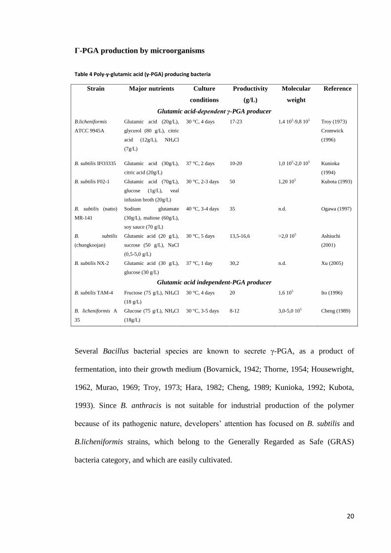
Γ-PGA production by microorganisms
Table 4 Poly-γ-glutamic acid (γ-PGA) producing bacteria
Strain Major nutrients Culture
conditions
Productivity
(g/L)
Molecular
weight
Reference
Glutamic acid-dependent γ-PGA producer
B.licheniformis
ATCC 9945A
Glutamic acid (20g/L),
glycerol (80 g/L), citric
acid (12g/L), NH4Cl
(7g/L)
30 °C, 4 days 17-23 1,4 105-9,8 105 Troy (1973)
Cromwick
(1996)
B. subtilis IFO3335 Glutamic acid (30g/L),
citric acid (20g/L)
37 °C, 2 days 10-20 1,0 105-2,0 105 Kunioka
(1994)
B. subtilis F02-1 Glutamic acid (70g/L),
glucose (1g/L), veal
infusion broth (20g/L)
30 °C, 2-3 days 50 1,20 105 Kubota (1993)
B. subtilis (natto)
MR-141
Sodium glutamate
(30g/L), maltose (60g/L),
soy sauce (70 g/L)
40 °C, 3-4 days 35 n.d. Ogawa (1997)
B. subtilis
(chungkoojan)
Glutamic acid (20 g/L),
sucrose (50 g/L), NaCl
(0,5-5,0 g/L)
30 °C, 5 days 13,5-16,6 >2,0 105 Ashiuchi
(2001)
B. subtilis NX-2 Glutamic acid (30 g/L),
glucose (30 g/L)
37 °C, 1 day 30,2 n.d. Xu (2005)
Glutamic acid independent-PGA producer
B. subtilis TAM-4 Fructose (75 g/L), NH4Cl
(18 g/L)
30 °C, 4 days 20 1,6 105 Ito (1996)
B. licheniformis A
35
Glucose (75 g/L), NH4Cl
(18g/L)
30 °C, 3-5 days 8-12 3,0-5,0 105 Cheng (1989)
Several Bacillus bacterial species are known to secrete γ-PGA, as a product of
fermentation, into their growth medium (Bovarnick, 1942; Thorne, 1954; Housewright,
1962, Murao, 1969; Troy, 1973; Hara, 1982; Cheng, 1989; Kunioka, 1992; Kubota,
1993). Since B. anthracis is not suitable for industrial production of the polymer
because of its pathogenic nature, developers’ attention has focused on B. subtilis and
B.licheniformis strains, which belong to the Generally Regarded as Safe (GRAS)
bacteria category, and which are easily cultivated.

21
Table 4 lists some significant examples, reporting the principal features of the most
useful strains in terms of industrial application, paying attention in particular to their
productivity and to the best culture conditions developed so far.
Extensive investigation has been performed on these strains, with the aim of optimizing
nutrient requirements and culture conditions to boost cell growth and γ-PGA
production. It must be noted that the overall approach has been, until recent times,
heuristic. Many questions remain open about genes and enzymes, as well as the
metabolic pathways involved and related to γ-PGA synthesis. Many aspects such as
variation in molecular weight and enantiomeric composition of the final product still
have to be clarified, too.
In fact, it has been found that nutrient requirements (carbon source, nitrogen sources,
metal ions) required for cell growth and γ-PGA production depend on the particular
bacterial strain used; factors such as ionic strength, aeration, pH of the medium also
play an important role and affect productivity and quality of the resulting polymer.
Culture conditions, specific for each strain, have in fact a strong impact on its amount,
molecular weight and enantiomeric composition (Sung, 2005; Jung, 2006).
Moreover, evidence suggests that also synthetic mechanism involved differ between
each strain.
According to nutrient necessity, bacteria capable of producing γ-PGA are divided in two
principal categories: the former requires the addition of L-glutamic acid to the medium
in order to let the cells grow and produce the polymer; the latter does not require its
addition for γ-PGA production.
The former group, exogenous L-glutamic acid dependent bacteria, include B. anthracis
(although, as already said above, this species has no applicative interest) (Thorne,

22
1953), B.licheniformis ATCC 9945A (Troy, 1973), B. subtilis IFO3335 (Kunioka,
1994), B. subtilis F02-1 (Kubota, 1993), B. subtilis (natto) MR-141 (Ogawa,1 997), B.
subtilis (chungkoojan) (Ashiuchi 2001), B. subtilis NX-2 (Xu, 2005).
The latter group, namely exogenous independent L-glutamic acid bacteria, comprise B.
subtilis TAM-4 (Ito 1996), B. licheniformis A 35 (Cheng 1989), as well as B. subtilis 5
E (Shih, 2001) and B. licheniformis S173 (Kambourova, 2001).
It is worth noting and remarked here that the study of carbon source utilization
demonstrated that in most γ-PGA producers glutamic acid monomers incorporated in
the polymer derive even, in part, for the exogenous L-glutamic acid dependent strains,
from a de novo biosynthetic pathway via tricarboxylic acid cycle, starting from citric
acid or glucose.
Intracellular L-glutamic acid is presumed to be produced from citric acid via isocitric
acid and subsequently α-ketoglutaric (2-oxoglutaric) acid, which, on his turn, may be
converted to the desired product in two different ways.
As a matter of fact, this hypothesis is consistent with experimental data: metabolic
investigation performed by Cromwick and Gross on the B. licheniformis ATCC 9945
strain (Cromwick, 1995) by means of NMR spectroscopy and 13
C labeled citrate
showed that an amount equal to 10 to 30 %, depending on culture conditions such as
pH, of the monomers incorporated in the polymer derived from citrate rather than
directly from L-glutamic acid present in the growth medium. Similar observations were
made for other γ-PGA producer strains.
Biosynthesis of γ-PGA
Although extensively studied, the biosynthesis of γ-PGA is still not totally understood.

23
Since both L- and D- monomers of glutamic acid may be included in the polymer
backbone, the racemisation of L-glutamic acid has been an intriguing and debated topic.
It has been supposedly explained by three distinct mechanism: the indirect action of an
aminotransferase (Thorne, 1954), the direct one of a glutamic acid racemase Glr
(Ashiuchi, 1998) and the direct action of a different glutamic acid racemase YrpC
(Ashiuchi, Misono, 2002), too. Evidence shows that the indirect process is probably not
adopted in vivo since the activity of the aminotransferase decreases during polymer
production (Shih, 2005a-b); it was also shown that Glr racemase, a cytosolic enzyme
highly selective for L-glutamic acid (Thorne, 1955; Ashiuchi, 1998; 2002) is the only
agent involved in monomer racemisation (Ashiuchi, 2002).
Polymerization happens in a ribosomal-independent way, by means of a membrane-
associated complex requiring L-glutamate, ATP and Mg2+
as cofactors. Two
mechanisms have been proposed. According to the first, proposed by Troy (Troy, 1973;
Gardner, 1979) who first identified a membrane associated enzymatic complex
responsible for PGA synthesis in 1973, the polymer is formed in four subsequent steps:
first, glutamate is activated by an ATP molecule (1) forming a γ-bond with AMP, which
is bond to the amino acid via a γ-linkage; then, activated glutamate is transferred to a S-
protein, generating a thioester (2); isomerisation may occur (3) and finally the glutamyl
residue is transferred to the growing PGA chain (4).
L–glutamic acid+ATP→γ–L–glutamyl–AMP+PPi (1)
γ–L–glutamyl–AMP+SH–enzyme→γ–X–glutamyl –S–enzyme+AMP (2)
γ–X–glutamyl–S–enzyme→γ–D–glutamyl–S–enzyme (3)
γ–D–glutamyl–S–enzyme+[γ–D–glutamyl]n→[γ–D–glutamyl]n+1+SH–enzyme (4)

24
This mechanism is often referred to as the thiotemplate mechanism.
One of the alleged evidences in favor of this mechanism was the fact that the incubation
of bacterial membranes, which the enzymatic complex is bound to, resulted, in presence
of hydroxylamine, in the formation of the respective γ-glutamyl derivative.
More recently, Ashiuchi proposed an entirely different mechanism, starting from the
observation that hydroxamate was actually not found after incubation of hydroxylamine
with the synthetic enzymatic complex prepared in vitro (Ashiuchi, 2001b, 2004).
According to this second mechanistic proposal, it is γ-PGA itself to be activated by an
ATP molecule; formation of an activated PGA species is then followed by glutamate
monomer transfer, in what has been defined as an amide ligation fashion. The driving
force of the process is provided by the cleavage of ATP to ADP.
The two mechanisms are incompatible, and the latter hypothesis seems to be more in
accordance with experimental data. Still, conclusive studies have to be performed.
Much still lies ahead to be elucidated on the topic; what has been ascertained, however,
is that the synthesis is catalyzed by an enzymatic complex with 4 subunits. The
membrane-bound γ-PGA enzymatic synthetic complex, called PgsBCA, has been also
studied in fine detail, but so far its isolation in the active state was impossible due to its
high unstable and hydrophobic nature (Ashiuchi, 2004). What has been ascertained is
that it is able to accept both D- and L- glutamic acid monomers as substrates and that it
has no racemase activity; the catalytic site is formed by both its subunits PgsB and
PgsC, while subunit PgsA is responsible for the removal of the chain from the active
site, allowing its elongation step by step. The most peculiar property of this enzymatic
complex is that it is not stereospecific, a unique feature among transpeptidases

25
(Ashiuchi, 2001; 2004). Presence of Mg2+
seems to exert an action on the enzymatic
complex activity.
Finally, it has to be noticed that little is known about how γ-PGA filaments are released
from the cell.
Figure 5: glutamic acid biosynthesis via tricarboxylic acid cycle and polymerization to give γ-PGA; the enzymes involved are (1) glutamate dehydrogenase (GD), (2) glutamate 2-oxoglutarate(α-ketoglutarate) aminotransferase (GOGAT), (3) glutamine synthetase (GS), (4) L-glutamic acid: pyruvic acid aminotransferase, (5) alanine racemase, (6) D-glutamic: pyruvic acid aminotransferase, (7) PGA synthetase. Source: Shih, 2001.
Finally, it also has to be remarked that the enantiomeric ratio between D- and L-
glutamic acid monomers may be influenced by a number of factors which are still not
fully understood. Strong differences seem to occur between natural strains; bivalent

26
metal cofactors such as Mn2+
, Co2+
, and Zn2+
(Leonard, 1958; Gardner, 1979) seem to
influence racemisation.
Figure 6: synthetic pathway of -PGA in Bacillus species; A, B, C, E represent the enzymes involved in the synthesis
of released -PGA; CapD the enzyme responsible for the formation of capsular -PGA in B. anthracis.
Degradation of -PGA
Bioproduced -PGA has generally an average molecular weight between 105
and 106 Da
and polydispersities between 2 and 5. Generally speaking the control of the molecular
sizes of a polymer is particularly significant in order to tailor its proprieties and so it is
important to know how -PGA can be degraded.
-PGA is stable below 60°C and in the presence of ordinary proteases (Oppermann-
Sanio, 2002); in order to degrade it harsh conditions, such as prolonged exposure to
extreme pH, high temperature or ultrasonic irradiation, or specific enzymes are
necessary.
As a matter of fact, -PGA is synthesized by producers in the early stationary phase, and
later degraded in the late stationary phase.
27
Producing bacteria express two different proteases able to degrade -PGA: endo--
glutamyl-peptidase and exo--glutamyl-peptidase.
Production of endo--glutamyl-peptidase was shown to occur in various Bacilli
(Kunioka, 1993; Makino, 1988; King, 2000; Obst, 2004) and in some other bacteria
such as the bacteriophage NIT1 (Kimura, 2003). This enzyme usually is secreted in
the medium and cleaves high molecular weight -PGA into fragments as small as 105
Da (Goto, 1992; Kunioka, 1993; Kimura, 2004).
Nowadays, is not clear if this enzyme targets -DL (Ashiuchi, 2003) or -DD (Suzuki,
2003) bonds, however it is known that it strictly splits endo--glutamyl-bonds. This fact
has been demonstrated by Suzuki (2003) working with a clonated endo--glutamyl-
peptidase from B. subtilis IFO16449. In this work the authors show how the enzyme is
able to degrade high molecular mass -PGA giving a 490 kDa mass product and an 11
kDa one. Neither free glutamic acid nor -glutamyl oligomers were detected indicating
that such enzyme cleaves only endo-bonds. Furthermore the authors found out that the
enzyme is stable and active between pH 4 and 11 and in a temperature range from 4°C
to 45°C. Its activity is not inhibited by the treatment with 5 mM EDTA, divalent cations
nor 1mM phenylmethylsulphonyl fluoride while 1mM 4-(hydroxymercury) benzoate, a
sulphydril inhibitor, remarkably slow the activity suggesting that the enzyme
mechanism may be cysteine based, such as in DL-endopeptidase II (Smith, 2000). It
was also reported that in this hydrolysis there is a linear correlation between the
decrease in molecular weight and time (Tayal, 1999).
Exo--glutamyl-peptidase, also known as GGT, is a key enzyme in glutathione
metabolism (Ogawa, 1991; Xu, 1996) but it does not seem to be involved in -PGA
synthesis in vivo (Williams, 1954; Ashiuchi, 2003). However there are some evidences

28
that it is involved in formation of B. anthracis capsula, binding -PGA to membrane
peptidoglycanes (Candela, 2005), and in bioproduced polymer cleavage in B. subtilis
(Kimura, 2004).
Degradation of -PGA can be carried out chemically by hydrolysis at extreme pH and
high temperature (Goto, 1992; Kubota, 1996) or physically using ultrasonic irradiation.
While hydrolytic methods are not deemed satisfactory because of the poor control
cleavage obtainable in the process and the high polydispersity of the resulting products
the use of ultrasonic waves seems to be an interesting alternative to enzymatic
hydrolysis. Pérez-Camero and co-workers (1998) have shown how exposure to 20000
Hz for 2 hours cleaves -PGA of 2100 KDa into 60 to 90 KDa fragments that exhibit a
narrowed polydispersity compared to the starting material. This method could be useful
to control the molecular weight of the bioproduced polymer optimizing its proprieties
for specific applications.
In conclusion, it has to be mentioned that, as it was reported by Muñoz-Guerra and
coworkers (Portilla-Arias, 2007), γ-PGA and its ester derivatives depolymerize when
heated over 200 °C in nitrogen atmosphere, releasing pyroglutamic acid and the
corresponding esters as volatiles; the residues, analyzed by NMR, were
spectroscopically indistinguishable from the polymer.

29
Figure 7 TGA profiles of γ-PGA (PGGA) and its methyl ester (PGGA-1), acquired at the heating rate of 10 °C min-1
under nitrogen atmosphere. The first modest weight loss under 200 °C is due to the release of water absorbed by the polymer during the preparation of the sample; polymer decomposition occurs in the range 250-300 °C, resulting in a loss of weight about 50 and 70 % of the initial mass, respectively. Source: Portilla-Arias, 2007.
Genetic aspects of -PGA synthesis
Poly--glutamic acid is unusual and it is different structurally and functionally from
proteins in that glutamate is polymerized via the -amide linkages, and thus should be
synthesized by a ribosome independent manner. Identification and analyses of genes
responsible for the synthesis of this polymer are extremely important to understand the
biosynthetic mechanism. Nowadays all the genes responsible for the three stages of -
PGA metabolism (racemisation, polymerization and degradation) have been identified
(Ashiuchi, 2002).
30
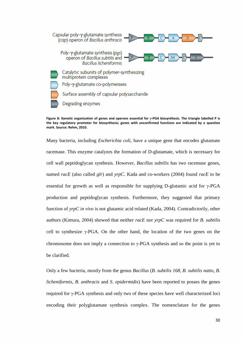
Figure 8: Genetic organization of genes and operons essential for -PGA biosynthesis. The triangle labelled P is the key regulatory promoter for biosynthesis; genes with unconfirmed functions are indicated by a question mark. Source: Rehm, 2010.
Many bacteria, including Escherichia coli, have a unique gene that encodes glutamate
racemase. This enzyme catalyzes the formation of D-glutamate, which is necessary for
cell wall peptidoglycan synthesis. However, Bacillus subtilis has two racemase genes,
named racE (also called glr) and yrpC. Kada and co-workers (2004) found racE to be
essential for growth as well as responsible for supplying D-glutamic acid for -PGA
production and peptidoglycan synthesis. Furthermore, they suggested that primary
function of yrpC in vivo is not glutamic acid related (Kada, 2004). Contradictorily, other
authors (Kimura, 2004) showed that neither racE nor yrpC was required for B. subtilis
cell to synthesize -PGA. On the other hand, the location of the two genes on the
chromosome does not imply a connection to -PGA synthesis and so the point is yet to
be clarified.
Only a few bacteria, mostly from the genus Bacillus (B. subtilis 168, B. subtilis natto, B.
licheniformis, B. anthracis and S. epidermidis) have been reported to posses the genes
required for -PGA synthesis and only two of these species have well characterized loci
encoding their polyglutamate synthesis complex. The nomenclature for the genes

31
involved in -PGA synthesis is one of two types, depending on whether the synthesized
-PGA is retained or released (Candela, 2005). If the -PGA is associated with the
bacterial surface and form a capsule, then the corresponding genes are named cap (for
capsule), whereas the corresponding genes are named pgs (for polyglutamate synthase)
if the -PGA is released.
In B. anthracis the cluster of genes encoding the -PGA synthesizing complex is located
on a plasmid (Uchida, 1987; Makino, 1988) while in other organism, the homologues
genes are located on the chromosome (Nagai, 1997; Ashiuchi, 2001). It is widely
accepted that all three pgsBCA genes are necessary and sufficient for -PGA production
in vivo (Ashiuchi, 1999; Ashiuchi, 2001); however it has also been observed that if one
of the three genes is disrupted, it can be complemented in a trans manner (Ashiuchi,
2004). Candela (2005) also outlined how even the previously overlooked capE is
essential for the production of -PGA in B. subtilis and B. anthracis. In opposition to the
findings of Ashiuchi, Urushibata and co-workers (2002) claimed that only pgsB and
pgsC genes are essential for the in vivo -PGA production by B. subtilis. A conclusive
study on this topic has yet to be published.
ComPA, degSU and degQ regulate -PGA production at transcriptional level in
response to quorum sensing (which is defined, according to Rehm, 2010, as “the
regulation of bacterial genes expression in response to fluctuation in cell population
density, mediated by release of chemical signal molecules”), osmolarity and phase
variation signals. The comPA system activates the transcription of the pgsBCA operon at
high cell density, while degSU has the same effect in response to an increase in salinity
and/or osmolarity (Ruzal, 1998). The mode of action of degQ has yet to be determined
32
even if it is known that its mutation severely compromises -PGA production (Msadek,
2002).
The gene encoding endo-γ-glutamyl-transpeptidase (ywtD, dep, pgds), is located
directly downstream from the pgsBCA operon in B. subtilis, as it happens in the
majority of γ-PGA produced bacteria. It also shares the same orientation of the pgsBCA
operon.
The gene product is similar to the DL-endopeptidase (II) family.
The gene encoding exo-γ-glutamyl-peptidase (ggt or capD) is instead usually located on
the chromosome distant form the pgdsBCA cluster; in B. anthracis it is found on a
plasmid downstream of it (Uchida, 1993). This enzyme is required in order to anchor γ-
PGA to the cell wall, but it is not involved in the polymer synthesis (Candela, 2005).
The expression of ggt, which occurs in the stationary phase, is reportedly controlled by
the ComQXPA quorum sensing system in B.subtilis (Kimura, 2004).
Applications of Γ-PGA
Among its other properties, -PGA is water soluble, biodegradable, edible, independent
of oil resources and non-toxic to humans and to the environment. Therefore potential
applications of -PGA and its derivates have been rising in interest during the past few
years for a broad range of industrial fields such as food, cosmetics, medicine, water
treatment and for other purpose (Shih, 2001; Gardner, 1979; Buescher, 2007). A
summary of the main ones is offered by Table 5.

33
Table 5: applications of γ-PGA
Field of
application
Applications Details
Water and
wastewater
treatment
Metal chelates or
absorbents
Removal of heavy metals and radionucleus
Bioflocculants Substitutes for non-biodegradable and toxic flocculants as PAMA
and PAC
Biodegradable
materials
Bioplastic Substitution for chemically synthesized non-biodegradable plastics
Hydrogels Excellent water absorbent; potential application in bioseparation,
controlled drug release, biosensors, diagnostics; desert greening;
substitutes for polyacrylate in napples
Food industry Thickener Viscosity enhancemecent for fruit juice beverages, sport drinks
Cryoprotectant Cryoprotectant for frozen foods
Bitterness-relieving
agents
Relief of bitter taste by amino acids, peptides, quinine, caffeine,
minerals, etc.
Ageing inhibitor or
texture enhancer
Ageing prevention and texture improvement of bakery products and
noodles
Mineral absorbents Promote mineral absorption in humans and animals, increase
eggshell strength, decrease body fat, prevent osteoporosis in humans
Cosmetics Humectant Use for skin care in cosmetics
Medical Drug and gene
delivery
Use as carrier for the improvement of anticancer drugs in gene and
cancer therapy
Vaccine Antigen for elicit antibody against anthrax; adjuvant for antigen
Medical adhesive Substitutes for fibrin in use as curable biological adhesive and
haemostatic or medical bonding kit, sulture thread
Others Dispersant Dispersing pigment and minerals in detergent cosmetics and paper
making
Tissue engineering Scaffolds with good mechanical strength and cytocompatibility for
tissue engineering
Biopolymer flocculant
Treatment of wastewater, both in industrial downstream process, and after domestic use,
usually makes use of several flocculants of synthetic origin. (Nakamura, 1976; Kurane,
1986).
These conventional materials are often not easily biodegradable; moreover, their
constituents, such as acryloamide, are recognized as neurotoxic and even carcinogenic
agents, thus representing a problem both on the environmental and on the health side
(Vanhorick, 1983; Dearfield, 1988).

34
n these grounds, -PGA has been proposed as a bioflucculant for wastewater
treatment, thanks to its constitutive biodegradability and to the innocuous nature of its
degradation products toward humans and the environment. Evidence proves that its high
flocculating activity can be tuned by the addition of multivalent cations (Ca2+
, Mg2+
,
Fe2+
, Al3+
and Fe3+
) as well as by pH adjustment: in fact, various inorganic (solid soil,
acid clay, active carbon, calcium and magnesium, among the others) and organic
suspensions (cellulose, yeast, etc) could be flocculated by preparations based on -PGA.
-PGA cross-linked by -irradiation properties as a flocculant agent were also positively
assessed when it was tried in order to clarify a suspension of kaolin, bentonite, and
E.coli, as well as turbid pond water (Kunioka, 2004; Taniguchi, 2005a; 2005b; 2005c).
-PGA also qualified for use in processing water suitable for human consumption or for
downstream treatment in food and fermentation industry.
Metal and radionuclide binding
Dedicated studies on known polymers of bacterial origin as-PGA have proved that it is
able to bind many metal ions such as Ni2+
, Cu2+
, Mn2+
, Al3+
and Cr3+
(Mc Lean, 1990;
1992); it also forms a binuclear, bidentate complex with the ionic radionuclide U4+
(He,
2000). Therefore, it has been proposed as a binding agent suitable for the remediation of
contaminated soils, sediments and waters. This is a topic of dramatic interest, given the
rising challenge of reducing the risks associated with heavy metals and radionuclides
spread as pollutants in the environment taking advantage of a biogeochemical process
(Macaskie, 1998).
35
Bioremediation
The use of -PGA has also been envisioned as a nitrogen reservoir which, together with
microbial biomass, could help in minimizing the drawback pollution effects of intensive
breeding (Pötter, 2001; Höppensack, 2003). In fact, the cultivation of B.licheniformis
strain S2 in swine manure and an optimised mineral salts medium had successfully
converted 28% (W/W) and 0.1% (W/W) of the total nitrogen into cellular biomass and
-PGA, respectively. This makes possible the conversion of the high quantity of
ammonium and other nutrients present in animal manure into -PGA and biomass,
preventing the eutrophication of soil and surface water, and the pollution of atmosphere,
due to the spread of excessive amounts of biological waste, endowed with high
quantities of nitrogen in the form of ammonium, in the environment. The resulting
biomass, as well as biopolymer, could be used as slow-release fertilizers, also capable of
storing and subsequently delivering cations precious for plants rizosphere (Kinnersly,
1994).
Drug carrier
In pharmaceutical technology, the use of -PGA has been proposed as a drug carrier or
as a scaffold for the construction of drug conjugates with increased efficacy, as well as
for the implementation of sustained release devices.
Taking advantage of its ability to form nanoparticles, -PGA has been tried in studies on
controlled drug release as a biocompatible and not cytotoxic matrix material. For
instance, nanoparticles of -PGA-L-fenylalanine esters were shown to prevent release of
an entrapped protein drug for ten days under physiological condition. The reported lack
of cytotoxicity of these nanoparticles represents a remarkable strongpoint (Akagi,
2005a; 2005b).

36
The conjugation of γ-PGA to cancer drugs, such as camptothecins (Singer, 2000) and
cis-diclorodiammine-platinum (II) (CDDP) (Avichezer, 1998); has also been tried,
resulting in good improvements in effectiveness and reduction of toxicity.
Figure 9: in this particolar elegant example, in vivo non-invasive NIR fluorescence images of real-time distribution
of γ-PGA-Citric acid-CDDP conjugate carryng a NIR fluorescent dye obtained at different time point on mice liver
cancer cell H22 mice and ex vivo images of dissected tissues after 8 h are illustrated (Xiong, 2012).
The most significant example of this strategy is perhaps represented by the construction,
by means of covalent bonding, of a PGA-taxol system. In fact, the use of water soluble
- or -PGA makes possible to overcome the major difficulty given by the insolubility
in water of the famous and potent anti-tumoral Paclitaxel (also known as taxol, TXL), a
natural anti-microtubule agent which is naturally found in the needles and bark of the
Pacific yew tree (Taxis brevifolia). It is effective, as chemotherapeutic agent, against
various forms of human neoplasm, including breast and ovarian cancer (Rowinsky,

37
1995; Holmes, 1995), the main limitation being its scarce solubility in water. The use of
PGA-TXL conjugates has, among its positive consequences, a significantly improved
anti-tumor activity, as well as fewer side-effects, and the increase of the maximum
tolerated dosage (MTD) as it was ascertained in preclinical studies. Phase I/II/III human
clinical trials are under way in the USA and Europe (Holmes, 1995). Γ-PGA-based
nanoparticles able to target liver cells (Liang, 2006a; 2006b) have also been conjugated
to TXL.
Biological adhesives
Modern surgery often recurs to adhesive material of synthetic or semi-synthetic origin
in all cases in which suturing, the most common technique for wound closure and
control of severe bleeding, is not possible, e.g. to seal aortic dissections, to stop fluid
leakage from the body, to prevent air to reach and contaminate tissues torn apart after a
traumatic event, and when effective haemostasis and control continuous blood drain is
needed.
These materials, however, may be cytotoxic and tend to degrade at low rate; moreover,
their degradation products can cause a chronic inflammatory response (Tseng, 1990;
Toriumi, 1996).
A new biological adhesive formed by cross-linking of gelatin and poly (L-glutamic)
acid has been proposed as a substitute for fibrin glue, the most widely used material as
surgical adhesive and haemostatic agent. With respect to the latter, the γ-PGA based
material has better haemostatic capability and better bonding strength to soft tissue, and
it is slowly degraded in the body without inducing any inflammatory response. On the
contrary, fibrin glue has bad mechanical properties, weakly adheres to tissues, and may
lead to a dangerous viral infection, being obtained from human blood (Otani, 1996;
1998a; 1998b).

38
Γ-PGA has also been used in odontology, in combination with glass powder, as self-
curing glass ionomer cement for dental restoration and implant fixation (Ledezma-
Pérez, 2005).
Vaccine
As it was mentioned above, a capsule made up of -D-PGA is a virulence factor in B.
anthracis, making the pathogen able to elude the host’s immune system. Thus, -D-
PGA has been suggested as an antigen to elicit antibodies against anthrax, but, since it is
non-immunogenic itself, for it to generate antibodies a protein carrier is requested
(Schneerson, 2003; 2005; Wang, 2005). An anti- anthrax vaccine that includes-D-
PGA as an active component has actually been prepared (Wimer-Mackin, 2005). The
antibody titer has been improved thanks to a novel triazine-based conjugation strategy
that could prevent fractioning of high molecular mass -D-PGA (Joyce, 2006).
Thermoplastics and hydrogels
Derivatives of -PGA could replace potentially polluting oil-derived polymers (such as
polyacrylates) for many technological purposes. In particular, some esters of -PGA can
be successfully used as thermoplastic (Kubota, 1993a; Borbély, 1994); they are able to
form fibers and films which are totally biodegradable. For instance, -PGA benzyl ester
can be manufactured in the form of a fibre or a membrane possessing good mechanical
strength, elasticity and transparency (Kubota, 1992; Yahata, 1992; Gross, 1995).
Γ-irradiation of a -PGA aqueous solution is one of the possible methods enabling the
formation of a hydrogel characterized by an impressive capability of water absorption,
up to a ratio between water weight and polymer weight of roughly 3500 (Kunioka 1993;
Choi, 1995a).

39
Combining -PGA with convenient concentrations of PEG-methacrylate thanks to a
photo-induced reaction makes possible the preparation of hydrogels characterized by the
proper kinetics for the release of particles of various size (Yang, 2002).
These constructs will be of great utility in agriculture as well as for the environmental
and biomedical fields; in fact, they will be possibly used as water reservoirs in
agriculture (Hara, 2002), as fertilizer, to prepare biodegradable diapers, as drug
controlled release devices (Park, 2001) and as scaffold for tissue engineering
(Matsusaki, 2005a; 2005b).
Food preservation and enrichment
Freezing is a well established and simple technique for preservation of materials of
biological origin, such as food; however, the freezing process, and subsequent thawing,
frequently causes unwanted deterioration biologically active substances and foods. The
addition of cryoprotectants has been widely used to protect them.
Differential scanning calorimetry (DSC) studies (Mitsuiki, 1998; Shih, 2003) showed
that oligo- and poly-γ-glutamic acids have an antifreeze activity that is higher than that
of glucose, a highly antifreeze substance, in the molecular weight range below 20,000.
Moreover, -PGA a weaker taste than the commonly used lower molecular weight
cryoprotectants, such as saccharides, inorganic salts and amino acids, so it can be added
to foods in larger quantities without a dramatic change in taste. Γ-PGA increased Ca2+
solubility in vitro and in vivo as well as intestinal Ca2+
absorption (Tanimoto, 2001).
Addition of -PGA to a food increases the uptake of minerals and other active
substances such as vitamins, polyphenols and carotenoids in the small intestine
(Kashima, 2006). Naturally occurring -PGA and foods enriched with it might, on these
basis, contribute to prevent disease like osteoporosis (Ashiuchi, 2004). Several other
applications of -PGA in food industries have been reported; for example it has been
40
used to relieve bitter taste caused by amino acids, peptides, quinine, caffeine, minerals,
etc (Sakai, 2000). The addition of the biopolymer, or of its edible salts, to the
manufacture of starch foods, such as bakery products and noodles, results in the
prevention of aging and the improvement of textures and shape retaining. Γ-PGA is also
industrially used as an ice cream stabilizer and a thickener for fruit juice and sport
drinks.
Other applications
Γ-PGA, in combination and a vegetable extract of aloe Vera, Matricariaa chamomilla,
or green tea is able to increase pyrrolidonecarboxylic acid on a skin epidermis,
enhancing the humectanct effects without affecting the quantitative balance and
constancy of a humectant ingredient essential to the skin. This preparation is considered
to be useful as a medicine for external use, a quasi-drug, a cosmetic and a bathing agent.
In cosmetics, the addition of -PGA to skin care products containing water-soluble as
well as insoluble vitamins increases the solubility of the latter, thus promoting their
absorption and their sustained-release, without any irritation of the skin. (Sung, 2006a;
2006b).
A hydrogel obtained from medium sized -PGA (MW=300 kDa) and poly
(vinylalcohol) (PVA) can be used to coat medical devices for improved blood
compatibility; water resistance, mechanical strength; protein adsorption properties can
be tailored by varying the -PGA to PVA ratio (Lin, 2006).
Finally, it has been demonstrated that -PGA can be a useful water-soluble template for
the immobilization of biomolecules to give polyvalent bioactive conjugates (King,
1998).

41
Chemical modification of γ-PGA
The chemical modification of γ-PGA represents a crucial issue in order to modulate its
chemical-physical properties and to shape new materials of practical use. In fact,
derivatisation of γ-PGA is a common strategy to reliably improve the material thermal
and mechanical properties. Taking advantage of the presence of a free carboxylic group
on the polymer backbone, the formation of esters or amides seems to be an adequate and
straightforward approach to the purpose. On the other hand, this is a quite challenging
topic, given the peculiarities of γ-PGA and the difficulties encountered in its
manipulation.
So far, γ-PGA derivatisation has been achieved by means of two main strategies.
According to the first, proposed by Kubota and Endo (Kubota, 1993b) the formation of
an ester is carried out via carboxylates, since standard Fisher’s conditions are not
feasible, resulting in depolymerization. Γ-PGA is therefore dissolved in solvents such as
1-methyl-2-pyrrolydone (NMP) or DMSO in presence of an inorganic base such as
sodium bicarbonate; an alkyl or benzyl halogenide (such as ethyl of benzyl bromide) is
subsequently added, and the reaction proceeds at relatively high temperature (60 °C).
The amphiphilic product is then isolated by precipitation in methanol, or in an acidic
aqueous solution (Morillo, 2001).
This approach permits the formation of copolymers, and has been particularly exploited
for the preparation of substances endowed with suitable handling and processing
properties and improved thermal stability, at the same time maintaining the
biodegradability proper of γ-PGA. In particular, a careful choice of the size of the side
chains determines the structural behavior of the product. Short alkyl lateral chains help
the crystallization of the copolymer, while long ones enhance the formation of two-

42
dimensional, periodic, biphasic layered structures in which the main chain is rodlike,
while the flexible side chains may stay disordered or crystallize in a separated phase,
according to temperature (Morillo, 2003). These materials, characterized by a very
distinctive self-organization at the nanometric scale, are of extreme academic as well as
applicative interest.
After the first derivatisation is performed, transesterification becomes possibly another
effective approach to the formation of γ-PGA derivatives (Melis, 2001). The first
generation derivative has to be dissolved, at high temperature (180 °C) in a strong
excess (up to 50 times) of an alcohol, in presence of a catalyst such as Ti (OtBu)4. The
degree of transesterification can be controlled by careful assessment of reaction time.
Moreover, the solubility in water of γ-PGA derivatives may be tuned by esterification
with biodegradable and biocompatible oligo(ethylene glycol)s (Pérez-Camero, 2001);
these kind of compounds can help in the assembly of stimulus responsive hydrogels,
able to predictably react to given changes of environmental conditions.
The drawbacks of the esterification strategy are that the reactivity appears to be strongly
sensitive of the alkyl group steric hindrance; therefore, the reaction often has to be
performed in successive steps to reach high conversion degrees. Transesterification
products, in turn, tend to be insoluble in working conditions, thus becoming inaccessible
to reagents and preventing complete conversion.
The second main strategy for γ-PGA chemical modification is represented by the
formation of amide derivatives.
This approach has been used mainly for the preparation of nanoparticles, which can be
prepared starting from an amphiphilic material such as a γ-PGA derivative endowed

43
with a hydrophilic backbone and a hydrophobic moiety such as L-phenylalanine ethyl
ester (Akagi, 2005b).
In fact, nanoparticles are essential in the construction of delivery systems able to modify
the distribution of molecules of biological relevance such as DNA; proteins or drugs in
cells and organs (J. Panyam, 2003). Properties of nanoparticles, in particular their
degradation rate (which affects the release rate) can be modulated according to the
molecular mass and the composition of the graft or block copolymers which they are
made of; among the materials commonly used to the purpose, we can mention
poly(lactide-co-glycolide) (PLGA), poly(lactic acid) (PLA) and poly(caprolactone)
(PCL) (Hans, 2002). Another crucial aspect in drug delivery systems preparation is their
entrapment efficiency, at the same time avoiding the denaturation or degradation of the
entrapped species (O’Hagan, 1994; Li, 2000).
Γ-PGA, thanks to its hydrophilic nature, is particularly suitable to be used as a scaffold
for the preparation, by means of covalent bonding to a hydrophobic species such as L-
phenylalanine-ethyl ester, of an amphiphilic graft copolymer; in aqueous solution, this
compounds tend to spontaneously form, in a self-assembled way, nano-sized aggregates
of micellar nature in which the hydrophobic block forms the inner core, able to easily
incorporate a drug, while the hydrophilic one forms the outer shell (Zhang, L., 1996);
Liu, 2004).
An amide derivative is prepared by means of a condensing agent such as a water soluble
carbodiimide (Kunioka, 1997). The degree of functionalisation may be controlled, in
principle, varying the WSC stoichoimetry. The product can be finally isolated by
dialysis (Matsusaki, 2002).

AIM OF THE THESIS

45
Although poly-γ-glutamic acid has been known for a long time, and considerable efforts
have been spent in investigating its chemical and biological properties, much lies ahead
to be elucidated about this polymer biosynthesis, reactivity and conformational aspects.
In addition, its potential applications appear to be still almost unexploited in the
Western world.
Aim of the present work, which was carried on in collaboration with the Genetics and
Microbiology Department and Pharmaceutical Chemistry Department of the University
of Pavia, was the production, isolation, characterization and chemical modification of γ-
PGA in order to obtain novel biomaterials possibly useful for biopackaging,
biomolecule immobilization, biocatalysis, and as drug carriers.
Moreover, the information acquired and the methodologies developed in the course of
the project will be of great help for future investigations dealing with the so far unclear
aspects of polymer biosynthesis and the influence of conformation on its peculiar
macroscopic properties and chemical reactivity.
For the industrial application of γ-PGA it is necessary to enhance the productivity of
this natural polymer and to find fermentation conditions innovative and optimal also
from the economic point of view. γ-PGA cannot be efficiently produced by chemical
synthesis; improving bacterial production represents the best and feasible method to
obtain the amount of biopolymer necessary to meeting industrial needs.
Γ-PGA is freely secreted in the fermentation medium by diverse Bacilli strains that,
although useful in terms of industrial application, are not well characterized from the
genetic point of view. In those wild isolates, enormous efforts have been devoted to
establish the culture conditions to obtain maximal productivity, which can vary between
8 to 50 g/l in function of the specific producer strain (Shih, 2009). On the other hand,

46
the Bacillus subtilis domestic laboratory strains, 168 and derivatives, as the PB5383
strain (Osera, 2009), offer the advantage of the deep knowledge of their genetic outfit,
defined in 1997 by sequencing of the 168 genome; besides, all genes have been
systematically analyzed from the functional point of view by a number of laboratories in
Europe and Japan in a Systematic Functional Analysis concerted effort (Schumann,
2001). In particular, in the course of this project, we dealt with the improvement of the
production yield of the identified γ-PGA producer strain PB5383 that was based on the
deep genetic knowledge of this strain and on the positive preliminary results obtained
by introducing selected mutations, already characterized in 168. A stable and well
defined genetic outfit has addressed us and our collaborators in the choice of the best
nutritional and cultural conditions, overcoming the heuristic approach thus far applied
with wild producers. Cost containment has been taken into account in selecting the best
fermentation conditions and the purification protocols. All of these aspects are crucial
for innovation and improvement of polymer production and represent a model
application of advanced genetic engineering technologies having a positive impact in
large-scale industrial production for a sustainable development.
Great efforts have also been made to synthesize derivatives of γ-PGA chemically. Ester
derivatives of γ-PGA have been investigated for their capability to form biodegradable
fibers and films that can replace currently used non-biodegradable polymers. It was
found that the esterified γ-PGA served as a good thermoplastic and that poly-(γ-
glutamic acid α-benzyl ester) could be processed into fibers or membranes with good
strength, transparency and elasticity by the standard methods used in polymer
processing (Buescher, 2007; Shih, 2001). The formation of hydrogels from γ-PGA with
or without additional polymers gives rise to many novel applications, as the physical
properties of a gel can be tailored to meet different requirements. Possible crosslinkers

47
include PEG-methacrylate, dihaloalkane or diamino compounds. A convenient method
for gelling γ-PGA by γ-irradiation without the addition of other polymers or crosslinkers
has been reported. Hydrogels are simply gels that swell strongly in aqueous media, and
are typically composed of a hydrophilic organic polymer component that is cross-linked
into a network by either covalent or non-covalent interactions. It is the crosslinking that
provides for dimensional stability, while the high solvent content gives rise to the fluid-
like transport properties. Development of new hydrogels from natural polymers has
been of interest in recent years because of their potential applications as materials for
controlled release, enzyme immobilization, or as moisture absorbents. Considering that
both biodegradability and biocompatibility are required for biomedical uses, the
development of biodegradable nanoparticles is indispensable for these applications. It is
known that amphiphilic block or graft copolymers consisting of hydrophilic and
hydrophobic segments are self-assembling materials, and are capable of forming
polymeric associates in aqueous solutions due to their intra- and/or intermolecular
hydrophobic interactions (Akagi, 2007a). Aggregates of various morphologies have
been observed in a number of self-assembled polymeric systems. The morphology of
the nanoparticles produced from amphiphilic block/graft copolymers can be varied by
changing the composition of the hydrophobic and hydrophilic blocks on the polymer
chains. The commonly used biodegradable polymers are aliphatic polyesters, such as
poly (lactic acid) (PLA), poly (glycolic acid) (PGA), poly (ε-caprolactone) (PCL), and
their copolymers. However, an important limitation in the use of these polyesters for
biomedical applications is their lack of reactive functional groups to which
biomolecules or drugs can be covalently immobilized. Recently, novel amphiphilic graft
copolymers composed of γ-PGA as the biodegradable hydrophilic backbone and
hydrophobic amino acid (HAA), such as L-phenylalanine ethyl ester, as the
hydrophobic segment were successfully synthesized by grafting L-HAA to γ-PGA. Due

48
to their amphiphilic properties, the γ-PGA–graft-L-HAA copolymers were able to form
nanoparticles in water. Using ovalbumin (OVA) as a model protein, OVA-loaded γ-
PGA nanoparticles were prepared by surface immobilization and encapsulation
methods. It was found that these OVA-encapsulated γ-PGA nanoparticles could be
preserved by freeze-drying process. Moreover, cytotoxicity tests showed that the γ-PGA
and γ-PGA nanoparticles did not cause any relevant cell damage. These results
demonstrated that γ-PGA nanoparticles have a great potential as protein carriers and
could have application in various technological and biomedical fields (Akagi, 2005,
2007b).
Another topic of extreme interest is the immobilization of enzymes (such as lipases and
Penicillin G acylase) and growth factors (such as the epidermal growth factor) to test the
feasibility of the new biomaterials as protein carriers in biocatalysis and tissue
engineering. Immobilized enzymes are bound to carriers, or are entrapped as soluble
form in devices, such as microcapsules or membranes, that are impermeable to the
enzyme, ensuring a continuous exchange of substrate or product. Immobilization on
solid support is a non-biological way which confers added stability to proteins towards
temperature and organic solvents, and provides a convenient mean to separate and reuse
the biocatalyst to improve process economics. The latter properties, namely noncatalytic
functions (NCFs), have to be performed by an immobilized enzyme by definition,
regardless of its nature or preparation, and naturally complete the enzyme catalytic
functions (CFs) (Cao, 2005). To design an immobilized biocatalyst, the main task is to
select a suitable carrier (defined as the noncatalytic part of an immobilized enzyme, on
which the catalytic part is constructed), experimental conditions (pH, temperature,
nature of medium) and enzyme itself (source, nature and purity). If the selected method
meets both the catalytic and the non-catalytic needs of the considered application, the
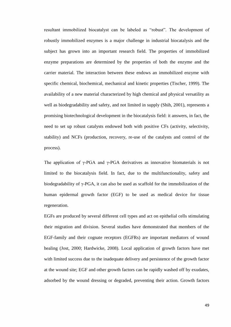
49
resultant immobilized biocatalyst can be labeled as “robust”. The development of
robustly immobilized enzymes is a major challenge in industrial biocatalysis and the
subject has grown into an important research field. The properties of immobilized
enzyme preparations are determined by the properties of both the enzyme and the
carrier material. The interaction between these endows an immobilized enzyme with
specific chemical, biochemical, mechanical and kinetic properties (Tischer, 1999). The
availability of a new material characterized by high chemical and physical versatility as
well as biodegradability and safety, and not limited in supply (Shih, 2001), represents a
promising biotechnological development in the biocatalysis field: it answers, in fact, the
need to set up robust catalysts endowed both with positive CFs (activity, selectivity,
stability) and NCFs (production, recovery, re-use of the catalysts and control of the
process).
The application of γ-PGA and γ-PGA derivatives as innovative biomaterials is not
limited to the biocatalysis field. In fact, due to the multifunctionality, safety and
biodegradability of γ-PGA, it can also be used as scaffold for the immobilization of the
human epidermal growth factor (EGF) to be used as medical device for tissue
regeneration.
EGFs are produced by several different cell types and act on epithelial cells stimulating
their migration and division. Several studies have demonstrated that members of the
EGF-family and their cognate receptors (EGFRs) are important mediators of wound
healing (Jost, 2000; Hardwicke, 2008). Local application of growth factors have met
with limited success due to the inadequate delivery and persistence of the growth factor
at the wound site; EGF and other growth factors can be rapidly washed off by exudates,
adsorbed by the wound dressing or degraded, preventing their action. Growth factors

50
have been covalently tethered to biomaterials, partly overcoming some of the
enunciated problems (Gu, 2004; Ehrbar, 2007).
It is therefore highly desiderable to develop novel scaffolds for EGF binding
characterized by high activity and able to protect them from environmental
denaturation. For tissue engineering applications, there are some special demands to
biodegradable polymers. Γ-PGA and its derivatives may answer most of the above
mentioned features (Matsusaki, 2002).
In spite of the potential of γ-PGA as protein carrier, only few reports have been
published so far about its use as matrix for enzymes immobilization. This might be
basically ascribed to three reasons: i) the still high costs of γ-PGA production since it
cannot be easily achieved by chemical synthesis (Buescher, 2007); ii) the extreme
variability in molecular weight and stereochemical composition of the naturally
produced biopolymer, that can obviously affect its physical and chemical properties;
and, finally, iii) the difficult handling of the natural biomaterial due to its high viscosity
(Shih, 2001).
In view of using γ-PGA as an enzyme carrier, some of the above mentioned drawbacks
have been partly overcome, but much effort is still needed to achieve an optimized,
usable biomaterial. For the γ-PGA large-scale production, many attempts have been
made to enhance bacterial productivity. The strategies were mostly based on the search
for suitable culture conditions (Ogawa, 1997; Yoon, 2000), on the identification of
better producer wild isolates (Kubota, 1993), or on both.
The above considerations contribute to justify our interest in the investigation of γ-PGA
production and characterization on one side, and on its reactivity and chemical
modification on the other; we devoted our efforts to the study of this material also in
51
view of its challenging applications in the field of tissue engineering, drug delivery, and
biocatalysis.

RESULTS AND DISCUSSION

53
Production
One of the first targets of the project was the improvement of γ-PGA production
applying biotechnological techniques to known Bacillus producer strains. This scientific
approach itself represents a novelty over the heuristic, semi-empirical ones mostly used
so far to the purpose.
In fact, most of γ-PGA producer strains are wild isolates, whose genetics and
physiology are not well defined; their use requires laborious customization of strain-
specific culturing conditions through heuristic approaches (Bajaj, 2009). This drawback
can be overcome by using derivatives of B. subtilis strain 168 as γ-PGA producers. B.
subtilis 168 is the model organism for Gram-positive bacteria: extensive scientific
knowledge has accumulated in over one hundred years of research on this specific
strain, and easy and standardized culturing conditions and genetic manipulation
protocols are available (Earl, 2008; Harwood, 1992; Volker, 2005). In strain 168, γ-
PGA biosynthetic genes are present but usually not transcribed (Urushibata, 2002),
unless two genetic variations are introduced: i) the degQ36 (Hy) or degU32 (Hy)
mutation (both leading to the same effect, i.e. an increased level of phosphorylation of
the transcription factor DegU) (Ohsawa, 2009); ii) the functional swrA allele (Osera,
2009; Stanley, 2005), whose function is still unclear (Patrick, 2012).
Improvement of microbial γ-PGA production has generally followed two parallel
routes: the identification of new and more efficient natural producer strains and the
development of better fermentation conditions (Bajaj, 2009; Shi, 2006). A third
potential route, so far poorly explored, is the control of the rate of γ-PGA post-synthesis
degradation by inactivation of specific γ-PGA degrading enzymes.
Our collaborators at the Genetics and Microbiology Department of the University of
Pavia could identify, among others, a mutant B. subtilis strain called PB5383, deriving

54
from the most common 169 B. subtilis laboratory strain and constitutively able to
produce high molecular weight γ-PGA, as early as 2009 (Osera, 2009).
It was conveniently possible to take advantage of the detailed knowledge of the genetic
asset and physiology of the producer bacterium as well as of the ease of manipulation of
the laboratory strain, to target specific pathways related to the biopolymer production.
Yields as good as those of the best natural known producers (Bajaj, 2011), were
achieved.
Advanced genetic engineering techniques made possible the construction and
subsequent selection of several mutant strains, with the aim to enhance productivity of
the biopolymer.
In particular (Scoffone, 2013), deletion of the genes related to the expression of
polymer- degrading enzymes endo-γ-glutamyl peptidase (pgdS) and exo-γ-glutamyl
hydrolase (γ-glutamyltranspeptidase GGT; ggt) was performed.
Comparison between the productivity of the parental PB5383 strain, and three mutants
in which the pgdS gene (PB5513, ΔpgdS), ggt gene (PB5518, Δggt) and both of them
(PB5522, ΔpgdS, Δggt) were respectively deleted, demonstrated that the effect of the
double deletion on the increase of polymer production is of substantial importance. In
fact, for the double deletion mutant, yield was reached about 40 g/L in 48 hours, almost
doubling the parental strain’s one, and above the average of known producer strains
(Bajaj, 2011). Quantification was performed drying polymer aliquots, extracted from at
least three independent cultures, to a constant weight.

55
Figure 10: electrophoretic separation of purified γ-PGA on SDS-page. Numbers above each lane correspond to the time points of sample collection, in hours. The yield increase in the case of the double deletion mutant (PB5522) is qualitatively evident.
Figure 11: comparison between B.subtilis PB5383, PB5513 (ΔpgdS), PB5518 (Δggt), PB5522 (ΔpgdS Δggt) strain productivity; samples collected at time cultures of 24, 48 and 60 hours, respectively.
Observation of the growth curves associated to each strain also showed that growth rate
between the mutants is comparable.
Figure 12: growth curves in E medium for B.subtilis PB5383, PB5513 (ΔpgdS), PB5518 (Δggt), PB5522 (ΔpgdS Δggt) strains; time is measured against Optical Density (OD) at 600 nm.

56
To make sure that the excellent improvement in yield for the double mutant was not due
to a second site mutation, the mutant strain itself was reconstructed starting from mutant
strains PB 5509 (ΔpgdS) and PB 5516 (Δggt) which were transformed by a plasmid
carrying the lacking deletion. Independent experiments performed in duplicate
confirmed the beneficial effect of the double gene deletion on γ-PGA productivity.
Purified biopolymer samples were analysed by SEC (Fig. 13A), and number average
molecular weight (Mn), weight average molecular weight (Mw), and polydispersity
(Mw/Mn) were calculated. Data in Table 6 show that the _pgdS _ggt double mutant
produced γ-PGA with lower Mn and Mw values and higher polydispersity than the other
samples; analysis of variance carried out on the independent samples collected at 24, 48
and 60 hours confirmed this observation (P<0.05). The _ggt mutant, impaired in the
end-chain degradation, that has a minor effect on polymer distribution, produced a
polymer that was similar to that produced by the wt strain in terms of Mn and Mw
(Kimura, 2004). The _pgdS mutant, missing the endo-hydrolase activity, produced γ-
PGA characterized by values of Mn and Mw higher than the wt sample, particularly at
earlier time points, although polydispersity did not notably vary.

57
Table 6: SEC measurements for γ-PGA produced by PB5383, PB5513, PB5518, PB5522 B. subtilis mutant strains
Sample Mn (106 Da) Mw (10
6 Da)
Polydispersity
(Mw/Mn)
PB5383 parental
24h 1,66± 0,075 2,23±0,098 1,34±0,032
48h 1,31 ± 0,066 1,87±0,110 1,43±0,012
60h 1,36 ± 0,111 1,92±0,221 1,41±0,067
PB5513 Δpgds
24h 1,93 ± 0,205 2,49±0,168 1,29±0,071
48h 1,46± 0,127 2,06±0,146 1,41±0,025
60h 1,40±0,108 2,00±0,158 1,43±0,012
PB5518 Δggt
24h 1,58±0,096 2,22±0,074 1,40±0,037
48h 1,35±0,046 1,94±0,023 1,44±0,037
60h 1,28±0,026 1,90±0,040 1,48±0,006
PB5522 Δpgds Δggt
24h 1,36±0,138 1,87±0,178 1,38±0,013
48h 1,01±0,152 1,55±0,172 1,54±0,063
60h 0,92±0,061 1,45±0,069 1,59±0,039
SEC traces (Fig. 13A) were plotted against the molecular weight calculated from the
standard calibration curve, in order to obtain the molecular weight distribution profiles
in the 0.1 - 5 MDa range (Fig. 13B); molecules lower than these values co-elute with
impurities and are below the resolving limit of our system (Yao, 2009). The distribution
curve showed that the increase in productivity of the _pgdS _ggt mutant was due to
accumulation of shorter polymer chains (0.1 - 2 MDa), whereas very high molecular
weight molecules (>3.5 MDa) were less abundant than in other strains.
58
Figure 13: SEC traces (A) and MWD profiles (B) for γ-PGA produced by PB5383, PB5513, PB5518, PB5522 B. subtilis mutant strains
Isolation
The first challenge encountered in the manipulation of γ-PGA was the isolation of the
polymer from its culture broth.
In order to solve the issue without recurring to chromatographic techniques (which may
represent a bottle neck for a possible scale-up of the process), several protocols were
tested, each of them based on the precipitation of the product, in its scarcely water-
soluble acidic form, with a cheap alcohol, such as methanol.
Best results, confirmed by NMR, were obtained by the removal of cells by
centrifugation or filtration, followed by a first precipitation of the product from the
culture broth with methanol; then acidification to pH 2-3 with an aqueous mineral acid
such as HCl, dialysis against water to remove the remaining impurities (mainly
represented by polysaccharides) and finally precipitation with i-propanol.

59
Figure 14: graphic representation of γ-PGA purification protocol by successive precipitations.
As an alternative, ultrafiltration with an Amicon device equipped with a 30 kDa cut-off
membrane, under Nitrogen atmosphere, was also successfully attempted.
This latter methodology proved, however, to be quite more time-consuming than the
former one, and to deliver reduced yields (less than 50% of those obtained by the
precipitation method).
Residual NMR
Solvent
A

60
Figure 15: Comparison between two γ-PGA samples, before (A) and after (B) purification; polysaccharides present in the crude have been eliminated.
Stereochemical L/D composition
In the introduction it has been discussed how the stereochemical composition of the
biopolymer, which affect its conformational as well as macroscopic properties, depends
on the bacterial strain responsible for its production.
Therefore we considered this to be a crucial part of our investigation, and decided to
analyze the stereochemical composition of the bioproduced (PB5383) as well as of that
a commercial available sample (Natto Bioscience, low molecular weight γ-PGA), for
comparison. The study could be possible thanks to our collaborators at the
Pharmaceutical Chemistry Department of the University of Pavia.
To the purpose, we followed the methodology proposed for γ-PGA by Cromwick e
Gross in 1995. According to this method, a sample of the biopolymer must be
completely hydrolyzed in acidic conditions; the resulting monomers are derivatized
with the so called Marfey’s reagent (1-fluoro-2, 4-dinitrophenyl-5-L-alanine amide,
FDAA) which can be regarded as a chiral version of the notorious Sanger’s reagent. The
B

61
so formed diastereoisomers can be finally separated and analysed in reverse phase
HPLC.
Scheme 1
Marfey’s reagent methodology
Derivatisation of D, L-glutamic acid was performed following the protocol reported by
Bhushan and Brückner in 2004, itself an adaptation of the original one described by
Marfey in 1984.
The reaction between Marfey’reagent (1-fluoro-2,4-dinitrophenyl-5-L-alanine amide,
FDAA) and a D,L amino acid results in the formation of L,L and L,D diastereoisomers
of the amino acid itself (Scheme 1), which can be separated and quantified in reverse-
phase HPLC.
The methodology was first developed for a in-house prepared mixture of L- and D-
glutamic acid, and then applied to the analysis of a commercial (Natto Bioscience, low
molecular weight) and a non-commercial (Genetics and Microbiology Department,
University of Pavia, mutant strain PB5383, purified with Amicon, cut-off 30kDa, and
lyophilized) sample. The diastereoisomers of D, L glutamic acid were analysed in

62
HPLC under conditions, which were properly adapted from those proposed by B’Hymer
in 2001.
All analyses were repeated twice.
The results obtained are shown in the following chromatograms (Fig. 16 and 17).
Figure 16: stereochemical composition of commercial γ-PGA: L-glutamic acid represents 48 % of total composition, D-glutamic acid 52%.
Figure 17: stereochemical composition of in-house polymer: D-glutamic acid isomer is prevalent (84% of total composition).

63
The order of elution, as was confirmed also by literature (B’Hymer, 2001), is the
following: derivatized L-glutamic acid, derivatized D-glutamic acid, Marfey’s reagent.
We could conclude that, while the commercial sample is composed by L- and D-
isomers of glutamic acid in 48:52 ratio respectively, on the contrary the polymer
produced by the Bacillus mutant strain at the University of Pavia is composed mainly
by D-glutamic acid (86%).
B. subtilis is known to be a poly-γ-DL-producer, but the prevalence of the D-isomer is a
peculiar characteristic of this particular mutant strain, which should make it non-
immunogenic -as it is the case, for instance, for the completely D-PGA produced by B.
anthracis (Keppie 1963), which contributes to the severity of its virulence. This feature,
together with its total atoxicity, may possibly make it interesting from a pharmaceutical
technology point of view, for the preparation of drug carrier and controlled delivery
constructs.
Molecular weight determination
Another fundamental parameter for the characterization of a polymer is its molecular
weight. The necessity of its measurement also arises from the inherent characteristics of
biosynthesis, which may bring a strong variability between each culture product.
To determine it, the technique of election is size-exclusion chromatography, coupled
with an in-line multi-angle-laser-light-scattering detector (MALS).
It is known from polymer chemistry that this technique is able, without the necessity of
any standard, to deliver a state-of-the art-accurate measurement of molecular weight
(M) and molecular weight distribution (MWD).
The response of the light-scattering detectors is in fact directly proportional to the
molecular weight of the polymer, following the equation (Mendichi, 2001):
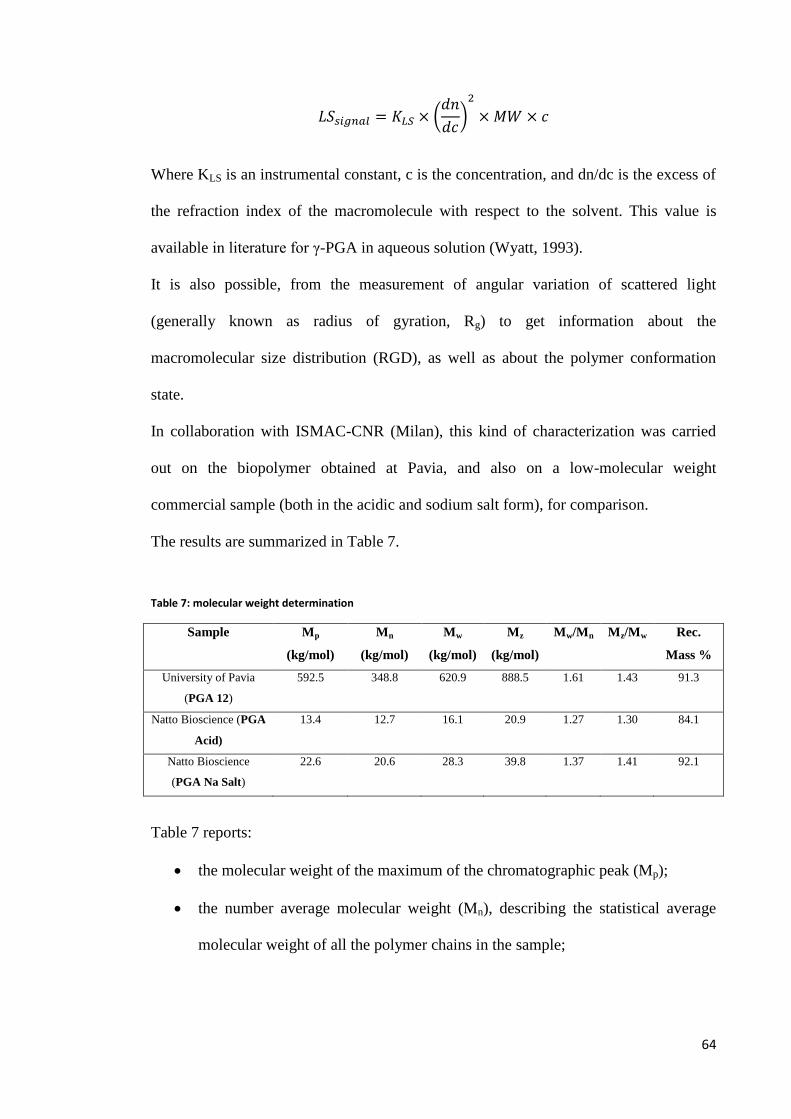
64
Where KLS is an instrumental constant, c is the concentration, and dn/dc is the excess of
the refraction index of the macromolecule with respect to the solvent. This value is
available in literature for γ-PGA in aqueous solution (Wyatt, 1993).
It is also possible, from the measurement of angular variation of scattered light
(generally known as radius of gyration, Rg) to get information about the
macromolecular size distribution (RGD), as well as about the polymer conformation
state.
In collaboration with ISMAC-CNR (Milan), this kind of characterization was carried
out on the biopolymer obtained at Pavia, and also on a low-molecular weight
commercial sample (both in the acidic and sodium salt form), for comparison.
The results are summarized in Table 7.
Table 7: molecular weight determination
Sample Mp
(kg/mol)
Mn
(kg/mol)
Mw
(kg/mol)
Mz
(kg/mol)
Mw/Mn Mz/Mw Rec.
Mass %
University of Pavia
(PGA 12)
592.5 348.8 620.9 888.5 1.61 1.43 91.3
Natto Bioscience (PGA
Acid)
13.4 12.7 16.1 20.9 1.27 1.30 84.1
Natto Bioscience
(PGA Na Salt)
22.6 20.6 28.3 39.8 1.37 1.41 92.1
Table 7 reports:
the molecular weight of the maximum of the chromatographic peak (Mp);
the number average molecular weight (Mn), describing the statistical average
molecular weight of all the polymer chains in the sample;

65
the weight average molecular mass (Mw), which, compared to Mn, takes into
account the molecular weight of a chain in determining contributions to the
molecular weght average;
the z average molecular mass (Mz),a higher molecular mass average which is
very sensitive to the higher molar mass species and therefore useful to describe
properties which strongly depend on them;
the polydispersity indexes Mw/Mn and Mz/Mw, which describe the amplitude
of the distribution of molecular mass;
percentage amount of sample mass recovered at the exit of the column with
respect to that injected is also shown.
Figure 18: superposition of the differential MWD of the samples

66
Figure 19: superposition of the three cumulative MWD of the samples
In Figures 18 and 19 the molecular weight distribution (MWD) of the polymer is
expressed in the differential and cumulative ways commonly used in the field of
polymer chemistry.
A brief analysis of the data let us make the following observations:
the biopolymer produced by the mutant B. subtilis PB5383 (PGA12) has a very
high molecular weight, around 620 kg/mol, and a wide MWD (Mw/Mn ≈1,6);
the commercial sample, on the contrary, has a low molecular weight
(≈16 kg/mole in the acidic form, ≈28 kg/mole in the sodium salt form) and a
relatively narrow MWD (≈1,3 in the acidic form, ≈1,4 in the sodium salt form).
These data are in contrast with those claimed by the producer, who stated the molecular
weight to be around 100 kg/mol.
This strong difference in molecular weight, as well as the different stereochemical
composition, may contribute to explain the difference between macroscopic properties
of the commercial and in-house produced biopolymer.

67
Figure 20: Conformation Plot
Figure 20 illustrates the Conformation Plot of the B.subitilis mutant- bioproduced
polymer and that of the Natto Bioscience commercial one, in the sodium salt form. The
Conformation Plot (analogous to the Mark-Houwink plot, which can be derived from
intrinsic viscosity measurements in function of molecular weight and which is
fundamental for polymer structure analysis) is constructed plotting radius of gyration
data acquired by means of a light scattering detector against MW data. Its slope
represents α parameter in the equation:
where Rg is the radius of gyration and M the molecular weight. The α parameter itself is
related with the conformation of the macromolecule. For the sample PGA12, its value is
≈ 0,58 which is typical of a stiff random coil conformation. The commercial sample
molecular weight is too low to permit a reliable estimation.

68
After measuring γ-PGA molecular weight by means of SEC-MALS both on commercial
and on laboratory produced samples, we directed our attention to other possible
methodologies able to provide us with an in-house means for molecular weight
measurement. Finding and developing a quick and easy method to the purpose, not
requiring high-specialized equipment and/or particularly expert and skilled personnel,
would compensate for the loss of analytical accuracy, and provide us with a first
evaluation ability, useful for routine work.
To reach this goal, we addressed the works reported by Park (2005) and Paraskevas
(2002). These authors developed an assay based on the use of Sanger’s reagent to
evaluate γ-PGA molecular weight. In fact, noticing that every polymeric filament has a
terminal amino group, it is possible to determine the numerical ratio between the amino
groups and the γ-glutamyl residues present in the polymer’s backbone. This ratio is,
obviously, related to the filament molecular weight. In order to estimate the amino
group number, the functionality can be, in principle, easily derivatized with Sanger’s
reagent. Thus, measuring the UV absorbance of Sanger-γ-PGA hydrolysates and
comparing it with an appropriate UV absorption standard curve of Sanger-glutamic
acid, it is possible to calculate the molecular weight of a polymeric sample.
Operatively, after the construction of the Sanger-glutamic acid standard UV absorption
curve, the polymeric sample must be derivatized, hydrolyzed in acidic conditions and
then measured at the UV spectrophotometer.
We therefore approached this methodology, trying the evaluation of commercial
samples whose molecular weight had been previously determined by means of SEC-
MALS, as well as of laboratory bioproduced samples. The method proved to be highly
sensitive, but not robust enough for our purposes. Our results were in good accordance
with those obtained thanks to more advanced methods when we analyzed commercial

69
samples, but this did not happen in the case of laboratory bioproduced ones. In fact, the
measurement of laboratory produced samples proved to be dramatically affected by the
presence of interferents, particularly glutamate monomers and oligomers. Park and
coworkers actually (Park, 2005) show how this method becomes extremely fragile when
the sample is not characterized by high purity grade; this analytical grade, according to
the authors, can be obtained only by the use of expensive, costly and time-consuming
purification techniques such as ion exchange chromatography, which is able to separate
oligomeric and monomeric fractions from high molecular weight ones. Therefore, this
method is unfit to be applied to our laboratory produced biopolymer samples, as long as
they reach just technical grade purification after having undergone a protocol based on
subsequent precipitations.
The application of the protocol on low molecular weight γ-PGA of commercial origin
provided us with an estimated value (ca 30 000 Da) in reasonable accord with the value
measured by SEC-MALS.
On the contrary, interferents heavily affected the protocol when we tried it on samples
produced by our strain, thus affording misleading results and confirming the limits of
this technique reported by literature.

70
Sonication
In collaboration with the Microbiology Department of the University of Pavia, we also
developed an effective method to control the polymer molecular weight. In fact, it was
found that sonication at fixed frequency (around 30 kHz) is able to degrade high
molecular weight polymer filaments to a limit value, in very brief times (≈ 200 s).
Figure 21: sonication of a sample of in house- produced γ-PGA; the MW drops from 700 kDa to less than 100 kDa
in around 200 s.
Figure 22: SEC analysis of the sonicated sample.

71
Rheology
Another important aspect in the characterization of a polymer is the study of its
rheological properties, both for elucidating the physical-chemical properties of the
material and to provide a reliable estimation of its molecular weight (Irurzun, 2001). On
this respect, it has to be noticed that viscosimetric studies on γ-PGA itself are scarce
(Kubota 1996); comparison between SEC-light scattering measurements and
viscosimetric ones has been performed, but a numerical correlation between these data
has not been proposed so far.
In collaboration with the Pharmaceutical Department of University of Pavia, we were
able to perform viscosimetric measurements, using a rotational rheometer, on three
representative samples: two commercial available ones (purchased from Natto
Bioscience and Biological Tech, Co), with low and high molecular weights,
respectively, and a laboratory bioproduced (University of Pavia) one.
Analyzing these measurements, it is possible to make some considerations about the
viscosimetric behaviour of our samples.
The low molecular weight (20 kDa, 100kDa declared by producer) commercial sample
(Natto Bioscience) is characterized by Newtonian behavior, i.e. its viscosity remains
constant with the increase of shear stress. Its viscosity is quite low (2,12 mPa s on
average). This value is in good accordance with that expected for a low molecular
weight polymer (Fig. 23).

72
Figure 23
On the contrary, the high molecular weight (700 kDa) commercial sample (Biological
Tech, Co.) is characterized by pseudo-plastic behavior, i.e. its viscosity tends to
decrease with shear stress increase. This is typical of polymeric solutions, because under
shear stress, the filaments tend to pass from a coiled conformation to a more ordered
one in which they stretch in the movement direction, increasing their hydrodinamicity.
For this sample, viscosity decreases from an initial value of 161 mPa s to 86 mPa s with
a shear rate increase from 10 to 300 /s (Fig. 24).
Figure 24
0
0,5
1
1,5
2
2,5
3
0 50 100 150 200 250 300
visc
osi
ty (
mP
a s)
shear rate (1/s)
Natto Bioscience (low MW)
0
50
100
150
200
0 100 200 300
visc
osi
ty (
mP
a s)
shear rate (1/s)
Biological Tech (high MW)

73
An analogous pseudo-plastic behavior is shown by the bioproduced at University of
Pavia polymer, whose MW is also around 700 kDa (determined by SEC-MALS
technique). Its viscosity decreases from 132 mPa s to 79 mPa s in the shear rate range
10-300 /s. This is consistent with the fact that the high molecular weight of this species
and that of the commercial high molecular weight samples are similar (Fig. 25).
Figure 25
Conformational aspects
Γ-PGA conformation and its influence on its chemical-physical behavior and reactivity
is still an open issue. In fact, the polymer filaments, can adopt several different
conformations in solution, which on their turn depend on numerous factors, including
the enantiomeric composition and the degree of ionization of the polymer, the pH
conditions, the nature of the solvent. Although a systematic study on the subject is still
lacking, some hypothesis have been formulated along the years.
First of all, a study performed on poly-γ-D-PGA conformation in aqueous solution
showed that considerable differences exist depending on the degree of ionization of the
polymer itself, with the un-ionized species preferably assuming an helical conformation
stabilized by hydrogen-bonds, while, on the contrary, the ionized polymer tend to a
0
50
100
150
0 50 100 150 200 250 300
visc
osi
ty (
mP
a s)
shear rate (1/s)
University of Pavia

74
random-coil state. In 1964, Rydon, on the basis of optical rotator dispersion (ORD)
measurements, proposed two possible structures for this kind of poly-γ-glutamate,
namely 317 helix and 319 helix, both of them stabilized by intramolecular hydrogen
bonds. More recently, molecular dynamics and quantum mechanical calculations led
Zanuy (Zanuy, 1998; 2001) to indicate the 319 left-handed helix as the most stable
conformation for this kind of polymer.
Figure 26: Equatorial and axial projections of the model conformationof un-ionized poly-γ-D-glutamic acid (Zanuy,
2001)
In 1973, Sawa and coworkers, after viscosity as well as ORD and IR spectroscopy
measurements, proposed that γ-PGA produced by B. subtilis was in a parallel β-sheet in

75
acidic solution, shifting to a more and more contracted random coil conformation with
the rise of pH to neutrality and then alkaline values.
In 2000, He and coworkers performed a conformational study on γ-PGA produced by
B.licheniformis by means of attenuated total reflectance FT-IR spectroscopy.
It is in fact known (Cantor, 1980) that the α-helix, β-sheet and random coil structures of
polypeptides and protein can be distinguished referring to the spectral bands at 1655 cm-
1 (amide I stretch) and 1550 cm
-1 (amide II stretch), which are diagnostic for α-helix,
while a shift of the amide I stretch to 1630 cm-1
is typical for β-sheet; random coils are
associated with a shift of the amide II stretch band to 1535 cm-1
.
The results collected by He’s group show that conformation of B.licheniformis γ-PGA
in aqueous solution is affected by pH, ionic strength, and concentration of the polymer
itself. In fact, at low pH, a low concentrated (0,1 % w/v) γ-PGA solution is protonated
and in helical conformation, as it is confirmed by the infrared bans at 1659 cm-1
(amide
I stretch) and 1558 cm-1
(amide II stretch), respectively. Rising the pH to neutrality, the
amide I shift to 1635 cm-1
, indicating a change of conformation to the β-sheet structure
in parallel with deprotonation. At higher pH, the polymer gets a random coil structure.
A similar trend is observed also with respect of ionic force, the increase of which makes
the conformation shift from α-helix to β-sheet in the range 0,1-0,5 M; α-helix is also
associated to polymer low concentrations; its increase to values as high as 25 mM as
glutamate cause a predominance of β-sheet, presumably because the intramolecular
interactions which maintain the helical structure are overwhelmed by intermolecular
ones.
Lately, the pH dependency of PGA helix folding dynamics has also been confirmed and
modeled by Krejtschi (2011) Gooding (2012) by means of CD and FTIR spectroscopy.

76
Moreover, in order to elucidate the stereochemical microstructure of the polymer,
Martínez de Ilarduya (2002) performed the synthesis of γ-PGA according to Sanda
(2001) in order to get standard sample suitable to conformational NMR studies.
Chemical synthesis
During our efforts for completing the chemical-physical characterization of biopolymer,
we realized that its peculiar macroscopic behavior (for instance its tendency to form
gels, its erratic solubility in organic and aqueous media, its high hygroscopicity), as well
as its chemical reactivity, should be related to its microscopic properties, and
particularly to molecular weight and conformation (this related, on its turn, to the
polymer stereochemical composition); studying these properties could help to elucidate
their influence on the material, and provide useful suggestions for its manipulation, not
to mention the possibility of future theoretical studies dealing with the so far unclear
stereochemical aspects of γ-PGA biosynthesis.
On these grounds, we decided to try the chemical synthesis of the polymer, in order to
obtain standard samples with a well defined enantiomeric structure and controlled
molecular weight; these will serve as model compounds to perform conformational
investigation on, taking advantage of circular dichroism spectroscopic techniques.
A preliminary study of the problem made immediately clear that direct polymerization
of glutamic acid was out of discussion. In fact, it is known that coupling of two α-
protected glutamic moieties only results in the formation of pyroglutamic acid
derivatives (Kajtár, 1969).
Other approaches involving protected lactame intermediates end up with no exception
in the formation of poly-α-glutamic acid or again in the formation of pyroglutamic
esters (Sun, 2011).
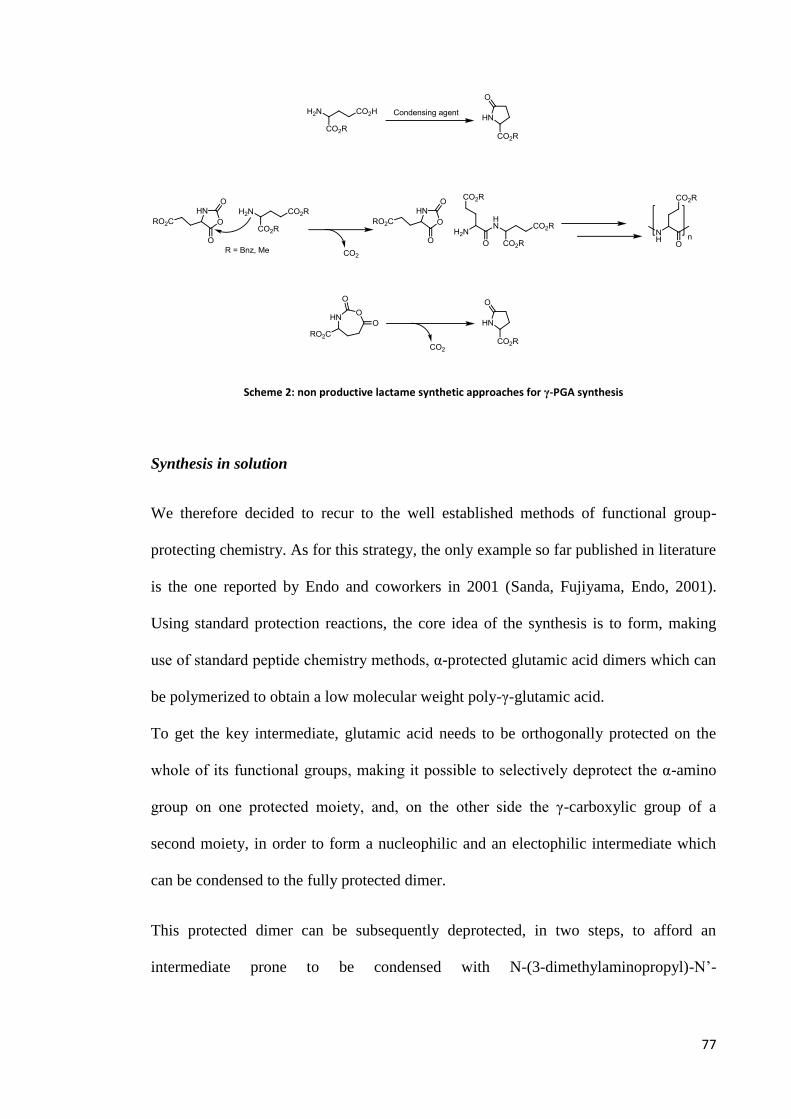
77
Scheme 2: non productive lactame synthetic approaches for γ-PGA synthesis
Synthesis in solution
We therefore decided to recur to the well established methods of functional group-
protecting chemistry. As for this strategy, the only example so far published in literature
is the one reported by Endo and coworkers in 2001 (Sanda, Fujiyama, Endo, 2001).
Using standard protection reactions, the core idea of the synthesis is to form, making
use of standard peptide chemistry methods, α-protected glutamic acid dimers which can
be polymerized to obtain a low molecular weight poly-γ-glutamic acid.
To get the key intermediate, glutamic acid needs to be orthogonally protected on the
whole of its functional groups, making it possible to selectively deprotect the α-amino
group on one protected moiety, and, on the other side the γ-carboxylic group of a
second moiety, in order to form a nucleophilic and an electophilic intermediate which
can be condensed to the fully protected dimer.
This protected dimer can be subsequently deprotected, in two steps, to afford an
intermediate prone to be condensed with N-(3-dimethylaminopropyl)-N’-

78
ehylcarbodiimide hydrochloride (EDC·HCl) and hydroxybenzotriazole (HOBt) as
coupling agents (Scheme 3).
Scheme 3
We were able to successfully reproduce this protocol, but we also experienced it as
being quite long and tedious. Nine steps are necessary, and the overall yield does not
exceed 5 %.
Thus, we tried an improvement of the procedure, to obtain more quickly the formation
of the key protected dimer intermediate. To reach this goal, we decided to avoid the
protection of one of the three groups leaving the amino or the γ-carboxylic group free,
so that we could spare one deprotection step before the formation of the dimer. A
consequence of this choice was the change of the protective group used for the amino
and γ-carboxylic functions of the electrophilic and nucleophilic units, respectively. Two
1 2
3
4
5
6 7
8
9
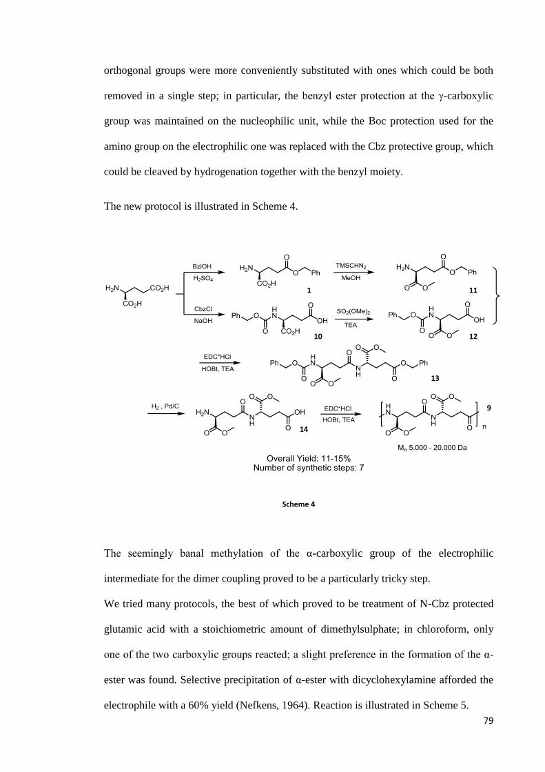
79
orthogonal groups were more conveniently substituted with ones which could be both
removed in a single step; in particular, the benzyl ester protection at the γ-carboxylic
group was maintained on the nucleophilic unit, while the Boc protection used for the
amino group on the electrophilic one was replaced with the Cbz protective group, which
could be cleaved by hydrogenation together with the benzyl moiety.
The new protocol is illustrated in Scheme 4.
Scheme 4
The seemingly banal methylation of the α-carboxylic group of the electrophilic
intermediate for the dimer coupling proved to be a particularly tricky step.
We tried many protocols, the best of which proved to be treatment of N-Cbz protected
glutamic acid with a stoichiometric amount of dimethylsulphate; in chloroform, only
one of the two carboxylic groups reacted; a slight preference in the formation of the α-
ester was found. Selective precipitation of α-ester with dicyclohexylamine afforded the
electrophile with a 60% yield (Nefkens, 1964). Reaction is illustrated in Scheme 5.
1
10
11
12
13
14
9
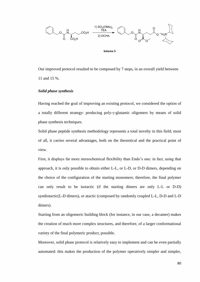
80
Scheme 5
Our improved protocol resulted to be composed by 7 steps, in an overall yield between
11 and 15 %.
Solid phase synthesis
Having reached the goal of improving an existing protocol, we considered the option of
a totally different strategy: producing poly-γ-glutamic oligomers by means of solid
phase synthesis techniques.
Solid phase peptide synthesis methodology represents a total novelty in this field; most
of all, it carries several advantages, both on the theoretical and the practical point of
view.
First, it displays far more stereochemical flexibility than Endo’s one: in fact, using that
approach, it is only possible to obtain either L-L, or L-D, or D-D dimers, depending on
the choice of the configuration of the starting monomers; therefore, the final polymer
can only result to be isotactic (if the starting dimers are only L-L or D-D)
syndiotactic(L-D dimers), or atactic (composed by randomly coupled L-L, D-D and L-D
dimers).
Starting from an oligomeric building block (for instance, in our case, a decamer) makes
the creation of much more complex structures, and therefore, of a larger conformational
variety of the final polymeric product, possible.
Moreover, solid phase protocol is relatively easy to implement and can be even partially
automated: this makes the production of the polymer operatively simpler and simpler,

81
and quite faster, than in the case of solution synthesis, requiring several laborious
protection, deprotection and purification steps.
We thus chose to implement a Fmoc procotol to generate a ten residue oligomer of γ-
glutamic acid, which could be subsequently polymerized in solution in standard
coupling conditions, working at high concentration to minimize the possibility of
ciclyzation. The overall synthetic strategy is illustrated in Scheme 6.
Scheme 6
The choice of the Fmoc protocol is due to its ease of implementation as well as to the
facile deprotection of the amino group in mild conditions, without affecting the most
common protections suitable for the carboxylic position. In fact, as it is widely known,
the Fmoc group can be removed by a piperidine solution.
The starting monomer was a commercial available amino acid, namely Fmoc-Glu-O-t-
Butyl-OH.
15
16
17
18

82
As supporting material, 2-chloro-trytil chloride was selected, for its robustness towards
several working conditions, commercial availability, and cheapness.
The first step of the process is the attachment of the N, α-carboxy-diprotected amino
acid monomer to the resin, which is possible by simple treatment with a base such as di-
isopropyl-ethyl-amine (DIPEA).
Once the first monomer has been attached to the resin, it can be put in an automatic
synthesizer, an instrument capable of performing all the necessary steps for the
condensation of amino acids, at each cycle deprotecting the amino group of the last
residue on the growing chain, and then letting it couple with a new amino acid thanks to
HOBt and HBTU or EDC or DCC reagents, in our case (Scheme 7).
Scheme 7: solid phase polymerization.
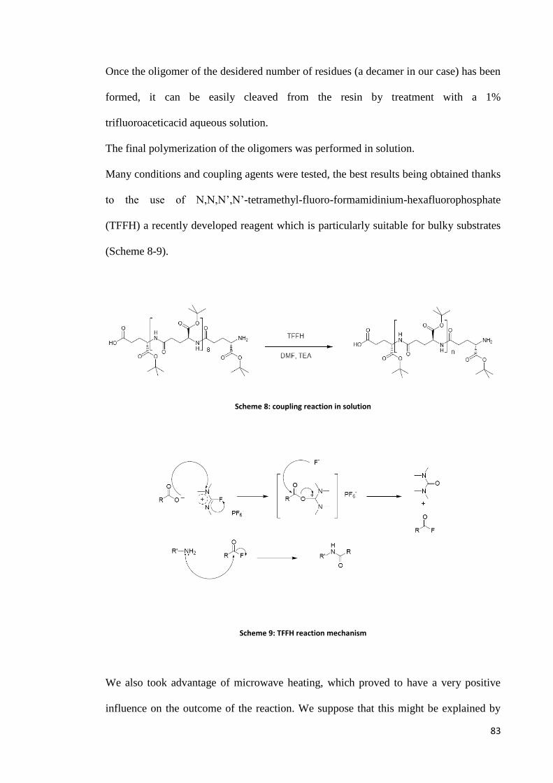
83
Once the oligomer of the desidered number of residues (a decamer in our case) has been
formed, it can be easily cleaved from the resin by treatment with a 1%
trifluoroaceticacid aqueous solution.
The final polymerization of the oligomers was performed in solution.
Many conditions and coupling agents were tested, the best results being obtained thanks
to the use of N,N,N’,N’-tetramethyl-fluoro-formamidinium-hexafluorophosphate
(TFFH) a recently developed reagent which is particularly suitable for bulky substrates
(Scheme 8-9).
Scheme 8: coupling reaction in solution
Scheme 9: TFFH reaction mechanism
We also took advantage of microwave heating, which proved to have a very positive
influence on the outcome of the reaction. We suppose that this might be explained by

84
the fact that microwaves, a form of coherent heat electromagnetic radiation, interact
with the peptide chain dipole moment, keeping the chains in a linear and therefore
reagent-accessible conformation, differently from what normally happens in solution,
when the chains tend to form secluded structures such as α-helix (thanks to hydrogen
bonding) or random coils.
The effectiveness of this approach was confirmed by conventional Size Exclusion
Chromatography (calibration operated with respect to Polystyrene standards)
measurement of the molecular weight of a sample taken form a crude polymerization
attempt. Although presented a relatively high polydispersity, a poly-γ-glutamic acid t-
butyl ester was formed (although it is easily deprotected with aqueous TFA, the
polymer was initially kept in the ester form to avoid the risk of contaminating the
sample), its molecular weight being comparable to that of a commercial sample such as
that acquired by Natto Bioscience Company.
Results are shown in the following table 8.
Table 8
Sample Mp g/mole Mn g/mole Mw g/mole Mz g/mole Mw/Mn Mz/Mw
Γ-PGA t-butyl ester 17,950 8,000 14,140 21,850 1.77 1.55
A possible drawback of this process is its relatively higher cost, compared with solution
phase protocols, but since for our purposes only little amounts of polymer are necessary,
this objection can be considered of marginal importance.
Derivatisation
The ability of chemically modifying γ-PGA is an essential requirement for the
technological valorization of the material. Every attempt to reach this goal must of
course start from the observation that the only functionality present in the

85
macromolecule is the α-carboxylic group pending from the polymer backbone. A
careful choice of the substituting group (which will be likely linked to the polymer in
the form of an ester, or an amide) will contribute to the modulation of its chemical-
physical properties; for instance, increasing its hydrophobicity, and therefore, the
possibility for it to be used as a biopackaging material, or as scaffold for the
construction of nanoparticles for biotechnological and pharmaceutical use.
However, evidence indeed proved that the inherent tendency of this group to react is far
from being excellent. In fact, the reactivity of the carboxylic groups pending from the
polymer backbone is unusual, and quite cumbersome.
So far, our efforts have aimed at the esterification of the biopolymer, both directly and
indirectly, via transesterification, as well as at the formation of amide derivatives.
Although the procedures involved may appear trivial at a first glance, their development
proved to be difficult and tedious, due to three main obstacles: the scarce chemical
reactivity of the material, and the difficulty encountered in isolation and purification of
the products.
To date, three methodologies have been attempted and developed for γ-PGA
esterification:
Nucleophilic substitution on carboxylates;
Direct esterification via previous acyl chloride formation;
Use of a carbodiimide as condensing agent.

86
Scheme 10: 1) RBr, NaHCO3, DMSO, 45 ºC, 5-8 days; 2) (COCl)2 or SOCl2, THF, DMF, ROH, 24-36 h, from 0 ºC to r.t.; 3) EDC or COMU or TFFH, DMSO, ROH, 24-36 h, r.t.
Our efforts made possible the preparation of the following derivates of γ-PGA:
γ-PGA-α-methyl- ester
γ-PGA-α-ethyl- ester
γ-PGA-α-isopropyl- ester
γ-PGA-α-n-butyl- ester
γ-PGA-α-tetradecanoyl- ester
γ-PGA-α-benzyl- ester
γ-PGA-α-ethyl-α’-poly (ethylene glycol)-yl ester
γ-PGA-α-benzyl-α’-poly (ethylene glycol)-yl ester
Nucleophilic substitution on carboxylates
The first method, reported by Kubota and Endo (Kubota, 1993b) and widely used by
Muñoz-Guerra has proven to be effective, but quite inefficient. To achieve a degree of
substitution exceeding 50 %, the procedure requires dissolving the polymer in a high-
boiling solvent such as DMSO and letting it react with alkyl or benzylic halide in basic
condition for times between 5 and 10 days, performing two subsequent derivatisations.
Afterwards, the product must be isolated by precipitation in cold acidic water. This
procedure is quite long and tricky, and skill is requested to get rid of solvent traces.
Following this approach, we have prepared benzyl- and ethyl- esters of γ-PGA.
The formation of the ethyl ester and its degree of functionalisation can be assessed by
1H-NMR; in particular, the signal belonging to the amide of the derivative falls at higher

87
fields than that belonging to the starting material; therefore, by the ratio of the two
signals, it is possible to evaluate the progress of the reaction.
In our case, after a first derivatisation in presence of a 5-fold excess of ethyl bromide,
which lasted 5 days, we were able to obtain a sticky, viscous product, with a degree of
functionalisation about 15-20%. Its isolation was possible by precipitation of the
reaction mixture, drop by drop, in a 15 fold quantity of acid water, whose pH was
carefully maintained around 1,5-2 by addition of HCl. This procedure is
straightforward, but slow, and the filtration of the product is quite tedious; in fact, its
gluish nature makes its manipulation difficult. Moreover, appreciable amounts of
DMSO tend to adhere to this first generation product; rinsing with acid water, and/or
organic solvents, are of little help in getting rid of solvent traces. Also a long (several
hours) run at the rotary evaporator, connected to an acetone/dry ice trap, did not
success.
We had therefore the material react a second time, in the same conditions. Second
generation product showed a quite higher degree of functionalisation (almost 70%),
after 3 days of reaction. Complete conversion was not reached even by further
prolonging reaction times, or increasing the reagent excess, or reaction temperature;
however, it is reported in literature that steric effects become significant with the
increase of functionalisation, affecting the reaction outcome. Moreover, the possibility
of a reduction in molecular weight after prolonged reaction in these conditions has also
been proposed by Muñoz-Guerra. The isolation of the second generation ester was
possible by the same protocol previously used; the material was quite easier to handle,
and less prone to contamination by DMSO.
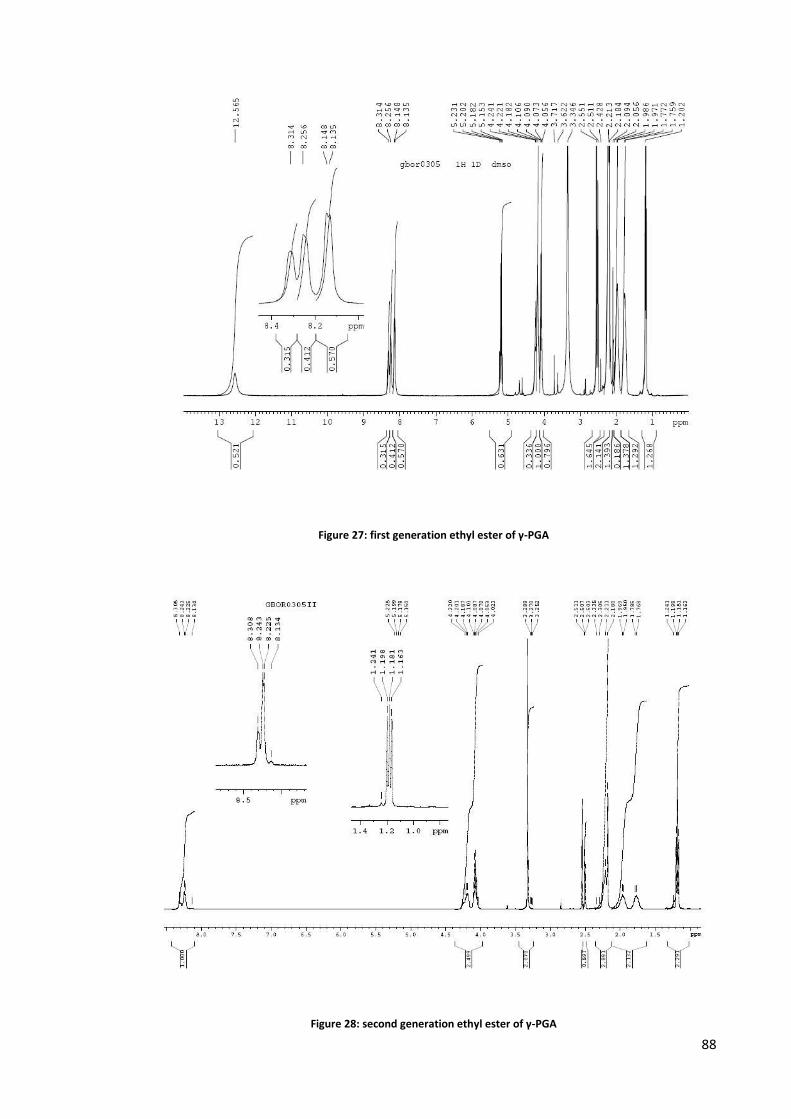
88
Figure 27: first generation ethyl ester of γ-PGA
Figure 28: second generation ethyl ester of γ-PGA
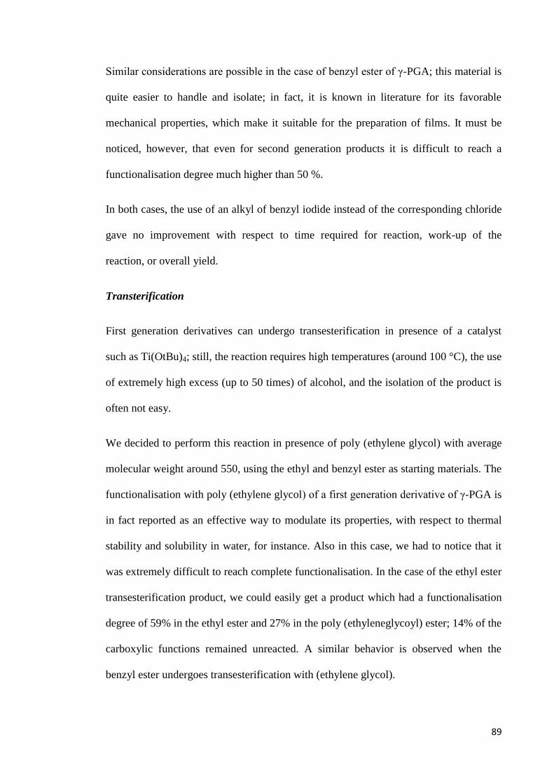
89
Similar considerations are possible in the case of benzyl ester of γ-PGA; this material is
quite easier to handle and isolate; in fact, it is known in literature for its favorable
mechanical properties, which make it suitable for the preparation of films. It must be
noticed, however, that even for second generation products it is difficult to reach a
functionalisation degree much higher than 50 %.
In both cases, the use of an alkyl of benzyl iodide instead of the corresponding chloride
gave no improvement with respect to time required for reaction, work-up of the
reaction, or overall yield.
Transterification
First generation derivatives can undergo transesterification in presence of a catalyst
such as Ti(OtBu)4; still, the reaction requires high temperatures (around 100 °C), the use
of extremely high excess (up to 50 times) of alcohol, and the isolation of the product is
often not easy.
We decided to perform this reaction in presence of poly (ethylene glycol) with average
molecular weight around 550, using the ethyl and benzyl ester as starting materials. The
functionalisation with poly (ethylene glycol) of a first generation derivative of γ-PGA is
in fact reported as an effective way to modulate its properties, with respect to thermal
stability and solubility in water, for instance. Also in this case, we had to notice that it
was extremely difficult to reach complete functionalisation. In the case of the ethyl ester
transesterification product, we could easily get a product which had a functionalisation
degree of 59% in the ethyl ester and 27% in the poly (ethyleneglycoyl) ester; 14% of the
carboxylic functions remained unreacted. A similar behavior is observed when the
benzyl ester undergoes transesterification with (ethylene glycol).

90
Figure 29: transesterification of the diethyl ester of γ-PGA with poly (ethylene glycol, Mn ≈ 550)
Figure 30: transesterification of the benzyl ester of γ-PGA with poly (ethylene glycol, Mn ≈ 550)
In an attempt to improve the scope of the nucleophilic substitution on carboxylates
approach, we also tried the well established methodology of phase transfer catalysis,
making use of tetraalkylammonium halides and dissolving γ-PGA in basic water and the
91
halide in dichlorometane, respectively. Unfortunately, our attempts were not successful.
Future investigation, however, should be devoted to a deeper exploration of this
effective and simple methodology, maybe concentrating the attention on the choice of
the proper catalyst to the purpose.
Direct esterification
The second methodology, which to the best of our knowledge has not been proposed
before, consists in the formation, facilitated by a catalytic amount of DMF, of an acyl
chloride in situ, followed by direct esterification, in an etheric solvent such as THF, or
directly in the alcohol corresponding to the desired ester. The reaction is quite faster
(24-36 hours), and the product is relatively easily isolated by precipitation in a cold
alcohol such as methanol and filtration. The drawback of the procedure is that also in
this case, the material has to react twice in a row to achieve some functionalisation in
the second generation product. Moreover, the functionalisation degree is not always
excellent.
It must be noted that an inherent limit in the chemical manipulation and modification of
γ-PGA lies in its scarce solubility in most organic solvents, and in their mixtures; this
fact negatively affects yields and purification, and strongly narrows down the possible
applicable reaction conditions.
Taking advantage of this novel methodology, we were able to synthesize some short
and long chain aliphatic esters of γ-PGA, namely methyl-, ethyl-, isopropyl, n-butyl-
and tetradecanoyl- esters.
The preparation of the methyl ester gave the better results in terms of product yield,
reaction time, easiness of the procedure and of the work-up. In fact, in 24 hours we were
able to obtain a degree of functionalisation higher than 95%, using a moderate excess (6

92
times) of oxalyl chloride; the work-up simply consists in neutralization of the excess
acid, and evaporation of the alcohol, resulting in the straightforward precipitation of the
product, which is very pure and not contaminated by high-boiling solvents such as
DMSO (a drawback of the carboxylates approach) or by other additives.
Figure 31: methyl ester of γ-PGA via direct esterification. The magnification shows the signal of the amide, which permits the calculation of functionalisation degree.
As for the other esters, we also had good results, but the functionalisation degree of first
generation products, in some cases, tends to remain stable around 30 % (except in the
case of the tetradecanoyl esters, when, it tends to drop to less than 20 %). In the case of
the isopropyl as well as in that of the n-butylester, the product smoothly reached 70%
functionalisation, while this curiously didn’t happen for the ethyl derivative; we
suppose that sterics may play a role, as well as, the solubility issues which deeply affect
the reactivity of the polymer. The latter factor is probably crucial in determining the
outcome of the reaction. The increase of oxalyl chloride excess (up to 20 times the
substrate) is not always beneficial, as well as the raise of the quantity of alcohol or the
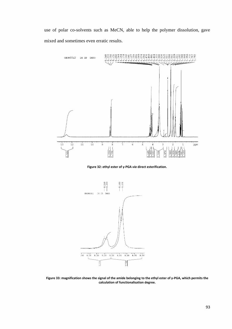
93
use of polar co-solvents such as MeCN, able to help the polymer dissolution, gave
mixed and sometimes even erratic results.
Figure 32: ethyl ester of γ-PGA via direct esterification.
Figure 33: magnification shows the signal of the amide belonging to the ethyl ester of γ-PGA, which permits the calculation of functionalisation degree.
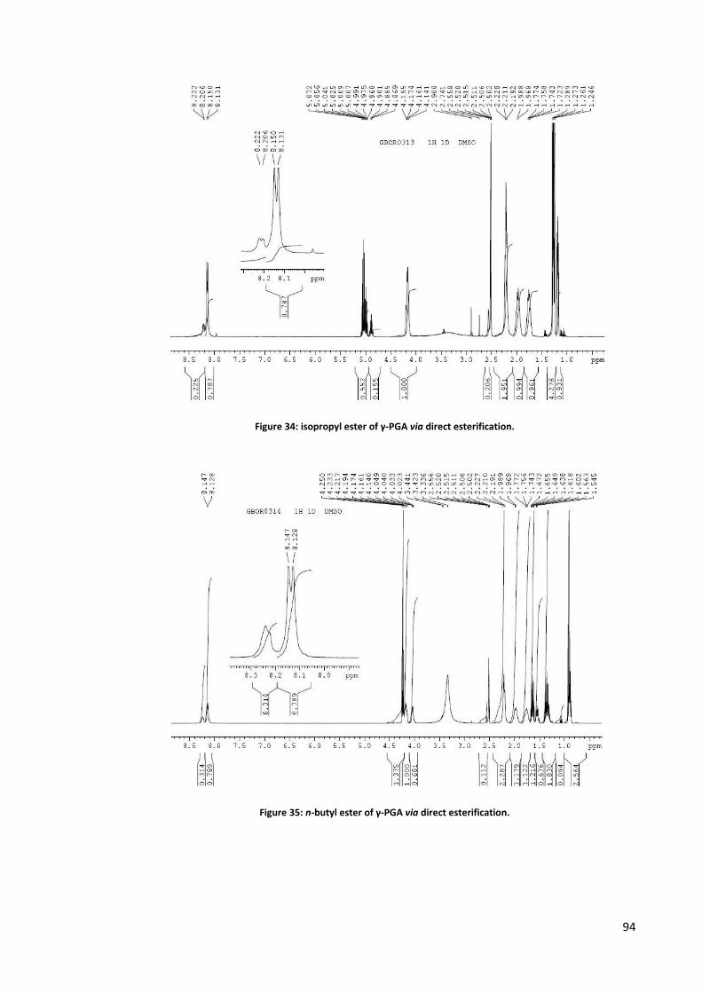
94
Figure 34: isopropyl ester of γ-PGA via direct esterification.
Figure 35: n-butyl ester of γ-PGA via direct esterification.

95
Use of condensing agents
The third methodology (Akagi, 2005b) has its origins in peptide coupling chemistry,
and makes use of condensing agents such as a carbodiimide (usually a water-soluble
one) or a stronger reagent such as COMU or TFFH. Operatively, the polymer is
dissolved or suspended in DMSO, and reacts for 24 -36 hours in presence of an organic
base (such as TEA, or DiPEA, or Py) and an alcohol, or an amine. The product has to be
isolated by means of dialysis against distilled water, followed by lyophilisation. The
bottle neck of the procedure is represented by the inherent slowness of the dialysis
procedure, and, more importantly, also by the difficulty in getting rid of the byproducts
of the coupling agent used. However, the method, when the proper conditions are
developed, has some effectiveness, permits the preparation of amides besides esters, and
opens many important possibilities, especially for biomedical and pharmaceutical
applications, where γ-PGA is conjugated to species such as amino acids and peptides.
Novel methodologies in course of development
We have also tried other methodologies, in particular the formation of an active ester in
situ, such as a pentafluorophenyl- or p-nitro-phenyl ester, as well as the activation of the
carboxylic group in the form of the corresponding succinimide. So far these approaches
have given mixed results, mainly because of the difficulty in product isolation. In fact,
the precipitation of the material of interest from the reaction solvent may be
cumbersome, and require days and sometimes even weeks, although efforts have been
put in screening the best precipitation procedures and conditions. Still, these
methodologies are currently in course of development. Our exploration in this field is
far from being over.

Γ-GLUTAMYL TRANSPEPTIDASE (γ-GGT) of B. subtilis

97
Γ-glutamyl transpeptidase (GGT, E.C. 2.3.2.2) is a widely distributed, heterodimeric
enzyme, found in organisms from bacteria to plants and mammals (Tate, 1981).
Figure 36: B. subtilis γ-GGT secondary structure (PDB id =3A75)
It cleaves glutathione, a "γ-glutamyl donor", releasing the cysteinylglycine portion and
forming a covalently bound γ-glutamyl-enzyme intermediate through an ester linkage
between the γ-carbon atom of the glutamyl group and the oxygen atom of the
catalytically active, conserved N-terminal threonine residue of the small enzyme subunit
(Inoue, 2000).

98
Figure 37: detail of B.subtilis γ-GGT active site (pdb id: 3A75); the catalytically active N-terminal threonine residue and a ligand (glutamic acid) are in evidence.
The γ-glutamyl moiety can then be transferred to an acceptor nucleophile in a second
step. If the acceptor is a water molecule, the hydrolysis of the γ-glutamyl-enzyme
adduct occurs with the release of free glutamic acid (Thomson, 1976; Allison, 1985;
Taniguchi, 1998). On the other hand, if the acceptor is the amino group of a second
species, a transpeptidation reaction ensues, affording the γ -glutamyl derivative of the
acceptor (Tate 1974; Thompson, 1977; Meister, 1981). A third reaction, namely an
autotranspeptidation, in which the same species behaves both as the donor and as the
acceptor, could also take place in vitro in the presence of a high concentration of the
donor substrate (Allison, 1985) (Scheme 11).
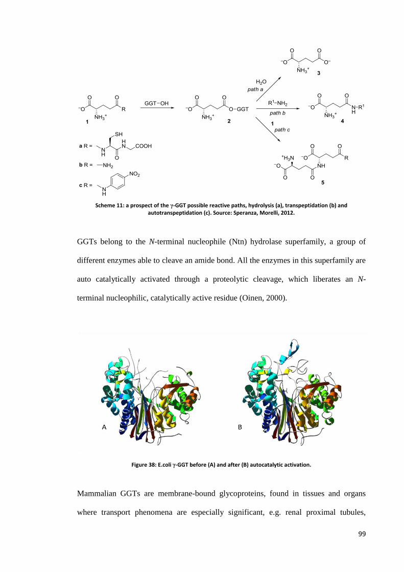
99
Scheme 11: a prospect of the γ-GGT possible reactive paths, hydrolysis (a), transpeptidation (b) and autotranspeptidation (c). Source: Speranza, Morelli, 2012.
GGTs belong to the N-terminal nucleophile (Ntn) hydrolase superfamily, a group of
different enzymes able to cleave an amide bond. All the enzymes in this superfamily are
auto catalytically activated through a proteolytic cleavage, which liberates an N-
terminal nucleophilic, catalytically active residue (Oinen, 2000).
Figure 38: E.coli γ-GGT before (A) and after (B) autocatalytic activation.
Mammalian GGTs are membrane-bound glycoproteins, found in tissues and organs
where transport phenomena are especially significant, e.g. renal proximal tubules,

100
jejunum, bile duct, epididymis, choroid plexus, ciliary body, liver, pancreas,
bronchioles. The enzymatic activity found in these tissues can vary over a broad range,
depending also on species. For example, almost no detectable enzymatic activity is
found in the blood serum of rats and mice, whereas the enzyme is detectable in human
serum, where its level is of clinical importance in the detection of liver diseases
(Rosalki, 1975; Goldberg, 1980).
In mammals, GGT is mainly involved in glutathione metabolism and cysteine salvage
(Tate, 1981; Taniguchi, 1998). In humans, GGT is also implicated in a number of
pathological processes, such as tumor progression (Hanigam, 1995) and
neurodegenerative deseases (Owen, 1996). Some mechanisms of drug resistance seem
to be also dependent on GGT activity (Godwin, 1992).
The observation that mammalian GGTs catalyze the transpeptidation reaction ca 180
fold faster than the hydrolysis reaction (Ikeda, 1995a, 1995b), together with the before
mentioned enzyme localization studies, is in favour of the physiologically significant
function of the GGT-catalyzed transpeptidation reaction in vivo through the so-called γ
-glutamyl cycle (Griffith, 1978; 1979). The γ-glutamyl cycle pictures the conversion of
amino acids into their γ-glutamyl derivatives in a GGT-catalyzed reaction using
glutathione as the γ-glutamyl donor. The γ-glutamylamino acids are then translocated
inside the cells, where the cyclotransferase enzyme catalyzes the intramolecular reaction
of the γ-glutamyl derivatives releasing the free amino acids and affording pyroglutamic
acid.
The occurrence of the γ -glutamyl cycle is however under debate, as the concentration
of some amino acids in vivo is well-below their KM values with respect to GGT (see for
example Elce, 1976).
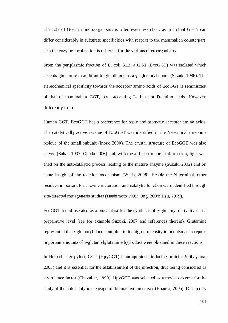
101
The role of GGT in microorganisms is often even less clear, as microbial GGTs can
differ considerably in substrate specificities with respect to the mammalian counterpart;
also the enzyme localization is different for the various microorganisms.
From the periplasmic fraction of E. coli K12, a GGT (EcoGGT) was isolated which
accepts glutamine in addition to glutathione as a γ -glutamyl donor (Suzuki 1986). The
stereochemical specificity towards the acceptor amino acids of EcoGGT is reminiscent
of that of mammalian GGT, both accepting L- but not D-amino acids. However,
differently from
Human GGT, EcoGGT has a preference for basic and aromatic acceptor amino acids.
The catalytically active residue of EcoGGT was identified in the N-terminal threonine
residue of the small subunit (Inoue 2000). The crystal structure of EcoGGT was also
solved (Sakai, 1993; Okada 2006) and, with the aid of structural information, light was
shed on the autocatalytic process leading to the mature enzyme (Suzuki 2002) and on
some insight of the reaction mechanism (Wada, 2008). Beside the N-terminal, other
residues important for enzyme maturation and catalytic function were identified through
site-directed mutagenesis studies (Hashimoto 1995; Ong, 2008; Hsu, 2009).
EcoGGT found use also as a biocatalyst for the synthesis of γ-glutamyl derivatives at a
preparative level (see for example Suzuki, 2007 and references therein). Glutamine
represented the γ-glutamyl donor but, due to its high propensity to act also as acceptor,
important amounts of γ-glutamylglutamine byproduct were obtained in these reactions.
In Helicobacter pylori, GGT (HpyGGT) is an apoptosis-inducing protein (Shibayama,
2003) and it is essential for the establishment of the infection, thus being considered as
a virulence factor (Chevalier, 1999). HpyGGT was selected as a model enzyme for the
study of the autocatalytic cleavage of the inactive precursor (Boanca, 2006). Differently

102
from human GGT and EcoGGT, Hpy GGT shows only a limited transpeptidase activity.
Structural studies based on x-ray crystallographic data demonstrated the formation of a
threonine-threonine catalytic dyad (Boanca, 2007) the involvement of the mobile C-
terminus of the protein in catalysis (Williams, 2009) and the presence of a lid loop
covering the glutamate-binding pocket of the mature enzyme during catalysis. In
particular, a tyrosine residue (Tyr433) positioned in the middle of the lid loop shields
the enzyme-bound glutamyl residue, allowing only the γ-carboxy group to be solvent-
exposed (Morrow, 2007).
GGT from B. subtilis is an extracellular protein. The gene encoding GGT in B. subtilis
was identified in 1996. It was expressed and the amino acid sequence was established
(Xu, 1996). B. subtilis GGT is a 587 amino acids protein, carrying a 28 residues N-
terminal portion signaling the extracellular localization. It shares 41% sequence
homology with E. coli GGT. The physiological significance of the enzyme remained
however unclear at that time.
The glutaminase activity and the salt tolerance of B. subtilis GGT were acknowledged
(Minami, 2003) and a technological use of this enzyme was suggested in the
manufacture of soy sauce, a traditional Japanese seasoning, the taste of which depends
mainly on its glutamic acid content (Kijima, 2007). The enzymatic activity of the
enzyme was investigated. pH, temperature optima and substrate specificities were
defined for both hydrolase and transpeptidase activity. As the enzyme showed
transpeptidase activity towards some acceptor amino acids, Authors proposed its use for
the enzymatic synthesis of γ-glutamyl compounds, although this possibility was not
experimentally demonstrated. Among the tested amino acid acceptors, glutamine and
glutamic acid showed the lowest activities and no experiments were carried out using
D-amino acids (Minami, 2003).

103
The GGT isolated from Bacillus subtilis 168 (BsubGGT) has some unique structural
features. Compared to the GGTs of other species, it possesses an extra sequence at the
C-terminus of the heavy subunit and it lacks the lid loop.
Figure 39: representation of E.coli γ-GGT (pdb id: 2DBX) (A) in comparison with B.subtilis one (pdb id: 3A75) (B); the superimposition of the two structures (C) clearly displays the difference represented by the absence, in the
latter, of the lid loop (red).
Differently from other GGT structures, the bound glutamic acid in the active site of B.
subtilis GGT is therefore fully solvent exposed (Wada, 2010). In this respect, B. subtilis
is similar to the enzyme CapD (Wu, 2009), a recognized virulence factor from Bacillus
anthracis (Scorpio, 2007) involved in anchoring and remodeling the poly-γ-D-glutamic
acid capsule surrounding the bacterium (Candela, 2005; Richter, 2009). There is
however little sequence homology between BsubGGT and CapD, as BsubGGT
resembles much more other microbial GGTs. In particular, the catalytically active
threonine residue at the N-terminus of the small subunit is highly conserved among all
known GGTs, as are conserved the residues that bind the γ-glutamyl moiety through

104
hydrogen bonds and electrostatic interactions involving the α-amino and the γ-carboxyl
groups. Differences in the active site architecture of B. subtilis GGT and B. anthracis
CapD were ascribed to the different size of their natural substrates. A large PGA
polymer was indeed recognized as the substrate of CapD, whereas the discrete, low-
molecular weight glutathione was assumed to be the substrate of B. subtilis GGT
(Wada, 2010).
However, also B. licheniformis and some strains of B. subtilis (Thorme, 1954) release a
polymer of glutamic acid units linked through γ -glutamylic bonds extracellularly. As a
consequence, the involvement of GGT in the biosynthesis of poly-γ-glutamic acid was
in a first time proposed for B. subtilis (natto) (Ogawa, 1991). Evidences for a
membrane-bound PGA-synthetase complex seem however to be more convincing,
although the biosynthesis of PGA may vary in different microorganisms (Kunioka,
1997). It was therefore proposed that in B. subtilis (natto), GGT is more likely involved
in PGA capsule hydrolysis for energy supply under carbon-limiting conditions, rather
than in its biosynthesis (Ogawa, 1997). This view was supported by some other
investigations carried out on B. subtilis GGT. It was indeed found that B. subtilis GGT
is able to hydrolyse PGA generating D- and L- glutamic acid in amounts corresponding
to their proportion in the PGA substrate. Using γ-glutamyltripeptides labelled with γ-D-
glutamic acid or α-L-glutamic acid at either the amino or the carboxy terminal, it was
demonstrated that the hydrolysis proceeds starting from the amino terminal, and the
enzyme shows no preference for L-glutamic acid over the D-stereoisomer (Kimura,
2004).
Under nitrogen-limited supply, B. subtilis mutants lacking GGT sporulate more
frequently than the wild type microorganism. Γ-PGA thus represents a source of
nutrients, in that it can be degraded and the constituent glutamic acid residues are

105
internalized by cells and used as nitrogen source. PGA degradation requires the
intervention of at least two enzymes. An endo-type PGA hydrolase generates PGA
fragments of approximately 105 Da. These fragments become in turn substrates of GGT,
which liberates one glutamic acid residue at a time starting from the amino terminal.
The action of the endo-type hydrolase is a required condition for the intervention of
GGT, in that it raises the molar concentration of the N-terminal ends of γ -PGA chains
up to the KM value of GGT. This observation, together with the high activity of B.
subtilis GGT towards γ -PGA, supports the hypotheses that
γ -PGA is the natural substrate of B. subtilis GGT and that γ -PGA hydrolysis is its
physiological role (Kimura, 2004).
Besides B. subtilis (natto), other GGTs have been isolated and characterized from
various B. subtilis strains. From B. subtilis TAM-4 two isozymes of GGT were isolated
with similar enzymatic properties. They showed glutaminase activity towards D- and L-
glutamine. The transpeptidase activity was tested using L-amino acids and some
glycinyl dipeptides as acceptors (Abe, 1997).
The GGT from B. subtilis NX-2, isolated from soil in China, revealed a broad substrate
stereospecificity, showing the same activities towards L-amino acids and their racemic
forms (Wu, 2006). This enzyme was used for the enzymatic synthesis of γ-D-glutamyl-
L-tryptophan (Wang, 2008), a promising immune regulation compound in the treatment
of tuberculosis (Orellana, 2002).
A recently discovered GGT from B. subtilis SK11.400 showed a maximum
transpeptidase activity at pH 10 and maintained its activity also towards the few D-
amino acids tested as acceptors (Shuai, 2011). The main goal of the studies about the
GGTs from B. subtilis strains was the characterization of the enzyme properties and
their possible role in the homeostasis of γ-PGA capsule of the bacterium was never

106
mentioned. Among the GGTs mentioned, the amino acid sequence is given only for
GGT from B. subtilis SK11.400, and comparison with the sequences of other bacterial
GGTs showed that it lacks the lid loop.
Enzymatic activity of B. subtilis GGT
The Bacillus subtilis 168 GGT gene, excluding the signal sequence, was PCR amplified
from genomic DNA and cloned in a pET vector in front of a His-tag. The construct was
transformed in BL21 (DE3) E. coli cells and gene expression was induced. The GGT
protein is retained in the cell because it lacks the signal sequence, and undergoes
autocatalytic cleavage producing the active large (L, 40 kDa) and small (S, 20 kDa)
subunits. The complex was purified with Ni-conjugated agarose beads. Different
amount of recombinant proteins (as indicated in the figure) were loaded on a 12% SDS-
PAGE gel (fig. 40), together with an aliquot of the flow-through and remaining beads.
The molecular weight markers (NEB, broad range) are loaded on the left.
Figure 40: SDS-PAGE illustrating Bacillus subtilis 168 GGT mutant protein expression
Enzymatic activity of B. subtilis GGT was measured by standard methods using the

107
chromogenic substrate γ-glutamyl-p-nitroanilide. The release of p-nitroaniline in the
GGT-catalyzed reaction can be determined spectrophotometrically at 410 nm. A
calibration curve was constructed by determining the absorbances of buffered solutions
of p-nitroaniline of increasing concentrations. This calibration curve was then used for
the calculation of the concentration of the same compound released in a GGT-catalyzed
hydrolysis of γ-glutamyl-p-nitroanilide carried out in standardized conditions. A unit
(U) of enzyme is defined as the amount of enzyme which liberates 1 μmol of p-
nitroaniline within 1 minute at 25 °C and pH 8,5. Because the enzyme shows two kind
of activities, i.e. hydrolase and transpeptidase, two values can be determined, one in the
presence of the standard acceptor compound glycylclycine (transpeptidase activity), and
one in its absence (hydrolase activity). We determined in a first experiment the
hydrolase activity, which was 16,4 U/mL of enzyme solution.
Hydrolase/transpeptidase ratio of B. subtilis GGT
Mammalian GGTs show a more marked transpeptidase activity with respect to the
hydrolase activity. Human GGT catalyzes indeed transpeptidation ca 180-fold faster
than hydrolysis (Ikeda, 1995). Moreover, the hydrolysis/transpeptidation rate is strongly
pH-dependent. More basic pH values favor transpeptidation reaction, while acidic pH
values are beneficial for the hydrolysis reaction. For bacterial GGTs the differences
between the reaction rates seems to be less distinct. E.coli GGT, for example, catalyzes
the transpeptidation reaction only two-fold faster than the hydrolysis (Boanca, 2006).
Evidences from the literature suggested that also for B. subtilis GGT the difference
between the two reaction rates could be small (Minami, 2003). With the aim of
measuring this difference more accurately, we compared the reaction rate of the GGT-

108
catalyzed reactions of γ-glutamyl-p-nitroanilide in the absence and in the presence of an
excess of the acceptor compound, glycylglycine. Reaction progress was monitored
spectrophotometrically at 410 nm for few minutes, during which time the liberation of
p-nitroaniline in the reaction media was linear. From the slopes of the two curves, the
ratio between transpeptidation and hydrolysis reactions was determined to be ca 1,3-1,5
at pH 8,5 and 25 °C (Fig. 41).
Figure 41: hydrolysis vs transpeptidation reaction effected by B. subtilis GGT
The experiment was repeated using L-methionine as the acceptor compound. In this
case, no appreciable differences between the two reactions rates were noticed.
Hydrolase activity of B. subtilis GGT towards γ-PGA
The ability of B. subtilis GGT to degrade γ-PGA by removing one glutamic acid residue
at time was also checked. Γ-PGA was dissolved in TRIS buffer at pH 8.5 and, after
addition of GGT, the reaction mixture was shaken at 25 °C for 24 hours. Glutamic acid
liberation was revealed by TLC analysis of the reaction mixture using an authentic
sample for comparison. Attempt to use L-methionine in a transpeptidation reaction as
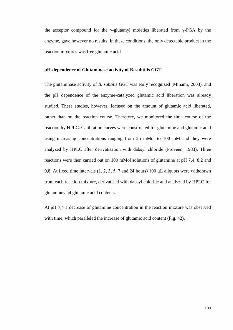
109
the acceptor compound for the γ-glutamyl moieties liberated from γ-PGA by the
enzyme, gave however no results. In these conditions, the only detectable product in the
reaction mixtures was free glutamic acid.
pH-dependence of Glutaminase activity of B. subtilis GGT
The glutaminase activity of B. subtilis GGT was early recognized (Minami, 2003), and
the pH dependence of the enzyme-catalyzed glutamic acid liberation was already
studied. These studies, however, focused on the amount of glutamic acid liberated,
rather than on the reaction course. Therefore, we monitored the time course of the
reaction by HPLC. Calibration curves were constructed for glutamine and glutamic acid
using increasing concentrations ranging from 25 mMol to 100 mM and they were
analyzed by HPLC after derivatisation with dabsyl chloride (Proveen, 1983). Three
reactions were then carried out on 100 mMol solutions of glutamine at pH 7,4, 8,2 and
9,8. At fixed time intervals (1, 2, 3, 5, 7 and 24 hours) 100 μL aliquots were withdrawn
from each reaction mixture, derivatised with dabsyl chloride and analyzed by HPLC for
glutamine and glutamic acid contents.
At pH 7,4 a decrease of glutamine concentration in the reaction mixture was observed
with time, which paralleled the increase of glutamic acid content (Fig. 42).

110
Figure 42: pH-dependence of Glutaminase activity of B. subtilis GGT- at pH 7,4, only hydrolysis occurs
Raising the pH to 8,2 a new peak in the chromatogram appeared transiently between 1
and 5 hours reaction time, which was attributed to γ-glutamylglutamine. HPLC-ESI MS
experiments confirmed our hypothesis of an autotranspeptidation reaction, attributing to
the new peak in the chromatogram a molecular mass of 562, corresponding to that of
the dabsyl derivative of γ-glutamylglutamine. Further evidence came from the use of an
authentic sample of γ-glutamylglutamine, on purpose synthesized and used for the
construction of a calibration curve aimed at its quantitative estimation in the reaction
mixture.
The most impressive result was however obtained at pH 9,8, in that glutamine
disappeared very quickly within two hours, and a series of little peaks appeared in the
chromatograms. The amounts of glutamic acid and γ-glutamylglutamine formed in the
same time were insufficient to explain a so rapid disappearance of glutamine (fig 43).

111
Figure 43: pH-dependence of Glutaminase activity of B. subtilis GGT- raising pH to 9,8, transpeptidation becomes significant
HPLC-ESI MS experiments showed that oligomeric species, containing up to four γ-
glutamyl residues linked to a single glutamine molecule, were formed (Scheme 12).
Scheme 12: oligomeric species formed by transpeptidation
We were unfortunately unable to quantify these species, but the result is similar to that
hypnotized using B. subtilis (natto) (Suzuki, 2002b).

112
The same experiment was repeated using D-glutamine, with identical outcomes.
The results obtained in these experiments are in our opinion noteworthy for at least
three reasons. Firstly, while on one hand they confirmed the glutaminase activity of B.
subtilis GGT, they showed also the ability of the enzyme to accept glutamine as a
substrate in a transpeptidation reaction. It is in apparent contrast with results previously
reported, in which the transpeptidase activity towards glutamine was referred to be very
low (Suzuki, 2002b). Those results were however obtained at a lower pH value, below
the pKa of the glutamine amino group. It was indeed proposed that an alkaline pH is
beneficial for the GGT-catalyzed transpeptidation reaction in that it increases the
concentration of unprotonated nucleophilic amino groups (Proveen, 1983). At more
acidic pH, the amino group of the acceptor molecule is protonated to a more extent, so
the hydrolysis reaction is favored. In our case, the autotranspeptidation reaction was
mainly observed at pH 9,8, slightly above 9,1, the pKa of the amino group of glutamine.
Secondly, the enzyme can accept as substrates not only discrete molecules, but also
oligomeric poly-γ-glutamyl derivatives, as showed by the HPLC-ESI MS experiments.
This is a peculiar characteristic, not shared with mammalian or other bacterial GGTs,
such as for example E. coli GGT. When E. coli GGT was used for synthetic purposes in
the presence of a high concentration of glutamine as donor at elevated pH, only the
formation of γ-glutamylglutamine was reported, but not the formation of oligomeric
compounds (Suzuki, 2002b). A similar behaviour was noted for rat kidney GGT
(Proeen, 1983). The ability of processing also oligomeric γ-glutamyl derivatives is
ascribable to the possible role of B. subtilis GGT in the homeostasis of γ-PGA, which
constitutes the cellular envelope of some strains of B. subtilis (Tate, 1974; Suzuki,
1986).
Thirdly, the ability of B. subtilis GGT to accept D-glutamine as an acceptor substrate
113
marks another difference from mammalian and E. coli GGT. The latter can indeed
accept γ-glutamyl compounds with D-configuration only as donors, but amino acid
acceptors have to be in the L-configuration (Tate, 1974; Proveen, 1983; Sukuzi, 2002b).
The similar results obtained subjecting L- and D-glutamine to the action of B. subtilis
GGT can be once again related to the involvement of the enzyme in the homeostasis of
extracellular γ-PGA, in that this polymeric material is usually formed by L- and D-
glutamic acid units in different proportion, depending on Bacillus strain and
environmental conditions (Suzuki, 1986).

EXPERIMENTAL PART
115
Materials and Methods
Glutamic acid, N-Fmoc-OtBut-glutamic acid, 2-chloro-trytil-chloride resin and all other
reagents were purchased from Sigma-Aldrich (Milan, Italy) and/or from VWR
International (Milan, Italy) and were used without further purification. All solvents were
of HPLC grade.
Analytical TLC was performed on silica gel F254 precoated aluminum sheets (0.2 mm
layer; Merck, Darmstadt, Germany); components were detected under an UV lamp (λ
254 nm) and by spraying with a ninhydrin 5% (w/v) in ethanol solution, followed by
heating to ca. 150 °C.
1H and
13C NMR spectra were recorded at 400,13 and 100,61 MHz, respectively, on a
Bruker AVANCE 400 spectrometer equipped with a XWIN-NMR software package
(Bruker, Karlsruhe, Germany) at 300 K. 1H and
13C chemical shifts (δ) are given in
parts per million and were referenced to the solvent signals. 13
C NMR signal
multiplicities were based on APT (attached proton test) spectra.
Matrix assisted laser desorption/ionization spectra (MALDI TOF) were acquired on a
Bruker Microflex LT Spectrometer.
UV measurements were carried out with a Jasco UV-vis/NIR V 630 instrument.
For HPLC runs, the instruments used was a HPLC Merck Hitachi: D-7000 interface,
UV detector L-7400, column oven L-7350, L-7200 programmable autosampler, L-7150
pump.
For solid phase synthesis a Milligen 9050 Peptide synthesizer and a Biotage Initiator+
were used
SpectraPor3 membranes, MWCO 3500 Da, were used for dialysis.
116
Ultrafiltration was performed by means of Amicon device (250 and 10 ml), equipped
with Millipore filtration membranes (regenerated cellulose, diameter 76-25 mm;
NMWL 30 000 Da).
Abbreviations used
Fmoc: fluorenylmethyloxycarbonyl
Boc: tert-butoxycarbonyl
BCC: benzylchlorocarbonate
Cbz: carboxybenzyl
DMF: dimethylformamide
HOBt: hydroxybenzotriazole
HBTU: O-benzotriazole-N,N,N’,N’-tetramethyl-uronium-hexafluoro-phosphate
DIPEA: N,N-diisopropylamine
DCHA: dicyclohexylamine
EDC: 1-ethyl-3-(3-dimethylaminopropyl)carbodiimide
TEA: triethylamine
TFFH: N,N,N’,N’-tetramethyl-fluoro-formamidinium-exafluorophosphate
TMSCH2N2: trimethylsilyldiazomethane

117
Bioproduction
A typical procedure is reported.
Cells growth and sporulation
B. subtilis PB5249 and PB5509 strains, grown overnight in 20 mL of LB medium, were
diluted to optical density 0.2 at 600nm (OD600) in rich Difco Sporulation Medium
(DSM) and in Spizizen’s Minimal Salt Medium (SMS) (Harwood and Cutting, 1990)
and incubated at 37 °C in an orbital shaker. After 90 min, aliquots were withdrawn
every 30 min for OD600 readings. After 24 h-incubation, cells were taken from cultures
grown in DSM medium and serially diluted. An equal volume was plated on LB plates
in duplicate, before and after incubation at 80 °C for 10 min (to kill vegetative cells).
After incubation for 16 h at 37 °C, CFUs (colony forming units) were counted and
sporulation efficiency was calculated as the ratio between heat resistant and total CFUs.
Experiments were independently repeated at least three times.
γ-PGA culture
Cells were grown overnight in LB medium at 37 °C in a shaking incubator; cultures
were diluted to OD600 0.2 in E medium adapted from Leonard et al. (1958) (L-glutamic
acid, 40.0 g/L; citric acid, 12.0 g/L; glucose, 80.0 g/L; NH4Cl, 7.0 g/L; K2HPO4, 0.5
g/L; MgSO4・7H2O, 0.5 g/L, FeCl3・6H2O, 0.04 g/L; CaCl2・2H2O, 0.15 g/L;
MnSO4・H2O, 0.104 g/L, pH 6.5) and incubated at 37 °C with shaking. Aliquots (500
μL in duplicate) were collected every 12 hours for OD600 readings and for γ-PGA
extraction. After OD600 readings, each sample was centrifuged at 16000 rcf for 10 min
at 4°C.

118
γ-PGA Separation on SDS-PAGE
To 10 μL γ-PGA samples, 2.5 μL loading buffer (Bromophenol Blue 5 mg/mL,
Glycerol 50%, running buffer 50%) was added before separation on 8% SDS-PAGE.
The gel was stained with 0.5% methylene blue in 3% acetic acid for 30 min and
destained in H2O.
Isolation
A typical procedure is given thereafter.
γ-PGA was precipitated from culture surnatant by addition of MeOH (3 volumes) at -20
°C overnight; the resulting crude product was suspended in milliQ water, and then
dialyzed (SpectraPor3 membranes, MWCO 3500 Da) against milliQ water for 12 hours;
the resulting solution was acidified to pH 2 by addition of concentrated HCl,
reprecipitated with 1-Propanol (3 volumes) and rinsed with acetone/diethyl ether.
1H NMR (δ ppm D2O): 1.35 [b, 1H, C
βH2], 1.50 [b, 1H, C
βH2], 1.78 [m, 2H, C
γH2],
3,71 [b, 1H, CH], 8.00 [d, 1H, -CONH].
Stereochemical L/D composition determination
Derivatisation method
A 1% solution of FDAA in acetone (200 L, 7.2 L) and 40 L of an aqueous 1%
NaHCO3 solution were added to a 50 mM solution of D,L-glutamic acid (100 L, 5
mol) in a 1mL brown vial. The vial was closed with a screw cap, covered with
aluminium sheet to protect it from light and put in an oven at 40 °C for 1 hour and 15
minutes, under magnetic stirring. After cooling at room temperature, a 2M HCl solution
(20 L, 40 mol) was added. The solution was subsequently stirred for some minutes
and then dried with a nitrogen flow in roughly 45 minutes.

119
The vial content was dissolved in 1mL of mobile phase A (see HPLC protocol) and
diluted in 1:5 proportion with mobile phase B to get a final concentration around 1 mM
which was injected in HPLC.
HPLC protocol
-column: RP Select B C18 (Merck) 5 m (250×4.6 mm)
-wavelenght: 340 nm
-flow: 1.2 mL/min
-temperature: 27 °C
-eluent:
-mobile phase A=acetonitrile/water (10:90 v/v) and sodium acetate 0.04 M,
final pH 5.3;
- mobile phase B=acetonitrile/water (50:50 v/v) and sodium acetate 0.04 M, final
pH 5.3.
-gradient elution:
- 0-20% B from 0 to 15.0 min;
- 20-75% B from 15.1 to 30.0 min;
- 20-75% B from min 30.1 to the end of the analysis.
It was verified that the analyte, if protected by light, maintains its stability for up to 5
days. The order of elution of peaks was in accordance with that reported in literature
(B’Hymer, 2001).

120
Commercial sample analysis
-PGA hydrolysis
A 0,3 molar solution of -PGA (sodium salt, molarity calculated on the monomer MW
(169,12, g mol-1
51,5 mg weighted) in 1mL of 2N HCl was put, with a magnetic stirrer,
in a 1,5 mL vial equipped with a sealed cap. The vial was put in a sand bath at 110 °C
and kept under stirring for 3,5 hours. It was afterwards let cool to room temperature;
solution was dried with a nitrogen flow (this operation took roughly 2 hours. A white
solid was obtained, which was stored at 4 °C.
Derivatisation
Reaction with Marfey’s reagent was carried out as previously described: 200 L of
Marfey’s reagent mother solution (3,6 mg in 355 L of acetone) were added to 100 L
of the sample’s mother solution (7,9 mg of acid hydrolisate, MW 147.13, in 1,1 mL).
HPLC run
Analysis was carried out the previously described, conditions but for the temperature,
which was raised from 27 °C to 35 °C. Dried reaction residue was dissolved with 1 mL
of mobile phase B, containing 50% of acetonitrile, differently from the D-L in house
prepared mixture, that was easily dissolved in mobile phase A, composed at 90% by
acetonitrile. Analysis was repeated twice.

121
Results are summarized in table 9.
Table 9
Sample 1 (dil. 1:5) Rt (min) Area (mV) % % L/D
17,95 1878426 14,20 (L) 47,97
24,90 2037731 15,40 (D) 52,03
35,21 8802742 66,53 (Marfey)
3,86 (other)
Sample 2 (dil. 1:4) Rt (min) Area (mV) % % L/D
16,60 2590954 14,68 (L) 48,24
22,66 2779724 15,75 (D) 51,76
35,04 12095685 68,54 (Marfey)
1,02 (other)
Non commercial sample analysis
-PGA hydrolysis
Procedure was the same used in the previous case. A 0,3 M solution of -PGA (sodium
salt) was prepared weighting 11,6 mg in 0,230 L of 2 M HCl. A brown, vitreous solid
was obtained and kept at 4 °C.
The product was quite rubbery and was tediously dissolved in 2M HCl, under stirring.
Solubility was complete after 10 minutes at 110 °C.
Derivatisation
Reaction was carried out as described above: 100 L of the mother solution were added
to 200 L of Marfey’s reagent mother solution (2,8 mg in 281 L of acetone). Since it
was not possible to take an exactly weighted aliquot of hydrolisate solution, the initial

122
weight of the polymer (11,6 mg in 1,6 mL) in its acidic form (MW 147,13) was taken
into account.
HPLC run
Analysis was carried out in the previously described, conditions but for the temperature,
which was raised from 27 °C to 35 °C. Dried reaction residue was dissolved with 1 mL
of mobile phase B, containing 50% of acetonitrile, as was done for the commercial
sample.
Results are summarized in table 10.
Table 10
Sample 1 (dil. 1:5) Rt (min) Area (mV) % % L/D
17,04 300053 4,010 (L) 14,33
22,81 1794414 23,98 (D) 85,67
35,15 4928788 65,87 (Marfey)
6,14 (other)
Sample 2 (dil. 1:1) Rt (min) Area (mV) % % L/D
15,99 1257696 4,21 (L) 14,33
22,11 7518484 25,16 (D) 85,67
35,03 19979016 66,87 (Marfey)
3,76 (other)
Determination of molecular weight
Chromatographic method
Chromatographic system
A modular Waters SEC System was used: Alliance 2695 (degassing unit, pump,
injector) column oven. Differential Refractometer (DRI) as concentration detector. To it
a MALS detector was connected in line, in the serial disposition: Alliance-MALS-DRI.
123
Since DRI detector suffers a slight delay with respect to the MALS one, signals were re-
aligned. DRI detector delay with respect to the MALS one was measured to be 0,270
mL.
Multi-Angle Laser Light Scattering (MALS)
A MALS photometer Dawn DSP-F (Wyatt) with K5 flow cell was used. The device
employs a He-Ne laser, λ=632,8 nm, with vertically polarized light. It measures the
intensity of scattered light in 18 angular positions at the same time.
0,1 M phosphate buffer (plus 0,1 M NaCl) at pH 7,0 at 35 °C was used as a solvent.
With the K5 cell used 16 angular positions (with values varying from 14,5° to 158,3°)
were available in this aqueous solvent.
MALS photometer was calibrated with Toluene, assuming a Rayleigh Factor
Rq=1,406·10-5
cm-1
.
Photodiodes were angularly normalized using a globular protein BSA (M=66.4
kg/mole, Rg=2.9 nm) assumed to isotropic.
dn/dc
The value of the dn/dc ratio (refraction index excess value of the macromolecule with
respect to the solvent) was obtained from the literature (Wyatt, 1993). For γ-PGA in the
mobile phase used for SEC at 25 °C it is equal to 0,140 mL/g.
Analysis conditions
Γ-PGA samples
–Columns: 2 Aquagel OH60 Polymer Laboratories (for sample PGA 12); 2
Ultrahydrogel (2000-500 Å) Waters (for Acid e Na Salt samples)
–Mobile phase: 0.1M NaCl + 0.1M Phosphate Buffer pH 7,0
– Temperature: 35 °C

124
– Flow: 0.8 mL/min
– Injection Volume: 150 μL
– Concentration: 1 or 5 mg/mL
Γ-PGA t-butyl ester
–Columns: 1 Mesopore, 1 Oligopore Polymer Laboratories
–Mobile phase: THF, stabilized with 0.025% BHT
–Temperature: 35 °C
–Flow: 0.8 mL/min
–Injection volume: 150 μL
–Concentration: 3 mg/mL
All samples were dissolved in the mobile phase chosen for SEC at the reported
concentration. Before injection all solutions were passed through 0,20 μm filters.
γ-PGA molecular weight assay by derivatisation with Sanger’s reagent and
hydrolysis
The following procedures were adapted from Park (Park, 2005) and Paraskevas
(Paraskevas, 2002).
Preparation of DNP-Glu
Glutamic acid (3,398 mmol, 0,500 g) was dissolved in an aqueous NaHCO3 solution
(13,60 mmol, 1,142 g in 20 ml). The solution was heated to 40 °C and then 1-fluoro-
2,4-dinitrobenzene (Sanger’s reagent) was added (3,504 mmol 0,44 ml) under stirring.
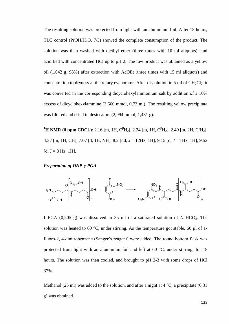
125
The resulting solution was protected from light with an aluminium foil. After 18 hours,
TLC control (PrOH/H2O, 7/3) showed the complete consumption of the product. The
solution was then washed with diethyl ether (three times with 10 ml aliquots), and
acidified with concentrated HCl up to pH 2. The raw product was obtained as a yellow
oil (1,042 g, 98%) after extraction with AcOEt (three times with 15 ml aliquots) and
concentration to dryness at the rotary evaporator. After dissolution in 5 ml of CH2Cl2, it
was converted in the corresponding dicyclohexylammonium salt by addition of a 10%
excess of dicyclohexylammine (3,660 mmol, 0,73 ml). The resulting yellow precipitate
was filtered and dried in desiccators (2,994 mmol, 1,481 g).
1H NMR (δ ppm CDCl3): 2.16 [m, 1H, C
βH2], 2.24 [m, 1H, C
βH2], 2.40 [m, 2H, C
γH2],
4.37 [m, 1H, CH], 7.07 [d, 1H, NH], 8.2 [dd, J = 12Hz, 1H], 9.15 [d, J =4 Hz, 1H], 9.52
[d, J = 8 Hz, 1H].
Preparation of DNP-γ-PGA
Γ-PGA (0,505 g) was dissolved in 35 ml of a saturated solution of NaHCO3. The
solution was heated to 60 °C, under stirring. As the temperature got stable, 60 µl of 1-
fluoro-2, 4-dinitrobenzene (Sanger’s reagent) were added. The round bottom flask was
protected from light with an aluminium foil and left at 60 °C, under stirring, for 18
hours. The solution was then cooled, and brought to pH 2-3 with some drops of HCl
37%.
Methanol (25 ml) was added to the solution, and after a night at 4 °C, a precipitate (0,31
g) was obtained.
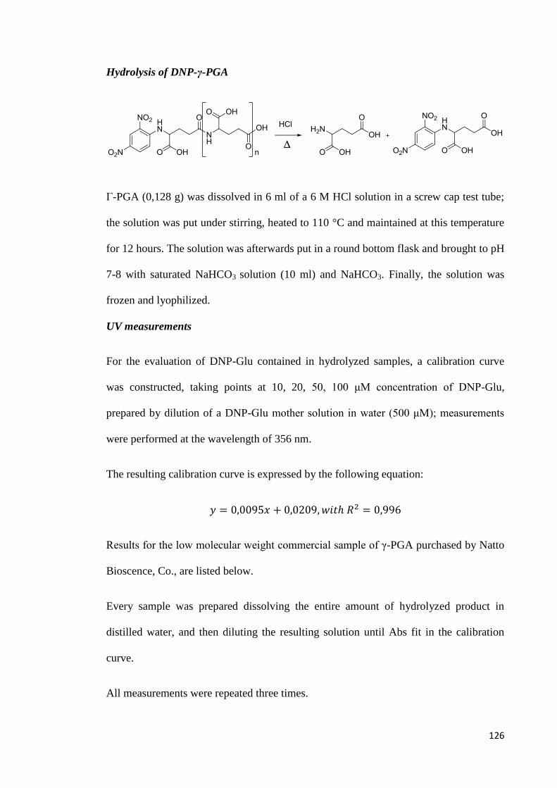
126
Hydrolysis of DNP-γ-PGA
Γ-PGA (0,128 g) was dissolved in 6 ml of a 6 M HCl solution in a screw cap test tube;
the solution was put under stirring, heated to 110 °C and maintained at this temperature
for 12 hours. The solution was afterwards put in a round bottom flask and brought to pH
7-8 with saturated NaHCO3 solution (10 ml) and NaHCO3. Finally, the solution was
frozen and lyophilized.
UV measurements
For the evaluation of DNP-Glu contained in hydrolyzed samples, a calibration curve
was constructed, taking points at 10, 20, 50, 100 μM concentration of DNP-Glu,
prepared by dilution of a DNP-Glu mother solution in water (500 μM); measurements
were performed at the wavelength of 356 nm.
The resulting calibration curve is expressed by the following equation:
Results for the low molecular weight commercial sample of γ-PGA purchased by Natto
Bioscence, Co., are listed below.
Every sample was prepared dissolving the entire amount of hydrolyzed product in
distilled water, and then diluting the resulting solution until Abs fit in the calibration
curve.
All measurements were repeated three times.
127
Representative results are reported thereafter.
Table 11
Abs (corrected for blank absorption) Molecular weight (g/mol)
0,248 30508
0,260 29742
0,249 28958
Average molecular weight estimated by this method results to be 29 736 g/mol.
Rheological measurements
All measurements were carried out using a rotational rheometer produced by Haake
(Karlsruke, Germany) model Rheostress RS600.
Working conditions are reported for each sample. Strumental parameters are listed as
follows.
Cone and plate:
these two parts of the sensor system are identified by their diameter and by
the angle occurring between them. For instance, the notation “60/1” indicates
a cone and plate system with a diameter of 60 mm, and an angle occurring
between the two of 1°.
Gap: distance between cone and plate, expressed in micrometers (μm).
Waiting time: delay between measurements.
Sample quantity deposed on plate.
Temperature.
Samples were prepared dissolving 25 mg of γ-PGA in 2,5 ml of milliQ water. All
measurements were repeated three times. Results are reported as the average.

128
Natto Bioscience sample
Working conditions:
Cone and plate: 60/1.
Gap: 52 μm.
Waiting time: 180 s.
Sample quantity: 1 ml.
Temperature: 25 °C.
Results
Table 12
Average
ds
τ (Pa) η (mPa s) shear rate (1/s) τ (Pa) η ( mPa s)
0,0262 2,619 10,0 0,0015 0,1485
0,1025 2,049 50,0 0,0007 0,0113
0,1545 2,061 75,0 0,0021 0,0361
0,2095 2,094 100 0,0021 0,0247
0,2580 2,062 125 0 0
0,3095 2,063 150 0,0078 0,0509
0,3600 2,058 175 0,0014 0,0099
0,4100 2,048 200 0,0057 0,0283
0,5140 2,057 250 0,0042 0,0156
0,6170 2,057 300 0,0014 0,0071
Biological Tech sample
Working conditions:
Cone and plate: 35/1.
Gap: 52 μm.
Waiting time: 180 s.
Sample quantity: 200 μl.
Temperature: 25 °C.

129
Results
Table 13
Average
ds
τ (Pa) η (mPa s) shear rate (1/s) τ (Pa) η (mPa s)
1,616 161,5 9,999 0,0340 3,383
6,958 139,2 49,99 0,1344 2,676
9,641 128,5 75,00 0,1439 1,950
12,01 120,1 100,0 0,1305 1,305
14,17 113,4 125,0 0,1852 1,474
16,19 108,0 150,0 0,2254 1,531
18,05 103,1 175,0 0,2309 1,626
19,82 99,12 200,0 0,2627 1,323
22,99 91,95 250,0 0,2893 1,164
25,90 86,34 300,0 0,3304 1,104
University of Pavia sample
Working conditions:
Cone and plate: 35/1.
Gap: 52 μm.
Waiting time: 180 s.
Sample quantity: 200 μl.
Temperature: 25 °C.
Results
Table 14
Average
ds
τ (Pa) η (mPa s) shear rate (1/s) τ (Pa) η (mPa s)
1,320 132,0 9,999 0,0332 3,318
5,758 115,2 50,00 0,0943 1,922
8,097 107,9 75,00 0,1507 2,021
10,11 101,1 100,0 0,1723 1,724
11,99 95,95 125,0 0,2346 1,883
13,80 92,01 150,0 0,1833 1,207
15,45 88,28 175,0 0,1258 0,715
16,98 84,89 200,0 0,2138 1,067
19,82 79,27 250,0 0,1947 0,782
22,40 74,66 300,0 0,2146 0,721
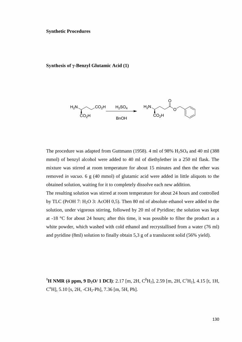
130
Synthetic Procedures
Synthesis of γ-Benzyl Glutamic Acid (1)
The procedure was adapted from Guttmann (1958). 4 ml of 98% H2SO4 and 40 ml (388
mmol) of benzyl alcohol were added to 40 ml of diethylether in a 250 ml flask. The
mixture was stirred at room temperature for about 15 minutes and then the ether was
removed in vacuo. 6 g (40 mmol) of glutamic acid were added in little aliquots to the
obtained solution, waiting for it to completely dissolve each new addition.
The resulting solution was stirred at room temperature for about 24 hours and controlled
by TLC (PrOH 7: H2O 3: AcOH 0,5). Then 80 ml of absolute ethanol were added to the
solution, under vigorous stirring, followed by 20 ml of Pyridine; the solution was kept
at -18 °C for about 24 hours; after this time, it was possible to filter the product as a
white powder, which washed with cold ethanol and recrystallised from a water (76 ml)
and pyridine (8ml) solution to finally obtain 5,3 g of a translucent solid (56% yield).
1H NMR (δ ppm, 9 D2O/ 1 DCl): 2.17 [m, 2H, C
βH2], 2.59 [m, 2H, C
γH2], 4.15 [t, 1H,
CαH], 5.10 [s, 2H, -CH2-Ph], 7.36 [m, 5H, Ph].
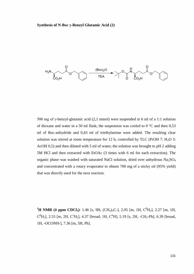
131
Synthesis of N-Boc γ-Benzyl Glutamic Acid (2)
500 mg of γ-benzyl-glutamic acid (2,1 mmol) were suspended in 6 ml of a 1:1 solution
of dioxane and water in a 50 ml flask; the suspension was cooled to 0 °C and then 0,53
ml of Boc-anhydride and 0,43 ml of triethylamine were added. The resulting clear
solution was stirred at room temperature for 12 h, controlled by TLC (PrOH 7: H2O 3:
AcOH 0,5) and then diluted with 5 ml of water; the solution was brought to pH 2 adding
5M HCl and then extracted with EtOAc (3 times with 6 ml for each extraction). The
organic phase was washed with saturated NaCl solution, dried over anhydrous Na2SO4
and concentrated with a rotary evaporator to obtain 700 mg of a sticky oil (95% yield)
that was directly used for the next reaction.
1H NMR (δ ppm CDCl3): 1.46 [s, 9H, (CH3)3C-], 2.05 [m, 1H, C
βH2], 2.27 [m, 1H,
CβH2], 2.55 [m, 2H, C
γH2], 4.37 [broad, 1H, C
αH], 5.19 [s, 2H, -CH2-Ph], 6.39 [broad,
1H, -OCONH-], 7.36 [m, 5H, Ph].

132
Synthesis of N-Boc-α-Methyl-γ-Benzyl Glutamic Acid (3)
700 mg of N-Boc-γ-benzyl-glutamic acid (2 mmol), previously dissolved in 6 ml of
DMF were loaded under N2 atmosphere in a 10 ml flask. 336 mg of NaHCO3 and then
0,2 μl of MeI were added under magnetic stirring to the resulting mixture. The solution
was stirred at room temperature for about 24 hours and controlled by TLC (CH2Cl2 9:
MeOH 1). Then it was diluted with 5 ml of water and extracted with diethyl ether (3
times with 6 ml for each extraction). The organic phase was washed with water, with a
saturated Na2S2O3 solution and finally with a saturated NaCl solution; it was then dried
over anhydrous Na2SO4 and concentrated with a rotary evaporator. The obtained oil was
dissolved in diethyl ether and subsequently treated with n-hexane to obtain, after 24
hours at -18 °C, a white crystalline solid that was filtered and washed with cold n-
hexane (470 mg, 67% yield).
1H NMR (δ ppm CDCl3): 1.46 [s, 9H, (CH3)3C-], 1.99 [m, 1H, C
βH2], 2.20 [m, 1H,
CβH2], 2.45 [m, 2H, C
γH2], 3.75 [s, 3H, -COOCH3], 4.37 [broad, 1H, CH], 5.14 [s, 2H, -
CH2-Ph], 7.38 [m, 5H, Ph].

133
Synthesis of α-Methyl-γ-Benzyl Glutamate·CF3CO2H (4)
400 mg of N-Boc-α-methyl-γ-benzyl glutamic acid (1,14 mmol) were dissolved in a 10
ml flask, in 3,5 ml of CH2Cl2 containing 1,75 ml of CF3CO2H at 0 °C. The mixture was
stirred at room temperature for about 1 hour and controlled by TLC (CH2Cl2 9: MeOH
1). After that the solution was concentrated at rotary evaporator and then at the pump to
completely remove the CF3CO2H. The residue was then dissolved in diethyl ether and
then treated with n-hexane to afford a precipitate after 24 hours at -18 °C. The white
solid was filtered and washed with cold n-hexane. (365 mg, 87% yield).
1H NMR (δ ppm CDCl3): 2.27 [m, 2H, C
βH2], 2.60 [m, 2H, C
γH2], 3.76 [s, 3H, -
COOCH3], 4.16 [broad, 1H, CH], 5.12 [s, 2H, -CH2-Ph], 7.35 [m, 5H, Ph].
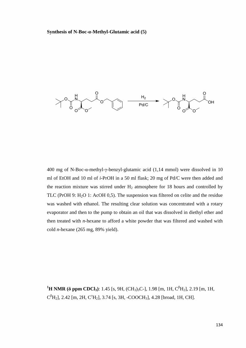
134
Synthesis of N-Boc-α-Methyl-Glutamic acid (5)
400 mg of N-Boc-α-methyl-γ-benzyl-glutamic acid (1,14 mmol) were dissolved in 10
ml of EtOH and 10 ml of i-PrOH in a 50 ml flask; 20 mg of Pd/C were then added and
the reaction mixture was stirred under H2 atmosphere for 18 hours and controlled by
TLC (PrOH 9: H2O 1: AcOH 0,5). The suspension was filtered on celite and the residue
was washed with ethanol. The resulting clear solution was concentrated with a rotary
evaporator and then to the pump to obtain an oil that was dissolved in diethyl ether and
then treated with n-hexane to afford a white powder that was filtered and washed with
cold n-hexane (265 mg, 89% yield).
1H NMR (δ ppm CDCl3): 1.45 [s, 9H, (CH3)3C-], 1.98 [m, 1H, C
βH2], 2.19 [m, 1H,
CβH2], 2.42 [m, 2H, C
γH2], 3.74 [s, 3H, -COOCH3], 4.28 [broad, 1H, CH].
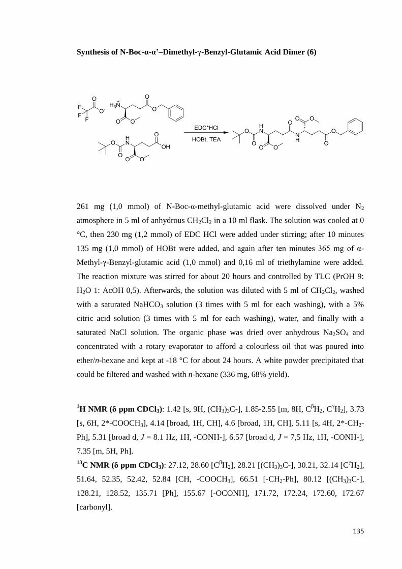
135
Synthesis of N-Boc-α-α’–Dimethyl-γ-Benzyl-Glutamic Acid Dimer (6)
261 mg (1,0 mmol) of N-Boc-α-methyl-glutamic acid were dissolved under N2
atmosphere in 5 ml of anhydrous CH2Cl2 in a 10 ml flask. The solution was cooled at 0
°C, then 230 mg (1,2 mmol) of EDC HCl were added under stirring; after 10 minutes
135 mg (1,0 mmol) of HOBt were added, and again after ten minutes 365 mg of α-
Methyl-γ-Benzyl-glutamic acid (1,0 mmol) and 0,16 ml of triethylamine were added.
The reaction mixture was stirred for about 20 hours and controlled by TLC (PrOH 9:
H2O 1: AcOH 0,5). Afterwards, the solution was diluted with 5 ml of CH2Cl2, washed
with a saturated NaHCO3 solution (3 times with 5 ml for each washing), with a 5%
citric acid solution (3 times with 5 ml for each washing), water, and finally with a
saturated NaCl solution. The organic phase was dried over anhydrous Na2SO4 and
concentrated with a rotary evaporator to afford a colourless oil that was poured into
ether/n-hexane and kept at -18 °C for about 24 hours. A white powder precipitated that
could be filtered and washed with n-hexane (336 mg, 68% yield).
1H NMR (δ ppm CDCl3): 1.42 [s, 9H, (CH3)3C-], 1.85-2.55 [m, 8H, C
βH2, C
γH2], 3.73
[s, 6H, 2*-COOCH3], 4.14 [broad, 1H, CH], 4.6 [broad, 1H, CH], 5.11 [s, 4H, 2*-CH2-
Ph], 5.31 [broad d, J = 8.1 Hz, 1H, -CONH-], 6.57 [broad d, J = 7,5 Hz, 1H, -CONH-],
7.35 [m, 5H, Ph].
13C NMR (δ ppm CDCl3): 27.12, 28.60 [C
βH2], 28.21 [(CH3)3C-], 30.21, 32.14 [C
γH2],
51.64, 52.35, 52.42, 52.84 [CH, -COOCH3], 66.51 [-CH2-Ph], 80.12 [(CH3)3C-],
128.21, 128.52, 135.71 [Ph], 155.67 [-OCONH], 171.72, 172.24, 172.60, 172.67
[carbonyl].

136
Synthesis of N-Boc-α-α’–Dimethyl-Glutamic Acid Dimer (7)
232 mg of N-Boc-α-α’-dimethyl-γ-benzyl-glutamic acid dimer (0,5 mmol) were
dissolved in10 ml of ethanol and 5 ml of i-PrOH in a 50 ml flask. Then, 13 mg of Pd/C
were added and the reaction mixture was stirred under H2 atmosphere for 15 hours
(TLC: PrOH 9: H2O 1: AcOH 0,5). The suspension was filtered on celite and the
residue was washed with ethanol. The resulting clear solution was concentrated with a
rotary evaporator and then to the pump to obtain 182 mg of a colourless oil (90% yield)
that was directly used for the next reaction.
1H NMR (δ ppm CDCl3): 1.45 [s, 9H, (CH3)3C-], 1.86-2.50 [m, 8H, C
βH2, C
γH2], 3.78
[s, 6H, 2*-COOCH3], 4.20 [m, 1H, CH], 4.63 [m, 1H, CH].
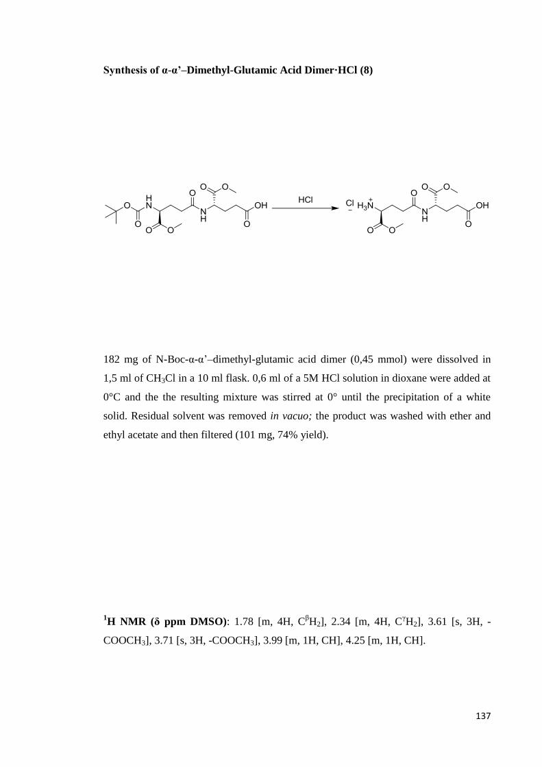
137
Synthesis of α-α’–Dimethyl-Glutamic Acid Dimer·HCl (8)
182 mg of N-Boc-α-α’–dimethyl-glutamic acid dimer (0,45 mmol) were dissolved in
1,5 ml of CH3Cl in a 10 ml flask. 0,6 ml of a 5M HCl solution in dioxane were added at
0°C and the the resulting mixture was stirred at 0° until the precipitation of a white
solid. Residual solvent was removed in vacuo; the product was washed with ether and
ethyl acetate and then filtered (101 mg, 74% yield).
1H NMR (δ ppm DMSO): 1.78 [m, 4H, C
βH2], 2.34 [m, 4H, C
γH2], 3.61 [s, 3H, -
COOCH3], 3.71 [s, 3H, -COOCH3], 3.99 [m, 1H, CH], 4.25 [m, 1H, CH].

138
Polycondensation (9)
160 mg (0,526 mmol) of α-α’-methyl glutamic acid dimer were dissolved in 1 ml of
DMF and loaded under N2 atmosphere in a 5 ml flask. The solution was cooled to 0 °C
and then, one after the other, 120 mg (0,631 mmol) of EDC·HCl, 71 mg (0,526 mmol)
of HOBt and 88 μl of triethylamine were added. The reaction mixture was stirred for 24
hours, controlled by TLC (PrOH 9: H2O 1: AcOH 0,5) and then concentrated with a
rotary evaporator. The residue was dissolved in 5 ml of water and extracted with
CH2Cl2 (3 times with 2 ml for each extraction); the organic phase was dried over
anhydrous Na2SO4 and concentrated to obtain a sticky oil; this was triturated in diethyl
ether until the formation of a pale yellow powder that was filtered, swiftly washed with
n-Hexane and put in a drier (60 mg, 38% yield).
1H NMR (δ ppm CDCl3): 1.68-2.38 [broad, 4H, C
βH2, C
γH2], 3.72-3.79 [broad, 3H, -
COOCH3], 4.59-4.78 [broad, 1H, CH].
Mass spectrum (MALDI-TOF): molecular weight distribution centered on m/z 3909.

139
Synthesis of N-Cbz-Glutamic Acid (10)
14 g of glutamic acid (95 mmol) were dissolved in 20 ml of water in a 50 ml flask. The
solution was basified at 0°C with 30 ml of 5M NaOH. 18 ml of benzylchlorocarbonate
(BCC) (125 mmol) were added in little aliquots keeping the pH high during the process
with 2M NaOH solution. The reaction mixture was stirred at 0 °C for 1,5 hours and then
let to room temperature; it was basified to pH 10 and washed with ethyl acetate. The
aqueous phase was acidified to pH 2 with a 5M HCl solution and left at -18 °C for 24
hours to afford a white solid that was filtered and washed with n-hexane (21 g, 80%
yield).
1H NMR (δ ppm DMSO): 1.75 [m, 1H, C
βH2], 1.97 [m, 1H, C
βH2], 2.29 [m, 2H,
CγH2], 3.99 [m, 1H, CH], 5.03 [s, 2H, -CH2-Ph], 7.35 [m, 5H, Ph], 7.59 [d, 1H, -CONH-
], 12.40 [broad, 2H, -COOH].

140
Synthesis of α-Methyl-γ-Benzyl Glutamic Acid (11)
γ-benzyl-glutamic acid (148 mg, 0,6 mmol) were dissolved in 7,2 ml of a mixture of
CH2Cl2 (6 ml) and MeOH (1,2 ml), under N2 flow. TMSCH2N2 (0,78 mmol, 0,375 ml
of a 2N solution in diethyl ether) was added; the solution immediately got an intense
yellow colour, tending to vanish with reaction’s progress, which was controlled by TLC
(PrOH 9: H2O 1: AcOH 0,5); loss of colour along some 6 hours indicated the
consumption of the reagent; solvent was afterwards evaporated, and the crude product
was dissolved in AcOEt and washed with saturated NaHCO3 solution (twice with 8 ml
each time); the aqueous phase was then extracted with AcOEt (twice with 4 ml each
time). Organic phases were pooled together, dried with saturated NaCl solution and
Na2SO4, and concentrated at the rotary evaporator to yield 105 mg (70%) of a white
solid product. This procedure was adapted from Kühnel, 2007.
1H NMR (δ ppm CDCl3): 1.99 [m, 1H, C
βH2], 2.20 [m, 1H, C
βH2], 2.45 [m, 2H, C
γH2],
3.75 [s, 3H, -COOCH3], 4.37 [broad, 1H, CH], 5.14 [s, 2H, -CH2-Ph], 7.38 [m, 5H, Ph].

141
Synthesis of N-Cbz-α-Methyl-Glutamic Acid (12)
562 mg of N-Cbz glutamic acid (2,0 mmol) were suspended under N2 atmosphere in 1
ml of distilled CH3Cl in a 25 ml flask; 0,28 ml of triethylamine (2,0 mmol) were added
to the suspension and the mixture was stirred at room temperature until the complete
dissolution of the solid, then 0,22 ml (2,2 mmol) of dimethylsulphate were added and
the reaction mixture was stirred at Room temperature for 4,5 hours and followed by
TLC (PrOH 9: H2O 1: AcOH 0,5). The solvent was removed with a rotary evaporator
and the residue was dissolved in 5 ml of water and extracted with ethyl acetate (3 times
with 3 ml for each extraction). The organic phase was then washed with water and a
saturated NaCl solution, dried over anhydrous Na2SO4 and concentrated with a rotary
evaporator. The resulting oil was dissolved in 20 ml of ethyl acetate and treated with
0,44 ml of diciclohexylamine to afford, after 24 hours at -18 °C, a white solid that was
filtered and washed with cold ethyl acetate (572 mg, 60% yield). The salt was then
dissolved in 10 ml of 0,5M HCl and the resulting solution was stirred at room
temperature for half an hour; then it was extracted with CH2Cl2 (3 times with 6 ml for
each extraction). The organic phase was washed with water and a saturated NaCl
solution, dried over Na2SO4 and concentrated with a rotary evaporator to give 333 mg
of a colourless oil (94% yield) that was directly used for the next reaction.
1H NMR (δ ppm CDCl3): 2.28 [m, 2H, C
βH2], 2.94 [m, 2H, C
γH2], 3.74 [s, 3H, -
COOCH3], 4.32 [m, 1H, CH], 5.10 [s, 2H, -CH2-Ph], 6.85 [broad, 1H, -CONH-], 3.23
[m, 5H, Ph], 8.5[broad, CO2H].

142
Synthesis of N-Cbz-α-α’–Dimethyl-γ-Benzyl-Glutamic Acid Dimer (13)
295 mg of N-Cbz α-methyl glutamic acid (1 mmol) were dissolved under N2
atmosphere in 5 ml of anhydrous CH2Cl2 in a 10 ml flask. The solution was cooled at 0
°C then 230 mg (1.2 mmol) of EDC·HCl were added under stirring; after 10 minutes
135 mg (1 mmol) of HOBt were added and again after ten minutes 365 mg of α-Methyl-
γ-Benzyl Glutamic Acid (1 mmol) and 0.16 ml of triethylamine. The reaction mixture
was stirred for about 16 hours and controlled by TLC (PrOH 9: H2O 1: AcOH 0,5).
Then the solution was diluted with 5 ml of CH2Cl2, washed with a saturated NaHCO3
solution (3 times with 6 ml each), with a 5% citric acid solution (3 times with 6 ml
each) water and finally with a saturated NaCl solution. The organic phase was dried
over anhydrous Na2SO4 and concentrated with a rotary evaporator to obtain a colourless
oil that was cooled at -18 °C to precipitate a white powder that was filtered and washed
with n-hexane (350 mg, 65% yield).
1H NMR (δ ppm CDCl3): 2.03 [m, 2H, C
βH2], 2.25 [m, 4H, C
βH2, C
γH2], 2.49 [m, 2H,
CγH2], 3.73 [s, 3H, -COOCH3], 3.74 [s, 3H, -COOCH3], 4.38 [broad, 1H, CH], 4.62
[broad, 1H, CH], 5.11 [s, 2H, -CH2-Ph], 5.13 [s, 2H, -CH2-Ph], 5.64 [broad, 1H, -
CONH-], 6.44 [broad, 1H, - CONH-], 7.33 [m, 10H, Ph].
13C NMR (δ ppm CDCl3): 27.19, 28.32 [CβH2], 30.09, 32.06 [C
γH2], 51.75 [CH],
52.53 [-COOCH3], 53.39 [CH], 66.58, 67.11 [-CH2-Ph], 129-127 [CH Ph], 135.74,
136.20 [C-Ph], 156.20 [CO-CH2Ph], 171.69, 172.24, 172. 34, 172. 65 [carbonyl].

143
Synthesis of α-α’–Dimethyl-Glutamic Acid Dimer (14)
300 mg (0.57 mmol) of N-Cbz α-α’ –methyl-γ-benzyl glutamic acid dimer were
dissolved in 10 ml of EtOH and 10 ml of i-PrOH in a 50 ml flask, then 15 mg of Pd/C
were added and the reaction mixture was stirred under H2 atmosphere for 18 hours and
controlled by TLC (PrOH 9: H2O 1: AcOH 0,5). The suspension was filtered on celite
and the residue was washed with ethanol. The resulting clear solution was concentrated
with a rotary evaporator and then to the pump to obtain 160 mg of a colourless oil (94%
yield) that was directly used for the polycondensation.
1H NMR (δ ppm DMSO): 1.77 [m, 4H, C
βH2], 2.34 [m, 4H, C
γH2], 3.60 [s, 3H, -
COOCH3], 3.72 [s, 3H, -COOCH3], 3.98 [m, 1H, CH], 4.25 [m, 1H, CH].

144
Functionalisation of 2-Chloro-Trytil-Chloride Resin (15)
213 mg of N-Fmoc-O-tert-butyl-glutamic acid (0,5 mmol) and 0,33 ml of DIPEA (2
mmol) were dissolved in 3 ml of dry CH2Cl2 and loaded in a fritted plastic tube. Then
312,5 mg of 2-chlorotrityl chloride resin (loading 1,6 mmol/g) were added and the tube
was kept under orbital stirring overnight. The resin was washed three times with 3 ml of
a CH2Cl2/CH3OH/DIPEA (17:2:1) solution for each washing, then three times with of
CH2Cl2,with 4 ml for each washing, two times with DMF, with 3 ml for each washing,
and finally two times of CH2Cl2, each time with 3 ml. The resin was dried overnight in
vacuo; 488 mg of loaded resin were obtained (96% recover).

145
Synthesis of α-tert-butyl-glutamic acid γ-decamer (16)
Nine vials containing 212 mg of N-Fmoc-O-tertbutyl-glutamic acid (0,5 mmol), 67 mg
of HOBt (0,5 mmol) and 189 mg of HBTU (0,5 mmol) were prepared. Then, 101 mg of
loaded resin (0,1 mmol of amino acid) were suspended in DMF and put in the
synthesizer reaction column. The couplings were performed in a 2M DIPEA in DMF
solution and the deprotection of the amino terminus was carried out with a 20%
piperidine in DMF solution. 175 mg of α-tert-butyl-glutamic acid γ-decamer linked to
the resin were obtained (71% recover).

146
Cleavage of the Resin (17)
175 mg of loaded resin (0,07 mmol of α-tert-butyl-glutamic acid γ-decamer) were
suspended in 4ml of CH2Cl2 in a fritted plastic tube; CH2Cl2 was filtered and then the
resin was washed ten times with 3 ml of a 1% TFA in CH2Cl2 solution, each time
collecting the filtered solvent in the same flask containing 20 ml of a 10% pyridine in
CH3OH solution. Eventually the resin was washed with CH2Cl2 (three times with 6 ml
for each washing), CH3OH (three times, each time with 6 ml), CH2Cl2 (twice, each time
with 6 ml) and CH3OH (with 6 ml, twice).The filtered fractions were combined with the
pyridine solution that was then evaporated, under reduced pressure, to 5% of the staring
volume. The residue was freeze-dried to give the decamer as a yellow powder (100 mg,
53% yield).
Mass spectrum (MALDI-TOF): m/z 1870 (MH22+
).

147
Coupling of the decamers (18)
89 mg (0,0047 mmol) of γ-PGA butyl ester decamer and 32 μl (0,188 mmol) of DIPEA
were dissolved in 4 ml of DMF; 12,5 mg (0,047 mmol) of TFFH were added to the
reaction mixture just before putting the reactor in the microwave oven. The mixture was
heated at 50 °C for 30 minutes (oven power: 200 Watt). Afterwards, it was diluted with
20 ml of distilled water, frozen and freeze dried. SEC-MALS analysis showed the
product had Mp= 17,950.
Mass spectrum (MALDI-TOF): m/z 5993, 5480, 5188, 4647, 4355, 3772, 2740 (base
peak)
Mp= 17,950 (SEC-MALS)
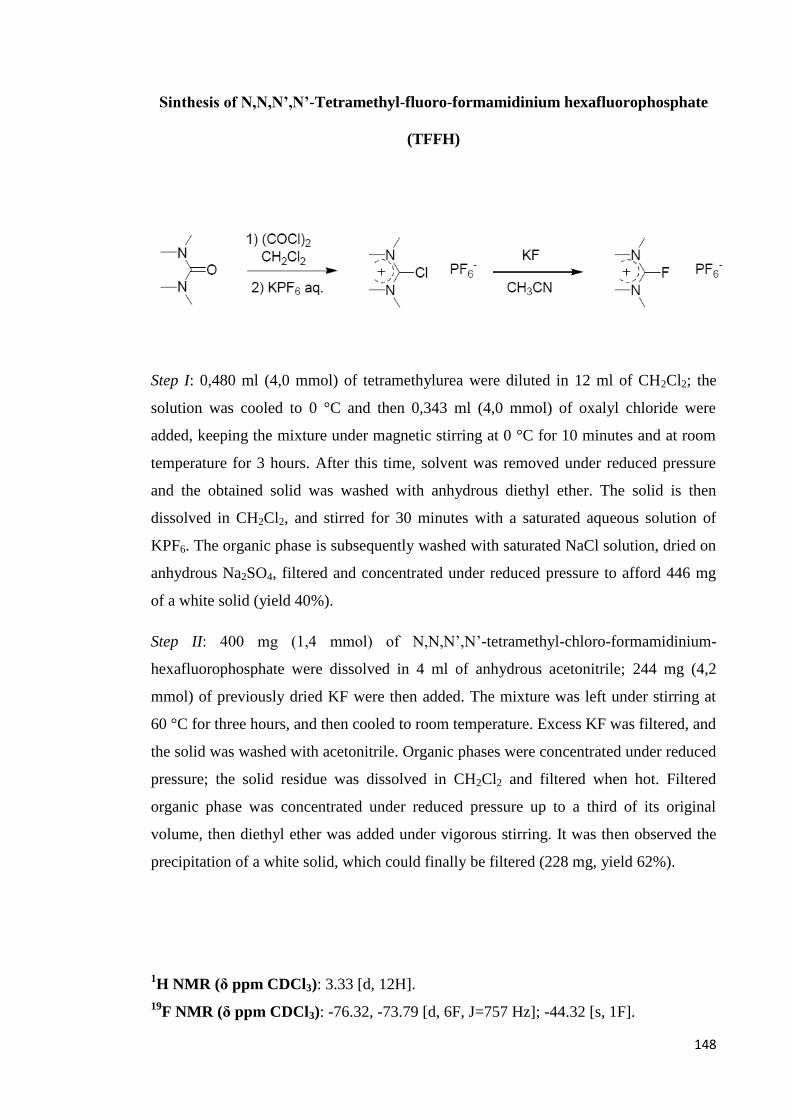
148
Sinthesis of N,N,N’,N’-Tetramethyl-fluoro-formamidinium hexafluorophosphate
(TFFH)
Step I: 0,480 ml (4,0 mmol) of tetramethylurea were diluted in 12 ml of CH2Cl2; the
solution was cooled to 0 °C and then 0,343 ml (4,0 mmol) of oxalyl chloride were
added, keeping the mixture under magnetic stirring at 0 °C for 10 minutes and at room
temperature for 3 hours. After this time, solvent was removed under reduced pressure
and the obtained solid was washed with anhydrous diethyl ether. The solid is then
dissolved in CH2Cl2, and stirred for 30 minutes with a saturated aqueous solution of
KPF6. The organic phase is subsequently washed with saturated NaCl solution, dried on
anhydrous Na2SO4, filtered and concentrated under reduced pressure to afford 446 mg
of a white solid (yield 40%).
Step II: 400 mg (1,4 mmol) of N,N,N’,N’-tetramethyl-chloro-formamidinium-
hexafluorophosphate were dissolved in 4 ml of anhydrous acetonitrile; 244 mg (4,2
mmol) of previously dried KF were then added. The mixture was left under stirring at
60 °C for three hours, and then cooled to room temperature. Excess KF was filtered, and
the solid was washed with acetonitrile. Organic phases were concentrated under reduced
pressure; the solid residue was dissolved in CH2Cl2 and filtered when hot. Filtered
organic phase was concentrated under reduced pressure up to a third of its original
volume, then diethyl ether was added under vigorous stirring. It was then observed the
precipitation of a white solid, which could finally be filtered (228 mg, yield 62%).
1H NMR (δ ppm CDCl3): 3.33 [d, 12H].
19F NMR (δ ppm CDCl3): -76.32, -73.79 [d, 6F, J=757 Hz]; -44.32 [s, 1F].

149
α-ethyl ester of γ-PGA
γ-PGA (2 g, 11, 83 mmol) was suspended in DMSO, under stirring; and was heated up
to reach the temperature of 45 °C. As it got homogenous, NaHCO3 (1,99 g, 23,66
mmol) was added, and after the salt got homogenously dispersed in the suspension,
EtBr (4,38 ml, 59,15 mmol) was poured in. The reaction was left at 45 °C for 5 days;
the solution was cooled to room temperature and then added dropwise in 750 ml of acid
water which was kept in the pH interval 1-2 with conc HCl. Evolution of CO2 was
observed. A sticky, gluish compound precipitated from the solution; after filtration, 1H-
NMR showed it to be the α-ethyl ester of γ-PGA, with a low (44%) functionalisation
degree. The material was again reacted in the same conditions for 3 more days; after
precipitation, the material was put at the rotary evaporator, which was connected to an
acetone/solid CO2 trap to eliminate DMSO traces, for some three hours. The second
generation product was finally obtained in the form of yellowish powder (525 mg).
Recover 25%, functionalisation 67%.
1H NMR (δ ppm DMSO): 1.8 [t, 3H, -COOCH2CH3], 1.77 [m, 1H, C
βH2], 1.97 [m,
1H, CβH2], 2.18 [m, 2H, C
γH2], 4.05 [q, 2H, -COOCH2CH3], 4.20 [m, 1H, CH], 8.22 [d,
1H, -CONH].

150
α-benzyl ester of γ-PGA
γ-PGA (2,00 g, 11, 83 mmol) was suspended in DMSO, under stirring; and was heated
up to reach the temperature of 45 °C. Once homogenous, NaHCO3 (1,99 g, 23,66 mmol)
was added; BnBr (7,0 ml, 59,15 mmol) was poured in only when the suspension got
homogenous again. The reaction was left at 45 °C for 5 days, afterewards the solution
was cooled to room temperature and subsequently added, dropwise, in 750 ml of acid
water which was kept in the pH interval 1-2 by addition of concentrated HCl. Evolution
of CO2 was observed. A yellow, translucent powder precipitated from the solution; 1H-
NMR revealed it to be α-benzyl ester of γ-PGA, with 50% functionalisation degree. The
material was again reacted in the same conditions for 3 more days; after precipitation, it
was put at the rotary evaporator, which was connected to an acetone/solid CO2 trap to
eliminate DMSO traces, for a couple of hours. The second generation product was
obtained in the form of brownish powder (1,71 g). Recover 61%, functionalisation
degree 70%.
1H NMR (δ ppm DMSO): 1.76 [m, 1H, C
βH2], 1.92 [m, 1H, C
βH2], 2.21 [m, 2H,
CγH2], 4.21 [m, 1H, CH], 5.11 [s, 2H, -CH2Ph], 7.37 [s, 5H Ph] 8.12 [d, 1H, -CONH].

151
α-methyl ester of γ-PGA
γ-PGA (500 mg, 3,40 mmol) was suspended in anhydrous methanol (8 ml), and cooled
to 0 °C in an ice bath. A catalytic amount of anhydrous DMF (0,1 ml) and oxalyl
chloride (1,75 ml, 20,33 mmol) were then cautiously added dropwise under nitrogen
flow, paying attention not to let the temperature increase above 5 °C. Evolution of gas
was observed. Once the additions were completed, the ice bath was removed and the
solution temperature was let raise to ambient value. After 24 hours, the solution was
diluted with methanol (16 ml) and neutralized with saturated NaHCO3 solution (20 ml).
After removal of methanol at the rotary evaporator, the formation of a white precipitate
was observed. The product, a white, sticky powder (101 mg) was filtered and washed
with little portions of distilled water and diethyl ether. Recover 25 %, functionalisation
degree 90%.
The general procedure used for the preparation of the next products follows the previous
one, used for the preparation of the methyl ester.
1H NMR (δ ppm DMSO): 1.75 [m, 1H, C
βH2], 1.95 [m, 1H, C
βH2], 2.21 [m, 2H,
CγH2], 3.62 [s, 3H, -COOCH3], 4.22 [m, 1H, CH], 8.27 [d, 1H, -CONH].b

152
α-ethyl ester of γ-PGA
γ-PGA (250 mg, 1,70 mmol) was suspended in anhydrous ethanol (4 ml), and cooled to
0 °C in an ice bath. A catalytic amount of anhydrous DMF (0,1 ml) and oxalyl chloride
(0,87 ml, 10,20 mmol) were then cautiously added dropwise under nitrogen flow,
paying attention not to let the temperature increase above 5 °C. Evolution of gas was
observed. Once the additions were completed, the ice bath was removed and the
solution temperature was let raise to ambient value. After 48 hours, the solution was
diluted with ethanol (14 ml) and neutralized with saturated NaHCO3 solution (20 ml).
After removal of methanol at the rotary evaporator, the formation of a white precipitate
was observed. The product, a white powder (101 mg) was filtered and washed with little
portions of distilled water, ethanol and diethyl ether. Recover 95 %, functionalisation
degree 25%.
1H NMR (δ ppm DMSO): 1.18 [t, 3H, -COOCH2CH3], 1.77 [m, 1H, C
βH2], 1.97 [m,
1H, CβH2], 2.18 [m, 2H, C
γH2], 4.05 [q, 2H, -COOCH2CH3], 4.20 [m, 1H, CH], 8.22 [d,
1H, -CONH].
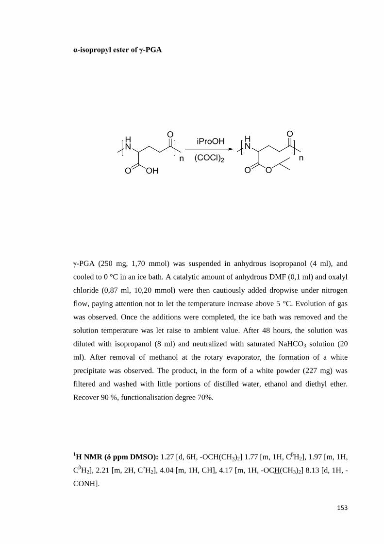
153
α-isopropyl ester of γ-PGA
γ-PGA (250 mg, 1,70 mmol) was suspended in anhydrous isopropanol (4 ml), and
cooled to 0 °C in an ice bath. A catalytic amount of anhydrous DMF (0,1 ml) and oxalyl
chloride (0,87 ml, 10,20 mmol) were then cautiously added dropwise under nitrogen
flow, paying attention not to let the temperature increase above 5 °C. Evolution of gas
was observed. Once the additions were completed, the ice bath was removed and the
solution temperature was let raise to ambient value. After 48 hours, the solution was
diluted with isopropanol (8 ml) and neutralized with saturated NaHCO3 solution (20
ml). After removal of methanol at the rotary evaporator, the formation of a white
precipitate was observed. The product, in the form of a white powder (227 mg) was
filtered and washed with little portions of distilled water, ethanol and diethyl ether.
Recover 90 %, functionalisation degree 70%.
1H NMR (δ ppm DMSO): 1.27 [d, 6H, -OCH(CH3)2] 1.77 [m, 1H, C
βH2], 1.97 [m, 1H,
CβH2], 2.21 [m, 2H, C
γH2], 4.04 [m, 1H, CH], 4.17 [m, 1H, -OCH(CH3)2] 8.13 [d, 1H, -
CONH].

154
α-n-butyl ester of γ-PGA
γ-PGA (250 mg, 1,70 mmol) was suspended in anhydrous n-butanol (4 ml), and cooled
to 0 °C in an ice bath. A catalytic amount of anhydrous DMF (0,1 ml) and oxalyl
chloride (0,87 ml, 10,20 mmol) were then cautiously added dropwise under nitrogen
flow, paying attention not to let the temperature increase above 5 °C. Evolution of gas
was observed. Once the additions were completed, the ice bath was removed and the
solution temperature was let raise to ambient value. After 48 hours, the solution was
diluted with methanol (12 ml) and neutralized with saturated NaHCO3 solution (20 ml).
After removal of methanol at the rotary evaporator, the formation of a white precipitate
was observed. The product, a white powder (126 mg) was filtered and washed with little
portions of methanol. Recover 50 %, functionalisation degree 25-70%, depending on
preparation.
1H NMR (δ ppm DMSO): 0,91 [t, 3H, -COOCH2CH2CH2CH3], 1.62 [m, 2 H, -
OCH2CH2CH2CH3] 1.39 [m, 2H, -OCH2CH2CH2CH3], 1.74 [m, 1H, CβH2], 1.97 [m,
1H, CβH2], 2.21 [b, 2H, C
γH2], 4.05 [m, 2H, -COOCH2CH2CH2CH3], 4.17 [m, 1H, CH],
8,13 [d, 1H, -CONH].

155
α-tetradecanoyl ester of γ-PGA
γ-PGA (500 mg, 3,40 mmol) were suspended in anhydrous THF (100 ml), and cooled to
0 °C in an ice bath. A catalytic amount of anhydrous DMF (0,3 ml) and oxalyl chloride
(5,2 ml, 61,2 mmol) were then cautiously added dropwise under nitrogen flow, paying
attention not to let the temperature increase above 5 °C. Evolution of gas was observed.
Once the additions were completed, the ice bath was removed and the solution
temperature got to ambient value, and then tetradecanol (14,6 g, 68,0 mmol) was added.
After 72 hours, the solution was neutralized with saturated NaHCO3 solution; after
removal of THF at the rotary evaporator, the formation of a white precipitate was
observed. The product, a white powder (133 mg) was filtered and washed with portions
of methanol, and then diethyl ether. Recover 53 %, functionalisation degree 4-25%
depending on preparation.
1H NMR (δ ppm DMSO): 0,85 [t, 3H, -COO(CH2)13CH3], 1.23 [s, 24H, -
OCH2(CH2)12CH3] 1.74 [m, 1H, CβH2], 1.95 [m, 1H, C
βH2], 2.19 [b, 2H, C
γH2], 4.06 [b,
2H, -COOCH2(CH2)12CH3], 4.16 [b, 1H, CH], 8,14 [d, 1H, -CONH].

156
α-ethyl-α’-poly(ethylene glycol)-yl ester of γ-PGA
α-ethyl ester of γ-PGA (175 mg, 1 mmol, 67% functionalisation) was suspended in poly
(ethylenglycol) (average molecular weight 550, 11 g, 20 mmol); Ti (Ot-Bu)4 was added
(68 mg, 0,2 mmol) under nitrogen flow and the reaction mixture was heated to 100 °C
and maintained at the same temperature for 3 hours, under stirring. It was then cooled to
room temperature and diluted, drop by drop, in methanol (25 ml). The formation of a
precipitate (47 mg) was observed, which was filtered off and washed with little aliquots
of methanol and diethyl ether. Recover 27 %, functionalisation degree 59% ethyl ester,
27% poly (ethylenglycol).
1H NMR (δ ppm DMSO): 1.21 [t, 3H, -COOCH2CH3], 1.74 [m, 1H, C
βH2], 1.95 [m,
1H, CβH2], 2.21 [m, 2H, C
γH2], 3.45 [s, (OCH2CH2)n], 3.53 [s, (OCH2CH2)n], 4.01 [q,
2H, -COOCH2CH3], 4.21 [m, 1H, CH], 8.26 [d, 1H, -CONH].

157
α-benzyl-α’-poly(ethylene glycol)-yl ester of γ-PGA
α-benzyl ester of γ-PGA (237 mg, 1 mmol, 50% functionalisation) was suspended in
poly (ethylenglycol) (average molecular weight 550, 11 g, 20 mmol); Ti (Ot-Bu)4 was
added (68 mg, 0,2 mmol) under nitrogen flow and the reaction mixture was heated to
100 °C and maintained at the same temperature for 3 hours, under stirring. It was then
cooled to room temperature and diluted, drop by drop, in methanol (25 ml). The
formation of a precipitate (107 mg) was observed, which was filtered off and washed
with little aliquots of methanol and diethyl ether. Recover 45 %, functionalisation
degree 42 % benzyl ester, 21% poly (ethylenglycol).
1H NMR (δ ppm DMSO): 1.77 [b, 1H, C
βH2], 1.99 [b, 1H, C
βH2], 2.51 [b, 2H, C
γH2],
4.21 [m, 1H, CH], 3.46 [s, (OCH2CH2)n], 3.52 [s, (OCH2CH2)n], 5.10 [s, 2H, -CH2Ph],
7.33 [s, 5H Ph] 8.31 [b, 1H, -CONH].

REFERENCES
159
Abe K., Ito Y., Ohmachi T., Asada Biosci Y., Biotech. Biochem., 1997, 61, 1621-1625
Aboulmagd E., Oppermann-Sanio F.B., Steinbüchel A., Arch. Microbiol., 2000, 174,
297-306
Aboulmagd E., Voss I., Oppermann-Sanio F.B., Steinbüchel A., Biomacromolecules,
2001, 2, 1338-1342
Akagi T., Higashi M., Kaneko T., Kida T., Akashi M., Macromol. Biosci., 2005a, 5,
598-602
Akagi T., Kaneko T., Kida T., Akashi M., Journal of Controlled Release, 2005b, 108,
226-236
Allen M.M., Methods Enzymol., 1988, 167, 207-213
Allison R.D., Methods Enzymol., 1985, 113, 419-437
Anderson A.J., Haywood G.W., Dawes E.A., Int. J. Biol. Macromol., 1990, 12, 102-
105
Aono R., Biochem. J., 1987, 245, 467-472
Ashiuchi M., Tani K., Soda K., Misono H., J. Biochemi (Tokyo), 1998, 123, 1156-
1163
Ashiuchi M., Soda K., Misono H., Biochem Biophys Res. Commun., 1999, 263, 6-12
Ashiuchi M., Kamei T., Baek D.H., Shin S.Y., Sung M.H., Soda K., Yagi T., Misono
H., Appl. Microbiol. Biotechnol., 2001, 57, 764-769
Ashiuchi M., Misono H., Appl. Microbiol. Biotechnol., 2002, 59, 9-14
Ashiuchi M., Kamei T., Misono H., J. Mol. Catal. B., 2003, 23, 101-106
Ashiuchi M., Shimanouchi K., Nakamura H., Kamei T., Soda K., Park C., Sung M.H.,
Misono H., Appl. Environ. Microbiol., 2004, 70, 4249-4255
Avichezer D., Schechter B., Arnon R., Reactive Functional Polym., 1998, 36, 59-69
Bajaj I.B., Lele S.S., Singhal R.S., Biores. Technol., 2009, 100, 826-832

160
Bajaj I., Singhal R., Bioresour. Technol., 2011, 102, 5551-5561
Berg H., Ziegler K., Piotukh K., Baier K., Lockau W., Volkmer-Engert R., Eur. J.
Biochem., 2000, 267, 5561-5570
B’Hymer C. J., Liq. Chrom & Rel. Technol., 2001, 24, 3085-3094.
Birrer G.A., Cromwick A.M., Gross R.A., Int. J. Biol. Macromol., 1994, 16, 265-275
Boanca G., Sand A., Barycki J.J., J. Biol. Chem., 2006, 281, 19029-19037
Boanca G., Sand A., Okada T., Suzuki I., Kumagai H., Fukuyama K., Barycki J.J., J.
Biol. Chem., 2007, 282, 534-541
Borbély G.A., Nagasaki Y, Borbély J., Fran K., Bhogle A., Sevoian M., Polymer
Bull., 1994, 32, 127-132
Bovarnick M., J. Biol. Chem., 1942, 145, 415-424
Bruckner V., Kovaca J., Denes G., Nature, 1953, 172, 508
Buescher J.M., Margaritis A., Crit. Rev. Biotechnol., 2007, 27, 1-19
Bhushan R., Brückner H. Amino Acids, 2004, 27, 231-247.
Bhushan R., Bruckner H., J. Chrom. B, 2011, 879, 3148-3161
Candela T., Mock M., Fouet A., J. Bacteriol., 2005, 187, 7765-7772
Candela T., Fouet. A., Mol. Microbiol., 2006, 60, 1091-1098
Cantor C.R., Schimmel P.R., Structure of polypeptide in Biophysical Chemistry, W.H.
Freeman & Co., New York, 1980
Carrasco F., Chornet E., Overend R.P., Costa, J.A., J. Appl. Polym. Sci., 1989, 37,
2087-2098
Cheng C., Asada Y., Aaida T., Agric. Biol. Chem., 1989, 53, 2369-2375
Chevalier C., Thiberge J.M., Ferrero L., Labigne A., Mol. Microbiol., 1999, 31, 1359-
1372
Chibnall A.C., Rees W.M., Richard F.M., Biochem. J., 1958, 68, 129-135

161
Choi H.J., Kunioka M., Radiat. Phys. Chem., 1995a, 46, 175-179
Choi H.J., Yang R., Kunioka M., J. Appl. Polym. Sci., 1995b, 58, 807-814
Cromwick A.M., Gross R.A., Int. J. Biol. Macromol., 1995a, 16, 265-275
Cromwick A.M., Gross R.A., Can. J. Microbiol., 1995b, 41, 902-909
Cromwick A.M., Gross R.A., Biotechnol. Bioeng., 1996, 50, 222-227
Dearfield K.L., Abermathy C.O., Mutant. Res., 1988, 195, 45-77
Drummelsmith J., Whitfield C., Mol. Microbiol., 1999, 31, 1321-1332
Earl A.M, Losick R., Kolter R., Trends Microbiol., 2008, 16, 269-275
Elce J.S., Broxmeyer B., Biochem. J., 1976, 153, 223-232
Fujii H., Nippon Nogeikagaku Kaishi, 1963, 37, 407-411
Gardner J.M., Troy F.A., J. Biol. Chem., 1979, 254, 6262-6269
Godwin A.K., Meister A., O'Dwyer P.J., Huang C.S., Hamilton T.C., Anderson M.E.,
Proc. Natl. Acad. Sci. USA, 1992, 89, 3070-3074
Goldberg D.M., Critical Reviews in Clinical Laboratory Sciences, 1980, 12, 1-58
Golecki J.R., Heinrich U.R., J. Microsc., 1991, 162, 147-154
Gooding E.A., Sharma S., Petty S.A., Fouts E. A., Palmer C. J., Nolan B. E, Volk M.,
Chemical Physics, (2012), in press
Goto A., Kunioka M., Biosci. Biotechnol. Biochem., 1992, 56, 1031-1035
Grage K., Jahns A.C., Parlane N., Palanisamy R., Rasiah I.A., Atwood J.A., Rehm
B.H.A., Biomacromol., 2009, 10, 660-669
Griffith O.W., Bridges R.J., Meister A., Proc. Natl. Acad. Sci. USA, 1978, 75, 5405-
5408
Griffith O.W., Bridges R.J., Meister A., Proc. Natl. Acad. Sci. USA, 1979, 76, 6319-
6322
162
Gross R.A., McCarthy S.P., Shah D.T., 1995, US Patent 5,378,807
Gross A., in Biopolymer from renewable resources, 1998, Springer, New York, pp.
195-219
Guttman S., Boissonnas R.A., Helvetica Chimica Acta, 1958, 199, 1852-1867
Hai T., Ahlers H., Gorenflo V., Steinbüchel A., Appl. Microbiol. Biotechnol., 2000,
53, 383-389
Hanby W., Rhydon H., Biochemistry, 1946, 40, 297-307
Hanby W.E., Waley S.G., Watson J., J. Chem. Soc. (London), 1950, 3239-3249
Hanigam N.H., Carcinogenesis, 1995, 16, 181-185
Hans M.L., Lowman A.M., Curr. Opin. Solid State Mater. Sci. 6 (2002) 319–327
Hara T., Ueda S., Agric. Biol. Chem., 1982a, 46, 2275-2281
Hara T., Aumayr A., Fujio Y., Ueda S., Appl. Environ. Microbiol., 1982b, 44, 1456-
1458
Hara T., Fujio Y., Ueda S., J. Apll. Biochem., 1982c, 4, 112-120
Hara T., Kobunshi, 2002, 49, 367-370
Harwood C.R., Trends Biotechnol, 1992, 10, 247-256.
Hashimoto W., Suzuki I., Yamamoto K., Kumagai H., J. Biochem., 1995, 118, 75-80
Haurowitz F., Bursa F., Biochem. J., 1949, 44, 509-512
He L.M., Neu M.P., Vanderberg L.A., Environ. Sci. Technol., 2000, 34, 1694-1701
Hezayen F.F., Rehm B.H.A., Eberhardt R., Steinbüchel A., Appl. Microbiol.
Biotechnol., 2000, 54, 319-325
Hezayen F.F., Rehm B.H., Tindall B.J., Steinbüchel A., Eberhardt R., Int. J. Syst.
Evol. Microbiol., 2001, 51, 1133-1142
Hirayama C., Sakata M., Nakamura M., Ihara H., Kunitake M., Todokora M., J.
Chromatogr. B, 1999, 721, 187-195
163
Holmes F.A., Kudelka A.P., Kavanagn J.J., Huber M.H., Ajani J.A., Valero V., in
Taxane Anticancer Agents: Basic Science and Current Status, 1995, American
Chemical Society, Washington, D.C., pp. 31-57
Höppensack A., Opperman-Sanio F.B., Steinbüchel A., FEMS Microbiol. Letters,
2003, 218, 39-45
Housewright R.D. in The Bacteria: a Tractise on Structure and Function (Vol. III),
1962, Academic Press, New York, pp. 389-412
Hsu W.H., Ong P.L., Chen S.C., Lin L.L., Indian J. Biochem. Biophys., 2009, 46, 281-
288
Inoue M., Hiratake J., Suzuki H., Kumagai H., Sakata K., Biochemistry 2000, 39,
7764-7771
Irurzun I., Bou J.J., Pérez-Camero G., Abad C., Campos A., Muñoz-Guerra S.,
Macromol Chem and Phys, 2001, 202, 3253-3256
Ito Y., Tanaka T., Homachi T., Asada Y., Biosci. Biotechnol. Biochem., 1996, 60,
1239-1242
Ikeda Y., Fujii J., Anderson M.E., Taniguchi N., Meister A., J. Biol. Chem., 1995,
270, 22223-22228
Ikeda Y., Fujii J., Taniguchi N., Meister A., Proc. Natl. Acad. Sci. USA, 1995, 92,
126-130
Ikeda Y., Adv. Enzymol. Relat. Areas Mol. Biol., 1998, 72, 239-278
Ivánovics G., Erdös L., Immunitatsforsh, 1937a, 90, 5-19
Ivánovics G., Bruckner V., Immunitatsforsh, 1937b, 90, 304-318
Jendrossek D., J. Bacteriol., 2009, 191, 3195-3202
Joentgen W., Groth T., Steinbüchel A., Hai T., Oppermann F.B., 1998, International
Patent Application WO 98/39090
164
Joyce J., Cook J., Chabot D., Hepler R., Shoop W., Xu Q., Stambaugh T., Aste-
Amezaga M., Wang S., Indrawati L., Bruner M., Friedlander A., Keller P., Caulfield
M., J. Biol. Chem., 2006, 281, 4831-4843
Jung D.K., Jung S., Sun J.S., Kim J.N., Wee Y.J., Jang H.G., Ryu H.W. Biotechnol.
Bioprocess. Eng, 2006, 10, 289-295
Kada S., Nanamiya H., Kawamura F., Horinouchi S., Microbiol. Lett., 2004, 236. 13-
20
Kambourova M., Tangnei M., Priest F.G., Appl. Environ. Microbiol., 2001, 67, 1004-
1007
Kameda A., Shiba T., Kawazoe Y., Satoh Y., Ihara Y., Munekata M., Ishige K.,
Noguchi T., J. Biosci. Bioeng., 2001, 91, 557-563
Kandler O., König H., Wiegel J., Claus D., Syst. Appl. Microbiol., 1983, 4, 34-41
Kashima N., Furuta K., Tanabe I., 2006, United States Patent 20060025346
Keppie J., Harris-Smith P.W., Smith H., Br. J. Exp. Pathol., 1963, 44, 446-453
Kessler B., Witholt B., J. Biotechnol., 2001, 86, 97-104
Kijima K., Suzuki H., Enz. Micr. Tech., 2007, 41, 80-84
Kim D., Robyt J.F., Enzyme Microb. Technol., 1994, 16, 659-664
Kimura K., Itoh Y., Appl. Environ. Microb., 2003, 69, 2491-2497
Kimura K., Tran L.S., Uchida I., Itoh Y., Microbiology, 2004, 150, 4115-4123
King E.C., Warkins W.J., Blacker A.J., Bugg T.D.H., J. Polymer Science Part A:
Polymer Chemistry, 1998, 36, 1995-1999
King E.C., Blacker, A.J., Bugg, T.D.H., Biomacromolecules, 2000, 1, 75-83
Kinnersly A., Strom D., Deah R.Y., Koskan C.P., 1994, WO Patent 94/09.628
Kocianova S., Wong C., Yao Y., Woyich J.M., Fischer E.R., DeLeo F.R., Otto M., J.
Clin. Invest., 2005, 115, 688-694

165
Krehenbrink M., Steinbüchel A., Microbiology, 2004, 150, 2599-2608.
Krejtschi, C. Hauser, K. European Biophysics Journal, 2011, 40,673-85
Kubota H., Nambu Y., Takeda H., Endo T., 1992, US Patent 5,118,784
Kubota H., Matsunobu T., Uotani K., Takebe H., Satoh A., Tanaka T., Taniguchi M.,
Biosci. Biotechnol. Biochem., 1993a, 57, 1212-1213
Kubota H., Nambu Y., Endo T., J. Poly. Sci. A: Pol. Chem., 1993b, 31, 2877-2878
Kubota H., Nambu Y., Endo T., J. Poly. Sci. A: Pol. Chem., 1996, 34, 1347-1351
Kühnel E., Laffan D.P.D., Lloyd-Jones G.C., Martínez del Campo T., Shepperson
I.R., Slaughter J.L., Angew. Chem. Int. Ed., 2007, 46, 7075–7078
Kunioka M., Kobunshi Ronbunshu, 1993, 50, 755-760
Kunioka M., Goro A., Appl. Microbiol Biotechnol., 1994, 40, 867-872
Kunioka M., Appl. Microbiol. Biotechnol., 1995, 44, 501-506
Kunioka M., Appl. Microbiol. Biotechnol., 1997, 47, 469-475
Kunioka M., Macromol. Biosci., 2004, 4, 324-329
Kurane R., Takeda K., Suzuki T., Agric. Biol. Chem., 1986, 50, 2301-2307
Lawry N.H., Simon R.D. J. Phycol., 1982, 18, 391-399
Leathers T.D. in Biopolymers for Medical and Pharmaceutical Applications, 2005,
Wiley-VCH, Weinheim, pp. 537-560
Ledezma-Pérez A.S., Romero-Garcia J., Vargas-Gutiérrez G., Arias-Main E.,
Materials Lett., 2005, 59, 3188-3191
Leonard C.G., Housewright R.D., Thorne C.B., J. Bacteriol., 1958a, 76, 499-503
Leonard C.G., Housewright R.D., Thorne C.B., Biochim. Biophys. Acta, 1958b, 62,
432-434
Li X., Deng X., Yuan M., Xiong C., Huang Z., Zhang Y., Jia W., J. Appl. Polym. Sci.
78, 2000, 140– 148

166
Liang H.F., Chen C.T., Chen S.C., Kulkani A.R., Chiu Y.L., Chen M.C., Sung H.W.,
Biomaterials, 2006a, 27, 2051-2059
Liang H.F., Chen S.C., Chen M.C., Lee P.W., Chen C.T., Sung H.W., Bioconjug.
Chem., 2006b, 17, 291-299
Lin W.C., Yu D.G., Yang M.C., Colloids Surf. B: Biointerfaces, 2006, 47, 43-49
Liotenberg S., Campbell D., Rippka R., Hourmard J., Tandeau-de-Marsac N.,
Microbiology, 1996, 142, 611-622
Liu X.M., Pramoda K.P., Yang Y.Y., Chow S.Y., He C., Biomaterials, 2004, 25,
2619– 2628
Macaskie L.E., Basnakova G., Environ. Sci. Technol., 1998, 32, 184-187
Makino S., Sasakawa C., Uchida I., Terakado N., Yoshikawa M., Mol. Microbiol.,
1988, 2, 371-376
Makino S., Uchida I., Terakado N., Sasakawa C., Yoshikawa M., J.Bacteriol., 1989,
171, 722-730
Marfey P., Carlsberg Res. Commun., 1984, 49, 591.
Matsusaki M., Serizawa T., Kishida A., Endo T., Akashi M., Bioconjugate Chem.,
2002, 13, 23-28.
Matsusaki M., Akashi M., Biomacromolecules, 2005a, 6, 3351-3356
Matsusaki M., Serizawa T., Kishida A., Akashi M., J. Biomed. Mater. Res. A, 2005b,
73, 485-491
McCloy E.W., J. Hyg., 1951, 49, 114-125
McLean R.C., Wolf D.C., Ferris F.G., Beveridge T.J., Appl. Environ. Microbiol.,
1990, 56, 3671-3677
McLean R.C., Beauchemin D., Beveridge T.J., Appl. Environ. Microbiol., 1992, 58,
405-408
Meister A., Tate S.S., Griffith O.W., Methods Enzymol., 1981, 77, 237-253
167
Melis J., Morillo M., Martínez de Ilarduya A., Muñoz-Guerra S., Polymer, 2001, 42,
9319-9327
Mesnage S., Tosi-Couture E., Gounon P., Mock M., Fouet A., J. Bacteriol., 1998, 180,
52-58
Minami H., Suzuki H., Kumagai H., Enz. Micr. Tech., 2003, 32, 431-438
Mitsuiki M., Mizuno A., Tanimoto H., Motoki M., J. Agric. Food Chem., 1998, 46,
891-895
Martínez de Ilarduya A., Ittobane N., Bermúdez M., Alla A., El Idrissi M., Muñoz-
Guerra S., Biomacromolecules 2002, 3, 1078-1086
Mendichi R., A. Giacometti Schieroni A., Current Trends in Polymer Science, 2001,
6, 17-32
Morillo M., Martínez de Ilarduya A., Muñoz-Guerra S., Macromolecules, 2001, 34,
7868-7875
Morillo M., Martínez de Ilarduya A., Muñoz-Guerra S., Polymer, 2003, 44, 7557-7564
Morrow A.L., Williams K., Sand A., Boanca G., Barycki J.J., Biochemistry, 2007, 46,
13407-13414
Murao S., Kobunshi, 1969, 16, 1204-1212
Msadek T., Kunst F., Klier A., Rapoport G., J. Bacteriol., 1991, 173, 2366-2377
Nagai T., Koguchi K., Itoh Y., J. Gen. Appl. Microbiol., 1997, 43, 139-143
Nakamura J., Miyashiro S., Hirose Y., Agric. Biol. Chem., 1976, 40, 1341-1347
Nefkens, Nivard R.J.F., Rec. Des Trav. Chim dans Pais Bas, 1964, 83, 199-207
Niemetz R., Kärcher U., Kandlera O., Tindall B.J., König H., Eur. J. Biochem., 1997,
249, 905-911
Nitecki D.E., Goodman J.W. in Chemistry and Biochemistry of Amino Acids, Peptides
and Proteins, Vol I, 1971, Marcel Dekker, New York, pp. 87-92
Obst M., Steinbüchel A., Biomacromolecules, 2004, 5, 1166-1176
168
Ogawa Y., Hosoyama H., Hamano M., Motai H., Agric. Biol. Chem., 1991, 55, 2971-
2977
Ogawa Y., Yamaguchi F., Yuasa K., Tahara Y., Biosci. Biotechnol. Biochem., 1997,
61, 1684-1687
Ogawa Y., Sugiura D., Motai H., Yuasa K., Tahara Y., Biosci. Biotech. Biochem.,
1997, 61, 1596-1600
O’Hagan D.T., Jeffery H., Davis S.S., Int. J. Pharm., 1994 ,103, 37– 45
Ohsawa T., Tsukahara K., Ogura M., Biosci. Biotechnol. Biochem., 2009, 73, 2096-
2102.
Oinonen C., Rouvinen J., Protein Sci., 2000, 9, 2329-2337
Okada T., Suzuki I., Wada K., Kumagai H., Fukuyama K., Proc. Natl. Acad. Sci. USA,
2006, 103, 6471-6476
Ong P.L., Yao Y.F., Weng Y.M., Hsu W.H., Lin L.L., Biochem. Biophys. Res.
Commun., 2008, 366, 294-300
Oppermann-Sanio F.B., Hai T., Aboulmagd E., Hezayen F.F., Jossek S., Steinbüchel
A., in Biochemical principles and mechanisms of biosynthesis and biodegradation of
polymers, 1999, Wiley-VCH, Weinheim, pp. 185-193
Oppermann-Sanio F.B., Steinbüchel A., Naturwissenschaften, 2002, 89, 11-22
Orellana C., Lancet Infect. Dis., 2002, 2, 711
Osera C., Amati G., Calvio C., Galizzi A., Microbiology, 2009, 155, 2282-2287.
Owen A.D., Shapira A.H., Jenner P., Marsden C.D., Ann. N. Y. Acad. Sci., 1996, 786,
217-223
Otani Y., Tabata Y., Ikada Y., J. Biomed. Mater. Res., 1996, 31, 157-166
Otani Y., Tabata Y., Ikada Y., Biomaterials, 1998a, 19, 2091-2098
Otani Y., Tabata Y., Ikada Y., Biomaterials, 1998b, 19, 2167-2173
Panyam J., Labhasetwar V., Adv. Drug Deliv.Rev., 2003,55, 329– 347
169
Paraskevas G., J. Atta-Politou J., M. Koupparis M., J. Pharm. Biomed. Anal., 2002,
29, 865-872
Park Y.J., Liang J., Yang Z., Yang V.C., J. Control. Release, 2001, 75, 37-44
Park C., Choi J.C., Choi Y.H., Nakamura H., Shimanouchi K., Horiuchi T., Misono
H., Sewaki T., Soda K., Ashiuchi M., Sung M.H., J. Mol. Cat B: Enz., 2005, 35, 128-
133
Patrick J.E., Kearns D.B., Mol. Microbiol., 2012, 83, 14-23.
Pérez-Camero G., Congregado F., Bou J.J., Muñoz-Guerra S., Biotechnol. Bioengin.,
1999, 63, 110-115
Pérez-Camero G., Vázquez B., Muñoz-Guerra S., J. Applied Polymer Science, 2001,
82, 2027-2036
Portilla-Arias J.A., García-Alvarez M., Martínez de Ilarduya A., Muñoz-Guerra S.,
Polym. Degrad. Stabilility, 2007, 92, 1916-1924
Pötter M., Opperman-Sanio F.B., Steinbüchel A., Appl. Environ. Microbiol., 2001, 67,
617-622
Proveen D., Life Sci., 1983, 33, 1757-1762
Rao N.N., Gomez-Garcia M.R., Kornberg A., Annu. Rev. Biochem., 2009, 78, 605-647
Rehm B.H.A., Steinbüchel A., Int. J. Biol. Macromol., 1999, 25, 3-19.
Rehm B.H.A., Nature Rev. Microbiol., 2010, 8, 578-592
Remminghorst U., Rehm B.H.A. Biotechnol. Lett., 2006, 28, 1701-1712
Reusch R.N., Sadoff H.L., Proc. Natl Acad. Sci. USA, 1988, 85, 4176-4180
Richter S., Anderson V.J., Garufi G., Lu L., Budzik J.M., Joachimiak A., He C.,
Schneewind O., Missiakas D., Mol. Microbiol,. 2009, 71, 404-420
Robyt J.F., Yoon S.H., Mukerjea R., Carbohydr. Res., 2008, 343, 3039-3048
Rosalki S.B., Adv. Clin. Chem., 1975, 17, 53-107
170
Rowinsky K.E., Donehower R.C., N. Engl. J. Med., 1995, 332, 1004-1014
Ruzal S.M., Sanchez-Rivas C., Curr. Microbiol., 1998, 37, 368-372
Rydon H.N., J. Chem. Soc., 1964, 1328-1333;
Sakai H., Sakabe N., Sasaki K., Hashimoto W., Suzuki I., Tachi H., Kumagai H.,
Sakabe K., J. Biochem., 1993, 120, 26-28
Sakai K., Sonoda C., Murase K., 2000, JP Patent WO0021390
Sanda F, Fukiyama T., Endo T., J. Polym. Sci. Pol. Chem , 2001, 39, 732-741
Sawa S., Murakawa T., Watanabe T., Murao S., Omata S., Nippon Nögeikagaku
Kaishi, 1973, 47, 159-165
Sawamura S., J. Coll. Agric. Tokyo, 1913, 5, 189-191
Schneerson R., Kubler-Kielb J., Lin T.Y., Dai Z.D., Leppla S.H., Yergey A.,
Backlund P., Shiloach J., Majaldly F., Robbins J.B., Proc. Natl. Acad, Sci. USA, 2003,
100, 8945-8950
Schneerson R., Leppla S., Robbins J.B., Shiloach J., Kubler-Kielb J., Liu D., Majadly
F., 2005, Patent Cooperation Treaty Appl. Number WO 2005000884
Schreier H.J. in Bacillus subtilis and other Gram positive bacteria, 1993, America
Society for Microbiology, Washigton DC, pp. 281-298
Schwamborn M., Polym. Degrad. Stab., 1998, 59, 39-45
Scoffone V., Dondi D., Biino G., Borghese G., Pasini D., Galizzi A., Calvio C.,
Biotechnology and Bioengineering, 2013, in press, doi: 10.1002/bit.24846
Scorpio A., Chabot D.J., Day W.A., O’Brien D.K., Vietri N.J., Itoh Y.,
Mohamadzadeh M., Friedlander A.M., Antimicrob. Ag. Chemother., 2007, 51, 215-
222
Shibayama K., Kamachi K., Nagata N., Yagi T., Nada T., Doi Y., Shibata N.,
Yokoyama K., Yamane K., Kato H., Inuma Y., Arakawa Y., Mol. Microbiol., 2003,
47, 443-451
171
Shih I.L., Van Y.T., Bioresour. Technol., 2001, 79, 207-225
Shih I.L., Van Y.T., Shen M.H., Biotechnol. Lett., 2003, 25, 1709-1712
Shi F., Xu Z., Cen P., Appl. Biochem. Biotechnol., 2006, 133, 271-282.
Shima S., Sakai H., Agric. Biol. Chem., 1977, 41, 1807-1809
Shima S., Sakai H., Agric. Biol. Chem., 1981a, 45, 2497-2502
Shima S., Sakai H., Agric. Biol. Chem., 1981b, 45, 2503-2508
Shima S., Fakuhara Y., Sakai H., Agric. Biol. Chem., 1982, 46, 1917-1919
Shima S., Matsuoka H., Iwamoto T., Sakai H., J. Antibiot., 1984, 37, 1449-1455
Shuai Y., Zhang T., Mu W., Jiang B., J. Agric. Food Chem., 2011, 59, 6233-6238
Simon R.D., Proc. Natl. Acad. Sci. USA, 1971, 68, 265-267
Simon R.D., J. Bacteriol., 1973a ,114, 1213-1216
Simon R.D., Arch. Microbiol., 1973b, 92, 115-122
Simon R.D., Biochim. Biophys. Acta, 1976, 422, 407-418
Simon R.D., Lawry N.H., McLendon G.L., Biochim. Biophys. Acta, 1980, 626, 277-
281
Simon R.D. in The cyanobacteria., 1987, Elsevier, Amsterdam, pp. 199-225
Singer J.W., DeVries P., Bhatt R., Tulinsky J., Klein P., Li C., Milas L., Lewis R.A.,
Wallace S., Ann. NY Acad. Sci., 2000, 922, 136-150
Smith T.J., Blackman S.A., Foster S.J., Microbiology, 2000, 146, 249-262
Speranza G., Morelli C.F., Journal of Molecular Catalysis B: Enzymatic, 2012, 84,
65– 71
Steigedal M., Sletta H., Moreno S., Maerk M., Christensen B.E., Bjerkan T., Ellingsen
T.E., Espìn G., Estervåg H., Valla S., Environ. Microbiol., 2008, 10, 1760-1770

172
Suarez C., Kohler S.J., Allen M.M., Kolodny N.H., Biochim. Biophys. Acta, 1999,
1426, 429-438
Sun H., Meng F., Dias A.A., Hendriks M., Feijen J., Zhong Z., Biomacromolecules,
2011, 12, 1937–1955
Sung M., Park C., Kim C.J., Poo H., Soda K., Ashiuchi M., Chem Rec, 2005, 5, 352-
366
Sung H.W., Liang H.F., Tu H., 2006a, United States Patent 20060073210
Sung M.H., Park C., Kim C.J., Park J.S., Uyama H., Poo H.R., Song J.J., 2006b,
Patent Cooperation Treaty Application Number WO2006001567
Suzuki H., Kumagai H., Tochikura T.J., Bacteriol., 1986, 168, 1325-1331
Suzuki I., Kumagai K., J. Biol. Chem., 2002, 277, 43536-43543
Suzuki H., Miyakawa N., Kumagai H., Enzyme Microb. Technol., 2002b, 30, 883-888
Suzuki T., Tahara Y., J. Bacteriol., 2003, 185, 2379-2382
Suzuki I., Yamada C., Kato K., Amino Acids, 2007, 32, 333-340
Tanaka T., Fujita K.I., Takenishi S., Taniguchi M., J. Fermen. Bioeng., 1997, 84, 361-
364
Taniguchi N., Ikeda Y., Adv. Enzymol. Relat. Areas Mol. Biol., 1998, 72, 239-278
Taniguchi M., Kato K., Matsui O., Ping X., Nakayama H., Usuki Y., Ichimura A.,
Fujita K., Tanaka T., Tarui Y., Hirasawa E., J.Biosci.Bioeng., 2005a, 99, 130-135
Taniguchi M., Kato K., Shimauchi A., Ping X., Nakayama H., Fujita K., TanakaT.,
Tarui Y., Hirasawa E., J.Biosci.Bioeng., 2005b, 99, 245-251
Taniguchi M., Kato K., Shimauchi A., Xu P., Fujita K., Tanaka T., Tarui Y., Hirasawa
E., J.Biosci.Bioeng., 2005c, 100, 207-211
Tanimoto H., Mori M., Motoki M., Torii K., Kadowaki M., Noguchi T., Biosci.
Biotechnol. Biochem, 2001, 65, 5516-521
Tate S.S., Meister A., J. Biol. Chem., 1974, 249, 7593-7602

173
Tate S.S., Meister A., Mol. Cell. Biochem., 1981, 39, 357
Tayal A., Kelly R.M:, Khan S.A., Macromolecules, 1999, 32, 294-300
Thompson G.A., Meister A., Biochem. Biophys. Res. Commun., 1976, 71, 32-36
Thompson G.A., Meister A., J. Biol. Chem., 1977, 252, 6792-6798.
Thorne C.B., Gómez C.G., Blind G.R., Housewright R.D., J. Bacteriol., 1953, 65,
472-478
Thorne C.B., Gómez C.G., Noyes H.E., Housewright R.D., J. Bacteriol., 1954, 68,
307-315
Tocilj A., Munger C., Proteau A., Morona R., Purins L., Ajamian E., Wagner J.,
Papadopoulos M., Van Den Bosh L., Rubinstein J.L., Féthière J., Matte A., Cygler M.,
Nature Struct. Mol. Biol., 2008, 15, 130-138
Torii M., Med. J. Osaka Univ., 1956, 6, 1043-1046
Toriumi D. in Surgical Adhesives and Sealants, Current Technology and Applications,
1996, Technomic USA, pp. 61-69
Troy F.A., J. Biol. Chem., 1973a, J. Biol. Chem., 248, 305-316
Troy F.A., J. Biol. Chem., 1973b, J. Biol. Chem., 248, 316-324
Tseng Y.C., Hyson S.H., Ikada Y., Shimidzu Y., Tamura K., Hitomi S., J. Appl.
Biomater., 1990, 1, 111-122
Uchida I., Hashimoto K., Makino S., Sasakawa C., Yoshikawa M., Terakado N.,
Plasmid, 1987, 18, 178-181
Urushibata Y., Tokuyama S., Tahara Y., J. Bacteriol, 2002, 184, 337-343
Urushibata Y., Tokuyama S., Tahara Y., J. Biosci. Bioeng., 2002, 93, 252-254.
Vanhorick M., Moens W., Carcinogenesis, 1983, 4, 1459
Völker U., Hecker M., Cell Microbiol., 2005, 7, 1077-1085
174
Wada K., Hiratake J., Irie M., Okada T., Yamada C., Kumagai H., Suzuki I.,
Fukuyama K,. J. Mol. Biol., 2008, 380, 361-372
Wada K., Irie M., Suzuki H., Fukuyama K., Febs J., 2010, 277, 1000-1009
Wang L., Liu D., Reeves P.R., J. Bacteriol., 1996, 178, 2598-2604
Wang L., Reeves P.R., J. Bacteriol., 1994, 176, 4348-4356
Wang J.Y., Mekalanos J., Rhie G.E., Collier J.R., 2005, Patent Cooperation Treaty
Appl. Number WO 2005007804
Wang Q., Yoa Z., Xun Z., Xu X., Xu H., Wei P., Front. Chem. Eng. China, 2008, 2,
456-461
Weber J., J. Biol. Chem., 1990, 265, 9664-9669
Williams W.J., Thorne C.B., J. Biol. Chem., 1954, 210, 4319-4322
Williams K., Cullati S., Sand A., Biterova E., Barycki J.J., Biocemistry, 2009, 48,
2459-2467
Wimer-Mackin S., 2005, Patent Cooperation Treaty Appl. Number WO 2005086637
Wu Q., Xu H., Zhang L., Yao J., Ouyang P., J. Mol. Catal. B: enzymatic, 2006, 43,
113-117
Wu R., Richter S., Zhang R.G., Anderson V.J., Missiakas D., Joachimiak A., J. Biol.
Chem., 2009, 284, 24406-24414
Wyatt P.J., Anal. Chim. Acta, 1993, 272, 1-40
Yahata K., Sadanobu J., Endo T., Polym. Prep. Jpn., 1992, 41, 1077
Yao J., Jing J., Xu H., Liang J., Wu Q., Feng X., Ouyang P., J. Mol. Catal. B: Enzym.,
2009,56, 158-164
Yang G., Chen J., Qu Y.B., Lun S.Y., Sheng Wu Gong Cheng Xue Bao, 2002, 17, 706-
709
Xiong, Y., Jiang W., Shen Y., Li H., Sun C., Ouahab A., Tu, J.,Biomaterials, 2012,
33, 7182-7193
175
Xu K., Strauch M.A., J. Bacteriol., 1996, 178, 4319-4322
Xu H., Jiang M., Li H., Lu D., Ouang P., Process Biochem., 2005, 40, 519-532
Zanuy D., Alemán C., Muñoz-Guerra S., Int. J. Biol. Macromol., 1998, 23, 175-184
Zanuy D., Alemán C., Biomacromolecules 2001, 2, 651-657
Zhang L., Eisenberg A., J. Am. Chem. Soc. 118 (1996) 3168– 3181
Ziegler K., Diener A., Herpin C., Richter R., Deutzmann R., Lockau W., Eur. J.
Biochem., 1998, 254, 154-159
Zwartouw H.T., Smith H., Biochem J., 1956, 63, 437-454


![Effect of Starch Physiology, Gelatinization and Retrogradation …...[16]. Starch amylose/amylopectin ratio, morphological attributes along with other biopolymers and plasticizers](https://static.fdocument.org/doc/165x107/60ef84ec794f946f0c2778b9/effect-of-starch-physiology-gelatinization-and-retrogradation-16-starch.jpg)