Salvianolic Acid A Protects the Kidney against Oxidative...
17
Research Article Salvianolic Acid A Protects the Kidney against Oxidative Stress by Activating the Akt/GSK-3β/Nrf2 Signaling Pathway and Inhibiting the NF-κB Signaling Pathway in 5/6 Nephrectomized Rats Hong-feng Zhang , 1 Jia-hong Wang , 1 Yan-li Wang , 2 Cheng Gao , 1 Yan-ting Gu , 1 Jian Huang, 2,3 Jin-hui Wang , 2,3 and Zhou Zhang 1 1 Department of Physiology, School of Life Science and Biopharmaceutics, Shenyang Pharmaceutical University, Shenyang 110016, China 2 Department of Medicinal Chemistry and Natural Medicine Chemistry, State-Province Key Laboratories of Biomedicine-Pharmaceutics of China, Harbin Medical University, Harbin 150081, China 3 Shenzhen Honghui Biopharmaceutical Co., Ltd. Shenzhen 518000, China Correspondence should be addressed to Jin-hui Wang; [email protected] and Zhou Zhang; [email protected] Received 20 September 2018; Accepted 12 December 2018; Published 18 March 2019 Academic Editor: Massimo Collino Copyright © 2019 Hong-feng Zhang et al. This is an open access article distributed under the Creative Commons Attribution License, which permits unrestricted use, distribution, and reproduction in any medium, provided the original work is properly cited. Salvianolic acid A (SAA) is a bioactive polyphenol extracted from Salviae miltiorrhizae Bunge, which possesses a variety of pharmacological activities. In our previous study, we have demonstrated that SAA effectively attenuates kidney injury and inflammation in an established animal model of 5/6 nephrectomized (5/6Nx) rats. However, there has been limited research regarding the antioxidative effects of SAA on chronic kidney disease (CKD). Here, we examined the antioxidative effects and underlying mechanisms of SAA in 5/6Nx rats. The rats were injected with SAA (2.5, 5, and 10 mg·kg -1 ·d -1 , ip) for 28 days. Biochemical, flow cytometry, and Western blot analyses showed that SAA significantly increased the activities of total superoxide dismutase (T-SOD), glutathione peroxidase (GPx), and catalase (CAT) and lowered the levels of malondialdehyde (MDA), reactive oxygen species (ROS), and NADPH oxidase 4 (NOX-4) in a dose-dependent manner in 5/6Nx rats and in H 2 O 2 -induced HK-2 cells in vitro. Moreover, SAA enhanced the activation of the protein kinase B/glycogen synthase kinase-3β/nuclear factor-erythroid- 2-related factor 2 (Akt/GSK-3β/Nrf2) signaling pathway in a dose-dependent manner and subsequently increased the expression of heme oxygenase-1 (HO-1) in the kidney of 5/6Nx rats, which were consistent with those obtained in H 2 O 2 -induced HK-2 cells in vitro shown by Western blot analysis. Furthermore, SAA significantly increased the expression of intranuclear Nrf2 and HO-1 proteins compared to HK-2 cells stimulated by LPS on the one hand, which can be enhanced by QNZ to some extent; on the other hand, SAA significantly lowered the expression of p-NF-κB p65 and ICAM-1 proteins compared to HK-2 cells stimulated by H 2 O 2 , which can be abrogated by ML385 to some extent. In conclusion, our results demonstrated that SAA effectively protects the kidney against oxidative stress in 5/6Nx rats. One of the pivotal mechanisms for the protective effects of SAA on kidney injury was mainly related with its antioxidative roles by activating the Akt/GSK-3β/Nrf2 signaling pathway and inhibiting the NF-κB signaling pathway. 1. Introduction Chronic kidney disease (CKD) has high morbidity and morta- lity and has become a serious public health concern world- wide [1–3]. Prevalence is estimated to be 8–16% worldwide [2]. According to a systematic analysis for the 2015 Global Burden of Disease Study, the mortality of CKD increased by about 30% and attributable mortality and disability- adjusted life-years also significantly increased between 2005 and 2015, which was due to low glomerular filtration rates [4]. Two important features of CKD are oxidative stress and inflammation, which are inseparably linked and play key roles in driving the development and progression of CKD and other complications [5]. CKD patients usually have Hindawi Oxidative Medicine and Cellular Longevity Volume 2019, Article ID 2853534, 16 pages https://doi.org/10.1155/2019/2853534
Transcript of Salvianolic Acid A Protects the Kidney against Oxidative...
HindawiOxidative Medicine and Cellular LongevityVolume 2019, Article ID 2853534, 16 pageshttps://doi.org/10.1155/2019/2853534
Research ArticleSalvianolic Acid A Protects the Kidney against Oxidative Stress byActivating the Akt/GSK-3β/Nrf2 Signaling Pathway and Inhibitingthe NF-κB Signaling Pathway in 5/6 Nephrectomized Rats
Hong-feng Zhang ,1 Jia-hong Wang ,1 Yan-li Wang ,2 Cheng Gao ,1 Yan-ting Gu ,1
Jian Huang,2,3 Jin-hui Wang ,2,3 and Zhou Zhang 1
1Department of Physiology, School of Life Science and Biopharmaceutics, Shenyang Pharmaceutical University,Shenyang 110016, China2Department of Medicinal Chemistry and Natural Medicine Chemistry, State-Province Key Laboratories ofBiomedicine-Pharmaceutics of China, Harbin Medical University, Harbin 150081, China3Shenzhen Honghui Biopharmaceutical Co., Ltd. Shenzhen 518000, China
Correspondence should be addressed to Jin-hui Wang; [email protected] and Zhou Zhang; [email protected]
Received 20 September 2018; Accepted 12 December 2018; Published 18 March 2019
Academic Editor: Massimo Collino
Copyright © 2019 Hong-feng Zhang et al. This is an open access article distributed under the Creative Commons Attribution License,which permits unrestricted use, distribution, and reproduction in any medium, provided the original work is properly cited.
Salvianolic acid A (SAA) is a bioactive polyphenol extracted from Salviae miltiorrhizae Bunge, which possesses a variety ofpharmacological activities. In our previous study, we have demonstrated that SAA effectively attenuates kidney injury andinflammation in an established animal model of 5/6 nephrectomized (5/6Nx) rats. However, there has been limited researchregarding the antioxidative effects of SAA on chronic kidney disease (CKD). Here, we examined the antioxidative effects andunderlying mechanisms of SAA in 5/6Nx rats. The rats were injected with SAA (2.5, 5, and 10mg·kg-1·d-1, ip) for 28 days.Biochemical, flow cytometry, and Western blot analyses showed that SAA significantly increased the activities of total superoxidedismutase (T-SOD), glutathione peroxidase (GPx), and catalase (CAT) and lowered the levels of malondialdehyde (MDA), reactiveoxygen species (ROS), and NADPH oxidase 4 (NOX-4) in a dose-dependent manner in 5/6Nx rats and in H2O2-induced HK-2 cellsin vitro. Moreover, SAA enhanced the activation of the protein kinase B/glycogen synthase kinase-3β/nuclear factor-erythroid-2-related factor 2 (Akt/GSK-3β/Nrf2) signaling pathway in a dose-dependent manner and subsequently increased the expression ofheme oxygenase-1 (HO-1) in the kidney of 5/6Nx rats, which were consistent with those obtained in H2O2-induced HK-2 cellsin vitro shown by Western blot analysis. Furthermore, SAA significantly increased the expression of intranuclear Nrf2 and HO-1proteins compared to HK-2 cells stimulated by LPS on the one hand, which can be enhanced by QNZ to some extent; on the otherhand, SAA significantly lowered the expression of p-NF-κB p65 and ICAM-1 proteins compared to HK-2 cells stimulated by H2O2,which can be abrogated by ML385 to some extent. In conclusion, our results demonstrated that SAA effectively protects the kidneyagainst oxidative stress in 5/6Nx rats. One of the pivotal mechanisms for the protective effects of SAA on kidney injury was mainlyrelatedwith its antioxidative roles by activating theAkt/GSK-3β/Nrf2 signaling pathway and inhibiting theNF-κBsignaling pathway.
1. Introduction
Chronic kidney disease (CKD) has highmorbidity andmorta-lity and has become a serious public health concern world-wide [1–3]. Prevalence is estimated to be 8–16% worldwide[2]. According to a systematic analysis for the 2015 GlobalBurden of Disease Study, the mortality of CKD increased
by about 30% and attributable mortality and disability-adjusted life-years also significantly increased between 2005and 2015, which was due to low glomerular filtration rates[4]. Two important features of CKD are oxidative stressand inflammation, which are inseparably linked and playkey roles in driving the development and progression ofCKD and other complications [5]. CKD patients usually have
2 Oxidative Medicine and Cellular Longevity
multiple cardiovascular risk factors including hypertension,diabetes mellitus, and dyslipidemia associated with oxidativestress, which can induce inflammatory processes and accele-rate the progression of CKD [6]. Thus, inhibiting oxidativestress may delay the progression of CKD. 5/6 nephrectomized(5/6Nx) rats were used to model chronic kidney disease andhave frequently been used to study the progression of CKD[7–9]. During CKD, reactive oxygen species (ROS) wereelevated in the proximal tubule and promoted lipid peroxida-tion and the progression of CKD [10]. HK-2 cells stimulatedin vitro with H2O2 were used to identify antioxidative effectsin the kidney [11–13].
Oxidative stress is a condition, in which the balancebetween the oxidative and antioxidative systems is destroyedand excessive ROS are produced. Researches have shown thatoxidative stress, increase of nicotinamide adenine dinucleo-tidephosphate (NADPH)oxidase, anddecrease of antioxidantenzymes are involved in the pathogenesis of CKD [14–18].
The nuclear factor-erythroid-2-related factor 2 (Nrf2) is atranscription factor that can regulate genes encoding antiox-idant and cytoprotective proteins, such as catalase (CAT),superoxide dismutase (SOD), heme oxygenase-1 (HO-1),and glutathione peroxidase (GPx) [5, 19, 20]. In basal condi-tions, Nrf2 is kept as an inactive complex and bound toKelch-like ECH-associated protein 1 (Keap1) in the cyto-plasm, which can facilitate the ubiquitination of Nrf2. OnceNrf2 is activated, it translocates into the nucleus and inducesthe transcription of its downstream genes when exposed tooxidative stress [5, 12]. Previous studies have suggested thatthe activity of Nrf2 and the expression of its downstreamproteins were markedly reduced in the kidney in CKDanimals [5, 9, 15, 21, 22]. Furthermore, evidence recentlyshowed that NF-κB directly represses Nrf2 signaling at thetranscription level by depriving CREB-binding protein(CBP) from Nrf2 or facilitating recruitment of histone deace-tylase 3 (HDAC3) to MafK (one of the small Maf proteins)[23, 24]. However, the latent crosstalk between these twosignaling pathways needs to be elucidated in more detail du-ring CKD. Nrf2 can be activated via phosphorylation of itsthreonine or serine residues by upstream kinases, such asmitogen-activated protein kinases (MAPK), protein kinase C(PKC), casein kinase-2, phosphatidylinositol-3-kinase/pro-tein kinase B (PI3K/Akt), and the endoplasmic reticulumenzyme PERK [5, 25, 26]. It has been reported that Puerarininduces thenuclear translocationofNrf2 for protectionof cellsagainst oxidative stress throughactivating thePI3K/Akt/GSK-3β signaling in PC12 cells exposed to lead [27]. Furthermore,another study suggested that fenofibrate prevents theprogress of diabetic nephropathy via upregulating FGF21and activating the PI3K/Akt/GSK-3β/Fyn/Nrf2 pathway[28]. However, the exact mechanism of the Akt/GSK3β/Nrf2signaling pathway during CKD remains not well understood.
Salvianolic acid A (SAA) is a bioactive polyphenolextracted from the root of Salviae Miltiorrhizae (Danshen),a versatile traditional Chinese herbal medicine. Studieshave shown that SAA is a multitarget agent possessing avariety of pharmacological activities, such as antioxidant[29], anti-inflammatory [30], antifibrotic [31], antiplatelet,and antithrombotic properties [32]. In our previous study,
we have demonstrated that SAA attenuates kidney injuryand inflammation in 5/6Nx rats [33]. Additionally, anotherstudy reported that SAA exhibits renoprotection indoxorubicin-induced nephropathy through its roles in anti-oxidation, anti-inflammation, and amelioration of podocyteinjury [34]. Furthermore, SAA has been reported to protectagainst oxidative stress via activating theNrf2/HO-1 signalingin retinal pigment epithelial (RPE) cells [20].
However, up to now, the antioxidative effects of SAA inCKD remain poorly understood. In our study, we evaluatedthe antioxidative effects and molecular mechanisms of SAAin an established model of 5/6Nx rats and further confirmedthe results in H2O2-induced HK-2 cells in vitro.
2. Materials and Methods
2.1. Materials. SAA (purity ≧ 98% by HPLC) was provided bythe laboratory of Dr. Jin-hui Wang (Shenyang Pharmaceuti-cal University). The antibodies used in this study were asfollows: anti-p-Nrf2 (abs137005a, Absin), anti-Nrf2 (16396-1-AP, Proteintech), anti-HO-1 (10701-1-AP, Proteintech),anti-NADPH oxidase 4 (NOX-4) (14347-1-AP, Proteintech),anti-ICAM-1 (#60299-1-lg, Proteintech), anti-β-actin(66009-1-lg, Proteintech), anti-p-Akt (#4060, CST), anti-Akt (#4691, CST), anti-p-GSK-3β (#5558, CST), anti-GSK-3β (#12456, CST), anti-p-NF-κB p65 (#3033, CST),anti-NF-κB p65 (#8242, CST), and anti-histone H3 (#4499,CST). The anti-rabbit and anti-mouse secondary antibodieswere also obtained from CST.
2.2. In Vivo Experiments
2.2.1. Animals and Experimental Design. Adult maleSprague-Dawley (SD) rats weighing 200–250 g were pur-chased from the Center for Experimental Animals ofShenyang Pharmaceutical University. Rats were maintainedunder a 12h light-dark cycle and fed standard rodent chowand tap water ad libitum in an environmentally controlledroom. Rats were acclimated for 1 week and then randomlydivided into five groups as follows: group 1: the 5/6 nephrec-tomized group (5/6Nx; n = 10), in which the upper and lowerpoles of the left kidney were ablated and subsequently, theright unilateral nephrectomy was performed 1 week later;groups 2, 3, and 4: SAA-treated 5/6Nx rats were injected with2.5mg/kg, 5mg/kg, and 10mg/kg SAA (5/6Nx+SAA (2.5, 5,and 10mg/kg), respectively, ip; n = 10); group 5: the controlgroup (n = 10) was subjected to sham operation. Surgerywas performed under anesthesia with administration of chlo-ral hydrate solution (350mg/kg, ip). All animal experimentswere carried out as per the National Institutes of HealthGuide for the Care and Use of Laboratory Animals, as wellas according to the guidelines of animal handling of our uni-versity authority.
2.2.2. Activity of Antioxidant Enzymes and LipidPeroxidation Analysis. After 28 days of administration, allrats were sacrificed with intraperitoneal injection of chloralhydrate (350mg/kg) and the kidney tissues were rapidlyremoved (isolated kidney tissue from adipose tissue). Kidneytissues were homogenized in PBS on ice followed by
3Oxidative Medicine and Cellular Longevity
centrifugation at 12000 × g for 15min at 4°C. The levels oftotal superoxide dismutase (T-SOD) activity, glutathioneperoxidase (GPx) activity, catalase (CAT) activity, and mal-ondialdehyde (MDA) in the supernatant were measuredusing commercial kits (Jiancheng Bioengineering Institute,Nanjing, China).
2.2.3. ROS Assay. As a marker of oxidative stress, the level ofROS was determined by 2′,7′-dichlorofluorescein diacetate(DCFH-DA) (Jiancheng Bioengineering Institute, Nanjing,China) in renal homogenates according to the manufac-turer’s instructions. Kidney tissues were lysed in RIPA LysisBuffer and homogenized on ice followed by centrifugationat 12000 × g for 15min at 4°C. A volume of 50μL of thesupernatant was placed in a 96-well plate and loaded with10μL of DCFH-DA solution for 25min at 37°C in the dark.The absorbance was measured using a microplate readerat 485/532 nm.
2.2.4. Protein Extraction and Western Blot Analysis. Kidneytissues were homogenized in RIPA Lysis Buffer on ice,followed by centrifugation at 12000 × g for 15min at 4°C.The BCA Protein Assay Kit (Beyotime Institute of Biotech-nology, Shanghai, China) was used to measure proteinconcentration. Protein samples were separated using 10%SDS-PAGE gels and electrotransferred onto PVDF mem-branes and blocked with 5% skimmed milk in Tris-bufferedsaline for 2 h at room temperature. The membranes wereincubated with primary antibodies (anti-p-Nrf2, anti-Nrf2,anti-HO-1, and anti-NOX-4 at 1 : 500 dilution; anti-p-Akt,anti-Akt, anti-p-GSK-3β, and anti-GSK-3β at 1 : 1000 dilu-tion) overnight at 4°C and then incubated with horseradishperoxidase-conjugated anti-rabbit and anti-mouse second-ary antibodies (1 : 5000 dilution). Proteins were visualizedusing ECL chemiluminescence agents (Wanlei Institute ofBiotechnology, Shenyang, China) and developed byautoluminography.
2.3. In Vitro Experiments
2.3.1. Cell Culture. Renal proximal tubule epithelial cells(HK-2 cells) were purchased from the American TypeCulture Collection (ATCC) (Manassas, VA, USA). HK-2cells were cultured in DMEM/F12 media containing 10%heat-inactivated FBS, 100U/mL penicillin, and 100μg/mLstreptomycin at 37°C in a 5% CO2-humidified incubator.The cells were treated with 300μmol/L H2O2 in the presenceor absence of SAA at the indicated concentrations andincubation periods. SAA was dissolved in DMSO, andthe final concentration of DMSO in media was under0.1% in all groups.
2.3.2. MTT Assay for Cell Viability. The 3-(4,5-dimethyl-2-thiazolyl)-2,5-diphenyl tetrazolium bromide (MTT) assaywas used to detect cell viability. HK-2 cells were seeded in96-well plates at 104 cells/well. Following 24 h incubation,cells were pretreated with or without different concentrationsof SAA (3, 10, and 30μmol/L) for 2 h and then coincubatedwith or without various concentrations of H2O2 for 24 h toassess the effects of SAA on cell viability in H2O2-induced
HK-2 cells. For detecting the effects of ML385 or QNZ on cellviability, cells were treated with or without different concen-trations of ML385 (an inhibitor of Nrf2, MedChemExpress)or QNZ (an inhibitor of NF-κB, Selleck) for 24h. Subse-quently, the supernatant was removed and replaced withserum-free media and 20μL of MTT (5mg/mL) and incu-bated for another 4 h. Next, the supernatant was removedand 100μL of DMSO was added to dissolve the formazan.The absorbance at λ = 490 nm was measured using a micro-plate reader.
2.3.3. Activity of Antioxidant Enzymes and Lipid PeroxidationAnalysis. HK-2 cells were seeded in 6-well plates at 2 × 10⁵cells/well. After 24 h of incubation, cells were pretreated withor without different concentrations of SAA (3, 10, and30μmol/L) for 2 h and then coincubated with or withoutH2O2 (300μmol/L) for 24 h. The cells were lysed in PBS byultrasound on ice followed by centrifugation at 12000 × g for15min at 4°C. The levels of T-SOD, GPx, CAT activity, andMDA in the supernatants were measured using commercialkits (Jiancheng Bioengineering Institute, Nanjing, China).
2.3.4. ROS Assay. Intracellular ROS was determined by 2′,7′-dichlorofluorescein diacetate (DCFH-DA) (Jiancheng Bio-engineering Institute, Nanjing, China) according to the ma-nufacturer’s instructions. Briefly, HK-2 cells were seeded in6-well plates at 2 × 10⁵ cells/well. After 24 h incubation, cellswere pretreated with or without various concentrations ofSAA (3, 10 and 30μmol/L) for 2 h and then coincubated withor without H2O2 (300μmol/L) for 12 h. Then HK-2 cellswere loaded with 5μmol/L DCFH-DA for 30min at 37°C inthe dark and washed with PBS three times. The cells weresent for flow cytometry analysis (BD Biosciences) and mea-sured using a 485/532 nm laser.
2.3.5. Protein Extraction and Western Blot Analysis. For eva-luating the effects of SAA on the Akt/GSK-3β/Nrf2 signalingpathway in H2O2-induced HK-2 cells, cells were pretreatedwith or without various concentrations of SAA (3, 10, and30μmol/L) for 2 h followed by coincubation with or withoutH2O2 (300μmol/L) for 4 h to detect the expression of p-Akt,Akt, p-GSK-3β, GSK-3β, nuclear Nrf2, and cytosolic Nrf2proteins or 24 h to detect the expression of HO-1 andNOX-4 before harvesting cells. To examine the relationshipbetween the NF-κB and Nrf2 signaling pathways, cells werepretreated with or without 10μmol/L ML385 or 0.5μmol/LQNZ for 10 h followed by coincubation with or without30μmol/L SAA for 2 h and then stimulated with or without300μmol/L H2O2 or 5μg/mL LPS until the cells were har-vested. The nuclear and cytosolic proteins were preparedusing a nuclear and cytoplasmic protein extraction kit (Beyo-time Institute of Biotechnology, Shanghai, China). The celltotal proteins were extracted in RIPA Lysis Buffer andhomogenized on ice followed by centrifugation at 12000 × gfor 15min at 4°C. The BCA Protein Assay Kit (BeyotimeInstitute of Biotechnology, Shanghai, China) was used tomeasure protein concentrations. Protein samples were sepa-rated using 10% SDS-PAGE, and gels were electrotransferredontoPVDFmembranes andblockedwith5%skimmedmilk in
4 Oxidative Medicine and Cellular Longevity
Tris-buffered saline for 2 h at room temperature. The mem-branes were incubated with primary antibodies (anti-Nrf2,anti-HO-1, anti-NOX-4, and anti-ICAM-1 at 1 : 500 dilution;anti-p-Akt, anti-Akt, anti-p-GSK-3β, anti-GSK-3β,anti-p-NF-κB p65, and anti-NF-κB p65 at 1 : 1000 dilution;anti-histone H3 at 1 : 2000 dilution) overnight at 4°C and thenincubatedwithhorseradishperoxidase-conjugatedanti-rabbitand anti-mouse secondary antibodies (1 : 5000 dilution). Pro-teins were visualized using ECL chemiluminescence agentsand developed by autoluminography.
2.4. Statistical Analysis. All results are expressed as themeans ± SD and analyzed by one-way analysis of variance(ANOVA) (SPSS version 17.0 software; SPSS Inc., Chicago,IL, USA). Quantitative data were tested for homogeneity ofvariance. If the variances were homogeneous, analysis ofvariance (ANOVA) followed by the post hoc least signifi-cant difference (LSD) test was used for multiple comparisons.If the variances were not homogeneous, a one-way ANOVAwas performed followed by the Dunnett’s T3 post hoc testfor multiple comparisons. A P value of <0.05 was consi-dered significant.
3. Results
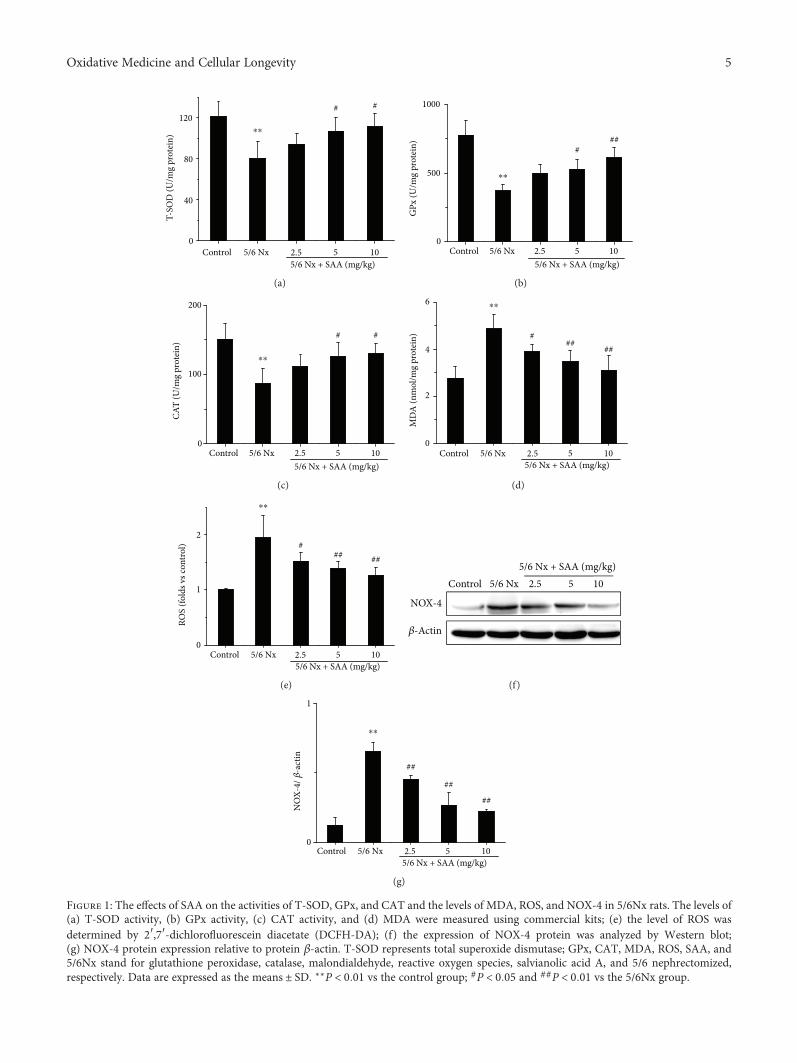
3.1. SAA Attenuates Oxidative Stress in 5/6Nx Rats. The5/6Nx rat model was successfully established in our previousstudy [33], in which all rats were subjected to 5/6 nephrec-tomy. To investigate the effects of SAA on oxidative stressin 5/6Nx rats, the levels of renal T-SOD, GPx, CAT activity,MDA, and ROS were tested. The results show that the levelsof MDA and ROS were significantly increased in 5/6Nx ratscompared to the control group, while the activities ofT-SOD, GPx, and CAT were all decreased, which suggeststhat oxidative stress was grievous in this model (Figure 1).Administration of SAA significantly increased the activitiesof T-SOD, GPx, and CAT compared to the untreated5/6Nx group and lowered the levels of MDA and ROS in adose-dependent manner (Figure 1) suggesting that SAAplays a key role in this model.
In addition, the renal level of NOX-4, which is a majorsource of ROS formation in renal pathogenic conditions,was also analyzed [35]. The NOX-4 protein level was signi-ficantly increased in 5/6Nx rats compared to the controlgroup rats. Treatment with SAA resulted in significantlylower protein expression of NOX-4 compared to theuntreated 5/6Nx group in a dose-dependent manner(Figures 1(f) and 1(g)).
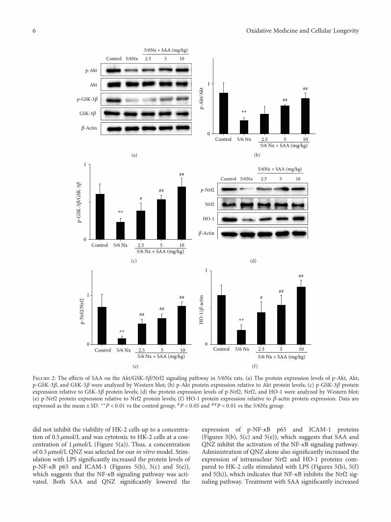
3.2. SAA Activates the Akt/GSK-3β/Nrf2 Signaling Pathwayin 5/6Nx Rats. Some studies suggested that the Akt/GSK-3β/Nrf2 signaling pathway is closely associated with kidneyoxidative stress [28, 36]. Thus, major proteins of theAkt/GSK-3β/Nrf2 signaling pathway were measured duringCKD. The protein levels of p-Akt, p-GSK-3β, p-Nrf2, andHO-1 in 5/6Nx rats were significantly decreased comparedto the control rats, which suggests that the Akt/GSK-3β/Nrf2signaling pathway was inhibited during renal oxidative stress.Treatment with SAA significantly increased the protein
expression of p-Akt, p-GSK-3β, p-Nrf2, and HO-1 comparedto the 5/6Nx group in a dose-dependent manner, which sug-gests that SAA enhances the activation of the Akt/GSK-3β/Nrf2 signaling pathway (Figure 2).
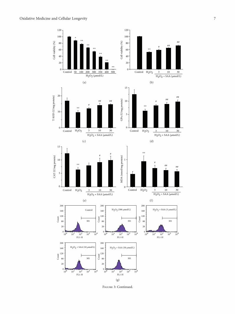
3.3. SAA Improves Cell Viability and Lowers Oxidative Stressin H2O2-Induced HK-2 Cells. To further confirm the effects ofSAA,we observed in vivo andwe subsequently determined theeffects of SAA in an in vitro model of kidney oxidative stress(H2O2-induced HK-2 cells). The results demonstrated thatH2O2 inhibited the viability of HK-2 cells in a concentration-dependent manner and induced approximately 50% of cellviability loss at the concentration of 300μmol/L. Thereby, weselected a concentration of 300μmol/L H2O2 for our in vitromodel. Although H2O2 had a cytotoxic effect on cells,treatment with SAA significantly enhanced the cell viabilityin a dose-dependent manner in H2O2-induced HK-2 cells(Figures 3(a) and 3(b)).
The levels of T-SOD, GPx, CAT activity, MDA, and ROSwere also examined in H2O2-induced HK-2 cells. The activi-ties of T-SOD, GPx, and CAT were all significantly decreasedin H2O2-induced HK-2 cells compared to H2O2-untreatedcontrol cells, while the levels ofMDAandROSwere increased,which indicates that themodel of oxidative stress is functionalin vitro. Administration of SAA significantly increased theactivities of T-SOD, GPx, and CAT and attenuated the levelsof MDA and ROS in H2O2-induced HK-2 cells in aconcentration-dependent manner (Figures 3(c)–3(h)).
Furthermore, the expression of NOX-4 was also detectedin H2O2-induced HK-2 cells. The NOX-4 protein level wassignificantly increased in H2O2-induced HK-2 cells com-pared to the control group. Treatment with SAA signifi-cantly lowered the level of NOX-4 protein in HK-2 cellsstimulated with H2O2 in a concentration-dependent man-ner (Figures 3(i) and 3(j)).
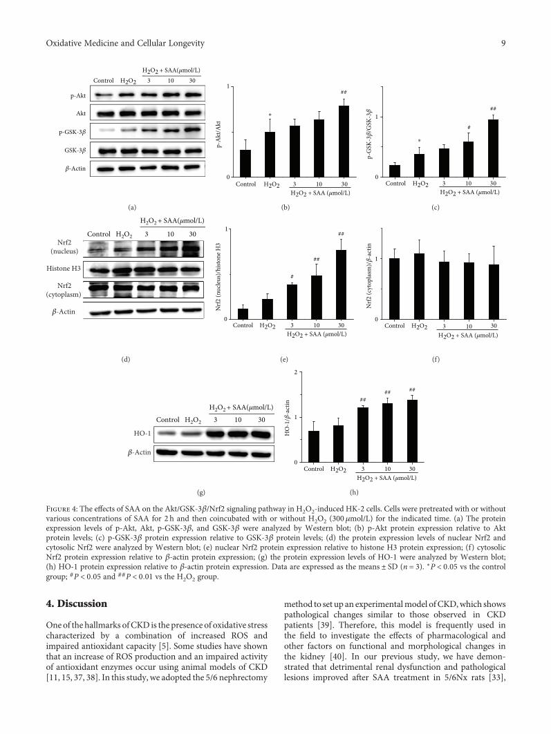
3.4. SAA Enhances the Activation of the Akt/GSK-3β/Nrf2Signaling Pathway in H2O2-Induced HK-2 Cells. Major pro-teins of the Akt/GSK-3β/Nrf2 signaling pathway were alsodetected in H2O2-induced HK-2 cells. Stimulation withH2O2 upregulated the protein levels of p-Akt, p-GSK-3β,intranuclear Nrf2, and HO-1 compared to H2O2-untreatedcontrol cells (Figure 4), which may be relevant to the cell-adaptive response. However, the level of cytoplasmic Nrf2remained relatively unaffected (Figures 4(d) and 4(f)).Treatment with SAA significantly increased the expression ofp-Akt, p-GSK-3β, intranuclear Nrf2, and HO-1 proteinscompared toHK-2 cells stimulatedbyH2O2 tovaryingdegreesdepending on the concentration of SAA. This suggests thatSAA enhances the activation of the Akt/GSK-3β/Nrf2 sig-naling pathway (Figure 4). All of the in vitro results areconsistent with the results obtained from the in vivomodel using 5/6Nx rats.
3.5. SAA Partly Enhances the Activation of the Nrf2 SignalingPathway through Inhibiting the NF-κB Signaling Pathway inLPS-Induced HK-2 Cells. To further study the relationshipbetween the NF-κB and Nrf2 signaling pathways, QNZ andML385 were used in vitro. The results revealed that QNZ
120⁎⁎
# #
80T-
SOD
(U/m
g pr
otei
n)
Control 5/6 Nx 2.55/6 Nx + SAA (mg/kg)
5 10
40
0
(a)
###
1000
500
0
GPx
(U/m
g pr
otei
n)
Control 5/6 Nx 2.55/6 Nx + SAA (mg/kg)
5 10
⁎⁎
(b)
# #
200
100
0
CAT
(U/m
g pr
otei
n)
Control 5/6 Nx 2.55/6 Nx + SAA (mg/kg)
5 10
⁎⁎
(c)
### ##
MD
A (n
mol
/mg
prot
ein)
Control 5/6 Nx 2.55/6 Nx + SAA (mg/kg)
5 10
6
4
2
0
⁎⁎
(d)
### ##
ROS
(fold
s vs c
ontro
l)
Control 5/6 Nx 2.55/6 Nx + SAA (mg/kg)
5 10
2
1
0
⁎⁎
(e)
NOX-4
�훽-Actin
Control 5/6 Nx 2.55/6 Nx + SAA (mg/kg)
5 10
(f)
##
##
##
Control 5/6 Nx 2.55/6 Nx + SAA (mg/kg)
5 10
1
0
NO
X-4/
�훽-a
ctin
⁎⁎
(g)
Figure 1: The effects of SAA on the activities of T-SOD, GPx, and CAT and the levels of MDA, ROS, and NOX-4 in 5/6Nx rats. The levels of(a) T-SOD activity, (b) GPx activity, (c) CAT activity, and (d) MDA were measured using commercial kits; (e) the level of ROS wasdetermined by 2′,7′-dichlorofluorescein diacetate (DCFH-DA); (f) the expression of NOX-4 protein was analyzed by Western blot;(g) NOX-4 protein expression relative to protein β-actin. T-SOD represents total superoxide dismutase; GPx, CAT, MDA, ROS, SAA, and5/6Nx stand for glutathione peroxidase, catalase, malondialdehyde, reactive oxygen species, salvianolic acid A, and 5/6 nephrectomized,respectively. Data are expressed as the means ± SD. ∗∗P < 0 01 vs the control group; #P < 0 05 and ##P < 0 01 vs the 5/6Nx group.
5Oxidative Medicine and Cellular Longevity
Control 5/6Nx 2.5 5 10
5/6Nx + SAA (mg/kg)
p-Akt
Akt
p-GSK-3�훽
GSK-3�훽
�훽-Actin
(a)
Control0
1
p-A
kt/A
kt
5/6 Nx 2.55/6 Nx + SAA (mg/kg)
5 10
##
##
⁎⁎
(b)
0
1
p-G
SK-3�훽
/GSK
-3�훽
Control 5/6 Nx 2.55/6 Nx + SAA (mg/kg)
5 10
##
##
#
⁎⁎
(c)
p-Nrf2
Nrf2
HO-1
�훽-Actin
Control 5/6Nx 2.5 5 10
5/6Nx + SAA (mg/kg)
(d)
0
1
p-N
rf2/N
rf2
Control 5/6 Nx 2.55/6 Nx + SAA (mg/kg)
5 10
####
##
⁎⁎
(e)
0
1
HO
-1/�훽
-act
in
Control 5/6 Nx 2.55/6 Nx + SAA (mg/kg)
5 10
##
##
#
⁎⁎
(f)
Figure 2: The effects of SAA on the Akt/GSK-3β/Nrf2 signaling pathway in 5/6Nx rats. (a) The protein expression levels of p-Akt, Akt,p-GSK-3β, and GSK-3β were analyzed by Western blot; (b) p-Akt protein expression relative to Akt protein levels; (c) p-GSK-3β proteinexpression relative to GSK-3β protein levels; (d) the protein expression levels of p-Nrf2, Nrf2, and HO-1 were analyzed by Western blot;(e) p-Nrf2 protein expression relative to Nrf2 protein levels; (f) HO-1 protein expression relative to β-actin protein expression. Data areexpressed as the mean ± SD. ∗∗P < 0 01 vs the control group; #P < 0 05 and ##P < 0 01 vs the 5/6Nx group.
6 Oxidative Medicine and Cellular Longevity
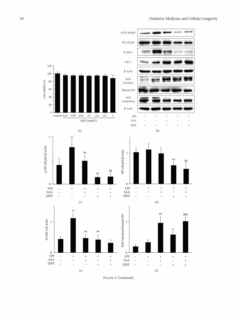
did not inhibit the viability of HK-2 cells up to a concentra-tion of 0.5μmol/L and was cytotoxic to HK-2 cells at a con-centration of 1μmol/L (Figure 5(a)). Thus, a concentrationof 0.5μmol/L QNZ was selected for our in vitromodel. Stim-ulation with LPS significantly increased the protein levels ofp-NF-κB p65 and ICAM-1 (Figures 5(b), 5(c) and 5(e)),which suggests that the NF-κB signaling pathway was acti-vated. Both SAA and QNZ significantly lowered the
expression of p-NF-κB p65 and ICAM-1 proteins(Figures 5(b), 5(c) and 5(e)), which suggests that SAA andQNZ inhibit the activation of the NF-κB signaling pathway.Administration of QNZ alone also significantly increased theexpression of intranuclear Nrf2 and HO-1 proteins com-pared to HK-2 cells stimulated with LPS (Figures 5(b), 5(f)and 5(h)), which indicates that NF-κB inhibits the Nrf2 sig-naling pathway. Treatment with SAA significantly increased
120
100 ⁎⁎⁎
⁎⁎
⁎⁎
⁎⁎
⁎⁎
⁎⁎
80
60
40
20
0
H2O2 (�휇mol/L)Control 50 100 200 300 350 400 500
Cel
l via
bilit
y (%
)
(a)
120
100
80
60
40
20
0
H2O2 + SAA (�휇mol/L)Control
Cel
l via
bilit
y (%
)
H2O2 3 10 30
# ####
⁎⁎
(b)
20
10
0Control
H2O2 + SAA (�휇mol/L)
T-SO
D (U
/mg
prot
ein)
H2O2 3 10 30
### ##
⁎⁎
(c)
15
10
5
0
GPx
(U/m
g pr
otei
n)
H2O2 + SAA (�휇mol/L)Control H2O2 3 10 30
# ####
⁎⁎
(d)
15
10
5
0
CAT
(U/m
g pr
otei
n)
H2O2 + SAA (�휇mol/L)Control H2O2 3 10 30
# #
⁎⁎
(e)
0
1
MD
A (n
mol
/mg
prot
ein)
H2O2 + SAA (�휇mol/L)Control H2O2 3 10 30
### ##
⁎⁎
(f)
H2O2 + SAA (3 �휇mol/L)
0
20
80Cou
nt
Cou
nt
Cou
nt120
M1
M1 M1
M1 M1
160
200
0
20
80Cou
nt 120
160
200
0
20
80Cou
nt 120
160
200
0
20
80
120
160
200
0
20
80
120
160
200
FL1-H FL1-H FL1-H
FL1-H FL1-H
H2O2 + SAA (30 �휇mol/L)H2O2 + SAA (10 �휇mol/L)
H2O2 (300 �휇mol/L)Control
100 101 102 103 104 100 101 102 103 104
100 101 102 103 104 100 101 102 103 104
100 101 102 103 104
(g)
Figure 3: Continued.
7Oxidative Medicine and Cellular Longevity
4
3
2
1
0
ROS
(fold
s vs.
cont
rol)
H2O2 + SAA (�휇mol/L)Control H2O2 3 10 30
###
⁎⁎
(h)
H2O2 + SAA (�휇mol/L)
Control
NOX-4
�훽-Actin
H2O2 3 10 30
(i)
Control0
1
3 10 30
###
##
H2O2 + SAA (�휇mol/L)
NO
X-4/�훽
-act
in
H2O2
⁎⁎
(j)
Figure 3: The effects of SAA on HK-2 cell viability and the levels of T-SOD, GPx, CAT activity, MDA, ROS, and NOX-4 in H2O2-inducedHK-2 cells. Cells were pretreated with or without various concentrations of SAA for 2 h and then coincubated with or without H2O2(300 μmol/L) for the indicated time. (a) Effects of various concentrations of H2O2 on HK-2 cell viability and (b) effects of SAA on300μmol/L H2O2-induced HK-2 cell viability were assessed by MTT assay. The levels of (c) T-SOD activity, (d) GPx activity, (e) CATactivity, and (f) MDA were measured using commercial kits; (g, h) the levels of ROS were determined by 2′,7′-dichlorofluoresceindiacetate (DCFH-DA); (i) the expression of NOX-4 protein was analyzed by Western blot; (j) NOX-4 protein expression relative toprotein β-actin expression. Data are expressed as the means ± SD (n = 3). ∗P < 0 05 and ∗∗P < 0 01 vs the control group; #P < 0 05 and##P < 0 01 vs the H2O2 group.
8 Oxidative Medicine and Cellular Longevity
the expression of intranuclear Nrf2 and HO-1 proteinscompared to HK-2 cells stimulated by LPS, which isenhanced by QNZ to some extent (Figures 5(b), 5(f) and5(h)). Thus, SAA activates the Nrf2 signaling pathway indi-rectly through inhibiting the NF-κB signaling pathway.Compared to the administration of QNZ, SAA increasedthe expression of intranuclear Nrf2 and HO-1 proteins inHK-2 cells stimulated by LPS (Figures 5(b), 5(f) and5(h)), which suggests that SAA activates the Nrf2 signalingpathway directly. Thus, SAA does activate not only directlybut also indirectly the Nrf2 signaling pathway based on theinhibition of NF-κB.
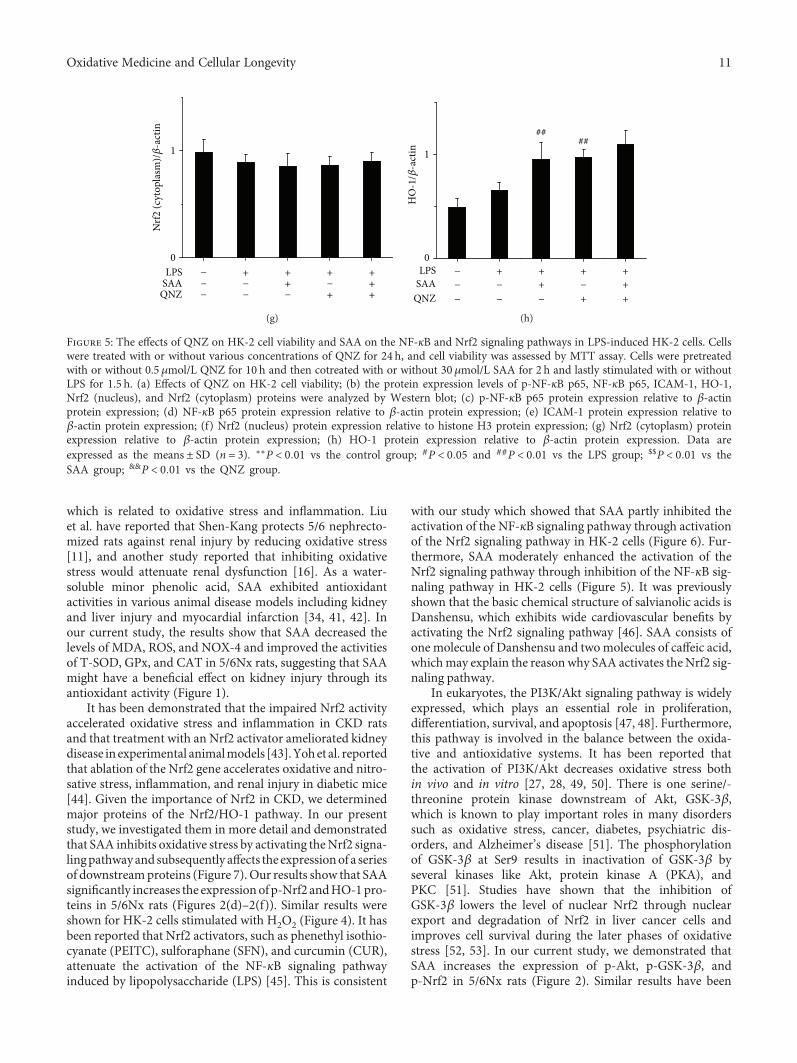
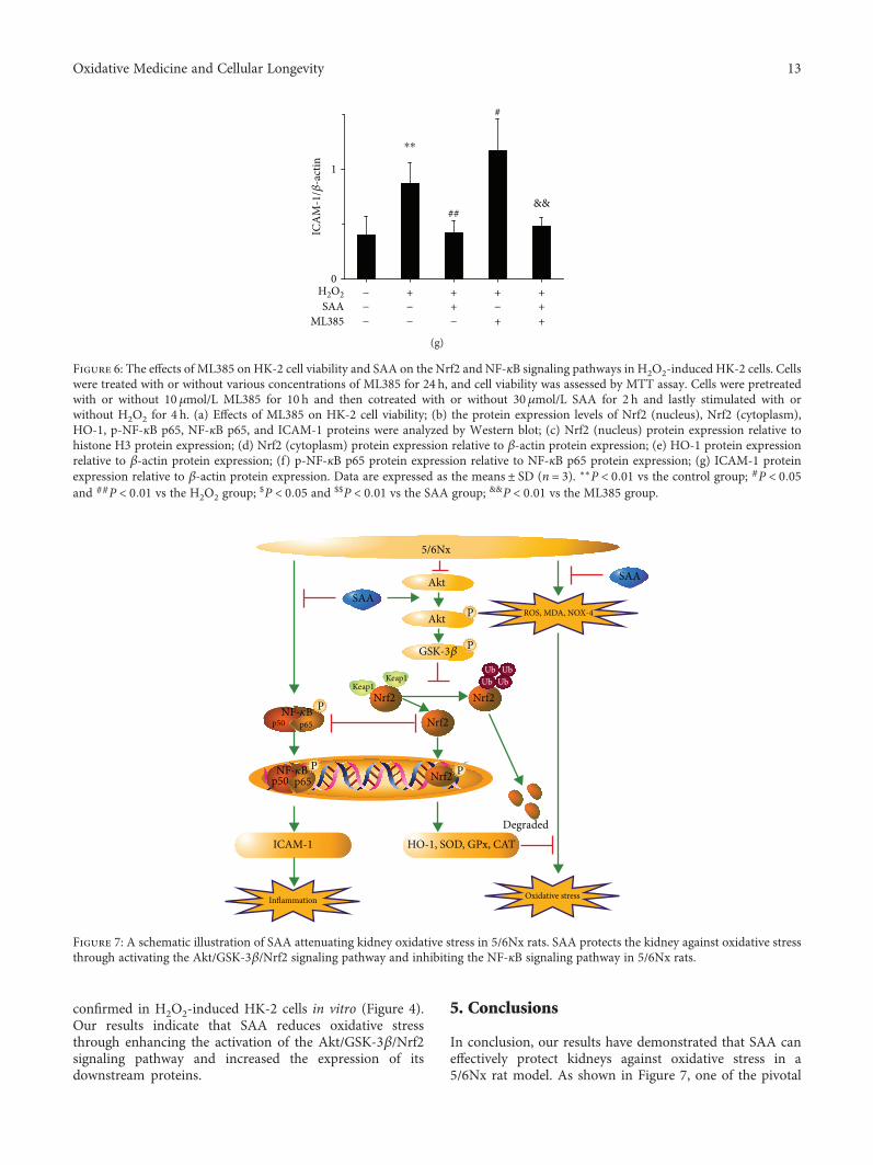
3.6. SAA Partly Inhibits the Activation of the NF-κB SignalingPathway through Activating the Nrf2 Signaling Pathway inH2O2-Induced HK-2 Cells. The results revealed that ML385did not inhibit the viability of HK-2 cells up to a concen-tration of 10μmol/L (Figure 6(a)). Therefore, 10μmol/L ofML385 was selected for our in vitromodel. SAA significantlyincreased the expression of intranuclear Nrf2 and HO-1 pro-teins in H2O2-induced HK-2 cells (Figures 6(b), 6(c) and
6(e)), which suggests that SAA enhances the activation ofthe Nrf2 signaling pathway. However, ML385 decreasedthe protein levels of Nrf2 and HO-1 (Figures 6(b)–6(e)),suggesting that ML385 significantly inhibits the Nrf2signaling pathway. Administration with ML385 alone sig-nificantly increased the expression of p-NF-κB p65 andICAM-1 proteins compared to HK-2 stimulated by H2O2(Figures 6(b), 6(f) and 6(g)), which indicates that Nrf2inhibits the NF-κB signaling pathway. SAA significantlylowered the expression of p-NF-κB p65 and ICAM-1 pro-teins compared to HK-2 cells stimulated by H2O2, which canbe abrogated by ML385 to some degree (Figures 6(b), 6(f)and 6(g)). Thus, SAA inhibits the NF-κB signaling pathwayindirectly through activating the Nrf2 signaling pathway.Compared to the administration with ML385, SAA signifi-cantly lowered the expression of p-NF-κB p65 and ICAM-1proteins in H2O2-treated HK-2 cells (Figures 6(b), 6(f) and6(g)), which shows that SAA inhibits the NF-κB signalingpathway directly. Thus, SAA does inhibit not only directlybut also indirectly the NF-κB signaling pathway based onthe activation of Nrf2.
p-Akt
p-GSK-3�훽
Akt
GSK-3�훽
�훽-Actin
Control H2O2 3 10 30H2O2 + SAA(�휇mol/L)
(a)
1
⁎
p-A
kt/A
kt
0Control 3 10 30
##
H2O2 + SAA (�휇mol/L)H2O2
(b)
p-G
SK-3�훽
/GSK
-3�훽
Control0
H2O2 3 10 30H2O2 + SAA (�휇mol/L)
#
##
⁎
1
(c)
Control H2O2 3 10 30
H2O2 + SAA(�휇mol/L)
Nrf2(nucleus)
Histone H3
Nrf2(cytoplasm)
�훽-Actin
(d)
Nrf2
(nuc
leus
)/hi
stone
H3
Control H2O2 3 10 30
##
1
#
##
0
H2O2 + SAA (�휇mol/L)
(e)
Control H2O20
1
3 10 30H2O2 + SAA (�휇mol/L)
Nrf2
(cyt
opla
sm)/�훽
-act
in(f)
Control H2O2 3 10 30
H2O2 + SAA(�휇mol/L)
HO-1
�훽-Actin
(g)
Control
2
1
0H2O2 3 10 30
#### ##
H2O2 + SAA (�휇mol/L)
HO
-1/�훽
-act
in
(h)
Figure 4: The effects of SAA on the Akt/GSK-3β/Nrf2 signaling pathway in H2O2-induced HK-2 cells. Cells were pretreated with or withoutvarious concentrations of SAA for 2 h and then coincubated with or without H2O2 (300 μmol/L) for the indicated time. (a) The proteinexpression levels of p-Akt, Akt, p-GSK-3β, and GSK-3β were analyzed by Western blot; (b) p-Akt protein expression relative to Aktprotein levels; (c) p-GSK-3β protein expression relative to GSK-3β protein levels; (d) the protein expression levels of nuclear Nrf2 andcytosolic Nrf2 were analyzed by Western blot; (e) nuclear Nrf2 protein expression relative to histone H3 protein expression; (f) cytosolicNrf2 protein expression relative to β-actin protein expression; (g) the protein expression levels of HO-1 were analyzed by Western blot;(h) HO-1 protein expression relative to β-actin protein expression. Data are expressed as the means ± SD (n = 3). ∗P < 0 05 vs the controlgroup; #P < 0 05 and ##P < 0 01 vs the H2O2 group.
9Oxidative Medicine and Cellular Longevity
4. Discussion
Oneof the hallmarks ofCKD is the presence of oxidative stresscharacterized by a combination of increased ROS andimpaired antioxidant capacity [5]. Some studies have shownthat an increase of ROS production and an impaired activityof antioxidant enzymes occur using animal models of CKD[11, 15, 37, 38]. In this study, we adopted the 5/6 nephrectomy
method to set up an experimentalmodel ofCKD,which showspathological changes similar to those observed in CKDpatients [39]. Therefore, this model is frequently used inthe field to investigate the effects of pharmacological andother factors on functional and morphological changes inthe kidney [40]. In our previous study, we have demon-strated that detrimental renal dysfunction and pathologicallesions improved after SAA treatment in 5/6Nx rats [33],
Cel
l via
bilit
y (%
)
120
100
80
60
40
20
0Control 0.01 0.02 0.05 0.1 0.2 0.5 1
⁎⁎
QNZ (�휇mol/L)
(a)
p-NF-�휅B p65
NF-�휅B p65
ICAM-1
HO-1
�훽-Actin
Nrf2(nucleus)
Histone H3
Nrf2(cytoplasm)
�훽-Actin
LPSSAA
QNZ
−−
−
+−
−
+ +
+
++
+
+ −
−
(b)
LPSSAAQNZ
−−−
+−−
+ +
+
+++
+ −−
##
⁎⁎
##$$
0
1
p-N
F-�휅
B p6
5/�훽
-act
in
(c)
LPS
1
0
SAAQNZ
−−−
+
−−
+ +
+
+
+
+
+ −−
## $$
NF-�휅
B p6
5/�훽
-act
in
(d)
LPSSAAQNZ
−−−
+
−−
+ +
+
+
+
+
+ −−
## ##
0
1
⁎⁎
ICA
M-1
/�훽-a
ctin
(e)
Nrf2
(nuc
leus
)/hi
stone
H3
LPSSAAQNZ
−−−
+
−−
+ +
+
+
+
+
+ −−
## &&
#
0
1
(f)
Figure 5: Continued.
10 Oxidative Medicine and Cellular Longevity
LPSSAA
QNZ
−−−
+−−
+ +
+
+++
+ −−
0
1N
rf2 (c
ytop
lasm
)/�훽
-act
in
(g)
LPSSAA
QNZ
−−−
+
−−
+ +
+
+
+
+
+ −−
0
1
####
HO
-1/�훽
-act
in
(h)
Figure 5: The effects of QNZ on HK-2 cell viability and SAA on the NF-κB and Nrf2 signaling pathways in LPS-induced HK-2 cells. Cellswere treated with or without various concentrations of QNZ for 24 h, and cell viability was assessed by MTT assay. Cells were pretreatedwith or without 0.5 μmol/L QNZ for 10 h and then cotreated with or without 30μmol/L SAA for 2 h and lastly stimulated with or withoutLPS for 1.5 h. (a) Effects of QNZ on HK-2 cell viability; (b) the protein expression levels of p-NF-κB p65, NF-κB p65, ICAM-1, HO-1,Nrf2 (nucleus), and Nrf2 (cytoplasm) proteins were analyzed by Western blot; (c) p-NF-κB p65 protein expression relative to β-actinprotein expression; (d) NF-κB p65 protein expression relative to β-actin protein expression; (e) ICAM-1 protein expression relative toβ-actin protein expression; (f) Nrf2 (nucleus) protein expression relative to histone H3 protein expression; (g) Nrf2 (cytoplasm) proteinexpression relative to β-actin protein expression; (h) HO-1 protein expression relative to β-actin protein expression. Data areexpressed as the means ± SD (n = 3). ∗∗P < 0 01 vs the control group; #P < 0 05 and ##P < 0 01 vs the LPS group; $$P < 0 01 vs theSAA group; &&P < 0 01 vs the QNZ group.
11Oxidative Medicine and Cellular Longevity
which is related to oxidative stress and inflammation. Liuet al. have reported that Shen-Kang protects 5/6 nephrecto-mized rats against renal injury by reducing oxidative stress[11], and another study reported that inhibiting oxidativestress would attenuate renal dysfunction [16]. As a water-soluble minor phenolic acid, SAA exhibited antioxidantactivities in various animal disease models including kidneyand liver injury and myocardial infarction [34, 41, 42]. Inour current study, the results show that SAA decreased thelevels of MDA, ROS, and NOX-4 and improved the activitiesof T-SOD, GPx, and CAT in 5/6Nx rats, suggesting that SAAmight have a beneficial effect on kidney injury through itsantioxidant activity (Figure 1).
It has been demonstrated that the impaired Nrf2 activityaccelerated oxidative stress and inflammation in CKD ratsand that treatment with an Nrf2 activator ameliorated kidneydisease in experimental animalmodels [43].Yohet al. reportedthat ablation of the Nrf2 gene accelerates oxidative and nitro-sative stress, inflammation, and renal injury in diabetic mice[44]. Given the importance of Nrf2 in CKD, we determinedmajor proteins of the Nrf2/HO-1 pathway. In our presentstudy, we investigated them in more detail and demonstratedthat SAA inhibits oxidative stress by activating theNrf2 signa-lingpathwayand subsequently affects the expressionof a seriesof downstreamproteins (Figure 7). Our results show that SAAsignificantly increases the expression ofp-Nrf2 andHO-1pro-teins in 5/6Nx rats (Figures 2(d)–2(f)). Similar results wereshown for HK-2 cells stimulated with H2O2 (Figure 4). It hasbeen reported that Nrf2 activators, such as phenethyl isothio-cyanate (PEITC), sulforaphane (SFN), and curcumin (CUR),attenuate the activation of the NF-κB signaling pathwayinduced by lipopolysaccharide (LPS) [45]. This is consistent
with our study which showed that SAA partly inhibited theactivation of the NF-κB signaling pathway through activationof the Nrf2 signaling pathway in HK-2 cells (Figure 6). Fur-thermore, SAA moderately enhanced the activation of theNrf2 signaling pathway through inhibition of the NF-κB sig-naling pathway in HK-2 cells (Figure 5). It was previouslyshown that the basic chemical structure of salvianolic acids isDanshensu, which exhibits wide cardiovascular benefits byactivating the Nrf2 signaling pathway [46]. SAA consists ofone molecule of Danshensu and twomolecules of caffeic acid,whichmay explain the reasonwhy SAA activates theNrf2 sig-naling pathway.
In eukaryotes, the PI3K/Akt signaling pathway is widelyexpressed, which plays an essential role in proliferation,differentiation, survival, and apoptosis [47, 48]. Furthermore,this pathway is involved in the balance between the oxida-tive and antioxidative systems. It has been reported thatthe activation of PI3K/Akt decreases oxidative stress bothin vivo and in vitro [27, 28, 49, 50]. There is one serine/-threonine protein kinase downstream of Akt, GSK-3β,which is known to play important roles in many disorderssuch as oxidative stress, cancer, diabetes, psychiatric dis-orders, and Alzheimer’s disease [51]. The phosphorylationof GSK-3β at Ser9 results in inactivation of GSK-3β byseveral kinases like Akt, protein kinase A (PKA), andPKC [51]. Studies have shown that the inhibition ofGSK-3β lowers the level of nuclear Nrf2 through nuclearexport and degradation of Nrf2 in liver cancer cells andimproves cell survival during the later phases of oxidativestress [52, 53]. In our current study, we demonstrated thatSAA increases the expression of p-Akt, p-GSK-3β, andp-Nrf2 in 5/6Nx rats (Figure 2). Similar results have been
Cel
l via
bilit
y (%
)
120
100
80
60
40
20
0Control 0.1 0.2 0.5 1 2 5 10
ML385 (�휇mol/L)
(a)
−−−
+−−
+ +
+
+++
+ −−
Nrf2(nucleus)
Histone H3
Nrf2 (cytoplasm)
�훽-Actin
HO-1
p-NF-�휅B p65
NF-�휅B p65
ICAM-1
�훽-Actin
SAA ML385
H2O2
(b)
SAAML385
0
1
Nrf2
(nuc
leus
)/hi
stone
H3
−−−
+
−−
+ +
+
+
+
+
+ −−
##
#
$
H2O2
(c)
SAAML385
−−−
+
##$$
−−
+ +
+
+
+
+
+ −−
Nrf2
(cyt
opla
sm)/�훽
-act
in
H2O2
1
0
(d)
0
1
SAAML385
−−−
+−−
+ +
+
+++
+ −−
$$##
##
H2O2
HO
-1/�훽
-act
in
(e)
SAA ML385
0
1
−−−
+−−
+ +
+
+++
+ −−
#
⁎⁎
#
&&
H2O2
p-N
F-�휅
B p6
5/N
F-�휅
B p6
5
(f)
Figure 6: Continued.
12 Oxidative Medicine and Cellular Longevity
0
1
SAAML385
H2O2 −−−
+
⁎⁎
−−
+
##
#
+
+
+++
+ −−
&&##
ICA
M-1
/�훽-a
ctin
(g)
Figure 6: The effects of ML385 on HK-2 cell viability and SAA on the Nrf2 and NF-κB signaling pathways in H2O2-induced HK-2 cells. Cellswere treated with or without various concentrations of ML385 for 24 h, and cell viability was assessed by MTT assay. Cells were pretreatedwith or without 10 μmol/L ML385 for 10 h and then cotreated with or without 30μmol/L SAA for 2 h and lastly stimulated with orwithout H2O2 for 4 h. (a) Effects of ML385 on HK-2 cell viability; (b) the protein expression levels of Nrf2 (nucleus), Nrf2 (cytoplasm),HO-1, p-NF-κB p65, NF-κB p65, and ICAM-1 proteins were analyzed by Western blot; (c) Nrf2 (nucleus) protein expression relative tohistone H3 protein expression; (d) Nrf2 (cytoplasm) protein expression relative to β-actin protein expression; (e) HO-1 protein expressionrelative to β-actin protein expression; (f) p-NF-κB p65 protein expression relative to NF-κB p65 protein expression; (g) ICAM-1 proteinexpression relative to β-actin protein expression. Data are expressed as the means ± SD (n = 3). ∗∗P < 0 01 vs the control group; #P < 0 05and ##P < 0 01 vs the H2O2 group;
$P < 0 05 and $$P < 0 01 vs the SAA group; &&P < 0 01 vs the ML385 group.
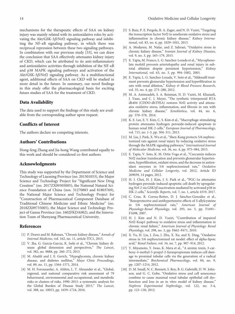
Nrf2
5/6Nx
ICAM-1
Inflammation Oxidative stress
ROS, MDA, NOX-4
HO-1, SOD, GPx, CATDegraded
SAA
SAA
p50 p65NF-�휅B
Nrf2
Nrf2 Nrf2
Akt
Akt
GSK-3�훽
Keap1Keap1
p65PP
P
P
P
Ub UbUbUb
p50NF-�휅B
Figure 7: A schematic illustration of SAA attenuating kidney oxidative stress in 5/6Nx rats. SAA protects the kidney against oxidative stressthrough activating the Akt/GSK-3β/Nrf2 signaling pathway and inhibiting the NF-κB signaling pathway in 5/6Nx rats.
13Oxidative Medicine and Cellular Longevity
confirmed in H2O2-induced HK-2 cells in vitro (Figure 4).Our results indicate that SAA reduces oxidative stressthrough enhancing the activation of the Akt/GSK-3β/Nrf2signaling pathway and increased the expression of itsdownstream proteins.
5. Conclusions
In conclusion, our results have demonstrated that SAA caneffectively protect kidneys against oxidative stress in a5/6Nx rat model. As shown in Figure 7, one of the pivotal
14 Oxidative Medicine and Cellular Longevity
mechanisms for the therapeutic effects of SAA on kidneyinjury was mainly related with its antioxidative roles by acti-vating the Akt/GSK-3β/Nrf2 signaling pathway and inhibi-ting the NF-κB signaling pathway, in which there wasreciprocal repression between these two signaling pathways.In combination with our previous study [33], we can drawthe conclusion that SAA effectively attenuates kidney injuryof CKD, which can be attributed to its anti-inflammatoryand antioxidative activities through inhibition of the NF-κBand p38 MAPK signaling pathways and activation of theAkt/GSK-3β/Nrf2 signaling pathway. As a multifunctionalagent, additional effects of SAA on CKD will be studied inmore detail in the future. In summary, our novel findingsin this study offer the pharmacological basis for excitingfuture studies of SAA for the treatment of CKD.
Data Availability
The data used to support the findings of this study are avail-able from the corresponding author upon request.
Conflicts of Interest
The authors declare no competing interests.
Authors’ Contributions
Hong-feng Zhang and Jia-hong Wang contributed equally tothis work and should be considered co-first authors.
Acknowledgments
This study was supported by the Department of Science andTechnology of Liaoning Province (no. 201501053), the MajorScience and Technology Project for “Significant New DrugCreation” (no. 2017ZX09305005), the National Natural Sci-ence Foundation of China (nos. 31270883 and 81803769),the National Major Science and Technology Project for“Construction of Pharmaceutical Component Database ofTraditional Chinese Medicine and Ethnic Medicine” (no.2018ZX09735005), the Major Science and Technology Pro-ject of Gansu Province (no. 1602FKDA002), and the Innova-tion Team of Shenyang Pharmaceutical University.
References
[1] P. Drawz andM. Rahman, “Chronic kidney disease,” Annals ofInternal Medicine, vol. 162, no. 11, article ITC1, 2015.
[2] V. Jha, G. Garcia-Garcia, K. Iseki et al., “Chronic kidney di-sease: global dimension and perspectives,” The Lancet,vol. 382, no. 9888, pp. 260–272, 2013.
[3] M. Alsahli and J. E. Gerich, “Hypoglycemia, chronic kidneydisease, and diabetes mellitus,” Mayo Clinic Proceedings,vol. 89, no. 11, pp. 1564–1571, 2014.
[4] M. H. Forouzanfar, A. Afshin, L. T. Alexander et al., “Global,regional, and national comparative risk assessment of 79behavioural, environmental and occupational, and metabolicrisks or clusters of risks, 1990–2015: a systematic analysis forthe Global Burden of Disease Study 2015,” The Lancet,vol. 388, no. 10053, pp. 1659–1724, 2016.
[5] S. Ruiz, P. E. Pergola, R. A. Zager, and N. D. Vaziri, “Targetingthe transcription factor Nrf2 to ameliorate oxidative stress andinflammation in chronic kidney disease,” Kidney Interna-tional, vol. 83, no. 6, pp. 1029–1041, 2013.
[6] A. Modaresi, M. Nafar, and Z. Sahraei, “Oxidative stress inchronic kidney disease,” Iranian Journal of Kidney Diseases,vol. 9, no. 3, pp. 165–179, 2015.
[7] E. Tapia, M. Franco, L. G. Sanchez-Lozada et al., “Mycopheno-late mofetil prevents arteriolopathy and renal injury in sub-total ablation despite persistent hypertension,” KidneyInternational, vol. 63, no. 3, pp. 994–1002, 2003.
[8] E. Tapia, L. G. Sanchez-Lozada, V. Soto et al., “Sildenafil treat-ment prevents glomerular hypertension and hyperfiltration inrats with renal ablation,” Kidney & Blood Pressure Research,vol. 35, no. 4, pp. 273–280, 2012.
[9] M. A. Aminzadeh, S. A. Reisman, N. D. Vaziri, M. Khazaeli,J. Yuan, and C. J. Meyer, “The synthetic triterpenoid RTAdh404 (CDDO-dhTFEA) restores Nrf2 activity and attenu-ates oxidative stress, inflammation, and fibrosis in rats withchronic kidney disease,” Xenobiotica, vol. 44, no. 6,pp. 570–578, 2014.
[10] K. E. Lee, E. Y. Kim, C. S. Kim et al., “Macrophage-stimulatingprotein attenuates hydrogen peroxide-induced apoptosis inhuman renal HK-2 cells,” European Journal of Pharmacology,vol. 715, no. 1-3, pp. 304–311, 2013.
[11] M. Liu, J. Park, X. Wu et al., “Shen-Kang protects 5/6 nephrec-tomized rats against renal injury by reducing oxidative stressthrough the MAPK signaling pathways,” International Journalof Molecular Medicine, vol. 36, no. 4, pp. 975–984, 2015.
[12] E. Tapia, V. Soto, K. M. Ortiz-Vega et al., “Curcumin inducesNrf2 nuclear translocation and prevents glomerular hyperten-sion, hyperfiltration, oxidant stress, and the decrease in antiox-idant enzymes in 5/6 nephrectomized rats,” OxidativeMedicine and Cellular Longevity, vol. 2012, Article ID269039, 14 pages, 2012.
[13] H. I. Choi, H. J. Kim, J. S. Park et al., “PGC-1α attenuateshydrogen peroxide-induced apoptotic cell death by upregulat-ing Nrf-2 via GSK3β inactivation mediated by activated p38 inHK-2 cells,” Scientific Reports, vol. 7, no. 1, article 4319, 2017.
[14] C. Cruz, R. Correa-Rotter, D. J. Sánchez-González et al.,“Renoprotective and antihypertensive effects of S-allylcysteinein 5/6 nephrectomized rats,” American Journal ofPhysiology-Renal Physiology, vol. 293, no. 5, pp. F1691–F1698, 2007.
[15] H. J. Kim and N. D. Vaziri, “Contribution of impairedNrf2-Keap1 pathway to oxidative stress and inflammation inchronic renal failure,” American Journal of Physiology. RenalPhysiology, vol. 298, no. 3, pp. F662–F671, 2010.
[16] X. Yu, H. Liu, J. Zou, J. Zhu, X. Xu, and X. Ding, “Oxidativestress in 5/6 nephrectomized rat model: effect of alpha-lipoicacid,” Renal Failure, vol. 34, no. 7, pp. 907–914, 2012.
[17] Y. Miyamoto, Y. Iwao, K. Mera et al., “A uremic toxin, 3-car-boxy-4-methyl-5-propyl-2-furanpropionate induces cell dam-age to proximal tubular cells via the generation of a radicalintermediate,” Biochemical Pharmacology, vol. 84, no. 9,pp. 1207–1214, 2012.
[18] D. M. Small, N. C. Bennett, S. Roy, B. G. Gabrielli, D. W. John-son, and G. C. Gobe, “Oxidative stress and cell senescencecombine to cause maximal renal tubular epithelial cell dys-function and loss in an in vitro model of kidney disease,”Nephron Experimental Nephrology, vol. 122, no. 3-4,pp. 123–130, 2012.
15Oxidative Medicine and Cellular Longevity
[19] P. Wu, Y. Yan, L. L. Ma et al., “Effects of the Nrf2 proteinmodulator salvianolic acid A alone or combined with met-formin on diabetes-associated macrovascular and renalinjury,” The Journal of Biological Chemistry, vol. 291,no. 42, pp. 22288–22301, 2016.
[20] H. Zhang, Y. Y. Liu, Q. Jiang et al., “Salvianolic acid A protectsRPE cells against oxidative stress through activation ofNrf2/HO-1 signaling,” Free Radical Biology & Medicine,vol. 69, pp. 219–228, 2014.
[21] H. J. Kim, T. Sato, B. Rodriguez-Iturbe, and N. D. Vaziri, “Roleof intrarenal angiotensin system activation, oxidative stress,inflammation, and impaired nuclear factor-erythroid-2-re-lated factor 2 activity in the progression of focal glomerulo-sclerosis,” The Journal of Pharmacology and ExperimentalTherapeutics, vol. 337, no. 3, pp. 583–590, 2011.
[22] M. A. Aminzadeh, S. B. Nicholas, K. C. Norris, and N. D.Vaziri, “Role of impaired Nrf2 activation in the pathogenesisof oxidative stress and inflammation in chronictubulo-interstitial nephropathy,” Nephrology, Dialysis, Trans-plantation, vol. 28, no. 8, pp. 2038–2045, 2013.
[23] W. Li, T. O. Khor, C. Xu et al., “Activation of Nrf2-antioxidantsignaling attenuates NFkappaB-inflammatory response andelicits apoptosis,” Biochemical Pharmacology, vol. 76, no. 11,pp. 1485–1489, 2008.
[24] G. H. Liu, J. Qu, and X. Shen, “NF-kappaB/p65 antagonizesNrf2-ARE pathway by depriving CBP from Nrf2 and facilitat-ing recruitment of HDAC3 toMafK,” Biochimica et BiophysicaActa, vol. 1783, no. 5, pp. 713–727, 2008.
[25] S. B. Cullinan and J. A. Diehl, “PERK-dependent activation ofNrf2 contributes to redox homeostasis and cell survival follow-ing endoplasmic reticulum stress,” The Journal of BiologicalChemistry, vol. 279, no. 19, pp. 20108–20117, 2004.
[26] Y. J. Surh, J. Kundu, and H. K. Na, “Nrf2 as a master redoxswitch in turning on the cellular signaling involved in theinduction of cytoprotective genes by some chemopreventivephytochemicals,” Planta Medica, vol. 74, no. 13, pp. 1526–1539, 2008.
[27] C. Li, Z. Pan, T. Xu, C. Zhang, Q. Wu, and Y. Niu, “Puerarininduces the upregulation of glutathione levels and nucleartranslocation of Nrf2 through PI3K/Akt/GSK-3β signalingevents in PC12 cells exposed to lead,” Neurotoxicology andTeratology, vol. 46, pp. 1–9, 2014.
[28] Y. Cheng, J. Zhang, W. Guo et al., “Up-regulation of Nrf2 isinvolved in FGF21-mediated fenofibrate protection againsttype 1 diabetic nephropathy,” Free Radical Biology & Medi-cine, vol. 93, pp. 94–109, 2016.
[29] G. T. Liu, T. M. Zhang, B. E. Wang, and Y. W. Wang,“Protective action of seven natural phenolic compoundsagainst peroxidative damage to biomembranes,” BiochemicalPharmacology, vol. 43, no. 2, pp. 147–152, 1992.
[30] K. S. Oh, B. K. Oh, J. Mun, H. W. Seo, and B. H. Lee, “Salvia-nolic acid A suppress lipopolysaccharide-induced NF-κBsignaling pathway by targeting IKKβ,” International Immu-nopharmacology, vol. 11, no. 11, pp. 1901–1906, 2011.
[31] C. H. Liu, Y. Y. Hu, X. L. Wang, P. Liu, and L. M. Xu, “Effectsof salvianolic acid-A on NIH/3T3 fibroblast proliferation, col-lagen synthesis and gene expression,”World Journal of Gastro-enterology, vol. 6, no. 3, pp. 361–364, 2000.
[32] H.–. Y. Fan, F.–. H. Fu, M.–. Y. Yang, H. Xu, A.–. H. Zhang,and K. Liu, “Antiplatelet and antithrombotic activities of sal-vianolic acid A,” Thrombosis Research, vol. 126, no. 1,pp. e17–e22, 2010.
[33] H. F. Zhang, Y. L. Wang, C. Gao et al., “Salvianolic acid Aattenuates kidney injury and inflammation by inhibitingNF-κB and p38 MAPK signaling pathways in 5/6 nephrecto-mized rats,” Acta Pharmacologica Sinica, vol. 39, no. 12,pp. 1855–1864, 2018.
[34] H. Y. Fan, M. Y. Yang, D. Qi et al., “Salvianolic acid A as a mul-tifunctional agent ameliorates doxorubicin-induced nephrop-athy in rats,” Scientific Reports, vol. 5, no. 1, article 12273, 2015.
[35] T. M. Paravicini and R. M. Touyz, “NADPH oxidases, reactiveoxygen species, and hypertension: clinical implications andtherapeutic possibilities,” Diabetes Care, vol. 31, Supplement2, pp. S170–S180, 2008.
[36] G. Zhang, Q. Wang, Q. Zhou et al., “Protective effect of tempolon acute kidney injury through PI3K/Akt/Nrf2 signaling path-way,” Kidney and Blood Pressure Research, vol. 41, no. 2,pp. 129–138, 2016.
[37] J. F. Chen, H. Liu, H. F. Ni et al., “Improved mitochondrialfunction underlies the protective effect of pirfenidone againsttubulointerstitial fibrosis in 5/6 nephrectomized rats,” PLoSOne, vol. 8, no. 12, article e83593, 2013.
[38] D. Wu, N. Luo, L. Wang et al., “Hydrogen sulfide ameliorateschronic renal failure in rats by inhibiting apoptosis and inflam-mation through ROS/MAPK and NF-κB signaling pathways,”Scientific Reports, vol. 7, no. 1, p. 455, 2017.
[39] D. T. Wang, R. H. Huang, X. Cheng, Z. H. Zhang, Y. J. Yang,and X. Lin, “Tanshinone IIA attenuates renal fibrosis andinflammation via altering expression of TGF-β/Smad andNF-κB signaling pathway in 5/6 nephrectomized rats,” Inter-national Immunopharmacology, vol. 26, no. 1, pp. 4–12, 2015.
[40] P. Kujal and Z. Vernerová, “5/6 nephrectomy as an experi-mental model of chronic renal failure and adaptation toreduced nephron number,” Ceskoslovenská Fysiologie, vol. 57,no. 4, pp. 104–109, 2008.
[41] Z.M.Wu, T.Wen, Y. F. Tan, Y. Liu, F. Ren, andH.Wu, “Effectsof salvianolic acid A on oxidative stress and liver injury inducedby carbon tetrachloride in rats,” Basic & Clinical Pharmacology& Toxicology, vol. 100, no. 2, pp. 115–120, 2007.
[42] S. B. Wang, S. Tian, F. Yang, H. G. Yang, X. Y. Yang, and G. H.Du, “Cardioprotective effect of salvianolic acid A onisoproterenol-inducedmyocardial infarction in rats,”EuropeanJournal of Pharmacology, vol. 615, no. 1-3, pp. 125–132, 2009.
[43] N. D. Vaziri, S. Liu, S. H. Farzaneh, S. Nazertehrani,M. Khazaeli, and Y. Y. Zhao, “Dose-dependent deleteriousand salutary actions of the Nrf2 inducer dh404 in chronickidney disease,” Free Radical Biology & Medicine, vol. 86,pp. 374–381, 2015.
[44] K. Yoh, A. Hirayama, K. Ishizaki et al., “Hyperglycemiainduces oxidative and nitrosative stress and increases renalfunctional impairment in Nrf2-deficient mice,” Genes to Cells,vol. 13, no. 11, pp. 1159–1170, 2008.
[45] W.-S. Jeong, I.-W. Kim, R. Hu, and A.-N. T. Kong, “Modula-tory properties of various natural chemopreventive agents onthe activation of NF-kappaB signaling pathway,” Pharmaceuti-cal Research, vol. 21, no. 4, pp. 661–670, 2004.
[46] J. Yu, L. Wang, M. Akinyi et al., “Danshensu protects iso-lated heart against ischemia reperfusion injury through acti-vation of Akt/ERK1/2/Nrf2 signaling,” International Journalof Clinical and Experimental Medicine, vol. 8, no. 9,pp. 14793–14804, 2015.
[47] Q. Chen, T. Xu, D. Li et al., “JNK/PI3K/Akt signaling path-way is involved in myocardial ischemia/reperfusion injury
16 Oxidative Medicine and Cellular Longevity
in diabetic rats: effects of salvianolic acid A intervention,”American Journal of Translational Research, vol. 8, no. 6,pp. 2534–2548, 2016.
[48] L. C. Cantley, “The phosphoinositide 3-kinase pathway,”Science, vol. 296, no. 5573, pp. 1655–1657, 2002.
[49] S. Saha, P. Sadhukhan, K. Sinha, N. Agarwal, and P. C. Sil,“Mangiferin attenuates oxidative stress induced renal celldamage through activation of PI3K induced Akt and Nrf-2mediated signaling pathways,” Biochemistry and BiophysicsReports, vol. 5, pp. 313–327, 2016.
[50] D. Jing, H. Bai, and S. Yin, “Renoprotective effects of emodinagainst diabetic nephropathy in rat models are mediated viaPI3K/Akt/GSK‑3β and Bax/caspase‑3 signaling pathways,”Experimental and Therapeutic Medicine, vol. 14, no. 5,pp. 5163–5169, 2017.
[51] M. Juhaszova, D. B. Zorov, Y. Yaniv, H. B. Nuss, S. Wang,and S. J. Sollott, “Role of glycogen synthase kinase-3beta incardioprotection,” Circulation Research, vol. 104, no. 11,pp. 1240–1252, 2009.
[52] P. Rada, A. I. Rojo, S. Chowdhry, M. McMahon, J. D. Hayes,and A. Cuadrado, “SCF/β-TrCP promotes glycogen synthasekinase 3-dependent degradation of the Nrf2 transcription fac-tor in a Keap1-independent manner,” Molecular and CellularBiology, vol. 31, no. 6, pp. 1121–1133, 2011.
[53] J. W. Kaspar and A. K. Jaiswal, “Tyrosine phosphorylationcontrols nuclear export of Fyn, allowing Nrf2 activation ofcytoprotective gene expression,” The FASEB Journal, vol. 25,no. 3, pp. 1076–1087, 2011.

Stem Cells International
Hindawiwww.hindawi.com Volume 2018
Hindawiwww.hindawi.com Volume 2018
MEDIATORSINFLAMMATION
of
EndocrinologyInternational Journal of
Hindawiwww.hindawi.com Volume 2018
Hindawiwww.hindawi.com Volume 2018
Disease Markers
Hindawiwww.hindawi.com Volume 2018
BioMed Research International
OncologyJournal of
Hindawiwww.hindawi.com Volume 2013
Hindawiwww.hindawi.com Volume 2018
Oxidative Medicine and Cellular Longevity
Hindawiwww.hindawi.com Volume 2018
PPAR Research
Hindawi Publishing Corporation http://www.hindawi.com Volume 2013Hindawiwww.hindawi.com
The Scientific World Journal
Volume 2018
Immunology ResearchHindawiwww.hindawi.com Volume 2018
Journal of
ObesityJournal of
Hindawiwww.hindawi.com Volume 2018
Hindawiwww.hindawi.com Volume 2018
Computational and Mathematical Methods in Medicine
Hindawiwww.hindawi.com Volume 2018
Behavioural Neurology
OphthalmologyJournal of
Hindawiwww.hindawi.com Volume 2018
Diabetes ResearchJournal of
Hindawiwww.hindawi.com Volume 2018
Hindawiwww.hindawi.com Volume 2018
Research and TreatmentAIDS
Hindawiwww.hindawi.com Volume 2018
Gastroenterology Research and Practice
Hindawiwww.hindawi.com Volume 2018
Parkinson’s Disease
Evidence-Based Complementary andAlternative Medicine
Volume 2018Hindawiwww.hindawi.com
Submit your manuscripts atwww.hindawi.com
![Gamma Radiation-Induced Disruption of Cellular Junctions ...downloads.hindawi.com/journals/omcl/2019/1486232.pdf · junction protein [13]. Connexins compose the gap junction channels](https://static.fdocument.org/doc/165x107/5f06b4cd7e708231d4195458/gamma-radiation-induced-disruption-of-cellular-junctions-junction-protein-13.jpg)