Humoral IgM and IgG against excretory-secretory antigens ...
Role of A20 in cIAP-2 Protection againstTumor Necrosis Factor α (TNF-α)-
Mediated Apoptosis in Endothelial CellsThe Harvard community has made this
article openly available. Please share howthis access benefits you. Your story matters
Citation Guo, Shuzhen, Angela F. Messmer-Blust, Jiaping Wu, Xiaoxiao Song,Melissa J. Philbrick, Jue-Lon Shie, Jamal S. Rana, and Jian Li. 2014.“Role of A20 in cIAP-2 Protection against Tumor Necrosis Factorα (TNF-α)-Mediated Apoptosis in Endothelial Cells.” InternationalJournal of Molecular Sciences 15 (3): 3816-3833. doi:10.3390/ijms15033816. http://dx.doi.org/10.3390/ijms15033816.
Published Version doi:10.3390/ijms15033816
Citable link http://nrs.harvard.edu/urn-3:HUL.InstRepos:12153039
Terms of Use This article was downloaded from Harvard University’s DASHrepository, and is made available under the terms and conditionsapplicable to Other Posted Material, as set forth at http://nrs.harvard.edu/urn-3:HUL.InstRepos:dash.current.terms-of-use#LAA
Int. J. Mol. Sci. 2014, 15, 3816-3833; doi:10.3390/ijms15033816
International Journal of
Molecular Sciences ISSN 1422-0067
www.mdpi.com/journal/ijms
Article
Role of A20 in cIAP-2 Protection against Tumor Necrosis Factor α (TNF-α)-Mediated Apoptosis in Endothelial Cells
Shuzhen Guo 1,2,†, Angela F. Messmer-Blust 2,†, Jiaping Wu 2,†, Xiaoxiao Song 2,†,
Melissa J. Philbrick 2, Jue-Lon Shie 2, Jamal S. Rana 2 and Jian Li 2,*
1 School of Preclinical Medicine, Beijing University of Chinese Medicine, Beijing 100029, China;
E-Mail: [email protected] 2 CardioVascular Institute, Beth Israel Deaconess Medical Center, Harvard Medical School,
Boston, MA 02215, USA; E-Mails: [email protected] (A.F.M.-B.);
[email protected] (J.W.); [email protected] (X.S.); [email protected] (M.J.P.);
[email protected] (J.-L.S.); [email protected] (J.S.R.)
† These authors contributed equally to this work.
* Author to whom correspondence should be addressed; E-Mail: [email protected].
Received: 12 December 2013; in revised form: 30 January 2014 / Accepted: 6 February 2014 /
Published: 3 March 2014
Abstract: Tumor necrosis factor α (TNF-α) influences endothelial cell viability by altering
the regulatory molecules involved in induction or suppression of apoptosis. However, the
underlying mechanisms are still not completely understood. In this study, we demonstrated
that A20 (also known as TNFAIP3, tumor necrosis factor α-induced protein 3, and an
anti-apoptotic protein) regulates the inhibitor of apoptosis protein-2 (cIAP-2) expression
upon TNF-α induction in endothelial cells. Inhibition of A20 expression by its siRNA
resulted in attenuating expression of TNF-α-induced cIAP-2, yet not cIAP-1 or XIAP.
A20-induced cIAP-2 expression can be blocked by the inhibition of phosphatidyl inositol-3
kinase (PI3-K), but not nuclear factor (NF)-κB, while concomitantly increasing the number
of endothelial apoptotic cells and caspase 3 activation. Moreover, TNF-α-mediated
induction of apoptosis was enhanced by A20 inhibition, which could be rescued by cIAP-2.
Taken together, these results identify A20 as a cytoprotective factor involved in cIAP-2
inhibitory pathway of TNF-α-induced apoptosis. This is consistent with the idea that
endothelial cell viability is dependent on interactions between inducers and suppressors of
apoptosis, susceptible to modulation by TNF-α.
OPEN ACCESS
Int. J. Mol. Sci. 2014, 15 3817
Keywords: cIAP-2; TNF-α; A20; endothelium; apoptosis
1. Introduction
Endothelial apoptosis has been implicated in the pathogenesis of diverse cardiovascular diseases,
such as arteriosclerosis, ischemia-reperfusion disorders, and congestive heart failure [1]. The vascular
endothelium is one of the major targets of tumor necrosis factor α (TNF-α) activity, which activates
both cell survival and cell death mechanisms simultaneously. Several TNF-α-induced proteins
including the Bcl-2 family [2], A1 and A20 [3,4], as well as inhibitors of apoptosis proteins (IAPs) [5]
have been identified as having a role in the resistance against TNF-α-initiated apoptosis in endothelial
cells. However, it is still not completely understood how these anti-apoptotic proteins are induced and
shift the balance towards a TNF-mediated survival pathway.
The IAPs are critical regulators of cell death and inflammation [6,7]. TNF receptor signaling
complexes contain these anti-apoptotic molecules including X-linked inhibitor of apoptosis protein
(XIAP), cellular inhibitor of apoptosis protein-1 (cIAP-1) and cellular inhibitor of apoptosis protein-2
(cIAP-2), which are characterized by the presence of three N-terminal baculovirus IAP repeat (BIR)
domains and a C-terminal RING domain that confers E3 ubiquitin ligase activity [8,9]. IAPs exert their
anti-apoptotic effect through the inhibition of caspases [10]. However, IAPs also influence a multitude
of other cellular processes, such as ubiquitin (Ub)-dependent signaling that regulates nuclear factor-κB
(NF-κB) transcription factors activation, which in turn drives the expression of genes which is important
for inflammation and cell survival [11,12]. In addition, other signaling pathways, including phosphatidyl
inositol-3 kinase (PI3-K), have the potential to concurrently mediate cIAP-2 upregulation [13,14].
A20, (also called TNFAIP3, tumor necrosis factor α-induced protein 3), is an 80 kDa zinc finger
protein consisting of seven Cys2/Cys2 zinc fingers [15], that binds to TRAF2 and is involved in the
TNF-induced activation of NF-κB and AP-1 [16,17]. Rapid induction of the A20 gene upon TNF
stimulation is suggested to involve the constitutive association of co-activators, such as CBP and p300,
on the A20 promoter, mediated by the transcription factor Sp-1 [18,19]. Additionally, A20 possesses a
dual ubiquitin editing function and regulates the NF-κB signaling pathway [20–22]. Besides TNF-α,
A20 can also protect endothelial cells from Fas, TRAIL and high glucose-induced apoptosis [23–27].
A20 plays an important role in the degradation of the endocytic microbial product, staphylococcal
enterotoxin B (SEB), in cardiac endothelial cells [24,28] and protect endothelial cells from
natural killer (NK)-mediated cell death. Interestingly, mice deficient for A20 die prematurely due to
severe inflammation and cachexia and are hypersensitive to TNF [29]. Subsequent analysis has
revealed that not only does A20 inhibits cell proliferation, but it has also been linked to the increased
angiogenesis [30,31]. Furthermore, A20 expression in human tumors has been suggested to be linked
to the enhanced tumorigenesis via resistance to apoptosis [32].
The precise mechanism by which either A20 or IAPs protects cells from apoptosis is not fully
understood. Therefore, we analyzed the anti-apoptotic effect of A20 on the endothelium. We studied
the effect of A20 on TNF-triggered apoptotic pathways. We have identified A20 as an important
mediator in the role of cIAP-2, yet not cIAP-1, in TNF-α-induced endothelial apoptosis. Furthermore,
Int. J. Mol. Sci. 2014, 15 3818
our data indicates that A20 protects endothelial cells from TNF-mediated apoptosis by signaling
through a PI3-K signaling pathway and inhibiting proteolytic cleavage of the effector caspase 3.
2. Results and Discussion
2.1. Expression of A20 Is Regulated by Tumor Necrosis Factor α (TNF-α)
Endothelial cells were exposed to TNF-α (20 ng/mL) stimulation for 4 h and examined by
quantitative polymerase chain reaction (qPCR). Increased A20 mRNA levels were observed (Figure 1A).
TNF-α-induced A20 upregulation in BAEC cells was also verified at the protein level by
immunoblotting (Figure 1B). Furthermore, human embryonic kidney 293 (HEK293) cells were
transiently transfected with a construct containing a sequence of the A20 promoter fragment and
analyzed for luciferase activity. A20 promoter activity was markedly increased in response to TNF-α
stimulation (Figure 1C), demonstrating that TNF-α mediated stimulation of A20 gene expression at the
transcriptional level.
Figure 1. A20 expression is upregulated by tumor necrosis factor α (TNF-α) in endothelial
cells. (A) Quantitative PCR analysis of A20 mRNA expression in both human aortic
endothelial cells (HAECs) and bovine aortic endothelial cells (BAECs) stimulated with TNF-α
(20 ng/mL) for 4 h. The data is presented from triplet tests as means ± SD. * p < 0.05;
(B) Expression of A20 was evaluated by immunoblot analysis in BAEC cells before and
after 20 ng/mL TNF-α treatment for 4 h. Data shown is a representative blot of three
experiments performed; and (C) The effect of TNF-α stimulation on A20 promoter activity
was examined by luciferase activity assay in HEK 293 cells. Analysis was performed after
4 h treatment of different doses of TNF-α. Results are expressed as luciferase activity from
triplicate tests, and are presented as means ± SD. Statistical significance was determined as
* p < 0.05 compared with control.
2.2. A20 Induces the Expression of Cellular Inhibitor of Apoptosis Protein (cIAP)-2 but not cIAP-1
To elucidate the role of A20 in TNF-α-related apoptotic pathways, A20 cDNA carried by a
retrovirus was generated in endothelial cells. A20 proteins were successfully expressed in HAEC and
BAEC cells. qPCR analysis demonstrated that cIAP-2 expression was increased by approximately
Int. J. Mol. Sci. 2014, 15 3819
2.2-fold in A20 over expression (o/e) endothelial cells, however, neither cIAP-1 nor XIAP was
significantly altered in BAEC cells infected with A20 retrovirus (Figure 2A). This was confirmed by
immunoblot analysis (Figure 2B). Using two different sequences of A20 siRNA to knockdown A20
expression in BAEC cells, both cIAP-2 mRNA and protein levels were significantly decreased; further
corroborated that A20 induces cIAP-2 expression (Figure 2C). In addition, A20 considerably increased
luciferase activity of the cIAP-2 promoter (Figure 2D) indicating that A20 induces expression of
cIAP-2 at the transcriptional level.
Figure 2. A20 induces the expression of cellular inhibitor of apoptosis protein (cIAP)-2 but
not cIAP-1. (A) Quantitative PCR analysis for cIAP-2 mRNA expression was performed in
BAEC cells. The data is presented from triplicate tests as means ± SD. * p < 0.05;
(B) Immunoblot analysis comparing cIAP-2, cIAP-1 and XIAP in control and A20
overexpressing BAEC and HAEC cells. Data shown is a representative blot of three
experiments performed (left) and the related statistical graph (right). * p < 0.05;
(C) Quantitative PCR analysis for cIAP-2 mRNA levels (left) and immunoblot analysis for
cIAP-2 protein expression (right) was performed in BAEC cells, after knocking down A20
expression by two different siRNA sequences targeted to A20. The data is presented from
triplicate tests as means ± SD. * p < 0.05; and (D) Effect of A20 on cIAP-2 promoter
activity was measured by luciferase assay in HEK293 cells transfected with A20 or control
plasmids. The data is presented from triplicate tests as means ± SD. Statistical significance
was determined as * p < 0.05 compared with control.
Int. J. Mol. Sci. 2014, 15 3820
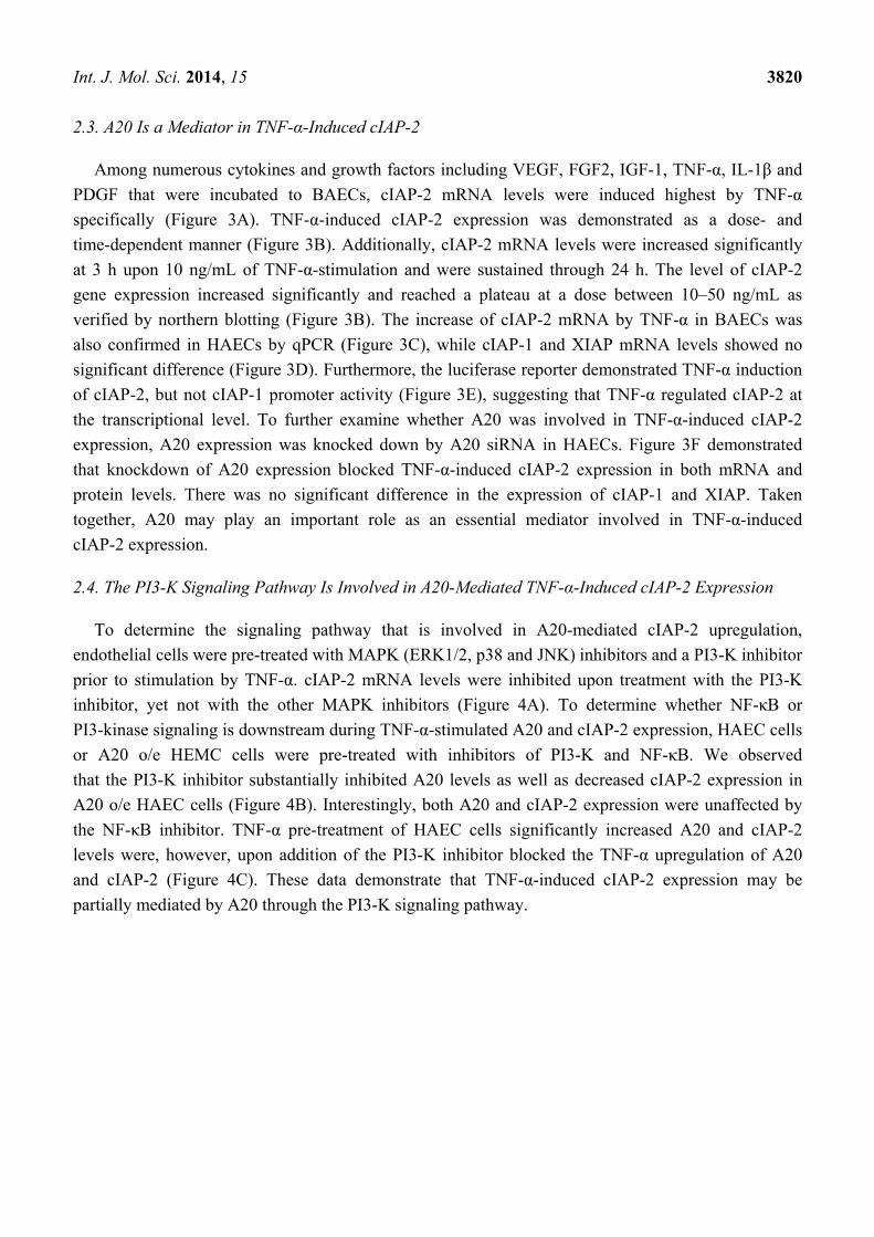
2.3. A20 Is a Mediator in TNF-α-Induced cIAP-2
Among numerous cytokines and growth factors including VEGF, FGF2, IGF-1, TNF-α, IL-1β and
PDGF that were incubated to BAECs, cIAP-2 mRNA levels were induced highest by TNF-α
specifically (Figure 3A). TNF-α-induced cIAP-2 expression was demonstrated as a dose- and
time-dependent manner (Figure 3B). Additionally, cIAP-2 mRNA levels were increased significantly
at 3 h upon 10 ng/mL of TNF-α-stimulation and were sustained through 24 h. The level of cIAP-2
gene expression increased significantly and reached a plateau at a dose between 10–50 ng/mL as
verified by northern blotting (Figure 3B). The increase of cIAP-2 mRNA by TNF-α in BAECs was
also confirmed in HAECs by qPCR (Figure 3C), while cIAP-1 and XIAP mRNA levels showed no
significant difference (Figure 3D). Furthermore, the luciferase reporter demonstrated TNF-α induction
of cIAP-2, but not cIAP-1 promoter activity (Figure 3E), suggesting that TNF-α regulated cIAP-2 at
the transcriptional level. To further examine whether A20 was involved in TNF-α-induced cIAP-2
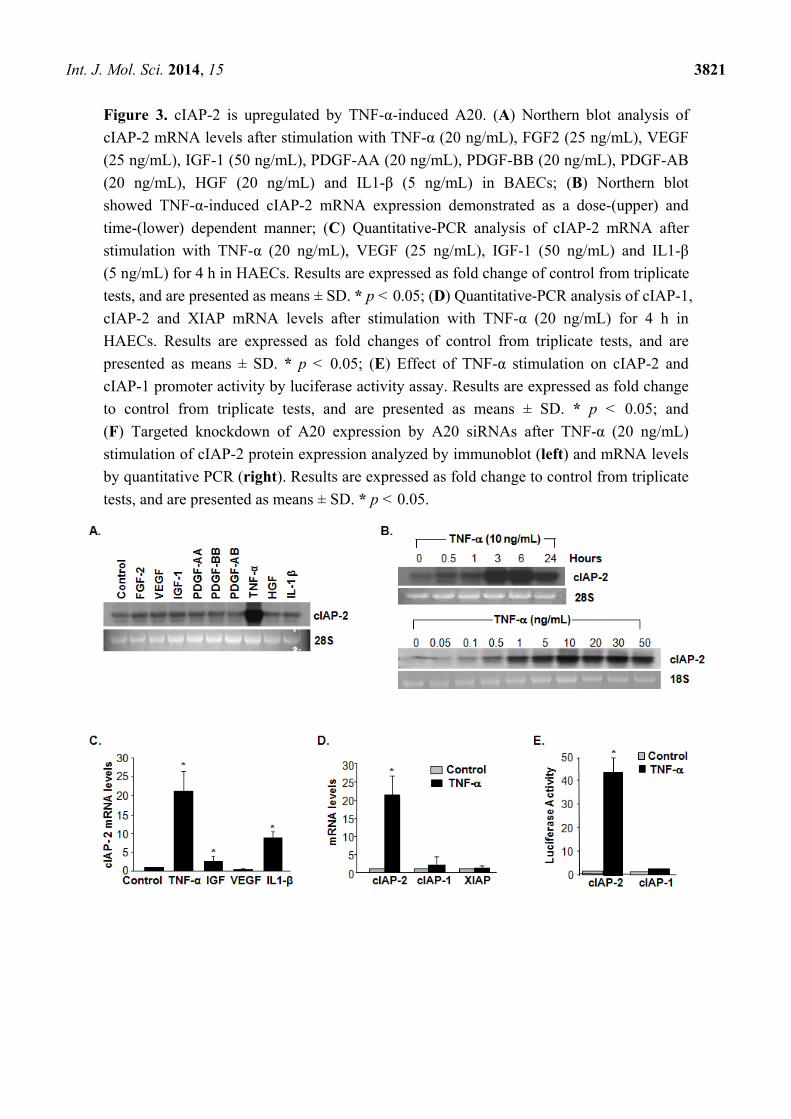
expression, A20 expression was knocked down by A20 siRNA in HAECs. Figure 3F demonstrated
that knockdown of A20 expression blocked TNF-α-induced cIAP-2 expression in both mRNA and
protein levels. There was no significant difference in the expression of cIAP-1 and XIAP. Taken
together, A20 may play an important role as an essential mediator involved in TNF-α-induced
cIAP-2 expression.
2.4. The PI3-K Signaling Pathway Is Involved in A20-Mediated TNF-α-Induced cIAP-2 Expression
To determine the signaling pathway that is involved in A20-mediated cIAP-2 upregulation,
endothelial cells were pre-treated with MAPK (ERK1/2, p38 and JNK) inhibitors and a PI3-K inhibitor
prior to stimulation by TNF-α. cIAP-2 mRNA levels were inhibited upon treatment with the PI3-K
inhibitor, yet not with the other MAPK inhibitors (Figure 4A). To determine whether NF-κB or
PI3-kinase signaling is downstream during TNF-α-stimulated A20 and cIAP-2 expression, HAEC cells
or A20 o/e HEMC cells were pre-treated with inhibitors of PI3-K and NF-κB. We observed
that the PI3-K inhibitor substantially inhibited A20 levels as well as decreased cIAP-2 expression in
A20 o/e HAEC cells (Figure 4B). Interestingly, both A20 and cIAP-2 expression were unaffected by
the NF-κB inhibitor. TNF-α pre-treatment of HAEC cells significantly increased A20 and cIAP-2
levels were, however, upon addition of the PI3-K inhibitor blocked the TNF-α upregulation of A20
and cIAP-2 (Figure 4C). These data demonstrate that TNF-α-induced cIAP-2 expression may be
partially mediated by A20 through the PI3-K signaling pathway.
Int. J. Mol. Sci. 2014, 15 3821
Figure 3. cIAP-2 is upregulated by TNF-α-induced A20. (A) Northern blot analysis of
cIAP-2 mRNA levels after stimulation with TNF-α (20 ng/mL), FGF2 (25 ng/mL), VEGF
(25 ng/mL), IGF-1 (50 ng/mL), PDGF-AA (20 ng/mL), PDGF-BB (20 ng/mL), PDGF-AB
(20 ng/mL), HGF (20 ng/mL) and IL1-β (5 ng/mL) in BAECs; (B) Northern blot
showed TNF-α-induced cIAP-2 mRNA expression demonstrated as a dose-(upper) and
time-(lower) dependent manner; (C) Quantitative-PCR analysis of cIAP-2 mRNA after
stimulation with TNF-α (20 ng/mL), VEGF (25 ng/mL), IGF-1 (50 ng/mL) and IL1-β
(5 ng/mL) for 4 h in HAECs. Results are expressed as fold change of control from triplicate
tests, and are presented as means ± SD. * p < 0.05; (D) Quantitative-PCR analysis of cIAP-1,
cIAP-2 and XIAP mRNA levels after stimulation with TNF-α (20 ng/mL) for 4 h in
HAECs. Results are expressed as fold changes of control from triplicate tests, and are
presented as means ± SD. * p < 0.05; (E) Effect of TNF-α stimulation on cIAP-2 and
cIAP-1 promoter activity by luciferase activity assay. Results are expressed as fold change
to control from triplicate tests, and are presented as means ± SD. * p < 0.05; and
(F) Targeted knockdown of A20 expression by A20 siRNAs after TNF-α (20 ng/mL)
stimulation of cIAP-2 protein expression analyzed by immunoblot (left) and mRNA levels
by quantitative PCR (right). Results are expressed as fold change to control from triplicate
tests, and are presented as means ± SD. * p < 0.05.
Int. J. Mol. Sci. 2014, 15 3822
Figure 3. Cont.
Figure 4. The PI3-K signaling pathway is involved in A20-mediated TNF-α-induced
cIAP-2 expression. (A) Quantitative-PCR analysis of cIAP-2 mRNA levels in HAECs
pre-treated with LY294002 (25 μM, PI3-K inhibitor), U0126 (10 μM, ERK1/2 inhibitor),
SB203580 (20 μM, p38 inhibitor), and SP600125 (20 μM, JNK inhibitor), respectively for
1 h. Results are expressed as fold change to control from triplicate tests, and are presented
as means ± SD. * p < 0.05; (B) Immunoblot analysis in A20 o/e HAECs pre-treated with
either wedelolactone (20 μM, IKKα/β inhibitor) or LY294002 (25 μM, PI3-K inhibitor) for
1 h and then immunoblotted for A20, cIAP-2 and vinculin. Representative blot is shown;
and (C) Immunoblot analysis in HAECs stimulated with TNF-α (20 ng/mL) and together
with either (20 μM, IKKα/β inhibitor) or LY294002 (25 μM, PI3-K inhibitor). A20, cIAP-1
and β-actin were blotted. Representative blot is shown.
Int. J. Mol. Sci. 2014, 15 3823
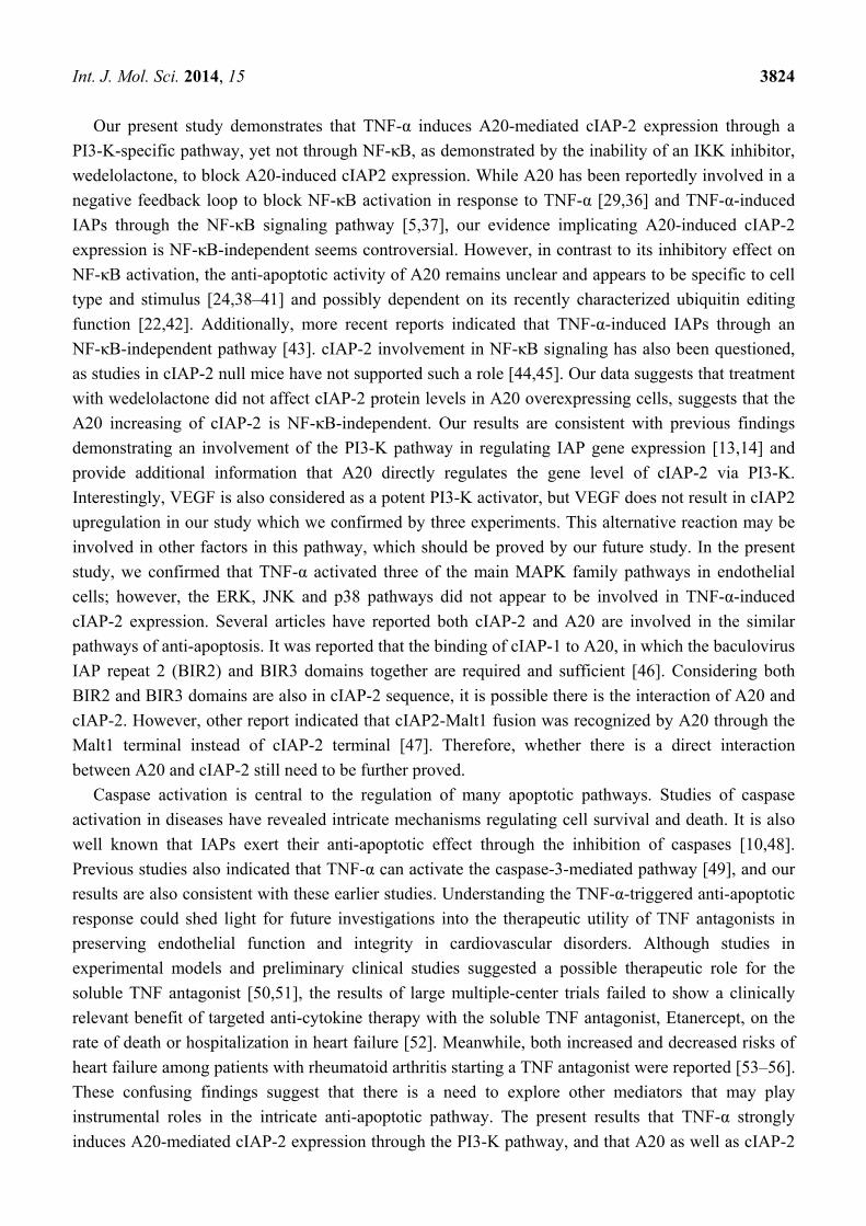
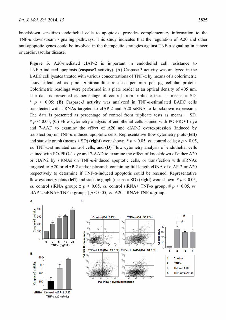
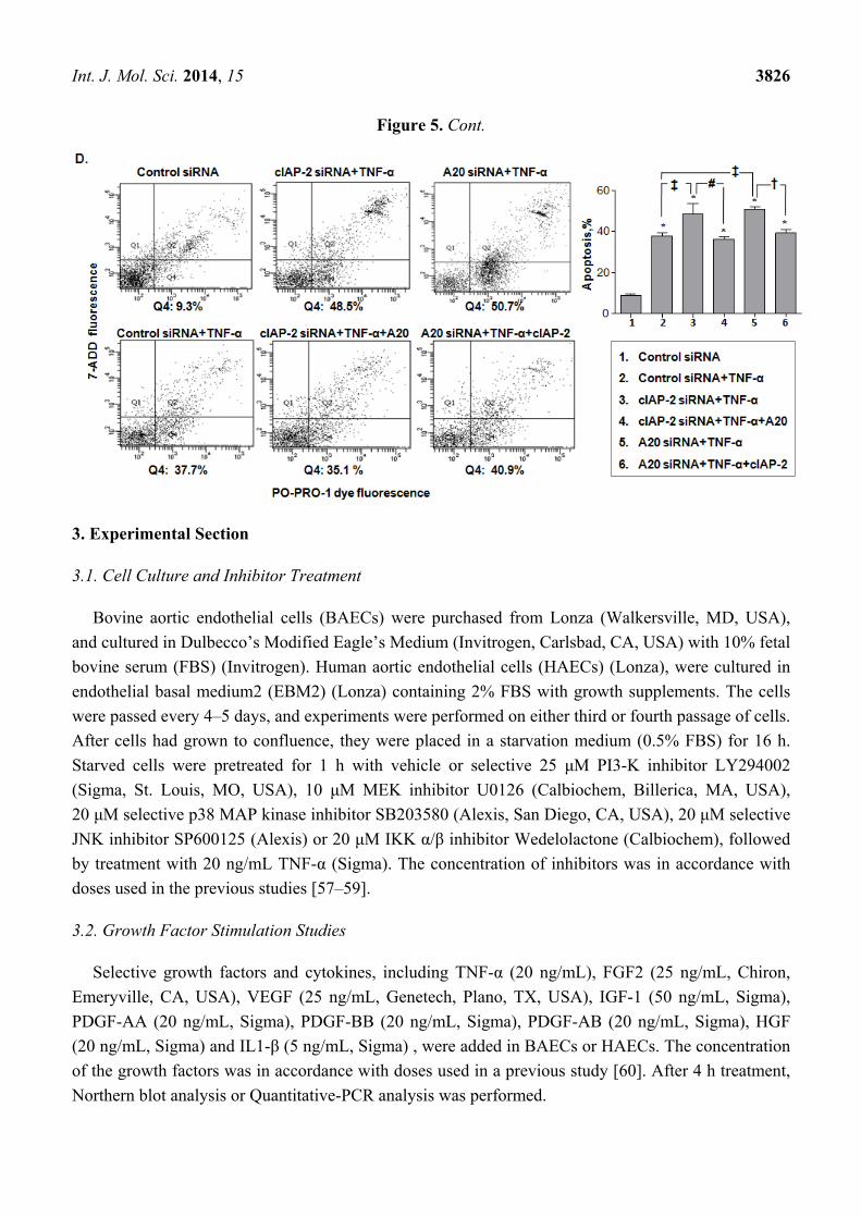
2.5. A20-Mediated cIAP-2 Is Important in Endothelial Cell Resistance to TNF-α-Induced Apoptosis
To assess the endothelial apoptotic effect of A20 in TNF-α-induced cIAP-2 expression, caspase-3
activity was measured in BAEC cells after TNF-α-stimulation. The caspase-3 activity increased by
4-fold upon stimulation with TNF-α as compared to controls (Figure 5A). Importantly, when either
cIAP-2 or A20 was knocked down using siRNAs, TNF-α-induced caspase-3 activation was increased
in comparison with control siRNA (Figure 5B), indicating an A20-mediated inhibitory effect of
cIAP-2 on caspase-3 activity induced by TNF-α. Using PO-PRO-1 dye and 7-AAD staining to identify
the consequences of TNF-α-induced apoptosis in endothelial cells, the results indicated that the
addition of A20 decreased the number of apoptotic cells that were induced by TNF-α (29.2 ± 1.8 vs.
36.1 ± 1.9), so did cIAP-2 (30.0 ± 1.9 vs. 36.1 ± 1.9) (Figure 5C). Moreover, there was a similar
increase in TNF-α-induced apoptotic cells when we knocked down expression of either A20 (50.7 ± 1.4
vs. 37.9 ± 1.3) or cIAP-2 (48.3 ± 5.2 vs. 37.9 ± 1.3) by their siRNA (Figure 5D). However, when cIAP-2
was knocked down, a decrease in apoptotic cells by additional A20 was observed (48.3 ± 5.2 vs.
36.1 ± 1.1). A20 siRNA increased the TNF-α-induced apoptotic cells could be rescued by additional
cIAP-2 (50.7 ± 1.4 vs. 39.1 ± 1.5), suggesting an inhibitory effect of A20 in apoptosis is, at least in part,
through induction of cIAP-2 (Figure 5D).
2.6. Discussion
Endothelial apoptosis results in vascular leakage, as well as the activation of inflammation and
coagulation pathways, and has been suggested to be an important mechanism of many diseases [1].
We aimed to determine the mechanisms that prevent endothelial cells from apoptotic damage, despite
being in continuous touch with hostile environments. Recent evidence indicates that IAPs are
frequently expressed in cancer, and their uncontrolled over-expression has been implicated in
contributing to tumorigenesis, chemoresistance, disease progression and poor patient survival [33].
In this study, we extend these findings by also showing that cIAP-2 induction is critical for the
resistance of endothelial cells to TNF-α-induced apoptosis. TNF-α stimulates cIAP-2 expression via
induction of A20, a protective gene in endothelial cells. Additionally, A20 plays an important role in
mediating this TNF-α-induced cIAP-2 expression. Our data also demonstrate that TNF-α-induced
cIAP-2 and A20 significantly prevented endothelial cells from undergoing TNF-α-induced apoptosis.
A20 was initially identified as a primary response gene following stimulation of human umbilical
vein endothelial cells (HUVEC) with TNF-α, IL-1 or LPS [32–34]. Based on references, TNF-α may
induce endothelial cells elongation [35] and A20 may inhibit the elongation of endothelial cells [18].
To observe whether A20 has impact on the TNF-α induced elongation of endothelial cells, we have
checked the cell morphology induced by A20 previously, but no obvious changes were found (data not
shown). Furthermore, the relationship between A20 and other anti-apoptotic genes, especially IAPs,
which also prevent endothelial cells from TNF-α-induced apoptosis, has not been well-investigated.
In the current study, we observed that A20 induced the expression of cIAP-2 but not that of cIAP-1
and XIAP. We also demonstrated that A20 knockdown reduced TNF-α-induced expression of cIAP-2
and effect TNF-α-induced apoptosis, suggesting that A20 is the mediator in TNF-α-induced cIAP-2.
Int. J. Mol. Sci. 2014, 15 3824
Our present study demonstrates that TNF-α induces A20-mediated cIAP-2 expression through a
PI3-K-specific pathway, yet not through NF-κB, as demonstrated by the inability of an IKK inhibitor,
wedelolactone, to block A20-induced cIAP2 expression. While A20 has been reportedly involved in a
negative feedback loop to block NF-κB activation in response to TNF-α [29,36] and TNF-α-induced
IAPs through the NF-κB signaling pathway [5,37], our evidence implicating A20-induced cIAP-2
expression is NF-κB-independent seems controversial. However, in contrast to its inhibitory effect on
NF-κB activation, the anti-apoptotic activity of A20 remains unclear and appears to be specific to cell
type and stimulus [24,38–41] and possibly dependent on its recently characterized ubiquitin editing
function [22,42]. Additionally, more recent reports indicated that TNF-α-induced IAPs through an
NF-κB-independent pathway [43]. cIAP-2 involvement in NF-κB signaling has also been questioned,
as studies in cIAP-2 null mice have not supported such a role [44,45]. Our data suggests that treatment
with wedelolactone did not affect cIAP-2 protein levels in A20 overexpressing cells, suggests that the
A20 increasing of cIAP-2 is NF-κB-independent. Our results are consistent with previous findings
demonstrating an involvement of the PI3-K pathway in regulating IAP gene expression [13,14] and
provide additional information that A20 directly regulates the gene level of cIAP-2 via PI3-K.
Interestingly, VEGF is also considered as a potent PI3-K activator, but VEGF does not result in cIAP2
upregulation in our study which we confirmed by three experiments. This alternative reaction may be
involved in other factors in this pathway, which should be proved by our future study. In the present
study, we confirmed that TNF-α activated three of the main MAPK family pathways in endothelial
cells; however, the ERK, JNK and p38 pathways did not appear to be involved in TNF-α-induced
cIAP-2 expression. Several articles have reported both cIAP-2 and A20 are involved in the similar
pathways of anti-apoptosis. It was reported that the binding of cIAP-1 to A20, in which the baculovirus
IAP repeat 2 (BIR2) and BIR3 domains together are required and sufficient [46]. Considering both
BIR2 and BIR3 domains are also in cIAP-2 sequence, it is possible there is the interaction of A20 and
cIAP-2. However, other report indicated that cIAP2-Malt1 fusion was recognized by A20 through the
Malt1 terminal instead of cIAP-2 terminal [47]. Therefore, whether there is a direct interaction
between A20 and cIAP-2 still need to be further proved.
Caspase activation is central to the regulation of many apoptotic pathways. Studies of caspase
activation in diseases have revealed intricate mechanisms regulating cell survival and death. It is also
well known that IAPs exert their anti-apoptotic effect through the inhibition of caspases [10,48].
Previous studies also indicated that TNF-α can activate the caspase-3-mediated pathway [49], and our
results are also consistent with these earlier studies. Understanding the TNF-α-triggered anti-apoptotic
response could shed light for future investigations into the therapeutic utility of TNF antagonists in
preserving endothelial function and integrity in cardiovascular disorders. Although studies in
experimental models and preliminary clinical studies suggested a possible therapeutic role for the
soluble TNF antagonist [50,51], the results of large multiple-center trials failed to show a clinically
relevant benefit of targeted anti-cytokine therapy with the soluble TNF antagonist, Etanercept, on the
rate of death or hospitalization in heart failure [52]. Meanwhile, both increased and decreased risks of
heart failure among patients with rheumatoid arthritis starting a TNF antagonist were reported [53–56].
These confusing findings suggest that there is a need to explore other mediators that may play
instrumental roles in the intricate anti-apoptotic pathway. The present results that TNF-α strongly
induces A20-mediated cIAP-2 expression through the PI3-K pathway, and that A20 as well as cIAP-2
Int. J. Mol. Sci. 2014, 15 3825
knockdown sensitizes endothelial cells to apoptosis, provides complementary information to the
TNF-α downstream signaling pathways. This study indicates that the regulation of A20 and other
anti-apoptotic genes could be involved in the therapeutic strategies against TNF-α signaling in cancer
or cardiovascular disease.
Figure 5. A20-mediated cIAP-2 is important in endothelial cell resistance to
TNF-α-induced apoptosis (caspase3 activity). (A) Caspase-3 activity was analyzed in the
BAEC cell lysates treated with various concentrations of TNF-α by means of a colorimetric
assay calculated as pmol p-nitroaniline released per min per μg cellular protein.
Colorimetric readings were performed in a plate reader at an optical density of 405 nm.
The data is presented as percentage of control from triplicate tests as means ± SD.
* p < 0.05; (B) Caspase-3 activity was analyzed in TNF-α-stimulated BAEC cells
transfected with siRNAs targeted to cIAP-2 and A20 siRNA to knockdown expression.
The data is presented as percentage of control from triplicate tests as means ± SD.
* p < 0.05; (C) Flow cytometry analysis of endothelial cells stained with PO-PRO-1 dye
and 7-AAD to examine the effect of A20 and cIAP-2 overexpression (induced by
transfection) on TNF-α-induced apoptotic cells. Representative flow cytometry plots (left)
and statistic graph (means ± SD) (right) were shown. * p < 0.05, vs. control cells; # p < 0.05,
vs. TNF-α-stimulated control cells; and (D) Flow cytometry analysis of endothelial cells
stained with PO-PRO-1 dye and 7-AAD to examine the effect of knockdown of either A20
or cIAP-2 by siRNAs on TNF-α-induced apoptotic cells, or transfection with siRNAs
targeted to A20 or cIAP-2 and/or plasmids containing full length cDNA of cIAP-2 or A20
respectively to determine if TNF-α-induced apoptotis could be rescued. Representative
flow cytometry plots (left) and statistic graph (means ± SD) (right) were shown. * p < 0.05,
vs. control siRNA group; ‡ p < 0.05, vs. control siRNA+ TNF-α group; # p < 0.05, vs.
cIAP-2 siRNA+ TNF-α group; † p < 0.05, vs. A20 siRNA+ TNF-α group.
Int. J. Mol. Sci. 2014, 15 3826
Figure 5. Cont.
3. Experimental Section
3.1. Cell Culture and Inhibitor Treatment
Bovine aortic endothelial cells (BAECs) were purchased from Lonza (Walkersville, MD, USA),
and cultured in Dulbecco’s Modified Eagle’s Medium (Invitrogen, Carlsbad, CA, USA) with 10% fetal
bovine serum (FBS) (Invitrogen). Human aortic endothelial cells (HAECs) (Lonza), were cultured in
endothelial basal medium2 (EBM2) (Lonza) containing 2% FBS with growth supplements. The cells
were passed every 4–5 days, and experiments were performed on either third or fourth passage of cells.
After cells had grown to confluence, they were placed in a starvation medium (0.5% FBS) for 16 h.
Starved cells were pretreated for 1 h with vehicle or selective 25 μM PI3-K inhibitor LY294002
(Sigma, St. Louis, MO, USA), 10 μM MEK inhibitor U0126 (Calbiochem, Billerica, MA, USA),
20 μM selective p38 MAP kinase inhibitor SB203580 (Alexis, San Diego, CA, USA), 20 μM selective
JNK inhibitor SP600125 (Alexis) or 20 μM IKK α/β inhibitor Wedelolactone (Calbiochem), followed
by treatment with 20 ng/mL TNF-α (Sigma). The concentration of inhibitors was in accordance with
doses used in the previous studies [57–59].
3.2. Growth Factor Stimulation Studies
Selective growth factors and cytokines, including TNF-α (20 ng/mL), FGF2 (25 ng/mL, Chiron,
Emeryville, CA, USA), VEGF (25 ng/mL, Genetech, Plano, TX, USA), IGF-1 (50 ng/mL, Sigma),
PDGF-AA (20 ng/mL, Sigma), PDGF-BB (20 ng/mL, Sigma), PDGF-AB (20 ng/mL, Sigma), HGF
(20 ng/mL, Sigma) and IL1-β (5 ng/mL, Sigma) , were added in BAECs or HAECs. The concentration
of the growth factors was in accordance with doses used in a previous study [60]. After 4 h treatment,
Northern blot analysis or Quantitative-PCR analysis was performed.
Int. J. Mol. Sci. 2014, 15 3827
3.3. Retroviral Construction
Control vector or Flag-HA-A20 (Addgene, Cambridge, MA, USA) was transfected to HEK293T
cells (American Type Culture Collection, Manassas, VA, USA) using polyethylenimine (PEI) with
pVSV-G, pJK3, and pCMVtat. The medium containing retrovirus-A20 was collected 48 h post
transfection and filtered before being used to infect HAECs and BAECs. The transfected gene
expression was confirmed using western blot analysis.
3.4. Western Blot Analysis
Whole-cell lysates were obtained from cultured cells with RIPA buffer (Boston Bioproducts Inc.,
Ashland, MA, USA) containing protease inhibitor cocktail (Roche, Basel, Switzerland) plus 10 mM
NaF and 10 mM Na3VO4. Proteins were fractionated by 10% SDS-polyacrylamide gel and transferred
to PVDF membranes (Millipore, Billerica, MA, USA). Membranes were incubated with the following
antibodies: Anti-A20 (Active Motif, Carlsbad, CA, USA); Anti-cIAP-1, Anti-cIAP-2, Anti-β-actin
(Santa Cruz Biotechnology, Santa Cruz, CA, USA); Anti-GAPDH (Millipore); Anti-XIAP (Transduction
Laboratories, San Jose, CA, USA); and Anti-vinculin (Sigma). Following incubation of anti-IgG,
the proteins were visualized using an ECL detection system (Amersham, Piscataway, NJ, USA).
3.5. Promoter Activity Analyses
A fragment of human cIAP-2 promoter (−1066 to +38, GeneBank NM_001165) and cIAP-1
promoter (−1083 to +79, GeneBank NM_001166) was cloned into a PGL-3 vector (Promega,
San Luis Obispo, CA, USA) containing the luciferase reporter. The A20 luciferase construct was a gift
from Dr. Kun-Sang Chang, from the University of Texas MD Anderson Cancer Center (Houston, TX,
USA) HEK293T cells (ATCC, Washington, DC, USA) were transfected with the cIAP-1, cIAP-2 and
A20 promoter constructors using PEI, Polyethyleneimine (Polysciences, Inc., Warrington, PA, USA).
20 ng/mL of TNF-α was added, cells were lysed 4 h later, and the luciferase activity was determined
with a dual-luciferase assay system (Promega).
3.6. siRNA Transfection
siRNAs targeting A20 (Shanghai GenePharma Co., Ltd., Shanghai, China), and cIAP-2
(Qiagen, Valencia, CA, USA) were prepared. The siRNAs were transfected using Lipofectamine 2000
(Invitrogen) according to manufacturer’s instructions into 30%–50% confluent HAECs or BAECs at a
final concentration of 20 nM. Seventy-two hours after transfection, the efficacy of knockdown was
assessed by Western blot assay and qPCR.
The sequences of the siRNAs are as the Table 1.
Table 1. The sequences of the siRNAs.
No. A20 siRNA cIAP-2 siRNA
1 5'-GAA GUG GAC UUC AGU ACA ATT-3' 5'-CCU CUA GUG UUC UAG UUA ATT-3'2 5'-GCG GAA AGC UGU GAA GAU ATT-3' 5'-GCU CCU UCU UUA AGA AAG UTT-3'
Int. J. Mol. Sci. 2014, 15 3828
3.7. RNA Analysis
Total RNA from BAECs and HAECs was extracted with TRIzol Reagent (Invitrogen), and cDNA
was synthesized by reverse transcription. RNA levels were measured by quantitative polymerase chain
reaction using the following six primers in Table 2.
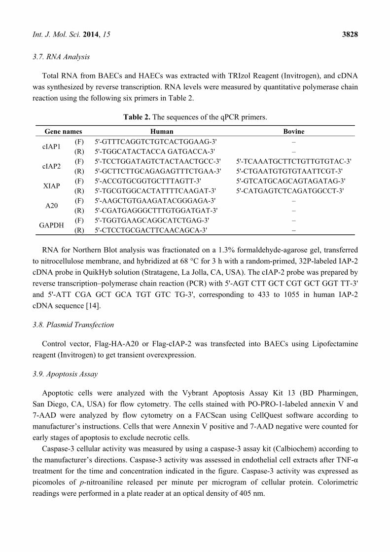
Table 2. The sequences of the qPCR primers.
Gene names Human Bovine
cIAP1 (F) 5'-GTTTCAGGTCTGTCACTGGAAG-3' – (R) 5'-TGGCATACTACCA GATGACCA-3' –
cIAP2 (F) 5'-TCCTGGATAGTCTACTAACTGCC-3' 5'-TCAAATGCTTCTGTTGTGTAC-3' (R) 5'-GCTTCTTGCAGAGAGTTTCTGAA-3' 5'-CTGAATGTGTGTAATTCGT-3'
XIAP (F) 5'-ACCGTGCGGTGCTTTAGTT-3' 5'-GTCATGCAGCAGTAGATAG-3' (R) 5'-TGCGTGGCACTATTTTCAAGAT-3' 5'-CATGAGTCTCAGATGGCCT-3'
A20 (F) 5'-AAGCTGTGAAGATACGGGAGA-3' – (R) 5'-CGATGAGGGCTTTGTGGATGAT-3' –
GAPDH (F) 5'-TGGTGAAGCAGGCATCTGAG-3' – (R) 5'-CTCCTGCGACTTCAACAGCA-3' –
RNA for Northern Blot analysis was fractionated on a 1.3% formaldehyde-agarose gel, transferred
to nitrocellulose membrane, and hybridized at 68 °C for 3 h with a random-primed, 32P-labeled IAP-2
cDNA probe in QuikHyb solution (Stratagene, La Jolla, CA, USA). The cIAP-2 probe was prepared by
reverse transcription–polymerase chain reaction (PCR) with 5'-AGT CTT GCT CGT GCT GGT TT-3'
and 5'-ATT CGA GCT GCA TGT GTC TG-3', corresponding to 433 to 1055 in human IAP-2
cDNA sequence [14].
3.8. Plasmid Transfection
Control vector, Flag-HA-A20 or Flag-cIAP-2 was transfected into BAECs using Lipofectamine
reagent (Invitrogen) to get transient overexpression.
3.9. Apoptosis Assay
Apoptotic cells were analyzed with the Vybrant Apoptosis Assay Kit 13 (BD Pharmingen,
San Diego, CA, USA) for flow cytometry. The cells stained with PO-PRO-1-labeled annexin V and
7-AAD were analyzed by flow cytometry on a FACScan using CellQuest software according to
manufacturer’s instructions. Cells that were Annexin V positive and 7-AAD negative were counted for
early stages of apoptosis to exclude necrotic cells.
Caspase-3 cellular activity was measured by using a caspase-3 assay kit (Calbiochem) according to
the manufacturer’s directions. Caspase-3 activity was assessed in endothelial cell extracts after TNF-α
treatment for the time and concentration indicated in the figure. Caspase-3 activity was expressed as
picomoles of p-nitroaniline released per minute per microgram of cellular protein. Colorimetric
readings were performed in a plate reader at an optical density of 405 nm.
Int. J. Mol. Sci. 2014, 15 3829
3.10. Statistics
Results are expressed as mean ± SD on the basis of triplicate experiments. Statistical analysis used
ANOVA and Student t test (2 tailed). A value of p < 0.05 was considered statistically significant.
4. Conclusions
In a word, these results identify A20 as a cytoprotective factor involved in cIAP-2 inhibitory pathway
of TNF-α-induced apoptosis. This is consistent with the idea that endothelial cell viability is dependent
on interactions between inducers and suppressors of apoptosis, susceptible to modulation by TNF-α.
Acknowledgments
We thank Kun-Sang Chang (University of Texas MD Anderson Cancer Center) for providing the A20
promoter constructer. This work was supported in part by NIHR01HL082837 (Li); NIHR21HL088219
(Li); China program NCET-13-0692 (Guo); and China Scholarship Council (Guo and Song).
Author Contributions
Study conception and design: J.L., A.M.-B. and S.G. Experiments: J.W., X.S. M.P., J.S., A.M.-B.
and S.G. Analysis and interpretation of data: J.L., A.M.-B. and S.G. Drafting of manuscript: A.M.-B.,
S.G., M.P., J.R. and J.L.
Conflicts of Interest
Disclosure: This work was prepared while Jian Li was employed at the CardioVascular Institute,
Beth Israel Deaconess Medical Center, Harvard Medical School. The opinions expressed in this article
are the author’s own and do not reflect the view of the National Institutes of Health, the Department of
Health and Human Services, or the United States Government. No conflicts of interest, financial or
otherwise, are declared by the authors.
References
1. Winn, R.K.; Harlan, J.M. The role of endothelial cell apoptosis in inflammatory and immune
diseases. J. Thromb. Haemost. 2005, 3, 1815–1824.
2. Badrichani, A.Z.; Stroka, D.M.; Bilbao, G.; Curiel, D.T.; Bach, F.H.; Ferran, C. Bcl-2 and bcl-xl
serve an anti-inflammatory function in endothelial cells through inhibition of NF-κB.
J. Clin. Investig. 1999, 103, 543–553.
3. He, K.L.; Ting, A.T. A20 inhibits tumor necrosis factor (TNF) α-induced apoptosis by disrupting
recruitment of tradd and rip to the tnf receptor 1 complex in jurkat T cells. Mol. Cell. Biol. 2002,
22, 6034–6045.
4. Karsan, A.; Yee, E.; Harlan, J.M. Endothelial cell death induced by tumor necrosis factor-α is
inhibited by the bcl-2 family member, a1. J. Biol. Chem. 1996, 271, 27201–27204.
Int. J. Mol. Sci. 2014, 15 3830
5. Stehlik, C.; de Martin, R.; Kumabashiri, I.; Schmid, J.A.; Binder, B.R.; Lipp, J. Nuclear factor
(NF)-κB-regulated X-chromosome-linked iap gene expression protects endothelial cells from
tumor necrosis factor α-induced apoptosis. J. Exp. Med. 1998, 188, 211–216.
6. Lopez, J.; Meier, P. To fight or die—Inhibitor of apoptosis proteins at the crossroad of innate
immunity and death. Curr. Opin. Cell Biol. 2010, 22, 872–881.
7. O’Riordan, M.X.; Bauler, L.D.; Scott, F.L.; Duckett, C.S. Inhibitor of apoptosis proteins in
eukaryotic evolution and development: A model of thematic conservation. Dev. Cell 2008, 15,
497–508.
8. Lorick, K.L.; Jensen, J.P.; Fang, S.; Ong, A.M.; Hatakeyama, S.; Weissman, A.M. Ring fingers
mediate ubiquitin-conjugating enzyme (e2)-dependent ubiquitination. Proc. Natl. Acad. Sci. USA
1999, 96, 11364–11369.
9. Vince, J.E.; Wong, W.W.; Khan, N.; Feltham, R.; Chau, D.; Ahmed, A.U.; Benetatos, C.A.;
Chunduru, S.K.; Condon, S.M.; McKinlay, M.; et al. Iap antagonists target ciap1 to induce
TNFα-dependent apoptosis. Cell 2007, 131, 682–693.
10. LeBlanc, A.C. Natural cellular inhibitors of caspases. Prog. Neuropsychopharmacol. Biol. Psychiatry
2003, 27, 215–229.
11. Gyrd-Hansen, M.; Darding, M.; Miasari, M.; Santoro, M.M.; Zender, L.; Xue, W.; Tenev, T.;
da Fonseca, P.C.; Zvelebil, M.; Bujnicki, J.M.; et al. Iaps contain an evolutionarily conserved
ubiquitin-binding domain that regulates NF-κB as well as cell survival and oncogenesis.
Nat. Cell Biol. 2008, 10, 1309–1317.
12. Gyrd-Hansen, M.; Meier, P. Iaps: From caspase inhibitors to modulators of NF-κB, inflammation
and cancer. Nat. Rev. 2010, 10, 561–574.
13. Petersen, S.L.; Peyton, M.; Minna, J.D.; Wang, X. Overcoming cancer cell resistance to smac
mimetic induced apoptosis by modulating ciap-2 expression. Proc. Natl. Acad. Sci. USA 2010,
107, 11936–11941.
14. Wu, J.; Parungo, C.; Wu, G.; Kang, P.M.; Laham, R.J.; Sellke, F.W.; Simons, M.; Li, J. Pr39
inhibits apoptosis in hypoxic endothelial cells: Role of inhibitor apoptosis protein-2. Circulation
2004, 109, 1660–1667.
15. Opipari, A.W., Jr.; Hu, H.M.; Yabkowitz, R.; Dixit, V.M. The a20 zinc finger protein protects
cells from tumor necrosis factor cytotoxicity. J. Biol. Chem. 1992, 267, 12424–12427.
16. Jaattela, M.; Mouritzen, H.; Elling, F.; Bastholm, L. A20 zinc finger protein inhibits TNF and IL-1
signaling. J. Immunol. 1996, 156, 1166–1173.
17. Song, H.Y.; Rothe, M.; Goeddel, D.V. The tumor necrosis factor-inducible zinc finger protein a20
interacts with traf1/traf2 and inhibits NF-κB activation. Proc. Natl. Acad. Sci. USA 1996, 93,
6721–6725.
18. Ainbinder, E.; Amir-Zilberstein, L.; Yamaguchi, Y.; Handa, H.; Dikstein, R. Elongation inhibition
by drb sensitivity-inducing factor is regulated by the a20 promoter via a novel negative element
and NF-κB. Mol. Cell. Biol. 2004, 24, 2444–2454.
19. Ainbinder, E.; Revach, M.; Wolstein, O.; Moshonov, S.; Diamant, N.; Dikstein, R. Mechanism of
rapid transcriptional induction of tumor necrosis factor α-responsive genes by NF-κB.
Mol. Cell. Biol. 2002, 22, 6354–6362.
Int. J. Mol. Sci. 2014, 15 3831
20. Evans, P.C.; Ovaa, H.; Hamon, M.; Kilshaw, P.J.; Hamm, S.; Bauer, S.; Ploegh, H.L.; Smith, T.S.
Zinc-finger protein a20, a regulator of inflammation and cell survival, has de-ubiquitinating
activity. Biochem. J. 2004, 378, 727–734.
21. Lin, S.C.; Chung, J.Y.; Lamothe, B.; Rajashankar, K.; Lu, M.; Lo, Y.C.; Lam, A.Y.; Darnay, B.G.;
Wu, H. Molecular basis for the unique deubiquitinating activity of the NF-κB inhibitor a20.
J. Mol. Biol. 2008, 376, 526–540.
22. Wertz, I.E.; O’Rourke, K.M.; Zhou, H.; Eby, M.; Aravind, L.; Seshagiri, S.; Wu, P.; Wiesmann, C.;
Baker, R.; Boone, D.L.; et al. De-ubiquitination and ubiquitin ligase domains of a20 downregulate
NF-κB signalling. Nature 2004, 430, 694–699.
23. Daniel, S.; Arvelo, M.; Ferran, C. Overexpression of a20 in endothelial cells of vascularized grafts
creates a protective barrier against TNF- and FAS-mediated apoptosis. Transplant. Proc. 2001,
33, 225.
24. Daniel, S.; Arvelo, M.B.; Patel, V.I.; Longo, C.R.; Shrikhande, G.; Shukri, T.; Mahiou, J.; Sun, D.W.;
Mottley, C.; Grey, S.T.; et al. A20 protects endothelial cells from TNF-, FAS-, and NK-mediated
cell death by inhibiting caspase 8 activation. Blood 2004, 104, 2376–2384.
25. Dong, B.; Lv, G.; Wang, Q.; Wei, F.; Bellail, A.C.; Hao, C.; Wang, G. Targeting a20 enhances
trail-induced apoptosis in hepatocellular carcinoma cells. Biochem. Biophys. Res. Commun. 2012,
418, 433–438.
26. Hou, C.L.; Zhang, W.; Wei, Y.; Mi, J.H.; Li, L.; Zhou, Z.H.; Zeng, W.; Ying, D.J. Zinc finger
protein a20 overexpression inhibits monocyte homing and protects endothelial cells from injury
induced by high glucose. Genet. Mol. Res. 2011, 10, 1050–1059.
27. Hou, C.L.; Huang, Q.; Wei, Y.; Zhang, W.; Mi, J.H.; Ying, D.J.; Zhou, Z.H. Protein transduction
domain-ha20 fusion protein protects endothelial cells against high glucose-induced injury.
Genet. Mol. Res. 2012, 11, 1899–1908.
28. Gu, G.; Zhang, Y.; Guo, L. Ubiquitin e3 ligase a20 is required in degradation of microbial
superantigens in vascular endothelial cells. Cell Biochem. Biophys. 2013, 66, 649–655.
29. Lee, E.G.; Boone, D.L.; Chai, S.; Libby, S.L.; Chien, M.; Lodolce, J.P.; Ma, A. Failure to regulate
TNF-induced NF-κB and cell death responses in a20-deficient mice. Science 2000, 289, 2350–2354.
30. Hovelmeyer, N.; Reissig, S.; Xuan, N.T.; Adams-Quack, P.; Lukas, D.; Nikolaev, A.; Schluter, D.;
Waisman, A. A20 deficiency in B cells enhances B-cell proliferation and results in the
development of autoantibodies. Eur. J. Immunol. 2011, 41, 595–601.
31. Chng, H.W.; Camplejohn, R.S.; Stone, M.G.; Hart, I.R.; Nicholson, L.J. A new role for the
anti-apoptotic gene a20 in angiogenesis. Exp. Cell Res. 2006, 312, 2897–2907.
32. Codd, J.D.; Salisbury, J.R.; Packham, G.; Nicholson, L.J. A20 RNA expression is associated with
undifferentiated nasopharyngeal carcinoma and poorly differentiated head and neck squamous cell
carcinoma. J. Pathol. 1999, 187, 549–555.
33. Hunter, A.M.; LaCasse, E.C.; Korneluk, R.G. The inhibitors of apoptosis (iaps) as cancer targets.
Apoptosis 2007, 12, 1543–1568.
34. Dixit, V.M.; Green, S.; Sarma, V.; Holzman, L.B.; Wolf, F.W.; O’Rourke, K.; Ward, P.A.;
Prochownik, E.V.; Marks, R.M. Tumor necrosis factor-α induction of novel gene products in
human endothelial cells including a macrophage-specific chemotaxin. J. Biol. Chem. 1990, 265,
2973–2978.
Int. J. Mol. Sci. 2014, 15 3832
35. Emmanuel, C.; Huynh, M.; Matthews, J.; Kelly, E.; Zoellner, H. TNF-α and TGF-β
synergistically stimulate elongation of human endothelial cells without transdifferentiation to
smooth muscle cell phenotype. Cytokine 2013, 61, 38–40.
36. Coornaert, B.; Carpentier, I.; Beyaert, R. A20: Central gatekeeper in inflammation and immunity.
J. Biol. Chem. 2009, 284, 8217–8221.
37. Rothe, M.; Pan, M.G.; Henzel, W.J.; Ayres, T.M.; Goeddel, D.V. The TNFR2-TRAF signaling
complex contains two novel proteins related to baculoviral inhibitor of apoptosis proteins. Cell
1995, 83, 1243–1252.
38. Slowik, M.R.; Min, W.; Ardito, T.; Karsan, A.; Kashgarian, M.; Pober, J.S. Evidence that tumor
necrosis factor triggers apoptosis in human endothelial cells by interleukin-1-converting
enzyme-like protease-dependent and -independent pathways. Lab. Investig. J. Tech. Methods Pathol.
1997, 77, 257–267.
39. Grey, S.T.; Arvelo, M.B.; Hasenkamp, W.; Bach, F.H.; Ferran, C. A20 inhibits cytokine-induced
apoptosis and nuclear factor κB-dependent gene activation in islets. J. Exp. Med. 1999, 190,
1135–1146.
40. Ferran, C.; Stroka, D.M.; Badrichani, A.Z.; Cooper, J.T.; Wrighton, C.J.; Soares, M.; Grey, S.T.;
Bach, F.H. A20 inhibits NF-κB activation in endothelial cells without sensitizing to tumor
necrosis factor-mediated apoptosis. Blood 1998, 91, 2249–2258.
41. Arvelo, M.B.; Cooper, J.T.; Longo, C.; Daniel, S.; Grey, S.T.; Mahiou, J.; Czismadia, E.;
Abu-Jawdeh, G.; Ferran, C. A20 protects mice from d-galactosamine/lipopolysaccharide acute
toxic lethal hepatitis. Hepatology 2002, 35, 535–543.
42. Hymowitz, S.G.; Wertz, I.E. A20: From ubiquitin editing to tumour suppression. Nat. Rev. 2010,
10, 332–341.
43. Grehan, J.F.; Levay-Young, B.K.; Benson, B.A.; Abrahamsen, M.S.; Dalmasso, A.P. α Gal
ligation of pig endothelial cells induces protection from complement and apoptosis independently
of NF-κB and inflammatory changes. Am. J. Transplant. 2005, 5, 712–719.
44. Conte, D.; Holcik, M.; Lefebvre, C.A.; Lacasse, E.; Picketts, D.J.; Wright, K.E.; Korneluk, R.G.
Inhibitor of apoptosis protein ciap2 is essential for lipopolysaccharide-induced macrophage
survival. Mol. Cell. Biol. 2006, 26, 699–708.
45. Conze, D.B.; Albert, L.; Ferrick, D.A.; Goeddel, D.V.; Yeh, W.C.; Mak, T.; Ashwell, J.D.
Posttranscriptional downregulation of c-iap2 by the ubiquitin protein ligase c-iap1 in vivo.
Mol. Cell. Biol. 2005, 25, 3348–3356.
46. Yamaguchi, N.; Oyama, M.; Kozuka-Hata, H.; Inoue, J. Involvement of a20 in the molecular
switch that activates the non-canonical NF-κB pathway. Sci. Rep. 2013, 3, 2568.
47. Kingeter, L.M.; Schaefer, B.C. Malt1 and ciap2-malt1 as effectors of NF-κB activation: Kissing
cousins or distant relatives? Cell. Signal. 2010, 22, 9–22.
48. Li, X.; Yang, Y.; Ashwell, J.D. TNF-RII and c-iap1 mediate ubiquitination and degradation of
TRAF2. Nature 2002, 416, 345–347.
49. Zhao, X.; Bausano, B.; Pike, B.R.; Newcomb-Fernandez, J.K.; Wang, K.K.; Shohami, E.;
Ringger, N.C.; DeFord, S.M.; Anderson, D.K.; Hayes, R.L. TNF-α stimulates caspase-3
activation and apoptotic cell death in primary septo-hippocampal cultures. J. Neurosci. Res. 2001,
64, 121–131.
Int. J. Mol. Sci. 2014, 15 3833
50. Bozkurt, B.; Torre-Amione, G.; Warren, M.S.; Whitmore, J.; Soran, O.Z.; Feldman, A.M.; Mann, D.L.
Results of targeted anti-tumor necrosis factor therapy with etanercept (enbrel) in patients with
advanced heart failure. Circulation 2001, 103, 1044–1047.
51. Deswal, A.; Bozkurt, B.; Seta, Y.; Parilti-Eiswirth, S.; Hayes, F.A.; Blosch, C.; Mann, D.L.
Safety and efficacy of a soluble p75 tumor necrosis factor receptor (enbrel, etanercept) in patients
with advanced heart failure. Circulation 1999, 99, 3224–3226.
52. Mann, D.L.; McMurray, J.J.; Packer, M.; Swedberg, K.; Borer, J.S.; Colucci, W.S.; Djian, J.;
Drexler, H.; Feldman, A.; Kober, L.; et al. Targeted anticytokine therapy in patients with chronic
heart failure: Results of the randomized etanercept worldwide evaluation (renewal). Circulation
2004, 109, 1594–1602.
53. Setoguchi, S.; Schneeweiss, S.; Avorn, J.; Katz, J.N.; Weinblatt, M.E.; Levin, R.; Solomon, D.H.
Tumor necrosis factor-α antagonist use and heart failure in elderly patients with rheumatoid
arthritis. Am. Heart J. 2008, 156, 336–341.
54. Solomon, D.H.; Curtis, J.R.; Saag, K.G.; Lii, J.; Chen, L.; Harrold, L.R.; Herrinton, L.J.;
Graham, D.J.; Kowal, M.K.; Kuriya, B.; et al. Cardiovascular risk in rheumatoid arthritis:
Comparing TNF-α blockade with nonbiologic dmards. Am. J. Med. 2013, 126, 730e9–730e17.
55. Solomon, D.H.; Rassen, J.A.; Kuriya, B.; Chen, L.; Harrold, L.R.; Graham, D.J.; Lewis, J.D.; Lii, J.;
Liu, L.; Griffin, M.R.; et al. Heart failure risk among patients with rheumatoid arthritis starting a
tnf antagonist. Ann. Rheum. Dis. 2013, 72, 1813–1818.
56. Kotyla, P.J.; Owczarek, A.; Rakoczy, J.; Lewicki, M.; Kucharz, E.J.; Emery, P. Infliximab
treatment increases left ventricular ejection fraction in patients with rheumatoid arthritis:
Assessment of heart function by echocardiography, endothelin 1, interleukin 6, and nt-pro brain
natriuretic peptide. J. Rheumatol. 2012, 39, 701–706.
57. Wu, G.; Luo, J.; Rana, J.S.; Laham, R.; Sellke, F.W.; Li, J. Involvement of COX-2 in
VEGF-induced angiogenesis via p38 and jnk pathways in vascular endothelial cells. Cardiovasc. Res.
2006, 69, 512–519.
58. Pan, C.; Lou, L.; Huo, Y.; Singh, G.; Chen, M.; Zhang, D.; Wu, A.; Zhao, M.; Wang, S.; Li, J.
Salvianolic acid B and tanshinone IIA attenuate myocardial ischemia injury in mice by no
production through multiple pathways. Ther. Adv. Cardiovasc. Dis. 2011, 5, 99–111.
59. Jin, Y.; Wu, J.; Song, X.; Song, Q.; Cully, B.L.; Messmer-Blust, A.; Xu, M.; Foo, S.Y.;
Rosenzweig, A.; Li, J. RTEF-1, an upstream gene of hypoxia-inducible factor-1α, accelerates
recovery from ischemia. J. Biol. Chem. 2011, 286, 22699–22705.
60. Wu, G.; Mannam, A.P.; Wu, J.; Kirbis, S.; Shie, J.L.; Chen, C.; Laham, R.J.; Sellke, F.W.; Li, J.
Hypoxia induces myocyte-dependent COX-2 regulation in endothelial cells: Role of VEGF.
Am. J. Physiol. Heart Circ. Physiol. 2003, 285, H2420–H2429.
© 2014 by the authors; licensee MDPI, Basel, Switzerland. This article is an open access article
distributed under the terms and conditions of the Creative Commons Attribution license
(http://creativecommons.org/licenses/by/3.0/).