Robert L. Moore and Douglas V. Faller Cancer Center and ...
45
SIRT1 REPRESSES ESTROGEN-SIGNALING, LIGAND-INDEPENDENT ERα-MEDIATED TRANSCRIPTION, AND CELL PROLIFERATION IN ESTROGEN-RESPONSIVE BREAST CELLS Robert L. Moore 1 and Douglas V. Faller 1,2 1 Cancer Center and 2 Departments of Medicine, Biochemistry, Pediatrics, Microbiology, Pathology and Laboratory Medicine, Boston University School of Medicine, Boston, MA 02118. CORRESPONDING AUTHOR: Dr. Douglas V. Faller 72 E. Concord St. Rm K-701 Boston, MA 02118 [email protected] (same address for both authors) Short Title: SIRT1 REPRESSESS LIGAND-INDEPENDENT ERα ACTIVITY Key Words: Sirtuin, estrogen receptor, SIRT1, ligand-independent, breast cancer Address Correspondence to: Douglas Faller, M.D., Ph.D., 72 East Concord Street, Room K-701, Boston, MA 02118-2307. Phone: (617)638-4173; Fax: (617)638-4176; Email: [email protected] This work was supported by a grant from the National Cancer Institute, R01-CA101992 (DVF) and CA101992S1 (RM), and by the Karin Grunebaum Cancer Research Foundation (DVF). The authors report no conflicts of interest. Page 1 of 45 Accepted Preprint first posted on 20 November 2012 as Manuscript JOE-12-0102 Copyright © 2012 by the Society for Endocrinology.
Transcript of Robert L. Moore and Douglas V. Faller Cancer Center and ...
SIRT1 REPRESSES ESTROGEN-SIGNALING, LIGAND-INDEPENDENT ERαααα-MEDIATED
TRANSCRIPTION, AND CELL PROLIFERATION IN ESTROGEN-RESPONSIVE BREAST
CELLS
Robert L. Moore1 and Douglas V. Faller
1,2
1Cancer Center and
2Departments of Medicine, Biochemistry, Pediatrics, Microbiology,
Pathology and Laboratory Medicine, Boston University School of Medicine, Boston, MA 02118.
CORRESPONDING AUTHOR:
Dr. Douglas V. Faller
72 E. Concord St. Rm K-701
Boston, MA 02118
(same address for both authors)
Short Title: SIRT1 REPRESSESS LIGAND-INDEPENDENT ERαααα ACTIVITY
Key Words: Sirtuin, estrogen receptor, SIRT1, ligand-independent, breast cancer
Address Correspondence to: Douglas Faller, M.D., Ph.D., 72 East Concord Street, Room K-701,
Boston, MA 02118-2307. Phone: (617)638-4173; Fax: (617)638-4176; Email: [email protected]
This work was supported by a grant from the National Cancer Institute, R01-CA101992 (DVF) and
CA101992S1 (RM), and by the Karin Grunebaum Cancer Research Foundation (DVF).
The authors report no conflicts of interest.
Page 1 of 45 Accepted Preprint first posted on 20 November 2012 as Manuscript JOE-12-0102
Copyright © 2012 by the Society for Endocrinology.
Abstract In prostate and breast cancer, the androgen and estrogen receptors mediate induction of androgen-
and estrogen-responsive genes respectively, and stimulate cell proliferation in response to the
binding of their cognate steroid hormones. Sirtuin 1 (SIRT1) is a nicotinamide adenosine
dinucleotide (NAD+)-dependent class III histone deacetylase (HDAC) that has been linked to gene
silencing, control of the cell cycle, apoptosis and energy homeostasis. In prostate cancer, SIRT1 is
required for androgen-antagonist-mediated transcriptional repression and growth suppression of
prostate cancer cells. Whether SIRT1 plays a similar role in the actions of estrogen or antagonists
had not been determined. We report here that SIRT1 represses the transcriptional and
proliferative response of breast cancer cells to estrogens, and this repression is estrogen receptor-
alpha (ERαααα)-dependent. Inhibition of SIRT1 activity results in the phosphorylation of ERαααα in an
AKT-dependent manner, and this activation requires phosphoinositide 3-kinase (PI3K) activity.
Phosphorylated ERαααα subsequently accumulates in the nucleus, where ERαααα binds DNA ER-response
elements and activates transcription of estrogen-responsive genes. This ER-dependent
transcriptional activation augments estrogen-induced signaling, but also activates ER-signaling in
the absence of estrogen, thus defining a novel and unexpected mechanism of ligand-independent
ERαααα-mediated activation and target gene transcription. Like ligand-dependent activation of ERαααα,
SIRT1 inhibition-mediated ERαααα activation in the absence of estrogen also results in breast cancer
cell proliferation. Together, these data demonstrate that SIRT1 regulates the most important cell
signaling pathway for the growth of breast cancer cells, both in the presence and the absence of
estrogen.
Page 2 of 45
Introduction 1
2
The sirtuins are a family of enzymes with increasingly-recognized relevance to cancer 3
(Moore 2011), but the full extent of their involvement in breast cancer genesis and evolution 4
remains to be elucidated. 5
SIRT1 is a nicotinamide adenosine dinucleotide (NAD+)-dependent histone deacetylase 6
(HDAC) that has been linked to longevity, gene silencing, control of the cell cycle, apoptosis and 7
energy homeostasis (Blander and Guarente 2004; Borra, et al. 2005; Dai, et al. 2007; Haigis and 8
Guarente 2006; Landry, et al. 2000; Yamamoto, et al. 2007). SIRT1 also has functions relating to 9
inflammation and neurodegeneration (Yamamoto et al. 2007). In addition, SIRT1 interacts with 10
PPARγ and PGC-α in the differentiation of muscle cells, adipogenesis, fat storage and 11
metabolism in the liver (Fulco, et al. 2003; Picard, et al. 2004; Puigserver, et al. 2005; Rodgers, et 12
al. 2005). SIRT1 is primarily a nuclear protein that targets the PGC-1α, FOXO and NFκB 13
families of transcription factors, among others (Haigis and Guarente 2006). SIRT1 also associates 14
with the tumor suppressor protein p53 (Dai et al. 2007; Haigis and Guarente 2006; Yamamoto et 15
al. 2007) and has been suggested to be a tumor suppressor protein itself (Dai et al. 2007; Jin, et al. 16
2007; Motta, et al. 2004; Pruitt, et al. 2006; Wang, et al. 2006). 17
SIRT1 appears to serve multiple functions in human prostate cancer cells. The enzyme is 18
over-expressed in prostate cancer cells and is present in both the nucleus and cytoplasm, 19
promoting cell survival (Byles, et al. 2010; Dai et al. 2007). However, enforced cytoplasmic 20
localization of SIRT1 enhanced sensitivity to apoptosis in one report (Jin et al. 2007). SIRT1 also 21
suppresses specific tumor suppressor genes in hormone-refractory prostate cancer cells (Fu, et al. 22
2006). Deacetylation of the androgen receptor (AR) by SIRT1 inactivates its ability to transform 23
prostate cells. SIRT1 binds to, and deacetylates, the AR at a conserved lysine motif, down-24
Page 3 of 45
regulating its levels in the cell and repressing androgen-induced AR transcription (Dai et al. 2007; 25
Dai, et al. 2008). Add: (Fu et al. 2006). 26
27
SIRT1 is required for the actions of endocrine therapy in prostate cancer as well. Androgen 28
antagonist-mediated transcriptional repression and growth suppression of prostate cancer cells 29
requires SIRT1 (Dai et al. 2007). It had not yet been determined whether SIRT1 plays a similar 30
role in the action of estrogen antagonists. We show here that SIRT1 is not required for estrogen 31
antagonist activity. Rather, SIRT1 functions to repress the estrogen response in the absence and in 32
the presence of estrogen, limiting ligand-independent signaling through ERα. 33
34
35
Page 4 of 45
Experimental Procedures 36
37
Cell Culture – The MCF-7 breast cancer cell line (ATC HTB-22) is an epithelial cell line derived 38
from an adenocarcinoma of the mammary gland. T47D cells (ATCC HTB-133) are a pTEN-39
negative breast cancer cell line that expresses the WNT7B oncogene. MDA-MB 231 cells (ATCC 40
HTB-26) are an ERα-negative, estrogen-independent breast cancer cell line. All cells were 41
cultured in phenol-red free DMEM (Invitrogen, Carlsbad, CA) plus 10% FBS or charcoal-treated 42
FBS (Hyclone, Erie, PA). Cells were treated with β-estradiol (R187933), sirtinol, splitomicin, 4-43
hydroxytamoxifen, faslodex (all from Sigma, St. Louis, MO) (Sigma), LY294002, Wortmannin 44
(both from CalBiochem, Gibbstown, NJ) or vehicle (EtOH or DMSO). 45
Transfections - Two micrograms of plasmid [ERE-luciferase reporter construct (Addgene 46
Cambridge, MA) (Hall and McDonnell 1999); SV40-β-gal reporter construct; D/N SIRT1 47
(H363Y); or ERα expression vector] were added to 100 µl of OPTI-MEM (Invitrogen) and 48
incubated for 15 min. Transfections were performed using Lipofectamine (Invitrogen), following 49
the manufacturer’s instructions. 50
Luciferase Assays - The Promega Dual luciferase reporter assay system was utilized. Cellular 51
harvest and assay were carried out according to manufacturer’s instructions. Luminescence was 52
quantitated in a Turner Designs 20/20 luminometer. Results were normalized with a β-53
galactosidase assay (Promega) . 54
Immunoblot and Chromatin Immunoprecipitation Assays – Cellular extracts were obtained by 55
harvesting cells using lysis buffer (20 mM Hepes [pH 7.4], 10% Glycerol, 2 mM EDTA, 2 mM 56
EGTA, 50 mM β-glycerophophate, 1% Triton X, 1 mM Dithiothreitol (DTT), 1 mM Sodium 57
Vanadate, and 1x Protease Inhibitor Cocktail (Roche Scientific #04693132001). Nuclear and 58
cytoplasmic fractions were prepared using the Nu-Per kit (Thermo Scientific #78833), according 59
to the manufacturer’s instruction. Immunoblots were performed using the following antibodies. 60
Page 5 of 45
Primary antibodies used were: α-SIRT1 (Upstate Biotechnology, Waltham, MA) 1:1000; α-61
ERα Η−184 1:500; β-actin 1:10,000; α-β-tubulin 1:1000 (all from Santa Cruz Biotechnologies, 62
Santa Cruz, CA); α-phospho-ERα [Ser118] 1:500; α-Lamin A/C 1:1000; α-AKT 1:2000; α-63
phospho-AKT [Ser473] 1:2000; α-Phospho AKT [Thr308] 1:2000; FOXO3a 1:1000 (all from 64
Cell Signaling Technology, Danvers, MA); Secondary antibodies were as follows: α-Mouse IgG-65
HRP; α-Rabbit IgG-HRP (both from (GE Healthcare UK Ltd. Little Chalfont Buckinghamshire, 66
UK). All ChIP experiments were carried out using a Fisher Scientific 550 Sonic Dismembrator 67
for chromatin shearing, and a ChampionChIP One-Day Kit (SABiosciences #GA101) according 68
to the manufacturer’s instructions. 69
siRNA - siRNA transfections were carried out according to manufacturer’s instructions (Thermo 70
Scientific Acell Smart Pool, LaFayette, CO). Cells were incubated with Smart Pool for 18 hr and 71
then placed in media containing 10% charcoal-stripped FBS for 72 hr. Cells were then treated for 72
24 or 48 hr and assayed for mRNA expression via quantitative RT-PCR, or for protein expression 73
via immunoblotting. Smart Pool sequences were as follows: siSIRT1: 74
GUCUUAUCCUCUAGUUCUU; GCAUCUUGCCUGAUUUGUA; 75
CUGUGAUGUCAUAAUUAAU; GUUCGGUGAUGAAAUUAUC: siERα: 76
GAUCAAACGCUCUAAGAAG; GAAUGUGCCUGGCUAGAGA; 77
GAUGAAAGGUGGGAUACGA; GCCAGCAGGUGCCCUACUA: siAKT: 78
CAUCACACCACCUGACCAA; ACAAGGACGGGCACAUUAA; 79
CAAGGGCACUUUCGGCAAG; UCACAGCCCUGAAGUACUC. 80
Real Time PCR - RNA was purified using the PureLink RNA Mini Kit (Invitrogen) according to 81
the manufacturer’s instructions. cDNA was made using the SuperScript III First-Strand Synthesis 82
System for RT-PCR (Invitrogen) according to the manufacturer’s instructions. RT-PCR was 83
carried out using the SYBR Green PCR Master Mix (Applied Biosystems Carlsbad, CA) 84
according to the manufacturer’s instructions. RT-PCR was carried out using an Applied 85
Page 6 of 45
Biosystems 7500 Fast RT-PCR machine; 50o 2’ 1 cycle: 95
o 10’ 1 cycle: 95
o 15”, 55
o 20”, 60
o 86
30” 45 cycles. All primers were acquired from Invitrogen and sequences are as follows: pS2 F/R: 87
TTGGAGCAGAGAGGAGGCAATGG; TGGTATTAGGATAGAAGCACCAGGG. SIRT1 F/R: 88
GGAATTGTTCCACCAGCATT; AACATTCCGATGGCTTTTTG. 89
ERα F/R: CCAGGGAAGCTACTGTTTGC; GATGTGGGAGAGGATGAGGA. 90
β-actin F/R: 5’-GCTCGTCGTCGACAACGGCTC-3’. 91
5’-CAAACATGATCTGGGTCATCTTCTC-3’ 92
Cell Proliferation Assays - 2.0 x 10⁵ cells were plated in a 6-well plate and incubated in media 93
containing 10% charcoal-treated FBS for 48 hr. Cells were then treated as indicated and 94
incubated for 72 hr. Viable cells were enumerated via a Trypan Blue (Invitrogen) exclusion assay 95
on a Countess Automated Cell Counter (Invitrogen). 96
97
98
99
Page 7 of 45
Results 100
101
SIRT1 represses basal and inducible expression of estrogen-responsive genes. 102
In a reporter assay using a transiently-transfected estrogen response element (ERE)-luciferase 103
reporter construct (wherein the promoter consists of three repeats of an ERα binding motif), 104
exposure to estrogen induced an approximately 4-fold increase in reporter activity. This induction 105
was ameliorated by 4-hydroxy tamoxifen (4HT), an estrogen antagonist, as expected, indicating 106
that the host MCF-7 cells are estrogen-responsive (Supplemental Fig. S1). Estrogen treatment 107
also induced mRNA expression of pS2, an endogenous estrogen-regulated gene, approximately 5-108
fold, in these cells, and this induction was blocked by co-exposure to 4HT, indicating that 109
endogenous estrogen-regulated genes are responsive to estrogen and 4HT in these cells, in a 110
pattern similar to the reporter gene (e.g., see Fig. 3A). 111
MCF-7 cells were exposed to sirtinol (Ota, et al. 2006), a small-molecule inhibitor of SIRT1 112
enzymatic activity, to examine the effect of SIRT1 inhibition on estrogen-regulated gene activity 113
(Supplemental Fig. S2A). Exposure to sirtinol consistently induced the activity of a transfected 114
estrogen-responsive reporter gene (Fig. 1A) and the mRNA levels of the endogenous pS2 gene 115
(Fig. 1B) in the absence of estrogen. Furthermore, combining estrogen and sirtinol exposure 116
produced a more-than-additive effect on estrogen-regulated gene activity, both for transfected and 117
for endogenous genes (Figs. 1A and B). These results indicate that SIRT1 activity is required for 118
basal repression of estrogen-regulated gene activity in the absence of estrogen. 119
Conversely, the effects of SIRT1 activation on basal expression levels of estrogen-regulated 120
genes was investigated via luciferase reporter assays and by measuring endogenous ER-regulated 121
mRNA gene expression (Supplemental Fig. S3A and B respectively) by treating MCF-7 cells 122
with resveratrol, a phytoestrogen and an activator of SIRT1 (albeit not a specific one). SIRT1 123
activation via resveratrol, in conjunction with exposure with estrogen or SIRT1 inhibitors did not 124
Page 8 of 45
significantly reduce ERα transcriptional activity when compared to estrogen- or SIRT1 inhibitor-125
treated controls (Supplemental Fig. S3A and B). 126
To confirm that this effect was not restricted to sirtinol, cells were exposed in parallel studies 127
to splitomycin (Neugebauer, et al. 2008), a chemically-distinct SIRT1 inhibitor (Supplemental 128
Fig. S2B). SIRT1 inhibition by splitomicin induced pS2 mRNA expression levels comparably to 129
estrogen treatment (Fig. 1C). This indicates that the induction of estrogen-responsive genes in 130
response to SIRT1 inhibition is not a SIRT1 inhibitor-specific effect. The effects of SIRT1 131
inhibition on an independent, endogenous estrogen-responsive gene, GREB-1 (Carroll, et al. 132
2006), was assessed. Upon exposure of MCF-7 cells to estrogen or sirtinol, GREB-1 mRNA 133
expression levels increased -5 and 4- fold, respectively (Fig. 1D), indicating that repression of 134
estrogen-responsive genes by SIRT1 is a generalizable effect These changes in endogenous 135
estrogen-responsive gene mRNA levels correlated with changes in estrogen-responsive luciferase 136
reporter gene expression in the same cells, indicating that they were the result of changes in 137
transcriptional activity. 138
To determine whether SIRT1 inhibition of the estrogen response is generalizable, T47D cells, 139
an estrogen-dependent breast cancer cell line known to express SIRT1 at high levels (Aoyagi and 140
Archer 2008) were exposed to estrogen and/or sirtinol at varying concentrations. Estrogen 141
induced pS2 mRNA expression levels approximately 6-fold, whereas sirtinol induced pS2 mRNA 142
expression levels by 3-fold. Treatment with both estrogen and sirtinol increased pS2 mRNA 143
expression more than exposure to either drug alone (e.g., see Fig. 3B). This indicates that SIRT1 144
inhibition of the estrogen response is not cell line-specific. Together, these data indicate that 145
SIRT1 activity represses basal and estrogen-inducible expression of estrogen-regulated genes. 146
As chemical inhibitors are never completely specific with respect to their enzymatic target, 147
more specific, genetically-based methods of inhibiting SIRT1 activity were also utilized. Cells 148
were transfected with a dominant-negative (D/N), deacetylase-defective SIRT1 mutant, together 149
with the ERE-luciferase reporter plasmid, to determine if the repression of estrogen-regulated 150
Page 9 of 45
gene activity is SIRT1-specific. In the absence of estrogen, expression of D/N SIRT1 induced 151
estrogen-regulated gene activity ~3 fold. When cells expressing D/N SIRT1 were exposed to 152
estrogen, estrogen-regulated gene activity was induced in an additive manner (Fig. 2A). These 153
results are consistent with the findings obtained using SIRT1 inhibitors. 154
As another independent and specific approach, SIRT1 levels in MCF-7 cells were knocked 155
down with SIRT1 siRNA. SIRT1 knockdown was verified by RT-PCR (data not shown) and 156
immunoblotting (Fig. 2B). Knockdown of SIRT1 in the absence of estrogen resulted in an 157
induction of pS2 mRNA by approximately 6-fold. pS2 induction by SIRT1 knockdown was 158
additive in the presence of estrogen, confirming that SIRT1 represses the estrogen response in the 159
absence and in the presence of estrogen, and that this effect is SIRT1 specific. 160
161
SIRT1 is not required for repression of estrogen-responsive genes by estrogen-antagonists. 162
To determine whether SIRT1 is required for the function of estrogen antagonists, MCF-7 and 163
T47D cells were exposed to estrogen in combination with sirtinol and 4HT (Fig. 3A) or faslodex 164
(Fig. 3B), a pure estrogen antagonist. SIRT1 inhibition did not affect the ability of 4HT or 165
faslodex to repress the estrogen response, indicating that SIRT1 is not required for estrogen 166
antagonist function (Figs. 3A-B), in contrast to its essential role in mediating the effects of 167
androgen antagonists (Dai et al. 2007; Dai et al. 2008). 168
169
ERα is required for regulation of estrogen-responsive genes by SIRT1 inhibition. 170
Interestingly, 4HT and other estrogen antagonists repressed the induction of estrogen-responsive 171
gene activity by SIRT1 inhibition as well as their induction by estrogen. One possible explanation 172
would be that the transcriptional activation of estrogen-regulated genes observed upon SIRT1 173
inhibition is also dependent upon the estrogen receptor, through which 4HT and faslodex inhibit 174
the estrogen response. 175
Page 10 of 45
As one test of the potential dependency of SIRT1 repression of the estrogen response on 176
ERα, MDA231 cells, which have silenced ERα expression and grow in an estrogen-independent 177
manner and are known to express high levels of SIRT1 (Alvala, et al. 2012), were exposed to 178
estrogen or sirtinol. pS2 mRNA expression was not induced by estrogen, as expected, nor by 179
sirtinol, compared to untreated control (data not shown, but see the empty-vector controls in Fig. 180
4A for a comparable experiment). ERα was then ectopically-expressed in the MDA231 cells by 181
transfection, and the effects of exposure to estrogen or sirtinol on a co-transfected ERE-luciferase 182
reporter gene was determined. In the absence of ERα (empty vector), neither estrogen nor sirtinol 183
induced estrogen-regulated gene activity (Fig. 4A). However, in the presence of ERα, estrogen-184
regulated gene activity increased 2- to 3-fold when exposed to sirtinol or estrogen, respectively. 185
ERα expression was verified by immunoblotting. 186
To demonstrate that the observed estrogen-regulated gene activity in MDA231 cells is due to 187
the inhibition of SIRT1, rather than a non-specific action of sirtinol, the cells were transfected 188
with the ERE-luciferase reporter, plus or minus ERα and D/N SIRT1 expression vectors. In the 189
absence of ERα, neither estrogen treatment nor expression of D/N SIRT1 induced estrogen-190
regulated gene activity. When ERα and D/N SIRT1 were expressed together, estrogen-regulated 191
gene activity increased 15- and 20-fold, in the absence and presence of estrogen, respectively 192
(data not shown). 193
As an alternative way of testing the requirement of ERα for SIRT1 activity, T47D cells were 194
pre-treated with faslodex, a pure anti-estrogen that targets the ERα for degradation, and then 195
exposed to estrogen, sirtinol, or both, in combination with faslodex. Pre-treatment with faslodex 196
reduced ERα protein levels, as verified by immunoblotting (Fig. 3B). pS2 mRNA expression 197
increased after estrogen or sirtinol exposure in vehicle-treated cells, but was this induction was 198
abrogated in cells where the ERα had been degraded by faslodex treatment (Fig. 3B). 199
Page 11 of 45
As a further test of the dependency of SIRT1 signaling on the ERα, knockdown of ERα by 200
siRNA was carried out in MCF-7 cells, and the cells were then exposed to estrogen, sirtinol or 201
both and assayed for estrogen-regulated gene induction. Knockdown of ERα was confirmed by 202
immunoblotting (Fig. 4B) and RT-PCR (not shown). In the presence of ERα, exposure to 203
estrogen or sirtinol caused significant induction of pS2 mRNA expression, as expected. 204
ERα knockdown effectively blunted pS2 mRNA induction by both estrogen and sirtinol. These 205
changes in endogenous estrogen-responsive gene mRNA levels correlated with changes in 206
estrogen-responsive luciferase reporter gene expression in the same cells, indicating that they 207
were the result of changes in transcriptional activity. Collectively, these findings indicate that 208
SIRT1 repression of the estrogen-regulated gene activity in the absence and presence of estrogen 209
is both SIRT1-specific and ERα−dependent. 210
211
Effect of SIRT1 inhibition on ERα localization. ERα is a steroid receptor that is present in 212
both the nucleus and cytoplasm, depending upon ligand availability and the metabolic state of the 213
cell. The nuclear fate and protein turnover of the receptor is dependent upon the chemical 214
structure of the agonist (or antagonist). Upon estrogen ligation, ERα accumulates in the nucleus. 215
In the cell lines used throughout this study, ERα has been shown to be expressed predominately 216
in the nucleus in cells growing in the presence of estrogen (Welsh, et al. 2012). However, ERα 217
expressed on the cell surface as well as in the cytoplasm can be readily detected by flow 218
cytometry as well as other analytical methods (Ford, et al. 2011). In order to determine whether 219
SIRT1 represses ligand-independent accumulation of ERα in the nucleus, MCF-7 cells were 220
exposed to estrogen or sirtinol, and cytoplasmic and nuclear fractions were prepared. The purity 221
of the subcellular fractions was determined by immunoblotting with β-tubulin (a cytoplasmic 222
protein) and Lamin A/C (a nuclear protein). In cells grown in media containing estrogen, we 223
found that ERα was predominately expressed in the nucleus (Supplemental Fig. S4), which is in 224
Page 12 of 45
agreement with the literature. However, after 24 hours of incubation in media containing charcoal 225
treated serum, which is devoid of all steroid hormones including estrogen, ERα was expressed 226
predominately in the cytoplasm (Fig. 5A, untreated lanes). In response to estrogen or sirtinol 227
exposure, ERα accumulated in the nucleus (Fig. 5A), indicating that SIRT1 regulates ligand-228
independent ERα nuclear accumulation. 229
230
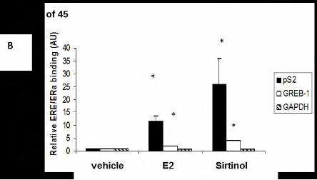
SIRT1 represses ligand-independent ERα activation and DNA binding. Once in the nucleus, 231
ERα binds to cognate sequences on the genome and activates the transcription of estrogen-232
regulated genes. To determine whether SIRT1 regulates ligand-independent binding of ERα to 233
estrogen-regulated promoters, MCF-7 cells were exposed to estrogen or sirtinol, and ERα or 234
RNA polymerase II binding to endogenous estrogen-regulated promoters was assessed via 235
chromatin immunoprecipitation (ChIP) assays. In response to estrogen, ERα association with the 236
estrogen-regulated gene pS2 promoter increased by approximately 15-fold, as expected. Exposure 237
to sirtinol alone also increased ERα binding to the endogenous pS2 promoter, by approximately 238
20-fold (Fig. 5B). To determine the generalizability of this effect, ERα binding to the promoter of 239
another estrogen-responsive gene, GREB-1, was investigated in parallel. Estrogen or sirtinol 240
exposure increased ERα−binding approximately 5-fold and 6–fold, respectively. In response to 241
estrogen or sirtinol exposure, the association of RNA polymerase II with these estrogen-242
responsive promoters increased approximately 9- and 7-fold respectively (Supplemental Fig. S5). 243
ERα binding to the promoter of GAPDH, a non-estrogen regulated gene, was not increased by 244
estrogen or sirtinol. 245
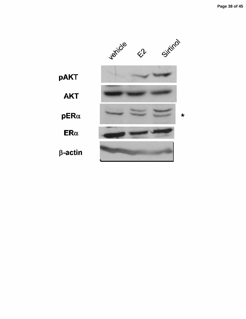
SIRT1 represses ligand-independent ERα activation. ERα is activated by a phosphorylation 246
event at serine118. To determine if this activating phosphorylation of ERα occurs after SIRT1 247
inhibition, MCF-7 cells were exposed to estrogen or sirtinol, and cell lystates were 248
immunoblotted for phospho-ser118-ERα (pERα). In vehicle-treated cells, pERα was undetectable. 249
Page 13 of 45
Upon exposure to estrogen, pERα levels accumulated in the cell, as expected (Fig. 5C). Exposure 250
to sirtinol alone produced a comparable rise in pERα levels. Collectively, these data indicate that 251
SIRT1 represses ligand-independent ERα activation, nuclear localization, and binding to 252
estrogen-regulated promoters. 253
SIRT1 represses estrogen-independent activation of AKT. ERα is phosphorylated at ser118 by 254
the serine/threonine kinase AKT1 in response to estrogen. To determine whether AKT is 255
activated in response to SIRT1 inhibition, lysates of MCF-7 cells exposed to estrogen or sirtinol 256
were blotted for phospho-ser473-AKT1 (pAKT), the activated form of the kinase. pAKT1 was 257
undetectable in the vehicle-treated cells. Upon exposure to estrogen, pAKT1 levels accumulated 258
in the cell, as expected (Fig. 5C). Exposure to sirtinol alone also produced a comparable rise in 259
activated AKT1. 260
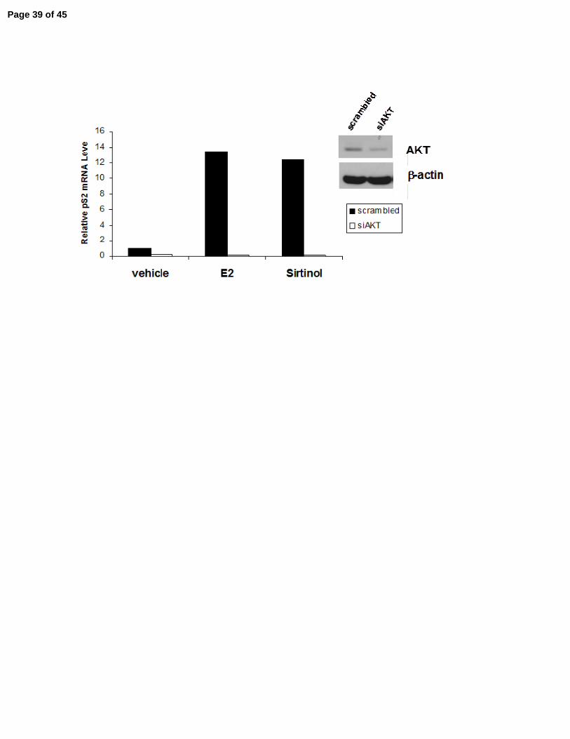
To determine whether SIRT1 repression of estrogen-regulated genes is AKT1-dependent, 261
MCF-7 cells were treated with siRNA directed against AKT1 or a scrambled siRNA, and the 262
treated cells were then exposed to estrogen or sirtinol. In the presence of AKT1, estrogen and 263
sirtinol increased pS2 mRNA expression approximately 12-fold (Fig. 5D). In cells where AKT1 264
was knocked down, neither estrogen nor sirtinol exposure increased pS2 mRNA expression. 265
AKT1 knock-down was confirmed by immunoblotting (Fig. 5D). Activation of estrogen-266
regulated genes by SIRT1 inhibition, like activation by estrogen, is therefore AKT1-dependent. 267
268
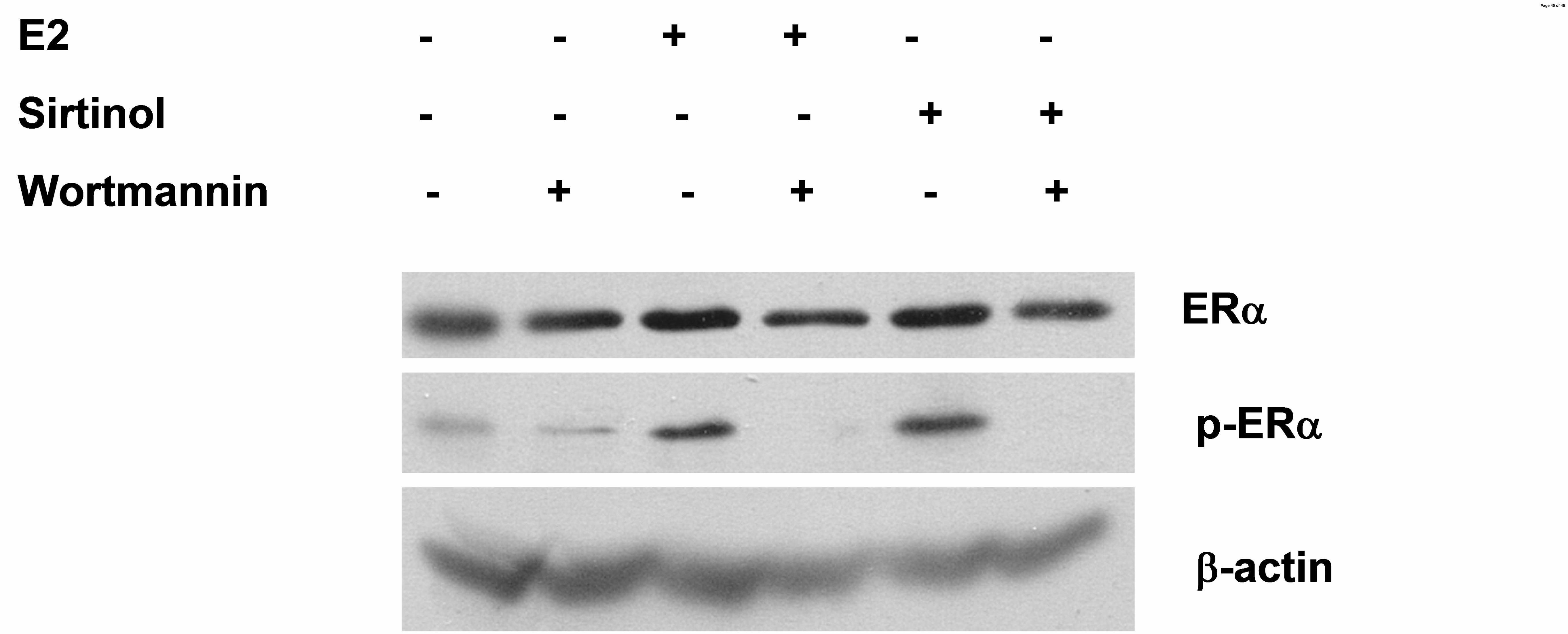
PI3K activity is required for activation of ERα signaling following SIRT1 inhibition or 269
estrogen exposure. Activation of AKT1 occurs through the phosphatidylinositol 3-kinase (PI3K) 270
pathway. To determine whether the ligand-independent activation of ERα following SIRT1 271
repression requires the PI3K activity, as does ligand-dependent activation, MCF-7 cells were 272
exposed to estrogen or sirtinol, in combination with wortmannin, a PI3K pathway inhibitor. In 273
cells exposed to estrogen or sirtinol, pERα accumulated in the cell. When cells were co-treated 274
Page 14 of 45
with wortmannin, pERα was undetectable (Fig. 5E). Together, these findings suggest that SIRT1 275
activity may represses estrogen-independent activation AKT1 by the PI3K pathway. 276
277
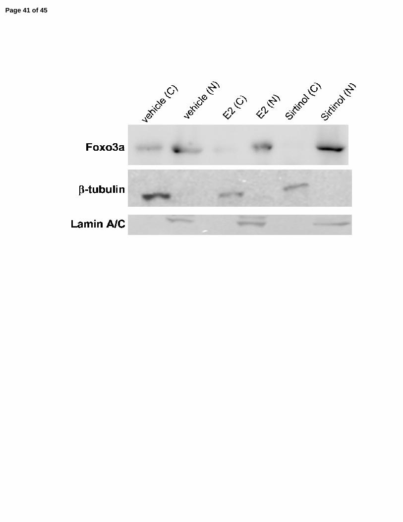
SIRT1 activity is required for FOXO3a expression in the cytoplasm of breast cancer cells. 278
The FOXO family of proteins play a role in ER signaling, as well as the regulation of PI3K 279
activity, and are in turn regulated by SIRT1 (Zou, et al. 2008). In order to investigate the link 280
between deregulation of the FOXO family of proteins by inhibition of SIRT1 activity and ligand-281
independent activation of ERα, AKT, and the PI3K pathway, MCF-7 cells were exposed to 282
estrogen or sirtinol, and cytoplasmic and nuclear fractions were prepared. The purity of the 283
subcellular fractions was determined by immunoblotting with β-tubulin (a cytoplasmic protein) 284
and Lamin A/C (a nuclear protein). In untreated cells, FOXO3a was relatively evenly distributed 285
between the cytoplasm and the nucleus. In response to estrogen or sirtinol exposure, FOXO3a 286
disappeared from the cytoplasm (Fig. 5F), indicating that SIRT1 regulates the cytoplasmic 287
expression of FOXO3a. Nuclear expression FOXO3a remained relatively unchanged. 288
289
SIRT1 represses estrogen-independent breast cancer cell growth. Activation of ERα by 290
estrogen results in cellular proliferation, as well as induction of ERα-responsive genes. To 291
determine if estrogen-independent activation of the ERα via repression of SIRT1 activity was 292
sufficient to induce estrogen-independent proliferation, estrogen-dependent MCF-7 cells were 293
exposed to estrogen or sirtinol for 72 hr and viable cells were enumerated. Compared to cells 294
incubated in charcoal-stripped media, estrogen-treated cells proliferated approximately 2.5-fold 295
over that interval. Cells exposed sirtinol alone showed comparable proliferation. Cells exposed to 296
estrogen plus sirtinol proliferated approximately 5-fold (Fig. 6). Cell viability remained at 96-297
99% throughout the study. 298
Page 15 of 45
To determine whether this ligand-independent cell proliferation in response to SIRT1 299
inhibition is ERα−dependent, cells were exposed to estrogen or sirtinol in combination with 4HT, 300
an estrogen antagonist. 4HT inhibited cell growth induced by either estrogen, or sirtinol, or the 301
combination (Fig. 6). Collectively, these findings indicate that SIRT1 normally functions to 302
repress estrogen-independent, ERα-dependent cell proliferation. 303
304
Page 16 of 45
Discussion 305
306
These studies demonstrate that the type III histone deacetylase SIRT1 serves an important 307
role in regulating estrogen receptor signaling. In MCF-7 cells, an estrogen-dependent breast 308
cancer cell line, we find that SIRT1 represses the basal expression levels of estrogen-regulated 309
genes, as well as their response to estrogen. In the absence of estrogen, SIRT1 repression by 310
small molecule inhibitors or a dominant-negative SIRT1, or siRNA knockdown, induces 311
estrogen-regulated gene activity to a level comparable to the induction seen when the cells are 312
exposed to estrogen. When SIRT1 is inhibited by various independent means in the presence of 313
estrogen, estrogen-regulated gene activity is induced in an additive manner. These results are 314
generalizable, and the effects are specific for SIRT1, as they are recapitulated by the specific 315
genetic techniques of siRNA knockdown of SIRT1 or expression of a dominant-negative SIRT1. 316
Interestingly, and in contrast to its function in androgen signaling (Dai et al. 2007), SIRT1 is not 317
required for estrogen antagonist activity. 318
The repression of estrogen-regulated genes by SIRT1 is dependent upon the ERα as 319
demonstrated by a number of independent approaches. 4HT and faslodex, which serve as ER-320
ligand antagonists, are able to suppress the induction of estrogen-responsive genes produced by 321
inhibition of SIRT1, both in the presence and absence of estrogen. Furthermore, MDA231 cells, 322
which lack ERα, do not show induction of ER-regulated genes upon exposure to SIRT1 inhibitors 323
(or to estrogen). When ERα is ectopically-expressed in MDA231 cells, however, SIRT1 324
inhibition stimulates the induction estrogen-responsive genes. 325
Ligand-independent activation of estrogen-responsive genes by SIRT1 inhibitors shares a 326
number of common elements with ligand (estrogen)-dependent activation signaling. SIRT1 327
inhibition results in ligand-independent phosphorylation and activation of ERα, accumulation of 328
Page 17 of 45
phospho-ERα in the nucleus, and subsequent association of ERα with the promoters of estrogen-329
responsive genes, in a pattern similar to that induced by estrogen. 330
ERα is known to be acetylated at multiple sites in vivo (Wang, et al. 2001). Mutational 331
modification of certain of these acetylation sites may modestly influence the activity of the 332
receptor, raising the possibility that the deacetylase SIRT1 might act on ERα directly, through 333
modulation of ERα acetylation status. Furthermore, the deacetylase activity of SITR1 is required 334
for its effects on ERα signaling, as the dominant-negative SIRT1 mutant we utilized is 335
deacetylase-deficient. We found, however, that the PI3K/AKT pathway is required for ligand-336
independent activation of ERα following SIRT1 inhibition, just as it is for activation by estrogen, 337
indicating that the actions of SIRT1 on ERα are likely indirect, rather than direct. Thus, SIRT1 338
inhibitors subvert the same pathway for ligand-independent activation of the ERα that estrogen 339
utilizes for ligand-dependent activation. 340
Under normal conditions, SIRT1 appears to repress ERα-mediated cell growth when estrogen 341
is absent, blocking estrogen-independent breast cancer cell proliferation. Inhibition of SIRT1 342
activity allows proliferation in the absence of estrogen. This proliferation can be blocked by 343
estrogen antagonists, indicating that this proliferation, like the induction of estrogen-regulated 344
genes, is dependent upon ERα. 345
A link between SIRT1 activity and the repression of estrogen-independent estrogen receptor-346
signaling has not been previously established. However, a recent report indicated that within the 347
nucleus, SIRT1 co-localizes and binds with ERα in several breast and breast cancer cell lines 348
(Elangovan, et al. 2011). One possible model is that SIRT1 may be inhibiting ERα, PI3K or AKT 349
activation directly. However, any direct link between SIRT1 and PI3K regulation, with 350
subsequent downstream effects on AKT-activity, or functionally-related acetylation of p85, p110 351
and AKT has not been identified to date. Furthermore, while ERα-SIRT1 binding requires SIRT1 352
catalytic activity, SIRT1 does not affect the acetylation status of ERα (Elangovan, et al. 2011). 353
Page 18 of 45
We speculate instead that SIRT1 regulation of ERα and PI3K/AKT activity is more likely carried 354
out in an indirect manner, through co-regulatory proteins. 355
One potential co-regulatory protein is phosphatase and tensin homolog (PTEN) (Ikenoue, et 356
al. 2008; Maehama and Dixon 1998; Myers, et al. 1998; Stambolic, et al. 1998). SIRT1 357
deacetylates PTEN at L402, thereby inactivating PTEN and relieving repression of PI3K signaling 358
(Ikenoue et al. 2008). However, our studies show an increase in PI3K signaling (AKT 359
phosphorylation) in response to SIRT1 inhibition, so it is unlikely that PTEN plays a role in 360
SIRT1-mediated repression of estrogen-independent PI3K activity. 361
The FOXO family of proteins play a role in ERα signaling, as well as the regulation of PI3K 362
activity, and are in turn regulated by SIRT1. Much of the activity seen in MCF-7 cells when 363
FOXO family members are inhibited mirrors the findings of this report. For example, the 364
silencing of endogenous FOXO3a increases expression of estrogen-regulated genes and can 365
convert non-tumorigenic, estrogen-dependent breast cancer cells into tumorigenic, estrogen-366
independent cells (Zou et al. 2008). Herein, we report that SIRT1 catalytic activity is required to 367
maintain FOXO3a expression in the cytoplasm. We hypothesize that it is this maintenance of 368
FOXO3a expression and activity in the cytoplasm that prevents FOXO3a from becoming 369
disassociated with ERα in the absence of ligand, thereby preventing the ligand-independent 370
activation of ERα, transcription of estrogen-regulated genes and breast cancer cell proliferation 371
(Fig. 7). We are currently further investigating a link between deregulation of the FOXO family 372
of proteins by inhibition of SIRT1 activity and ligand-independent activation of ERα, AKT, and 373
the PI3K pathway. 374
The findings presented in this report differ from those of several previous reports. Other 375
studies have found that sirtuin inhibition results in cell death and p53 acetylation (Peck, et al. 376
2010), or cell senescence (Ota et al. 2006), or that it represses expression levels of ERα (Yao, et 377
al. 2010). Elangovan et al. suggests that SIRT1 is required for oncogenic signaling in breast 378
Page 19 of 45
cancer cells. Furthermore, Yao, et al. indicated that SIRT1 inhibition decreases levels of ERα 379
protein expression. Those studies differ from this report in one major way, in that the previously 380
mentioned studies did not starve the breast cancer cells of steroid hormones prior to SIRT1 381
inhibition. Rather, SIRT1 was inhibited while the breast cancer cells were growing 382
logarithmically in the presence of estrogen with ERα engaged by estrogen ligand. In the present 383
study, however, cells were incubated in charcoal-treated media, thereby depriving the cells of any 384
steroidal hormone signaling, disengaging the Ras-MAPK pathway (Ota et al. 2006), halting cell 385
growth without effects on cell viability (as shown in Fig. 6), and allowing study of the effects of 386
SIRT1 depletion on quiescent cells with unengaged ERα. The Ras-MAPK pathway (Ota et al. 387
2006), as well as p53 (Peck et al. 2010) may contribute to differential regulation of estrogen 388
signaling by SIRT1 in the presence or absence of hormonal signaling. Another methodological 389
difference between our approach and some of the papers cited above is our use of reagents 390
highly-specific for SIRT1 (shRNA and dominant-negative proteins) to confirm the role of SIRT1 391
rather than relying only on chemical inhibitors, which target SIRT2 as well as having other off-392
target effects. (Peck et al. 2010) showed that the effects they observed of SIRT inhibitors on p53 393
and cell death required inhibition of both SIRT1 and SIRT2. Lastly, as shown in Fig. 5A, C and E, 394
we did not detect any significant change in ERα protein expression in response to the 395
experimental conditions presented herein. It is important to note that we used 2-3 fold less of the 396
chemical SIRT1 inhibitors in these studies than those used in Yao, et al.. It is possible that the 397
differences in ERα expression in response to treatment may be the result of cell toxicity or 398
unintended off-target effects of using inhibitors higher concentrations. Cell viability remained 399
between 96-99% as measured by trypan blue exclusion assay throughout the experiments 400
presented herein. Significantly, ERα expression levels did not change in same cells when SIRT1 401
was depleted by shRNA, again indicating that any reduction of ERα expression levels may have 402
been an unintended side effect due to non-specific actions of chemical inhibitors at higher 403
Page 20 of 45
concentrations. The data presented here therefore highlight a potentially significant difference 404
between the regulation of estrogen receptor signaling in the presence of estrogen compared with 405
ligand-independent signaling in the absence of estrogen. The results presented here, therefore, 406
describe an important mechanism by which breast cancer cells might transition from an estrogen-407
dependent to an estrogen-refractory state, particularly in the setting of estrogen depletion or 408
estrogen antagonists. 409
The findings presented in this report support the concept that SIRT1 serves as a tumor 410
suppressor gene in breast cancer cells (Jin et al. 2007; Moore 2011). These findings also 411
highlight the potential role of SIRT1 in regulating breast cancer cell dependency upon estrogen. 412
When coupled with the findings that SIRT1 increases the expression of drug-resistance genes 413
(Chu, et al. 2005), SIRT1 may have the potential to be an important biomarker for breast cancer 414
prognosis or future patient-specific tailored chemotherapeutic strategies (Bhat-Nakshatri, et al. 415
2008; Chu et al. 2005). Similarly, modulation of SIRT1 activity may prove useful for next-416
generation breast cancer therapeutics. 417
In view of the major regulatory effects of SIRT1 on ERα signaling in breast cancer, further 418
study of the transcriptional and post-translational regulation of SIRT1 in breast cancer cells will 419
likely prove relevant to our understanding of the genesis of breast cancer, and its evolution to a 420
hormone-independent state. 421
422
423
ACKNOWLEDGEMENTS 424
425
This work was supported by a grant from the National Cancer Institute, R01-CA101992 (DVF) 426
and CA101992S1 (RM), and by the Karin Grunebaum Cancer Research Foundation (DVF). We 427
Page 21 of 45

thank Dr. Yan Dai for reagents and advice, and Ms. Lora Forman for technical advice (all at 428
Boston University). 429
Page 22 of 45
REFERENCES
Alvala M, Bhatnagar S, Ravi A, Jeankumar VU, Manjashetty TH, Yogeeswari P &
Sriram D 2012 Novel acridinedione derivatives: Design, synthesis, SIRT1 enzyme and
tumor cell growth inhibition studies. Bioorganic & Medicinal Chemistry Letters 22 3256-
3260.
Aoyagi S & Archer TK 2008 Nicotinamide uncouples hormone-dependent chromatin
remodeling from transcription complex assembly. Molecular and Cellular Biology 28 30-
39.
Bhat-Nakshatri P, Wang GH, Appaiah H, Luktuke N, Carroll JS, Geistlinger TR, Brown
M, Badve S, Liu YL & Nakshatri H 2008 AKT Alters Genome-Wide Estrogen Receptor
alpha Binding and Impacts Estrogen Signaling in Breast Cancer. Molecular and Cellular
Biology 28 7487-7503.
Blander G & Guarente L 2004 The Sir2 family of protein deacetylases. Annual Review of
Biochemistry 73 417-435.
Borra MT, Smith BC & Denu JM 2005 Mechanism of human SIRT1 activation by
resveratrol. Journal of Biological Chemistry 280 17187-17195.
Byles V, Chmilewski LK, Wang J, Zhu L, Forman LW, Faller DV & Dai Y 2010
Aberrant cytoplasm localization and protein stability of SIRT1 is regulated by PI3K/IGF-
1R signaling in human cancer cells. Int J Biol Sci 6 599-612.
Carroll JS, Meyer CA, Song J, Li W, Geistlinger TR, Eeckhoute J, Brodsky AS, Keeton
EK, Fertuck KC, Hall GF, et al. 2006 Genome-wide analysis of estrogen receptor binding
sites. Nature Genetics 38 1289-1297.
Chu F, Chou PM, Zheng X, Mirkin BL & Rebbaa A 2005 Control of multidrug resistance
gene mdr1 and cancer resistance to chemotherapy by the longevity gene sirt1. Cancer
Research 65 10183-10187.
Dai Y, Ngo D, Forman LW, Qin DC, Jacob J & Faller DV 2007 Sirtuin 1 is required for
antagonist-induced transcriptional repression of androgen-responsive genes by the
androgen receptor. Molecular Endocrinology 21 1807-1821.
Dai Y, Ngo D, Jacob J, Forman LW & Faller DV 2008 Prohibitin and the SWI/SNF
ATPase subunit BRG1 are required for effective androgen antagonist-mediated
transcriptional repression of androgen receptor-regulated genes. Carcinogenesis 29 1725-
1733.
Elangovan S, Ramachandran S, Venkatesan N, Ananth S, Gnana-Prakasam JP, Martin
PM, Browning DD, Schoenlein PV, Prasad PD, Ganapathy V, et al. 2011 SIRT1 Is
Essential for Oncogenic Signaling by Estrogen/Estrogen Receptor alpha in Breast Cancer.
Cancer Research 71 6654-6664.
Ford CHJ, Al-Bader M, Al-Ayadhi B & Francis I 2011 Reassessment of Estrogen
Receptor Expression in Human Breast Cancer Cell Lines. Anticancer Research 31.
Fu MF, Liu MR, Sauve AA, Jiao XM, Zhang XP, Wu XF, Powell MJ, Yang TL, Gu W,
Avantaggiati ML, et al. 2006 Hormonal control of androgen receptor function through
SIRT1. Molecular and Cellular Biology 26 8122-8135.
Page 23 of 45
Fulco M, Schiltz RL, Iezzi S, King MT, Zhao P, Kashiwaya Y, Hoffman E, Veech RL &
Sartorelli V 2003 Sir2 regulates skeletal muscle differentiation as a potential sensor of the
redox state. Molecular Cell 12 51-62.
Haigis MC & Guarente LP 2006 Mammalian sirtuins - emerging roles in physiology,
aging, and calorie restriction. Genes & Development 20 2913-2921.
Hall JM & McDonnell DP 1999 The estrogen receptor beta-isoform (ER beta) of the
human estrogen receptor modulates ER alpha transcriptional activity and is a key
regulator of the cellular response to estrogens and antiestrogens. Endocrinology 140
5566-5578.
Ikenoue T, Inoki K, Zhao B & Guan KL 2008 PTEN acetylation modulates its interaction
with PDZ domain. Cancer Research 68 6908-6912.
Jin QH, Yan TT, Ge XJ, Sun C, Shi XG & Zhai QW 2007 Cytoplasm-localized SIRT1
enhances apoptosis. Journal of Cellular Physiology 213 88-97.
Landry J, Slama JT & Sternglanz R 2000 Role of NAD(+) in the deacetylase activity of
the SIR2-like proteins. Biochemical and Biophysical Research Communications 278 685-
690.
Maehama T & Dixon JE 1998 The tumor suppressor, PTEN/MMAC1, dephosphorylates
the lipid second messenger, phosphatidylinositol 3,4,5-trisphosphate. Journal of
Biological Chemistry 273 13375-13378.
Moore RL 2011 Sirtuin (SIRT) 1 and Steriod Hormone Activity in Cancer. Ed Y Dai:
Journal of Endocrinology.
Motta MC, Divecha N, Lemieux M, Kamel C, Chen D, Gu W, Bultsma Y, McBurney M
& Guarente L 2004 Mammalian SIRT1 represses forkhead transcription factors. Cell 116
551-563.
Myers MP, Pass I, Batty IH, Van der Kaay J, Stolarov JP, Hemmings BA, Wigler MH,
Downes CP & Tonks NK 1998 The lipid phosphatase activity of PTEN is critical for its
tumor suppressor function. Proceedings of the National Academy of Sciences of the
United States of America 95 13513-13518.
Neugebauer RC, Uchiechowska U, Meier R, Hruby H, Valkov V, Verdin E, Sippl W &
Jung M 2008 Structure-activity studies on splitomicin derivatives as sirtuin inhibitors and
computational prediction of binding mode. Journal of Medicinal Chemistry 51 1203-
1213.
Ota H, Tokunaga E, Chang K, Hikasa M, Iijima K, Eto M, Kozaki K, Akishita M, Ouchi
Y & Kaneki M 2006 Sirt1 inhibitor, Sirtinol, induces senescence-like growth arrest with
attenuated Ras-MAPK signaling in human cancer cells. Oncogene 25 176-185.
Peck B, Chen C-Y, Ho K-K, Di Fruscia P, Myatt SS, Coombes RC, Fuchter MJ, Hsiao C-
D & Lam EWF 2010 SIRT Inhibitors Induce Cell Death and p53 Acetylation through
Targeting Both SIRT1 and SIRT2. Molecular Cancer Therapeutics 9 844-855.
Picard F, Kurtev M, Chung NJ, Topark-Ngarm A, Senawong T, de Oliveira RM, Leid M,
McBurney MW & Guarente L 2004 Sirt1 promotes fat mobilization in white adipocytes
by repressing PPAR-gamma. Nature 429 771-776.
Pruitt K, Zinn RL, Ohm JE, McGarvey KM, Kang SHL, Watkins DN, Herman JG &
Baylin SB 2006 Inhibition of SIRT1 reactivates silenced cancer genes without loss of
promoter DNA hypermethylation. Plos Genetics 2 344-352.
Puigserver P, Rodgers J, Lerin C, Haas W, Gygi S & Spiegelman B 2005 Nutrient control
of glucose metabolism through PGC-1?/SIRT1 complex. Gerontologist 45 56-56.
Page 24 of 45
Rodgers JT, Lerin C, Haas W, Gygi SP, Spiegelman BM & Puigserver P 2005 Nutrient
control of glucose homeostasis through a complex of PGC-1 alpha and SIRT1. Nature
434 113-118.
Stambolic V, Suzuki A, de la Pompa JL, Brothers GM, Mirtsos C, Sasaki T, Ruland J,
Penninger JM, Siderovski DP & Mak TW 1998 Negative regulation of PKB/Akt-
dependent cell survival by the tumor suppressor PTEN. Cell 95 29-39.
Wang CG, Chen LH, Hou XH, Li ZY, Kabra N, Ma YH, Nemoto S, Finkel T, Gu W,
Cress WD, et al. 2006 Interactions between E2F1 and SirT1 regulate apoptotic response
to DNA damage. Nature Cell Biology 8 1025-U1109.
Wang CG, Fu MF, Angeletti RH, Siconolfi-Baez L, Reutens AT, Albanese C, Lisanti MP,
Katzenellenbogen BS, Kato S, Hopp T, et al. 2001 Direct acetylation of the estrogen
receptor alpha hinge region by p300 regulates transactivation and hormone sensitivity.
Journal of Biological Chemistry 276 18375-18383.
Welsh AW, Lannin DR, Young GS, Sherman ME, Figueroa JD, Henry NL, Ryden L,
Kim C, Love RR, Schiff R, et al. 2012 Cytoplasmic Estrogen Receptor in Breast Cancer.
Clinical Cancer Research 18 118-126.
Yamamoto H, Schoonjans K & Auwerx J 2007 Sirtuin functions in health and disease.
Molecular Endocrinology 21 1745-1755.
Yao Y, Li HZ, Gu YS, Davidson NE & Zhou Q 2010 Inhibition of SIRT1 deacetylase
suppresses estrogen receptor signaling. Carcinogenesis 31 382-387.
Zou YY, Tsai WB, Cheng CJ, Hsu C, Chung YM, Li PC, Lin SH & Hu MCT 2008
Forkhead box transcription factor FOXO3a suppresses estrogen-dependent breast cancer
cell proliferation and tumorigenesis. Breast Cancer Research 10.
Page 25 of 45
FIGURE LEGENDS
Fig. 1. SIRT1 inhibition stimulates estrogen-responsive gene activity. MCF-7 cells were plated to
60% confluence in a 10 centimeter dish (6 x 10⁵ cells per dish) in charcoal-treated (C/T) media,
incubated for 24 hr, and treated as follows: (A) transfection with an ERE-luciferase reporter
plasmid and RSV-β-gal, then exposed to either vehicle, or E2 (100 nM), or sirtinol (50 µM) or
both, and assayed for reporter gene activity via luciferase assay. Results are normalized to β-
galactosidase activity as a transfection control. In panels B, C and D, cells were exposed to either
vehicle, or E2 (100 nM), or sirtinol (50 µM) or splitomicin (200 µM) or combinations thereof,
and mRNA levels of the endogenous estrogen-regulated genes pS2 (B and C) and GREB-1 (D)
were assayed via quantitative RT-PCR and normalized to β-actin transcript levels. The data
presented is the average (+/- standard error) of 4 independent experiments. * indicates p<0.002
(A and B) and <0.01 (C and D), compared to vehicle controls. (AU: arbitrary units)
Fig. 2. Repression of estrogen-regulated gene activity is SIRT1-specific. MCF-7 cells were plated
to 60% confluence in a 10 centimeter dish (6 x 10⁵ cells per dish) in C/T media, incubated for 24
hr, then transfected with ERE-luciferase reporter plasmid plus either empty vector or H343Y D/N
SIRT and RSV-β-gal (A) or with 500 ng of scrambled siRNA or SIRT1-siRNA (B) and incubated
for 18 hr, then incubated for a further 24 hr (A) or 72 hr (B) in fresh C/T media. The cells were
then exposed to vehicle or E2 (100 nM), as indicated, for 24 hr. Estrogen-regulated gene activity
was determined via luciferase assay normalized to β-galactosidase activity (A), or mRNA
expression, as determined via quantitative RT-PCR and normalized to β-actin transcript levels
(B). The efficiency of SIRT1 knockdown was determined by analyzing protein expression via
immunoblot (B, inset). The data presented is the average (+/- standard error) of 3 independent
experiments. * indicates p<0.005.
Page 26 of 45
Fig. 3. SIRT1 is not required for estrogen-antagonist function. MCF-7 (A) or T47D (B) cells were
plated to 60% confluence in a 10 centimeter dish (6 x 10⁵ cells per dish) in C/T media and
incubated for 24 hr, then exposed to vehicle, E2 (100 nM) and/or sirtinol (50 µM), 4HT (1µM)
(A) and/or faslodex (100nM) (B), as indicated, incubated an additional 24 hr, and mRNA was
harvested. pS2 mRNA expression was determined via quantitative RT-PCR and normalized to β-
actin transcript levels. ERα protein expression was determined via immunoblot (B, inset). The
data presented is the average (+/- standard error) of 6 independent experiments (A) and 3
independent experiments (B). The increases in mRNA levels induced by the treatments were
significant compared to controls [p<0.001 (A) and <0.01 (B)]. Inhibition of mRNA levels by the
estrogen antagonists was significant [* p<0.001 (A) and <0.01 (B)]
Fig. 4. SIRT1 repression of estrogen-regulated gene activity is ERα-dependent. MDA231 (A) or
MCF-7 (B) cells were plated to 60% confluence in a 10 centimeter dish (6 x 10⁵ cells per dish) in
C/T media, incubated for 24 hr, then transfected with an ERE-luciferase reporter plasmid plus
either empty vector or an ERα-expression vector (A) or 500 ng of scrambled siRNA or
ERα−specific siRNA (B) and incubated for 18 hr, then incubated for 24 hr (A) or 72 hr (B) in
fresh C/T media. The cells were then exposed to either vehicle, or E2 (100 nM), or sirtinol (50
µM), or both, as indicated, for 24 hr. Cells were then harvested and estrogen-regulated gene
activity was determined via luciferase assay normalized to β-galactosidase activity (A), or mRNA
expression via RT-PCR normalized to β-actin transcript levels (B). In panel A, ectopic ERα
expression was verified by immunoblot (inset). In panel B, the efficiency of ERα knockdown was
determined by analyzing ERα protein expression via immunoblot (inset). For panel A, the data
presented is the average (+/- standard error) of 3 independent experiments. * indicates p<0.001.
For panel B, the experiment was repeated five times with comparable relative ERα knockdown.
Page 27 of 45
In each experiment, pS2 mRNA expression levels increased in response to E2 or sirtinol
treatment or the combination, and were significantly reduced in cells in which ERα had been
knocked-down. A representative experiment is presented (B). * p<0.01 compared to paired black
bar from scrambled siRNA-transfected cells.
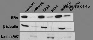
Fig. 5. SIRT1 represses estrogen-independent ERα nuclear accumulation, promoter binding, and
activation, in an AKT-dependent manner. (A) MCF-7 cells were plated to 60% confluence in a 10
centimeter dish (6 x 10⁵ cells per dish) in C/T media and incubated for 24 hr, then exposed to
vehicle, E2 or sirtinol as indicated and incubated for an additional 24 hr. The cells were then
harvested and Levels of ERα protein were analyzed by immunoblotting. β-tubulin and lamin A/C
were used as markers to determine the purity of the cytoplasmic and nuclear extracts, respectively.
The figure presented is representative of 3 independent experiments. B) MCF-7 cells were plated
to 40% confluence in a 10 centimeter dish (4 x 10⁵ cells per dish) in C/T media, incubated for 48
hr, then exposed to vehicle, E2 or sirtinol for 24 hr, harvested and ChIP analysis was performed.
Immunoprecipitation was carried out using α-ERα or –Pol II antibodies (see Supplemental Fig.
3). The promoter regions of the pS2 and GREB-1 genes were amplified via quantitative PCR. The
GAPDH promoter regions was also amplified, as a specificity control. The data is the average
(+/- standard error) of 3 independent experiments. Immunoprecipitations carried out using non-
specific antibodies or pooled antisera did not yield products up to 45 cycles of amplification (not
shown). * p<0.01 compared to corresponding vehicle controls. C) MCF-7 cells were plated to
60% confluence in a 10 centimeter dish (6 x 10⁵ cells per dish) in C/T media and incubated for
24 hr, then treated with vehicle, E2 or sirtinol as indicated, and incubated an additional 24 hr,
then harvested. Protein expression levels of pERα, ERα, pAKT, AKT and β-actin were analyzed
via immunoblotting. A blot representative of 3 independent experiments is shown. (* indicates a
non-specific band seen even in cell lacking ERα.) D) MCF-7 cells were plated at 60% confluence
Page 28 of 45
in a 10 centimeter dish (6 x 10⁵ cells per dish) in C/T media, incubated for 24 hr, then
transfected with 500 ng of scrambled siRNA or AKT1-specific siRNA and incubated for 18 hr,
then incubated for 72 hr in fresh C/T media. The cells were then exposed to vehicle, E2 or sirtinol
for 24 hr, harvested and mRNA was collected. pS2 mRNA expression levels were determined via
quantitative RT-PCR and normalized to β-actin transcript levels. The efficacy of AKT
knockdown was determined by immunoblot (insert). The experiment was repeated 4 times with
similar relative AKT knockdown and consistent effects on pS2 mRNA induction. Shown is a
representative experiment. E) MCF-7 cells were plated to 60% confluence in a 10 centimeter dish
(6 x 10⁵ cells per dish) in C/T media, incubated for 24 hr, then exposed to vehicle, E2, or sirtinol,
and/or wortmannin (100 nM) (a PI3K inhibitor), as indicated, and incubated an additional 12 hr.
The cells were then harvested and protein levels of pERα, ERα and β-actin were analyzed via
immunoblotting. The experiment was repeated three times with comparable results. A
representative blot is shown. F) MCF-7 cells were plated to 60% confluence in a 10 centimeter
dish (6 x 10⁵ cells per dish) in C/T media and incubated for 24 hr, then exposed to vehicle, E2 or
sirtinol as indicated and incubated for an additional 24 hr. The cells were then harvested and
levels of FOXO3a protein were analyzed by immunoblotting. β-tubulin and lamin A/C were used
as markers to determine the purity of the cytoplasmic and nuclear extracts, respectively. The
figure presented is representative of 3 independent experiments.
Fig. 6. SIRT1 inhibition induces estrogen-independent, ERα-dependent breast cancer cell
proliferation. 2x10⁵ MCF-7 cells were incubated in phenol red-free medium containing C/T
serum for 24 hr, then exposed to either vehicle, estrogen (100 nM), or sirtinol (50 µM), and/or
4HT (1 µM), as indicated, for 72 hr. Viable cells were then enumerated. The data presented is the
average (+/- standard error) of 4 independent experiments. Increases in cell number in response to
Page 29 of 45
estrogen and/or sirtinol are significant (p<0.01). * p<0.03 compared to paired open bars (vehicle-
treated cells).
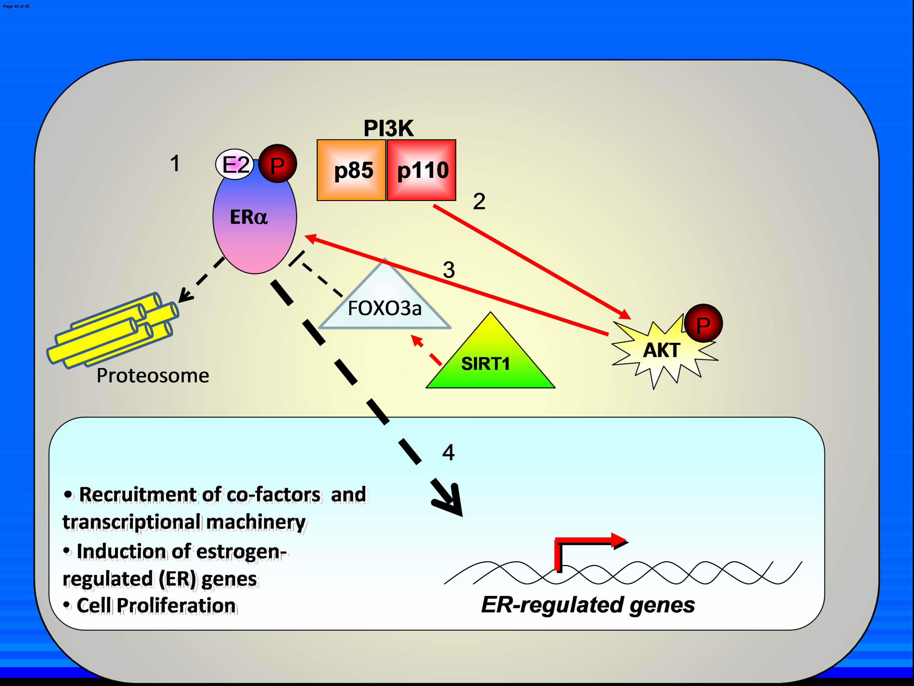
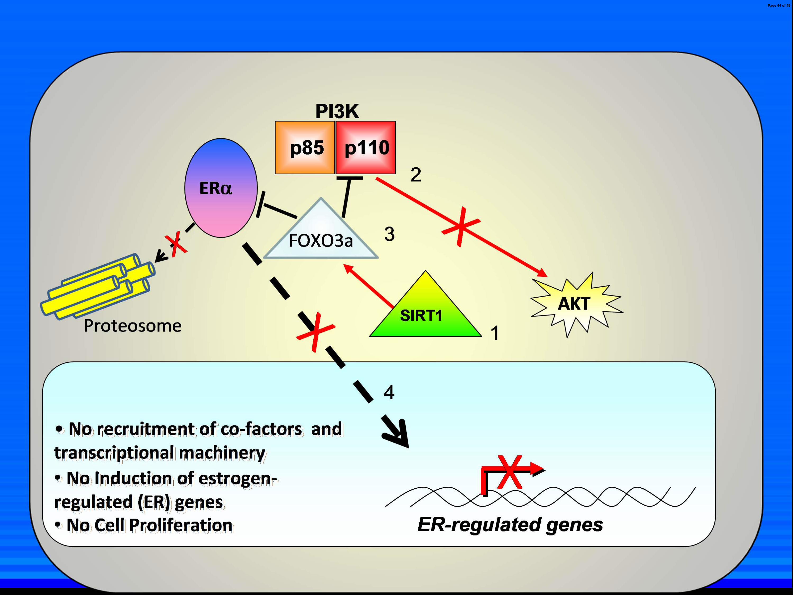
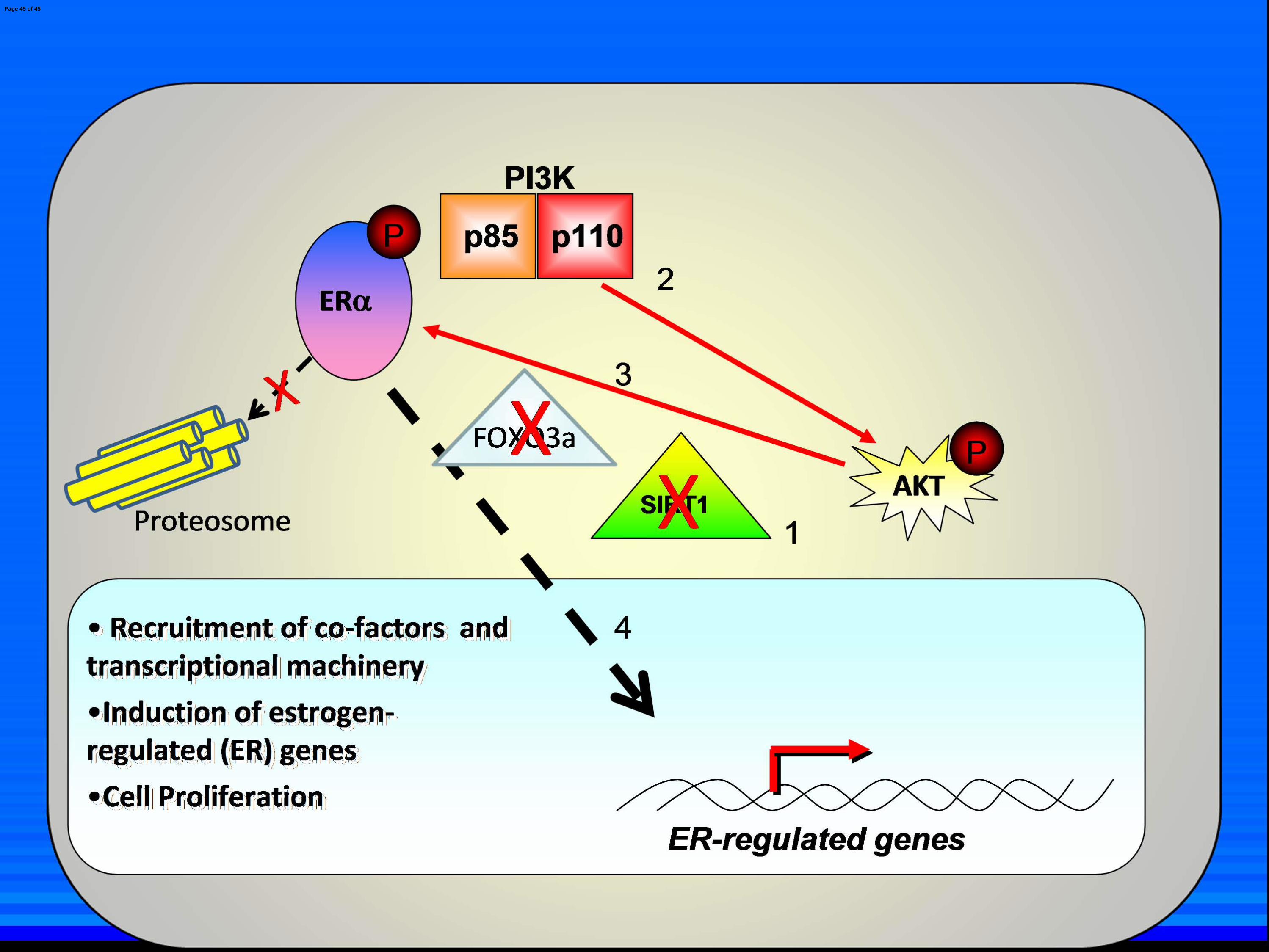
Fig. 7. Model of SIRT1 repression of ligand-independent ERαααα activation. A) Normal
estrogen signaling. Estrogen (E2) enters the cell and binds to ERα in the cytoplasm (1). This
activates PI3K which phosphorylates AKT [long red arrow] (2). This in turn phosphorylates ERα
(3) which then translocates to the nucleus [large black arrow] (4), where it recruits cofactors and
other transcriptional machinery, induces ER-regulated genes and leads to cancer cell proliferation.
SIRT1 and FOXO3a act as tonic inhibitors of ERα and PI3K in the presence of E2, as is
demonstrated by the fact that SIRT1 inhibition in addition to E2 treatment yields additive
induction of ER-regulated genes [dashed red arrow and black line]. B) SIRT1 inhibition in the
absence of E2. SIRT1 stabilizes FOXO3a in the cytoplasm through SIRT1 deacetylase activity
[small red arrow] (1) which in turn represses ligand-independent ERα, PI3K and AKT activation
[solid black line] (2 and 3). The ERα does not translocate to the nucleus (4) and is not degraded
in the cytoplasm via the proteosome. ER-regulated genes are not induced nor does the cell
proliferate. C) Effect of SIRT1 inhibition on ligand-independent ERα activation. SIRT1
inhibition (1) destabilizes FOXO3a cytoplasmic localization, which relieves ligand-independent
repression of PI3K and ERα (2). This leads to PI3K-mediated activation of AKT which in turn
phosphorylates and activates ERa (3). ERα translocates to the nucleus where it recruits co-factors
and transcriptional machinery and initiates transcription of ER-regulated genes, leading to cancer
cell proliferation (4). Due to a lack of E2 ligation, ERα is not degraded via the proteosome.
Page 30 of 45
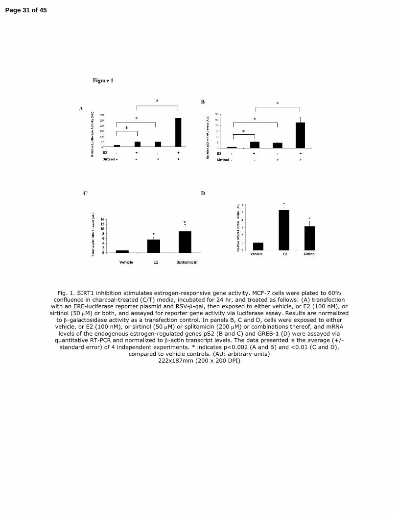
Fig. 1. SIRT1 inhibition stimulates estrogen-responsive gene activity. MCF-7 cells were plated to 60% confluence in charcoal-treated (C/T) media, incubated for 24 hr, and treated as follows: (A) transfection with an ERE-luciferase reporter plasmid and RSV-β-gal, then exposed to either vehicle, or E2 (100 nM), or
sirtinol (50 µM) or both, and assayed for reporter gene activity via luciferase assay. Results are normalized
to β-galactosidase activity as a transfection control. In panels B, C and D, cells were exposed to either vehicle, or E2 (100 nM), or sirtinol (50 µM) or splitomicin (200 µM) or combinations thereof, and mRNA
levels of the endogenous estrogen-regulated genes pS2 (B and C) and GREB-1 (D) were assayed via quantitative RT-PCR and normalized to β-actin transcript levels. The data presented is the average (+/-
standard error) of 4 independent experiments. * indicates p<0.002 (A and B) and <0.01 (C and D), compared to vehicle controls. (AU: arbitrary units)
222x187mm (200 x 200 DPI)
Page 31 of 45
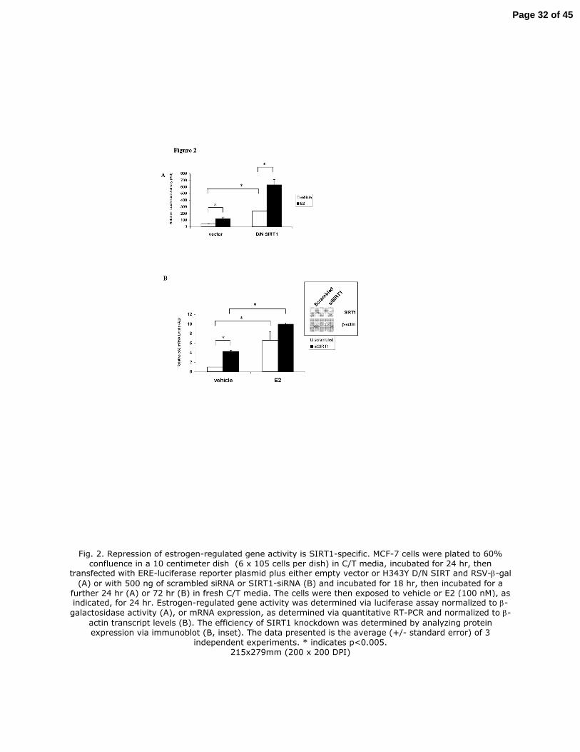
Fig. 2. Repression of estrogen-regulated gene activity is SIRT1-specific. MCF-7 cells were plated to 60% confluence in a 10 centimeter dish (6 x 10⁵ cells per dish) in C/T media, incubated for 24 hr, then
transfected with ERE-luciferase reporter plasmid plus either empty vector or H343Y D/N SIRT and RSV-β-gal
(A) or with 500 ng of scrambled siRNA or SIRT1-siRNA (B) and incubated for 18 hr, then incubated for a further 24 hr (A) or 72 hr (B) in fresh C/T media. The cells were then exposed to vehicle or E2 (100 nM), as indicated, for 24 hr. Estrogen-regulated gene activity was determined via luciferase assay normalized to β-
galactosidase activity (A), or mRNA expression, as determined via quantitative RT-PCR and normalized to β-
actin transcript levels (B). The efficiency of SIRT1 knockdown was determined by analyzing protein expression via immunoblot (B, inset). The data presented is the average (+/- standard error) of 3
independent experiments. * indicates p<0.005. 215x279mm (200 x 200 DPI)
Page 32 of 45
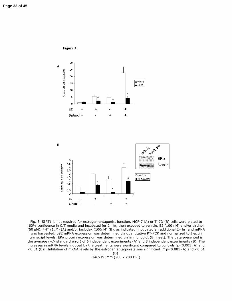
Fig. 3. SIRT1 is not required for estrogen-antagonist function. MCF-7 (A) or T47D (B) cells were plated to 60% confluence in C/T media and incubated for 24 hr, then exposed to vehicle, E2 (100 nM) and/or sirtinol (50 µM), 4HT (1µM) (A) and/or faslodex (100nM) (B), as indicated, incubated an additional 24 hr, and mRNA
was harvested. pS2 mRNA expression was determined via quantitative RT-PCR and normalized to β-actin
transcript levels. ERα protein expression was determined via immunoblot (B, inset). The data presented is
the average (+/- standard error) of 6 independent experiments (A) and 3 independent experiments (B). The increases in mRNA levels induced by the treatments were significant compared to controls [p<0.001 (A) and <0.01 (B)]. Inhibition of mRNA levels by the estrogen antagonists was significant [* p<0.001 (A) and <0.01
(B)] 146x193mm (200 x 200 DPI)
Page 33 of 45
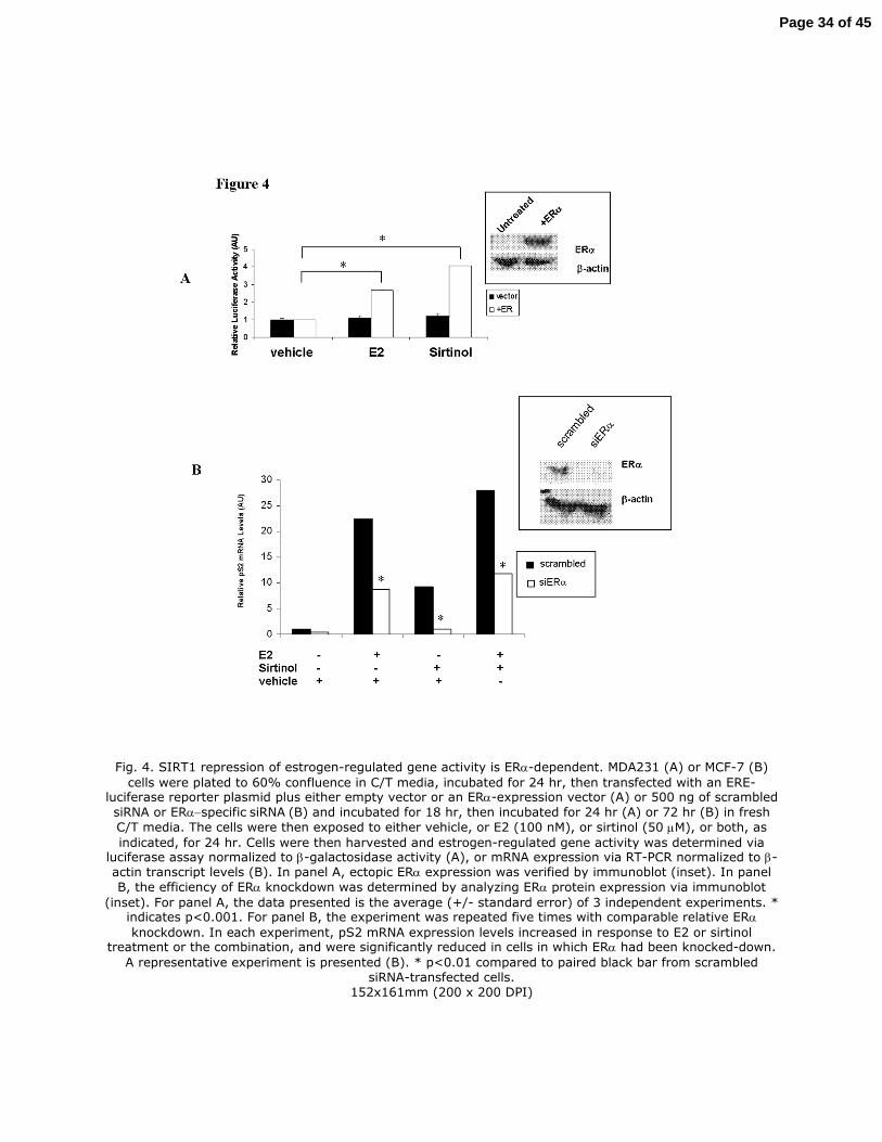
Fig. 4. SIRT1 repression of estrogen-regulated gene activity is ERα-dependent. MDA231 (A) or MCF-7 (B)
cells were plated to 60% confluence in C/T media, incubated for 24 hr, then transfected with an ERE-luciferase reporter plasmid plus either empty vector or an ERα-expression vector (A) or 500 ng of scrambled
siRNA or ERα−specific siRNA (B) and incubated for 18 hr, then incubated for 24 hr (A) or 72 hr (B) in fresh
C/T media. The cells were then exposed to either vehicle, or E2 (100 nM), or sirtinol (50 µM), or both, as
indicated, for 24 hr. Cells were then harvested and estrogen-regulated gene activity was determined via luciferase assay normalized to β-galactosidase activity (A), or mRNA expression via RT-PCR normalized to β-actin transcript levels (B). In panel A, ectopic ERα expression was verified by immunoblot (inset). In panel
B, the efficiency of ERα knockdown was determined by analyzing ERα protein expression via immunoblot
(inset). For panel A, the data presented is the average (+/- standard error) of 3 independent experiments. * indicates p<0.001. For panel B, the experiment was repeated five times with comparable relative ERα
knockdown. In each experiment, pS2 mRNA expression levels increased in response to E2 or sirtinol treatment or the combination, and were significantly reduced in cells in which ERα had been knocked-down.
A representative experiment is presented (B). * p<0.01 compared to paired black bar from scrambled siRNA-transfected cells.
152x161mm (200 x 200 DPI)
Page 34 of 45

Page 35 of 45
Page 36 of 45
Page 37 of 45
Page 38 of 45
Page 39 of 45
Page 40 of 45
Page 41 of 45
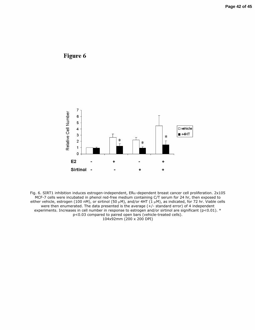
Fig. 6. SIRT1 inhibition induces estrogen-independent, ERα-dependent breast cancer cell proliferation. 2x10⁵
MCF-7 cells were incubated in phenol red-free medium containing C/T serum for 24 hr, then exposed to either vehicle, estrogen (100 nM), or sirtinol (50 µM), and/or 4HT (1 µM), as indicated, for 72 hr. Viable cells
were then enumerated. The data presented is the average (+/- standard error) of 4 independent experiments. Increases in cell number in response to estrogen and/or sirtinol are significant (p<0.01). *
p<0.03 compared to paired open bars (vehicle-treated cells). 104x92mm (200 x 200 DPI)
Page 42 of 45
Page 43 of 45
Page 44 of 45
Page 45 of 45