REVIEW Open Access Alpha-synuclein and tau: teammates in ...
14
REVIEW Open Access Alpha-synuclein and tau: teammates in neurodegeneration? Simon Moussaud 1 , Daryl R Jones 1 , Elisabeth L Moussaud-Lamodière 1 , Marion Delenclos 1 , Owen A Ross 1,2 and Pamela J McLean 1,2* Abstract The accumulation of α-synuclein aggregates is the hallmark of Parkinson’s disease, and more generally of synucleinopathies. The accumulation of tau aggregates however is classically found in the brains of patients with dementia, and this type of neuropathological feature specifically defines the tauopathies. Nevertheless, in numerous cases α-synuclein positive inclusions are also described in tauopathies and vice versa, suggesting a co-existence or crosstalk of these proteinopathies. Interestingly, α-synuclein and tau share striking common characteristics suggesting that they may work in concord. Tau and α-synuclein are both partially unfolded proteins that can form toxic oligomers and abnormal intracellular aggregates under pathological conditions. Furthermore, mutations in either are responsible for severe dominant familial neurodegeneration. Moreover, tau and α-synuclein appear to promote the fibrillization and solubility of each other in vitro and in vivo. This suggests that interactions between tau and α-synuclein form a deleterious feed-forward loop essential for the development and spreading of neurodegeneration. Here, we review the recent literature with respect to elucidating the possible links between α-synuclein and tau. Keywords: Tau, MAPT, Synuclein, SNCA, Oligomers, Tangles, Synucleinopathy, Tauopathy, Parkinson’s disease, Alzheimer’s disease Introduction Age-related neurodegenerative disorders like Alzheimer’ s disease (AD) and Parkinson’ s disease (PD) take an over- whelming toll on individuals and society [1]. AD and PD are the two most frequent neurodegenerative diseases (www.who.org). To date, PD and AD remain incurable and only very limited palliative treatment options exist [2]. The etiology of PD and AD is not fully understood, but appears to involve a complex combination of envir- onmental and genetic factors [3]. Interestingly, at the molecular level, protein misfolding, accumulation, aggregation and subsequently the forma- tion of amyloid deposits are common features in many neurological disorders including AD and PD. Thus neuro- degenerative diseases are sometimes referred to as protei- nopathies [4]. The existence of a common mechanism suggests that neurodegenerative disorders likely share a common trigger and that the nature of the pathology is determined by the type of the aggregated protein and the localization of the cell affected (Figures 1, 2 and 3). PD is pathologically characterized by the presence of Lewy bodies in the subcortical regions of the brain, which are composed of aggregated and phosphorylated alpha- synuclein protein (αsyn) (Figures 2 and 3) [5]. Hence PD belongs to a cluster of neurodegenerative disorders called synucleinopathies, which also includes Parkinson’ s disease with dementia (PDD), dementia with Lewy bodies (DLB) and multiple system atrophy (MSA) [6,7] (Figure 1). AD can be classified as a tauopathy (as well as an amyloidopa- thy); a class of disorders with intracellular inclusions com- posed of hyperphosphorylated and aggregated tau protein in the form of neurofibrillary tangles or Pick’ s bodies (Figures 2 and 3) [8]. Tauopathies also include fronto- temporal dementia with parkinsonism linked to tau muta- tions on chromosome 17 (FTDP-17 T), Pick’ s disease (PiD), progressive supranuclear palsy (PSP) and corticoba- sal degeneration (CBD) (Figure 1). * Correspondence: [email protected] 1 Mayo Clinic Jacksonville, 4500 San Pablo Road, Jacksonville, FL 32224, USA 2 Mayo Graduate School, Mayo Clinic College of Medicine, 200 1st St SW, Rochester, MN 55905, USA © 2014 Moussaud et al.; licensee BioMed Central Ltd. This is an Open Access article distributed under the terms of the Creative Commons Attribution License (http://creativecommons.org/licenses/by/4.0), which permits unrestricted use, distribution, and reproduction in any medium, provided the original work is properly credited. The Creative Commons Public Domain Dedication waiver (http://creativecommons.org/publicdomain/zero/1.0/) applies to the data made available in this article, unless otherwise stated. Moussaud et al. Molecular Neurodegeneration 2014, 9:43 http://www.molecularneurodegeneration.com/content/9/1/43
Transcript of REVIEW Open Access Alpha-synuclein and tau: teammates in ...
Moussaud et al. Molecular Neurodegeneration 2014, 9:43http://www.molecularneurodegeneration.com/content/9/1/43
REVIEW Open Access
Alpha-synuclein and tau: teammates inneurodegeneration?Simon Moussaud1, Daryl R Jones1, Elisabeth L Moussaud-Lamodière1, Marion Delenclos1, Owen A Ross1,2
and Pamela J McLean1,2*
Abstract
The accumulation of α-synuclein aggregates is the hallmark of Parkinson’s disease, and more generally ofsynucleinopathies. The accumulation of tau aggregates however is classically found in the brains of patientswith dementia, and this type of neuropathological feature specifically defines the tauopathies. Nevertheless, innumerous cases α-synuclein positive inclusions are also described in tauopathies and vice versa, suggesting aco-existence or crosstalk of these proteinopathies. Interestingly, α-synuclein and tau share striking commoncharacteristics suggesting that they may work in concord. Tau and α-synuclein are both partially unfolded proteinsthat can form toxic oligomers and abnormal intracellular aggregates under pathological conditions. Furthermore,mutations in either are responsible for severe dominant familial neurodegeneration. Moreover, tau and α-synucleinappear to promote the fibrillization and solubility of each other in vitro and in vivo. This suggests that interactionsbetween tau and α-synuclein form a deleterious feed-forward loop essential for the development and spreadingof neurodegeneration. Here, we review the recent literature with respect to elucidating the possible links betweenα-synuclein and tau.
Keywords: Tau, MAPT, Synuclein, SNCA, Oligomers, Tangles, Synucleinopathy, Tauopathy, Parkinson’s disease,Alzheimer’s disease
IntroductionAge-related neurodegenerative disorders like Alzheimer’sdisease (AD) and Parkinson’s disease (PD) take an over-whelming toll on individuals and society [1]. AD and PDare the two most frequent neurodegenerative diseases(www.who.org). To date, PD and AD remain incurableand only very limited palliative treatment options exist[2]. The etiology of PD and AD is not fully understood,but appears to involve a complex combination of envir-onmental and genetic factors [3].Interestingly, at the molecular level, protein misfolding,
accumulation, aggregation and subsequently the forma-tion of amyloid deposits are common features in manyneurological disorders including AD and PD. Thus neuro-degenerative diseases are sometimes referred to as protei-nopathies [4]. The existence of a common mechanismsuggests that neurodegenerative disorders likely share a
* Correspondence: [email protected] Clinic Jacksonville, 4500 San Pablo Road, Jacksonville, FL 32224, USA2Mayo Graduate School, Mayo Clinic College of Medicine, 200 1st St SW,Rochester, MN 55905, USA
© 2014 Moussaud et al.; licensee BioMed CenCreative Commons Attribution License (http:/distribution, and reproduction in any mediumDomain Dedication waiver (http://creativecomarticle, unless otherwise stated.
common trigger and that the nature of the pathology isdetermined by the type of the aggregated protein and thelocalization of the cell affected (Figures 1, 2 and 3).PD is pathologically characterized by the presence of
Lewy bodies in the subcortical regions of the brain, whichare composed of aggregated and phosphorylated alpha-synuclein protein (αsyn) (Figures 2 and 3) [5]. Hence PDbelongs to a cluster of neurodegenerative disorders calledsynucleinopathies, which also includes Parkinson’s diseasewith dementia (PDD), dementia with Lewy bodies (DLB)and multiple system atrophy (MSA) [6,7] (Figure 1). ADcan be classified as a tauopathy (as well as an amyloidopa-thy); a class of disorders with intracellular inclusions com-posed of hyperphosphorylated and aggregated tau proteinin the form of neurofibrillary tangles or Pick’s bodies(Figures 2 and 3) [8]. Tauopathies also include fronto-temporal dementia with parkinsonism linked to tau muta-tions on chromosome 17 (FTDP-17 T), Pick’s disease(PiD), progressive supranuclear palsy (PSP) and corticoba-sal degeneration (CBD) (Figure 1).
tral Ltd. This is an Open Access article distributed under the terms of the/creativecommons.org/licenses/by/4.0), which permits unrestricted use,, provided the original work is properly credited. The Creative Commons Publicmons.org/publicdomain/zero/1.0/) applies to the data made available in this
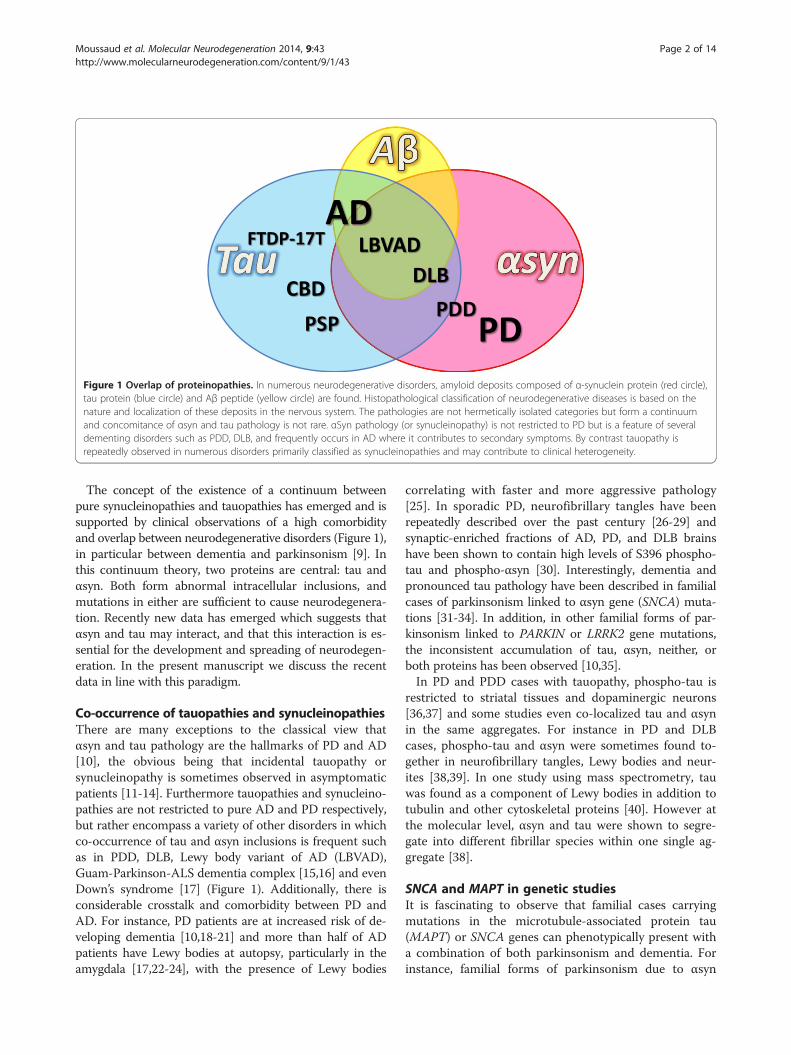
Figure 1 Overlap of proteinopathies. In numerous neurodegenerative disorders, amyloid deposits composed of α-synuclein protein (red circle),tau protein (blue circle) and Aβ peptide (yellow circle) are found. Histopathological classification of neurodegenerative diseases is based on thenature and localization of these deposits in the nervous system. The pathologies are not hermetically isolated categories but form a continuumand concomitance of αsyn and tau pathology is not rare. αSyn pathology (or synucleinopathy) is not restricted to PD but is a feature of severaldementing disorders such as PDD, DLB, and frequently occurs in AD where it contributes to secondary symptoms. By contrast tauopathy isrepeatedly observed in numerous disorders primarily classified as synucleinopathies and may contribute to clinical heterogeneity.
Moussaud et al. Molecular Neurodegeneration 2014, 9:43 Page 2 of 14http://www.molecularneurodegeneration.com/content/9/1/43
The concept of the existence of a continuum betweenpure synucleinopathies and tauopathies has emerged and issupported by clinical observations of a high comorbidityand overlap between neurodegenerative disorders (Figure 1),in particular between dementia and parkinsonism [9]. Inthis continuum theory, two proteins are central: tau andαsyn. Both form abnormal intracellular inclusions, andmutations in either are sufficient to cause neurodegenera-tion. Recently new data has emerged which suggests thatαsyn and tau may interact, and that this interaction is es-sential for the development and spreading of neurodegen-eration. In the present manuscript we discuss the recentdata in line with this paradigm.
Co-occurrence of tauopathies and synucleinopathiesThere are many exceptions to the classical view thatαsyn and tau pathology are the hallmarks of PD and AD[10], the obvious being that incidental tauopathy orsynucleinopathy is sometimes observed in asymptomaticpatients [11-14]. Furthermore tauopathies and synucleino-pathies are not restricted to pure AD and PD respectively,but rather encompass a variety of other disorders in whichco-occurrence of tau and αsyn inclusions is frequent suchas in PDD, DLB, Lewy body variant of AD (LBVAD),Guam-Parkinson-ALS dementia complex [15,16] and evenDown’s syndrome [17] (Figure 1). Additionally, there isconsiderable crosstalk and comorbidity between PD andAD. For instance, PD patients are at increased risk of de-veloping dementia [10,18-21] and more than half of ADpatients have Lewy bodies at autopsy, particularly in theamygdala [17,22-24], with the presence of Lewy bodies
correlating with faster and more aggressive pathology[25]. In sporadic PD, neurofibrillary tangles have beenrepeatedly described over the past century [26-29] andsynaptic-enriched fractions of AD, PD, and DLB brainshave been shown to contain high levels of S396 phospho-tau and phospho-αsyn [30]. Interestingly, dementia andpronounced tau pathology have been described in familialcases of parkinsonism linked to αsyn gene (SNCA) muta-tions [31-34]. In addition, in other familial forms of par-kinsonism linked to PARKIN or LRRK2 gene mutations,the inconsistent accumulation of tau, αsyn, neither, orboth proteins has been observed [10,35].In PD and PDD cases with tauopathy, phospho-tau is
restricted to striatal tissues and dopaminergic neurons[36,37] and some studies even co-localized tau and αsynin the same aggregates. For instance in PD and DLBcases, phospho-tau and αsyn were sometimes found to-gether in neurofibrillary tangles, Lewy bodies and neur-ites [38,39]. In one study using mass spectrometry, tauwas found as a component of Lewy bodies in addition totubulin and other cytoskeletal proteins [40]. However atthe molecular level, αsyn and tau were shown to segre-gate into different fibrillar species within one single ag-gregate [38].
SNCA and MAPT in genetic studiesIt is fascinating to observe that familial cases carryingmutations in the microtubule-associated protein tau(MAPT) or SNCA genes can phenotypically present witha combination of both parkinsonism and dementia. Forinstance, familial forms of parkinsonism due to αsyn
B -Synuclein protein
Alternatively spliced regions
Constant regions
exonic missense mutations
phosphorylation sites
Repeated domains
Proposed disease-associated
2HN
A53
T/E
G51
DH
50Q
1 65 95 140
Important region for
polymerization
Ligand binding domain
Amphipathic N-terminal region
NAC region
Acidic C-terminal region
COOH
A30
P
E46
K
Binding site to phospholipid
membranes
Exon 5103-130
Exon 341-54
K29
8E
A Tau proteinTubulin binding regionAcidic N-terminal region
2HN COOHN1 N2 R1 R2 R3 R4
Repeated domainsProline rich region
K36
9I1 441
G27
2V
P30
1L /
S /
T
G33
5S /
V
R40
6W
N27
9K
S30
5N /
S /
IR5H
/L
K25
7TI2
60V
L266
V
N29
6 / N
/ H
K28
0L2
84L
/ R
L315
R
S32
0F
P33
2S
S35
2L
G38
9R
G27
3R
G30
3VG
304S
K31
7M
V33
7M
Q33
6RE
342V S35
6TV
363I
P36
4SG
366R
E37
2G
N41
0HT
427M
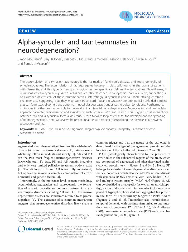
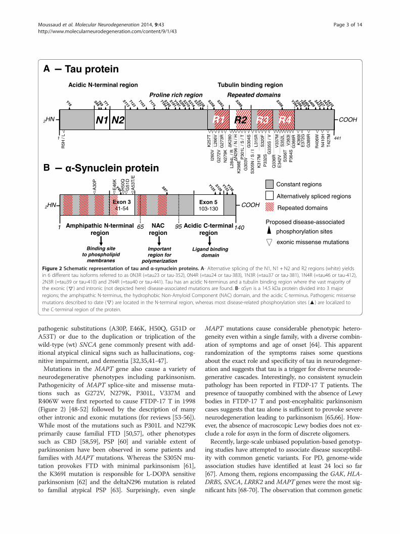
Figure 2 Schematic representation of tau and α-synuclein proteins. A- Alternative splicing of the N1, N1 + N2 and R2 regions (white) yieldsin 6 different tau isoforms referred to as 0N3R (=tau23 or tau-352), 0N4R (=tau24 or tau-383), 1N3R (=tau37 or tau-381), 1N4R (=tau46 or tau-412),2N3R (=tau39 or tau-410) and 2N4R (=tau40 or tau-441). Tau has an acidic N-terminus and a tubulin binding region where the vast majority ofthe exonic (▽) and intronic (not depicted here) disease-associated mutations are found. B- αSyn is a 14.5 kDa protein divided into 3 majorregions; the amphipathic N-terminus, the hydrophobic Non-Amyloid Component (NAC) domain, and the acidic C-terminus. Pathogenic missensemutations described to date (▽) are located in the N-terminal region, whereas most disease-related phosphorylation sites (▲) are localized tothe C-terminal region of the protein.
Moussaud et al. Molecular Neurodegeneration 2014, 9:43 Page 3 of 14http://www.molecularneurodegeneration.com/content/9/1/43
pathogenic substitutions (A30P, E46K, H50Q, G51D orA53T) or due to the duplication or triplication of thewild-type (wt) SNCA gene commonly present with add-itional atypical clinical signs such as hallucinations, cog-nitive impairment, and dementia [32,35,41-47].Mutations in the MAPT gene also cause a variety of
neurodegenerative phenotypes including parkinsonism.Pathogenicity of MAPT splice-site and missense muta-tions such as G272V, N279K, P301L, V337M andR406W were first reported to cause FTDP-17 T in 1998(Figure 2) [48-52] followed by the description of manyother intronic and exonic mutations (for reviews [53-56]).While most of the mutations such as P301L and N279Kprimarily cause familial FTD [50,57], other phenotypessuch as CBD [58,59], PSP [60] and variable extent ofparkinsonism have been observed in some patients andfamilies with MAPT mutations. Whereas the S305N mu-tation provokes FTD with minimal parkinsonism [61],the K369I mutation is responsible for L-DOPA sensitiveparkinsonism [62] and the deltaN296 mutation is relatedto familial atypical PSP [63]. Surprisingly, even single
MAPT mutations cause considerable phenotypic hetero-geneity even within a single family, with a diverse combin-ation of symptoms and age of onset [64]. This apparentrandomization of the symptoms raises some questionsabout the exact role and specificity of tau in neurodegener-ation and suggests that tau is a trigger for diverse neurode-generative cascades. Interestingly, no consistent synucleinpathology has been reported in FTDP-17 T patients. Thepresence of tauopathy combined with the absence of Lewybodies in FTDP-17 T and post-encephalitic parkinsonismcases suggests that tau alone is sufficient to provoke severeneurodegeneration leading to parkinsonism [65,66]. How-ever, the absence of macroscopic Lewy bodies does not ex-clude a role for αsyn in the form of discrete oligomers.Recently, large-scale unbiased population-based genotyp-
ing studies have attempted to associate disease susceptibil-ity with common genetic variants. For PD, genome-wideassociation studies have identified at least 24 loci so far[67]. Among them, regions encompassing the GAK, HLA-DRB5, SNCA, LRRK2 and MAPT genes were the most sig-nificant hits [68-70]. The observation that common genetic
A
B
C
D
100µm 100µm
50µm 50µm
Tauopathy: Synucleinopathy:
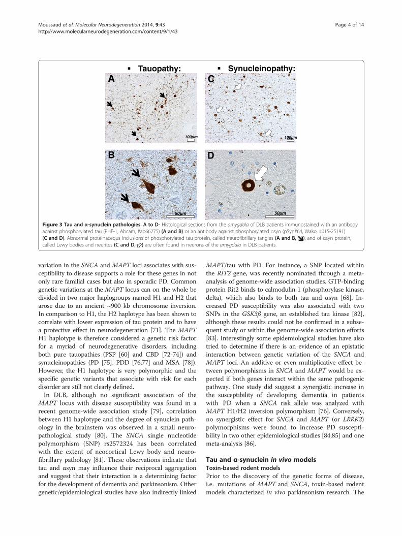
Figure 3 Tau and α-synuclein pathologies. A to D- Histological sections from the amygdala of DLB patients immunostained with an antibodyagainst phosphorylated tau (PHF-1, Abcam, #ab66275) (A and B) or an antibody against phosphorylated αsyn (pSyn#64, Wako, #015-25191)(C and D). Abnormal proteinaceous inclusions of phosphorylated tau protein, called neurofibrillary tangles (A and B, ), and of αsyn protein,called Lewy bodies and neurites (C and D, ) are often found in neurons of the amygdala in DLB patients.
Moussaud et al. Molecular Neurodegeneration 2014, 9:43 Page 4 of 14http://www.molecularneurodegeneration.com/content/9/1/43
variation in the SNCA and MAPT loci associates with sus-ceptibility to disease supports a role for these genes in notonly rare familial cases but also in sporadic PD. Commongenetic variations at the MAPT locus can on the whole bedivided in two major haplogroups named H1 and H2 thatarose due to an ancient ~900 kb chromosome inversion.In comparison to H1, the H2 haplotype has been shown tocorrelate with lower expression of tau protein and to havea protective effect in neurodegeneration [71]. The MAPTH1 haplotype is therefore considered a genetic risk factorfor a myriad of neurodegenerative disorders, includingboth pure tauopathies (PSP [60] and CBD [72-74]) andsynucleinopathies (PD [75], PDD [76,77] and MSA [78]).However, the H1 haplotype is very polymorphic and thespecific genetic variants that associate with risk for eachdisorder are still not clearly defined.In DLB, although no significant association of the
MAPT locus with disease susceptibility was found in arecent genome-wide association study [79], correlationbetween H1 haplotype and the degree of synuclein path-ology in the brainstem was observed in a small neuro-pathological study [80]. The SNCA single nucleotidepolymorphism (SNP) rs2572324 has been correlatedwith the extent of neocortical Lewy body and neuro-fibrillary pathology [81]. These observations indicate thattau and αsyn may influence their reciprocal aggregationand suggest that their interaction is a determining factorfor the development of dementia and parkinsonism. Othergenetic/epidemiological studies have also indirectly linked
MAPT/tau with PD. For instance, a SNP located withinthe RIT2 gene, was recently nominated through a meta-analysis of genome-wide association studies. GTP-bindingprotein Rit2 binds to calmodulin 1 (phosphorylase kinase,delta), which also binds to both tau and αsyn [68]. In-creased PD susceptibility was also associated with twoSNPs in the GSK3β gene, an established tau kinase [82],although these results could not be confirmed in a subse-quent study or within the genome-wide association efforts[83]. Interestingly some epidemiological studies have alsotried to determine if there is an evidence of an epistaticinteraction between genetic variation of the SNCA andMAPT loci. An additive or even multiplicative effect be-tween polymorphisms in SNCA and MAPT would be ex-pected if both genes interact within the same pathogenicpathway. One study did suggest a synergistic increase inthe susceptibility of developing dementia in patientswith PD when a SNCA risk allele was analyzed withMAPT H1/H2 inversion polymorphism [76]. Conversely,no synergistic effect for SNCA and MAPT (or LRRK2)polymorphisms were found to increase PD suscepti-bility in two other epidemiological studies [84,85] and onemeta-analysis [86].
Tau and α-synuclein in vivo modelsToxin-based rodent modelsPrior to the discovery of the genetic forms of disease,i.e. mutations of MAPT and SNCA, toxin-based rodentmodels characterized in vivo parkinsonism research. The
Moussaud et al. Molecular Neurodegeneration 2014, 9:43 Page 5 of 14http://www.molecularneurodegeneration.com/content/9/1/43
discovery that dopaminergic mid-brain neurons areespecially sensitive to oxidative stress inducers suchas 1-methyl-4-phenyl-1,2,3,6-tetrahydropyridine (MPTP),6-hydroxydopamine (6-OHDA), rotenone, and paraquatresulted in the creation of toxin-based rodent models tostudy parkinsonian phenotype in vivo (for review [87]).Interestingly, some studies also reported the accumulationof hyperphosphorylated tau in rodents after the systemicdelivery of rotenone, paraquat, and MPTP, but not maneb[88-90]. In rotenone treated rats, Hoglinger and colleaguesdescribed fibrillar structures composed of 15 nm straightfilaments positive for phospho-tau, thioflavin S, nitrosa-mine, and ubiquitin [89]. Insoluble and phosphorylatedtau has been described in mice treated with MPTP [88]and Duka and colleagues demonstrated that MPTP treat-ment induces tau hyperphosphorylation on the S396 andS404 residues via GSK3β kinase in wt but not in αsynknock-out (KO) mice [91]. Later, Qureshi and Paudel con-firmed that αsyn presence is required for the MPTP-induced phosphorylation of tau at S214, S262, S396 andS404 residues and identified GSK3β and PKA as the re-sponsible kinases [92]. However, a connection betweentauopathy and synucleinopathy has not been consistentlyobserved in these PD toxin-based models. In an interest-ing study by Morris and colleagues, a reduction of tau ex-pression did not prevent 6-OHDA neurotoxicity [93].These apparently contradictory results demonstrate thatthe interplay between tau and αsyn is complex.
αSyn and tau viral overexpression in rodentsThe effect of targeted human αsyn and tau protein ex-pression has been investigated using viral vectors-basedmodels. In these models, co-occurrence of tau and αsynpathologies has also been observed. For instance, in rats,αsyn overexpression induced by stereotaxic injection oflentivirus increases phospho-tau levels [94]. On the con-trary, rats transduced with tau and mutant P301L taudisplay an increase of αsyn and phospho-αsyn levels[95]. In another study using adeno-associated vectors forgene transfer into the substantia nigra of rats, overex-pression of human wt and P301L tau, but not αsyn, pro-voked dopaminergic neurodegeneration, reduced striataldopamine content, and motor deficit as measured byamphetamine-stimulated rotational behavior [96]. In thisstudy behavioral dysfunction preceded the formation ofneurofibrillary tangles suggesting that mature neurofib-rillary tangles are not required for tau-induced disrup-tion of dopaminergic transmission [96].
Tau and αsyn genetic mouse modelsNumerous genetically modified mice that overexpressthe human tau and/or αsyn proteins have been gener-ated and are used to model specific aspects of the hu-man diseases. Interestingly, tau transgenic models not
only develop cognitive changes but also motor dysfunc-tion. Mice overexpressing the mutant K369I tau developL-DOPA sensitive parkinsonism [62], and overexpres-sion of the pathogenic P301L and P301S forms of tau inmice provoke severe motor dysfunctions that recapitu-late some of their effects in humans [97,98]. In theP301L tau overexpressing mouse line, inhibition of tauhyperphosphorylation by treatment with a non-specificprotein kinase inhibitor also prevents the motor impair-ments suggesting that tau could be a target in degenera-tive movement disorders [99].Likewise, cognitive deficits and tauopathy have been
observed in αsyn-overexpressing PD models [100-103].Interestingly different extents of tauopathy and cognitiveimpairment were observed depending on the promotertype used for αsyn overexpression and the species ofαsyn expressed. The presence of hyperphosphorylatedtau was clearly identified in a wt αsyn overexpressingtransgenic mouse line using the PDGF-β promoter at11 months of age [101,102]. Nonetheless, no tauopathywas observed at 18 months in a transgenic line overex-pressing wt αsyn under the prion promoter unless this linewas crossed with a P301L tau mouse [100]. Interestingly,in two other lines also using the prion promoter to over-express the A53T and E46K mutant forms of αsyn, abun-dant tau inclusions were observed without the need ofcrossing with the P301L tau expressing line [100,104,105].The E46K line was reported to have more tau threads thanthe A53T αsyn overexpressing transgenic line suggestingmutation-induced differences [104]. In these mice, tauopa-thy was restricted to areas with abundant αsyn expressionand initiated simultaneously with synucleinopathy in anage-dependent fashion, although not always localizedwithin the same cells [100]. In the PDGF-β-wt-αsyn mice,hyperphosphorylated tau was primarily found in thebrainstem and in the striatum [101,102]. Kaul and col-leagues correlated phospho-tau occurrence with the acti-vation of ERK and JNK but not of GSK3β and p38MAPKkinases, whereas Haggerty and colleagues noted a matchbetween the presence of phospho-tau and phospho-GSK3β. Similarly, in the prion promoter driven A53Tαsyn mice, as well as in patients harboring the A53Tmutation, hyperphosphorylated and non-soluble tau ac-cumulated in the striatum and was correlated with in-creased levels of phospho-GSK3β [105,106]. In contrast,in a Thy-1 promoter driven A30P αsyn overexpressingtransgenic mouse line, hyperphosphorylated and non-soluble tau accumulated in the brainstem in correlationwith increased phospho-JNK level [107]. Jointly, theseobservations suggest the existence of complex region- andtime-dependent interactions between kinases, αsyn andtau.Some groups also crossed different transgenic mouse
models to trigger the co-occurrence of tauopathy and
Moussaud et al. Molecular Neurodegeneration 2014, 9:43 Page 6 of 14http://www.molecularneurodegeneration.com/content/9/1/43
synucleinopathy with the final aim to better model com-plex human disorders like DLB. For instance, a quadrupletransgenic mouse line has been generated by crossing atriple transgenic mouse that overexpresses human AD-causing M146V presenilin-1, APP Swedish mutation, andthe FTDP-17 T-causing P301L tau with a transgenicmouse that overexpresses human PD-causing A53T αsyn[108]. Co-overexpression of these pathogenic proteins hada strong synergistic effect on neurodegeneration, proteinaggregation, and on cognitive and motor deficits. In con-trast, crossing of a Thy1 promoter driven human wt αsynoverexpressing mouse line with a tau KO or tau condi-tional KO mouse did not prevent neurotoxicity indicatingthat αsyn also acts independently from tau [93].Several tau- and αsyn- deficient mouse lines have been
generated to determine if any particular phenotype or re-sistance to neurodegeneration might be present. Gener-ally, tau- or αsyn-deficient mice are viable with onlyminor phenotypic differences [109-112]. Remarkably, cog-nitive alterations were observed in an α- and γ-synucleindouble KO mouse line suggesting that both proteins havea compensatory role on cognition [103]. Conversely, inaged tau-deficient animals minor motor deficits were ob-served in correlation with an iron accumulation and lossof dopaminergic neurons in the substantia nigra [113],but could not be reproduced in a subsequent study [112].Together these data demonstrate that the absence ofboth proteins does not appear to have observable sig-nificant impact, perhaps due to compensatory mecha-nisms, whereas their overexpression, in particular intheir mutated forms, recapitulates some aspects of thehuman pathologies. This is in line with a gain of toxicfunction mechanism and validates therapeutic strategiesaimed at clearing tau and/or αsyn for parkinsonism and/or dementia.
Non-vertebrate modelsIn addition to rodents, non-vertebrate αsyn and/or tautransgenic in vivo models have been developed. Thesemodels work surprisingly well and present many prac-tical aspects [114,115]. For example, in Caenorhabditiselegans, expression of the human αsyn or tau protein inneurons recapitulates key features of the human diseasessuch as motor deficits and neuronal and dendritic loss[116]. Human αsyn expression in Drosophila melanoga-ster also induces neurotoxicity as well as L-DOPA-sensitive motor deficits and formation of αsyn-positivefibrils [117]. Remarkably, whereas αsyn expression pro-vokes the formation of Lewy body-like aggregates inD. melanogaster but not in C. elegans, tau expressionconversely leads to the formation of insoluble inclusionsin C. elegans but not in D. melanogaster [115,116].Nonetheless, both proteins are neurotoxic in bothmodels demonstrating that the formation of protein
aggregates is fundamentally unnecessary for toxicity. Inline with this idea, dopaminergic neurons in αsyn express-ing D. melanogaster were rescued without suppressing thepresence of αsyn inclusions by co-expression of the Hsp70chaperone [118]. More recently, co-expression of tauand αsyn in D. melanogaster has been shown to inducemotor dysfunction, dopaminergic denervation, cyto-toxicity, formation of abnormal ubiquitin positive in-clusions, axonal transport disruption, and cytoskeletal andsynaptic disorganization [119]. Tau affected dopaminergiccell count only when co-expressed with αsyn, demonstrat-ing the existence of a synergistic deleterious effect be-tween tau and αsyn once more. However, in contrast towhat was observed in rodent models, the mechanism oftoxicity in D. melanogaster was linked to severe cytoskel-etal and axonal disorganization and subsequent synapticalterations rather than αsyn–promoted tau hyperpho-sphorylation [119].Interesting findings have also been made in yeast models.
In Saccharomyces cerevisiae, overexpressing human taudoes not induce significant toxicity [120,121]. However, co-expression of tau with human αsyn leads to greater toxicitythan αsyn expression alone, and also leads to the formationof insoluble hyperphosphorylated tau and αsyn aggregates.The synergistic deleterious effects were increased by ex-pression of A53T αsyn or P301L tau instead of their wtforms [120,122]. Finally, in these models, yeast orthologs ofthe cyclin-dependent kinase 5 and GSK3β kinases wereshown to be involved in the phosphorylation of tau and inthe αsyn plus tau induced-toxicity [120,122].In various transgenic or toxin-induced PD models ran-
ging from mice to yeast, the existence of a deleterious andemulative action between tau and αsyn has been repeti-tively shown. This corroborates what has been observed inhumans (see Co-occurrence of tauopathies and synucleino-pathies and SNCA and MAPT in genetic studies sections)and reinforces the idea that the interplay between αsynand tau are pivotal in the neurodegenerative process.Nonetheless, results from these in vivo models have to beinterpreted with caution. For instance, there is no orthologgene of the human SNCA in the fly, worm or yeast,whereas in rodents the endogenous wt αsyn carries theA53T mutation without deleterious effect [123,124]. More-over, in rodents, tau hyperphosphorylation can occur ininstances of hibernation or starvation, making this patho-logical hallmark difficult to interpret [125,126].
Tau and α-synuclein in molecular studiesThe fact that αsyn and tau can physically interact witheach other was demonstrated by Jensen and colleaguesin 1999. In this pioneering study, tau protein from brainlysates was pulled down by αsyn affinity chromatog-raphy. The authors also noted a strong effect of ionicstrength on the binding, indicating the implication of
Moussaud et al. Molecular Neurodegeneration 2014, 9:43 Page 7 of 14http://www.molecularneurodegeneration.com/content/9/1/43
salt bridges in the interaction [127]. Moreover, in linewith an interaction under physiological conditions, abinding IC50 value of 50pM was calculated between tauand αsyn using plasmon surface resonance and a radio-active binding assay [127]. At the cellular level, tau andαsyn were co-localized in the same cellular compart-ments and in particular in axons [127]. This was con-firmed by Förster resonance energy transfer (FRET) inmore recent studies [128,129].
Docking sites and effects of mutations on tau – αsyninteractionsSeveral studies have tried to identify the exact regions andthe critical amino acid residues by which tau and αsyninteract. Using protein fragmentation and recombinantpeptides, Jensen and colleagues found that the interactingdomains are localized to the C-terminus of αsyn (55 to140) and the microtubule binding region of tau (192 to383) [127]. Accordingly two subsequent studies found thatthe N-terminal (1 to 153) and C-terminal (352 to 441)fragments of tau do not interact with αsyn [129,130]. Thequestion of the role of phosphorylation and disease-related mutations in the tau and αsyn interaction has alsobeen addressed and investigated in vitro. Phosphorylationof the serine 214 residue of tau was identified to increaseαsyn binding [92]. In contrast, phospho-mimic/dead mu-tations at the serine 129 residue of αsyn had no effect[129]. No effect of A30P and A53T αsyn disease-relatedmutations was initially reported [127], but later in a studyusing FRET, the αsyn mutation A30P, but not the A53Tand E46K, was shown to reduce association of αsynwith tau [129]. Conversely, in a third study using co-immunoprecipitation, αsyn mutations A30P, A53T, E46Kbut not E83P increased binding with tau [92], E83P beingan artificial mutation in the NAC domain that blocks αsynaggregation [131]. On the contrary, the P301L tau muta-tion was found to reduce interaction with αsyn [130].Consequently the exact role of the disease-related muta-tions on tau and αsyn interaction still needs to be clarifiedespecially since in vivo observations suggest that they mayplay a role.
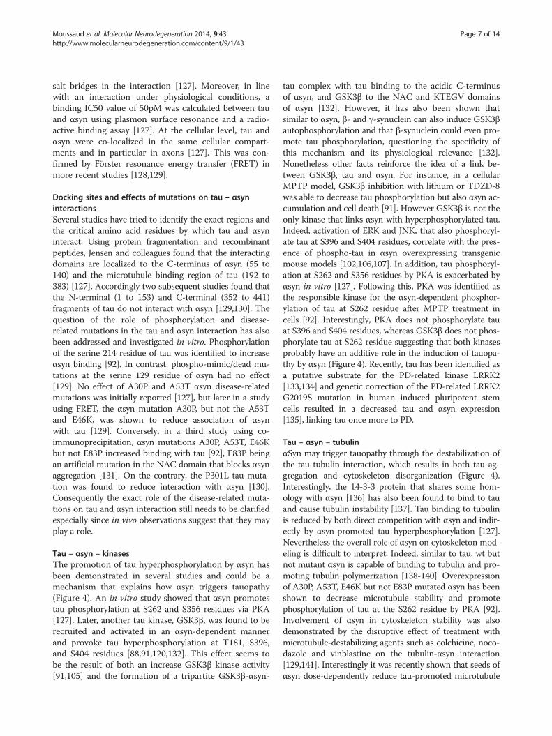
Tau – αsyn – kinasesThe promotion of tau hyperphosphorylation by αsyn hasbeen demonstrated in several studies and could be amechanism that explains how αsyn triggers tauopathy(Figure 4). An in vitro study showed that αsyn promotestau phosphorylation at S262 and S356 residues via PKA[127]. Later, another tau kinase, GSK3β, was found to berecruited and activated in an αsyn-dependent mannerand provoke tau hyperphosphorylation at T181, S396,and S404 residues [88,91,120,132]. This effect seems tobe the result of both an increase GSK3β kinase activity[91,105] and the formation of a tripartite GSK3β-αsyn-
tau complex with tau binding to the acidic C-terminusof αsyn, and GSK3β to the NAC and KTEGV domainsof αsyn [132]. However, it has also been shown thatsimilar to αsyn, β- and γ-synuclein can also induce GSK3βautophosphorylation and that β-synuclein could even pro-mote tau phosphorylation, questioning the specificity ofthis mechanism and its physiological relevance [132].Nonetheless other facts reinforce the idea of a link be-tween GSK3β, tau and αsyn. For instance, in a cellularMPTP model, GSK3β inhibition with lithium or TDZD-8was able to decrease tau phosphorylation but also αsyn ac-cumulation and cell death [91]. However GSK3β is not theonly kinase that links αsyn with hyperphosphorylated tau.Indeed, activation of ERK and JNK, that also phosphoryl-ate tau at S396 and S404 residues, correlate with the pres-ence of phospho-tau in αsyn overexpressing transgenicmouse models [102,106,107]. In addition, tau phosphoryl-ation at S262 and S356 residues by PKA is exacerbated byαsyn in vitro [127]. Following this, PKA was identified asthe responsible kinase for the αsyn-dependent phosphor-ylation of tau at S262 residue after MPTP treatment incells [92]. Interestingly, PKA does not phosphorylate tauat S396 and S404 residues, whereas GSK3β does not phos-phorylate tau at S262 residue suggesting that both kinasesprobably have an additive role in the induction of tauopa-thy by αsyn (Figure 4). Recently, tau has been identified asa putative substrate for the PD-related kinase LRRK2[133,134] and genetic correction of the PD-related LRRK2G2019S mutation in human induced pluripotent stemcells resulted in a decreased tau and αsyn expression[135], linking tau once more to PD.
Tau – αsyn – tubulinαSyn may trigger tauopathy through the destabilization ofthe tau-tubulin interaction, which results in both tau ag-gregation and cytoskeleton disorganization (Figure 4).Interestingly, the 14-3-3 protein that shares some hom-ology with αsyn [136] has also been found to bind to tauand cause tubulin instability [137]. Tau binding to tubulinis reduced by both direct competition with αsyn and indir-ectly by αsyn-promoted tau hyperphosphorylation [127].Nevertheless the overall role of αsyn on cytoskeleton mod-eling is difficult to interpret. Indeed, similar to tau, wt butnot mutant αsyn is capable of binding to tubulin and pro-moting tubulin polymerization [138-140]. Overexpressionof A30P, A53T, E46K but not E83P mutated αsyn has beenshown to decrease microtubule stability and promotephosphorylation of tau at the S262 residue by PKA [92].Involvement of αsyn in cytoskeleton stability was alsodemonstrated by the disruptive effect of treatment withmicrotubule-destabilizing agents such as colchicine, noco-dazole and vinblastine on the tubulin-αsyn interaction[129,141]. Interestingly it was recently shown that seeds ofαsyn dose-dependently reduce tau-promoted microtubule
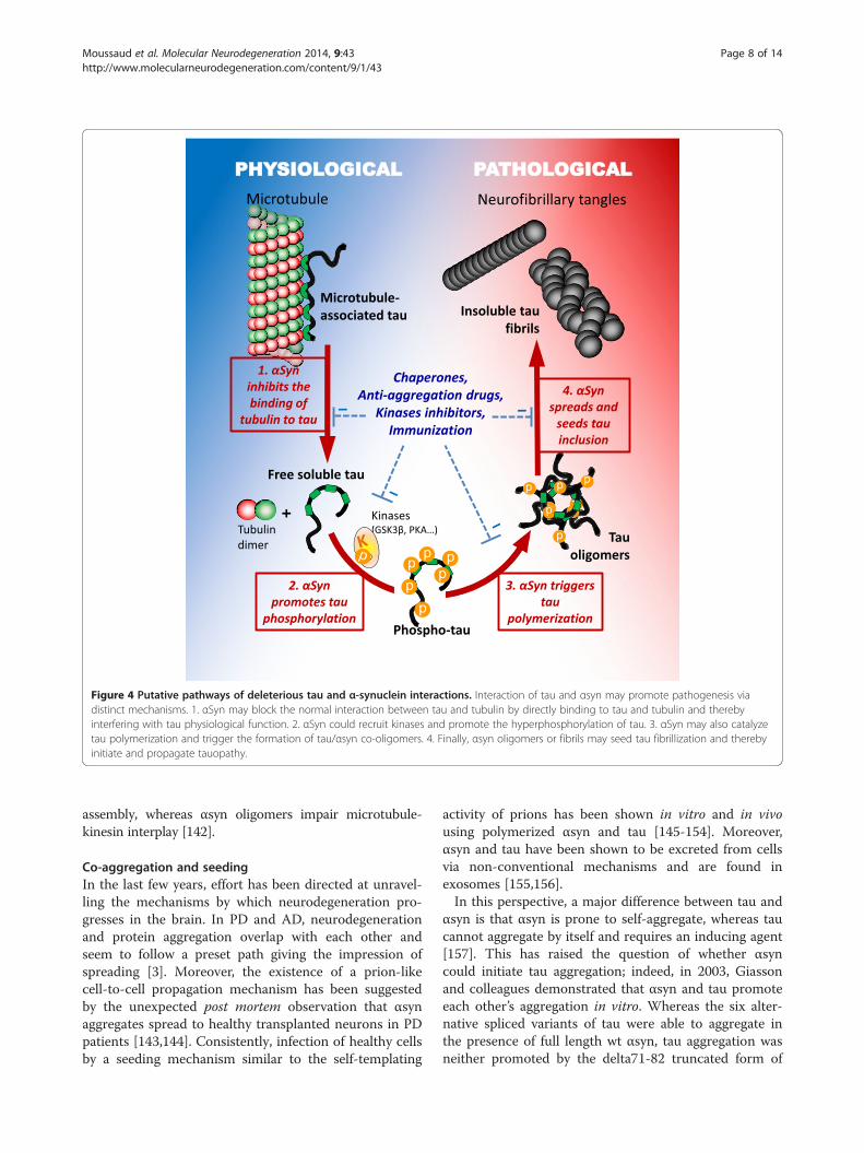
Figure 4 Putative pathways of deleterious tau and α-synuclein interactions. Interaction of tau and αsyn may promote pathogenesis viadistinct mechanisms. 1. αSyn may block the normal interaction between tau and tubulin by directly binding to tau and tubulin and therebyinterfering with tau physiological function. 2. αSyn could recruit kinases and promote the hyperphosphorylation of tau. 3. αSyn may also catalyzetau polymerization and trigger the formation of tau/αsyn co-oligomers. 4. Finally, αsyn oligomers or fibrils may seed tau fibrillization and therebyinitiate and propagate tauopathy.
Moussaud et al. Molecular Neurodegeneration 2014, 9:43 Page 8 of 14http://www.molecularneurodegeneration.com/content/9/1/43
assembly, whereas αsyn oligomers impair microtubule-kinesin interplay [142].
Co-aggregation and seedingIn the last few years, effort has been directed at unravel-ling the mechanisms by which neurodegeneration pro-gresses in the brain. In PD and AD, neurodegenerationand protein aggregation overlap with each other andseem to follow a preset path giving the impression ofspreading [3]. Moreover, the existence of a prion-likecell-to-cell propagation mechanism has been suggestedby the unexpected post mortem observation that αsynaggregates spread to healthy transplanted neurons in PDpatients [143,144]. Consistently, infection of healthy cellsby a seeding mechanism similar to the self-templating
activity of prions has been shown in vitro and in vivousing polymerized αsyn and tau [145-154]. Moreover,αsyn and tau have been shown to be excreted from cellsvia non-conventional mechanisms and are found inexosomes [155,156].In this perspective, a major difference between tau and
αsyn is that αsyn is prone to self-aggregate, whereas taucannot aggregate by itself and requires an inducing agent[157]. This has raised the question of whether αsyncould initiate tau aggregation; indeed, in 2003, Giassonand colleagues demonstrated that αsyn and tau promoteeach other’s aggregation in vitro. Whereas the six alter-native spliced variants of tau were able to aggregate inthe presence of full length wt αsyn, tau aggregation wasneither promoted by the delta71-82 truncated form of
Moussaud et al. Molecular Neurodegeneration 2014, 9:43 Page 9 of 14http://www.molecularneurodegeneration.com/content/9/1/43
αsyn, nor by β-synuclein, nor by amyloidogenic Aβpeptide [100]. These observations demonstrate that tauaccelerates αsyn polymerization and that αsyn can actas an inducing agent of tau polymerization through itshydrophobic NAC region. Interestingly, mutant A53Tαsyn was shown to have increased tau fibrillizationproperties in vitro when compared to wt αsyn [33]. Con-versely, tau expression enhanced toxicity and secretion,and changed the pattern of αsyn aggregation by promot-ing the formation of smaller inclusions in cellularmodels [128].Using the fluorescent intensity distribution analysis
technique (FIDA), Nübling and colleagues have shownthat tau and αsyn can form co-oligomers and that co-aggregation happens even at nanomolar concentrationsbut only in the presence of a cationic aggregation in-ducer such as Al3+ and Fe3+ or DMSO [158]. Moreover,tau phosphorylation by GSK3β strongly enhanced theformation of mixed oligomers [158]. However electronmicroscopy revealed that co-incubation of monomerictau and αsyn mainly leads to the formation of homopol-ymeric fibrils [100]. This is consistent with the observa-tions made in DLB cases [38] and suggests that αsynand tau predominantly interact with each other at themonomeric and oligomeric stages. More recently, a seriesof studies tried to reproduce these in vitro findings in vivoby demonstrating that exogenous αsyn can be taken up byneurons and induce the formation of intracellular Lewybody-like structures [159,160] and also hyperphosphory-lated tau aggregates [151,161]. For instance extracellulartreatment with polymerized recombinant human αsyninduced the formation of insoluble phosphorylated tauin cellular models [104,161]. Counterintuitively, wt αsynfibrils were more efficient than E46K αsyn fibrils atcross-seeding tau [104].
Conclusions and future directionsThe overlap and numerous similarities between synuclei-nopathies and tauopathies suggest that therapeutic strat-egies that target common processes of tau and αsynaggregation could benefit patients across a spectrum ofneurodegenerative disorders, and may be particularlyrelevant for the treatment of secondary symptoms suchas cognitive impairment in PD or secondary parkinson-ism in dementia. In the present review we have compileddata from the literature linking tau and αsyn. Therepeated in vitro and in vivo observations that tau andαsyn interact highly suggest that αsyn and tau play asteammates, however how this interaction occurs andaffects neurodegenerative processes is still not fullyelucidated and several scenarios are possible (Figure 4).αSyn was initially shown to bind to tau and interfere
with the normal interaction between tau and tubulin[127]. The disruption of the normal physiological
interaction between tau and tubulin for a pathologicalinteraction between tau and αsyn could explain whyαsyn and tau interaction seems to be deleterious.However, more recent data suggest that the role of tauand αsyn interaction on cytoskeleton modeling, axonaldevelopment and synaptic activity in neurons may bemore complex as first thought [119,140,142]. Additionalmechanisms, acting together in a vicious cycle, mayexplain how αsyn triggers tau aggregation and vice versa.Reciprocal promotion of phosphorylation is probably akey player, suggesting that kinases could be used as targets.GSK3β inhibition for instance was concomitantly able todecrease tau phosphorylation, αsyn accumulation andcell death in a cellular MPTP model [91]. In a P301L tauoverexpressing mouse line, inhibition of tau hyperpho-sphorylation by treatment with a non-specific proteinkinase inhibitor prevented motor impairments [99].A mechanistic cross-seeding effect based on templat-
ing of a pathological β-sheet conformation is also highlysuspected [151,159-161]. Indeed, αsyn and tau can formco-oligomers that catalyze aggregation and finally lead tothe formation of pure homofibrils [100,158]. This resem-bles the prion self-propagation mechanism and thisparallel is now commonly drawn in the literature, even ifthere is no evidence of human-to-human transmissionfor αsyn or tau [162,163]. However, mutual misfoldingis probably the first event that leads to αsyn and tausynergistic co-aggregation. The fact that αsyn has hom-ology with the 14-3-3 co-chaperone protein, a knownpartner of tau and αsyn, and is able to substitute 14-3-3co-chaperone activity on 14-3-3 targets supports thishypothesis [136,137]. In this regard upregulation ofchaperone proteins is another promising strategy that ispresently being investigated to restore the normalconformation of αsyn and tau (Figure 4). Heat shockproteins (HSPs) increase the association of tau with mi-crotubules and regulate tau degradation, ubiquitinationand phosphorylation [164,165]. Hsp70 preferentiallybinds to tau oligomers and restores anterograde fastaxonal transport [166]. Our group has demonstratedthat αsyn aggregation can be blocked by modulatingdifferent chaperones including Hsp27, Hsp70, Hsp90,torsinA and CHIP [167-175]. HSPs have been shownto positively act on αsyn or on tau independently, butthey may also have a neuroprotective effect by restoringand regulating the normal interaction between bothproteins [130,132].Observations suggest that αsyn being a trigger of tauo-
pathy is more plausible than the opposite scenario. Forinstance, whereas the presence of tau only acceleratedαsyn polymerization, co-incubation with αsyn was ne-cessary to trigger tau aggregation in vitro [100]. Consist-ently, tau ablation failed to prevent neurotoxicity in the6-OHDA or wt αsyn overexpressing mouse models
Moussaud et al. Molecular Neurodegeneration 2014, 9:43 Page 10 of 14http://www.molecularneurodegeneration.com/content/9/1/43
[125]. Furthermore, pronounced tauopathy has beendescribed in αsyn transgenic mice [100-102,104,105] andin PD patients harboring the A53T mutation [31,33,34],whereas no consistent αsyn pathology has been reportedin tau transgenic mice or FTDP-17 T patients [66].However, this is somewhat contradicted by the observa-tion that αsyn pathology in AD is more pronouncedthan the tau pathology in PD [16,22-24,26-29,37].Nonetheless, even if some gray areas persist regarding
the mechanisms and roles of the interaction between tauand αsyn, applications and future directions are alreadyemerging. One future development is the identificationof reliable biomarkers for efficient diagnosis of neurode-generative disorders at the prodromal stage. In additionto other proteins, αsyn and tau are presently being de-veloped as cerebrospinal fluid biomarkers for improvedclinical diagnoses. Cerebrospinal fluid levels of Aβ, totaland phospho-αsyn, and total and phospho-tau changedifferentially depending on the nature of the disease.Consequently, looking at the ratios between theseproteins could enable clinicians to determine the risk ofdeveloping PD, AD, or a mixed disorder such as DLBand provide them the needed therapeutic window tostart preventive and tailor-made treatments [176].
AbbreviationsAD: Alzheimer’s disease; ALS: Amyotrophic lateral sclerosis; αsyn: Alpha-synucleinprotein; DLB: Dementia with Lewy bodies; CBD: Corticobasal degeneration;FRET: Förster resonance energy transfer; FTDP-17 T: Frontotemporal dementiawith parkinsonism linked to tau mutations on chromosome 17; GSK3β: Glycogensynthase kinase 3 beta; Hsps: Heat shock proteins; JNK: c-Jun N-terminal kinases;KO: Knock-out; LRRK2: Leucine reach repeat kinase 2; MAPK: Mitogen-activatedprotein kinase; MAPT: Tau gene; MPTP: 1-methyl-4-phenyl-1:2:3:6-tetrahydropyridine; MSA: Multiple system atrophy; PD: Parkinson’s disease;PDD: Parkinson’s disease with dementia; PiD: Pick’s disease; PKA: Protein kinase A;PSP: Progressive supranuclear palsy; SNCA: αsyn gene; SNP: Single nucleotidepolymorphisms; wt: Wild-type.
Competing interestsThe authors declare that they have no competing interests.
Authors’ contributionsSM and PJM wrote and drafted the manuscript. DRJ, ELML, MD and OARadded intellectual input and editorial suggestions, and assisted in figurepreparation. All authors read and approved the final manuscript.
AcknowledgementsThis work was supported by the Mayo Clinic and the NIH NS063963. OAR ispartially supported by NINDS R01# NS078086 and Mayo Clinic Florida is aMorris K. Udall Parkinson's Disease Research Center of Excellence (NINDS P50#NS072187). The authors would like to thank A.-M. Baine, A. Bradshaw, A.Bronnhuber, Dr. M. Gan, Dr. W. Springer, Dr. F. Fiesel, Dr. C. Labbé, Dr. P. M.Tacik , Dr. M. G. Plumot and Dr. K. F. Bieniek for passionate scientificdiscussions.
Received: 3 April 2014 Accepted: 16 October 2014Published: 29 October 2014
References1. Weintraub D, Comella CL, Horn S: Parkinson's disease–part 1: pathophysiology,
symptoms, burden, diagnosis, and assessment. Am J Manag Care 2008,14:S40–S48.
2. Weintraub D, Comella CL, Horn S: Parkinson's disease–part 2: treatment ofmotor symptoms. Am J Manag Care 2008, 14:S49–S58.
3. Braak H, Rub U, Gai WP, Del Tredici K: Idiopathic Parkinson's disease: possibleroutes by which vulnerable neuronal types may be subject to neuroinvasionby an unknown pathogen. J Neural Transm 2003, 110:517–536.
4. Jones DR, Moussaud S, McLean P: Targeting heat shock proteins tomodulate alpha-synuclein toxicity. Ther Adv Neurol Disord 2014, 7:33–51.
5. Spillantini MG, Schmidt ML, Lee VM, Trojanowski JQ, Jakes R, Goedert M:Alpha-synuclein in Lewy bodies. Nature 1997, 388:839–840.
6. Irizarry MC, Growdon W, Gomez-Isla T, Newell K, George JM, Clayton DF,Hyman BT: Nigral and cortical Lewy bodies and dystrophic nigralneurites in Parkinson's disease and cortical Lewy body disease containalpha-synuclein immunoreactivity. J Neuropathol Exp Neurol 1998,57:334–337.
7. Spillantini MG, Crowther RA, Jakes R, Cairns NJ, Lantos PL, Goedert M:Filamentous alpha-synuclein inclusions link multiple system atrophy withParkinson's disease and dementia with Lewy bodies. Neurosci Lett 1998,251:205–208.
8. Brion JP, Couck AM, Passareiro E, Flament-Durand J: Neurofibrillary tanglesof Alzheimer's disease: an immunohistochemical study. J Submicrosc Cytol1985, 17:89–96.
9. Irwin DJ, Lee VM, Trojanowski JQ: Parkinson's disease dementia: convergenceof alpha-synuclein, tau and amyloid-beta pathologies. Nat Rev Neurosci 2013,14:626–636.
10. Galpern WR, Lang AE: Interface between tauopathies and synucleinopathies:a tale of two proteins. Ann Neurol 2006, 59:449–458.
11. Dickson DW, Fujishiro H, DelleDonne A, Menke J, Ahmed Z, Klos KJ, JosephsKA, Frigerio R, Burnett M, Parisi JE, Ahlskog JE: Evidence that incidental Lewybody disease is pre-symptomatic Parkinson's disease. Acta Neuropathol 2008,115:437–444.
12. Evidente VG, Adler CH, Sabbagh MN, Connor DJ, Hentz JG, Caviness JN,Sue LI, Beach TG: Neuropathological findings of PSP in the elderlywithout clinical PSP: possible incidental PSP? Parkinsonism Relat Disord2011, 17:365–371.
13. Mikolaenko I, Pletnikova O, Kawas CH, O'Brien R, Resnick SM, Crain B,Troncoso JC: Alpha-synuclein lesions in normal aging, Parkinson disease,and Alzheimer disease: evidence from the Baltimore Longitudinal Studyof Aging (BLSA). J Neuropathol Exp Neurol 2005, 64:156–162.
14. Saito Y, Kawashima A, Ruberu NN, Fujiwara H, Koyama S, Sawabe M, Arai T,Nagura H, Yamanouchi H, Hasegawa M, Iwatsubo T, Murayama S:Accumulation of phosphorylated alpha-synuclein in aging human brain.J Neuropathol Exp Neurol 2003, 62:644–654.
15. Forman MS, Schmidt ML, Kasturi S, Perl DP, Lee VM, Trojanowski JQ: Tauand alpha-synuclein pathology in amygdala of Parkinsonism-dementiacomplex patients of Guam. Am J Pathol 2002, 160:1725–1731.
16. Lippa CF, Fujiwara H, Mann DM, Giasson B, Baba M, Schmidt ML, Nee LE,O'Connell B, Pollen DA, St George-Hyslop P, Ghetti B, Nochlin D, Bird TD,Cairns NJ, Lee VM, Iwatsubo T, Trojanowski JQ: Lewy bodies containaltered alpha-synuclein in brains of many familial Alzheimer's diseasepatients with mutations in presenilin and amyloid precursor protein genes.Am J Pathol 1998, 153:1365–1370.
17. Lippa CF, Schmidt ML, Lee VM, Trojanowski JQ: Antibodies to alpha-synucleindetect Lewy bodies in many Down's syndrome brains with Alzheimer'sdisease. Ann Neurol 1999, 45:353–357.
18. Aarsland D, Zaccai J, Brayne C: A systematic review of prevalence studiesof dementia in Parkinson's disease. Mov Disord 2005, 20:1255–1263.
19. Braak H, Rub U, Jansen Steur EN, Del Tredici K, de Vos RA: Cognitive statuscorrelates with neuropathologic stage in Parkinson disease. Neurology2005, 64:1404–1410.
20. Hurtig HI, Trojanowski JQ, Galvin J, Ewbank D, Schmidt ML, Lee VM, ClarkCM, Glosser G, Stern MB, Gollomp SM, Arnold SE: Alpha-synuclein corticalLewy bodies correlate with dementia in Parkinson's disease. Neurology2000, 54:1916–1921.
21. Leverenz JB, Quinn JF, Zabetian C, Zhang J, Montine KS, Montine TJ:Cognitive impairment and dementia in patients with Parkinson disease.Curr Top Med Chem 2009, 9:903–912.
22. Hamilton RL: Lewy bodies in Alzheimer's disease: a neuropathologicalreview of 145 cases using alpha-synuclein immunohistochemistry.Brain Pathol 2000, 10:378–384.
23. Leverenz JB, Fishel MA, Peskind ER, Montine TJ, Nochlin D, Steinbart E,Raskind MA, Schellenberg GD, Bird TD, Tsuang D: Lewy body pathology infamilial Alzheimer disease: evidence for disease- and mutation-specificpathologic phenotype. Arch Neurol 2006, 63:370–376.
Moussaud et al. Molecular Neurodegeneration 2014, 9:43 Page 11 of 14http://www.molecularneurodegeneration.com/content/9/1/43
24. Leverenz JB, Hamilton R, Tsuang DW, Schantz A, Vavrek D, Larson EB, KukullWA, Lopez O, Galasko D, Masliah E, Kaye J, Woltjer R, Clark C, TrojanowskiJQ, Montine TJ: Empiric refinement of the pathologic assessment ofLewy-related pathology in the dementia patient. Brain Pathol 2008,18:220–224.
25. Kraybill ML, Larson EB, Tsuang DW, Teri L, McCormick WC, Bowen JD, KukullWA, Leverenz JB, Cherrier MM: Cognitive differences in dementia patientswith autopsy-verified AD, Lewy body pathology, or both. Neurology 2005,64:2069–2073.
26. Bancher C, Braak H, Fischer P, Jellinger KA: Neuropathological staging ofAlzheimer lesions and intellectual status in Alzheimer's and Parkinson'sdisease patients. Neurosci Lett 1993, 162:179–182.
27. Joachim CL, Morris JH, Kosik KS, Selkoe DJ: Tau antisera recognizeneurofibrillary tangles in a range of neurodegenerative disorders.Ann Neurol 1987, 22:514–520.
28. Lewy FH: Paralysis agitans. Pathologische Anatomie Handbuch derNeurologie (Ed M Lewandowsky) Julius Springer 1912, 3:920–933.
29. Schneider JA, Li JL, Li Y, Wilson RS, Kordower JH, Bennett DA: Substantianigra tangles are related to gait impairment in older persons. Ann Neurol2006, 59:166–173.
30. Muntane G, Dalfo E, Martinez A, Ferrer I: Phosphorylation of tau andalpha-synuclein in synaptic-enriched fractions of the frontal cortexin Alzheimer's disease, and in Parkinson's disease and relatedalpha-synucleinopathies. Neuroscience 2008, 152:913–923.
31. Duda JE, Giasson BI, Mabon ME, Miller DC, Golbe LI, Lee VM, Trojanowski JQ:Concurrence of alpha-synuclein and tau brain pathology in the Contursikindred. Acta Neuropathol 2002, 104:7–11.
32. Fujioka S, Ogaki K, Tacik PM, Uitti RJ, Ross OA, Wszolek ZK: Update on novelfamilial forms of Parkinson's disease and multiple system atrophy.Parkinsonism Relat Disord 2014, 20(Suppl 1):S29–S34.
33. Kotzbauer PT, Giasson BI, Kravitz AV, Golbe LI, Mark MH, Trojanowski JQ,Lee VM: Fibrillization of alpha-synuclein and tau in familial Parkinson'sdisease caused by the A53T alpha-synuclein mutation. Exp Neurol 2004,187:279–288.
34. Yamaguchi K, Cochran EJ, Murrell JR, Polymeropoulos MH, Shannon KM,Crowther RA, Goedert M, Ghetti B: Abundant neuritic inclusions andmicrovacuolar changes in a case of diffuse Lewy body disease with theA53T mutation in the alpha-synuclein gene. Acta Neuropathol 2005,110:298–305.
35. Poulopoulos M, Levy OA, Alcalay RN: The neuropathology of geneticParkinson's disease. Mov Disord 2012, 27:831–842.
36. Jellinger KA: Interaction between alpha-synuclein and tau in Parkinson'sdisease comment on Wills et al.: elevated tauopathy and alpha-synucleinpathology in postmortem Parkinson's disease brains with and withoutdementia. Exp Neurol 2010; 225: 210-218. Exp Neurol 2011, 227:13–18.
37. Wills J, Jones J, Haggerty T, Duka V, Joyce JN, Sidhu A: Elevated tauopathyand alpha-synuclein pathology in postmortem Parkinson's disease brainswith and without dementia. Exp Neurol 2010, 225:210–218.
38. Arima K, Mizutani T, Alim MA, Tonozuka-Uehara H, Izumiyama Y, Hirai S,Ueda K: NACP/alpha-synuclein and tau constitute two distinctive subsetsof filaments in the same neuronal inclusions in brains from a family ofparkinsonism and dementia with Lewy bodies: double-immunolabelingfluorescence and electron microscopic studies. Acta Neuropathol 2000,100:115–121.
39. Ishizawa T, Mattila P, Davies P, Wang D, Dickson DW: Colocalization of tauand alpha-synuclein epitopes in Lewy bodies. J Neuropathol Exp Neurol2003, 62:389–397.
40. Leverenz JB, Umar I, Wang Q, Montine TJ, McMillan PJ, Tsuang DW, Jin J,Pan C, Shin J, Zhu D, Zhang J: Proteomic identification of novel proteinsin cortical lewy bodies. Brain Pathol 2007, 17:139–145.
41. Appel-Cresswell S, Vilarino-Guell C, Encarnacion M, Sherman H, Yu I, Shah B,Weir D, Thompson C, Szu-Tu C, Trinh J, Aasly JO, Rajput A, Rajput AH,Jon Stoessl A, Farrer MJ: Alpha-synuclein p.H50Q, a novel pathogenicmutation for Parkinson's disease. Mov Disord 2013, 28:811–813.
42. Chartier-Harlin MC, Kachergus J, Roumier C, Mouroux V, Douay X, Lincoln S,Levecque C, Larvor L, Andrieux J, Hulihan M, Waucquier N, Defebvre L,Amouyel P, Farrer M, Destee A: Alpha-synuclein locus duplication as acause of familial Parkinson's disease. Lancet 2004, 364:1167–1169.
43. Kiely AP, Asi YT, Kara E, Limousin P, Ling H, Lewis P, Proukakis C, Quinn N,Lees AJ, Hardy J, Revesz T, Houlden H, Holton JL: alpha-Synucleinopathyassociated with G51D SNCA mutation: a link between Parkinson's
disease and multiple system atrophy? Acta Neuropathol 2013,125:753–769.
44. Kruger R, Kuhn W, Muller T, Woitalla D, Graeber M, Kosel S, Przuntek H,Epplen JT, Schols L, Riess O: Ala30Pro mutation in the gene encodingalpha-synuclein in Parkinson's disease. Nat Genet 1998, 18:106–108.
45. Polymeropoulos MH, Lavedan C, Leroy E, Ide SE, Dehejia A, Dutra A, Pike B,Root H, Rubenstein J, Boyer R, Stenroos ES, Chandrasekharappa S,Athanassiadou A, Papapetropoulos T, Johnson WG, Lazzarini AM, DuvoisinRC, Di Iorio G, Golbe LI, Nussbaum RL: Mutation in the alpha-synucleingene identified in families with Parkinson's disease. Science 1997,276:2045–2047.
46. Singleton AB, Farrer M, Johnson J, Singleton A, Hague S, Kachergus J,Hulihan M, Peuralinna T, Dutra A, Nussbaum R, Lincoln S, Crawley A, HansonM, Maraganore D, Adler C, Cookson MR, Muenter M, Baptista M, Miller D,Blancato J, Hardy J, Gwinn-Hardy K: alpha-Synuclein locus triplicationcauses Parkinson's disease. Science 2003, 302:841.
47. Zarranz JJ, Alegre J, Gomez-Esteban JC, Lezcano E, Ros R, Ampuero I, Vidal L,Hoenicka J, Rodriguez O, Atares B, Llorens V, Gomez Tortosa E, del Ser T,Munoz DG, de Yebenes JG: The new mutation, E46K, of alpha-synucleincauses Parkinson and Lewy body dementia. Ann Neurol 2004, 55:164–173.
48. Clark LN, Poorkaj P, Wszolek Z, Geschwind DH, Nasreddine ZS, Miller B, Li D,Payami H, Awert F, Markopoulou K, Andreadis A, D'Souza I, Lee VM, Reed L,Trojanowski JQ, Zhukareva V, Bird T, Schellenberg G, Wilhelmsen KC:Pathogenic implications of mutations in the tau gene in pallido-ponto-nigral degeneration and related neurodegenerative disorders linked tochromosome 17. Proc Natl Acad Sci U S A 1998, 95:13103–13107.
49. Dumanchin C, Camuzat A, Campion D, Verpillat P, Hannequin D, Dubois B,Saugier-Veber P, Martin C, Penet C, Charbonnier F, Agid Y, Frebourg T,Brice A: Segregation of a missense mutation in the microtubule-associatedprotein tau gene with familial frontotemporal dementia and parkinsonism.Hum Mol Genet 1998, 7:1825–1829.
50. Hutton M, Lendon CL, Rizzu P, Baker M, Froelich S, Houlden H, Pickering-Brown S, Chakraverty S, Isaacs A, Grover A, Hackett J, Adamson J, Lincoln S,Dickson D, Davies P, Petersen RC, Stevens M, de Graaff E, Wauters E, vanBaren J, Hillebrand M, Joosse M, Kwon JM, Nowotny P, Che LK, Norton J,Morris JC, Reed LA, Trojanowski J, Basun H, et al: Association of missenseand 5'-splice-site mutations in tau with the inherited dementia FTDP-17.Nature 1998, 393:702–705.
51. Poorkaj P, Bird TD, Wijsman E, Nemens E, Garruto RM, Anderson L,Andreadis A, Wiederholt WC, Raskind M, Schellenberg GD: Tau is acandidate gene for chromosome 17 frontotemporal dementia. AnnNeurol 1998, 43:815–825.
52. Spillantini MG, Murrell JR, Goedert M, Farlow MR, Klug A, Ghetti B: Mutationin the tau gene in familial multiple system tauopathy with preseniledementia. Proc Natl Acad Sci U S A 1998, 95:7737–7741.
53. D'Souza I, Schellenberg GD: Regulation of tau isoform expression anddementia. Biochim Biophys Acta 2005, 1739:104–115.
54. Gendron TF, Petrucelli L: The role of tau in neurodegeneration.Mol Neurodegener 2009, 4:13.
55. Liu F, Gong CX: Tau exon 10 alternative splicing and tauopathies.Mol Neurodegener 2008, 3:8.
56. Spillantini MG, Goedert M: Tau pathology and neurodegeneration.Lancet Neurol 2013, 12:609–622.
57. Arima K, Kowalska A, Hasegawa M, Mukoyama M, Watanabe R, Kawai M,Takahashi K, Iwatsubo T, Tabira T, Sunohara N: Two brothers withfrontotemporal dementia and parkinsonism with an N279K mutation ofthe tau gene. Neurology 2000, 54:1787–1795.
58. Di Maria E, Tabaton M, Vigo T, Abbruzzese G, Bellone E, Donati C, Frasson E,Marchese R, Montagna P, Munoz DG, Pramstaller PP, Zanusso G, Ajmar F,Mandich P: Corticobasal degeneration shares a common genetic backgroundwith progressive supranuclear palsy. Ann Neurol 2000, 47:374–377.
59. Kouri N, Carlomagno Y, Baker M, Liesinger AM, Caselli RJ, Wszolek ZK,Petrucelli L, Boeve BF, Parisi JE, Josephs KA, Uitti RJ, Ross OA, Graff-RadfordNR, Deture MA, Dickson DW, Rademakers R: Novel mutation in MAPT exon13 (p.N410H) causes corticobasal degeneration. Acta Neuropathol 2014,127:271–282.
60. Baker M, Litvan I, Houlden H, Adamson J, Dickson D, Perez-Tur J, Hardy J, LynchT, Bigio E, Hutton M: Association of an extended haplotype in the tau genewith progressive supranuclear palsy. Hum Mol Genet 1999, 8:711–715.
61. Iijima M, Tabira T, Poorkaj P, Schellenberg GD, Trojanowski JQ, Lee VM,Schmidt ML, Takahashi K, Nabika T, Matsumoto T, Yamashita Y, Yoshioka S,
Moussaud et al. Molecular Neurodegeneration 2014, 9:43 Page 12 of 14http://www.molecularneurodegeneration.com/content/9/1/43
Ishino H: A distinct familial presenile dementia with a novel missensemutation in the tau gene. Neuroreport 1999, 10:497–501.
62. Ittner LM, Fath T, Ke YD, Bi M, van Eersel J, Li KM, Gunning P, Gotz J:Parkinsonism and impaired axonal transport in a mouse model offrontotemporal dementia. Proc Natl Acad Sci U S A 2008, 105:15997–16002.
63. Rossi G, Gasparoli E, Pasquali C, Di Fede G, Testa D, Albanese A, Bracco F,Tagliavini F: Progressive supranuclear palsy and Parkinson's disease in afamily with a new mutation in the tau gene. Ann Neurol 2004, 55:448.
64. van Herpen E, Rosso SM, Serverijnen LA, Yoshida H, Breedveld G, van deGraaf R, Kamphorst W, Ravid R, Willemsen R, Dooijes D, Majoor-Krakauer D,Kros JM, Crowther RA, Goedert M, Heutink P, van Swieten JC: Variablephenotypic expression and extensive tau pathology in two families withthe novel tau mutation L315R. Ann Neurol 2003, 54:573–581.
65. Jellinger KA: Absence of alpha-synuclein pathology in postencephaliticparkinsonism. Acta Neuropathol 2009, 118:371–379.
66. Lee VM, Giasson BI, Trojanowski JQ: More than just two peas in a pod:common amyloidogenic properties of tau and alpha-synuclein inneurodegenerative diseases. Trends Neurosci 2004, 27:129–134.
67. Nalls MA, Pankratz N, Lill CM, Do CB, Hernandez DG, Saad M, DeStefano AL,Kara E, Bras J, Sharma M, Schulte C, Keller MF, Arepalli S, Letson C, Edsall C,Stefansson H, Liu X, Pliner H, Lee JH, Cheng R, Ikram MA, Ioannidis JP,Hadjigeorgiou GM, Bis JC, Martinez M, Perlmutter JS, Goate A, Marder K,Fiske B, Sutherland M, et al: Large-scale meta-analysis of genome-wideassociation data identifies six new risk loci for Parkinson's disease.Nat Genet 2014, 46:989–993.
68. Labbe C, Ross OA: Association studies of sporadic Parkinson's disease inthe genomic era. Curr Genomics 2014, 15:2–10.
69. Simon-Sanchez J, Schulte C, Bras JM, Sharma M, Gibbs JR, Berg D, Paisan-RuizC, Lichtner P, Scholz SW, Hernandez DG, Kruger R, Federoff M, Klein C, Goate A,Perlmutter J, Bonin M, Nalls MA, Illig T, Gieger C, Houlden H, Steffens M, OkunMS, Racette BA, Cookson MR, Foote KD, Fernandez HH, Traynor BJ, Schreiber S,Arepalli S, Zonozi R, et al: Genome-wide association study reveals genetic riskunderlying Parkinson's disease. Nat Genet 2009, 41:1308–1312.
70. Spencer CC, Plagnol V, Strange A, Gardner M, Paisan-Ruiz C, Band G, BarkerRA, Bellenguez C, Bhatia K, Blackburn H, Blackwell JM, Bramon E, Brown MA,Burn D, Casas JP, Chinnery PF, Clarke CE, Corvin A, Craddock N, Deloukas P,Edkins S, Evans J, Freeman C, Gray E, Hardy J, Hudson G, Hunt S, JankowskiJ, Langford C, Lees AJ, et al: Dissection of the genetics of Parkinson'sdisease identifies an additional association 5' of SNCA and multipleassociated haplotypes at 17q21. Hum Mol Genet 2011, 20:345–353.
71. Wade-Martins R: Genetics: the MAPT locus-a genetic paradigm in diseasesusceptibility. Nat Rev Neurol 2012, 8:477–478.
72. Caffrey TM, Wade-Martins R: The role of MAPT sequence variation inmechanisms of disease susceptibility. Biochem Soc Trans 2012,40:687–692.
73. Houlden H, Baker M, Morris HR, MacDonald N, Pickering-Brown S, AdamsonJ, Lees AJ, Rossor MN, Quinn NP, Kertesz A, Khan MN, Hardy J, Lantos PL, StGeorge-Hyslop P, Munoz DG, Mann D, Lang AE, Bergeron C, Bigio EH, LitvanI, Bhatia KP, Dickson D, Wood NW, Hutton M: Corticobasal degenerationand progressive supranuclear palsy share a common tau haplotype.Neurology 2001, 56:1702–1706.
74. Pittman AM, Myers AJ, Abou-Sleiman P, Fung HC, Kaleem M, Marlowe L,Duckworth J, Leung D, Williams D, Kilford L, Thomas N, Morris CM,Dickson D, Wood NW, Hardy J, Lees AJ, de Silva R: Linkage disequilibriumfine mapping and haplotype association analysis of the tau gene inprogressive supranuclear palsy and corticobasal degeneration. J Med Genet2005, 42:837–846.
75. Zabetian CP, Hutter CM, Factor SA, Nutt JG, Higgins DS, Griffith A, RobertsJW, Leis BC, Kay DM, Yearout D, Montimurro JS, Edwards KL, Samii A,Payami H: Association analysis of MAPT H1 haplotype and subhaplotypesin Parkinson's disease. Ann Neurol 2007, 62:137–144.
76. Goris A, Williams-Gray CH, Clark GR, Foltynie T, Lewis SJ, Brown J, Ban M,Spillantini MG, Compston A, Burn DJ, Chinnery PF, Barker RA, Sawcer SJ:Tau and alpha-synuclein in susceptibility to, and dementia in, Parkinson'sdisease. Ann Neurol 2007, 62:145–153.
77. Williams-Gray CH, Evans JR, Goris A, Foltynie T, Ban M, Robbins TW, BrayneC, Kolachana BS, Weinberger DR, Sawcer SJ, Barker RA: The distinctcognitive syndromes of Parkinson's disease: 5 year follow-up of theCamPaIGN cohort. Brain 2009, 132:2958–2969.
78. Vilarino-Guell C, Soto-Ortolaza AI, Rajput A, Mash DC, Papapetropoulos S,Pahwa R, Lyons KE, Uitti RJ, Wszolek ZK, Dickson DW, Farrer MJ, Ross OA:
MAPT H1 haplotype is a risk factor for essential tremor and multiplesystem atrophy. Neurology 2011, 76:670–672.
79. Bras J, Guerreiro R, Darwent L, Parkkinen L, Ansorge O, Escott-Price V,Hernandez DG, Nalls MA, Clark LN, Honig LS, Marder K, Van Der Flier WM,Lemstra A, Scheltens P, Rogaeva E, St George-Hyslop P, Londos E,Zetterberg H, Ortega-Cubero S, Pastor P, Ferman TJ, Graff-Radford NR,Ross OA, Barber I, Braae A, Brown K, Morgan K, Maetzler W, Berg D, TroakesC, et al: Genetic analysis implicates APOE, SNCA and suggests lysosomaldysfunction in the etiology of dementia with Lewy bodies. Hum MolGenet 2014, in press.
80. Colom-Cadena M, Gelpi E, Marti MJ, Charif S, Dols-Icardo O, Blesa R,Clarimon J, Lleo A: MAPT H1 haplotype is associated with enhancedalpha-synuclein deposition in dementia with Lewy bodies. NeurobiolAging 2013, 34:936–942.
81. Peuralinna T, Oinas M, Polvikoski T, Paetau A, Sulkava R, Niinisto L, Kalimo H,Hernandez D, Hardy J, Singleton A, Tienari PJ, Myllykangas L: Neurofibrillarytau pathology modulated by genetic variation of alpha-synuclein.Ann Neurol 2008, 64:348–352.
82. Kwok JB, Hallupp M, Loy CT, Chan DK, Woo J, Mellick GD, Buchanan DD,Silburn PA, Halliday GM, Schofield PR: GSK3B polymorphisms altertranscription and splicing in Parkinson's disease. Ann Neurol 2005,58:829–839.
83. Wider C, Vilarino-Guell C, Heckman MG, Jasinska-Myga B, Ortolaza-Soto AI,Diehl NN, Crook JE, Cobb SA, Bacon JA, Aasly JO, Gibson JM, Lynch T, UittiRJ, Wszolek ZK, Farrer MJ, Ross OA: SNCA, MAPT, and GSK3B in Parkinsondisease: a gene-gene interaction study. Eur J Neurol 2011, 18:876–881.
84. Biernacka JM, Armasu SM, Cunningham JM, Ahlskog JE, Chung SJ,Maraganore DM: Do interactions between SNCA, MAPT, and LRRK2 genescontribute to Parkinson's disease susceptibility? Parkinsonism Relat Disord2011, 17:730–736.
85. Mamah CE, Lesnick TG, Lincoln SJ, Strain KJ, de Andrade M, Bower JH,Ahlskog JE, Rocca WA, Farrer MJ, Maraganore DM: Interaction ofalpha-synuclein and tau genotypes in Parkinson's disease. Ann Neurol2005, 57:439–443.
86. Elbaz A, Ross OA, Ioannidis JP, Soto-Ortolaza AI, Moisan F, Aasly J, Annesi G,Bozi M, Brighina L, Chartier-Harlin MC, Destee A, Ferrarese C, Ferraris A,Gibson JM, Gispert S, Hadjigeorgiou GM, Jasinska-Myga B, Klein C, Kruger R,Lambert JC, Lohmann K, van de Loo S, Loriot MA, Lynch T, Mellick GD,Mutez E, Nilsson C, Opala G, Puschmann A, Quattrone A, et al: Independentand joint effects of the MAPT and SNCA genes in Parkinson disease.Ann Neurol 2011, 69:778–792.
87. Bove J, Perier C: Neurotoxin-based models of Parkinson's disease.Neuroscience 2012, 211:51–76.
88. Duka T, Rusnak M, Drolet RE, Duka V, Wersinger C, Goudreau JL, Sidhu A:Alpha-synuclein induces hyperphosphorylation of Tau in the MPTPmodel of parkinsonism. Faseb J 2006, 20:2302–2312.
89. Hoglinger GU, Lannuzel A, Khondiker ME, Michel PP, Duyckaerts C, Feger J,Champy P, Prigent A, Medja F, Lombes A, Oertel WH, Ruberg M, Hirsch EC:The mitochondrial complex I inhibitor rotenone triggers a cerebraltauopathy. J Neurochem 2005, 95:930–939.
90. Wills J, Credle J, Oaks AW, Duka V, Lee JH, Jones J, Sidhu A: Paraquat, butnot maneb, induces synucleinopathy and tauopathy in striata of micethrough inhibition of proteasomal and autophagic pathways. PLoS One2012, 7:e30745.
91. Duka T, Duka V, Joyce JN, Sidhu A: Alpha-Synuclein contributes toGSK-3beta-catalyzed Tau phosphorylation in Parkinson's disease models.Faseb J 2009, 23:2820–2830.
92. Qureshi HY, Paudel HK: Parkinsonian neurotoxin 1-methyl-4-phenyl-1,2,3,6-tetrahydropyridine (MPTP) and alpha-synuclein mutations promote Tauprotein phosphorylation at Ser262 and destabilize microtubulecytoskeleton in vitro. J Biol Chem 2011, 286:5055–5068.
93. Morris M, Koyama A, Masliah E, Mucke L: Tau reduction does not preventmotor deficits in two mouse models of Parkinson's disease. PLoS One2011, 6:e29257.
94. Khandelwal PJ, Dumanis SB, Feng LR, Maguire-Zeiss K, Rebeck G, LashuelHA, Moussa CE: Parkinson-related parkin reduces alpha-Synucleinphosphorylation in a gene transfer model. Mol Neurodegener 2010, 5:47.
95. Khandelwal PJ, Dumanis SB, Herman AM, Rebeck GW, Moussa CE:Wild type and P301L mutant Tau promote neuro-inflammation andalpha-Synuclein accumulation in lentiviral gene delivery models.Mol Cell Neurosci 2012, 49:44–53.
Moussaud et al. Molecular Neurodegeneration 2014, 9:43 Page 13 of 14http://www.molecularneurodegeneration.com/content/9/1/43
96. Klein RL, Dayton RD, Lin WL, Dickson DW: Tau gene transfer, but notalpha-synuclein, induces both progressive dopamine neuron degenerationand rotational behavior in the rat. Neurobiol Dis 2005, 20:64–73.
97. Allen B, Ingram E, Takao M, Smith MJ, Jakes R, Virdee K, Yoshida H,Holzer M, Craxton M, Emson PC, Atzori C, Migheli A, Crowther RA, Ghetti B,Spillantini MG, Goedert M: Abundant tau filaments and nonapoptoticneurodegeneration in transgenic mice expressing human P301S tauprotein. J Neurosci 2002, 22:9340–9351.
98. Lewis J, McGowan E, Rockwood J, Melrose H, Nacharaju P, Van SlegtenhorstM, Gwinn-Hardy K, Paul Murphy M, Baker M, Yu X, Duff K, Hardy J, Corral A,Lin WL, Yen SH, Dickson DW, Davies P, Hutton M: Neurofibrillary tangles,amyotrophy and progressive motor disturbance in mice expressingmutant (P301L) tau protein. Nat Genet 2000, 25:402–405.
99. Le Corre S, Klafki HW, Plesnila N, Hubinger G, Obermeier A, Sahagun H,Monse B, Seneci P, Lewis J, Eriksen J, Zehr C, Yue M, McGowan E, DicksonDW, Hutton M, Roder HM: An inhibitor of tau hyperphosphorylationprevents severe motor impairments in tau transgenic mice. Proc NatlAcad Sci U S A 2006, 103:9673–9678.
100. Giasson BI, Forman MS, Higuchi M, Golbe LI, Graves CL, Kotzbauer PT,Trojanowski JQ, Lee VM: Initiation and synergistic fibrillization of tau andalpha-synuclein. Science 2003, 300:636–640.
101. Haggerty T, Credle J, Rodriguez O, Wills J, Oaks AW, Masliah E, Sidhu A:Hyperphosphorylated Tau in an alpha-synuclein-overexpressing transgenicmodel of Parkinson's disease. Eur J Neurosci 2011, 33:1598–1610.
102. Kaul T, Credle J, Haggerty T, Oaks AW, Masliah E, Sidhu A: Region-specifictauopathy and synucleinopathy in brain of the alpha-synuclein overexpressingmouse model of Parkinson's disease. BMC Neurosci 2011, 12:79.
103. Magen I, Chesselet MF: Mouse models of cognitive deficits due toalpha-synuclein pathology. J Parkinsons Dis 2011, 1:217–227.
104. Emmer KL, Waxman EA, Covy JP, Giasson BI: E46K human alpha-synucleintransgenic mice develop Lewy-like and tau pathology associated withage-dependent, detrimental motor impairment. J Biol Chem 2011,286:35104–35118.
105. Wills J, Credle J, Haggerty T, Lee JH, Oaks AW, Sidhu A: Tauopathic changesin the striatum of A53T alpha-synuclein mutant mouse model ofParkinson's disease. PLoS One 2011, 6:e17953.
106. Oaks AW, Frankfurt M, Finkelstein DI, Sidhu A: Age-dependent effects ofA53T alpha-synuclein on behavior and dopaminergic function. PLoS One2013, 8:e60378.
107. Frasier M, Walzer M, McCarthy L, Magnuson D, Lee JM, Haas C, Kahle P,Wolozin B: Tau phosphorylation increases in symptomatic miceoverexpressing A30P alpha-synuclein. Exp Neurol 2005, 192:274–287.
108. Clinton LK, Blurton-Jones M, Myczek K, Trojanowski JQ, LaFerla FM:Synergistic interactions between Abeta, tau, and alpha-synuclein:acceleration of neuropathology and cognitive decline. J Neurosci 2010,30:7281–7289.
109. Abeliovich A, Schmitz Y, Farinas I, Choi-Lundberg D, Ho WH, Castillo PE,Shinsky N, Verdugo JM, Armanini M, Ryan A, Hynes M, Phillips H, Sulzer D,Rosenthal A: Mice lacking alpha-synuclein display functional deficits inthe nigrostriatal dopamine system. Neuron 2000, 25:239–252.
110. Avila J, de Barreda EG, Fuster-Matanzo A, Simon D, Llorens-Martin M, EngelT, Lucas JJ, Diaz-Hernandez M, Hernandez F: Looking for novel functions oftau. Biochem Soc Trans 2012, 40:653–655.
111. Harada A, Oguchi K, Okabe S, Kuno J, Terada S, Ohshima T, Sato-YoshitakeR, Takei Y, Noda T, Hirokawa N: Altered microtubule organization insmall-calibre axons of mice lacking tau protein. Nature 1994, 369:488–491.
112. Morris M, Hamto P, Adame A, Devidze N, Masliah E, Mucke L: Age-appropriatecognition and subtle dopamine-independent motor deficits in aged tauknockout mice. Neurobiol Aging 2013, 34:1523–1529.
113. Lei P, Ayton S, Finkelstein DI, Spoerri L, Ciccotosto GD, Wright DK, Wong BX,Adlard PA, Cherny RA, Lam LQ, Roberts BR, Volitakis I, Egan GF, McLean CA,Cappai R, Duce JA, Bush AI: Tau deficiency induces Parkinsonism withdementia by impairing APP-mediated iron export. Nat Med 2012,18:291–295.
114. Dawson TM, Ko HS, Dawson VL: Genetic animal models of Parkinson'sdisease. Neuron 2010, 66:646–661.
115. Prussing K, Voigt A, Schulz JB: Drosophila melanogaster as a modelorganism for Alzheimer's disease. Mol Neurodegener 2013, 8:35.
116. Teschendorf D, Link CD: What have worm models told us about themechanisms of neuronal dysfunction in human neurodegenerativediseases? Mol Neurodegener 2009, 4:38.
117. Feany MB, Bender WW: A Drosophila model of Parkinson's disease.Nature 2000, 404:394–398.
118. Auluck PK, Chan HY, Trojanowski JQ, Lee VM, Bonini NM: Chaperonesuppression of alpha-synuclein toxicity in a Drosophila model forParkinson's disease. Science 2002, 295:865–868.
119. Roy B, Jackson GR: Interactions between Tau and alpha-synucleinaugment neurotoxicity in a Drosophila model of Parkinson's disease.Hum Mol Genet 2014.
120. Ciaccioli G, Martins A, Rodrigues C, Vieira H, Calado P: A powerful yeastmodel to investigate the synergistic interaction of alpha-synuclein andtau in neurodegeneration. PLoS One 2013, 8:e55848.
121. Vandebroek T, Terwel D, Vanhelmont T, Gysemans M, Van Haesendonck C,Engelborghs Y, Winderickx J, Van Leuven F: Microtubule binding andclustering of human Tau-4R and Tau-P301L proteins isolated from yeastdeficient in orthologues of glycogen synthase kinase-3beta or cdk5.J Biol Chem 2006, 281:25388–25397.
122. Zabrocki P, Pellens K, Vanhelmont T, Vandebroek T, Griffioen G, Wera S,Van Leuven F, Winderickx J: Characterization of alpha-synucleinaggregation and synergistic toxicity with protein tau in yeast. Febs J2005, 272:1386–1400.
123. Hamilton BA: alpha-Synuclein A53T substitution associated withParkinson disease also marks the divergence of Old World and NewWorld primates. Genomics 2004, 83:739–742.
124. Larsen K, Hedegaard C, Bertelsen MF, Bendixen C: Threonine 53 inalpha-synuclein is conserved in long-living non-primate animals.Biochem Biophys Res Commun 2009, 387:602–605.
125. Arendt T, Stieler J, Strijkstra AM, Hut RA, Rudiger J, Van der Zee EA,Harkany T, Holzer M, Hartig W: Reversible paired helical filament-likephosphorylation of tau is an adaptive process associated with neuronalplasticity in hibernating animals. J Neurosci 2003, 23:6972–6981.
126. Yanagisawa M, Planel E, Ishiguro K, Fujita SC: Starvation induces tauhyperphosphorylation in mouse brain: implications for Alzheimer'sdisease. FEBS Lett 1999, 461:329–333.
127. Jensen PH, Hager H, Nielsen MS, Hojrup P, Gliemann J, Jakes R: alpha-synucleinbinds to Tau and stimulates the protein kinase A-catalyzed tauphosphorylation of serine residues 262 and 356. J Biol Chem 1999,274:25481–25489.
128. Badiola N, de Oliveira RM, Herrera F, Guardia-Laguarta C, Goncalves SA, PeraM, Suarez-Calvet M, Clarimon J, Outeiro TF, Lleo A: Tau enhances alpha-synuclein aggregation and toxicity in cellular models of synucleinopathy.PLoS One 2011, 6:e26609.
129. Esposito A, Dohm CP, Kermer P, Bahr M, Wouters FS: alpha-Synuclein andits disease-related mutants interact differentially with the microtubuleprotein tau and associate with the actin cytoskeleton. Neurobiol Dis 2007,26:521–531.
130. Benussi L, Ghidoni R, Paterlini A, Nicosia F, Alberici AC, Signorini S, BarbieroL, Binetti G: Interaction between tau and alpha-synuclein proteins isimpaired in the presence of P301L tau mutation. Exp Cell Res 2005,308:78–84.
131. Koo HJ, Lee HJ, Im H: Sequence determinants regulating fibrillation ofhuman alpha-synuclein. Biochem Biophys Res Commun 2008, 368:772–778.
132. Kawakami F, Suzuki M, Shimada N, Kagiya G, Ohta E, Tamura K, Maruyama H,Ichikawa T: Stimulatory effect of alpha-synuclein on the tau-phosphorylationby GSK-3beta. Febs J 2011, 278:4895–4904.
133. Bailey RM, Covy JP, Melrose HL, Rousseau L, Watkinson R, Knight J, Miles S,Farrer MJ, Dickson DW, Giasson BI, Lewis J: LRRK2 phosphorylates noveltau epitopes and promotes tauopathy. Acta Neuropathol 2013, 126:809–827.
134. Kawakami F, Yabata T, Ohta E, Maekawa T, Shimada N, Suzuki M, MaruyamaH, Ichikawa T, Obata F: LRRK2 phosphorylates tubulin-associated tau butnot the free molecule: LRRK2-mediated regulation of the tau-tubulinassociation and neurite outgrowth. PLoS One 2012, 7:e30834.
135. Reinhardt P, Schmid B, Burbulla LF, Schondorf DC, Wagner L, Glatza M,Hoing S, Hargus G, Heck SA, Dhingra A, Wu G, Muller S, Brockmann K, KlubaT, Maisel M, Kruger R, Berg D, Tsytsyura Y, Thiel CS, Psathaki OE, Klingauf J,Kuhlmann T, Klewin M, Muller H, Gasser T, Scholer HR, Sterneckert J:Genetic correction of a LRRK2 mutation in human iPSCs linksparkinsonian neurodegeneration to ERK-dependent changes in geneexpression. Cell Stem Cell 2013, 12:354–367.
136. Ostrerova N, Petrucelli L, Farrer M, Mehta N, Choi P, Hardy J, Wolozin B:alpha-Synuclein shares physical and functional homology with 14-3-3proteins. J Neurosci 1999, 19:5782–5791.
Moussaud et al. Molecular Neurodegeneration 2014, 9:43 Page 14 of 14http://www.molecularneurodegeneration.com/content/9/1/43
137. Hashiguchi M, Sobue K, Paudel HK: 14-3-3zeta is an effector of tau proteinphosphorylation. J Biol Chem 2000, 275:25247–25254.
138. Alim MA, Hossain MS, Arima K, Takeda K, Izumiyama Y, Nakamura M, Kaji H,Shinoda T, Hisanaga S, Ueda K: Tubulin seeds alpha-synuclein fibrilformation. J Biol Chem 2002, 277:2112–2117.
139. Alim MA, Ma QL, Takeda K, Aizawa T, Matsubara M, Nakamura M, Asada A,Saito T, Kaji H, Yoshii M, Hisanaga S, Ueda K: Demonstration of a rolefor alpha-synuclein as a functional microtubule-associated protein.J Alzheimers Dis 2004, 6:435–442. discussion 443-439.
140. Zhou Y, Gu G, Goodlett DR, Zhang T, Pan C, Montine TJ, Montine KS,Aebersold RH, Zhang J: Analysis of alpha-synuclein-associated proteins byquantitative proteomics. J Biol Chem 2004, 279:39155–39164.
141. Wersinger C, Sidhu A: Disruption of the interaction of alpha-synucleinwith microtubules enhances cell surface recruitment of the dopaminetransporter. Biochemistry 2005, 44:13612–13624.
142. Prots I, Veber V, Brey S, Campioni S, Buder K, Riek R, Bohm KJ, Winner B:alpha-Synuclein oligomers impair neuronal microtubule-kinesininterplay. J Biol Chem 2013, 288:21742–21754.
143. Kordower JH, Chu Y, Hauser RA, Freeman TB, Olanow CW: Lewy body-likepathology in long-term embryonic nigral transplants in Parkinson'sdisease. Nat Med 2008, 14:504–506.
144. Li JY, Englund E, Holton JL, Soulet D, Hagell P, Lees AJ, Lashley T, Quinn NP,Rehncrona S, Bjorklund A, Widner H, Revesz T, Lindvall O, Brundin P: Lewybodies in grafted neurons in subjects with Parkinson's disease suggesthost-to-graft disease propagation. Nat Med 2008, 14:501–503.
145. Clavaguera F, Akatsu H, Fraser G, Crowther RA, Frank S, Hench J, Probst A,Winkler DT, Reichwald J, Staufenbiel M, Ghetti B, Goedert M, Tolnay M:Brain homogenates from human tauopathies induce tau inclusions inmouse brain. Proc Natl Acad Sci U S A 2013, 110:9535–9540.
146. Clavaguera F, Bolmont T, Crowther RA, Abramowski D, Frank S, Probst A,Fraser G, Stalder AK, Beibel M, Staufenbiel M, Jucker M, Goedert M, TolnayM: Transmission and spreading of tauopathy in transgenic mouse brain.Nat Cell Biol 2009, 11:909–913.
147. Clavaguera F, Hench J, Lavenir I, Schweighauser G, Frank S, Goedert M,Tolnay M: Peripheral administration of tau aggregates triggersintracerebral tauopathy in transgenic mice. Acta Neuropathol 2014,127:299–301.
148. Danzer KM, Haasen D, Karow AR, Moussaud S, Habeck M, Giese A, Kretzschmar H,Hengerer B, Kostka M: Different species of alpha-synuclein oligomers inducecalcium influx and seeding. J Neurosci 2007, 27:9220–9232.
149. Danzer KM, Krebs SK, Wolff M, Birk G, Hengerer B: Seeding induced byalpha-synuclein oligomers provides evidence for spreading ofalpha-synuclein pathology. J Neurochem 2009, 111:192–203.
150. Frost B, Jacks RL, Diamond MI: Propagation of tau misfolding from theoutside to the inside of a cell. J Biol Chem 2009, 284:12845–12852.
151. Guo JL, Covell DJ, Daniels JP, Iba M, Stieber A, Zhang B, Riddle DM,Kwong LK, Xu Y, Trojanowski JQ, Lee VM: Distinct alpha-synuclein strainsdifferentially promote tau inclusions in neurons. Cell 2013, 154:103–117.
152. Iba M, Guo JL, McBride JD, Zhang B, Trojanowski JQ, Lee VM: Synthetic taufibrils mediate transmission of neurofibrillary tangles in a transgenic mousemodel of Alzheimer's-like tauopathy. J Neurosci 2013, 33:1024–1037.
153. Volpicelli-Daley LA, Luk KC, Patel TP, Tanik SA, Riddle DM, Stieber A, MeaneyDF, Trojanowski JQ, Lee VM: Exogenous alpha-synuclein fibrils induceLewy body pathology leading to synaptic dysfunction and neuron death.Neuron 2011, 72:57–71.
154. Watts JC, Giles K, Oehler A, Middleton L, Dexter DT, Gentleman SM,DeArmond SJ, Prusiner SB: Transmission of multiple system atrophyprions to transgenic mice. Proc Natl Acad Sci U S A 2013, 110:19555–19560.
155. Danzer KM, Kranich LR, Ruf WP, Cagsal-Getkin O, Winslow AR, Zhu L,Vanderburg CR, McLean PJ: Exosomal cell-to-cell transmission of alphasynuclein oligomers. Mol Neurodegener 2012, 7:42.
156. Simon D, Garcia-Garcia E, Royo F, Falcon-Perez JM, Avila J: Proteostasis oftau: Tau overexpression results in its secretion via membrane vesicles.FEBS Lett 2012, 586:47–54.
157. Goedert M, Jakes R, Spillantini MG, Hasegawa M, Smith MJ, Crowther RA:Assembly of microtubule-associated protein tau into Alzheimer-likefilaments induced by sulphated glycosaminoglycans. Nature 1996,383:550–553.
158. Nubling G, Bader B, Levin J, Hildebrandt J, Kretzschmar H, Giese A:Synergistic influence of phosphorylation and metal ions on tau
oligomer formation and coaggregation with alpha-synuclein at thesingle molecule level. Mol Neurodegener 2012, 7:35.
159. Luk KC, Kehm V, Carroll J, Zhang B, O'Brien P, Trojanowski JQ, Lee VM:Pathological alpha-synuclein transmission initiates Parkinson-likeneurodegeneration in nontransgenic mice. Science 2012, 338:949–953.
160. Masuda-Suzukake M, Nonaka T, Hosokawa M, Oikawa T, Arai T, Akiyama H,Mann DM, Hasegawa M: Prion-like spreading of pathologicalalpha-synuclein in brain. Brain 2013, 136:1128–1138.
161. Waxman EA, Giasson BI: Induction of intracellular tau aggregation ispromoted by alpha-synuclein seeds and provides novel insights into thehyperphosphorylation of tau. J Neurosci 2011, 31:7604–7618.
162. Brown P, Gajdusek DC, Gibbs CJ Jr, Asher DM: Potential epidemic ofCreutzfeldt-Jakob disease from human growth hormone therapy.N Engl J Med 1985, 313:728–731.
163. Irwin DJ, Abrams JY, Schonberger LB, Leschek EW, Mills JL, Lee VM,Trojanowski JQ: Evaluation of potential infectivity of Alzheimer andParkinson disease proteins in recipients of cadaver-derived humangrowth hormone. JAMA Neurol 2013, 70:462–468.
164. David DC, Layfield R, Serpell L, Narain Y, Goedert M, Spillantini MG:Proteasomal degradation of tau protein. J Neurochem 2002, 83:176–185.
165. Petrucelli L, Dickson D, Kehoe K, Taylor J, Snyder H, Grover A, De Lucia M,McGowan E, Lewis J, Prihar G, Kim J, Dillmann WH, Browne SE, Hall A,Voellmy R, Tsuboi Y, Dawson TM, Wolozin B, Hardy J, Hutton M: CHIP andHsp70 regulate tau ubiquitination, degradation and aggregation. HumMol Genet 2004, 13:703–714.
166. Patterson KR, Ward SM, Combs B, Voss K, Kanaan NM, Morfini G, Brady ST,Gamblin TC, Binder LI: Heat shock protein 70 prevents both tauaggregation and the inhibitory effects of preexisting tau aggregates onfast axonal transport. Biochemistry 2011, 50:10300–10310.
167. Klucken J, Shin Y, Masliah E, Hyman BT, McLean PJ: Hsp70 Reduces alpha-Synuclein Aggregation and Toxicity. J Biol Chem 2004, 279:25497–25502.
168. McFarland NR, Dimant H, Kibuuka L, Ebrahimi-Fakhari D, Desjardins CA,Danzer KM, Danzer M, Fan Z, Schwarzschild MA, Hirst W, McLean PJ:Chronic treatment with novel small molecule Hsp90 inhibitors rescuesstriatal dopamine levels but not alpha-synuclein-induced neuronalcell loss. PLoS One 2014, 9:e86048.
169. McLean PJ, Kawamata H, Shariff S, Hewett J, Sharma N, Ueda K, BreakefieldXO, Hyman BT: TorsinA and heat shock proteins act as molecularchaperones: suppression of alpha-synuclein aggregation. J Neurochem2002, 83:846–854.
170. McLean PJ, Klucken J, Shin Y, Hyman BT: Geldanamycin induces Hsp70and prevents alpha-synuclein aggregation and toxicity in vitro. BiochemBiophys Res Commun 2004, 321:665–669.
171. Outeiro TF, Klucken J, Strathearn KE, Liu F, Nguyen P, Rochet JC, Hyman BT,McLean PJ: Small heat shock proteins protect against alpha-synuclein-induced toxicity and aggregation. Biochem Biophys Res Commun 2006,351:631–638.
172. Putcha P, Danzer KM, Kranich LR, Scott A, Silinski M, Mabbett S, Hicks CD,Veal JM, Steed PM, Hyman BT, McLean PJ: Brain-permeable small-moleculeinhibitors of Hsp90 prevent alpha-synuclein oligomer formation andrescue alpha-synuclein-induced toxicity. J Pharmacol Exp Ther 2010,332:849–857.
173. Sharma N, Hewett J, Ozelius LJ, Ramesh V, McLean PJ, Breakefield XO,Hyman BT: A close association of torsinA and alpha-synuclein in Lewybodies: a fluorescence resonance energy transfer study. Am J Pathol2001, 159:339–344.
174. Shin Y, Klucken J, Patterson C, Hyman BT, McLean PJ: The co-chaperonecarboxyl terminus of Hsp70-interacting protein (CHIP) mediates alpha-synuclein degradation decisions between proteasomal and lysosomalpathways. J Biol Chem 2005, 280:23727–23734.
175. Tetzlaff JE, Putcha P, Outeiro TF, Ivanov A, Berezovska O, Hyman BT,McLean PJ: CHIP targets toxic alpha-Synuclein oligomers for degradation.J Biol Chem 2008, 283:17962–17968.
176. Parnetti L, Castrioto A, Chiasserini D, Persichetti E, Tambasco N, El-Agnaf O,Calabresi P: Cerebrospinal fluid biomarkers in Parkinson disease. Nat RevNeurol 2013, 9:131–140.
doi:10.1186/1750-1326-9-43Cite this article as: Moussaud et al.: Alpha-synuclein and tau: teammatesin neurodegeneration? Molecular Neurodegeneration 2014 9:43.