RESPIRATION Dr. Zainab H.H Dept. of Physiology Lec. 8.
23
RESPIRATION Dr. Zainab H.H Dept. of Physiology Lec. 8
-
Upload
evan-crawford -
Category
Documents
-
view
221 -
download
1
Transcript of RESPIRATION Dr. Zainab H.H Dept. of Physiology Lec. 8.

RESPIRATION
Dr. Zainab H.HDept. of Physiology
Lec. 8
objectives
Discriminate between O2 & CO2 transport.
Describe O2- Hb dissociation curve
Transport of O2 & CO2

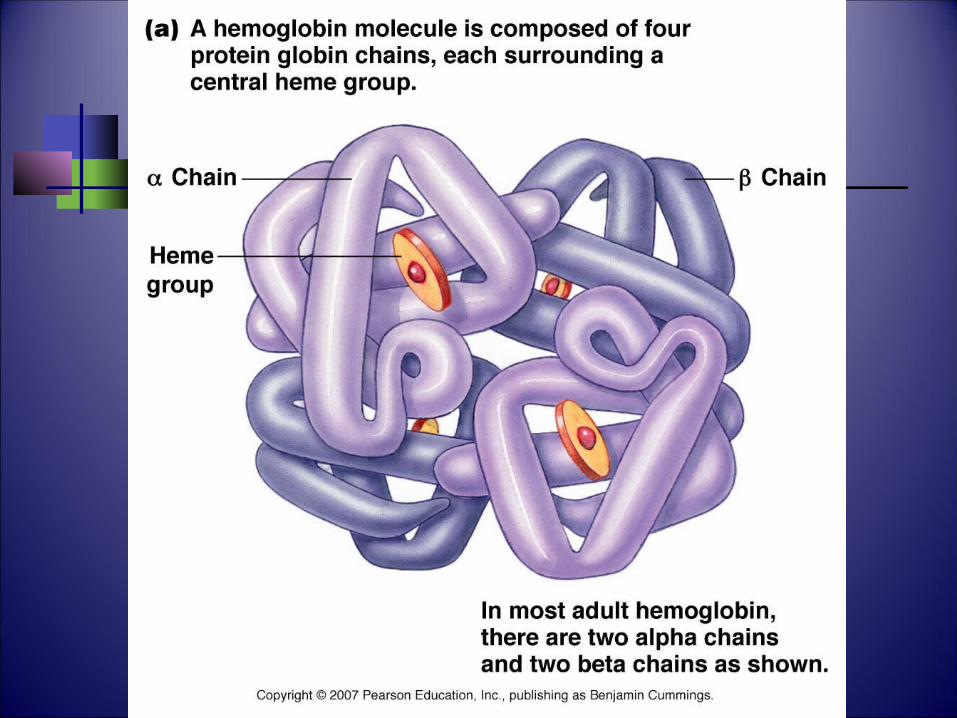
Hb and O2 Transport
Each Hb has 4 polypeptide (2 α and 2 ) chains, each combined with heme group.
In the center of each heme group is 1 atom of iron that can combine with 1 molecule O2.
One Hb molecule thus combine with four O2 molecules.
There are about 280 million Hb molecules/RBC
each RBC can carry over a billion molecules of O2
Hemoglobin
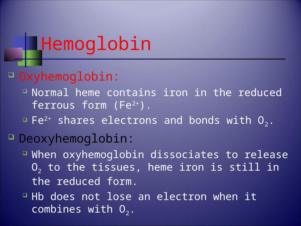
Oxyhemoglobin: Normal heme contains iron in the reduced
ferrous form (Fe2+). Fe2+ shares electrons and bonds with O2.
Deoxyhemoglobin: When oxyhemoglobin dissociates to release O2
to the tissues, heme iron is still in the reduced form.
Hb does not lose an electron when it combines with O2.
Hemoglobin (continued)
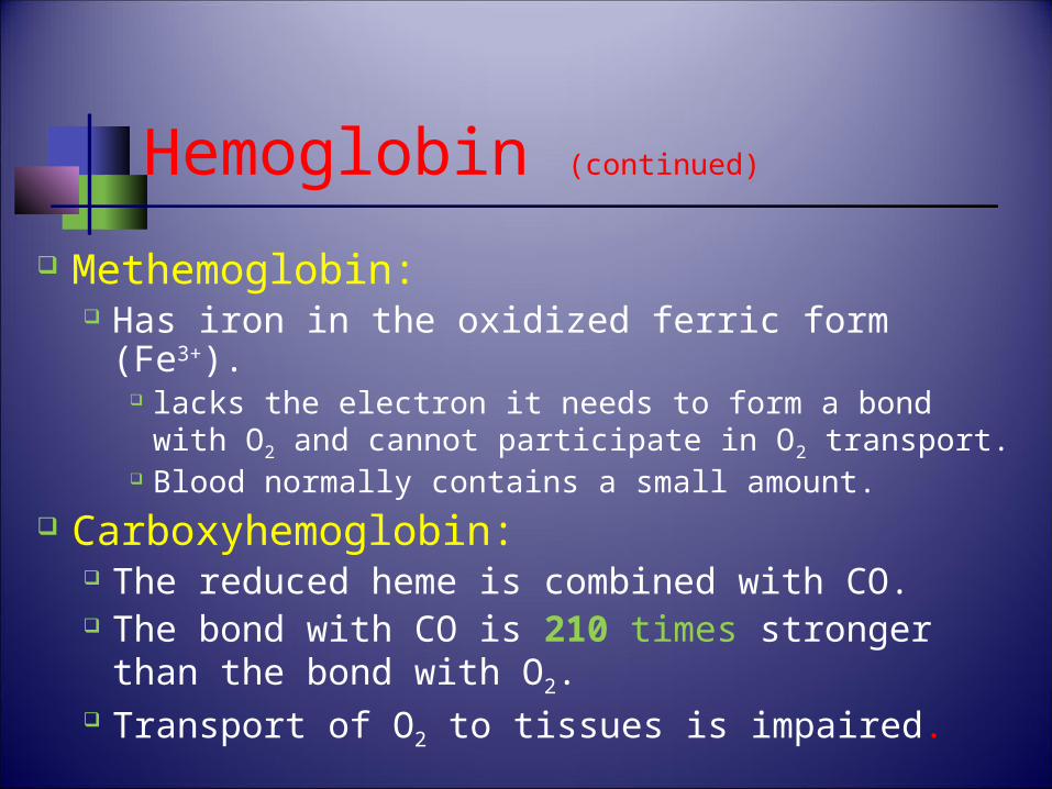
Methemoglobin: Has iron in the oxidized ferric form (Fe3+).
lacks the electron it needs to form a bond with O2 and cannot participate in O2 transport.
Blood normally contains a small amount. Carboxyhemoglobin:
The reduced heme is combined with CO. The bond with CO is 210 times stronger
than the bond with O2. Transport of O2 to tissues is impaired.
Hb Concentration
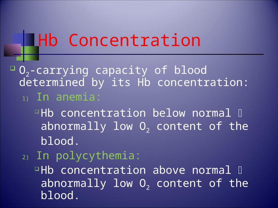
O2-carrying capacity of blood determined by its Hb concentration:1) In anemia:
Hb concentration below normal abnormally low O2 content of the blood.
2) In polycythemia:Hb concentration above normal abnormally low O2 content of the blood.
Hb Concentration
Erythropoietin: hormone produced by the kidneys controls production of RBCs in the bone marrow.
If the amount of O2 delivered to the kidneys is lower than normal stimulates secretion of erythropoietin production of RBCs.
Androgens: promotes RBCs production explains why the Hb concentration in men is
from 1 to 2 g per 100 ml higher than in women.
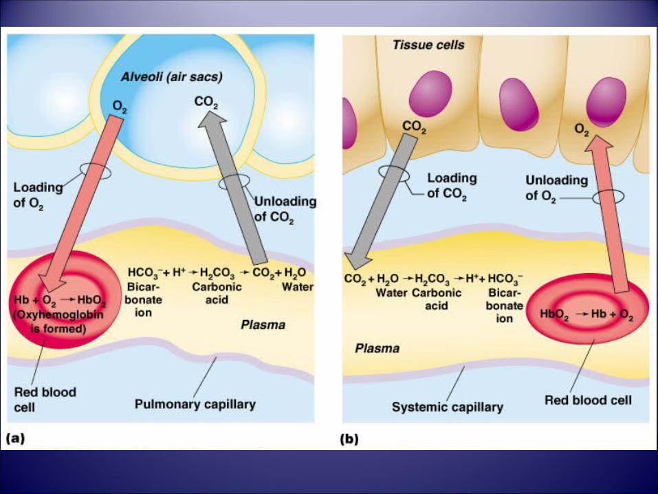
The Loading and Unloading Reactions
Deoxyhemoglobin + O2 Oxyhemoglobin (loading reaction) occurs in the lungs
Oxyhemoglobin deoxyhemoglobin + O2 (Unloading reaction) occurs in the tissues.
Loading and unloading shown as a reversible reaction:
(tissues)Deoxyhemoglobin + O2 Oxyhemoglobin (lungs)
The Loading and Unloading Reactions
The extent to which the reaction will go in each direction depends on:
1) PO2 of the environment High PO2 drives the equation to the Rt. (favors
the loading reaction). Low PO2 in the systemic capillaries drives the
reaction in the opposite direction (promote unloading).
The extent of this unloading depends on how low the PO2 values are.
The Loading and Unloading Reactions
2) affinity (bond strength) between Hb and O2. Very strong bond would favor loading but
inhibit unloading Weak bond would hinder loading but improve
unloading. The bond strength between Hb and O2 is
normally strong enough so that 97% of the Hb leaving the lungs is in the form of oxyHb.
BUT the bond is sufficiently weak so that adequate amounts of O2 are unloaded to sustain aerobic respiration in the tissues.
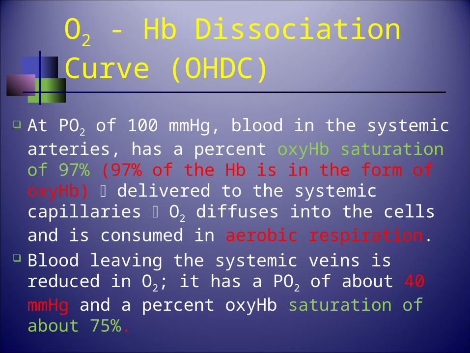
O2 - Hb Dissociation Curve (OHDC)
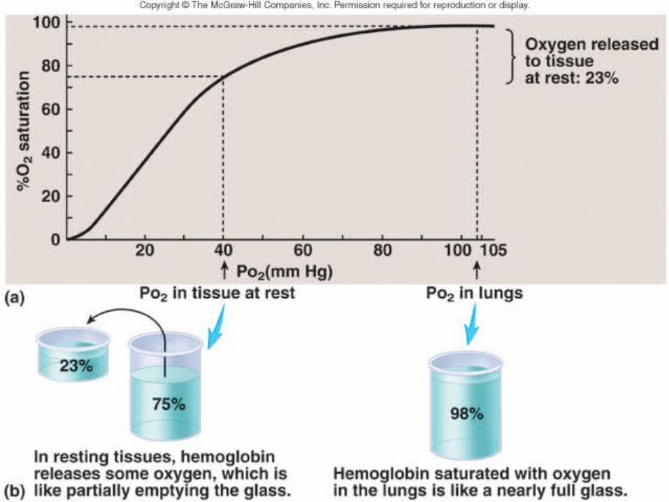
At PO2 of 100 mmHg, blood in the systemic arteries, has a percent oxyHb saturation of 97% (97% of the Hb is in the form of oxyHb) delivered to the systemic capillaries O2 diffuses into the cells and is consumed in aerobic respiration.
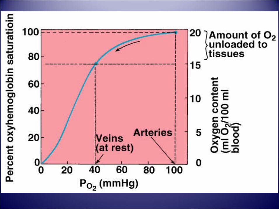
Blood leaving the systemic veins is reduced in O2; it has a PO2 of about 40 mmHg and a percent oxyHb saturation of about 75%.
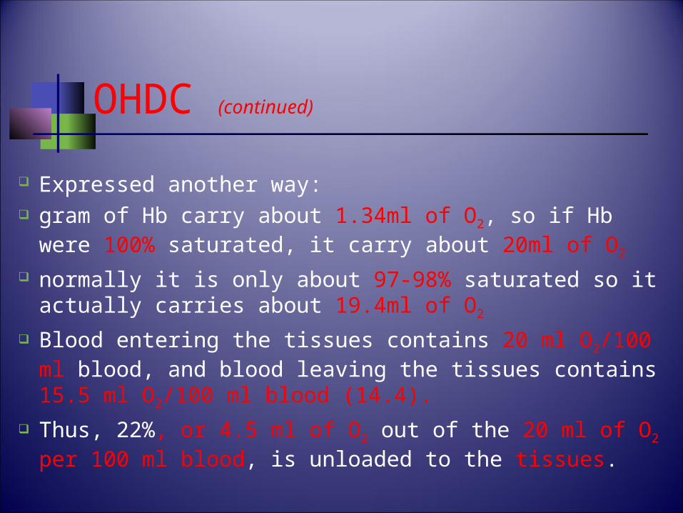
OHDC (continued)
Expressed another way: gram of Hb carry about 1.34ml of O2, so if Hb were
100% saturated, it carry about 20ml of O2 normally it is only about 97-98% saturated so it
actually carries about 19.4ml of O2
Blood entering the tissues contains 20 ml O2/100 ml blood, and blood leaving the tissues contains 15.5 ml O2/100 ml blood (14.4).
Thus, 22%, or 4.5 ml of O2 out of the 20 ml of O2 per 100 ml blood, is unloaded to the tissues.
OHDC (continued)
The relatively large amount of oxyHb remaining in the venous blood at rest serves as an O2 reserve.
If a person stops breathing, a sufficient reserve of O2 in the blood will keep the brain and heart alive for about 4 to 5 minutes without using cardiopulmonary resuscitation (CPR) techniques.
This reserve supply of O2 can also be tapped when a tissue’s requirements for O2 are raised.
OHDC (continued)
A graphic illustration of the percent oxyH saturation at different values of PO2
The values in this graph are obtained by subjecting samples of blood in vitro to different PO2.
The percent oxyHb saturations obtained can be used to predict what the unloading percentages would be in vivo with a given difference in arterial and venous PO2 values.

OHDC (continued)
It is S-shaped, or sigmoidal due to Heme- Heme interaction.
A. Plateau portion of the curve: Range that exists at the pulmonary
capillaries. relatively flat at high PO2 values indicates
that changes in PO2 within this range have little effect on the unloading reaction.
Minimal O2 transported until the PO2 falls below 60 mm Hg.
OHDC (continued)
B. Steep portion of the curve: Range that exists at the systemic
capillaries small changes in PO2 produce large
differences in % saturation (unload more O2)

A 36-year-old woman is found comatose at her home and is life-flighted to the nearest regional medical center. Blood gases reveal a normal PaO2 but a lower-than normal arterial O2 saturation. Which of the following conditions is most consistent with the findings?
a. Anemia b. Carbon monoxide poisoning c. Hypoventilation d. Low V/Q ratio e. Right-to-left shunt