RESEARCH Open Access 4-desaturation pathway for DHA ...
10
RESEARCH Open Access The Δ4-desaturation pathway for DHA biosynthesis is operative in the human species: Differences between normal controls and children with the Zellweger syndrome Manuela Martinez 1* , Natalia Ichaso 1 , Fernando Setien 2 , Nuria Durany 3 , Xiao Qiu 4 , William Roesler 5 Abstract Background: Docosahexaenoic acid (DHA, 22:6ω3) is a fundamental component of cell membranes, especially in the brain and retina. In the experimental animal, DHA deficiency leads to suboptimal neurological performance and visual deficiencies. Children with the Zellweger syndrome (ZS) have a profound DHA deficiency and symptoms that can be attributed to their extremely low DHA levels. These children seem to have a metabolic defect in DHA biosynthesis, which has never been totally elucidated. Treatment with DHA ethyl ester greatly improves these patients, but if we could normalize their endogenous DHA production we could get additional benefits. We examined whether DHA biosynthesis by Δ4-desaturation could be enhanced in the human species by transfecting the enzyme, and if this could normalize the DHA levels in cells from ZS patients. Results: We showed that the Δ4-desaturase gene (Fad4) from Thraustochytrium sp, which can be expressed by heterologous transfection in other plant and yeast cells, can also be transfected into human lymphocytes, and that it expresses the enzyme (FAD4, Δ4-desaturase) by producing DHA from direct Δ4-desaturation of 22:5ω3. We also found that the other substrate for Δ4-desaturase, 22:4ω6, was parallely desaturated to 22:5ω6. Conclusions: The present “in vitro” study demonstrates that Δ4-desaturase can be transfected into human cells and synthesize DHA (as well as 22:5ω6, DPA) from 22:5ω3 and 22:4ω6, respectively, by putative Δ4-desaturation. Even if this pathway may not be the physiological route for DHA biosynthesis “in vivo”, the present study opens new perspectives for the treatment of patients within the ZS spectrum. Background Docosahexaenoic acid (DHA, 22:6ω3) is a polyunsatu- rated fatty acid (PUFA) of fundamental importance in cell membranes, especially in nerve endings and the photoreceptor cells in the retina [1,2]. DHA is consid- ered essential during brain development, especially after 31 weeks of gestation, when its accretion is maximal [3]. In the clinical setting, DHA deficiency has been related to several human diseases [4]. In the human species, it is generally agreed that synth- esis of DHA from its essential precursor 18:3ω3(a-lino- lenic acid) is inadequate, especially in the premature infant, where it does not cover the daily needs of this important PUFA. Because of that, DHA is being added to many infant formulas during the last years. Even in the adult, DHA is being increasingly recommended to improve several conditions, with more or less convin- cing basis and results. Thus, based on its marginally decreased levels, DHA is currently being recommended as a supplement for such varied diseases as attention deficit hyperactivity disorder [5], fetal alcohol syndrome [6], phenylketonuria [7], schizophrenia [8], unipolar depression [9], aggressive behavior [10], Alzheimer’s dis- ease [11] and diabetes [12]. However, it is only in one group of diseases -the Zellweger syndrome and its related phenotypes- where the DHA levels are dramati- cally diminished in all tissues, including the brain and retina [13,14]. * Correspondence: [email protected] 1 Manuela Martinez Foundation for Children with Metabolic Diseases. Research Laboratory. Plaza Karl Marx 1, Barcelona 08042, Spain Full list of author information is available at the end of the article Martinez et al. Lipids in Health and Disease 2010, 9:98 http://www.lipidworld.com/content/9/1/98 © 2010 Martinez et al; licensee BioMed Central Ltd. This is an Open Access article distributed under the terms of the Creative Commons Attribution License (http://creativecommons.org/licenses/by/2.0), which permits unrestricted use, distribution, and reproduction in any medium, provided the original work is properly cited.
Transcript of RESEARCH Open Access 4-desaturation pathway for DHA ...
RESEARCH Open Access
The Δ4-desaturation pathway for DHAbiosynthesis is operative in the human species:Differences between normal controls andchildren with the Zellweger syndromeManuela Martinez1*, Natalia Ichaso1, Fernando Setien2, Nuria Durany3, Xiao Qiu4, William Roesler5
Abstract
Background: Docosahexaenoic acid (DHA, 22:6ω3) is a fundamental component of cell membranes, especially inthe brain and retina. In the experimental animal, DHA deficiency leads to suboptimal neurological performanceand visual deficiencies. Children with the Zellweger syndrome (ZS) have a profound DHA deficiency and symptomsthat can be attributed to their extremely low DHA levels. These children seem to have a metabolic defect in DHAbiosynthesis, which has never been totally elucidated. Treatment with DHA ethyl ester greatly improves thesepatients, but if we could normalize their endogenous DHA production we could get additional benefits. Weexamined whether DHA biosynthesis by Δ4-desaturation could be enhanced in the human species by transfectingthe enzyme, and if this could normalize the DHA levels in cells from ZS patients.
Results: We showed that the Δ4-desaturase gene (Fad4) from Thraustochytrium sp, which can be expressed byheterologous transfection in other plant and yeast cells, can also be transfected into human lymphocytes, and thatit expresses the enzyme (FAD4, Δ4-desaturase) by producing DHA from direct Δ4-desaturation of 22:5ω3. We alsofound that the other substrate for Δ4-desaturase, 22:4ω6, was parallely desaturated to 22:5ω6.
Conclusions: The present “in vitro” study demonstrates that Δ4-desaturase can be transfected into human cellsand synthesize DHA (as well as 22:5ω6, DPA) from 22:5ω3 and 22:4ω6, respectively, by putative Δ4-desaturation.Even if this pathway may not be the physiological route for DHA biosynthesis “in vivo”, the present study opensnew perspectives for the treatment of patients within the ZS spectrum.
BackgroundDocosahexaenoic acid (DHA, 22:6ω3) is a polyunsatu-rated fatty acid (PUFA) of fundamental importance incell membranes, especially in nerve endings and thephotoreceptor cells in the retina [1,2]. DHA is consid-ered essential during brain development, especially after31 weeks of gestation, when its accretion is maximal [3].In the clinical setting, DHA deficiency has been relatedto several human diseases [4].In the human species, it is generally agreed that synth-
esis of DHA from its essential precursor 18:3ω3 (a-lino-lenic acid) is inadequate, especially in the premature
infant, where it does not cover the daily needs of thisimportant PUFA. Because of that, DHA is being addedto many infant formulas during the last years. Even inthe adult, DHA is being increasingly recommended toimprove several conditions, with more or less convin-cing basis and results. Thus, based on its marginallydecreased levels, DHA is currently being recommendedas a supplement for such varied diseases as attentiondeficit hyperactivity disorder [5], fetal alcohol syndrome[6], phenylketonuria [7], schizophrenia [8], unipolardepression [9], aggressive behavior [10], Alzheimer’s dis-ease [11] and diabetes [12]. However, it is only in onegroup of diseases -the Zellweger syndrome and itsrelated phenotypes- where the DHA levels are dramati-cally diminished in all tissues, including the brain andretina [13,14].
* Correspondence: [email protected] Martinez Foundation for Children with Metabolic Diseases.Research Laboratory. Plaza Karl Marx 1, Barcelona 08042, SpainFull list of author information is available at the end of the article
Martinez et al. Lipids in Health and Disease 2010, 9:98http://www.lipidworld.com/content/9/1/98
© 2010 Martinez et al; licensee BioMed Central Ltd. This is an Open Access article distributed under the terms of the CreativeCommons Attribution License (http://creativecommons.org/licenses/by/2.0), which permits unrestricted use, distribution, andreproduction in any medium, provided the original work is properly cited.
Generalized peroxisomal disorders within the Zellwe-ger syndrome spectrum, usually called peroxisomalbiogenesis disorders (PBD), are lethal congenital diseases,characterized by the lack of functional peroxisomes inthe cells of the body [15]. As a consequence, several bio-chemical reactions related to lipid metabolism, whichnormally occur in the peroxisome, are affected. Amongthem, b-oxidation of pristanic and very long chain fattyacids (VLCFA) [16], as well as a-oxidation of phytanicacid [17], are defective. Biosynthesis of plasmalogens[18] and biliary acids is also defective [19]. In addition,we found that these children show a profound DHAdeficiency [13,14]. In classic Zellweger’s syndrome (ZS),the prototype of these disorders, brain DHA levels maybe so low as to only account for 20-25% of the normalvalues [3,13], and in the Zellweger retina, DHA isvirtually absent [14].Clinically, these patients become blind, deaf, and
mentally retarded very early in life. Myelin is alwaysdefective in classic Zellweger’s syndrome (ZS), in bothamount and quality (dysmyelination), and liver functionis consistently impaired. Renal cysts are commonlyadded, which gave the name of cerebro-hepato-renalsyndrome to the first ZS patients described [20,21] Thelife span of these patients is very short, especially inclassic ZS, where survival rarely surpasses the first yearof age. There are milder Zellweger variants, known asneonatal adrenoleukodystrophy (NALD) and infantileRefsum’s disease (IRD). These relatively milder pheno-types, however, present most of the symptoms of classicZS, including profound mental retardation with myelina-tion delay, followed later by demyelination, visual andhearing sensorineural defects and liver involvement.There is no means to differentiate the various ZS phe-notypes, when based on genotype or biochemicalabnormalities alone. They are mainly clinical entities,with extremely variable organ involvement and prog-nosis. Without a treatment and special care, however,all these patients live very short lives, rarely reachingolder childhood. In spite of that, correcting their DHAdeficiency produces substantial benefits in their myelina-tion [22], liver function [23] and vision [24], thusprolonging survival and quality of life markedly.We believe that DHA deficiency plays a crucial role inthis group of congenital disorders, as most of thepatients’ symptoms are consistent with what is foundin DHA-deficient animals [25] and humans [26]. ZSpatients - even more than normal children - are prob-ably incapable of producing all the DHA required by thefast development of their brain and retina.There is some controversy about the mechanism of
DHA biosynthesis in mammals. Classically, it wasaccepted that DHA is formed by front-end, putativeΔ4-desaturation of its immediate precursor 22:5ω3, by
introducing a double bond in position Δ4 (countingfrom the carboxyl end, IUPAC nomenclature). In paral-lel, this enzyme would also convert 22:4ω6 to 22:5ω6(ω6 docosapentaeoic acid, DPA). In lower eukaryotes,like the Thraustochytrium sp, the existence of aΔ4-desaturase has been demonstrated. When expressedin yeast (Saccharomyces cerevisiae) and plants (Brassicajuncea), this enzyme Δ4-desaturates 22:5ω3 to 22:6ω3[27]. This results in the controlled production of doco-sahexaenoic acid, an interesting method for the nutra-ceutical industry, given the decreasing supply of DHAfrom natural, marine products.In mammals, this pathway has practically been aban-
doned and an alternate route involving the peroxisomehas been generally accepted [28]. This route wouldimply the microsomal elongation of 22:5ω3 to 24:5ω3,followed by a second Δ6-desaturation step to 24:6ω3,which would finally be b-oxidated in the peroxisome toproduce DHA. Again, the same route would work forthe ω6 family, finally yielding 22:5ω6 from 24:5ω6.Whether or not humans have an intrinsic Δ4-desaturaseremains unknown. Nevertheless, the present papershows for the first time that the Δ4-desaturation path-way is functional in the human species when cells aretransfected with the enzyme. We show that when thegene that codes for this enzyme (Fad4) is transfectedinto human lymphocytes immortalized with the Epstein-Barr virus (EBV), putative Δ4-desaturation of both sub-strates, 22:5ω3 and 22:4ω6, is produced, yielding DHAand DPA, respectively, as their products.
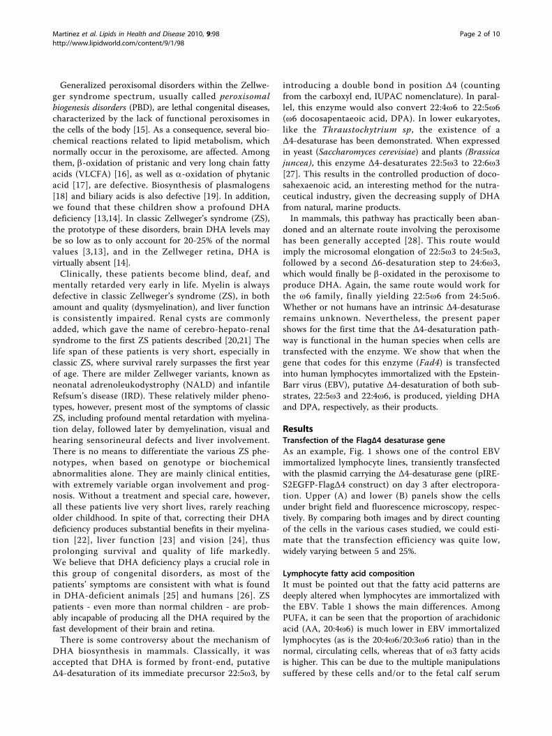
ResultsTransfection of the FlagΔ4 desaturase geneAs an example, Fig. 1 shows one of the control EBVimmortalized lymphocyte lines, transiently transfectedwith the plasmid carrying the Δ4-desaturase gene (pIRE-S2EGFP-FlagΔ4 construct) on day 3 after electropora-tion. Upper (A) and lower (B) panels show the cellsunder bright field and fluorescence microscopy, respec-tively. By comparing both images and by direct countingof the cells in the various cases studied, we could esti-mate that the transfection efficiency was quite low,widely varying between 5 and 25%.
Lymphocyte fatty acid compositionIt must be pointed out that the fatty acid patterns aredeeply altered when lymphocytes are immortalized withthe EBV. Table 1 shows the main differences. AmongPUFA, it can be seen that the proportion of arachidonicacid (AA, 20:4ω6) is much lower in EBV immortalizedlymphocytes (as is the 20:4ω6/20:3ω6 ratio) than in thenormal, circulating cells, whereas that of ω3 fatty acidsis higher. This can be due to the multiple manipulationssuffered by these cells and/or to the fetal calf serum
Martinez et al. Lipids in Health and Disease 2010, 9:98http://www.lipidworld.com/content/9/1/98
Page 2 of 10
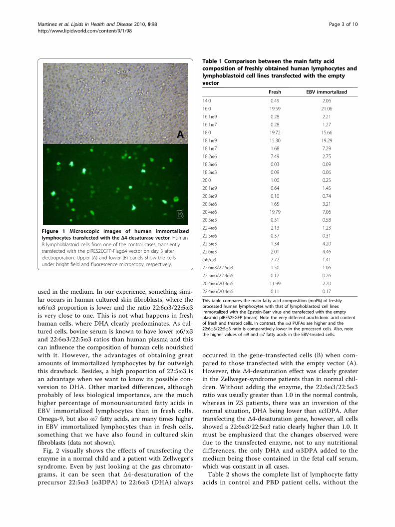
used in the medium. In our experience, something simi-lar occurs in human cultured skin fibroblasts, where theω6/ω3 proportion is lower and the ratio 22:6ω3/22:5ω3is very close to one. This is not what happens in freshhuman cells, where DHA clearly predominates. As cul-tured cells, bovine serum is known to have lower ω6/ω3and 22:6ω3/22:5ω3 ratios than human plasma and thiscan influence the composition of human cells nourishedwith it. However, the advantages of obtaining greatamounts of immortalized lymphocytes by far outweighthis drawback. Besides, a high proportion of 22:5ω3 isan advantage when we want to know its possible con-version to DHA. Other marked differences, althoughprobably of less biological importance, are the muchhigher percentage of monounsaturated fatty acids inEBV immortalized lymphocytes than in fresh cells.Omega-9, but also ω7 fatty acids, are many times higherin EBV immortalized lymphocytes than in fresh cells,something that we have also found in cultured skinfibroblasts (data not shown).Fig. 2 visually shows the effects of transfecting the
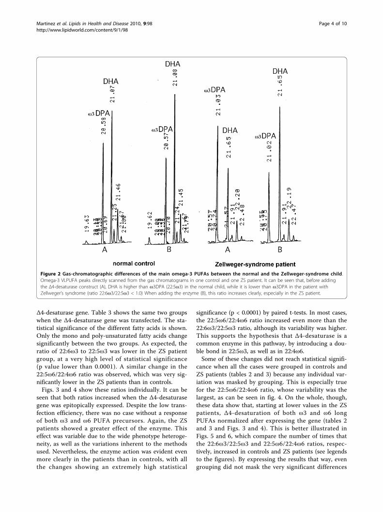
enzyme in a normal child and a patient with Zellweger’ssyndrome. Even by just looking at the gas chromato-grams, it can be seen that Δ4-desaturation of theprecursor 22:5ω3 (ω3DPA) to 22:6ω3 (DHA) always
occurred in the gene-transfected cells (B) when com-pared to those transfected with the empty vector (A).However, this Δ4-desaturation effect was clearly greaterin the Zellweger-syndrome patients than in normal chil-dren. Without adding the enzyme, the 22:6ω3/22:5ω3ratio was usually greater than 1.0 in the normal controls,whereas in ZS patients, there was an inversion of thenormal situation, DHA being lower than ω3DPA. Aftertransfecting the Δ4-desaturation gene, however, all cellsshowed a 22:6ω3/22:5ω3 ratio clearly higher than 1.0. Itmust be emphasized that the changes observed weredue to the transfected enzyme, not to any nutritionaldifferences, the only DHA and ω3DPA added to themedium being those contained in the fetal calf serum,which was constant in all cases.Table 2 shows the complete list of lymphocyte fatty
acids in control and PBD patient cells, without the
Figure 1 Microscopic images of human immortalizedlymphocytes transfected with the Δ4-desaturase vector. HumanB lymphoblastoid cells from one of the control cases, transientlytransfected with the pIRES2EGFP-FlagΔ4 vector on day 3 afterelectroporation. Upper (A) and lower (B) panels show the cellsunder bright field and fluorescence microscopy, respectively.
Table 1 Comparison between the main fatty acidcomposition of freshly obtained human lymphocytes andlymphoblastoid cell lines transfected with the emptyvector
Fresh EBV immortalized
14:0 0.49 2.06
16:0 19.59 21.06
16:1ω9 0.28 2.21
16:1ω7 0.28 1.27
18:0 19.72 15.66
18:1ω9 15.30 19.29
18:1ω7 1.68 7.29
18:2ω6 7.49 2.75
18:3ω6 0.03 0.09
18:3ω3 0.09 0.06
20:0 1.00 0.25
20:1ω9 0.64 1.45
20:3ω9 0.10 0.74
20:3ω6 1.65 3.21
20:4ω6 19.79 7.06
20:5ω3 0.31 0.58
22:4ω6 2.13 1.23
22:5ω6 0.37 0.31
22:5ω3 1.34 4.20
22:6ω3 2.01 4.46
ω6/ω3 7.72 1.41
22:6ω3/22:5ω3 1.50 1.06
22:5ω6/22:4ω6 0.17 0.26
20:4ω6/20:3ω6 11.99 2.20
22:4ω6/20:4ω6 0.11 0.17
This table compares the main fatty acid composition (mol%) of freshlyprocessed human lymphocytes with that of lymphoblastoid cell linesimmortalized with the Epstein-Barr virus and transfected with the emptyplasmid pIRES2EGFP (mean). Note the very different arachidonic acid contentof fresh and treated cells. In contrast, the ω3 PUFAs are higher and the22:6ω3/22:5ω3 ratio is comparatively lower in the processed cells. Also, notethe higher values of ω9 and ω7 fatty acids in the EBV-treated cells.
Martinez et al. Lipids in Health and Disease 2010, 9:98http://www.lipidworld.com/content/9/1/98
Page 3 of 10
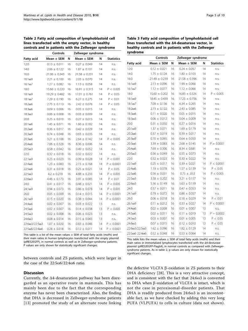
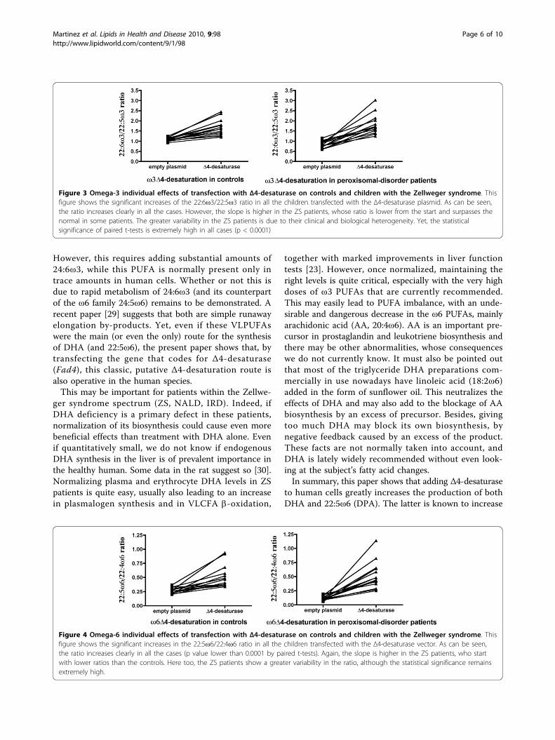
Δ4-desaturase gene. Table 3 shows the same two groupswhen the Δ4-desaturase gene was transfected. The sta-tistical significance of the different fatty acids is shown.Only the mono and poly-unsaturated fatty acids changesignificantly between the two groups. As expected, theratio of 22:6ω3 to 22:5ω3 was lower in the ZS patientgroup, at a very high level of statistical significance(p value lower than 0.0001). A similar change in the22:5ω6/22:4ω6 ratio was observed, which was very sig-nificantly lower in the ZS patients than in controls.Figs. 3 and 4 show these ratios individually. It can be
seen that both ratios increased when the Δ4-desaturasegene was epitopically expressed. Despite the low trans-fection efficiency, there was no case without a responseof both ω3 and ω6 PUFA precursors. Again, the ZSpatients showed a greater effect of the enzyme. Thiseffect was variable due to the wide phenotype heteroge-neity, as well as the variations inherent to the methodsused. Nevertheless, the enzyme action was evident evenmore clearly in the patients than in controls, with allthe changes showing an extremely high statistical
significance (p < 0.0001) by paired t-tests. In most cases,the 22:5ω6/22:4ω6 ratio increased even more than the22:6ω3/22:5ω3 ratio, although its variability was higher.This supports the hypothesis that Δ4-desaturase is acommon enzyme in this pathway, by introducing a dou-ble bond in 22:5ω3, as well as in 22:4ω6.Some of these changes did not reach statistical signifi-
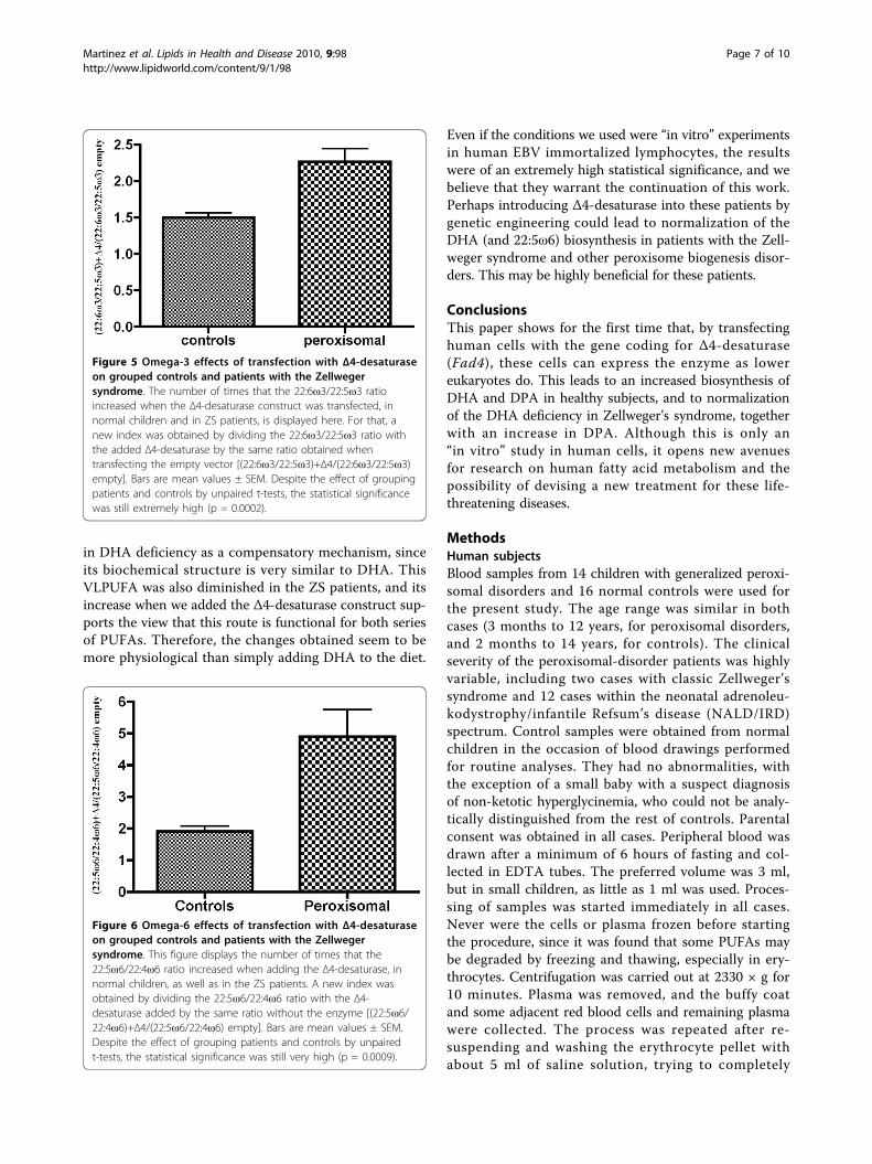
cance when all the cases were grouped in controls andZS patients (tables 2 and 3) because any individual var-iation was masked by grouping. This is especially truefor the 22:5ω6/22:4ω6 ratio, whose variability was thelargest, as can be seen in fig. 4. On the whole, though,these data show that, starting at lower values in the ZSpatients, Δ4-desaturation of both ω3 and ω6 longPUFAs normalized after expressing the gene (tables 2and 3 and Figs. 3 and 4). This is better illustrated inFigs. 5 and 6, which compare the number of times thatthe 22:6ω3/22:5ω3 and 22:5ω6/22:4ω6 ratios, respec-tively, increased in controls and ZS patients (see legendsto the figures). By expressing the results that way, evengrouping did not mask the very significant differences
Figure 2 Gas-chromatographic differences of the main omega-3 PUFAs between the normal and the Zellweger-syndrome child.Omega-3 VLPUFA peaks directly scanned from the gas chromatograms in one control and one ZS patient. It can be seen that, before addingthe Δ4-desaturase construct (A), DHA is higher than ω3DPA (22:5ω3) in the normal child, while it is lower than ω3DPA in the patient withZellweger’s syndrome (ratio 22:6ω3/22:5ω3 < 1.0) When adding the enzyme (B), this ratio increases clearly, especially in the ZS patient.
Martinez et al. Lipids in Health and Disease 2010, 9:98http://www.lipidworld.com/content/9/1/98
Page 4 of 10
between controls and ZS patients, which were larger inthe case of the 22:5ω6/22:4ω6 ratio.
DiscussionCurrently, the Δ4-desaturation pathway has been disre-garded as an operative route in mammals. This hasmainly been due to the fact that the correspondingenzyme has never been characterized. Also, the findingthat DHA is decreased in Zellweger-syndrome patients[13] promoted the study of an alternate route linking
the defective VLCFA b-oxidation in ZS patients to theirDHA deficiency [28]. This is a very attractive concept,and is consistent with the fact that 24:6ω3 is convertedto DHA when b-oxidation of VLCFA is intact, which isnot the case in peroxisomal-disorder patients. ThatDHA is readily produced from 24:6ω3 is an incontest-able fact, as we have checked by adding this very longPUFA (VLPUFA) to cells in culture (data not shown).
Table 2 Fatty acid composition of lymphoblastoid celllines transfected with the empty vector, in healthycontrols and in patients with the Zellweger syndrome
Controls Zellweger syndrome
Fatty acid Mean ± SEM N Mean ± SEM N Statistics
12:0 0.13 ± 0.011 16 0.27 ± 0.049 14 n.s.
14:0 2.06 ± 0.122 16 1.87 ± 0.101 14 n.s.
16:0 21.06 ± 0.343 16 21.58 ± 0.231 14 n.s.
16:1ω9 2.21 ± 0.100 16 2.03 ± 0.070 14 n.s.
16:1ω7 1.27 ± 0.082 16 1.13 ± 0.058 14 n.s.
18:0 15.66 ± 0.220 16 16.91 ± 0.315 14 P < 0.005
18:1ω9 19.29 ± 0.482 16 17.01 ± 0.761 14 P < 0.05
18:1ω7 7.29 ± 0.190 16 6.27 ± 0.275 14 P < 0.01
18:2ω6 2.75 ± 0.113 16 2.42 ± 0.076 14 P < 0.05
18:3ω6 0.09 ± 0.006 16 0.05 ± 0.015 14 n.s.
18:3ω3 0.06 ± 0.008 16 0.03 ± 0.009 14 n.s.
20:0 0.25 ± 0.010 16 0.27 ± 0.015 14 n.s.
20:1ω6 1.45 ± 0.071 16 1.66 ± 0.182 14 n.s.
20:2ω6 0.36 ± 0.017 16 0.42 ± 0.029 14 n.s.
20:3ω9 0.74 ± 0.048 16 0.65 ± 0.035 14 n.s.
20:3ω6 3.21 ± 0.108 16 2.64 ± 0.142 14 P < 0.005
20:4ω6 7.06 ± 0.328 16 8.36 ± 0.696 14 n.s.
20:5ω3 0.58 ± 0.042 16 0.49 ± 0.052 14 n.s.
22:0 0.32 ± 0.018 16 0.32 ± 0.022 14 n.s.
22:1ω9 0.25 ± 0.023 16 0.39 ± 0.028 14 P < 0.001
22:4ω6 1.23 ± 0.083 16 2.15 ± 0.168 14 P < 0.0001
22:5ω6 0.31 ± 0.018 16 0.25 ± 0.021 14 P < 0.05
22:5ω3 4.2 ± 0.218 16 4.88 ± 0.233 14 P < 0.005
22:6ω3 4.46 ± 0.173 16 3.91 ± 0.085 14 P < 0.01
24:0 0.41 ± 0.017 16 0.48 ± 0.021 14 P < 0.005
24:1ω9 0.94 ± 0.073 16 0.86 ± 0.078 14 P < 0.005
26:0 0.05 ± 0.009 16 0.14 ± 0.015 14 P < 0.0001
26:1ω9 0.15 ± 0.020 16 0.38 ± 0.044 14 P < 0.0001
24:4ω6 0.02 ± 0.007 16 0.03 ± 0.022 13 n.s.
24:5ω6 0.02 ± 0.007 16 0.14 ± 0.038 13 P < 0.005
24:5ω3 0.02 ± 0.008 16 0.06 ± 0.023 13 n.s.
24:6ω3 0.08 ± 0.014 16 0.15 ± 0.043 13 n.s.
22:6ω3/22:5ω3 1.07 ± 0.020 16 0.83 ± 0.046 14 P < 0.0001
22:5ω6/22:4ω6 0.28 ± 0.018 16 0.12 ± 0.011 14 P < 0.0001
This table is a list of the mean values ± SEM of total fatty acids (mol%) andtheir main ratios in human lymphocytes transfected with the empty plasmid(pIRES2EGFP), in normal controls as well as in Zellweger-syndrome patients.P values are only shown for statistically significant changes.
Table 3 Fatty acid composition of lymphoblastoid celllines transfected with the Δ4-desaturase vector, inhealthy controls and in patients with the Zellwegersyndrome
Controls Zellweger syndrome
Fatty acid Mean ± SEM N Mean ± SEM N Statistics
12:0 0.12 ± 0.021 16 0.24 ± 0.057 14 n.s.
14:0 1.75 ± 0.124 16 1.80 ± 0.103 14 n.s.
16:0 21.49 ± 0.239 16 21.58 ± 0.396 14 n.s.
16:1ω9 2.13 ± 0.096 16 1.99 ± 0.066 14 n.s.
16:1ω7 1.12 ± 0.077 16 1.12 ± 0.066 14 n.s.
18:0 15.60 ± 0.202 16 16.83 ± 0.326 14 P < 0.005
18:1ω9 18.45 ± 0.439 16 17.25 ± 0.756 14 n.s.
18:1ω7 7.09 ± 0.136 16 6.34 ± 0.265 14 n.s.
18:2ω6 2.73 ± 0.122 16 2.43 ± 0.085 14 n.s.
18:3ω6 0.11 ± 0.020 16 0.05 ± 0.015 14 n.s.
18:3ω3 0.06 ± 0.012 16 0.04 ± 0.009 14 n.s.
20:0 0.31 ± 0.050 16 0.27 ± 0.016 14 n.s.
20:1ω9 1.37 ± 0.071 16 1.69 ± 0.179 14 n.s.
20:2ω6 0.37 ± 0.019 16 0.39 ± 0.017 14 n.s.
20:3ω9 0.70 ± 0.065 16 0.64 ± 0.050 14 n.s.
20:3ω6 3.39 ± 0.083 16 2.68 ± 0.145 14 P = 0.0001
20:4ω6 7.69 ± 0.306 16 8.34 ± 0.681 14 n.s.
20:5ω3 0.56 ± 0.049 16 0.55 ± 0.073 14 n.s.
22:0 0.32 ± 0.023 16 0.30 ± 0.022 14 n.s.
22:1ω9 0.25 ± 0.017 16 0.39 ± 0.021 14 P < 0.0001
22:4ω6 1.19 ± 0.078 16 1.57 ± 0.139 14 P < 0.05
22:5ω6 0.56 ± 0.031 16 0.75 ± .053 14 P < 0.005
22:5ω3 3.58 ± 0.202 16 3.21 ± 0.157 14 n.s.
22:6ω3 5.56 ± 0.149 16 5.63 ± 0.159 14 n.s.
24:0 0.37 ± 0.011 16 0.47 ± 0.053 14 n.s.
24:1ω9 0.79 ± 0.072 16 0.86 ± 0.057 14 n.s.
26:0 0.06 ± 0.018 16 0.16 ± 0.029 14 P < 0.01
26:1ω9 0.11 ± 0.012 16 0.33 ± 0.027 14 P < 0.0001
24:4ω6 0.02 ± 0.008 16 0.01 ± 0.007 13 n.s.
24:5ω6 0.02 ± 0.011 16 0.11 ± 0.019 13 P = 0,0002
24:5ω3 0.03 ± 0.007 16 0.01 ± 0.005 13 P < 0.05
24:6ω3 0.07 ± 0.015 16 0.12 ± 0.010 13 P < 0.05
22:6ω3/22:5ω3 1.62 ± 0.096 16 1.82 ± 0.129 14 n.s.
22:5ω6 22:4ω6 0.52 ± 0.048 16 0.53 ± 0.064 14 n.s.
This table lists the mean values ± SEM of total fatty acids (mol%) and theirmain ratios in immortalized lymphocytes transfected with the Δ4-desturaseplasmid (pIRES2EGFP-FlagΔ4), in normal controls as compared with Zellweger-syndrome patients. As in table 2, p values are only shown for statisticallysignificant changes.
Martinez et al. Lipids in Health and Disease 2010, 9:98http://www.lipidworld.com/content/9/1/98
Page 5 of 10
However, this requires adding substantial amounts of24:6ω3, while this PUFA is normally present only intrace amounts in human cells. Whether or not this isdue to rapid metabolism of 24:6ω3 (and its counterpartof the ω6 family 24:5ω6) remains to be demonstrated. Arecent paper [29] suggests that both are simple runawayelongation by-products. Yet, even if these VLPUFAswere the main (or even the only) route for the synthesisof DHA (and 22:5ω6), the present paper shows that, bytransfecting the gene that codes for Δ4-desaturase(Fad4), this classic, putative Δ4-desaturation route isalso operative in the human species.This may be important for patients within the Zellwe-
ger syndrome spectrum (ZS, NALD, IRD). Indeed, ifDHA deficiency is a primary defect in these patients,normalization of its biosynthesis could cause even morebeneficial effects than treatment with DHA alone. Evenif quantitatively small, we do not know if endogenousDHA synthesis in the liver is of prevalent importance inthe healthy human. Some data in the rat suggest so [30].Normalizing plasma and erythrocyte DHA levels in ZSpatients is quite easy, usually also leading to an increasein plasmalogen synthesis and in VLCFA b-oxidation,
together with marked improvements in liver functiontests [23]. However, once normalized, maintaining theright levels is quite critical, especially with the very highdoses of ω3 PUFAs that are currently recommended.This may easily lead to PUFA imbalance, with an unde-sirable and dangerous decrease in the ω6 PUFAs, mainlyarachidonic acid (AA, 20:4ω6). AA is an important pre-cursor in prostaglandin and leukotriene biosynthesis andthere may be other abnormalities, whose consequenceswe do not currently know. It must also be pointed outthat most of the triglyceride DHA preparations com-mercially in use nowadays have linoleic acid (18:2ω6)added in the form of sunflower oil. This neutralizes theeffects of DHA and may also add to the blockage of AAbiosynthesis by an excess of precursor. Besides, givingtoo much DHA may block its own biosynthesis, bynegative feedback caused by an excess of the product.These facts are not normally taken into account, andDHA is lately widely recommended without even look-ing at the subject’s fatty acid changes.In summary, this paper shows that adding Δ4-desaturase
to human cells greatly increases the production of bothDHA and 22:5ω6 (DPA). The latter is known to increase
Figure 3 Omega-3 individual effects of transfection with Δ4-desaturase on controls and children with the Zellweger syndrome. Thisfigure shows the significant increases of the 22:6ω3/22:5ω3 ratio in all the children transfected with the Δ4-desaturase plasmid. As can be seen,the ratio increases clearly in all the cases. However, the slope is higher in the ZS patients, whose ratio is lower from the start and surpasses thenormal in some patients. The greater variability in the ZS patients is due to their clinical and biological heterogeneity. Yet, the statisticalsignificance of paired t-tests is extremely high in all cases (p < 0.0001)
Figure 4 Omega-6 individual effects of transfection with Δ4-desaturase on controls and children with the Zellweger syndrome. Thisfigure shows the significant increases in the 22:5ω6/22:4ω6 ratio in all the children transfected with the Δ4-desaturase vector. As can be seen,the ratio increases clearly in all the cases (p value lower than 0.0001 by paired t-tests). Again, the slope is higher in the ZS patients, who startwith lower ratios than the controls. Here too, the ZS patients show a greater variability in the ratio, although the statistical significance remainsextremely high.
Martinez et al. Lipids in Health and Disease 2010, 9:98http://www.lipidworld.com/content/9/1/98
Page 6 of 10
in DHA deficiency as a compensatory mechanism, sinceits biochemical structure is very similar to DHA. ThisVLPUFA was also diminished in the ZS patients, and itsincrease when we added the Δ4-desaturase construct sup-ports the view that this route is functional for both seriesof PUFAs. Therefore, the changes obtained seem to bemore physiological than simply adding DHA to the diet.
Even if the conditions we used were “in vitro” experimentsin human EBV immortalized lymphocytes, the resultswere of an extremely high statistical significance, and webelieve that they warrant the continuation of this work.Perhaps introducing Δ4-desaturase into these patients bygenetic engineering could lead to normalization of theDHA (and 22:5ω6) biosynthesis in patients with the Zell-weger syndrome and other peroxisome biogenesis disor-ders. This may be highly beneficial for these patients.
ConclusionsThis paper shows for the first time that, by transfectinghuman cells with the gene coding for Δ4-desaturase(Fad4), these cells can express the enzyme as lowereukaryotes do. This leads to an increased biosynthesis ofDHA and DPA in healthy subjects, and to normalizationof the DHA deficiency in Zellweger’s syndrome, togetherwith an increase in DPA. Although this is only an“in vitro” study in human cells, it opens new avenuesfor research on human fatty acid metabolism and thepossibility of devising a new treatment for these life-threatening diseases.
MethodsHuman subjectsBlood samples from 14 children with generalized peroxi-somal disorders and 16 normal controls were used forthe present study. The age range was similar in bothcases (3 months to 12 years, for peroxisomal disorders,and 2 months to 14 years, for controls). The clinicalseverity of the peroxisomal-disorder patients was highlyvariable, including two cases with classic Zellweger’ssyndrome and 12 cases within the neonatal adrenoleu-kodystrophy/infantile Refsum’s disease (NALD/IRD)spectrum. Control samples were obtained from normalchildren in the occasion of blood drawings performedfor routine analyses. They had no abnormalities, withthe exception of a small baby with a suspect diagnosisof non-ketotic hyperglycinemia, who could not be analy-tically distinguished from the rest of controls. Parentalconsent was obtained in all cases. Peripheral blood wasdrawn after a minimum of 6 hours of fasting and col-lected in EDTA tubes. The preferred volume was 3 ml,but in small children, as little as 1 ml was used. Proces-sing of samples was started immediately in all cases.Never were the cells or plasma frozen before startingthe procedure, since it was found that some PUFAs maybe degraded by freezing and thawing, especially in ery-throcytes. Centrifugation was carried out at 2330 × g for10 minutes. Plasma was removed, and the buffy coatand some adjacent red blood cells and remaining plasmawere collected. The process was repeated after re-suspending and washing the erythrocyte pellet withabout 5 ml of saline solution, trying to completely
Figure 5 Omega-3 effects of transfection with Δ4-desaturaseon grouped controls and patients with the Zellwegersyndrome. The number of times that the 22:6ω3/22:5ω3 ratioincreased when the Δ4-desaturase construct was transfected, innormal children and in ZS patients, is displayed here. For that, anew index was obtained by dividing the 22:6ω3/22:5ω3 ratio withthe added Δ4-desaturase by the same ratio obtained whentransfecting the empty vector [(22:6ω3/22:5ω3)+Δ4/(22:6ω3/22:5ω3)empty]. Bars are mean values ± SEM. Despite the effect of groupingpatients and controls by unpaired t-tests, the statistical significancewas still extremely high (p = 0.0002).
Figure 6 Omega-6 effects of transfection with Δ4-desaturaseon grouped controls and patients with the Zellwegersyndrome. This figure displays the number of times that the22:5ω6/22:4ω6 ratio increased when adding the Δ4-desaturase, innormal children, as well as in the ZS patients. A new index wasobtained by dividing the 22:5ω6/22:4ω6 ratio with the Δ4-desaturase added by the same ratio without the enzyme [(22:5ω6/22:4ω6)+Δ4/(22:5ω6/22:4ω6) empty]. Bars are mean values ± SEM.Despite the effect of grouping patients and controls by unpairedt-tests, the statistical significance was still very high (p = 0.0009).
Martinez et al. Lipids in Health and Disease 2010, 9:98http://www.lipidworld.com/content/9/1/98
Page 7 of 10
recover the white cell layer. A Lymphocyte IsolationSolution Gradient (Rafer) was used to isolate peripheralblood mononuclear cells (PBMC) from the white celllayer, according to the manufacturer’s instructions. Thefatty acid composition of the removed plasma andwashed erythrocytes was separately determined as theroutine control of the patients (data not shown), as wellas to check the fatty acid status of the normal subjects.
Establishment of the B lymphoblastoid cell lines (BLCL)Lymphoblastoid cell lines were established by Epstein-Barr Virus (EBV) transformation/infection of peripheralblood mononuclear cells (PBMC) in the presence ofphytohemagglutinin (PHA) [31]. PBMCs were incubatedwith B95-8 cell supernatant (containing Epstein-Barrvirus (EBV) in the presence of 10 μg/mL of PHA inRPMI1640 supplemented with 20% (v/v) heat inacti-vated, fetal calf serum (FCS), 2 mM of L-Glutamine, 100units/mL of penicillin, 100 μg/mL of streptomycin and0.5 μg/mL of Amphotericin B (Gibco, Invitrogen). Cellswere plated on 24-well plates and were incubated at37°C, 5% CO2 until the growth of the B lymphoblastoidcell lines (BLCL) was established. Thereafter, cells wereexpanded and routinely grown in RPMI1640 culturemedium supplemented with 10% heat inactivated FCS,2 mM of L-Glutamine, 100 units/mL of penicillin and100 μg/mL of streptomycin.
Generation of the pM2Δ4 vectorThe open reading frame of the Thraustochytrium spΔ4 desaturase (Fad4) was identified and cloned from acDNA library of Thraustochytrium sp [27]. To generatepM2Δ4, the cDNA fragment was released by a BamHI/EcoRI double digestion and inserted into the BglII/EcoRI double digested eukaryotic expression vector pM2under the control of the constitutive SV40 earlypromoter.
Generation of pIRES2EGFP-FlagΔ4 vectorPreliminary experiments were performed co-trasfectingthe pM2Δ4 or empty pM2 together with the pIRES2EGFP vector as a control for efficency of transfection.Initially, Lipofectamine LTX (Invitrogen) was used asthe transfection reagent. This produced a slight increasein the desaturase activity in the pM2Δ4 transfected cellswith respect to the ones transfected with the emptypM2 vector. However, with these conditions, theΔ4-desaturase activity was too small to reach statisticalsignificance. Moreover, the fatty acid composition of thecells was affected by the use of lipofectamine LTX, asshown in the empty pM2 vector transfected cells (datanot shown). To circunvent this, electroporation waschosen as the method for transfection.
To better monitor the transfection efficiency, the cod-ing sequence of Fad4 was cloned into the pIRES2EGFPvector. The mammalian expression vector pIRES2EGFPcontains an Internal Ribosome Entry Site (IRES) betweenthe MCS and the EGFP coding region that would permitboth the Fad4 and the EGFP to be translated from a sin-gle bicistronic mRNA. Moreover, it also contains theHuman cytomegalovirus (CMV) immediate early promo-ter. The CMV promoter has been shown to be strongerin B lymphoid cell lines than the SV40 promoter [32]. Tofurther increase the translation efficiency in eukaryoticcells, a Kozak consensus translation initiation site wasincluded. A flag sequence was added to allow the detec-tion of the expressed FAD4 desaturase in the transfectedcells. To achieve that, the Thraustochytrium sp Δ4 cDNAwas amplified by PCR using the plasmid pM2Δ4 as atemplate. Restriction sites were added to the 5′ ends ofboth PCR primers to allow further cloning. The forwardprimer was designed to contain a BglII restriction site(underlined), a Kozak consensus sequence, the ATG startcodon (bold), and a Flag tag sequence (italic), as follows:5′-AAAGATCTCCACCATGGACTACAAGGACGAC-
GATGACAAGGGCAGCGGCACGGTCGGCTACGAC-GAG-3′. Reverse primer contained a EcoRI site(underlined) and a stop codon (bold), and it was:5′-AGGAATTCAGGCAGCGCGCTGC-3′. The PCR
reaction was performed using Phusion High-FidelityDNA Polymerase (Finnzymes). The conditions were: aninitial denaturation of 1 min. at 98 °C, followed by 30cycles of 10 s at 98°C, 12 s at 54°C, and 1 min at 72°C,and a final extension of 7 min at 72°C. The 1.6 kb PCR-amplified DNA fragment was isolated by electrophoresisusing 0.8% agarose, gel-purified using NucleoSpinExtract II (MACHEREY-NAGEL), and, following diges-tion with BglII and EcoRI restriction enzymes (Takara),cloned into BglI/EcoRI-digested pIRES2EGFP to gener-ate the construct pIRES2EGFP-FlagΔ4. To rule out thepresence of PCR-generated mutations, the entire insertand flanking regions were sequenced using the BigDye®Terminator v3.1 Cycle Sequencing Kit (Applied Biosys-tems), and an Applied Biosystems Model 3730 DNAAnalyzer. The CodonCode Aligner software was usedfor alignment and analysis of the sequences. The codingsequence was identical to that in pM2Δ4.
Expression of FlagΔ4-desaturase proteinTo confirm the functionality of the pIRES2EGFP-FlagΔ4vector, the recombinant plasmid was used to transfectthe colon cancer cell line HCT116, and overexpressionof the 60 kDa Flag-FAD4 was confirmed by Westernblotting (data not shown). Functional activity of theΔ4-desaturase was not affected by the Flag tag, since thepIRES2EGFP-FlagΔ4 construct restored desaturase
Martinez et al. Lipids in Health and Disease 2010, 9:98http://www.lipidworld.com/content/9/1/98
Page 8 of 10
activity to the same extent as a similar construct with-out the tag (pIRES2EGFP-Δ4) (data not shown).
Transfection of the B lymphoblastoid cell linesPlasmid pIRES2EGFP-FlagΔ4 was used to transfectBLCLs. 40 μg of DNA were electroporated at 250 V and950 μF into 6-10 × 107 cells in 0.4 cm cuvettes (GenePulser × cell, BioRad). Cells were plated into 6-wellplates and grown in RPMI-1640 medium supplementedwith 20% FCS. In each case, electroporation of pIRE-S2EGFP was done in parallel and used as a control. Onday 3 after transfection, EGFP expression was confirmedusing an inverse fluorescence microscope (Leica DM ILLED), and alive cells were purified by centrifugationupon Lymphocytes Isolation Solution Gradient (Rafer).A small fraction was separated for protein quantifica-tion, and the rest of the cells were pelleted andsubjected to fatty acid analysis. Protein quantificationwas carried out using the BCA Protein Assay Kit(Pierce) with Bovine serum albumin (BSA) as theprotein standard.
Fatty acid analysisThe final lymphocyte pellet was directly used for fattyacid analysis. In our experience, measuring the totalfatty acid content of a sample is the best to evaluate itsPUFA composition. This allows direct methanolysis ofthe sample, and loses are minimized.Thus, total lipids (and plasmalogens) were directly
transmethylated from the washed lymphocyte pelletwith HCl-methanol, basically by the method of Lepageand Roy [33] with slight variations [14]. In order tocheck the linearity of the flame ionization detector (FID)in each analysis, two internal standards of widely differ-ent molecular weights (13:0 and 23:0) were used insteadof one. Only when the areas of the two internal stan-dards were close enough (within ≤ 10% variation) werethe results considered acceptable.Briefly, 1.95 ml of methanol-benzene 4:1 (v/v) and
50 μl of the internal standard mixture (in the same sol-vent), containing 25 μg of each internal standard, wasadded to the cell pellet in a methanolysis, Corning tube.After slowly adding 0.2 ml of acetyl chloride while con-tinuously vortexing the tube, the tightly stopped tubewas placed in a dry bath at 100°C for an hour. Aftercooling, 5 ml of 6% K2CO3 was added to neutralize theacid. After centrifuging at 2330 × g for 10 minutes, theclear supernatant was ready for direct injection intothe gas chromatograph. When the protein content ofthe sample was too low, the supernatant was concen-trated under a stream of N2 in a small glass tube andinjected immediately. Never was the concentrate storedin the freezer or refrigerator for later use, since this wasfound to decrease the PUFA levels.
The total fatty acid methyl esters (FAME) were sepa-rated on a 30 m long, 0.25 mm ID RTX-2330 capillarycolumn, programmed between 140°C and 200-220°C, atvariable rates (2°-4° C/minute) according to the state ofthe column, until the whole spectrum of fatty acids wasresolved. When necessary, a second RTX-2330 and a30 m long 0.2 mm ID, BPX70 column was used tocheck the results. Three different Hewlett Packard gaschromatographs were used for these columns: one 6890and two 5890-II. The carrier gas was helium, at rates of0.8-1.2, depending on the column type and state. Injec-tor and detector temperatures were 250°C and 260°C,respectively. When necessary, the identity of some peaksof interest was confirmed by mass spectrometry using aHewlett Packard, 5970B mass spectrometer. Quantita-tion of fatty acids was performed with Merck Hitachi,2000 and 2500, computer-integrators. Quantitative datawere referred to mg of protein. However, percent valueswere preferred, due to the large protein variability of thecells.
Statistical analysisThe statistical differences were analyzed by Studentt-tests. Two types of analyses were performed, pairedand unpaired t tests. To estimate the Δ4-desaturaseactivity we used simple product/precursor ratios, that is,22:6ω3/22:5ω3 for DHA synthesis, and 22:5ω6/22:4ω6for formation of the ω6 docosapentaenoic acid (DPA).These two ratios were evaluated in the cells transfectedwith the empty plasmid (pIRES2EGFP) and in thosetransfected with the Δ4 construct (pIRES2EGFP-FlagΔ4). To diminish the individual variability a pairedt-test was used for this evaluation (Figs. 3 and 4).Unpaired t-tests were used for grouping comparisonsbetween patients and controls (Tables 2 ad 3, andFigs. 5 and 6). GraphPad Prism 4 and t-test calculator(GraphPad Sofware) were used as the software.
AcknowledgementsThe authors are greatly indebted to Dr. Manel Esteller for his disinterestedcession of personnel, equipment and reagents. Without his help the presentstudy could not have been completed.
Author details1Manuela Martinez Foundation for Children with Metabolic Diseases.Research Laboratory. Plaza Karl Marx 1, Barcelona 08042, Spain. 2CancerEpigenetics and Biology Program (PEBC), Av. Gran Vía s/n, km. 2.7L’Hospitalet de Llobregat, Barcelona 08907, Spain. 3International University ofCatalunya, Department of Molecular and Cell Biology, Hospital General deCatalunya, Josep Trueta, s/n, Sant Cugat del Vallès 08195, Spain.4Department of Food & Bioproduct Science, University of Saskatchewan, 51Campus Drive, Saskatoon, Saskatchewan S7N 5A8, Canada. 5Department ofBiochemistry, University of Saskatchewan, Saskatoon, Saskatchewan S7N 5E5,Canada.
Authors’ contributionsMM devised the original project on fatty acid synthesis and its possibleimpact on Zellweger-syndrome patients, performed all fatty acid analyses,
Martinez et al. Lipids in Health and Disease 2010, 9:98http://www.lipidworld.com/content/9/1/98
Page 9 of 10

evaluation and statistics, and wrote most of the manuscript. NI carried outthe molecular biology and transfection studies, and participated in theestablishment of the B lymphoblastoid cell lines, in the design of themolecular part of the study, and in the drafting of the manuscript. FSparticipated both theoretically and manually in the establishment of the Blymphoblastoid cell lines. ND performed the preliminary transfectionexperiments and provided ideas regarding the molecular methods to beused. XQ discussed with MM the original project and prepared the Fad4plasmid from Thraustochytrium sp. WR provided the original pM2Δ4construct for its use in mammals and participated in helpful discussionsabout the project. All authors read and approved the final manuscript.
Competing interestsThe authors declare that they have no competing interests.
Received: 29 July 2010 Accepted: 9 September 2010Published: 9 September 2010
References1. Salem N Jr, Niebylski CD: The nervous system has an absolute molecular
species requirement for proper function. Mol Membr Biol 1995, 12:131-4.2. Anderson RE, Benolken RM, Dudley PA, Landis DJ, Wheeler TG:
Proceedings: Polyunsaturated fatty acids of photoreceptor membranes.Exp Eye Res 1974, 18:205-13.
3. Martinez M: Tissue levels of polyunsaturated fatty acids during earlyhuman development. J Pediat 1992, 120:S129-138.
4. Horrocks LA, Farooqui AA: Docosahexaenoic acid in the diet: itsimportance in maintenance and restoration of neural membranefunction. Prostaglandins Leukot Essent Fatty Acids 2004, 70:361-72.
5. Burgess JR, Stevens L, Zhang W, Peck L: Long-chain polyunsaturated fattyacids in children with attention-deficit hyperactivity disorder. AmericanJournal of Clinical Nutrition 2000, 71(suppl):327S-30S.
6. Beblo S, Stark KD, Murthy M, Janisse J, Rockett H, Whitty JE, Buda-Abela M,Martier SS, Sokol RJ, Hannigan JH, Salem N Jr: Effects of alcohol intakeduring pregnancy on docosahexaenoic acid and arachidonic acid inumbilical cord vessels of black women. Pediatrics 2005, 115:e194-203.
7. Koletzko B, Beblo S, Demmelmair H, Müller-Felber W, Hanebutt FL: Doesdietary DHA improve neural function in children? Observations inphenylketonuria. Prostaglandins Leukot Essent Fatty Acids 2009, 81:159-64.
8. Mahadik SP, Mukherjee S, Horrobin DF, Jenkins K, Correnti EE, Scheffer RE:Plasma membrane phospholipid fatty acid composition of cultured skinfibroblasts from schizophrenic patients: comparison with bipolarpatients and normal subjects. Prostaglandins Leukot Essent Fatty Acids1996, 55:119-22.
9. Hibbeln JR, Salem N Jr: Dietary polyunsaturated fatty acids anddepression: when cholesterol does not satisfy. Am J Clin Nutr 1995, 6:1-9.
10. Hamazaki T, Sawazaki S, Itomura M, Asaoka E, Nagao Y, Nishimura N,Yazawa K, Kuwamori T, Kobayashi M: The effect of docosahexaenoic acidon aggression in young adults. A placebo-controlled double-blind study.J Clin Invest 1996, 97:1129-33.
11. Hashimoto M, Hossain S, Shimada T, Sugioka K, Yamasaki H, Fujii Y,Ishibashi Y, Oka J, Shido O: Docosahexaenoic acid provides protectionfrom impairment of learning ability in Alzheimer’s disease model rats. JNeurochem 2002, 81:1084-91.
12. Woodman RJ, Mori TA, Burke V: Effects of purified eicosapentaenoic anddocosahexaenoic acid on glycemic control, blood pressure, and serumlipid in type 2 diabetic patients with treated hypertension. Am J Clin Nutr2002, 76:1007-1015.
13. Martinez M: Polyunsaturated fatty acid changes suggesting a newenzymatic defect in Zellweger syndrome. Lipids 1989, 24:261-5.
14. Martinez M: Abnormal profiles of polyunsaturated fatty acids in thebrain, liver, kidney and retina of patients with peroxisomal disorders.Brain Res 1992, 583:171-82.
15. Goldfisher S, Moore CL, Johnson AB, Spiro AJ, Valsamis MP, Wisniewski HK,Ritch RH, Norton WT, Rapin I, Gartner LM: Peroxisomal and mitochondrialdefects in the cerebro-hepato-renal syndrome. Science 1973, 182:62-64.
16. Moser AE, Singh I, Brown FR, Solish GI, Kelley RI, Benke PJ, Moser HW: Thecerebro-hepato-renal (Zellweger) syndrome. Increased levels andimpaired degradation of very long chain fatty acids and their use inprenatal diagnosis. N Engl J Med 1984, 310:1141-6.
17. Poulos A, Sharp P, Fellenberg AJ, Danks DM: Cerebro-hepato-renal(Zellweger) syndrome, adrenoleukodystrophy and Refsum’s disease:plasma changes and skin fibroblast phytanic acid oxidase. Hum Genet1985, 70:172-7.
18. Hajra AK, Bishop JE: Glycerolipid biosynthesis in peroxisomes via the acyldihydroxyacetonephosphate pathway. Ann N Y Acad Sci 1982, 386:170-82.
19. Hanson RF, Szczepanik-VanLeeuwen P, Williams GC, Grabowski G, Sharp HL:Defects of bile acids synthesis in Zellweger’s syndrome. Science 1979,203:1107-8.
20. Zellweger H: The cerebro-hepato-renal (Zellweger) syndrome and otherperoxisomal disorders. Dev Med Child Neurol 1987, 29:821-829.
21. Brown FR, McAdams AJ, Cumins JW, Konkol R, Singh I, Moser AB,Moser HW: Cerebro-hepato-renal (Zellweger) syndrome and neonataladrenoleukodystrophy: similarities in phenotype and accumulation ofvery long chain fatty acids. Johns Hopkins Med J 1982, 151:344-361.
22. Martinez M, Vazquez. E: MRI evidence that docosahexaenoic acid ethylester improves myelination in generalized peroxisomal disorders.Neurology 1998, 51:26-32.
23. Martinez M: Restoring the DHA levels in the brains of Zellweger patients.Journal of Molecular Neuroscience 2001, 16:225-232.
24. Noguer MT, Martinez M: Visual follow-up in peroxisomal-disorder patientstreated with docosahexaenoic acid ethyl ester. Invest Ophthalmol Vis Sci2010, 51:2277-2285.
25. Neuringer M, Connor WE, Lin DS, Barstad L, Luck S: Biochemical andfunctional effects of prenatal and postnatal omega 3 fatty aciddeficiency on retina and brain in rhesus monkeys. Proc Natl Acad Sci US1986, 83:4021-4025.
26. Holman RT, Johnson SB, Hatch TF: A case of human linolenic aciddeficiency involving neurological abnormalities. Am J Clin Nutr 1982,35:617-623.
27. Qiu X, Hong H, MacKenzie SL: Identification of a Delta 4 fatty aciddesaturase from Thraustochytrium sp. involved in the biosynthesis ofdocosahexanoic acid by heterologous expression in Saccharomycescerevisiae and Brassica juncea. J Biol Chem 2001, 276:31561-31566.
28. Voss A, Reinhart M, Sankarappa S, Sprecher H: The metabolism of7,10,13,16,19 docosapentaenoic acid to 4,7,10,13,16,19-docosahexaenoicacid in rat liver is independent of a 4-desaturase. J Biol Chem 1991,266:19995-20000.
29. Infante JP, Tschanz CL, Shaw N, Michaud AL, Lawrence P, Brenna JT:Straight-chain acyl-CoA oxidase knockout mouse accumulates extremelylong chain fatty acids from alpha-linolenic acid: evidence for runawaycarousel-type enzyme kinetics in peroxisomal beta-oxidation diseases.Mol Genet Metab 2002, 75:108-119.
30. Rapoport SI, Igarashi M: Can the rat liver maintain normal brain DHAmetabolism in the absence of dietary DHA? Prostaglandins Leukot EssentFatty Acids 2009, 81(2-3):119-23.
31. Bolton BJ, Spurr NK: B-Lymphocytes. In Culture of Immortalized Cells. Editedby: Freshney RI, Freshney MG. New York: Wiley-Liss; 1996:283-297.
32. Zarrin AA, Malkin L, Fong I, Luk KD, Ghose A, Berinstein NL: Comparison ofCMV, RSV, SV40 viral and Vlambda1 cellular promoters in B and Tlymphoid and non-lymphoid cell lines. Biochim Biophys Acta 1999,1446:135-139.
33. Lepage G, Roy CC: Direct transesterification of all classes of lipids in aone-step reaction. J Lipid Res 1986, 27:114-120.
doi:10.1186/1476-511X-9-98Cite this article as: Martinez et al.: The Δ4-desaturation pathway forDHA biosynthesis is operative in the human species: Differencesbetween normal controls and children with the Zellweger syndrome.Lipids in Health and Disease 2010 9:98.
Martinez et al. Lipids in Health and Disease 2010, 9:98http://www.lipidworld.com/content/9/1/98
Page 10 of 10