RESEARCH ARTICLE Open Access Dependence of α-helical …
15
RESEARCH ARTICLE Open Access Dependence of α-helical and β-sheet amino acid propensities on the overall protein fold type Kazuo Fujiwara * , Hiromi Toda and Masamichi Ikeguchi Abstract Background: A large number of studies have been carried out to obtain amino acid propensities for α-helices and β-sheets. The obtained propensities for α-helices are consistent with each other, and the pair-wise correlation coefficient is frequently high. On the other hand, the β-sheet propensities obtained by several studies differed significantly, indicating that the context significantly affects β-sheet propensity. Results: We calculated amino acid propensities for α-helices and β-sheets for 39 and 24 protein folds, respectively, and addressed whether they correlate with the fold. The propensities were also calculated for exposed and buried sites, respectively. Results showed that α-helix propensities do not differ significantly by fold, but β-sheet propensities are diverse and depend on the fold. The propensities calculated for exposed sites and buried sites are similar for α-helix, but such is not the case for the β-sheet propensities. We also found some fold dependence on amino acid frequency in β-strands. Folds with a high Ser, Thr and Asn content at exposed sites in β-strands tend to have a low Leu, Ile, Glu, Lys and Arg content (correlation coefficient = -0.90) and to have flat β-sheets. At buried sites in β-strands, the content of Tyr, Trp, Gln and Ser correlates negatively with the content of Val, Ile and Leu (correlation coefficient = -0.93). "All-β" proteins tend to have a higher content of Tyr, Trp, Gln and Ser, whereas "α/β" proteins tend to have a higher content of Val, Ile and Leu. Conclusions: The α-helix propensities are similar for all folds and for exposed and buried residues. However, β-sheet propensities calculated for exposed residues differ from those for buried residues, indicating that the exposed-residue fraction is one of the major factors governing amino acid composition in β-strands. Furthermore, the correlations we detected suggest that amino acid composition is related to folding properties such as the twist of a β-strand or association between two β sheets. Background In 1974, Chou and Fasman published the calculated fre- quency of occurrence and conformational propensity of each amino acid in the secondary structures of 15 pro- teins, consisting of 2473 amino acid residues [1]. Since then, a vast number of protein structures have been determined and classified to reflect both structural and evolutionary relatedness [2,3]. SCOP classification (Structural Classification of Protein) is one of the major database which provides a detailed and compre- hensive description of the relationships of all known proteins structures. The classification is on hierarchical levels: the first two levels, family and superfamily, de- scribe near and far evolutionary relationships; the third, fold, describes geometrical relationships. Most of the folds (899/1086) are assigned to one of the four structural classes; “all-α”, “all-β”, “α/β” (for proteins with α-helices and β-strands that are largely interspersed) and “α + β” (for those in which α-helices and β-strands are largely segregated). Remaining folds are assigned to "Multi-domain", "Membrane and cell surface" or "Small" proteins classes. In 2009, we developed a quaternary structural database for proteins, OLIGAMI [4] in which the oligomer information was added to the SCOP classi- fication [2], to allow an exhaustive survey of tertiary or quaternary structures of proteins. A large number of studies have been carried out to ob- tain amino acid propensities for α-helix and β-sheet [1,5-28]. The propensities have been estimated from statistical analysis of three-dimensional structures [1,6- 15], experimental determination of α-helix or β-sheet * Correspondence: [email protected] Department of Bioinformatics, Soka University, 1-236 Tangi-cho, Hachioji, Tokyo 192-8577, Japan © 2012 Fujiwara et al.; licensee BioMed Central Ltd. This is an Open Access article distributed under the terms of the Creative Commons Attribution License (http://creativecommons.org/licenses/by/2.0), which permits unrestricted use, distribution, and reproduction in any medium, provided the original work is properly cited. Fujiwara et al. BMC Structural Biology 2012, 12:18 http://www.biomedcentral.com/1472-6807/12/18
Transcript of RESEARCH ARTICLE Open Access Dependence of α-helical …
Fujiwara et al. BMC Structural Biology 2012, 12:18http://www.biomedcentral.com/1472-6807/12/18
RESEARCH ARTICLE Open Access
Dependence of α-helical and β-sheet amino acidpropensities on the overall protein fold typeKazuo Fujiwara*, Hiromi Toda and Masamichi Ikeguchi
Abstract
Background: A large number of studies have been carried out to obtain amino acid propensities for α-helices andβ-sheets. The obtained propensities for α-helices are consistent with each other, and the pair-wise correlationcoefficient is frequently high. On the other hand, the β-sheet propensities obtained by several studies differedsignificantly, indicating that the context significantly affects β-sheet propensity.Results: We calculated amino acid propensities for α-helices and β-sheets for 39 and 24 protein folds, respectively,and addressed whether they correlate with the fold. The propensities were also calculated for exposed and buriedsites, respectively. Results showed that α-helix propensities do not differ significantly by fold, but β-sheetpropensities are diverse and depend on the fold. The propensities calculated for exposed sites and buried sites aresimilar for α-helix, but such is not the case for the β-sheet propensities. We also found some fold dependence onamino acid frequency in β-strands. Folds with a high Ser, Thr and Asn content at exposed sites in β-strands tend tohave a low Leu, Ile, Glu, Lys and Arg content (correlation coefficient = −0.90) and to have flat β-sheets. At buriedsites in β-strands, the content of Tyr, Trp, Gln and Ser correlates negatively with the content of Val, Ile and Leu(correlation coefficient = −0.93). "All-β" proteins tend to have a higher content of Tyr, Trp, Gln and Ser, whereas"α/β" proteins tend to have a higher content of Val, Ile and Leu.
Conclusions: The α-helix propensities are similar for all folds and for exposed and buried residues. However,β-sheet propensities calculated for exposed residues differ from those for buried residues, indicating that theexposed-residue fraction is one of the major factors governing amino acid composition in β-strands. Furthermore,the correlations we detected suggest that amino acid composition is related to folding properties such as the twistof a β-strand or association between two β sheets.
BackgroundIn 1974, Chou and Fasman published the calculated fre-quency of occurrence and conformational propensity ofeach amino acid in the secondary structures of 15 pro-teins, consisting of 2473 amino acid residues [1]. Sincethen, a vast number of protein structures have beendetermined and classified to reflect both structuraland evolutionary relatedness [2,3]. SCOP classification(Structural Classification of Protein) is one of themajor database which provides a detailed and compre-hensive description of the relationships of all knownproteins structures. The classification is on hierarchicallevels: the first two levels, family and superfamily, de-scribe near and far evolutionary relationships; the
* Correspondence: [email protected] of Bioinformatics, Soka University, 1-236 Tangi-cho, Hachioji,Tokyo 192-8577, Japan
© 2012 Fujiwara et al.; licensee BioMed CentraCommons Attribution License (http://creativecreproduction in any medium, provided the or
third, fold, describes geometrical relationships. Most ofthe folds (899/1086) are assigned to one of the fourstructural classes; “all-α”, “all-β”, “α/β” (for proteins withα-helices and β-strands that are largely interspersed) and“α+ β” (for those in which α-helices and β-strands arelargely segregated). Remaining folds are assigned to"Multi-domain", "Membrane and cell surface" or "Small"proteins classes. In 2009, we developed a quaternarystructural database for proteins, OLIGAMI [4] in whichthe oligomer information was added to the SCOP classi-fication [2], to allow an exhaustive survey of tertiary orquaternary structures of proteins.A large number of studies have been carried out to ob-
tain amino acid propensities for α-helix and β-sheet[1,5-28]. The propensities have been estimated fromstatistical analysis of three-dimensional structures [1,6-15], experimental determination of α-helix or β-sheet
l Ltd. This is an Open Access article distributed under the terms of the Creativeommons.org/licenses/by/2.0), which permits unrestricted use, distribution, andiginal work is properly cited.
Fujiwara et al. BMC Structural Biology 2012, 12:18 Page 2 of 15http://www.biomedcentral.com/1472-6807/12/18
content in peptides [16-23], and experimental deter-mination of the thermodynamic stability of mutantproteins [23-28]. The obtained propensities for α-helixare consistent between studies, with the pair-wise cor-relation coefficient (R) frequently being >0.8, althoughRichardson et al. [7] and Engel et al. [12] showed thatamino acid propensities are different for specific loca-tions of α-helix depending on amino acids. Engel et al.also show that most helices are amphiphilic and havea strong tendency to both begin and end on thesolvent-inaccessible face of the α-helix, suggesting thatthe propensities for α-helix differ between solvent-accessible and solvent-inaccessible faces. On the otherhand, the β-sheet propensities obtained by severalstudies differ significantly, indicating that the contextsignificantly affects β-sheet propensity. β-sheets consistof various combination of β-strands; the number ofstrands, parallel, anti-parallel, mixed β-sheet and so on.For IgG-binding domain from protein G, which havefour antiparallel β-strands, Minor and Kim showedthat β-sheet propensity measured at the center strand[27] differs significantly from that measured at an edgestrand [28]. This context-dependent nature of the β-sheet propensity may be reflected in its dependence onoverall protein fold. Previously, Jiang et al. [10] andCostantini et al. [13] calculated the secondary structurepropensities for four protein structural classes; “all-α”,“all-β”, “α/β”, and “α+ β” and showed that β-sheet pro-pensity depends on these structural classes. However,it has not been clarified that their dependencies resultfrom the difference in what kind of context, since eachfolding class contains various folds that have differentcontext. So it is interesting to address whether theamino acid propensity of each amino acid vary de-pending on the fold type.In this study, to clarify the relationship between the
amino acid propensity and the context in more detail,we calculated the occurrence of each amino acid resi-due in α-helical and β-strand conformations as a func-tion of the SCOP fold of the protein (i.e. lowerstructural level than previously addressed), and categor-ized the residues as exposed to solvent or buried inter-ior. The results indicate that α-helix propensities donot differ significantly by fold but that β-sheet propen-sities are diverse and indeed depend on the fold. Fur-thermore, we found the some relationships between astructural feature and an amino acid composition byanalyzing correlations between a protein fold and anamino acid propensity.
MethodsSelecting protein structures to be included in the datasetThis study uses sets of non-redundant PDB entries(three-dimensional coordinates) in each fold type. To
facilitate the analysis, we wanted to extract monomericor homo-oligomeric and single-domain proteins fromPDB. This has been accomplished in OLIGAMI (http://protein.t.soka.ac.jp/oligami/) [4] which is databasecombined SCOP database (Structural Classification ofProteins) [2] and oligomeric information. From thesecoordinates, a non-redundant subset of PDB entries(in which no pair of structures had >60% sequence iden-tity) was created for each fold of the four main SCOPclasses of proteins: “all-α”, “all-β”, “α/β”, and “α+ β”. Thenumber of proteins (or protein domains) classifiedin each SCOP fold varies; for example, the SCOP fold“dipeptide transport proteins” contains only one entry,that of D-amino peptidase (PDB, 1HI9). This enzyme isa decamer of identical subunits, each with 88 and 68residues in α-helical and β-strand conformations, re-spectively. Because the number of residues in this SCOPfold category is too small to extract statistically mean-ingful results, we selected only those SCOP foldsthat contained at least 2,000 residues in an α-helical,β-strand, or other conformation (Table 1). Consequently,we identified 39 (2,029 PDB entries) of 899 SCOP foldsfor the dataset of α-helices and 24 (1,879 PDB entries)of 899 SCOP folds for the dataset of β-strands. Twelveof these SCOP folds, such as the TIM barrel andRossmann fold—both examples of α/β proteins—wereincluded in the dataset for both α-helices and β-strands,and consequently we used 51 SCOP folds. We alsoidentified 39 of 51folds for the dataset of other con-formation as a control. SCOP release 1.73 was used forall calculations.
Determining amino acid propensities in the secondarystructure elementsThe propensity, Pij, of amino acid, i, for SCOP fold, j,in α-helices (Pαij) or β-strands (Pβij) was calculated asfollows:
PSij ¼
f Sijfi
ð1Þ
where fSij is the frequency of the amino acid i occur-ring in SCOP fold j in the secondary structure S (fSij=NSij/N
Sj ), and fi is the frequency of the amino acid i
occurring in the protein (fi=Ni/Nt). NSij and NS
j arethe number of amino acid i, and the number of allamino acids in the secondary structure S in SCOPfold j. Ni is the number of amino acid i, and Nt isthe total number of amino acids in all 51 SCOP folds.Therefore, the propensity means a relative quantity ofthe frequency of the amino acid i occurring in a sec-ondary structure in a specific fold divided by the fre-quency of the amino acid i occurring in all proteins.If Pαi = 1, the amino acid i is contained equally in both
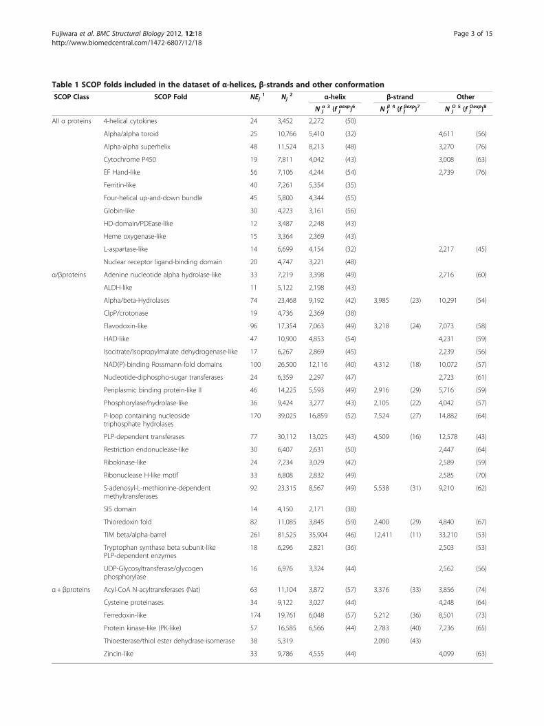
Table 1 SCOP folds included in the dataset of α-helices, β-strands and other conformation
SCOP Class SCOP Fold NEj1 Nj
2 α-helix β-strand Other
N αj3 (f αexp
j )6 N βj4 (f βexp
j )7 N Oj
5 (f Oexpj )8
All α proteins 4-helical cytokines 24 3,452 2,272 (50)
Alpha/alpha toroid 25 10,766 5,410 (32) 4,611 (56)
Alpha-alpha superhelix 48 11,524 8,213 (48) 3,270 (76)
Cytochrome P450 19 7,811 4,042 (43) 3,008 (63)
EF Hand-like 56 7,106 4,244 (54) 2,739 (76)
Ferritin-like 40 7,261 5,354 (35)
Four-helical up-and-down bundle 45 5,800 4,344 (55)
Globin-like 30 4,223 3,161 (56)
HD-domain/PDEase-like 12 3,487 2,248 (43)
Heme oxygenase-like 15 3,364 2,369 (43)
L-aspartase-like 14 6,699 4,154 (32) 2,217 (45)
Nuclear receptor ligand-binding domain 20 4,747 3,221 (48)
α/βproteins Adenine nucleotide alpha hydrolase-like 33 7,219 3,398 (49) 2,716 (60)
ALDH-like 11 5,122 2,198 (43)
Alpha/beta-Hydrolases 74 23,468 9,192 (42) 3,985 (23) 10,291 (54)
ClpP/crotonase 19 4,736 2,369 (38)
Flavodoxin-like 96 17,354 7,063 (49) 3,218 (24) 7,073 (58)
HAD-like 47 10,900 4,853 (54) 4,231 (59)
Isocitrate/Isopropylmalate dehydrogenase-like 17 6,267 2,869 (45) 2,239 (56)
NAD(P)-binding Rossmann-fold domains 100 26,500 12,116 (40) 4,312 (18) 10,072 (57)
Nucleotide-diphospho-sugar transferases 24 6,359 2,297 (47) 2,723 (61)
Periplasmic binding protein-like II 46 14,225 5,593 (49) 2,916 (29) 5,716 (59)
Phosphorylase/hydrolase-like 36 9,424 3,277 (43) 2,105 (22) 4,042 (57)
P-loop containing nucleosidetriphosphate hydrolases
170 39,025 16,859 (52) 7,524 (27) 14,882 (64)
PLP-dependent transferases 77 30,112 13,025 (43) 4,509 (16) 12,578 (43)
Restriction endonuclease-like 30 6,407 2,631 (50) 2,447 (64)
Ribokinase-like 24 7,234 3,029 (42) 2,589 (59)
Ribonuclease H-like motif 33 6,808 2,832 (49) 2,585 (70)
S-adenosyl-L-methionine-dependentmethyltransferases
92 23,315 8,567 (49) 5,538 (31) 9,210 (62)
SIS domain 14 4,150 2,171 (38)
Thioredoxin fold 82 11,085 3,845 (59) 2,400 (29) 4,840 (67)
TIM beta/alpha-barrel 261 81,525 35,904 (46) 12,411 (11) 33,210 (53)
Tryptophan synthase beta subunit-likePLP-dependent enzymes
18 6,296 2,821 (36) 2,503 (53)
UDP-Glycosyltransferase/glycogenphosphorylase
16 6,976 3,324 (44) 2,562 (56)
α+ βproteins Acyl-CoA N-acyltransferases (Nat) 63 11,104 3,872 (57) 3,376 (33) 3,856 (74)
Cysteine proteinases 34 9,122 3,027 (44) 4,248 (64)
Ferredoxin-like 174 19,761 6,048 (57) 5,212 (36) 8,501 (73)
Protein kinase-like (PK-like) 57 16,585 6,566 (44) 2,783 (40) 7,236 (65)
Thioesterase/thiol ester dehydrase-isomerase 38 5,319 2,090 (43)
Zincin-like 33 9,786 4,555 (44) 4,099 (63)
Fujiwara et al. BMC Structural Biology 2012, 12:18 Page 3 of 15http://www.biomedcentral.com/1472-6807/12/18
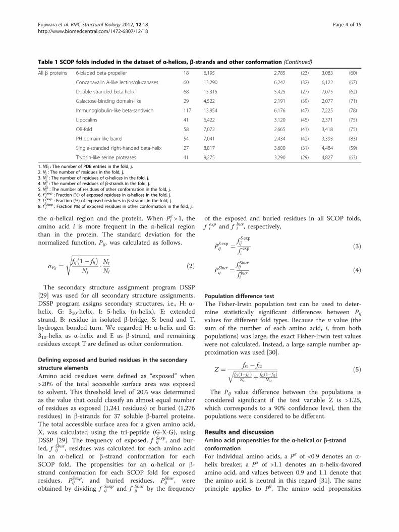
Table 1 SCOP folds included in the dataset of α-helices, β-strands and other conformation (Continued)
All β proteins 6-bladed beta-propeller 18 6,195 2,785 (23) 3,083 (60)
Concanavalin A-like lectins/glucanases 60 13,290 6,242 (32) 6,122 (67)
Double-stranded beta-helix 68 15,315 5,425 (27) 7,075 (62)
Galactose-binding domain-like 29 4,522 2,191 (39) 2,077 (71)
Immunoglobulin-like beta-sandwich 117 13,954 6,176 (47) 7,225 (78)
Lipocalins 41 6,422 3,120 (45) 2,371 (75)
OB-fold 58 7,072 2,665 (41) 3,418 (75)
PH domain-like barrel 54 7,041 2,434 (42) 3,393 (83)
Single-stranded right-handed beta-helix 27 8,817 3,600 (31) 4,484 (59)
Trypsin-like serine proteases 41 9,275 3,290 (29) 4,827 (63)
1. NEj : The number of PDB entries in the fold, j.2. Nj : The number of residues in the fold, j.3. Nα
j : The number of residues of α-helices in the fold, j.4. Nβ
j : The number of residues of β-strands in the fold, j.5. NO
j : The number of residues of other conformation in the fold, j.6. f αexpj : Fraction (%) of exposed residues in α-helices in the fold, j.7. f βexpj : Fraction (%) of exposed residues in β-strands in the fold, j.8. f Oexpj : Fraction (%) of exposed residues in other conformation in the fold, j.
Fujiwara et al. BMC Structural Biology 2012, 12:18 Page 4 of 15http://www.biomedcentral.com/1472-6807/12/18
the α-helical region and the protein. When Pαi > 1, theamino acid i is more frequent in the α-helical regionthan in the protein. The standard deviation for thenormalized function, Pij, was calculated as follows.
σPij ¼ffiffiffiffiffiffiffiffiffiffiffiffiffiffiffiffiffiffiffiffiffiffiffiffiffiffiffiffifij 1� fij� �Nj
� Nt
Ni
sð2Þ
The secondary structure assignment program DSSP[29] was used for all secondary structure assignments.DSSP program assigns secondary structures, i.e., H: α-helix, G: 310-helix, I: 5-helix (π-helix), E: extendedstrand, B: residue in isolated β-bridge, S: bend and T,hydrogen bonded turn. We regarded H: α-helix and G:310-helix as α-helix and E as β-strand, and remainingresidues except T are defined as other conformation.
Defining exposed and buried residues in the secondarystructure elementsAmino acid residues were defined as “exposed” when>20% of the total accessible surface area was exposedto solvent. This threshold level of 20% was determinedas the value that could classify an almost equal numberof residues as exposed (1,241 residues) or buried (1,276residues) in β-strands for 37 soluble β-barrel proteins.The total accessible surface area for a given amino acid,X, was calculated using the tri-peptide (G-X-G), usingDSSP [29]. The frequency of exposed, f Sexp
ij , and bur-ied, f Sbur
ij , residues was calculated for each amino acidin an α-helical or β-strand conformation for eachSCOP fold. The propensities for an α-helical or β-strand conformation for each SCOP fold for exposedresidues, PSexpij , and buried residues, PSburij , wereobtained by dividing f Sexp
ij and f Sburij by the frequency
of the exposed and buried residues in all SCOP folds,f expi and f bur
i , respectively,
PS expij ¼ f S expij
f expi
ð3Þ
PSburij ¼ f Sburij
f buri
ð4Þ
Population difference testThe Fisher-Irwin population test can be used to deter-mine statistically significant differences between Pijvalues for different fold types. Because the n value (thesum of the number of each amino acid, i, from bothpopulations) was large, the exact Fisher-Irwin test valueswere not calculated. Instead, a large sample number ap-proximation was used [30].
Z ¼ fi1 � fi2ffiffiffiffiffiffiffiffiffiffiffiffiffiffiffiffiffiffiffiffiffiffiffiffiffiffiffiffiffiffiffiffifi1 1�fi1ð Þ
Ni1þ fi2 1�fi2ð Þ
Ni2
q ð5Þ
The Pij value difference between the populations isconsidered significant if the test variable Z is >1.25,which corresponds to a 90% confidence level, then thepopulations were considered to be different.
Results and discussionAmino acid propensities for the α-helical or β-strandconformationFor individual amino acids, a Pα of <0.9 denotes an α-helix breaker, a Pα of >1.1 denotes an α-helix-favoredamino acid, and values between 0.9 and 1.1 denote thatthe amino acid is neutral in this regard [31]. The sameprinciple applies to Pβ. The amino acid propensities
Fujiwara et al. BMC Structural Biology 2012, 12:18 Page 5 of 15http://www.biomedcentral.com/1472-6807/12/18
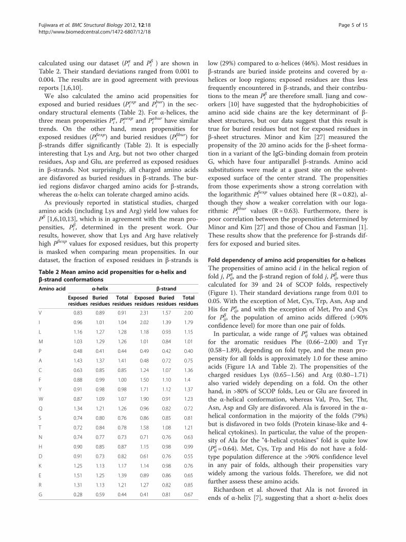
calculated using our dataset (Pαi and Pβi ) are shown inTable 2. Their standard deviations ranged from 0.001 to0.004. The results are in good agreement with previousreports [1,6,10].We also calculated the amino acid propensities for
exposed and buried residues (Pexpi and Pburi ) in the sec-ondary structural elements (Table 2). For α-helices, thethree mean propensities Pαi , P
αexpi and Pαburi have similar
trends. On the other hand, mean propensities forexposed residues (Pβexpi ) and buried residues (Pβburi ) forβ-strands differ significantly (Table 2). It is especiallyinteresting that Lys and Arg, but not two other chargedresidues, Asp and Glu, are preferred as exposed residuesin β-strands. Not surprisingly, all charged amino acidsare disfavored as buried residues in β-strands. The bur-ied regions disfavor charged amino acids for β-strands,whereas the α-helix can tolerate charged amino acids.As previously reported in statistical studies, charged
amino acids (including Lys and Arg) yield low values forPβ [1,6,10,13], which is in agreement with the mean pro-pensities, Pβi , determined in the present work. Ourresults, however, show that Lys and Arg have relativelyhigh Pβexp values for exposed residues, but this propertyis masked when comparing mean propensities. In ourdataset, the fraction of exposed residues in β-strands is
Table 2 Mean amino acid propensities for α-helix andβ-strand conformations
Amino acid α-helix β-strand
Exposedresidues
Buriedresidues
Totalresidues
Exposedresidues
Buriedresidues
Totalresidues
V 0.83 0.89 0.91 2.31 1.57 2.00
I 0.96 1.01 1.04 2.02 1.39 1.79
L 1.16 1.27 1.28 1.18 0.93 1.15
M 1.03 1.29 1.26 1.01 0.84 1.01
P 0.48 0.41 0.44 0.49 0.42 0.40
A 1.43 1.37 1.41 0.48 0.72 0.75
C 0.63 0.85 0.85 1.24 1.07 1.36
F 0.88 0.99 1.00 1.50 1.10 1.4
Y 0.91 0.98 0.98 1.71 1.12 1.37
W 0.87 1.09 1.07 1.90 0.91 1.23
Q 1.34 1.21 1.26 0.96 0.82 0.72
S 0.74 0.80 0.76 0.86 0.85 0.81
T 0.72 0.84 0.78 1.58 1.08 1.21
N 0.74 0.77 0.73 0.71 0.76 0.63
H 0.90 0.85 0.87 1.15 0.98 0.99
D 0.91 0.73 0.82 0.61 0.76 0.55
K 1.25 1.13 1.17 1.14 0.98 0.76
E 1.51 1.25 1.39 0.89 0.86 0.65
R 1.31 1.13 1.21 1.27 0.82 0.85
G 0.28 0.59 0.44 0.41 0.81 0.67
low (29%) compared to α-helices (46%). Most residues inβ-strands are buried inside proteins and covered by α-helices or loop regions; exposed residues are thus lessfrequently encountered in β-strands, and their contribu-tions to the mean Pβi are therefore small. Jiang and cow-orkers [10] have suggested that the hydrophobicities ofamino acid side chains are the key determinant of β-sheet structures, but our data suggest that this result istrue for buried residues but not for exposed residues inβ-sheet structures. Minor and Kim [27] measured thepropensity of the 20 amino acids for the β-sheet forma-tion in a variant of the IgG-binding domain from proteinG, which have four antiparallel β-strands. Amino acidsubstitutions were made at a guest site on the solvent-exposed surface of the center strand. The propensitiesfrom those experiments show a strong correlation withthe logarithmic Pβexpi values obtained here (R = 0.82), al-though they show a weaker correlation with our loga-rithmic Pβburi values (R = 0.63). Furthermore, there ispoor correlation between the propensities determined byMinor and Kim [27] and those of Chou and Fasman [1].These results show that the preference for β-strands dif-fers for exposed and buried sites.
Fold dependency of amino acid propensities for α-helicesThe propensities of amino acid i in the helical region offold j, Pαij, and the β-strand region of fold j, Pβij, were thuscalculated for 39 and 24 of SCOP folds, respectively(Figure 1). Their standard deviations range from 0.01 to0.05. With the exception of Met, Cys, Trp, Asn, Asp andHis for Pαij, and with the exception of Met, Pro and Cysfor Pβij, the population of amino acids differed (>90%confidence level) for more than one pair of folds.In particular, a wide range of Pαij values was obtained
for the aromatic residues Phe (0.66–2.00) and Tyr(0.58–1.89), depending on fold type, and the mean pro-pensity for all folds is approximately 1.0 for these aminoacids (Figure 1A and Table 2). The propensities of thecharged residues Lys (0.65–1.56) and Arg (0.80–1.71)also varied widely depending on a fold. On the otherhand, in >80% of SCOP folds, Leu or Glu are favored inthe α-helical conformation, whereas Val, Pro, Ser, Thr,Asn, Asp and Gly are disfavored. Ala is favored in the α-helical conformation in the majority of the folds (79%)but is disfavored in two folds (Protein kinase-like and 4-helical cytokines). In particular, the value of the propen-sity of Ala for the "4-helical cytokines" fold is quite low(Pαij= 0.64). Met, Cys, Trp and His do not have a fold-type population difference at the >90% confidence levelin any pair of folds, although their propensities varywidely among the various folds. Therefore, we did notfurther assess these amino acids.Richardson et al. showed that Ala is not favored in
ends of α-helix [7], suggesting that a short α-helix does
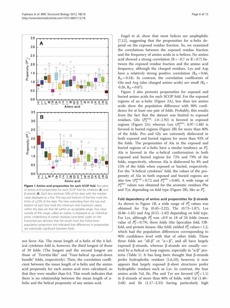
Figure 1 Amino acid propensities for each SCOP fold. Box plotsof amino acid propensities for each SCOP fold for α-helices (A) andβ-strands (B). Each box encloses 50% of the data with the medianvalue displayed as a line. The top and bottom of the box mark thelimits of ±25% of the data. The lines extending from the top andbottom of each box mark the minimum and maximum valueswithin the data set that fall within an acceptable range. Any valueoutside of this range, called an outlier, is displayed as an individualpoint. Underlining of certain residues (one-letter code) on thehorizontal axis denotes that the results from the Fisher-Irwinpopulation proportion test indicated that differences in propensitiesare statistically significant between folds.
Fujiwara et al. BMC Structural Biology 2012, 12:18 Page 6 of 15http://www.biomedcentral.com/1472-6807/12/18
not favor Ala. The mean length of α-helix of the 4 hel-ical cytokines fold is, however, the third longest of thoseof 39 folds (The longest and the second longest arethose of "Ferritin-like" and "Four-helical up-and-downbundle" folds, respectively). Then, the correlation coeffi-cient between the mean length of α-helix and the aminoacid propensity for each amino acid were calculated, sothat they were smaller than 0.4. This result indicates thatthere is no relationship between the mean length of α-helix and the helical propensity of any amino acid.
Engel et al. show that most helices are amphiphilic[7,12], suggesting that the propensities for α-helix de-pend on the exposed residue fraction. So, we examinedthe correlations between the exposed residue fractionand the frequency of amino acids in α-helices. No aminoacid showed a strong correlation (R <−0.7 or R > 0.7) be-tween the exposed residue fraction and the amino acidfrequency, although the charged residues, Lys and Asphave a relatively strong positive correlation (RK = 0.66,RD = 0.54). In contrast, the correlation coefficients ofGlu and Arg (also charged amino acids) are small (RE =0.26, RR = 0.07).Figure 2 also presents propensities for exposed and
buried amino acids for each SCOP fold. For the exposedregions of an α-helix (Figure 2A), less than ten aminoacids show the population difference with 90% confi-dence for at least one pair of folds. Probably, this resultsfrom the fact that the dataset was limited to exposedresidues. Glu (Pαexpij : 1.0–1.92) is favored in exposedregions (Figure 2A) whereas Leu (Pαburij : 0.97–1.88) isfavored in buried regions (Figure 2B) for more than 80%of the folds. Pro and Gly are extremely disfavored inboth exposed and buried regions for more than 92% ofthe folds. The propensities of Ala in the exposed andburied regions of α-helix have a similar tendency as Pαij.Ala is favored in the α-helical conformation in bothexposed and buried regions for 72% and 79% of thefolds, respectively, whereas Ala is disfavored by 8% and13% of the folds when exposed or buried, respectively.For the "4-helical cytokines" fold, the values of the pro-pensity of Ala in both exposed and buried regions arealso low (Pαexpij = 0.72 and Pαburij = 0.60). A wide range ofPαburij values was obtained for the aromatic residues Pheand Tyr, depending on fold type (Figure 2B), like as Pαij.
Fold dependency of amino acid propensities for β-strandsAs shown in Figure 1B, a wide range of Pβij values wasobtained for Trp (0.45–2.22), Thr (0.73–1.87), Lys(0.46–1.45) and Arg (0.51–1.42) depending on fold type.For Lys, although Pβij was <0.9 in 18 of 24 folds (meanvalue of Pβij= 0.79), three folds (the lipocalins fold, OB-fold, and protein kinase–like fold) yielded Pβij values > 1.2,which had the population differences corresponding to90% confidence level with that of other folds. Thesethree folds are “all-β” or “α+ β”, and all have largelyexposed β-strands, whereas β-strands are usually cov-ered by α-helical or loop regions, especially in “α/β” pro-teins (Table 1). It has long been thought that β-strandsprefer hydrophobic residues [1,6,10]; however, it nowappears that largely exposed β-sheet structures preferhydrophilic residues such as Lys. In contrast, the fouramino acids Val, Ile, Phe and Tyr are favored (Pβij > 1.1)in β-strands of more than 80% of folds, with Val (1.40–2.68) and Ile (1.17–2.33) having particularly high
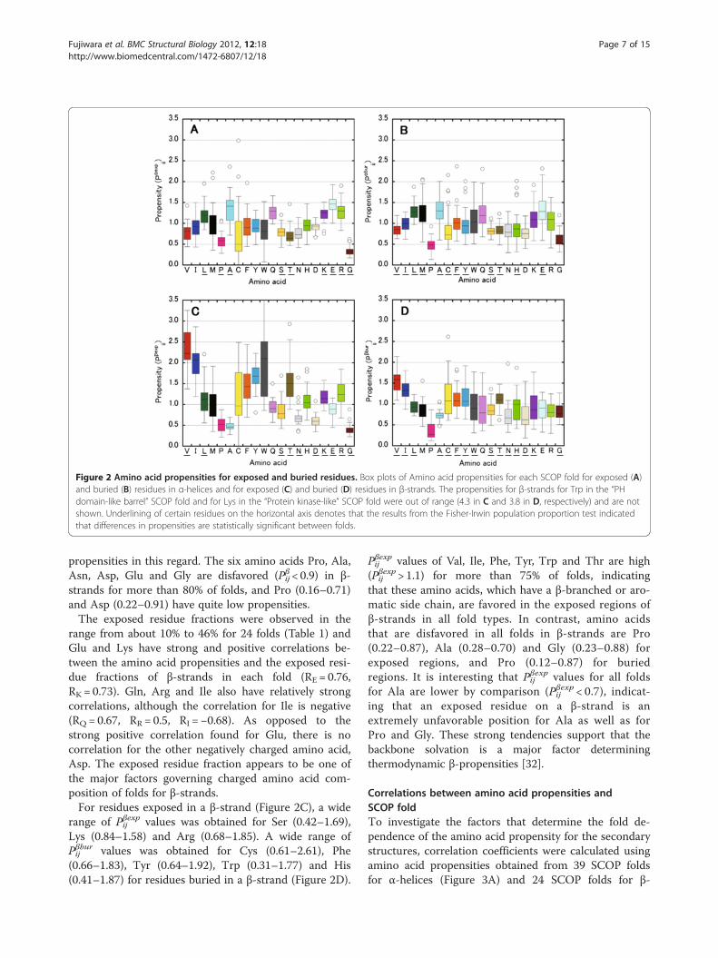
Figure 2 Amino acid propensities for exposed and buried residues. Box plots of Amino acid propensities for each SCOP fold for exposed (A)and buried (B) residues in α-helices and for exposed (C) and buried (D) residues in β-strands. The propensities for β-strands for Trp in the “PHdomain-like barrel” SCOP fold and for Lys in the “Protein kinase-like” SCOP fold were out of range (4.3 in C and 3.8 in D, respectively) and are notshown. Underlining of certain residues on the horizontal axis denotes that the results from the Fisher-Irwin population proportion test indicatedthat differences in propensities are statistically significant between folds.
Fujiwara et al. BMC Structural Biology 2012, 12:18 Page 7 of 15http://www.biomedcentral.com/1472-6807/12/18
propensities in this regard. The six amino acids Pro, Ala,Asn, Asp, Glu and Gly are disfavored (Pβij < 0.9) in β-strands for more than 80% of folds, and Pro (0.16–0.71)and Asp (0.22–0.91) have quite low propensities.The exposed residue fractions were observed in the
range from about 10% to 46% for 24 folds (Table 1) andGlu and Lys have strong and positive correlations be-tween the amino acid propensities and the exposed resi-due fractions of β-strands in each fold (RE = 0.76,RK = 0.73). Gln, Arg and Ile also have relatively strongcorrelations, although the correlation for Ile is negative(RQ = 0.67, RR = 0.5, RI =−0.68). As opposed to thestrong positive correlation found for Glu, there is nocorrelation for the other negatively charged amino acid,Asp. The exposed residue fraction appears to be one ofthe major factors governing charged amino acid com-position of folds for β-strands.For residues exposed in a β-strand (Figure 2C), a wide
range of Pβexpij values was obtained for Ser (0.42–1.69),Lys (0.84–1.58) and Arg (0.68–1.85). A wide range ofPβburij values was obtained for Cys (0.61–2.61), Phe(0.66–1.83), Tyr (0.64–1.92), Trp (0.31–1.77) and His(0.41–1.87) for residues buried in a β-strand (Figure 2D).
Pβexpij values of Val, Ile, Phe, Tyr, Trp and Thr are high(Pβexpij > 1.1) for more than 75% of folds, indicatingthat these amino acids, which have a β-branched or aro-matic side chain, are favored in the exposed regions ofβ-strands in all fold types. In contrast, amino acidsthat are disfavored in all folds in β-strands are Pro(0.22–0.87), Ala (0.28–0.70) and Gly (0.23–0.88) forexposed regions, and Pro (0.12–0.87) for buriedregions. It is interesting that Pβexpij values for all foldsfor Ala are lower by comparison (Pβexpij < 0.7), indicat-ing that an exposed residue on a β-strand is anextremely unfavorable position for Ala as well as forPro and Gly. These strong tendencies support that thebackbone solvation is a major factor determiningthermodynamic β-propensities [32].
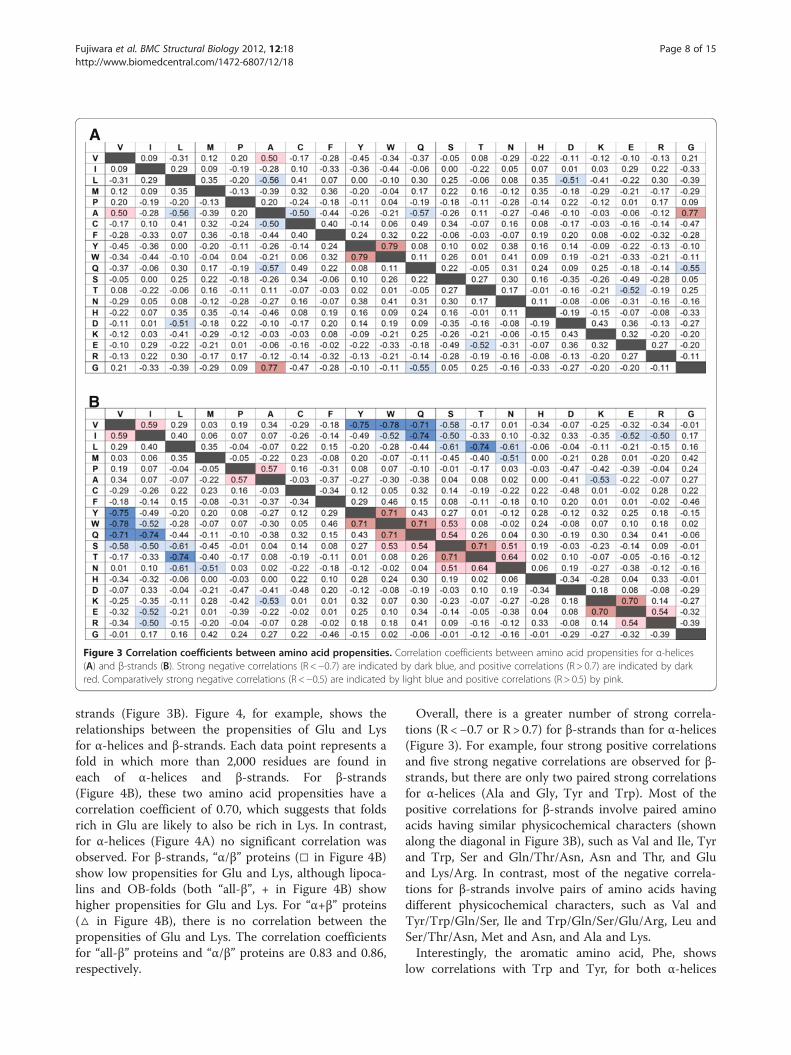
Correlations between amino acid propensities andSCOP foldTo investigate the factors that determine the fold de-pendence of the amino acid propensity for the secondarystructures, correlation coefficients were calculated usingamino acid propensities obtained from 39 SCOP foldsfor α-helices (Figure 3A) and 24 SCOP folds for β-
A
B
Figure 3 Correlation coefficients between amino acid propensities. Correlation coefficients between amino acid propensities for α-helices(A) and β-strands (B). Strong negative correlations (R <−0.7) are indicated by dark blue, and positive correlations (R > 0.7) are indicated by darkred. Comparatively strong negative correlations (R <−0.5) are indicated by light blue and positive correlations (R > 0.5) by pink.
Fujiwara et al. BMC Structural Biology 2012, 12:18 Page 8 of 15http://www.biomedcentral.com/1472-6807/12/18
strands (Figure 3B). Figure 4, for example, shows therelationships between the propensities of Glu and Lysfor α-helices and β-strands. Each data point represents afold in which more than 2,000 residues are found ineach of α-helices and β-strands. For β-strands(Figure 4B), these two amino acid propensities have acorrelation coefficient of 0.70, which suggests that foldsrich in Glu are likely to also be rich in Lys. In contrast,for α-helices (Figure 4A) no significant correlation wasobserved. For β-strands, “α/β” proteins (□ in Figure 4B)show low propensities for Glu and Lys, although lipoca-lins and OB-folds (both “all-β”, + in Figure 4B) showhigher propensities for Glu and Lys. For “α+β” proteins(△ in Figure 4B), there is no correlation between thepropensities of Glu and Lys. The correlation coefficientsfor “all-β” proteins and “α/β” proteins are 0.83 and 0.86,respectively.
Overall, there is a greater number of strong correla-tions (R <−0.7 or R > 0.7) for β-strands than for α-helices(Figure 3). For example, four strong positive correlationsand five strong negative correlations are observed for β-strands, but there are only two paired strong correlationsfor α-helices (Ala and Gly, Tyr and Trp). Most of thepositive correlations for β-strands involve paired aminoacids having similar physicochemical characters (shownalong the diagonal in Figure 3B), such as Val and Ile, Tyrand Trp, Ser and Gln/Thr/Asn, Asn and Thr, and Gluand Lys/Arg. In contrast, most of the negative correla-tions for β-strands involve pairs of amino acids havingdifferent physicochemical characters, such as Val andTyr/Trp/Gln/Ser, Ile and Trp/Gln/Ser/Glu/Arg, Leu andSer/Thr/Asn, Met and Asn, and Ala and Lys.Interestingly, the aromatic amino acid, Phe, shows
low correlations with Trp and Tyr, for both α-helices
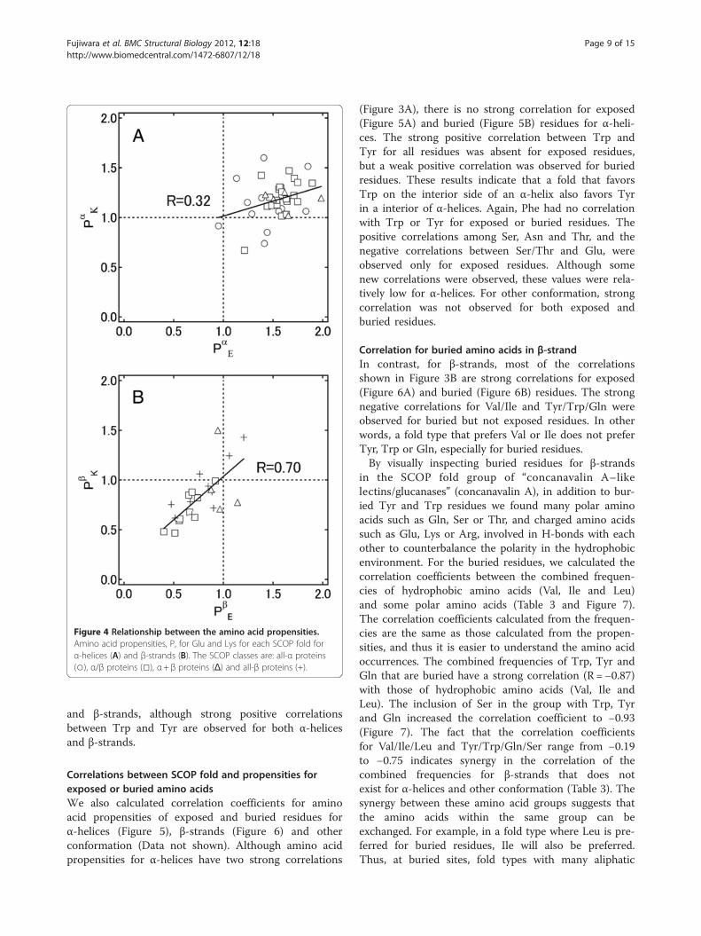
Figure 4 Relationship between the amino acid propensities.Amino acid propensities, P, for Glu and Lys for each SCOP fold forα-helices (A) and β-strands (B). The SCOP classes are: all-α proteins(○), α/β proteins (□), α+ β proteins (Δ) and all-β proteins (+).
Fujiwara et al. BMC Structural Biology 2012, 12:18 Page 9 of 15http://www.biomedcentral.com/1472-6807/12/18
and β-strands, although strong positive correlationsbetween Trp and Tyr are observed for both α-helicesand β-strands.
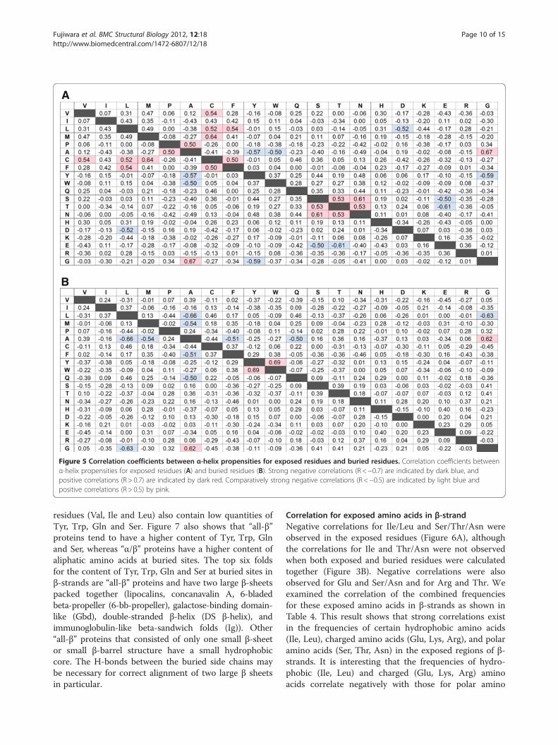
Correlations between SCOP fold and propensities forexposed or buried amino acidsWe also calculated correlation coefficients for aminoacid propensities of exposed and buried residues forα-helices (Figure 5), β-strands (Figure 6) and otherconformation (Data not shown). Although amino acidpropensities for α-helices have two strong correlations
(Figure 3A), there is no strong correlation for exposed(Figure 5A) and buried (Figure 5B) residues for α-heli-ces. The strong positive correlation between Trp andTyr for all residues was absent for exposed residues,but a weak positive correlation was observed for buriedresidues. These results indicate that a fold that favorsTrp on the interior side of an α-helix also favors Tyrin a interior of α-helices. Again, Phe had no correlationwith Trp or Tyr for exposed or buried residues. Thepositive correlations among Ser, Asn and Thr, and thenegative correlations between Ser/Thr and Glu, wereobserved only for exposed residues. Although somenew correlations were observed, these values were rela-tively low for α-helices. For other conformation, strongcorrelation was not observed for both exposed andburied residues.
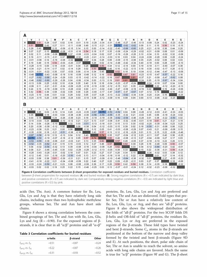
Correlation for buried amino acids in β-strandIn contrast, for β-strands, most of the correlationsshown in Figure 3B are strong correlations for exposed(Figure 6A) and buried (Figure 6B) residues. The strongnegative correlations for Val/Ile and Tyr/Trp/Gln wereobserved for buried but not exposed residues. In otherwords, a fold type that prefers Val or Ile does not preferTyr, Trp or Gln, especially for buried residues.By visually inspecting buried residues for β-strands
in the SCOP fold group of “concanavalin A–likelectins/glucanases” (concanavalin A), in addition to bur-ied Tyr and Trp residues we found many polar aminoacids such as Gln, Ser or Thr, and charged amino acidssuch as Glu, Lys or Arg, involved in H-bonds with eachother to counterbalance the polarity in the hydrophobicenvironment. For the buried residues, we calculated thecorrelation coefficients between the combined frequen-cies of hydrophobic amino acids (Val, Ile and Leu)and some polar amino acids (Table 3 and Figure 7).The correlation coefficients calculated from the frequen-cies are the same as those calculated from the propen-sities, and thus it is easier to understand the amino acidoccurrences. The combined frequencies of Trp, Tyr andGln that are buried have a strong correlation (R =−0.87)with those of hydrophobic amino acids (Val, Ile andLeu). The inclusion of Ser in the group with Trp, Tyrand Gln increased the correlation coefficient to −0.93(Figure 7). The fact that the correlation coefficientsfor Val/Ile/Leu and Tyr/Trp/Gln/Ser range from −0.19to −0.75 indicates synergy in the correlation of thecombined frequencies for β-strands that does notexist for α-helices and other conformation (Table 3). Thesynergy between these amino acid groups suggests thatthe amino acids within the same group can beexchanged. For example, in a fold type where Leu is pre-ferred for buried residues, Ile will also be preferred.Thus, at buried sites, fold types with many aliphatic
A
B
Figure 5 Correlation coefficients between α-helix propensities for exposed residues and buried residues. Correlation coefficients betweenα-helix propensities for exposed residues (A) and buried residues (B). Strong negative correlations (R <−0.7) are indicated by dark blue, andpositive correlations (R > 0.7) are indicated by dark red. Comparatively strong negative correlations (R <−0.5) are indicated by light blue andpositive correlations (R > 0.5) by pink.
Fujiwara et al. BMC Structural Biology 2012, 12:18 Page 10 of 15http://www.biomedcentral.com/1472-6807/12/18
residues (Val, Ile and Leu) also contain low quantities ofTyr, Trp, Gln and Ser. Figure 7 also shows that “all-β”proteins tend to have a higher content of Tyr, Trp, Glnand Ser, whereas “α/β” proteins have a higher content ofaliphatic amino acids at buried sites. The top six foldsfor the content of Tyr, Trp, Gln and Ser at buried sites inβ-strands are “all-β” proteins and have two large β-sheetspacked together (lipocalins, concanavalin A, 6-bladedbeta-propeller (6-bb-propeller), galactose-binding domain-like (Gbd), double-stranded β-helix (DS β-helix), andimmunoglobulin-like beta-sandwich folds (Ig)). Other“all-β” proteins that consisted of only one small β-sheetor small β-barrel structure have a small hydrophobiccore. The H-bonds between the buried side chains maybe necessary for correct alignment of two large β sheetsin particular.
Correlation for exposed amino acids in β-strandNegative correlations for Ile/Leu and Ser/Thr/Asn wereobserved in the exposed residues (Figure 6A), althoughthe correlations for Ile and Thr/Asn were not observedwhen both exposed and buried residues were calculatedtogether (Figure 3B). Negative correlations were alsoobserved for Glu and Ser/Asn and for Arg and Thr. Weexamined the correlation of the combined frequenciesfor these exposed amino acids in β-strands as shown inTable 4. This result shows that strong correlations existin the frequencies of certain hydrophobic amino acids(Ile, Leu), charged amino acids (Glu, Lys, Arg), and polaramino acids (Ser, Thr, Asn) in the exposed regions of β-strands. It is interesting that the frequencies of hydro-phobic (Ile, Leu) and charged (Glu, Lys, Arg) aminoacids correlate negatively with those for polar amino
A
B
Figure 6 Correlation coefficients between β-sheet propensities for exposed residues and buried residues. Correlation coefficientsbetween β-sheet propensities for exposed residues (A) and buried residues (B). Strong negative correlations (R <−0.7) are indicated by dark blue,and positive correlations (R > 0.7) are indicated by dark red. Comparatively strong negative correlations (R <−0.5) are indicated by light blue andpositive correlations (R > 0.5) by pink.
Fujiwara et al. BMC Structural Biology 2012, 12:18 Page 11 of 15http://www.biomedcentral.com/1472-6807/12/18
acids (Ser, Thr, Asn). A common feature for Ile, Leu,Glu, Lys and Arg is that they have relatively long sidechains, including more than two hydrophobic methylenegroups, whereas Ser, Thr and Asn have short sidechains.Figure 8 shows a strong correlation between the com-
bined groupings of Ser, Thr and Asn with Ile, Leu, Glu,Lys and Arg (R =−0.90). For the exposed regions of β-strands, it is clear that in all “α/β” proteins and all “α+β”
Table 3 Correlation coefficients for buried residues
α-helix β-strand Other
fWYQ vs. fVI −0.51 −0.87 −0.24
fWYQ vs. fVIL −0.22 −0.87 −0.26
fWYQS vs. fVIL −0.31 −0.93 −0.52
proteins, Ile, Leu, Glu, Lys and Arg are preferred andthat Ser, Thr and Asn are disfavored. Fold types that pre-fer Ser, Thr or Asn have a relatively low content ofIle, Leu, Glu, Lys, or Arg, and they are “all-β” proteins.Figure 8 also shows the widespread distribution ofthe folds of “all-β” proteins. For the two SCOP folds DSβ-helix and OB-fold of “all-β” proteins, the residues Ile,Leu, Glu, Lys or Arg are preferred in the exposedregions of the β-strands. These fold types have twistedand bent β-strands. Some Cα atoms in the β-strands arepositioned at the bottom of the narrow and deep valleyformed by the twisted and bent β-strands (Figure 9Dand E). At such positions, the short, polar side chain ofSer, Thr or Asn is unable to reach the solvent, so aminoacids with long side chains are favored. Much the sameis true for “α/β” proteins (Figure 9F and G). The β-sheet
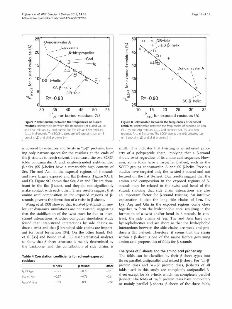
β
β
β
β
Figure 7 Relationship between the frequencies of buriedresidues. Relationship between the frequencies of buried Val, Ileand Leu residues, fVIL, and buried Trp, Tyr, Gln and Ser residues,fWYQS, in β-strands. The SCOP classes are: α/β proteins (□), α+ βproteins (Δ) and all-β proteins (+).
β
β
β
β
Figure 8 Relationship between the frequencies of exposedresidues. Relationship between the frequencies of exposed Ile, Leu,Glu, Lys and Arg residues, fILEKR, and exposed Ser, Thr and Asnresidues, fSTN, in β-strands. The SCOP classes are: α/β proteins (□),α+ β proteins (Δ) and all-β proteins (+).
Fujiwara et al. BMC Structural Biology 2012, 12:18 Page 12 of 15http://www.biomedcentral.com/1472-6807/12/18
is covered by α-helices and twists in “α/β” proteins, leav-ing only narrow spaces for the residues at the ends ofthe β-strands to reach solvent. In contrast, the two SCOPfolds concanavalin A and single-stranded right-handedβ-helix (SS β-helix) have a remarkably high content ofSer, Thr and Asn in the exposed regions of β-strandsand have largely exposed and flat β-sheets (Figure 9A, Band C). Figure 9C shows that Ser, Asn and Thr are dom-inant in the flat β-sheet, and they do not significantlymake contact with each other. These results suggest thatamino acid composition in the exposed regions of β-strands governs the formation of a twist in β-sheets.Wang et al. [33] showed that isolated β-strands in mo-
lecular dynamics simulations are not twisted, suggestingthat the stabilization of the twist must be due to inter-strand interactions. Another computer simulation studyfound that inter-strand interactions by side chains in-duce a twist and that β-branched side chains are import-ant for twist formation [34]. On the other hand, Kohet al. [35] and Bosco et al. [36] used statistical analysesto show that β-sheet structure is mainly determined bythe backbone, and the contribution of side chains is
Table 4 Correlation coefficients for solvent-exposedresidues
α-helix β-strand Other
fIL vs. fSTN −0.21 −0.79 −0.51
fEKR vs. fSTN −0.57 −0.76 −0.61
fILEKR vs. fSTN −0.59 −0.90 −0.66
small. This indicates that twisting is an inherent prop-erty of a polypeptide chain, implying that a β-strandshould twist regardless of its amino acid sequence. How-ever, some folds have a large/flat β-sheet, such as theSCOP groups concanavalin A and SS β-helix. Previousstudies have targeted only the twisted β-strand and notfocused on the flat β-sheet. Our results suggest that theamino acid composition in the exposed regions of β-strands may be related to the twist and bend of thestrand, showing that side chain interactions are alsoan important factor for β-strand twisting. An intuitiveexplanation is that the long side chains of Leu, Ile,Lys, Arg and Glu in the exposed regions come closetogether to form the hydrophobic core, resulting in theformation of a twist and/or bend in β-strands. In con-trast, the side chains of Ser, Thr and Asn have lowhydrophobicities and are short so that the hydrophobicinteractions between the side chains are weak and pro-duce a flat β-sheet. Therefore, it seems that the strainwithin a β-sheet is one of the major factors governingamino acid propensities of folds for β-strands.
The types of β-sheets and the amino acid propensityThe folds can be classified by their β-sheet types intothree; parallel, antiparallel and mixed β-sheet. For "all-β"protein class and "α+ β" protein class, β-sheets of allfolds used in this study are completely antiparallel β-sheet except for SS β-helix which has completely parallelβ-sheet. The folds of "α/β" protein class have completelyor mainly parallel β-sheets. β-sheets of the three folds,
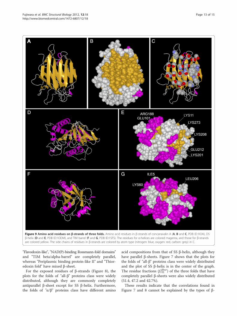
AA B C
D E
F G
Figure 9 Amino acid residues on β-strands of three folds. Amino acid residues in β-strands of concanavalin A (A, B and C, PDB ID:1IOA), DSβ-helix (D and E, PDB ID:1ODM), and TIM barrel (F and G, PDB ID:1SFS). The residues for α-helices are colored magenta, and those for β-strandsare colored yellow. The side chains of residues in β-strands are colored by atom type (nitrogen: blue, oxygen: red, carbon: grey) in C.
Fujiwara et al. BMC Structural Biology 2012, 12:18 Page 13 of 15http://www.biomedcentral.com/1472-6807/12/18
"Flavodoxin-like", "NAD(P)-binding Rossmann-fold domains"and "TIM beta/alpha-barrel" are completely parallel,whereas "Periplasmic binding protein-like II" and "Thior-edoxin fold" have mixed β-sheet.For the exposed residues of β-strands (Figure 8), the
plots for the folds of "all-β" proteins class were widelydistributed, although they are commonly completelyantiparallel β-sheet except for SS β-helix. Furthermore,the folds of "α/β" proteins class have different amino
acid compositions from that of SS β-helix, although theyhave parallel β-sheets. Figure 7 shows that the plots forthe folds of "all-β" proteins class were widely distributedand the plot of SS β-helix is in the center of the graph.The residue fractions (f βburVIL ) of the three folds that havecompletely parallel β-sheets were also widely distributed(51.4, 47.2 and 42.7%).These results indicate that the correlations found in
Figure 7 and 8 cannot be explained by the types of β-
Fujiwara et al. BMC Structural Biology 2012, 12:18 Page 14 of 15http://www.biomedcentral.com/1472-6807/12/18
sheets. Consequently, we think that the propensities donot depend on the types of β-sheets.
Robustness of the datasetWe checked the robustness of our results using thedataset of more than 1,500 residues and less than 2,000residues, which is not included in the dataset usedin this study; six folds for α-helix and eight folds forβ-strands. For β-strands, strong correlations werealso observed for buried residues (RWYQS-VIL = −0.81)and for exposed residues (RILEKR-STN = −0.78). Thereare no strong correlations for buried residues(RWYQS-VIL = −0.64) and for exposed residues (RILEKR-
STN = −0.48) in α-helices. These results are the sameas those obtained for the dataset containing morethan 2,000 residues. Therefore, the results presentedhere seem to be independent of the dataset selection.
ConclusionThe amino acid propensities for secondary structureswere investigated for each SCOP fold. The helix propen-sities calculated for exposed and buried residues are alsosimilar to each other. For β-sheet propensities, however,propensities calculated for exposed residues are remark-ably different from those of buried residues, whichare similar to those calculated for all residues becauseβ-sheets tend to be located in the interior of proteins.We also detected correlations between amino acid
compositions in β-strands. At buried sites, the contentof Tyr, Trp, Gln and Ser correlates negatively with thecontent of the aliphatic amino acids Val, Ile and Leu.All-β proteins tend to have a higher content of Tyr, Trp,Gln and Ser, whereas α/β proteins tend to have a highercontent of aliphatic amino acids at buried sites. In all-βproteins, the H-bonds between buried side chains maybe necessary for correct alignment of two large β sheets.For exposed residues, there is a tendency that a fold witha high content of Ile, Leu, Glu, Lys and Arg would havea low content of Ser, Thr and Asn. Generally, α/β pro-teins have twisted and bent β-strands and favor longerside chains at exposed sites.These findings are very useful for the design of
β-sheet. They are especially effective when there is struc-tural information such as whether a residue is exposedor buried, two large β-sheets are packed together, aβ-sheet has α-helices at least one side of β-sheets anda β-strand is twisted or not. Hecht and coworkershave succeeded in designing de novo proteins withbinary patterning techniques, in which polar and non-polar amino acids are placed at desired sites alongthe sequence by synthesizing DNA with degeneratedcodon [37]. If one desire to design a de novo proteinlibrary of SS β-helix, for example, he should considerto bias in favor of Ser, Thr, and Asn rather than Glu,
Lys, Arg for exposed sites on β-strands because thefrequency of Ser, Thr, and Asn is relatively high andconversely the frequency of Ile, Leu, Glu, Lys, Arg islow for exposed sites on β-strands of SS β-helix folds(Figure 8).
AbbreviationsR: Correlation coefficient.
Competing interestsThe authors declare that they have no competing interests.
Authors’ contributionsKF conceived the project and wrote the manuscript. HT wrote the programsand performed all analyses. MI participated the discussion of the project andwas involved the revision of the manuscript. All authors read and approvedthe final manuscript.
Received: 24 March 2012 Accepted: 19 July 2012Published: 2 August 2012
References1. Chou PY, Fasman GD: Conformational parameters for amino acids in
helical, beta-sheet, and random coil regions calculated from proteins.Biochemistry 1974, 13(2):211–222.
2. Hubbard TJ, Ailey B, Brenner SE, Murzin AG, Chothia C: SCOP: a StructuralClassification of Proteins database. Nucleic Acids Res 1999, 27(1):254–256.
3. Orengo CA, Pearl FM, Bray JE, Todd AE, Martin AC, Lo Conte L, Thornton JM:The CATH Database provides insights into protein structure/functionrelationships. Nucleic Acids Res 1999, 27(1):275–279.
4. Fujiwara K, Ikeguchi M: OLIGAMI: OLIGomer Architecture and MolecularInterface. The Open Bioinformatics Journal 2008, 2:50–53.
5. Serrano L: The relationship between sequence and structure inelementary folding units. Adv Protein Chem 2000, 53:49–85.
6. Williams RW, Chang A, Juretic D, Loughran S: Secondary structurepredictions and medium range interactions. Biochim Biophys Acta 1987,916(2):200–204.
7. Richardson JS, Richardson DC: Amino acid preferences for specificlocations at the ends of alpha helices. Science 1988, 240(4859):1648–1652.
8. Munoz V, Serrano L: Intrinsic secondary structure propensities of theamino acids, using statistical phi-psi matrices: comparison withexperimental scales. Proteins 1994, 20(4):301–311.
9. Swindells MB, MacArthur MW, Thornton JM: Intrinsic phi, psi propensitiesof amino acids, derived from the coil regions of known structures. NatStruct Biol 1995, 2(7):596–603.
10. Jiang B, Guo T, Peng L, Sun Z: Folding Type-Specific Secondary StructurePropensities of Amino Acids, Derived from a-Helical, b-Sheet, a/b, anda +b Proteins of Known Structures. Biopolymers 1998, 45:35–49.
11. Pal D, Chakrabarti P: beta-sheet propensity and its correlation withparameters based on conformation. Acta Crystallogr D: Biol Crystallogr2000, 56(Pt 5):589–594.
12. Engel DE, DeGrado WF: Amino acid propensities are position-dependentthroughout the length of alpha-helices. J Mol Biol 2004, 337(5):1195–1205.
13. Costantini S, Colonna G, Facchiano AM: Amino acid propensities forsecondary structures are influenced by the protein structural class.Biochem Biophys Res Commun 2006, 342(2):441–451.
14. Malkov SN, Zivkovic MV, Beljanski MV, Hall MB, Zaric SD: A reexamination ofthe propensities of amino acids towards a particular secondarystructure: classification of amino acids based on their chemical structure.J Mol Model 2008, 14(8):769–775.
15. Bhattacharjee N, Biswas P: Position-specific propensities of amino acids inthe beta-strand. BMC Struct Biol 2010, 10:29.
16. O'Neil KT, DeGrado WF: A thermodynamic scale for the helix-formingtendencies of the commonly occurring amino acids. Science 1990,250(4981):646–651.
17. Park SH, Shalongo W, Stellwagen E: Residue helix parameters obtainedfrom dichroic analysis of peptides of defined sequence. Biochemistry1993, 32(27):7048–7053.
Fujiwara et al. BMC Structural Biology 2012, 12:18 Page 15 of 15http://www.biomedcentral.com/1472-6807/12/18
18. Rohl CA, Chakrabartty A, Baldwin RL: Helix propagation and N-cappropensities of the amino acids measured in alanine-based peptides in40 volume percent trifluoroethanol. Protein Sci 1996, 5(12):2623–2637.
19. Yang J, Spek EJ, Gong Y, Zhou H, Kallenbach NR: The role of context onalpha-helix stabilization: host-guest analysis in a mixed backgroundpeptide model. Protein Sci 1997, 6(6):1264–1272.
20. Lacroix E, Viguera AR, Serrano L: Elucidating the folding problem of alpha-helices: local motifs, long-range electrostatics, ionic-strengthdependence and prediction of NMR parameters. J Mol Biol 1998,284(1):173–191.
21. Kim CA, Berg JM: Thermodynamic beta-sheet propensities measuredusing a zinc-finger host peptide. Nature 1993, 362(6417):267–270.
22. Myers JK, Pace CN, Scholtz JM: Trifluoroethanol effects on helix propensityand electrostatic interactions in the helical peptide from ribonucleaseT1. Protein Sci 1998, 7(2):383–388.
23. Myers JK, Pace CN, Scholtz JM: Helix propensities are identical in proteinsand peptides. Biochemistry 1997, 36(36):10923–10929.
24. Horovitz A, Matthews JM, Fersht AR: Alpha-helix stability in proteins. II.Factors that influence stability at an internal position. J Mol Biol 1992,227(2):560–568.
25. Blaber M, Zhang XJ, Matthews BW: Structural basis of amino acid alphahelix propensity. Science 1993, 260(5114):1637–1640.
26. Blaber M, Baase WA, Gassner N, Matthews BW: Alanine scanningmutagenesis of the alpha-helix 115–123 of phage T4 lysozyme: effectson structure, stability and the binding of solvent. J Mol Biol 1995,246(2):317–330.
27. Minor DL Jr, Kim PS: Measurement of the beta-sheet-forming propensitiesof amino acids. Nature 1994, 367(6464):660–663.
28. Minor DL Jr, Kim PS: Context is a major determinant of beta-sheetpropensity. Nature 1994, 371(6494):264–267.
29. Kabsch W, Sander C: Dictionary of protein secondary structure: patternrecognition of hydrogen-bonded and geometrical features. Biopolymers1983, 22(12):2577–2637.
30. Warren GL, Petsko GA: Composition analysis of alpha-helices inthermophilic organisms. Protein Eng 1995, 8(9):905–913.
31. Chou PY, Fasman GD: Prediction of the secondary structure of proteinsfrom their amino acid sequence. Adv Enzymol Relat Areas Mol Biol 1978,47:45–148.
32. Avbelj F, Baldwin RL: Role of backbone solvation in determiningthermodynamic beta propensities of the amino acids. Proc Natl Acad SciU S A 2002, 99(3):1309–1313.
33. Wang L, O'Connell T, Tropsha A, Hermans J: Molecular simulations ofbeta-sheet twisting. J Mol Biol 1996, 262(2):283–293.
34. Chou KC, Nemethy G, Scheraga HA: Role of interchain interactions in thestabilization of the right-handed twist of beta-sheets. J Mol Biol 1983,168(2):389–407.
35. Koh E, Kim T, Cho HS: Mean curvature as a major determinant of beta-sheet propensity. Bioinformatics 2006, 22(3):297–302.
36. Ho BK, Curmi PM: Twist and shear in beta-sheets and beta-ribbons. J MolBiol 2002, 317(2):291–308.
37. Hecht MH, Das A, Go A, Bradley LH, Wei Y: De novo proteins fromdesigned combinatorial libraries. Protein Sci 2004, 13(7):1711–1723.
doi:10.1186/1472-6807-12-18Cite this article as: Fujiwara et al.: Dependence of α-helical and β-sheetamino acid propensities on the overall protein fold type. BMC StructuralBiology 2012 12:18.
Submit your next manuscript to BioMed Centraland take full advantage of:
• Convenient online submission
• Thorough peer review
• No space constraints or color figure charges
• Immediate publication on acceptance
• Inclusion in PubMed, CAS, Scopus and Google Scholar
• Research which is freely available for redistribution
Submit your manuscript at www.biomedcentral.com/submit