RESEARCH ARTICLE Open Access Aspergillus giganteus ... · for N. crassa and 1 μg/ml for A. niger....
13
RESEARCH ARTICLE Open Access The Aspergillus giganteus antifungal protein AFP NN5353 activates the cell wall integrity pathway and perturbs calcium homeostasis Ulrike Binder 1,5 , Mojca Bencina 2,3 , Andrea Eigentler 1 , Vera Meyer 4 and Florentine Marx 1* Abstract Background: The antifungal protein AFP NN5353 is a defensin-like protein of Aspergillus giganteus. It belongs to a group of secretory proteins with low molecular mass, cationic character and a high content of cysteine residues. The protein inhibits the germination and growth of filamentous ascomycetes, including important human and plant pathogens and the model organsims Aspergillus nidulans and Aspergillus niger. Results: We determined an AFP NN5353 hypersensitive phenotype of non-functional A. nidulans mutants in the protein kinase C (Pkc)/mitogen-activated protein kinase (Mpk) signalling pathway and the induction of the a- glucan synthase A (agsA) promoter in a transgenic A. niger strain which point at the activation of the cell wall integrity pathway (CWIP) and the remodelling of the cell wall in response to AFP NN5353 . The activation of the CWIP by AFP NN5353 , however, operates independently from RhoA which is the central regulator of CWIP signal transduction in fungi. Furthermore, we provide evidence that calcium (Ca 2+ ) signalling plays an important role in the mechanistic function of this antifungal protein. AFP NN5353 increased about 2-fold the cytosolic free Ca 2+ ([Ca 2+ ] c ) of a transgenic A. niger strain expressing codon optimized aequorin. Supplementation of the growth medium with CaCl 2 counteracted AFP NN5353 toxicity, ameliorated the perturbation of the [Ca 2+ ] c resting level and prevented protein uptake into Aspergillus sp. cells. Conclusions: The present study contributes new insights into the molecular mechanisms of action of the A. giganteus antifungal protein AFP NN5353 . We identified its antifungal activity, initiated the investigation of pathways that determine protein toxicity, namely the CWIP and the Ca 2+ signalling cascade, and studied in detail the cellular uptake mechanism in sensitive target fungi. This knowledge contributes to define new potential targets for the development of novel antifungal strategies to prevent and combat infections of filamentous fungi which have severe negative impact in medicine and agriculture. Background All organisms have evolved several defence systems in order to protect themselves against bacteria, fungi and viruses. Higher organisms have developed a complex network of humoral and cellular responses, called adap- tive immunity. A second defence system, the innate immunity, consists of many components, including small peptides with a broad antimicrobial spectrum [1,2]. The production of such proteins with antimicrobial activity is not limited to higher eukaryotes, but also found in microorganisms, including fungi. The diversity of these proteins is reflected in their mode of action and their species-specificity. Some of them form pores in the membrane, others are known to inhibit cell wall synthesis or interfere with nucleic acids and their synthesis [3,4]. They can be involved in the inhibition of protein synthesis or interfere with cell cycle control [3,4]. A relatively new group of antimicrobial proteins secreted by filamentous ascomycetes includes small, cationic and cysteine-rich proteins. So far, only few anti- fungal proteins have been characterized, namely AFP from Aspergillus giganteus , ANAFP from Aspergillus * Correspondence: [email protected] 1 Biocenter, Division of Molecular Biology, Innsbruck Medical University, Fritz- Pregl Strasse 3, Innsbruck, A-6020, Austria Full list of author information is available at the end of the article Binder et al. BMC Microbiology 2011, 11:209 http://www.biomedcentral.com/1471-2180/11/209 © 2011 Binder et al; licensee BioMed Central Ltd. This is an Open Access article distributed under the terms of the Creative Commons Attribution License (http://creativecommons.org/licenses/by/2.0), which permits unrestricted use, distribution, and reproduction in any medium, provided the original work is properly cited.
Transcript of RESEARCH ARTICLE Open Access Aspergillus giganteus ... · for N. crassa and 1 μg/ml for A. niger....
RESEARCH ARTICLE Open Access
The Aspergillus giganteus antifungal proteinAFPNN5353 activates the cell wall integritypathway and perturbs calcium homeostasisUlrike Binder1,5, Mojca Bencina2,3, Andrea Eigentler1, Vera Meyer4 and Florentine Marx1*
Abstract
Background: The antifungal protein AFPNN5353 is a defensin-like protein of Aspergillus giganteus. It belongs to agroup of secretory proteins with low molecular mass, cationic character and a high content of cysteine residues.The protein inhibits the germination and growth of filamentous ascomycetes, including important human andplant pathogens and the model organsims Aspergillus nidulans and Aspergillus niger.
Results: We determined an AFPNN5353 hypersensitive phenotype of non-functional A. nidulans mutants in theprotein kinase C (Pkc)/mitogen-activated protein kinase (Mpk) signalling pathway and the induction of the a-glucan synthase A (agsA) promoter in a transgenic A. niger strain which point at the activation of the cell wallintegrity pathway (CWIP) and the remodelling of the cell wall in response to AFPNN5353. The activation of the CWIPby AFPNN5353, however, operates independently from RhoA which is the central regulator of CWIP signaltransduction in fungi.Furthermore, we provide evidence that calcium (Ca2+) signalling plays an important role in the mechanisticfunction of this antifungal protein. AFPNN5353 increased about 2-fold the cytosolic free Ca2+ ([Ca2+]c) of a transgenicA. niger strain expressing codon optimized aequorin. Supplementation of the growth medium with CaCl2counteracted AFPNN5353 toxicity, ameliorated the perturbation of the [Ca2+]c resting level and prevented proteinuptake into Aspergillus sp. cells.
Conclusions: The present study contributes new insights into the molecular mechanisms of action of the A.giganteus antifungal protein AFPNN5353. We identified its antifungal activity, initiated the investigation of pathwaysthat determine protein toxicity, namely the CWIP and the Ca2+ signalling cascade, and studied in detail the cellularuptake mechanism in sensitive target fungi. This knowledge contributes to define new potential targets for thedevelopment of novel antifungal strategies to prevent and combat infections of filamentous fungi which havesevere negative impact in medicine and agriculture.
BackgroundAll organisms have evolved several defence systems inorder to protect themselves against bacteria, fungi andviruses. Higher organisms have developed a complexnetwork of humoral and cellular responses, called adap-tive immunity. A second defence system, the innateimmunity, consists of many components, includingsmall peptides with a broad antimicrobial spectrum[1,2]. The production of such proteins with
antimicrobial activity is not limited to higher eukaryotes,but also found in microorganisms, including fungi. Thediversity of these proteins is reflected in their mode ofaction and their species-specificity. Some of them formpores in the membrane, others are known to inhibit cellwall synthesis or interfere with nucleic acids and theirsynthesis [3,4]. They can be involved in the inhibition ofprotein synthesis or interfere with cell cycle control[3,4]. A relatively new group of antimicrobial proteinssecreted by filamentous ascomycetes includes small,cationic and cysteine-rich proteins. So far, only few anti-fungal proteins have been characterized, namely AFPfrom Aspergillus giganteus, ANAFP from Aspergillus
* Correspondence: [email protected], Division of Molecular Biology, Innsbruck Medical University, Fritz-Pregl Strasse 3, Innsbruck, A-6020, AustriaFull list of author information is available at the end of the article
Binder et al. BMC Microbiology 2011, 11:209http://www.biomedcentral.com/1471-2180/11/209
© 2011 Binder et al; licensee BioMed Central Ltd. This is an Open Access article distributed under the terms of the Creative CommonsAttribution License (http://creativecommons.org/licenses/by/2.0), which permits unrestricted use, distribution, and reproduction inany medium, provided the original work is properly cited.
niger, PAF from Penicillium chrysogenum and NAF fromPenicillium nalgiovense [5-8].The mode of action of these proteins is not fully
understood. Nevertheless, there is evidence, that theirtoxicity is mediated by interaction with distinct mole-cules or receptors at the outer layers of the cell, e.g. cellwall or plasma membrane. Deleterious effects can thenbe induced either by transmitting signals from the outerlayers into the cell, or by internalization of the proteinand interaction with internal molecules [9-15]. Similarto substances that perturb the cell wall, such as caspo-fungin, congo red or calcofluor white (CFW) [10,16],the A. giganteus antifungal protein AFP was found tomodulate the cell wall composition by enhancing theexpression of the a-1,3-glucan synthase A gene (agsA),possibly by the activation of the cell wall integrity path-way (CWIP), and inhibiting chitin synthesis in sensitivefungi [10]. This, however, stands in contrast to themode of action of the P. chrysogenum antifungal proteinPAF which fails to activate the CWIP [9]. However, thecentral players that trigger cell wall remodelling in AFP-sensitive fungi have not been investigated so far.Another mechanistic function of antifungal proteins is
the interference with ion, especially Ca2+ ion homeosta-sis and signalling [15,17,18]. We could recently showthat the P. chrysogenum antifungal protein PAF severelyperturbed the Ca2+ homeostasis of Neurospora crassa byrapidly elevating the cytoplasmic Ca2+ [Ca2+]c restinglevel [17]. Numerous reports indicate that the activity ofantifungal proteins can be antagonized by the externaladdition of Ca2+ ions to the test medium [15,17-21]pointing towards the induction of adaptive responseswhich may be triggered by Ca2+ signalling [15,17].The aim of this study was to characterize in more
detail the mode of action of the A. giganteus AFP var-iant protein AFPNN5353 and to investigate the pathwaysthat might be affected/modulated by this antifungal pro-tein. Therefore, we focussed our interest on the involve-ment of the CWIP and the Ca2+ signalling in thetoxicity of AFPNN5353. To address these questions, weused the highly AFPNN5353 sensitive model organisms A.nidulans and A. niger for which appropriate mutantstrains were available.
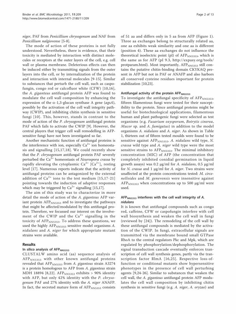
ResultsIn silico analysis of AFPNN5353CLUSTALW amino acid (aa) sequence analysis ofAFPNN5353 with other known antifungal proteinsrevealed that AFPNN5353 from A. giganteus strain A3274is a protein homologous to AFP from A. giganteus strainMDH 18894 [8,22]. AFPNN5353 exhibits > 90% identitywith AFP, but only 42% identity with the P. chryso-genum PAF and 27% identity with the A. niger ANAFP.In fact, the secreted mature form of AFPNN5353 consists
of 51 aa and differs only in 5 aa from AFP (Figure 1).Three aa exchanges belong to structurally related aa,one aa exhibits weak similarity and one aa is different(position 4). These aa exchanges do not influence thetheoretical isoelectric point (pI) of AFPNN5353, which isthe same as for AFP (pI 9.3, http://expasy.org/tools/protparam.html). Most importantly, AFPNN5353 still con-tains the putative chitin-binding domain CKYKAQ pre-sent in AFP but not in PAF or ANAFP and also harborsall conserved cysteine residues important for proteinstabilization [10,23].
Antifungal activity of the protein AFPNN5353To investigate the antifungal specificity of AFPNN5353,fifteen filamentous fungi were tested for their suscept-ibility to the protein. Since antifungal proteins might beuseful for biotechnological applications, filamentoushuman and plant pathogenic fungi were selected as testorganisms (e.g. Fusarium oxysporum, Botrytis cinerea,Mucor sp. and A. fumigatus) in addition to the modelorganisms A. nidulans and A. niger. As shown in Table1, thirteen out of fifteen tested moulds were found to besensitive against AFPNN5353. A. nidulans wild type, N.crassa wild type and A. niger wild type were the mostsensitive strains to AFPNN5353. The minimal inhibitoryconcentration (MIC) of AFP (the concentration thatcompletely inhibited conidial germination in liquidgrowth assays) was 0.2 μg/ml for A. nidulans, 0.5 μg/mlfor N. crassa and 1 μg/ml for A. niger. Two strains wereunaffected at the protein concentrations tested: M. circe-nelloides and M. genevensis were insensitive againstAFPNN5353 when concentrations up to 500 μg/ml wereused.
AFPNN5353 interferes with the cell wall integrity of A.nidulansIt is known that antifungal compounds such as congored, caffeine, CFW or caspofungin interfere with cellwall biosynthesis and weaken the cell wall in fungi(reviewed by [24]). The remodeling of the cell wall bythese antifungal compounds is mediated by the activa-tion of the CWIP. In fungi, extracellular signals aretransmitted via the membrane bound small GTPaseRhoA to the central regulators Pkc and Mpk, which areregulated by phosphorylation/dephosphorylation. Thesignal transduction cascade eventually enforces tran-scription of cell wall synthesis genes, partly via the tran-scription factor RlmA [16,25]. Respective loss-of-function or conditional mutants show hypersensitivephenotypes in the presence of cell wall perturbingagents [9,24-26]. Similar to substances that weaken thecell wall, the A. giganteus antifungal protein AFP modu-lates the cell wall composition by inhibiting chitinsynthesis in sensitive fungi (e.g. A. niger, A. oryzae) and
Binder et al. BMC Microbiology 2011, 11:209http://www.biomedcentral.com/1471-2180/11/209
Page 2 of 13
inducing the expression of agsA most likely by the acti-vation of the CWIP [10].To study the involvement of the CWIP in AFPNN5353
toxicity, we first tested whether the osmotic stabilizersorbitol counteracts the toxicity of AFPNN5353. In theabsence of AFPNN5353 A. nidulans proliferated less wellin the presence of 1 M sorbitol and reached only 30%growth compared to the growth in standard medium(100%). Nevertheless, the addition of 1 M sorbitol to thegrowth medium strongly reduced the activity ofAFPNN5353 on A. nidulans wild type. The osmotic stabi-lizer ameliorated growth in the presence of 0.05 μg/mlAFPNN5353 by 80% compared to a 10% growth rate inthe absence of sorbitol (Table 2). This was even moreaccentuated when 0.1 and 0.2 μg/ml AFPNN5353 wereapplied, suggesting that AFPNN5353 indeed weakens thecell wall of A. nidulans.To investigate whether AFPNN5353 induces agsA gene
transcription similar to AFP via the Pkc/Mpk signallingpathway, we tested the effect of the antifungal proteinon the transgenic A. niger strain RD6.47 which expresses
a nuclear-targeted GFP protein fused to the A. nigeragsA promoter. RD6.47 germlings were treated withAFPNN5353 (conc. 10 to 100 μg/ml) for 2 h and analyzedmicroscopically. As shown in Additional file 1, a nuclearsignal was clearly detectable in germlings of RD6.47treated with ≥ 50 μg/ml AFPNN5353, similar to that whenexposed to 10 μg/ml caspofungin. In untreated germl-ings, however, no signal could be observed. These obser-vations perfectly match with the data obtained for AFP[10]. It has to be noted here that antifungal protein con-centrations higher than the MIC determined for conidia(> 10-50 fold) are needed to inhibit the growth ofgermlings or hyphae of sensitive fungi [10,27] (data notshown).Next, we tested several A. nidulans mutant strains
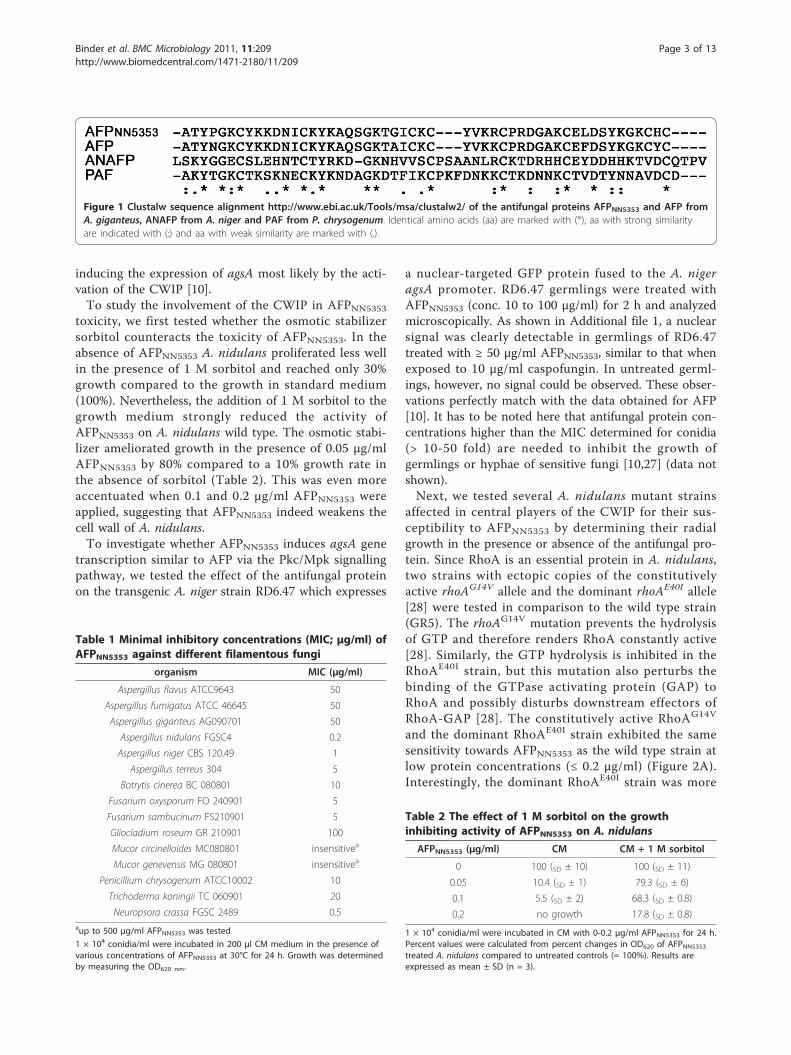
affected in central players of the CWIP for their sus-ceptibility to AFPNN5353 by determining their radialgrowth in the presence or absence of the antifungal pro-tein. Since RhoA is an essential protein in A. nidulans,two strains with ectopic copies of the constitutivelyactive rhoAG14V allele and the dominant rhoAE40I allele[28] were tested in comparison to the wild type strain(GR5). The rhoAG14V mutation prevents the hydrolysisof GTP and therefore renders RhoA constantly active[28]. Similarly, the GTP hydrolysis is inhibited in theRhoAE40I strain, but this mutation also perturbs thebinding of the GTPase activating protein (GAP) toRhoA and possibly disturbs downstream effectors ofRhoA-GAP [28]. The constitutively active RhoAG14V
and the dominant RhoAE40I strain exhibited the samesensitivity towards AFPNN5353 as the wild type strain atlow protein concentrations (≤ 0.2 μg/ml) (Figure 2A).Interestingly, the dominant RhoAE40I strain was more
Figure 1 Clustalw sequence alignment http://www.ebi.ac.uk/Tools/msa/clustalw2/ of the antifungal proteins AFPNN5353 and AFP fromA. giganteus, ANAFP from A. niger and PAF from P. chrysogenum. Identical amino acids (aa) are marked with (*), aa with strong similarityare indicated with (:) and aa with weak similarity are marked with (.).
Table 1 Minimal inhibitory concentrations (MIC; μg/ml) ofAFPNN5353 against different filamentous fungi
organism MIC (μg/ml)
Aspergillus flavus ATCC9643 50
Aspergillus fumigatus ATCC 46645 50
Aspergillus giganteus AG090701 50
Aspergillus nidulans FGSC4 0.2
Aspergillus niger CBS 120.49 1
Aspergillus terreus 304 5
Botrytis cinerea BC 080801 10
Fusarium oxysporum FO 240901 5
Fusarium sambucinum FS210901 5
Gliocladium roseum GR 210901 100
Mucor circinelloides MC080801 insensitivea
Mucor genevensis MG 080801 insensitivea
Penicillium chrysogenum ATCC10002 10
Trichoderma koningii TC 060901 20
Neuropsora crassa FGSC 2489 0.5aup to 500 μg/ml AFPNN5353 was tested
1 × 10⁴ conidia/ml were incubated in 200 μl CM medium in the presence ofvarious concentrations of AFPNN5353 at 30°C for 24 h. Growth was determinedby measuring the OD620 nm.
Table 2 The effect of 1 M sorbitol on the growthinhibiting activity of AFPNN5353 on A. nidulans
AFPNN5353 (μg/ml) CM CM + 1 M sorbitol
0 100 (SD ± 10) 100 (SD ± 11)
0.05 10.4 (SD ± 1) 79.3 (SD ± 6)
0.1 5.5 (SD ± 2) 68.3 (SD ± 0.8)
0.2 no growth 17.8 (SD ± 0.8)
1 × 10⁴ conidia/ml were incubated in CM with 0-0.2 μg/ml AFPNN5353 for 24 h.Percent values were calculated from percent changes in OD620 of AFPNN5353treated A. nidulans compared to untreated controls (= 100%). Results areexpressed as mean ± SD (n = 3).
Binder et al. BMC Microbiology 2011, 11:209http://www.biomedcentral.com/1471-2180/11/209
Page 3 of 13
resistant to AFPNN5353 than the wild type strain or theRhoAG14V strain at higher protein concentrations (1 μg/ml) (Figure 2A). Therefore, we suggest that the toxicityof AFPNN5353 is transmitted by RhoA-GAP targets andnot by RhoA itself. These mutants performed similarlywhen exposed to the orthologous P. chrysogenum anti-fungal protein PAF [9].In addition, mutants defective in PkcA and MpkA
activity were tested for their AFPNN5353 susceptibility.As pkcA is an essential gene in A. nidulans, a condi-tional alcA-PKC mutant strain was used, where thepkcA gene was put under the control of the alcA
promoter, which is repressed by glucose but derepressedby glycerol [26]. Both the conditional alcA-PKC mutant(cultivated under repressive conditions) and a ΔmpkAmutant were hypersensitive to AFPNN5353 compared totheir recipient strains R153 and GR5, respectively, indi-cating that the activity of PkcA and MpkA confers acertain resistance to AFPNN5353 (Figure 2A). The hyper-sensitive phenotype of the ΔmpkA mutant was also con-firmed by liquid growth inhibitory assays. Inunchallenged liquid condition, the GR5 and the ΔmpkAmutant showed a comparable proliferation rate (Figure2B). In the presence of 0.05 μg/ml AFPNN5353, however,
Figure 2 AFPNN5353 susceptibility of A. nidulans mutants RhoAG14V, RhoAE40I, alcA-PkcA and ΔmpkA compared to the respectiverecipient strains GR5 and R153. (A) A total of 2 × 10³ conidia were point inoculated on agar plates (CM for GR5, RhoAG14V, RhoAE40I andΔmpkA, repressive MM containing 1% glucose according to [26] for R135 and alcA-PkcA) containing the appropriate supplements and 0, 0.2 and1 μg/ml AFPNN5353 for GR5, RhoA
G14V, RhoAE40I, R135 and alcA-PkcA. The ΔmpkA mutant and its reference strain GR5 were exposed to 0, 0.5 and1 μg/ml AFPNN5353. The plates were incubated at 37°C for 48 h. (B) 1 × 10⁴ conidia/ml of the ΔmpkA mutant and GR5 were treated with 0.05μg/ml AFPNN5353 or without protein (controls) in a total volume of 200 μl of appropriately supplemented CM in 96-well plates.
Binder et al. BMC Microbiology 2011, 11:209http://www.biomedcentral.com/1471-2180/11/209
Page 4 of 13

the mpkA deletion strain did not germinate whereas theGR5 strain still exhibited 11% growth. Note that growthinhibition in liquid conditions requires less antifungalprotein to monitor its toxicity than on solid media prob-ably due to less diffusion in the latter case (data notshown).From these data we conclude that AFPNN5353 inter-
feres with the cell wall homeostasis of A. nidulans andthat this interaction is mediated by PkcA/MpkA signal-ling, although independently from RhoA.
AFPNN5353 disrupts calcium homeostasis in A. nigerSupplements other than osmotic stabilizers can alsoantagonize the activity of antifungal proteins from plantsand ascomycetes. For example, the addition of cationssuch as Ca2+ ions to the growth medium reversed theantifungal activity of the P. chrysogenum PAF [17], theA. giganteus AFP [15,21] and of plant defensins [29,30]which are usually positively charged due to their highpI. A cation-sensitive antifungal mode of action can forexample be associated with the perturbation of theintracellular Ca2+ homeostasis by antifungal peptides[17,18] but might also result from the interference ofcations with antifungal-target interaction(s).Therefore, we tested to which extend these effects also
account for the antifungal activity of AFPNN5353. To thisend, we selected A. niger as model organism because thismould was highly sensitive to AFPNN5353 and a trans-genic strain was available that expressed the recombinantcodon optimized Ca2+-sensitive photoprotein aequorinfor measuring the [Ca2+]c resting level in response toAFPNN5353 [31]. First, we tested the activity of AFPNN5353
in Vogels* medium supplemented with 5-20 mM CaCl2or without CaCl2 as a control (data not shown). Additionof CaCl2 did not influence the growth of A. niger up to aconcentration of 20 mM. The growth of A. niger exposedto AFPNN5353, however, ameliorated in the presence ofincreasing concentrations of CaCl2. 20 mM CaCl2 neu-tralized the toxicity of 0.5-1.0 μg/ml AFPNN5353 and thetreated samples resumed growth to 100% (Table 3).Next, we determined the influence of AFPNN5353 on
the intracellular Ca2+ signature. Before AFPNN5353
addition, the resting level of the intracellular Ca2+ was0.08 μM. We could show, however, that the [Ca2+]c rest-ing level was significantly increased in twelve h old A.niger cultures that were treated with 20 μg/mlAFPNN5353. The [Ca2+]c resting level rose to a maximumof 0.19 μM within the first 8 min and stayed elevatedthroughout the time of measurement (60 min), whereasthe Ca2+ level of the untreated control remained at 0.08μM (Figure 3). This indicated that AFPNN5353 indeeddisrupts Ca2+ homeostasis in A. niger.To exclude the possibility that the AFPNN5353 induced
rise in the [Ca2+]c resting level is due to membrane per-meabilization and/or pore formation, we studied theeffects of AFPNN5353 on germlings in the presence ofCMFDA, a membrane permeant dye that is metabolizedby viable cells, and the membrane impermeant dye pro-pidium iodide (PI). Additional file 2 shows that samplestreated with 20 μg/ml AFPNN5353 for 10 min metabo-lized CMFDA but did not take up PI, resulting in greenbut no red fluorescence, similar to untreated controls.This indicated that the plasma membrane was still intactafter 10 min of protein treatment. Samples exposed toethanol did not metabolize CMFDA but appeared brightred due to PI internalization, indicating that here themembrane was permeabilized. We therefore concludethat the rapid increase in [Ca2+]c within the first 10 minof protein treatment is not the result of uncontrolledCa2+ influx due to plasma membrane permeabilization.
The calcium chelator BAPTA abrogates the AFPNN5353-induced calcium signatureThe increased [Ca2+]c in response to AFPNN5353 treatmentcould originate from extracellular and/or from intracellu-lar Ca2+ stores, such as mitochondria, vacuoles,
Table 3 The effect of 20 mM external CaCl2 (in Vogels*medium) on the growth inhibitory activity of AFPNN5353on A. niger strain A533.
AFPNN5353 (μg/ml) Vogels* Vogels* + 20 mM Ca2+
0 100 (SD ± 10) 100 (SD ± 8)
0.5 12 (SD ± 3) 101 (SD ± 9)
1.0 no growth 105 (SD ± 6)
OD620 was measured after 24 h of incubation. The growth of untreatedcontrols was normalized to 100% to evaluate the percent growth of samplesin the presence of AFPNN5353. Vogels* medium without CaCl2 supplementationcontains 0.7 mM Ca2+. Results are expressed as mean ± SD (n = 3).
Figure 3 Increase in resting [Ca2+]c of twelve h old A. nigergermlings treated with AFPNN5353 or no protein (controls).Measurements were taken every 1.4 minutes. Values representaverage of six samples.
Binder et al. BMC Microbiology 2011, 11:209http://www.biomedcentral.com/1471-2180/11/209
Page 5 of 13
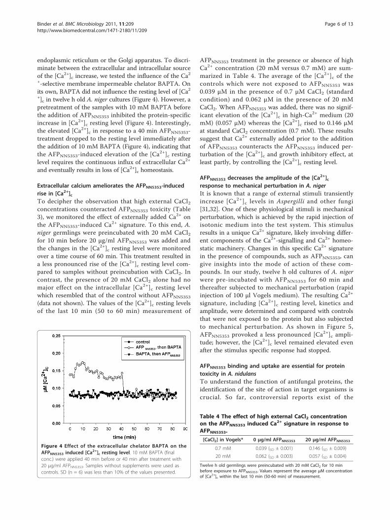
endoplasmic reticulum or the Golgi apparatus. To discri-minate between the extracellular and intracellular sourceof the [Ca2+]c increase, we tested the influence of the Ca2+-selective membrane impermeable chelator BAPTA. Onits own, BAPTA did not influence the resting level of [Ca2+]c in twelve h old A. niger cultures (Figure 4). However, apretreatment of the samples with 10 mM BAPTA beforethe addition of AFPNN5353 inhibited the protein-specificincrease in [Ca2+]c resting level (Figure 4). Interestingly,the elevated [Ca2+]c in response to a 40 min AFPNN5353-treatment dropped to the resting level immediately afterthe addition of 10 mM BAPTA (Figure 4), indicating thatthe AFPNN5353-induced elevation of the [Ca2+]c restinglevel requires the continuous influx of extracellular Ca2+
and eventually results in loss of [Ca2+]c homeostasis.
Extracellular calcium ameliorates the AFPNN5353-inducedrise in [Ca2+]cTo decipher the observation that high external CaCl2concentrations counteracted AFPNN5353 toxicity (Table3), we monitored the effect of externally added Ca2+ onthe AFPNN5353-induced Ca2+ signature. To this end, A.niger germlings were preincubated with 20 mM CaCl2for 10 min before 20 μg/ml AFPNN5353 was added andthe changes in the [Ca2+]c resting level were monitoredover a time course of 60 min. This treatment resulted ina less pronounced rise of the [Ca2+]c resting level com-pared to samples without preincubation with CaCl2. Incontrast, the presence of 20 mM CaCl2 alone had nomajor effect on the intracellular [Ca2+]c resting levelwhich resembled that of the control without AFPNN5353
(data not shown). The values of the [Ca2+]c resting levelsof the last 10 min (50 to 60 min) measurement of
AFPNN5353 treatment in the presence or absence of highCa2+ concentration (20 mM versus 0.7 mM) are sum-marized in Table 4. The average of the [Ca2+]c of thecontrols which were not exposed to AFPNN5353 was0.039 μM in the presence of 0.7 μM CaCl2 (standardcondition) and 0.062 μM in the presence of 20 mMCaCl2. When AFPNN5353 was added, there was no signif-icant elevation of the [Ca2+]c in high-Ca2+ medium (20mM) (0.057 μM) whereas the [Ca2+]c rised to 0.146 μMat standard CaCl2 concentration (0.7 mM). These resultssuggest that Ca2+ externally added prior to the additionof AFPNN5353 counteracts the AFPNN5353 induced per-turbation of the [Ca2+]c and growth inhibitory effect, atleast partly, by controlling the [Ca2+]c resting level.
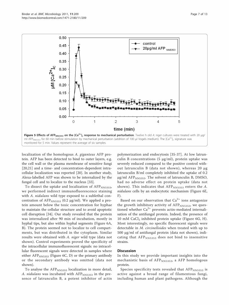
AFPNN5353 decreases the amplitude of the [Ca2+]cresponse to mechanical perturbation in A. nigerIt is known that a range of external stimuli transientlyincrease [Ca2+]c levels in Aspergilli and other fungi[31,32]. One of these physiological stimuli is mechanicalperturbation, which is achieved by the rapid injection ofisotonic medium into the test system. This stimulusresults in a unique Ca2+ signature, likely involving differ-ent components of the Ca2+-signalling and Ca2+ homeo-static machinery. Changes in this specific Ca2+ signaturein the presence of compounds, such as AFPNN5353, cangive insights into the mode of action of these com-pounds. In our study, twelve h old cultures of A. nigerwere pre-incubated with AFPNN5353 for 60 min andthereafter subjected to mechanical perturbation (rapidinjection of 100 μl Vogels medium). The resulting Ca2+
signature, including [Ca2+]c resting level, kinetics andamplitude, were determined and compared with controlsthat were not exposed to the protein but also subjectedto mechanical perturbation. As shown in Figure 5,AFPNN5353 provoked a less pronounced [Ca2+]c ampli-tude; however, the [Ca2+]c level remained elevated evenafter the stimulus specific response had stopped.
AFPNN5353 binding and uptake are essential for proteintoxicity in A. nidulansTo understand the function of antifungal proteins, theidentification of the site of action in target organisms iscrucial. So far, controversial reports exist of the
Figure 4 Effect of the extracellular chelator BAPTA on theAFPNN5353 induced [Ca2+]c resting level. 10 mM BAPTA (finalconc.) were applied 40 min before or 40 min after treatment with20 μg/ml AFPNN5353. Samples without supplements were used ascontrols. SD (n = 6) was less than 10% of the values presented.
Table 4 The effect of high external CaCl2 concentrationon the AFPNN5353 induced Ca2+ signature in response toAFPNN5353.
[CaCl2] in Vogels* 0 μg/ml AFPNN5353 20 μg/ml AFPNN5353
0.7 mM 0.039 (SD ± 0.001) 0.146 (SD ± 0.009)
20 mM 0.062 (SD ± 0.003) 0.057 (SD ± 0.004)
Twelve h old germlings were preincubated with 20 mM CaCl2 for 10 minbefore exposure to AFPNN5353. Values represent the average μM concentrationof [Ca2+]c within the last 10 min (50-60 min) of measurement.
Binder et al. BMC Microbiology 2011, 11:209http://www.biomedcentral.com/1471-2180/11/209
Page 6 of 13
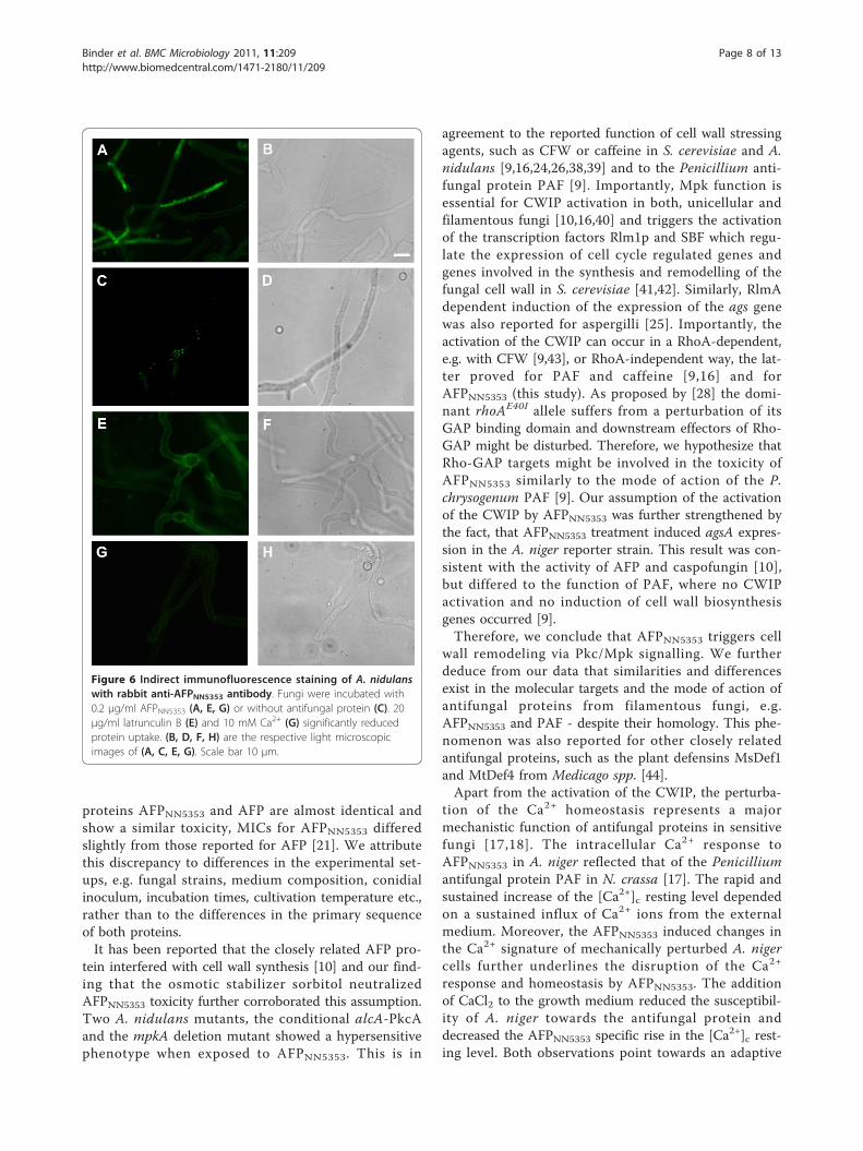
localization of the homologous A. giganteus AFP pro-tein. AFP has been detected to bind to outer layers, e.g.the cell wall or the plasma membrane of sensitive fungi[20,21] and a time- and concentration-dependent intra-cellular localization was reported [20]. In another study,Alexa-labelled AFP was shown to be internalized by thefungal cell and to localize to the nucleus [33].To dissect the uptake and localization of AFPNN5353,
we performed indirect immunofluorescence stainingwith A. nidulans wild type exposed to a sublethal con-centration of AFPNN5353 (0.2 μg/ml). We applied a pro-tein amount below the toxic concentration for hyphaeto maintain the cellular structure and to avoid apoptoticcell disruption [34]. Our study revealed that the proteinwas internalized after 90 min of incubation, mostly inhyphal tips, but also within hyphal segments (Figure 6A,B). The protein seemed not to localize to cell compart-ments, but was distributed in the cytoplasm. Similarresults were obtained with A. niger wild type (data notshown). Control experiments proved the specificity ofthe intracellular immunofluorescent signals: no intracel-lular fluorescent signals were detected in samples whereeither AFPNN5353 (Figure 6C, D) or the primary antibodyor the secondary antibody was omitted (data notshown).To analyse the AFPNN5353 localization in more detail,
A. nidulans was incubated with AFPNN5353 in the pre-sence of latrunculin B, a potent inhibitor of actin
polymerization and endocytosis [35-37]. At low latrun-culin B concentrations (5 μg/ml), protein uptake wasseverely reduced compared to the positive control with-out latrunculin B (data not shown), whereas 20 μglatrunculin B/ml completely inhibited the uptake of 0.2μg/ml AFPNN5353. The solvent of latrunculin B, DMSO,had no adverse effect on protein uptake (data notshown). This indicates that AFPNN5353 enters the A.nidulans cells by an endocytotic mechanism (Figure 6E,F).Based on our observation that Ca2+ ions antagonize
the growth inhibitory activity of AFPNN5353, we ques-tioned whether Ca2+ prevents actin-mediated internali-sation of the antifungal protein. Indeed, the presence of10 mM CaCl2 inhibited protein uptake (Figure 6G, H).Most interestingly, no specific fluorescent signals weredetectable in M. circinelloides when treated with up to500 μg/ml of antifungal protein (data not shown), indi-cating that AFPNN5353 does not bind to insensitivestrains.
DiscussionIn this study we provide important insights into themechanistic basis of AFPNN5353, a AFP homologousprotein.Species specificity tests revealed that AFPNN5353 is
active against a broad range of filamentous fungi,including human and plant pathogens. Although the
Figure 5 Effects of AFPNN5353 on the [Ca2+]c response to mechanical perturbation. Twelve h old A. niger cultures were treated with 20 μg/ml AFPNN5353 for 60 min before stimulation by mechanical perturbation (addition of 100 μl Vogels medium). The [Ca2+]c signature wasmonitored for 5 min. Values represent the average of six samples.
Binder et al. BMC Microbiology 2011, 11:209http://www.biomedcentral.com/1471-2180/11/209
Page 7 of 13
proteins AFPNN5353 and AFP are almost identical andshow a similar toxicity, MICs for AFPNN5353 differedslightly from those reported for AFP [21]. We attributethis discrepancy to differences in the experimental set-ups, e.g. fungal strains, medium composition, conidialinoculum, incubation times, cultivation temperature etc.,rather than to the differences in the primary sequenceof both proteins.It has been reported that the closely related AFP pro-
tein interfered with cell wall synthesis [10] and our find-ing that the osmotic stabilizer sorbitol neutralizedAFPNN5353 toxicity further corroborated this assumption.Two A. nidulans mutants, the conditional alcA-PkcAand the mpkA deletion mutant showed a hypersensitivephenotype when exposed to AFPNN5353. This is in
agreement to the reported function of cell wall stressingagents, such as CFW or caffeine in S. cerevisiae and A.nidulans [9,16,24,26,38,39] and to the Penicillium anti-fungal protein PAF [9]. Importantly, Mpk function isessential for CWIP activation in both, unicellular andfilamentous fungi [10,16,40] and triggers the activationof the transcription factors Rlm1p and SBF which regu-late the expression of cell cycle regulated genes andgenes involved in the synthesis and remodelling of thefungal cell wall in S. cerevisiae [41,42]. Similarly, RlmAdependent induction of the expression of the ags genewas also reported for aspergilli [25]. Importantly, theactivation of the CWIP can occur in a RhoA-dependent,e.g. with CFW [9,43], or RhoA-independent way, the lat-ter proved for PAF and caffeine [9,16] and forAFPNN5353 (this study). As proposed by [28] the domi-nant rhoAE40I allele suffers from a perturbation of itsGAP binding domain and downstream effectors of Rho-GAP might be disturbed. Therefore, we hypothesize thatRho-GAP targets might be involved in the toxicity ofAFPNN5353 similarly to the mode of action of the P.chrysogenum PAF [9]. Our assumption of the activationof the CWIP by AFPNN5353 was further strengthened bythe fact, that AFPNN5353 treatment induced agsA expres-sion in the A. niger reporter strain. This result was con-sistent with the activity of AFP and caspofungin [10],but differed to the function of PAF, where no CWIPactivation and no induction of cell wall biosynthesisgenes occurred [9].Therefore, we conclude that AFPNN5353 triggers cell
wall remodeling via Pkc/Mpk signalling. We furtherdeduce from our data that similarities and differencesexist in the molecular targets and the mode of action ofantifungal proteins from filamentous fungi, e.g.AFPNN5353 and PAF - despite their homology. This phe-nomenon was also reported for other closely relatedantifungal proteins, such as the plant defensins MsDef1and MtDef4 from Medicago spp. [44].Apart from the activation of the CWIP, the perturba-
tion of the Ca2+ homeostasis represents a majormechanistic function of antifungal proteins in sensitivefungi [17,18]. The intracellular Ca2+ response toAFPNN5353 in A. niger reflected that of the Penicilliumantifungal protein PAF in N. crassa [17]. The rapid andsustained increase of the [Ca2+]c resting level dependedon a sustained influx of Ca2+ ions from the externalmedium. Moreover, the AFPNN5353 induced changes inthe Ca2+ signature of mechanically perturbed A. nigercells further underlines the disruption of the Ca2+
response and homeostasis by AFPNN5353. The additionof CaCl2 to the growth medium reduced the susceptibil-ity of A. niger towards the antifungal protein anddecreased the AFPNN5353 specific rise in the [Ca2+]c rest-ing level. Both observations point towards an adaptive
Figure 6 Indirect immunofluorescence staining of A. nidulanswith rabbit anti-AFPNN5353 antibody. Fungi were incubated with0.2 μg/ml AFPNN5353 (A, E, G) or without antifungal protein (C). 20μg/ml latrunculin B (E) and 10 mM Ca2+ (G) significantly reducedprotein uptake. (B, D, F, H) are the respective light microscopicimages of (A, C, E, G). Scale bar 10 μm.
Binder et al. BMC Microbiology 2011, 11:209http://www.biomedcentral.com/1471-2180/11/209
Page 8 of 13
response which is mediated most probably via Ca2+ sig-nalling. First, high extracellular Ca2+ concentrations trig-ger chitin synthesis in A. niger and thereby conferincreased protection against antifungal proteins as shownfor AFP [15]. Second, it primes the Ca2+ homeostaticmachinery to better maintain a low [Ca2+]c resting levelwhen challenged with the antifungal protein, e.g. by (i)the increase of the activity of existing Ca2+ pumps/trans-porters to counteract the AFPNN5353-specific intracellularCa2+ perturbation, or (ii) the modulation of the expres-sion of Ca2+ channels/pumps/exchangers [17]. The for-mer hypothesis (i) might be supported by the observationthat the addition of CaCl2 only 10 min before A. nigerwas challenged with AFPNN5353 restored the low [Ca2+]cresting level. However, the perturbation of the Ca2+
homeostasis by a sustained elevation of the [Ca2+]c rest-ing level indicates that A. niger is not able to restore thelow [Ca2+]c resting level after exposure to AFPNN5353 andthis might trigger programmed cell death (PCD) on thelong term as it was shown to occur in A. nidulans inresponse to the P. chrysogenum PAF [34].Since AFP was shown to cause membrane permeabili-
zation [21], the influx of Ca2+ might be due to changesin membrane permeability for this ion, if not the forma-tion of pores. However, our staining experiments withCMFDA and PI exclude this possibility at least in thefirst 10 min of exposure to AFPNN5353 when the [Ca2+]cresting level reaches its maximum. This result is furthercorroborated by the fact that higher external concentra-tions of Ca2+ reduced the AFPNN5353 specific rise in[Ca2+]c resting level which - in our opinion - would notoccur with leaky membranes. However, we do notexclude changes in membrane permeability at longerexposure times to this antifungal protein and more stu-dies are needed to answer this question.Finally, we observed that the internalization of
AFPNN5353 is characteristic for sensitive but not resistantmoulds. A lack of binding of AFPNN5353 to insensitivefungi might point towards the absence or inaccessibilityof a putative interacting molecule at the cell surface.AFPNN5353 localized to the cytoplasm of target fungi onlywhen actin filaments were formed. This is in agreementwith the endocytotic uptake and intracellular localizationof the P. chrysogenum antifungal protein PAF in sensitivefilamentous fungi [14,45]. Importantly, we observed thatAFPNN5353 was internalized by hyphae even under sub-inhibitory concentrations (0.2 μg/ml for A. nidulans)which suggests that a threshold concentration is requiredto cause severe growth defects in target fungi.The presence of high concentrations of extracellular Ca2
+ counteracted AFPNN5353 uptake. This finding parallelswell with the report of [20] that the presence of cations,such as Ca2+, interfered with the binding of AFP to thesurface of F. oxysporum and with our observations made
with the Penicillium PAF (unpublished data). One possibleexplanation might be that extracellular Ca2+ ions competewith AFPNN5353 for the same molecular target on the fun-gal surface which might represent a first binding receptoror even a “gate” for protein uptake [20,21] or, alternatively,that the interacting target is repressed under these condi-tions [17]. An additional explanation might be that the pri-mary cell-surface localized AFPNN5353 target might bemasked due to a Ca2+-dependent stimulation of chitinsynthesis and cell wall remodeling as recently observed forAFP in A. niger [15]. This further suggests that the activa-tion of the CWIP and the agsA induction does not med-iate sufficient resistance to survive the toxic effects ofAFPNN5353. Instead, according to the “damage-responseframework of AFP-fungal interactions” [15], the chitinresponse might represent the better strategy for fungi tosurvive the antifungal attack.
ConclusionsBased on the growth inhibitory activity, antifungal pro-teins like AFPNN5353 can be well considered as promis-ing candidates for future antimycotic drugdevelopments. However, for biotechnological exploita-tion, the detailed knowledge on the mode of action isdemanded. Our study shows that the detrimental effectscaused by the A. giganteus antifungal protein AFPNN5353
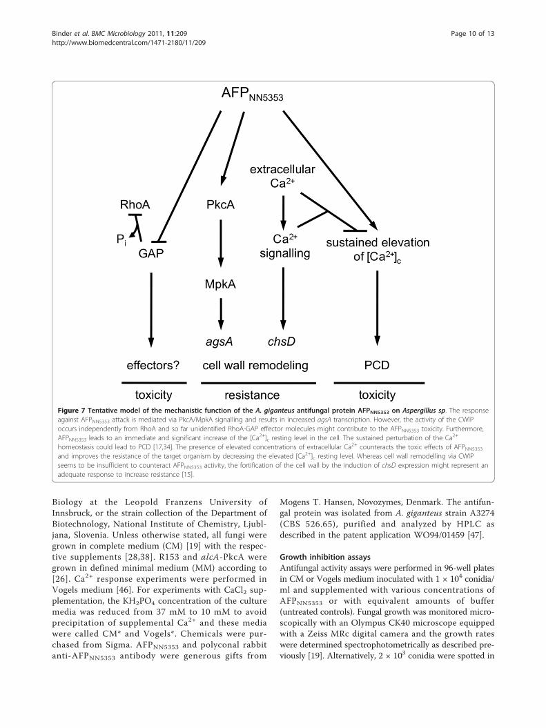
in sensitive target aspergilli are based on the interactionof this protein with more than one signalling pathway.In Figure 7, we present a tentative working model. Thetoxicity of AFPNN5353 is mediated via PkcA/MpkA sig-nalling which occurs independently from RhoA. Instead,so far unidentified RhoA-GAP effector molecules mightcontribute to AFPNN5353 toxicity. The activation of theCWIP by AFPNN5353 induces the agsA gene expressionwhich is, however, insufficient to counteract toxicity ofthe protein. Furthermore, AFPNN5353 leads to animmediate and significant increase of the [Ca2+]c restinglevel in the cell. This sustained perturbation of the Ca2+
homeostasis could lead to PCD [17,34]. The presence ofextracellular Ca2+ neutralizes the toxic effects ofAFPNN5353 and improves the resistance of the targetorganism possibly by decreasing the elevated [Ca2+]cresting level and stimulating the fortification of the cellwall by the induction of chsD expression as shown forAFP [15]. Further investigations are in progress to clar-ify how these pathways are interconnected and interferewith each other on the molecular level.
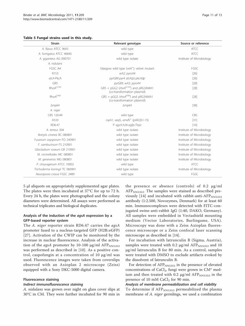
MethodsStrains, Media and ChemicalsFungal strains used in this study are listed in Table 5.All strains were obtained from the culture collectionsFGSC, ATCC, CBS, from the Institute of Microbiology,Division of Systematics, Taxonomy and Evolutionary
Binder et al. BMC Microbiology 2011, 11:209http://www.biomedcentral.com/1471-2180/11/209
Page 9 of 13
Biology at the Leopold Franzens University ofInnsbruck, or the strain collection of the Department ofBiotechnology, National Institute of Chemistry, Ljubl-jana, Slovenia. Unless otherwise stated, all fungi weregrown in complete medium (CM) [19] with the respec-tive supplements [28,38]. R153 and alcA-PkcA weregrown in defined minimal medium (MM) according to[26]. Ca2+ response experiments were performed inVogels medium [46]. For experiments with CaCl2 sup-plementation, the KH2PO4 concentration of the culturemedia was reduced from 37 mM to 10 mM to avoidprecipitation of supplemental Ca2+ and these mediawere called CM* and Vogels*. Chemicals were pur-chased from Sigma. AFPNN5353 and polyconal rabbitanti-AFPNN5353 antibody were generous gifts from
Mogens T. Hansen, Novozymes, Denmark. The antifun-gal protein was isolated from A. giganteus strain A3274(CBS 526.65), purified and analyzed by HPLC asdescribed in the patent application WO94/01459 [47].
Growth inhibition assaysAntifungal activity assays were performed in 96-well platesin CM or Vogels medium inoculated with 1 × 10⁴ conidia/ml and supplemented with various concentrations ofAFPNN5353 or with equivalent amounts of buffer(untreated controls). Fungal growth was monitored micro-scopically with an Olympus CK40 microscope equippedwith a Zeiss MRc digital camera and the growth rateswere determined spectrophotometrically as described pre-viously [19]. Alternatively, 2 × 10³ conidia were spotted in
Figure 7 Tentative model of the mechanistic function of the A. giganteus antifungal protein AFPNN5353 on Aspergillus sp. The responseagainst AFPNN5353 attack is mediated via PkcA/MpkA signalling and results in increased agsA transcription. However, the activity of the CWIPoccurs independently from RhoA and so far unidentified RhoA-GAP effector molecules might contribute to the AFPNN5353 toxicity. Furthermore,AFPNN5353 leads to an immediate and significant increase of the [Ca2+]c resting level in the cell. The sustained perturbation of the Ca2+
homeostasis could lead to PCD [17,34]. The presence of elevated concentrations of extracellular Ca2+ counteracts the toxic effects of AFPNN5353and improves the resistance of the target organism by decreasing the elevated [Ca2+]c resting level. Whereas cell wall remodelling via CWIPseems to be insufficient to counteract AFPNN5353 activity, the fortification of the cell wall by the induction of chsD expression might represent anadequate response to increase resistance [15].
Binder et al. BMC Microbiology 2011, 11:209http://www.biomedcentral.com/1471-2180/11/209
Page 10 of 13
5 μl aliquots on appropriately supplemented agar plates.The plates were then incubated at 37°C for up to 72 h.Every 24 h, the plates were photographed and the colonydiameters were determined. All assays were performed astechnical triplicates and biological duplicates.
Analysis of the induction of the agsA expression by aGFP-based reporter systemThe A. niger reporter strain RD6.47 carries the agsApromoter fused to a nucleus-targeted GFP (H2B::eGFP)[27]. Activation of the CWIP can be monitored by theincrease in nuclear fluorescence. Analysis of the activa-tion of the agsA promoter by 10-100 μg/ml AFPNN5353
was performed as described in [10]. As a positive con-trol, caspofungin at a concentration of 10 μg/ml wasused. Fluorescence images were taken from coverslipsobserved with an Axioplan 2 microscope (Zeiss)equipped with a Sony DKC-5000 digital camera.
Fluorescence stainingIndirect immunofluorescence stainingA. nidulans was grown over night on glass cover slips at30°C in CM. They were further incubated for 90 min in
the presence or absence (controls) of 0.2 μg/mlAFPNN5353. The samples were stained as described pre-viously [14] and incubated with rabbit-anti-AFPNN5353
antibody (1:2.500, Novozymes, Denmark) for at least 60min. Immunocomplexes were detected with FITC-con-jugated swine-anti-rabbit IgG (1:40, DAKO, Germany).All samples were embedded in Vectashield mountingmedium (Vector Laboratories, Burlingame, USA).Microscopy was done with a Zeiss Axioplan fluores-cence microscope or a Zeiss confocal laser scanningmicroscope as described in [14].For incubation with latrunculin B (Sigma, Austria),
samples were treated with 0.2 μg/ml AFPNN5353 and 10μg/ml latrunculin B for 80 min. As a control, sampleswere treated with DMSO to exclude artifacts evoked bythe dissolvent of latrunculin B.For detection of AFPNN5353 in the presence of elevated
concentrations of CaCl2, fungi were grown in CM* med-ium and then treated with 0.2 μg/ml AFPNN5353 in thepresence of 10 mM CaCl2 for 90 min.Analysis of membrane permeabilization and cell viabilityTo determine if AFPNN5353 permeabilized the plasmamembrane of A. niger germlings, we used a combination
Table 5 Fungal strains used in this study.
Strain Relevant genotype Source or reference
A. flavus ATCC 9643 wild type ATCC
A. fumigatus ATCC 46645 wild type ATCC
A. giganteus AG 090701 wild type isolate Institute of Microbiology
A. nidulans
FGSC A4 Glasgow wild type (veA+); velvet mutant FGSC
R153 wA2; pyroA4 [26]
alcA-PkcA pyrG89::pyr4 alcA(p)::pkcAΔp [26]
GR5 pyrG89; wA3; pyroA4 [28]
RhoAG14V GR5 + pGG2 (rhoAG14V) and pRG3AMA1(co-transformation plasmid)
[28]
RhoAE40I GR5 + pGG5 (rhoAE40I) and pRG3AMA1(co-transformation plasmid)
[28]
ΔmpkA ΔmpkA [38]
A. niger
CBS 120.49 wild type CBS
A533 cspA1, aeqS, amdS+ (pAEQS1-15) [31]
RD6.47 P agsA::h2b::egfp::Ttrpc [10]
A. terreus 304 wild type isolate Institute of Microbiology
Botrytis cinerea BC 080801 wild type isolate Institute of Microbiology
Fusarium oxysporum FO 240901 wild type isolate Institute of Microbiology
F. sambucinum FS 210901 wild type isolate Institute of Microbiology
Gliocladium roseum GR 210901 wild type isolate Institute of Microbiology
M. circinelloides MC 080801 wild type isolate Institute of Microbiology
M. genevensis MG 080801 wild type isolate Institute of Microbiology
P. chrysogenum ATCC 10002 wild type ATCC
Trichoderma koningii TC 060901 wild type isolate Institute of Microbiology
Neuropsora crassa FGSC 2489 wild type FGSC
Binder et al. BMC Microbiology 2011, 11:209http://www.biomedcentral.com/1471-2180/11/209
Page 11 of 13
of propidium iodide (PI) and fluorescein diacetate (celltracker, CMFDA green, Invitrogen) according to [48].Twelve h old A. niger germlings were grown in Vogelsmedium and pretreated with the two dyes (final conc. 5μg/ml each) for 15 min before AFPNN5353 was added toa final concentration of 20 μg/ml. Samples withoutAFPNN5353 served as controls for positive CMFDA stain-ing, while ethanol (70%) was used to permeabilize themembrane for positive PI staining.Analysis of the calcium response to AFPNN5353 application105 conidia/ml of the A. niger strain A533 expressingcodon optimized aequorin were grown in Vogels* med-ium containing 10 μM coelenterazine (Biosynth, Swit-zerland) at 30°C for twelve h in the dark. The [Ca2+]cresting level and mechanical perturbation experimentsand the calibration of [Ca2+]c were performed asdescribed in [17].
Additional material
Additional file 1: The expression of nucleus-targeted GFP under thecontrol of the agsA promoter in A. niger in response to cell wallinterfering substances. Differential interfering contrast images andcorresponding fluorescence images of A. niger RD6.47 indicate theexpression of a nucleus-targeted GFP under the control of the A. nigeragsA promoter. Five h old germlings were (A) left untreated (negativecontrol), (B) treated with 50 μg/ml AFPNN5353 and (C) with 10 μg/mlcaspofungin (positive control) as described in Materials and Methods.Scale bar, 20 μm.
Additional file 2: Viability staining of A. niger germlings afterAFPNN5353 exposure. Twelve h old A. niger germlings were stained withfluorescein diacetate (CMFDA, middle pannels) and propidium iodide(right pannels). The left panels show the respective light micrographs. Allsamples were pretreated with the dyes for 15 min before 20 μg/mlAFPNN5353 was added (B). Controls remained untreated (A) or wereexposed to 70% ethanol (C). Scale bar, 50 μm.
AcknowledgementsWe thank Mogens T. Hansen (Novozymes, Denmark) for the generous gift ofAFPNN5353 and the polyclonal rabbit anti-AFPNN5353 antibody. We gratefullyacknowledge Renate Weiler-Görz for technical assistance.This study was financially supported by the Austrian Science Fund FWF(P19970-B11) and the Österreichischer Austauschdienst ÖAD(Wissenschaftlich-Technische Zusammenarbeit Österreich und Slowenien,SI15/2009).
Author details1Biocenter, Division of Molecular Biology, Innsbruck Medical University, Fritz-Pregl Strasse 3, Innsbruck, A-6020, Austria. 2Department of Biotechnology,National Institute of Chemistry, Hajdrihova 19, Ljubljana, SI-1000, Slovenia.3Excellent NMR, Future Innovation for Sustainable Technologies Centre ofExcellence, Hajdrihova 19, Ljubljana, SI-1000, Slovenia. 4Department ofApplied and Molecular Microbiology, Institute of Biotechnology, BerlinUniversity of Technology, Gustav-Meyer-Allee 25, Berlin, D-13355, Germany.5Department of Hygiene, Microbiology and Social Medicine, InnsbruckMedical University, Fritz-Pregl Strasse 3, Innsbruck, A-6020, Austria.
Authors’ contributionsUB carried out the growth inhibition assays, the indirectimmunofluorescence stainings, the Ca2+ measurements and the calculationsto convert the luminescence units into the [Ca2+]c levels. She alsoperformed the statistical analysis and helped to draft the manuscript. MB
contributed the A. niger A533 strain, helped with the Ca2+ measurementsand participated in the design of the study. AE contributed to the indirectimmunofluorescence stainings. VM contributed the A. niger RD6.47 strainand performed the agsA induction assays. FM conceived of the study,participated in its design and coordination and drafted the manuscript. Allauthors read and approved the final manuscript.
Received: 25 May 2011 Accepted: 23 September 2011Published: 23 September 2011
References1. Hancock RE, Scott MG: The role of antimicrobial peptides in animal
defenses. Proc Natl Acad Sci USA 2000, 97(16):8856-8861.2. Kamysz W, Okroj M, Lukasiak J: Novel properties of antimicrobial peptides.
Acta Biochim Pol 2003, 50(2):461-469.3. Aerts AM, Francois IE, Cammue BP, Thevissen K: The mode of antifungal
action of plant, insect and human defensins. Cell Mol Life Sci 2008,65(13):2069-2079.
4. Gupte MD, Kulkarni PR: A study of antifungal antibiotic production byStreptomyces chattanoogensis MTCC 3423 using full factorial design. LettAppl Microbiol 2002, 35(1):22-26.
5. Geisen R: P. nalgiovense carries a gene which is homologous to the pafgene of P. chrysogenum which codes for an antifungal peptide. Int JFood Microbiol 2000, 62(1-2):95-101.
6. Lee GD, Shin SY, Maeng CY, Jin ZZ, Kim KL, Hahm KS: Isolation andcharacterization of a novel antifungal peptide from Aspergillus niger.Biochem Biophys Res Commun 1999, 263:646-651.
7. Marx F, Haas H, Reindl M, Stoffler G, Lottspeich F, Redl B: Cloning,structural organization and regulation of expression of the Penicilliumchrysogenum paf gene encoding an abundantly secreted protein withantifungal activity. Gene 1995, 167(1-2):167-171.
8. Wnendt S, Ulbrich N, Stahl U: Molecular cloning, sequence analysis andexpression of the gene encoding an antifungal-protein from Aspergillusgiganteus. Curr Genet 1994, 25(6):519-523.
9. Binder U, Oberparleiter C, Meyer V, Marx F: The antifungal protein PAFinterferes with PKC/MPK and cAMP/PKA signalling of Aspergillusnidulans. Mol Microbiol 2010, 75(2):294-307.
10. Hagen S, Marx F, Ram AF, Meyer V: The antifungal protein AFP fromAspergillus giganteus inhibits chitin synthesis in sensitive fungi. ApplEnviron Microbiol 2007, 73(7):2128-2134.
11. Lobo DS, Pereira IB, Fragel-Madeira L, Medeiros LN, Cabral LM, Faria J,Bellio M, Campos RC, Linden R, Kurtenbach E: Antifungal Pisum sativumdefensin 1 interacts with Neurospora crassa cyclin F related to the cellcycle. Biochemistry 2007, 46(4):987-996.
12. Marx F, Binder U, Leiter E, Pocsi I: The Penicillium chrysogenum antifungalprotein PAF, a promising tool for the development of new antifungaltherapies and fungal cell biology studies. Cell Mol Life Sci 2008,65(3):445-454.
13. Moreno AB, Penas G, Rufat M, Bravo JM, Estopa M, Messeguer J, SanSegundo B: Pathogen-induced production of the antifungal AFP proteinfrom Aspergillus giganteus confers resistance to the blast fungusMagnaporthe grisea in transgenic rice. Mol Plant Microbe Interact 2005,18(9):960-972.
14. Oberparleiter C, Kaiserer L, Haas H, Ladurner P, Andratsch M, Marx F: Activeinternalization of the Penicillium chrysogenum antifungal protein PAF insensitive aspergilli. Antimicrob Agents Chemother 2003, 47(11):3598-3601.
15. Ouedraogo JP, Hagen S, Spielvogel A, Engelhardt S, Meyer V: Survivalstrategies of yeast and filamentous fungi against the antifungal proteinAFP. J Biol Chem 2011, 286(16):13859-13868.
16. Fujioka T, Mizutani O, Furukawa K, Sato N, Yoshimi A, Yamagata Y,Nakajima T, Abe K: MpkA-Dependent and -independent cell wall integritysignaling in Aspergillus nidulans. Eukaryot Cell 2007, 6(8):1497-1510.
17. Binder U, Chu M, Read ND, Marx F: The antifungal activity of thePenicillium chrysogenum protein PAF disrupts calcium homeostasis inNeurospora crassa. Eukaryot Cell 2010, 9(9):1374-1382.
18. Thevissen K, Ghazi A, De Samblanx GW, Brownlee C, Osborn RW,Broekaert WF: Fungal membrane responses induced by plant defensinsand thionins. J Biol Chem 1996, 271(25):15018-15025.
19. Kaiserer L, Oberparleiter C, Weiler-Gorz R, Burgstaller W, Leiter E, Marx F:Characterization of the Penicillium chrysogenum antifungal protein PAF.Arch Microbiol 2003, 180(3):204-210.
Binder et al. BMC Microbiology 2011, 11:209http://www.biomedcentral.com/1471-2180/11/209
Page 12 of 13

20. Martin-Urdiroz M, Martinez-Rocha AL, Di Pietro A, Martinez-del-Pozo A,Roncero MI: Differential toxicity of antifungal protein AFP againstmutants of Fusarium oxysporum. Int Microbiol 2009, 12(2):115-121.
21. Theis T, Wedde M, Meyer V, Stahl U: The antifungal protein fromAspergillus giganteus causes membrane permeabilization. AntimicrobAgents Chemother 2003, 47(2):588-593.
22. Wnendt S, Felske-Zech H, Henze PP, Ulbrich N, Stahl U: Characterization ofthe gene encoding alpha-sarcin, a ribosome-inactivating proteinsecreted by Aspergillus giganteus. Gene 1993, 124(2):239-244.
23. Meyer V: A small protein that fights fungi: AFP as a new promisingantifungal agent of biotechnological value. Appl Microbiol Biotechnol 2008,78(1):17-28.
24. Levin DE: Cell wall integrity signaling in Saccharomyces cerevisiae.Microbiol Mol Biol Rev 2005, 69(2):262-291.
25. Damveld RA, Arentshorst M, Franken A, vanKuyk PA, Klis FM, van denHondel CA, Ram AF: The Aspergillus niger MADS-box transcription factorRlmA is required for cell wall reinforcement in response to cell wallstress. Mol Microbiol 2005, 58(1):305-319.
26. Ronen R, Sharon H, Levdansky E, Romano J, Shadkchan Y, Osherov N: TheAspergillus nidulans pkcA gene is involved in polarized growth,morphogenesis and maintenance of cell wall integrity. Curr Genet 2007,51(5):321-329.
27. Meyer V, Damveld RA, Arentshorst M, Stahl U, van den Hondel CA, Ram AF:Survival in the presence of antifungals: genome-wide expressionprofiling of Aspergillus niger in response to sublethal concentrations ofcaspofungin and fenpropimorph. J Biol Chem 2007, 282(45):32935-32948.
28. Guest GM, Lin X, Momany M: Aspergillus nidulans RhoA is involved inpolar growth, branching, and cell wall synthesis. Fungal Genet Biol 2004,41(1):13-22.
29. Terras FR, Schoofs HM, De Bolle MF, Van Leuven F, Rees SB, Vanderleyden J,Cammue BP, Broekaert WF: Analysis of two novel classes of plantantifungal proteins from radish (Raphanus sativus L.) seeds. J Biol Chem1992, 267(22):15301-15309.
30. Terras FR, Torrekens S, Van Leuven F, Osborn RW, Vanderleyden J,Cammue BP, Broekaert WF: A new family of basic cysteine-rich plantantifungal proteins from Brassicaceae species. FEBS Lett 1993,316(3):233-240.
31. Bencina M, Legisa M, Read ND: Cross-talk between cAMP and calciumsignalling in Aspergillus niger. Mol Microbiol 2005, 56(1):268-281.
32. Nelson G, Kozlova-Zwinderman O, Collis AJ, Knight MR, Fincham JR,Stanger CP, Renwick A, Hessing JG, Punt PJ, van den Hondel CA, Read ND:Calcium measurement in living filamentous fungi expressing codon-optimized aequorin. Mol Microbiol 2004, 52(5):1437-1450.
33. Moreno AB, Martinez Del Pozo A, San Segundo B: Biotechnologicallyrelevant enzymes and proteins. Antifungal mechanism of the Aspergillusgiganteus AFP against the rice blast fungus Magnaporthe grisea. ApplMicrobiol Biotechnol 2006, 72(5):883-895.
34. Leiter E, Szappanos H, Oberparleiter C, Kaiserer L, Csernoch L, Pusztahelyi T,Emri T, Pocsi I, Salvenmoser W, Marx F: Antifungal protein PAF severelyaffects the integrity of the plasma membrane of Aspergillus nidulans andinduces an apoptosis-like phenotype. Antimicrob Agents Chemother 2005,49(6):2445-2453.
35. Morton WM, Ayscough KR, McLaughlin PJ: Latrunculin alters the actin-monomer subunit interface to prevent polymerization. Nat Cell Biol 2000,2(6):376-378.
36. Conner SD, Schmid SL: Regulated portals of entry into the cell. Nature2003, 422(6927):37-44.
37. Lamaze C, Fujimoto LM, Yin HL, Schmid SL: The actin cytoskeleton isrequired for receptor-mediated endocytosis in mammalian cells. J BiolChem 1997, 272(33):20332-20335.
38. Bussink HJ, Osmani SA: A mitogen-activated protein kinase (MPKA) isinvolved in polarized growth in the filamentous fungus, Aspergillusnidulans. FEMS Microbiol Lett 1999, 173(1):117-125.
39. Ichinomiya M, Uchida H, Koshi Y, Ohta A, Horiuchi H: A protein kinase C-encoding gene, pkcA, is essential to the viability of the filamentousfungus Aspergillus nidulans. Biosci Biotechnol Biochem 2007,71(11):2787-2799.
40. Reinoso-Martin C, Schuller C, Schuetzer-Muehlbauer M, Kuchler K: The yeastprotein kinase C cell integrity pathway mediates tolerance to theantifungal drug caspofungin through activation of Slt2p mitogen-activated protein kinase signaling. Eukaryot Cell 2003, 2(6):1200-1210.
41. Igual JC, Johnson AL, Johnston LH: Coordinated regulation of geneexpression by the cell cycle transcription factor Swi4 and the proteinkinase C MAP kinase pathway for yeast cell integrity. EMBO J 1996,15(18):5001-5013.
42. Jung US, Levin DE: Genome-wide analysis of gene expression regulatedby the yeast cell wall integrity signalling pathway. Mol Microbiol 1999,34(5):1049-1057.
43. Kuranda K, Leberre V, Sokol S, Palamarczyk G, Francois J: Investigating thecaffeine effects in the yeast Saccharomyces cerevisiae brings newinsights into the connection between TOR, PKC and Ras/cAMP signallingpathways. Mol Microbiol 2006, 61(5):1147-1166.
44. Ramamoorthy V, Zhao X, Snyder AK, Xu JR, Shah DM: Two mitogen-activated protein kinase signalling cascades mediate basal resistance toantifungal plant defensins in Fusarium graminearum. Cell Microbiol 2007,9(6):1491-1506.
45. Batta G, Barna T, Gaspari Z, Sandor S, Kover KE, Binder U, Sarg B, Kaiserer L,Chhillar AK, Eigentler A, Leiter É, Hgedüs N, Pócsi I, Lindner H, Marx F:Functional aspects of the solution structure and dynamics of PAF - ahighly-stable antifungal protein from Penicillium chrysogenum. FEBS J2009, 276(10):2875-2890.
46. Vogel HJA: Convenient growth medium for Neurospora (Medium N).Microbiological Genetics Bulletin 1956, 13:42-43.
47. Pedersen AH, Halkier T, Nielsen BA, Lange L, Mikkelsen JM, Rasmussen G,Hansen MT: A fungicidally active compound Novo Nordisk A/S, Denmark;Patent WO 94/01459; 1994.
48. Palma-Guerrero J, Huang IC, Jansson HB, Salinas J, Lopez-Llorca LV,Read ND: Chitosan permeabilizes the plasma membrane and kills cells ofNeurospora crassa in an energy dependent manner. Fungal Genet Biol2009, 46(8):585-594.
doi:10.1186/1471-2180-11-209Cite this article as: Binder et al.: The Aspergillus giganteus antifungalprotein AFPNN5353 activates the cell wall integrity pathway and perturbscalcium homeostasis. BMC Microbiology 2011 11:209.
Submit your next manuscript to BioMed Centraland take full advantage of:
• Convenient online submission
• Thorough peer review
• No space constraints or color figure charges
• Immediate publication on acceptance
• Inclusion in PubMed, CAS, Scopus and Google Scholar
• Research which is freely available for redistribution
Submit your manuscript at www.biomedcentral.com/submit
Binder et al. BMC Microbiology 2011, 11:209http://www.biomedcentral.com/1471-2180/11/209
Page 13 of 13