Regulation of Ion Channels by Integrins - Texas A&M...
26
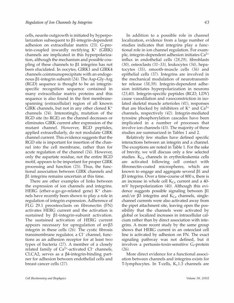
INTRODUCTION Ion channels are the targets of many intra- cellular signaling pathways, including pro- tein phosphorylation and dephosphorylation. Indeed, nearly every type of voltage-gated K + , Ca 2+ , and Na + channel is regulated to some extent by phosphorylation of serine/threonine residues on intracellular domains of the channel (1,2). Phosphorylation can alter channel gating properties, including voltage sensitivity and cal- cium sensitivity, and thereby dramatically con- trol the electrophysiological properties of a cell. In addition to serine/threonine phosphoryla- tion, considerable recent evidence suggests that ion channels are also regulated by phosphoryla- tion on tyrosine residues (1,3–7). Evidence for regulation of ion channels by tyrosine phosphorylation comes primarily from investigations of the effects of growth factors. Growth factors, which act through receptor protein tyrosine kinases (PTKs), regulate the long-term expression of ion channels (8,9) but Regulation of Ion Channels by Integrins Michael J. Davis, *,1 Xin Wu, 1 Timothy R. Nurkiewicz, 1 Junya Kawasaki, 1 Peichun Gui, 1 Michael A. Hill, 2 and Emily Wilson 1 1 Department of Medical Physiology, Cardiovascular Research Institute, Texas A&M University System Health Science Center, College Station, TX; and 2 Department of Human Biology, RMIT University, Bundoora, Victoria, Australia Abstract Ion channels are regulated by protein phosphorylation and dephosphorylation of serine, threo- nine, and tyrosine residues. Evidence for regulation of channels by tyrosine phosphorylation comes primarily from investigations of the effects of growth factors, which act through receptor tyrosine kinases. The purpose of the present work is to summarize evidence for the regulation of ion channels by integrins, through their downstream, nonreceptor tyrosine kinases. We review both direct and indirect evidence for this regulation, with particular emphasis on Ca 2+ -activated K + and voltage-gated Ca 2+ channels. We then discuss the critical roles that cytoskeletal, focal-adhe- sion, and channel-associated scaffolding proteins may play in localizing nonreceptor tyrosine kinases to the vicinity of ion channels. We conclude by speculating on the physiological signifi- cance of these regulatory pathways. Index Entries: Receptor tyrosine kinase; nonreceptor tyrosine kinase; integrins; cytoskeleton; focal adhesion; growth factor receptors; scaffolding proteins; Src; FAK; AKAP; SH3. * Author to whom all correspondence and reprint requests should be addressed. E-mail: [email protected] REVIEW ARTICLE © Copyright 2002 by Humana Press Inc. All rights of any nature whatsoever reserved. 1085-9195/02/36/41–66/$16.50 Cell Biochemistry and Biophysics 41 Volume 36, 2002
Transcript of Regulation of Ion Channels by Integrins - Texas A&M...
INTRODUCTION
Ion channels are the targets of many intra-cellular signaling pathways, including pro-tein phosphorylation and dephosphorylation.Indeed, nearly every type of voltage-gated K+,Ca2+, and Na+ channel is regulated to someextent by phosphorylation of serine/threonineresidues on intracellular domains of the channel(1,2). Phosphorylation can alter channel gating
properties, including voltage sensitivity and cal-cium sensitivity, and thereby dramatically con-trol the electrophysiological properties of a cell.In addition to serine/threonine phosphoryla-tion, considerable recent evidence suggests thation channels are also regulated by phosphoryla-tion on tyrosine residues (1,3–7).
Evidence for regulation of ion channels bytyrosine phosphorylation comes primarily frominvestigations of the effects of growth factors.Growth factors, which act through receptorprotein tyrosine kinases (PTKs), regulate thelong-term expression of ion channels (8,9) but
Regulation of Ion Channels by IntegrinsMichael J. Davis,*,1 Xin Wu,1 Timothy R. Nurkiewicz,1
Junya Kawasaki,1 Peichun Gui,1 Michael A. Hill,2and Emily Wilson1
1Department of Medical Physiology, Cardiovascular Research Institute, Texas A&M UniversitySystem Health Science Center, College Station, TX; and 2Department of Human Biology,
RMIT University, Bundoora, Victoria, Australia
Abstract
Ion channels are regulated by protein phosphorylation and dephosphorylation of serine, threo-nine, and tyrosine residues. Evidence for regulation of channels by tyrosine phosphorylationcomes primarily from investigations of the effects of growth factors, which act through receptortyrosine kinases. The purpose of the present work is to summarize evidence for the regulation ofion channels by integrins, through their downstream, nonreceptor tyrosine kinases. We reviewboth direct and indirect evidence for this regulation, with particular emphasis on Ca2+-activatedK+ and voltage-gated Ca2+ channels. We then discuss the critical roles that cytoskeletal, focal-adhe-sion, and channel-associated scaffolding proteins may play in localizing nonreceptor tyrosinekinases to the vicinity of ion channels. We conclude by speculating on the physiological signifi-cance of these regulatory pathways.
Index Entries: Receptor tyrosine kinase; nonreceptor tyrosine kinase; integrins; cytoskeleton;focal adhesion; growth factor receptors; scaffolding proteins; Src; FAK; AKAP; SH3.
* Author to whom all correspondence and reprintrequests should be addressed. E-mail: [email protected]
REVIEW ARTICLE
© Copyright 2002 by Humana Press Inc.All rights of any nature whatsoever reserved.1085-9195/02/36/41–66/$16.50
Cell Biochemistry and Biophysics 41 Volume 36, 2002
also have acute actions on channel activity.Receptor PTKs are characterized by an extracel-lular, ligand-binding domain, a transmembranedomain, a kinase-catalytic domain and cyto-plasmic regions responsible for coordinatingthe subsequent activity of signaling molecules.Signal transduction involves growth factor(ligand) binding to the extracellular domain,dimerization of the receptor proteins, andautophosphorylation of the receptor. Receptorautophosphorylation then creates phosphory-lated tyrosine residues on the cytoplasmic tailof the receptor, which form docking sites forsignaling molecules. The combination of thesesignaling molecules determines the specificityof individual receptor PTKs (10).
Nonreceptor PTKs can also regulate ion chan-nels. These enzymes play a prominent role insignaling pathways downstream from integrinsand other adhesion molecules. NonreceptorPTKs are found in both the cytoplasm andnuclei of cells, but the largest family is the cyto-plasmic Src (sarcoma virus tyrosine kinase) fam-ily (11), consisting of eight members, includingSrc, Fyn, and Yes, that are ubiquitouslyexpressed. Regulation of Src family members ishighly conserved: Autophosphorylation of akinase domain tyrosine leads to increased kinaseactivity, whereas phosphorylation of a tyrosineresidue near the C terminus represses activity(12). Many stimuli, including receptor PTKs, G-protein coupled receptors, and integrins havebeen implicated in Src activation, suggestingthat this family of kinases is a key point of inte-gration for many signal transduction pathways.Another relevant nonreceptor PTK is pp125FAK,which is discretely localized to cellular focaladhesions and has been shown to colocalizewith integrins. FAK is a substrate for integrin-dependent tyrosine phosphorylation andbecomes enzymatically active upon phosphory-lation, serving as a scaffold for the binding andlocalization of other proteins to the focal adhe-sion (13). Activation of Src family members andFAK serves as a key integration mechanism for anumber of extracellular signaling pathways.
The regulation of ion channels by growthfactors and receptor PTKs has been reviewed,
to some extent, previously (3,4,6,7). The pur-pose of the present work is to summarize evi-dence for the regulation of ion channels byintegrins and integrin-linked tyrosine kinases.We review both direct and indirect evidence forthis regulation. We then discuss the criticalroles that the cytoskeleton and channel-associ-ated scaffolding proteins may play in localiz-ing PTKs to the vicinity of ion channels. Whenpossible, we speculate on the physiological sig-nificance of these regulatory pathways.
INTERACTIONS BETWEENINTEGRINS AND ION CHANNELS
Integrins are a family of membrane-span-ning glycoproteins that link the extracellularmatrix (ECM) to the cytoskeleton. Integrins arecomposed of α–β heterodimers with extracellu-lar domains that bind ECM proteins and shortcytoplasmic tails that associate with focal adhe-sion proteins (14,15). As mentioned earlier,integrin activation is a well-known trigger ofintracellular tyrosine phosphorylation cas-cades. Integrin engagement by multivalent lig-ands, including extracellular matrix proteins,induces receptor clustering, the recruitment ofcytoskeletal proteins to the focal adhesion, andthe activation of nonreceptor PTKs (16,17).Because integrins lack intrinsic enzymaticactivity, they rely on the activation of othercytoplasmic signaling molecules, includingFAK and Src. Integrin interaction with FAKleads to FAK autophosphorylation, to the cre-ation of a binding site for the Src SH2 domain,and, ultimately, to Src activation (18). Src thenphosphorylates additional sites on FAK toallow binding of other signaling molecules andscaffolding proteins (19,20). This process leadsto the assembly of complex signaling mole-cules at the focal adhesion site and organizesfurther downstream signaling events (15).Many of the signaling pathways activated byreceptor PTKs overlap with integrin-mediatedsignaling pathways, utilizing the same PTKsthrough different adaptor proteins (21,22).
Integrins may play a role in directing thelocalization of ion channels. In neuroblastoma
42 Davis et al.
Cell Biochemistry and Biophysics Volume 36, 2002
cells, neurite outgrowth is initiated by hyperpo-larization subsequent to β1-integrin-dependentadhesion on extracellular matrix (23). G-pro-tein-coupled inwardly rectifying K+ (GIRK)channels are implicated in this hyperpolariza-tion, although the mechanism and possible cou-pling of these channels to β1 integrins has notbeen elucidated. In oocytes, GIRK1 and GIRK4channels coimmunoprecipitate with an endoge-nous β1-integrin subunit (24). The Asp-Gly-Arg(RGD) sequence is thought to be an integrin-specific recognition sequence contained inmany extracellular matrix proteins and thissequence is also found in the first membrane-spanning (extracellular) region of all knownGIRK channels, but not in any other cloned K+
channels (24). Interestingly, mutation of theRGD site (to RGE) on the channel decreases oreliminates GIRK current after expression of themutant channel. However, RGD peptides,applied extracellularly, do not modulate GIRKchannel current. This evidence suggests that theRGD site is important for insertion of the chan-nel into the cell membrane, rather than foracute regulation of the channel (24). However,only the aspartate residue, not the entire RGDmotif, appears to be important for proper GIRKprocessing and function (25). Thus, the func-tional association between GIRK channels andβ1 integrins remains uncertain at this time.
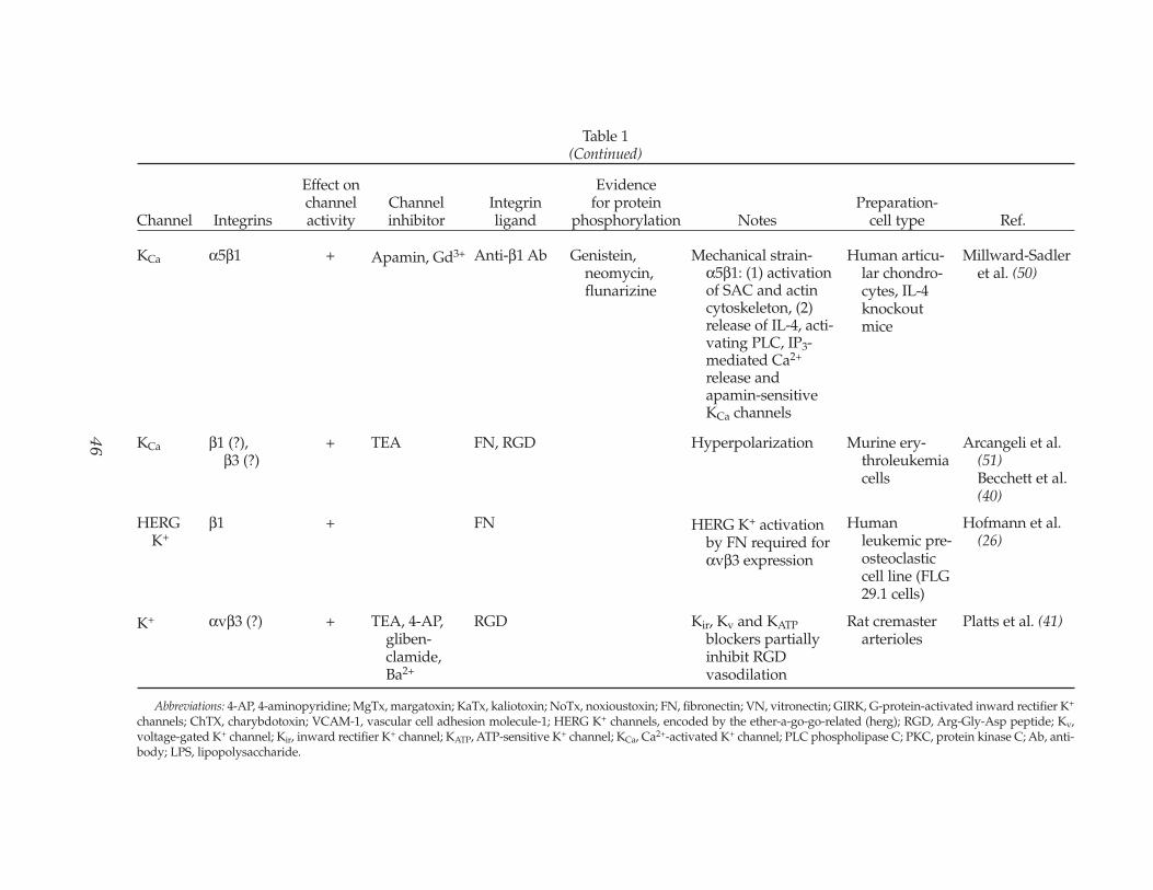
There are other examples of links betweenthe expression of ion channels and integrins.HERG (ether-a-go-go-related gene) K+ chan-nels have recently been shown to play a role inregulation of integrin expression. Adherence ofFLG 29.1 preosteoclasts on fibronectin (FN)activates HERG current and the activation issustained by β1-integrin-subunit activation.The sustained activation of HERG currentappears necessary for upregulation of αvβ3integrin in these cells (26). The cystic fibrosistransmembrane regulator, a Cl– channel, func-tions as an adhesion receptor for at least twotypes of bacteria (27). A member of a closelyrelated family of Ca2+-activated Cl– channels,CLCA2, serves as a β4-integrin-binding part-ner for adhesion between endothelial cells andbreast cancer cells (27).
In addition to a possible role in channellocalization, evidence from a large number ofstudies indicates that integrins play a func-tional role in ion channel regulation. For exam-ple, integrin-dependent adhesion initiates Ca2+
influx in endothelial cells (28,29), fibroblasts(30), osteoclasts (31–33), leukocytes (34), hepa-tocytes (35), smooth-muscle cells (36) andepithelial cells (37). Integrins are involved inthe mechanical modulation of neurotransmit-ter release (38,39). Integrin-dependent adhe-sion inititiates hyperpolarization in neurons(23,40). Integrin-specific peptides (RGD, LDV)cause vasodilation and vasoconstriction in iso-lated skeletal muscle arterioles (41), responsesthat are blocked by inhibitors of K+ and Ca2+
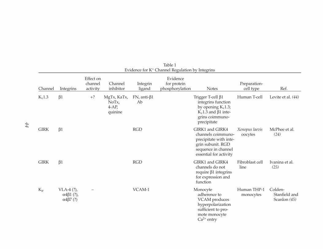
channels, respectively (42). Integrin-mediatedtyrosine phosphorylation cascades have beenimplicated in a number of processes thatinvolve ion channels (43). The majority of thesestudies are summarized in Tables 1 and 2.
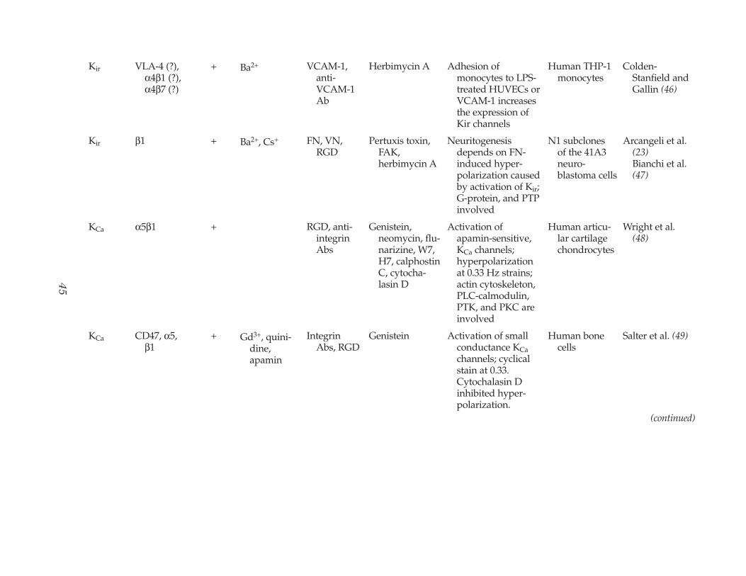
Relatively few studies have defined specificinteractions between an integrin and a channel.The exceptions are noted in Table 1. For the sakeof brevity, we will discuss only a few selectedstudies. KCa channels in erythroleukemia cellsare activated following cell contact withfibronectin-coated microspheres, which areknown to engage and aggregate several β1 andβ3 integrins. Over a time-course of 800 s, there isan increase in whole cell KCa current and a 40-mV hyperpolarization (40). Although this evi-dence suggests possible signaling between β1and/or β3 integrins and KCa channels, single-channel currents were also activated away fromthe pipet attachment site, leaving open the pos-sibility that the channels were activated byglobal or localized increases in intracellular cal-cium rather than by direct association with inte-grins. A more recent study by the same groupshows that HERG current in an osteoclast cellline is activated by adhesion on FN. The exactsignaling pathway was not defined, but itinvolves a pertussis-toxin-sensitive Gi-protein(26).
More direct evidence for a functional associ-ation between channels and integrins exists forT-lymphocytes. In T-cells, Kv1.3 channels are
Regulation of Ion Channels by Integrins 43
Cell Biochemistry and Biophysics Volume 36, 2002
44
Table 1Evidence for K+ Channel Regulation by Integrins
Effect on Evidence channel Channel Integrin for protein Preparation-
Channel Integrins activity inhibitor ligand phosphorylation Notes cell type Ref.
Kv1.3 β1 +? MgTx, KaTx,NoTx,4-AP,quinine
FN, anti-β1Ab
Trigger T-cell β1integrins functionby opening Kv1.3;Kv1.3 and β1 inte-grins coimmuno-precipitate
Human T-cell Levite et al. (44)
GIRK β1 RGD GIRK1 and GIRK4 channels coimmuno-precipitate with inte-grin subunit. RGDsequence in channelessential for activity
Xenopus laevisoocytes
McPhee et al. (24)
GIRK β1 RGD GIRK1 and GIRK4 channels do notrequire β1 integrinsfor expression andfunction
Fibroblast cell line
Ivanina et al. (25)
Kir VLA-4 (?),α4β1 (?),α4β7 (?)
– VCAM-1 Monocyteadherence toVCAM produceshyperpolarizationsufficient to pro-mote monocyteCa2+ entry
Human THP-1 monocytes
Colden-Stanfield andScanlon (45)
45
Kir VLA-4 (?), α4β1 (?),α4β7 (?)
+ Ba2+ VCAM-1,anti-VCAM-1Ab
Herbimycin A Adhesion of monocytes to LPS-treated HUVECs orVCAM-1 increasesthe expression ofKir channels
Human THP-1 monocytes
Colden-Stanfield andGallin (46)
Kir β1 + Ba2+, Cs+ FN, VN, RGD
Pertuxis toxin, FAK, herbimycin A
Neuritogenesisdepends on FN-induced hyper-polarization causedby activation of Kir;G-protein, and PTPinvolved
N1 subclones of the 41A3neuro-blastoma cells
Arcangeli et al. (23)Bianchi et al.(47)
KCa α5β1 + RGD, anti-integrinAbs
Genistein,neomycin, flu-narizine, W7,H7, calphostinC, cytocha-lasin D
Activation of apamin-sensitive,KCa channels;hyperpolarizationat 0.33 Hz strains;actin cytoskeleton,PLC-calmodulin,PTK, and PKC areinvolved
Human articu-lar cartilagechondrocytes
Wright et al. (48)
KCa CD47, α5,β1
+ Gd3+, quini-dine,apamin
IntegrinAbs, RGD
Genistein Activation of small conductance KCachannels; cyclicalstain at 0.33.Cytochalasin Dinhibited hyper-polarization.
Human bone cells
Salter et al. (49)
(continued)
46
Abbreviations: 4-AP, 4-aminopyridine; MgTx, margatoxin; KaTx, kaliotoxin; NoTx, noxioustoxin; FN, fibronectin; VN, vitronectin; GIRK, G-protein-activated inward rectifier K+
channels; ChTX, charybdotoxin; VCAM-1, vascular cell adhesion molecule-1; HERG K+ channels, encoded by the ether-a-go-go-related (herg); RGD, Arg-Gly-Asp peptide; Kv,voltage-gated K+ channel; Kir, inward rectifier K+ channel; KATP, ATP-sensitive K+ channel; KCa, Ca2+-activated K+ channel; PLC phospholipase C; PKC, protein kinase C; Ab, anti-body; LPS, lipopolysaccharide.
KCa α5β1 + Apamin, Gd3+ Anti-β1 Ab Genistein,neomycin,flunarizine
Mechanical strain-α5β1: (1) activationof SAC and actincytoskeleton, (2)release of IL-4, acti-vating PLC, IP3-mediated Ca2+
release andapamin-sensitiveKCa channels
Human articu-lar chondro-cytes, IL-4knockoutmice
Millward-Sadler et al. (50)
KCa β1 (?), β3 (?)
+ TEA FN, RGD Hyperpolarization Murine ery-throleukemiacells
Arcangeli et al. (51)Becchett et al.(40)
HERGK+
β1 + FN HERG K+ activationby FN required forαvβ3 expression
Humanleukemic pre-osteoclasticcell line (FLG29.1 cells)
Hofmann et al. (26)
K+ αvβ3 (?) + TEA, 4-AP, gliben-clamide,Ba2+
RGD Kir, Kv and KATPblockers partiallyinhibit RGDvasodilation
Rat cremaster arterioles
Platts et al. (41)
Table 1(Continued)
Effect on Evidence channel Channel Integrin for protein Preparation-
Channel Integrins activity inhibitor ligand phosphorylation Notes cell type Ref.
47
Na+ orNSC
α5β1 + TTX, Gd3+ Anti-α5,anti-β1 Ab
Genistein,neomycin
Depolarization at 0.33 Hz strain; PLC andPTK are involved
Osteoarthriticarticularcartilagechondrocytes
Millward-Sadler et al. (52)
SAC(NSC?)
α5β1 + Hyperpolerization to mechanical stimula-tion is mediated byparacrine factor;blocked by IL-4 Ab
Humanarticularchondrocytes
Millward-Sadler et al. (53)
SAC(NSC?)
α5β1 Gd3+ Oligopep-tides (con-tainingRGD)
FAK, β-catenin,paxillin
Tyrosine phosphory-lation of FAK,paxillin, β-cateninthrough α5β1requires SACactivity
Humanarticularchondrocytes
Lee et al. (54)
SOC? β1, αv + Ca2+-free bath FN, VN, anti-αvβ3Ab
Adhesion-dependent[Ca2+]i increase
Humanumbilical veinendothelium
Schwartz (55)Schwartz andDenninghoff(28)
SOC? αIIbβ3 + GPIIb-IIIaAb, RGD
GPIIb–IIIa is involved in Ca2+ channelactivation
Platelet plasma membrane
Fujimoto et al. (56)
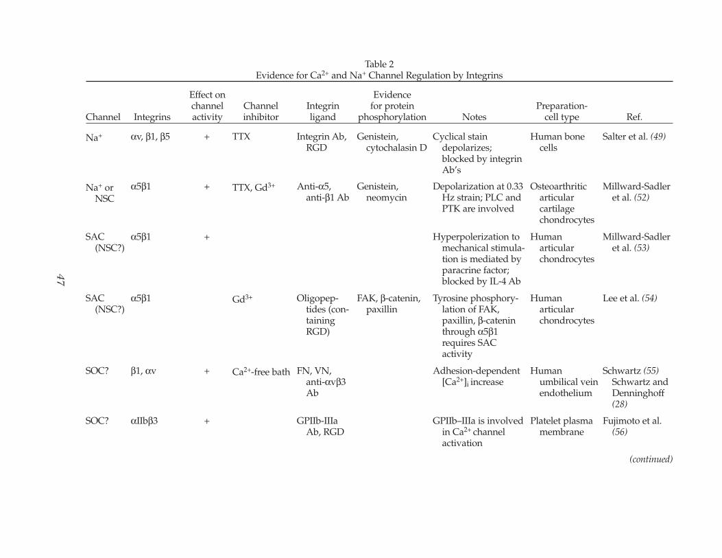
Table 2Evidence for Ca2+ and Na+ Channel Regulation by Integrins
Effect on Evidence channel Channel Integrin for protein Preparation-
Channel Integrins activity inhibitor ligand phosphorylation Notes cell type Ref.
Na+ αv, β1, β5 + TTX Integrin Ab, RGD
Genistein,cytochalasin D
Cyclical stain depolarizes;blocked by integrinAb’s
Human bone cells
Salter et al. (49)
(continued)
48
SOC? αvβ3, αvβ5 + Ni2+ RGD RGD peptides on beads induce Ca2+
entry; this Ca2+
signal promotesadhesion
Madin–Darbycanine kidneyepithelial cells
Sjaastad et al. (57)Sjaastad et al. (37)
SOC? αIIbβ3 + αIIbβ3 Ab, RGDpeptide
VWF binding to GPIb is responsible forαIIbβ3-dependentCa2+ influx, αIIbβ3complex mayfunction as Ca2+
channel
Platelet Bertolino et al. (58)
SOC? αvβ3 + Ca2+-free bath, thap-sigargin
LM609, VN Genistein [Ca2+]i increase evoked by αvβ3clustering; 70%Ca2+ entry; 30%Ca2+ release
Bovinepulmonaryarteryendothelium
Bhattacharyaet al. (29)
CaL αvβ3 – Nifedipine FN, VN, anti-β3Abs, RGD
VN and αvβ3–Abinhibit current; FNpotentiates current
Rat cremaster artetriolarsmoothmuscle
Wu et al. (59)
CaL α5β1 + Nifedipine FN, anti-α5Ab
FAK–Ab, Src–Ab, genistein,piceatannol
Potentiation of current by α5β1;blocked by PP2, SrcSH2 inhibitor,FRNK, paxillin Ab,vinculin Ab, pep-tides for PTP site onchannel C terminus
Rat cremaster arteriolarsmoothmuscle
Wu et al. (59)Wu et al. (60)
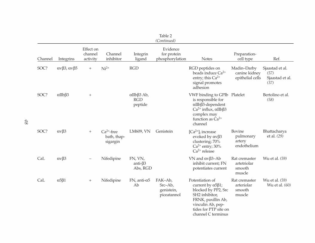
Table 2(Continued)
Effect on Evidence channel Channel Integrin for protein Preparation-
Channel Integrins activity inhibitor ligand phosphorylation Notes cell type Ref.
49
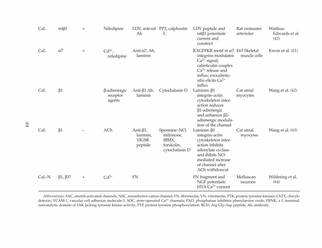
CaL α4β1 + Nifedipine LDV, anti-α4Ab
PP2, calphostin C
LDV peptide and α4β1 potentiatecurrent and constrict
Rat cremasterarteriolar
Waitkus-Edwards et al.(42)
CaL α7 + Cd2+,nifedipine
Anti-α7, Ab,laminin
KXGFFKR motif in α7integrins modulatesCa2+ signal;calreticulin couplesCa2+ release andinflux; exocalretic-ulin elicits Ca2+
influx
E63 Skeletal muscle cells
Kwon et al. (61)
CaL β1 β-adrenergicreceptoragents
Anti-β1 Ab,laminin
Cytochalasin D Laminin–β1integrin–actincytoskeleton inter-action reducesβ1-adrenergic and enhances β2-adrenergic modula-tion of the channel
Cat atrialmyocytes
Wang et al. (62)
CaL β1 – ACh Anti-β1,laminin,YIGSRpeptide
Spermine–NO,milrinone,IBMX,forskolin,cytochalasin D
Laminin–β1integrin–actincytoskeleton inter-action inhibitsadenylate cyclaseand ihibits NO-mediated increaseof channel afterACh withdrawal
Cat atrial myocytes
Wang et al. (63)
CaL-N β1, β3? + Cd2+ FN FN fragment and NGF potentiateHVA Ca2+ current
Molluscanneurons
Wildering et al. (64)
Abbreviations: SAC, stretch-activated channels; NSC, nonselective cation channel; FN, fibronectin; VN, vitronectin; PTK, protein tyrosine kinases; ChTX, charyb-dotoxin; VCAM-1, vascular cell adhesion molecule-1; SOC, store-operated Ca2+ channels; PAO, phosphatase inhibitor phenylarsine oxide; FRNK, a C-terminal,noncatalytic domain of FAK lacking tyrosine kinase activity; PTP, protein tyrosine phosphorylation; RGD, Arg-Gly-Asp peptide; Ab, antibody.
necessary for activation of β1 integrins andsubsequent integrin-dependent adhesion andmigration (44). This mechanism underlies theactivation of T-cell adhesion by elevation ofextracellular K+ in the absence of any specificreceptor-mediated event and accounts for theability of substance P, which inhibits Kv1.3channels, to inhibit T-cell adhesion. Although ithas not yet been tested whether the signalingbetween Kv1.3 channels and β1 integrins worksin reverse, Kv1.3 and β1 integrins coimmuno-precipitate, suggesting that direct physicalassociation of these molecules underlies theirfunctional interaction (44).
Several lines of evidence suggest that cal-cium channels are acutely regulated by inte-grin-dependent signaling. Most of thisevidence is summarized in Table 2. In manycell types, integrin engagement triggers bothintracellular Ca2+ release and Ca2+ influxacross the plasma membrane (65), and thesetwo processes are often coupled. This phe-nomenon can be illustrated by a discussion ofendothelial cell–integrin interactions. Integrin-dependent adhesion initiates Ca2+ influx inendothelial cells (28). Application of the ECMprotein, vitronectin, or a polyclonal antibodyto αvβ3 integrin stimulates endothelial cellCa2+ release and Ca2+ influx (29). These lig-ands also stimulate tyrosine phosphorylationof multiple endothelial cell proteins (66). BothCa2+ influx and protein phosphorylation areblocked by soluble tyrosine kinase inhibitors(29). The ion channel responsible for Ca2+
influx has not been identified, but it is likely tobe the so-called “store-operated Ca2+ influxchannel.” The current most likely to be associ-ated with this process is a small inward, Ca2+-selective current in nonexcitable cells, termedIcrac, for Ca2+-release-activated Ca2+ current(67). Molecularly, the best candidates for Icracare the Trp (transient receptor potential)family of proteins (68) and the channellikeintestinal calcium transporter CaT1 (69,70).Recombinant Trp channels share many, but notall, characteristics of Icrac (71,72), although het-erologous combinations of Trp isoforms innative cells may account for the unique char-
acteristics of some endogenous store-operatedcurrents (73,74). A recent study shows that het-erologously expressed CaT1 protein is nearlyindistinguishable from Icrac (70). Of relevanceto the possible regulation of store-operatedCa2+ current by integrins is the recent findingthat αvβ3 ligands activate store-operated Ca2+
current in human umbilical vein endothelialcells (75). Additional evidence indicates thatIcrac can be regulated by tyrosine kinases(76–79) and that its full activation requirescytoskeletal proteins (80,81) and smallGTPases (82).
In contrast to the circumstantial evidence forregulation of Ca2+ current in nonexcitable cellsby integrins, the evidence for regulation of volt-age-gated calcium channels is much stronger. Invascular smooth muscle, the L-type calciumchannel is regulated by at least three differentintegrins. Whole-cell recordings from vascularmyocytes show that soluble ligands of the αvβ3integrin, such as RGD peptides, vitronectin, andfibronectin fragments, inhibit L-type current(59). Interestingly, insoluble αvβ3 ligands (i.e.,ligands attached to microspheres) also inhibitcurrent (59). This is somewhat counter to whatmight be expected, because soluble (monova-lent, nonclustering) integrin ligands do not trig-ger the entire ensemble of downstream signalingevents (i.e., cytoskeletal protein redistributionand tyrosine phosphorylation) typical of inte-grin–multivalent ligand interaction (83). Rather,monovalent integrin ligands are thought to actby disrupting existing interactions between inte-grins and their insoluble ligands. Because bothsoluble and insoluble αvβ3 integrin ligands pro-duce the same effects on smooth-muscle-cell cal-cium current, both may be triggering the samesignaling pathway involving Ca2+ channels. Thesignaling pathway by which αvβ3 integrin regu-lates the L-type channel is not known, but itappears to be insensitive to tyrosine kinaseinhibitors (Wu, unpublished observations). Incontrast to inhibition of Ca2+ current by αvβ3 lig-ands, soluble ligands (LDV peptide) of thelaminin receptor, α4β1, enhance L-type calciumcurrent (84). However, the α4 integrin antibodydoes not modulate current, although pretreat-
50 Davis et al.
Cell Biochemistry and Biophysics Volume 36, 2002
ment with the antibody blocks the LDVresponse. These observations suggest that thesignaling pathways between these two integrinsand the L-type Ca2+ channel in this tissue arefundamentally different. Whether either signal-ing pathway involves changes in the phosphory-lation state of the channel is not known.
Substantially more information is availableabout how insoluble α5β1 integrin ligandsmodulate the L-type calcium channel. L-Typecalcium current in vascular smooth muscle ispotentiated up to twofold by insoluble α5β1ligands, and this potentiation is blocked bysoluble tyrosine kinase inhibitors, includinggenistein, piceatannol, and the Src-family-specific inhibitor, PP2. Tyrosine phosphataseinhibition increases basal L-type calcium cur-rent (60,85). Although it might be argued thatthese inhibitors lack specificity, cell dialysiswith antibodies to c-Src or focal adhesionkinase (FAK) also block potentiation of cur-rent, with a lesser but significant effect onbasal current. Recent experiments demon-strate many of the same effects of α5β1 lig-ands in heterologously expressed L-type Ca2+
channels (Gui et al., unpublished observa-tions), indicating that these conclusions donot only apply to freshly dissociated smooth-muscle cells, which could potentially bealtered by enzyme treatment. These observa-tions also fit with data from other types ofsmooth muscle where Ca2+ current isincreased by intracellular application of con-stitutively active Src kinase (86), increased byc-Src activating peptide (87), and inhibited bya c-Src monoclonal antibody (88).
Similarities between the actions of integrinligands and the growth factor PDGF (platelet-derived growth factor) on the L-type Ca2+ chan-nel also support a role for regulation of thechannel by tyrosine phosphorylation. PDGF,which stimulates tyrosine phosphorylation ofmultiple smooth-muscle proteins, enhances L-type calcium current (88,89) and increases tyro-sine phosphorylation of the pore-formingchannel subunit α1C (Cav1.2b). In PDGF-stimu-lated smooth muscle, α1C coimmunoprecipi-tates with c-Src (88). c-Src also interacts with the
neuronal isoform of the α1 Ca2+ channel sub-unit. In cerebellar granule neurons, α1C is tyro-sine phosphorylated in response to theinsulin-like growth factor-1 (IGF-1), resulting inpotentiation of L-type current (90,91). Thiseffect can be duplicated with recombinant α1C(Cav1.2c) expressed in SH-SY5Y cells. c-Srcmediates this response because expression ofkinase-dead Src, or application of PP2, blockspotentiation of current (92). Kinase assays usinglysates from neuroblastoma cells expressingα1C show that purified Src kinase phosphory-lates Y2122 on the α1C C terminus. Furthermore,point mutation of Y2122F prevents tyrosinephosphorylation and IGF-1 potentiation of cur-rent (92). Because Cav1.2b and Cav1.2c shareidentical sequences in this region, it is likelythat Y2122 also mediates potentiation of currentin smooth muscle by both PDGF and integrinligands. In fact, a peptide designed to competewith phosphorylation of this region of the chan-nel prevents potentiation of current in vascularsmooth muscle by α5β1 integrin ligands (60)and in Cav1.2b channels expressed in HEK 293cells (Gui et al., unpublished observations).
In smooth-muscle cells, intracellular dialy-sis with antibodies to two integrin-associatedcytoskeletal proteins, paxillin and vinculin,also blocks regulation of L-type current byα5β1 ligands. The ability of vinculin and pax-illin antibodies to do this is likely the result oftheir interference with the assembly of Src oranother nonreceptor PTK on an intracellularscaffold of focal adhesion proteins, ratherthan a direct interaction with the channel. Srchomology 2 (SH2) and Src homology 3 (SH3)domains in these proteins enable them toassociate with PTKs (14). As mentioned ear-lier, the C termini of Cav1.2b and Cav1.2c con-tain at least two proline-rich domains thatmay interact with SH3 domains in Src, Lyn,and Hck tyrosine kinases (93) and possiblywith SH3 domains in docking, adaptor, orscaffolding proteins. Deletion of one proline-rich region results in increased channel cur-rent, suggesting that it is constitutivelyinvolved in channel inhibition (94). However,it is not yet known whether phosphorylation
Regulation of Ion Channels by Integrins 51
Cell Biochemistry and Biophysics Volume 36, 2002
of the α1C C terminus by any of these PTKsalters this inhibitory property.
Collectively, these observations suggest thatL-type Ca2+ channels are regulated by multipleintracellular pathways downstream from αvβ3,α5β1, and α4β1 integrins. What is the physiolog-ical relevance of these observations? We proposethat the extracellular matrix exerts constitutivecontrol over the major calcium-permeable ionchannel in vascular smooth muscle and thatacute regulation of calcium signaling by inte-grins may occur under physiological and patho-logical conditions in blood vessels. Thisconclusion is consistent with evidence that inte-grin ligands and ECM proteins can acutely regu-late vascular tone. For example, peptidescontaining the integrin-specific RGD (Arg-Gly-Asp) and LDV (Leu-Asp-Val) amino acidsequences cause dilation and constriction,respectively, of isolated, pressurized skeletalmuscle arterioles (42,95). The dilation to RGDpeptides is associated with a fall in intracellularcalcium (96) and is blocked by a β3-integrin anti-body (95). In contrast, α5β1- and α4β1-integrinligands produce constriction of the same arteri-oles. This evidence suggests that extracellularmatrix proteins have the potential to influencevascular tone through an interaction with inte-grins, pointing to a possible role for these mech-anisms in tissue injury responses (97). Whetherthis occurs in other cell types is not known, but itis interesting to note that the ECM protein, FN,and the nerve growth factor (NGF) acutelyenhance high-voltage-activated Ca2+ currents inmolluscan neurons (64). FN also enhances thefrequency of action potential firing (W.Wildering, see Note Added in Proof) in that tis-sue, suggesting that a matrix–integrin–calciumchannel signaling axis may acutely regulate elec-trical excitability of those cells.
Another extracellular matrix protein, laminin,modulates L-type Ca2+ current in atrialmyocytes by a different mechanism. Interactionsbetween laminin and β1 integrins reduces β1-adrenergic modulation of L-type Ca2+ currentbut enhances β2-adrenergic modulation of cur-rent (62). Additionally, the laminin–β1 integrininteraction inhibits adenylate cyclase and
thereby alters L-type Ca2+ current (63). The sig-naling pathways involved in these actions oflaminin remain to be elucidated; however, botheffects require an intact actin cytoskeleton(62,63). Muscarinic inhibition of L-type Ca2+ cur-rent, and muscarinic activation of K+ current, isabsent in β1-integrin–/– cardiomyocytes (98).Interestingly, β-adrenergic modulation of theCa2+ current is unaffected in this knockout (98),an observation that conflicts somewhat with theabove reports (62,63). It is possible that thismode of regulation in cardiomyocytes compen-sates for lack of direct tyrosine phosphorylationby Src of the cardiac L-type Ca2+ channel(Cav1.2a), which has a substantially different Cterminus and does not contain the critical tyro-sine residue required for Cav1.2b and Cav1.2cpotentiation (92).
Other adhesion proteins are also known tointeract with ion channels. For example, theadhesion molecule PECAM, is constituitivelyexpressed on endothelial cells, and a PECAMantibody evokes [Ca2+]i increases in HUVECs(99) through activation of a nonselective cationcurrent. Activation of this current requires anintact PECAM-1 cytoplasmic domain and Srckinase activity (100). Tenascin-C is an extracellu-lar adhesive glycoprotein composed of a seriesof epidermal growth factor (EGF)-like repeats,a fibrinogen-like region, and a series offibronectin-like regions (101). Neuronal Na+
channels bind to tenascin-C with high affinity aswell as to the related protein, tenascin-R, whichlacks several of the fibronectin repeats (102). TheNa+ channel β2-subunit contains an Ig domainwith close sequence similarity to the neural celladhesion molecule contactin/F3 (103).Contactin/F3 binds tenascin and related ECMproteins. Additionally, the EGF-like domains oftenascin-R have the potential to regulate Na+
channels through interactions with PTKs.Although a functional role for these extracellu-lar proteins in the regulation of the Na+ channelhas not been demonstrated, it has been specu-lated that secretion of the proteins by neuronalsupport cells, such as oligodendrocytes, maydirect localization of Na+ channels on the corre-sponding neuronal cell membrane (102).
52 Davis et al.
Cell Biochemistry and Biophysics Volume 36, 2002
THE CHANNEL-PROTEIN KINASEREGULATORY COMPLEX
An emerging concept in the field of ion chan-nel regulation is that modulation of channelfunction by phosphorylation requires the for-mation of a multiprotein complex. The pore-forming α-subunits of many channels bind toauxilliary channel subunits, but they also asso-ciate with scaffolding proteins that play essen-tial roles in channel localization and activity (3).Scaffolding proteins link signaling enzymes,substrates, and potential effectors (such aschannels) into a multiprotein signaling complexthat may be anchored to the cytoskeleton. Inaddition to an obvious role in targeting thechannel to a particular location on the cellmembrane, there are at least three advantagesto having an ion channel in a multiprotein com-plex. First, there is a large increase in efficiencyof the kinetic reaction when an enzyme is local-ized with its substrate and effector in amicroenvironment with restricted diffusion(104). Second, the anchoring of an enzyme com-plex to a channel may be necessary for theextremely rapid transmission of signalsrequired to regulate some channels (105). Third,compartmentalization may be essential fordetermining specificity in signal transductionpathways (106).
There appear to be many families of scaffold-ing and adaptor proteins that potentially couldbe involved in organizing ion channels into sig-naling complexes and regulating function bycoupling those channels to protein kinases. Forserine–threonine kinases, prominent familiesare the MAGUK (membrane-associated guany-late kinase), AKAP (A-kinase associated) pro-teins, and GKAP (G-kinase associated) proteins(107). INAD (inactivation-no-after potential D)is another example of a multiprotein signalingcomplex. INAD is a Drosophila protein contain-ing five PDZ domains (PSD-95/SAPSO, Dlgand 20-1 domains) that link together most ofthe proteins involved directly in phototrans-duction. This multiprotein complex includesrhodopsin, calmodulin, the putative store-oper-ated Ca2+ channels Trp and Trpl, and the protein
kinases PLC and PKC (105,108). INAD mayserve as a template for understanding howother channels are regulated in a multiproteincomplex.
Evidence that scaffolding proteins can medi-ate kinase-channel interactions is perhaps bestestablished for the AKAP family of proteins(109). AKAPs serve to localize both kinases andphosphatases to multiprotein effector com-plexes that include K+ and Ca2+ channels (110).A conserved AKAP anchoring motif directsdimerized PKA subunits to a particular subcel-lular target (111). For example, forskolin andcAMP potentiate ROMK1 (a Kir1.x subfamily ofchannels expressed in epithelial cells) current innative renal secretory cells (112). This potentia-tion is lost when ROMK1 is expressed in oocytesbut restored if AKAP79 is coexpressed with thechannel (112). AKAP15/18 is required for pro-tein kinase A (PKA) potentiation of L-type Ca2+
channels in cardiac (113), skeletal (114), and vas-cular smooth muscle (115). AKAPs have alsobeen implicated in the regulation of KCa chan-nels (116) and the cystic fibrosis transmembraneconductance regulator (CFTR) (117). AKAP79associates with the β2-adrenergic receptor andwith MAGUK proteins, which, in turn, are cou-pled to glutamate-gated ion channels. AKAP79has the potential to form part of a scaffold uponwhich PKA and PP2B (protein phosphatase 2B)may dually regulate the coupling of β2-adrener-gic receptors and glutamate channels (110).Thus, AKAPs appear to be capable of assem-bling signaling complexes by virtue of theirassociations with ion channels and other scaf-folding proteins. It is possible that AKAPs rep-resent a general scheme for kinase regulation ofchannels and that similar families of adaptorproteins associated with other kinases, perhapsPTKs, will soon be identified.
ROLE OF THE CYTOSKELETON INREGULATION OF ION CHANNELS
The cytoskeleton provides a backbone uponwhich scaffolding proteins are organized andthereby positioned to link kinases to ion chan-
Regulation of Ion Channels by Integrins 53
Cell Biochemistry and Biophysics Volume 36, 2002
nels. The cytoskeleton is a highly structured,three-dimensional network composed of threemain components: actin filaments, micro-tubules (containing tubulin), and intermediatefilaments (containing vimentin, neurofilament,etc.). Each component has a unique functionaland structural role in the cell and the syncitialnature of the cytoskeletal network implies theexistence of complex interrelationships amongthe various components. A large number ofaccessory proteins are involved in assembly,disassembly, and crosslinking of each of theseelements. The prevailing view of the cytoskele-ton, the tensegrity model (118), proposes asyncitium of compression-resistant struts(microtubules) suspended between variouselastic elements (actin and intermediate fila-ments). Other structural proteins, includingezrin, ankyrin, spectrin, filamin, α-actinin, andtalin, are required for anchoring the cytoskele-ton into the plasma membrane and/or tether-ing it to other plasma membrane proteins.
There is increasing evidence to support acritical role for the cytoskeleton in the regula-tion of ion channels. One line of evidence isthat cytoskeletal proteins are directly associ-ated with ion channels. For example, thecGMP-gated cation channel of rod photorecep-tors associates with spectrin (119). Accessoryproteins associated with the N-methyl-D-aspar-tate (NMDA) receptor attach to the cytoskele-ton (e.g., SAP97 binds band 4.1, an actin, andspectrin-binding protein [120]). The epithelialNa+ channel associates with spectrin (121).NR1 and NR2 subunits of the NMDA recep-tor/channel bind to soluble tubulin and α-actinin (122,123). The list of these interactionsis extensive and the reader is referred to severalrecent reviews for more detail (3,111,124–126).A second line of evidence is that cytoskeletalproteins are involved in various aspects of ionchannel function. For example, the Kv4.2 chan-nel interacts with the actin-binding protein fil-amin in cerebellar and hippocampal neurons(127), such that the magnitude of current ismuch greater when filamin is coexpressed withthe channel than when the channel isexpressed alone (127). At least one β-subunit of
the Kv1.1 channel confers fast inactivation tothe channel’s α-subunit (128,129) and thisproperty depends on the interaction of the sub-units with F-actin and the phosphorylationstate of the α-subunit (130). For Kv1.5 channels,disruption of the actin network using cytocha-lasin D or antisense constructs to α-actinin-2result in increased Kv1.5 current, either by con-trolling channel gating or expression (131).Disruption of actin filaments in retinal bipolarcells also leads to activation of voltage-gatedK+ current (132). Collectively, these observa-tions suggest that the actin cytoskeleton exertsa tonic, inhibitory effect on at least severaltypes of K+ channels.
The actin network appears to exhibit a dif-ferent effect on Na+ and Ca2+ channels. Actinfilament disruption with cytochalasin Dinhibits L-type Ca2+ current in vascular smoothmuscle (133) and alters the time-course of acti-vation of the cardiac Na+ channel (134). The neteffect of actin filament disruption on Na+ andCa2+ currents is to enhance current and alterthe shape of the cardiac cell Ca2+ transient(135). Antibodies to F-actin are reported to alterthe gating kinetics of the cardiac Na+ channelby inducing a second open state and causingprolonged opening bursts (134). Fast inactiva-tion of the Na+ channel has been proposed tobe controlled by a triplet of amino acidresidues at the cytoplasmic II–IV linker region(136). Whether this region of the Na+ channel isdirectly associated with cytoskeletal or scaf-folding proteins is not yet clear.
An indirect way in which the actin networkcan influence ion channel gating is by control-ling the translocation of protein kinases (133).Actin and vinculin are involved in transloca-tion of c-Src (137) in response to growth factorsor thrombin (138,139). Because Src has beenshown to phosphorylate K+ (140–143) and Ca2+
channels (60,88,92), disruption of actin fila-ments could alter the constitutive phosphory-lation of those channels by Src or otherSrc-family kinases.
In addition to actin, other cytoskeletal pro-teins are implicated in the regulation of ionchannels. Ankyrin is a large intracellular attach-
54 Davis et al.
Cell Biochemistry and Biophysics Volume 36, 2002
ment protein involved in connecting spectrinand actin to the cell membrane. Ankryin andspectrin associate with voltage-gated Na+ chan-nels in neurons (144). Ankyrin is required forclustering of Na+ channels at nodes of Ranvier(145), axon hillocks (146), and in retinal ganglia(147). Cerebellum-specific knockout ofankyrinG results in an increased threshold foraction potential firing in cerebellar neurons(146). Mouse cardiac myocytes lackingankyrinB show reduced Na+ channel currentdensity and exhibit a variety of alterations inthe function of the remaining cardiac Na+ chan-nels, most notably slowed recovery from inacti-vation (148). The defect resembles, in somerespects, the action of selective Na+ channelblockers (149). Intracellular dialysis with anti-bodies to β-spectrin or ankyrin is reported toalter gating kinetics of the cardiac Na+ channel(134). Therefore, coupling of the Na+ channel toankyrin appears to be required for normallocalization and function of this channel inheart and brain.
Ezrin–radixin–moesin (ERM) proteins arethought to serve as regulatable scaffolds thatanchor actin filaments to the plasma mem-brane (150). Ezrin is identical to the 78-kDaAKAP that regulates type-II A-kinase in gastricparietal cells (151). Ezrin has also been identi-fied as the AKAP that links PKA II to the CFTRchloride channel in secretory cells (117). It isnoteworthy that ezrin is a well-known target ofprotein phosphorylation: It contains a PIP2binding site (152) and is a substrate for EGFRtyrosine kinase (153).
The ERM proteins also appear to be essen-tial for both Rho- and Rac-induced cytoskeletaleffects (154). Rho, Rac and Cdc42 belong to afamily of low-molecular-weight GTPases thatinteract with ERM proteins and play essentialroles in organizing the actin cytoskeleton. Onedownstream target of Rho, p160ROCK, isknown to phosphorylate myosin light-chainkinase and phosphatase to regulate assemblyof actin–myosin filament bundles (155). Thisprocess is critical for reorganization of focaladhesions and adhesion-molecule clustering.These small GTPases are therefore down-
stream targets of integrin and growth factorsignaling pathways (156) and are implicated asmediators of inside-out integrin signaling(157). Over a dozen target proteins have beenidentified for Rac and Cdc42 (150), includingion channels. For example, Rac 1 and Cdc42are involved in the regulation of voltage-gatedCa2+ current by bradykinin (158) and in theregulation of Icrac (82).
Other downstream products of integrin andRas-MAPK (mitogen-activated protein kinase)signaling (159) are also known to modulate ionchannels. In cortical neurons, the L-type calciumchannel is phosphorylated in response to β-amyloid, which accumulates extracellularly(160). This process is not sensitive toserine–threonine kinase inhibitors but is attenu-ated by PD98059, an inhibitor of MAPK (160).The use of antisense oligonucleotides to modifyMAPK expression also reduces β-amyloid-induced Ca2+ accumulation, presumablythrough the L-type Ca2+ channel (160). Similarly,another small GTP-binding protein, Ras, medi-ates enhancement of mesangial cell Ca2+ currentby Src and PDGF (161). This effect is specific forRas, but not Rho or Rap 1. Several other studiessupport a role for Ras in the regulation of ionchannels. Injection of H-Ras oncogenes intoneurons enhances calcium currents (162–164),transfection of AtT-20 cells with Ras alters K+
channel current, and tetrodotoxin (TTX) sensi-tivity of Na+ channels (164,166), p21ras inhibitscoupling of muscarinic receptors to inwardlyrectifying K+ (Kir) channels in atrial cells(167–169), and Ras mediates acute inhibition ofPC12 cell Na+ channels by growth factors (170).Ras is necessary for the assembly of a signalingcomplex involving a mesangial cell Ca2+ chan-nel, the PDGF-β receptor, and the adaptor pro-teins Grb2 and SOS (Son-of-sevenless) (161). Rasmay mediate the enhancement of voltage-gatedCa2+ current by NGF and Src kinase in dorsalroot ganglion neurons (171). Future studies areneeded to elucidate how these small GTP-binding proteins interact with PTKs, proteintyrosine phosphatases (PTPs), and their puta-tive multiprotein signaling complexes that regu-late ion channels.
Regulation of Ion Channels by Integrins 55
Cell Biochemistry and Biophysics Volume 36, 2002
The other major component of the cytoskele-ton, the microtubule system, has also beenimplicated in the regulation of ion channels.Colchicine, a microtubule disrupter, decreasesthe inactivation time constant of the cardiac L-type channel, thereby increasing the probabilitythat the channel is in its closed state (172). Themicrotubule stabilizer taxol shifts the activationof cardiac Na+ channels in such a way as todecrease the threshold for channel activation,which would potentially produce prematurecardiac contractions (134). Microtubules appearto be involved in the inactivation of Ca2+ cur-rents in snail neurons (173) but not in cardiacmyocytes (133,174). Because microtubules arethought to represent compression-resistantstruts that counteroppose contractile forcesdirected through the actin filament network(175), it is therefore likely that their disruptioncould result in secondary rearrangement of actinfilaments and associated actin-binding proteins.
It should be noted that the specificity ofcytoskeleton-disrupting agents must be con-sidered when interpreting electrophysiologicalstudies in which cytochalasin, colchicine, andso forth have been used. For example, lowdoses of colchicine that do not disrupt micro-tubules nevertheless inhibit the L-type Ca2+
channel in cardiac myocytes (albeit with lessinhibition than at higher doses), implying thatthis agent works by an additional mechanism,perhaps by a direct block of the channel (133).Colchicine is known to competitively antago-nize glycine receptors (176). Actin filament andmicrotubule disruption also produce a widevariety of effects on cell function that are unre-lated to ion channels. For example, colchicinestimulates PKA activity by disrupting micro-tubules (177), which would indirectly alter thephosphorylation state of several types of chan-nels. Many studies of the role of actin andtubulin in the regulation of particular channelsdo not appear to have addressed this issue.Therefore, elucidation of a role for specificcytoskeletal elements in the regulation of ionchannels awaits the development of moreselective tools to alter the function of thesecytoskeletal proteins.
In summary, the regulation of ion channelsby integrins most likely requires multiplecytoskeletal and focal adhesion proteins. Thefocal adhesion represents an insertion point foractin stress fibers in the cell membrane and anintracellular scaffold for the assemblage ofmany signaling and cytoskeletal components(14). Following integrin-dependent adhesion,kinases such as FAK, Src, phospholipase C(PLC)-γ, and Rho GTPase, as well as adaptorproteins such as Grb2, Sos, and Shc, arerecruited to the focal adhesion underneath theECM–integrin binding site (14,156). Theprocess of recruitment requires, at a minimum,several cytoskeletal proteins and the small G-proteins that assemble them. Focal adhesionsalso contain a number of proteins such as pax-illin, α-actinin, vinculin, talin, Cas, Crk, and soforth that are necessary for the association andregulation of PTKs and their targets. Althoughit is speculative at this time, it is possible todraw many parallels between the function ofthe focal adhesion and the function of chan-nel–protein kinase complexes documented ear-lier in other systems.
CONCLUSIONS AND PHYSIOLOGICAL RELEVANCE
Evidence is accumulating to suggest thatmany types of ion channels are regulated, notonly by growth factors through receptor PTKsbut also by integrins through nonreceptorPTKs. Clearly, the study of interactionsbetween integrins, or other adhesion proteins,and ion channels is in its infancy. Nevertheless,several studies already provide strong evi-dence that ECM proteins and other integrin lig-ands can modulate ion channels throughnonreceptor PTKs. Given the increasing evi-dence for synergy between receptor PTKs andintegrin signaling pathways, this suggests aparadigm whereby growth factors and cell–celland cell–substrate interactions might acutelyregulate cell function through ion channels.
The relevance of most of these interactionsremains to be determined. It is possible that
56 Davis et al.
Cell Biochemistry and Biophysics Volume 36, 2002
integrins only regulate ion channels underpathological conditions such as tissueischemia, reperfusion injury, wound repair,and vascular wall remodeling. However, thewell-established role for integrins in transduc-ing mechanical force across the cell membrane(30,178,179), leaves open the possibility thatmechanical forces may be constantly transmit-ted through integrins or other cell-adhesionproteins to regulate ion channels. Both possi-bilities need to be more thoroughly investi-gated with electrophysiological studies.
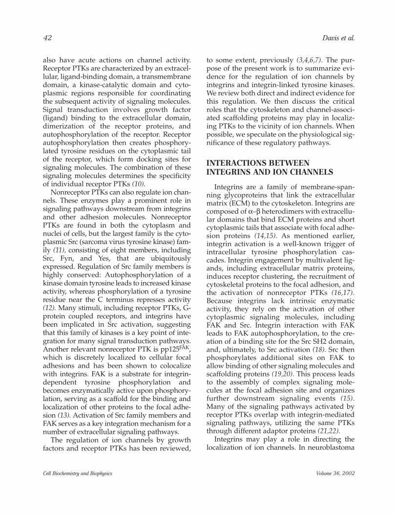
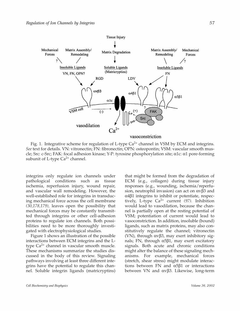
Figure 1 shows an illustration of the possibleinteractions between ECM integrins and the L-type Ca2+ channel in vascular smooth muscle.These mechanisms summarize the studies dis-cussed in the body of this review. Signalingpathways involving at least three different inte-grins have the potential to regulate this chan-nel. Soluble integrin ligands (matricryptins)
that might be formed from the degradation ofECM (e.g., collagen) during tissue injuryresponses (e.g., wounding, ischemia/reperfu-sion, neutrophil invasion) can act on αvβ3 andα4β1 integrins to inhibit or potentiate, respec-tively, L-type Ca2+ current (97). Inhibitionwould lead to vasodilation, because the chan-nel is partially open at the resting potential ofVSM; potentiation of current would lead tovasoconstriction. In addition, insoluble (bound)ligands, such as matrix proteins, may also con-stitutively regulate the channel; vitronectin(VN), through αvβ3, may exert inhibitory sig-nals; FN, through α5β1, may exert excitatorysignals. Both acute and chronic conditionsmight alter the balance of these signaling mech-anisms. For example, mechanical forces(stretch, shear stress) might modulate interac-tions between FN and α5β1 or interactionsbetween VN and αvβ3. Likewise, long-term
Regulation of Ion Channels by Integrins 57
Cell Biochemistry and Biophysics Volume 36, 2002
Fig. 1. Integrative scheme for regulation of L-type Ca2+ channel in VSM by ECM and integrins.See text for details. VN: vitronectin; FN: fibronectin; OPN: osteopontin; VSM: vascular smooth mus-cle; Src: c-Src; FAK: focal adhesion kinase; Y-P: tyrosine phosphorylation site; α1c: α1 pore-formingsubunit of L-type Ca2+ channel.
changes in the assembly or expression of ECMor integrins could shift the balance of these sig-naling mechanisms that converge on the pri-mary Ca2+ influx pathway in VSM.
Given the above discussion, the potentialexists for a number of common pathophysio-logical states to demonstrate involvement ofan extracellular matrix–integrin–ion channelaxis. Examples pertaining to vascular physiol-ogy include dysfunctions associated with dia-betes mellitus and hypertension. Both stateshave been shown to be associated with alter-ations to the extracellular matrix componentof the blood vessel wall, including increaseddeposition of fibronectin (180–183). An alteredcomplement of matrix proteins could conceiv-ably alter cell signaling initiated through inte-grin binding and/or by alterations in themechanical properties of the vessel wall. Inexperimental diabetes, accumulation ofmatrix proteins in the arteriolar wall has beensuggested to decrease distensibility and, con-sequently, impair smooth-muscle Ca2+ entryand mechanotransduction (183). Similarly,impaired shear-stress-mediated mechan-otransduction in endothelial cells could limitCa2+ entry via pathways such as store-deple-tion-dependent Ca2+ entry, thus contributingto the decreased nitric oxide production seenin hypertension (184).
In addition to an involvement of the extra-cellular matrix component, per se, disorderssuch as hypertension have been shown to beassociated with altered expression of integrins.Arterial vessels from the spontaneously hyper-tensive rat exhibit age-dependent increases inαVβ3 and α5β1 integrins (181,185). As voltage-gated Ca2+ channels appear to be regulated byactivation of such integrins (see the sectionInteractions Between Ion Channels andIntegrins), it is an attractive hypothesis thataltered vascular reactivity and, hence, resis-tance, in hypertension may result from alteredintegrin expression. Further studies should bedirected at understanding the functional con-sequences of pathological alterations in inter-actions among extracellular matrix proteins,integrins, and ion channels.
ACKNOWLEDGMENTS
We thank Judy Davidson for her extensiveassistance in preparing and proofreading themanuscript. This work was supported by NIHgrants HL-46502 and HL-60180 to MJD.
NOTE ADDED IN PROOF
HVA current in molluscan neurons is modu-lated by FN and RGD peptides, with low dosesof cRGD reducing current and high dosesincreasing current (186).
REFERENCES
1. Davis, M. J., Wu, X., Nurkiewicz, T. R.,Kawasaki, J., Gui, P., Hill, M. A., et al. (2001)Regulation of ion channels by protein tyro-sine phosphorylation. Am. J. Physiol, 281,H1835–H1862.
2. Ismailov, I. I. and Benos, D. J. (1995) Effects ofphosphorylation on ion channel function.Kidney Int. 48, 1167–1179.
3. Levitan, I. B. (1999) Modulation of ion chan-nels by protein phosphorylation—how thebrain works. Adv. Second MessengerPhosphoprotein Res. 33, 3–22.
4. Siegelbaum, S. A. (1994) Channel regulation:ion channel control by tyrosine phosphoryla-tion. Curr. Biol. 4, 242–245.
5. Tsunoda, Y. (1998) Receptor-operated calciuminflux mediated by protein tyrosine kinase path-ways. J. Recept. Signal Transduct. Res. 18, 281–310.
6. Jonas, E. A. and Kaczmarek, L. K. (1996)Regulation of potassium channels by proteinkinases. Curr. Opin. Neurobiol. 6, 318–323.
7. Levitan, I. B. (1994) Modulation of ion chan-nels by protein phosphorylation and dephos-phorylation. Annu. Rev. Physiol. 56, 193–212.
8. Pollock, J. D., Krempin, M., and Rudy, B. (1990)Differential effects of NGF, FGF, EGF, cAMP,and dexamethasone on neurite outgrowth andsodium channel expression in PC12 cells. J.Neurosci. 10, 2626–2637.
9. Baldelli, P., Magnelli, V., and Carbone, E.(1999) Selective up-regulation of P- and R-typeCa2+ channels in rat embryo motoneurons byBDNF. Eur. J. Neurosci. 11, 1127–1133.
58 Davis et al.
Cell Biochemistry and Biophysics Volume 36, 2002
10. Fantl, W. J., Johnson, D. E., and Williams, L. T.(1993) Signalling by receptor tyrosine kinases.Annu. Rev. Biochem. 62, 453–481.
11. Abram, C. L. and Courtneidge, S. A. (2000) Srcfamily tyrosine kinases and growth factor sig-naling. Exp. Cell Res. 254, 1–13.
12. Okada, M. and Nakagawa, H. (1989) A proteintyrosine kinase involved in regulation of pp60c-src function. J. Biol. Chem. 264, 20,886–20,893.
13. Schlaepfer, D. D., Hauck, C. R., and Sieg, D. J.(1999) Signaling through focal adhesionkinase. Prog. Biophys. Mol. Biol. 71, 435–478.
14. Vuori, K. (1998) Integrin signaling: tyrosinephosphorylation events in focal adhesions. J.Membr. Biol. 165, 191–199.
15. van der Flier, A. and Sonnenberg, A. (2001)Function and interactions of integrins. CellTissue Res. 305, 285–298.
16. Clark, E. A. and Brugge, J. S. (1995) Integrinsand signal transduction pathways: the roadtaken. Science 268, 233–239.
17. Miyamoto, S., Teramoti, H., Coso, O. A.,Gutkind, J. S., Burbelo, P. D., Akiyama, S. K., etal. (1995) Integrin function: molecular hierar-chies of cytoskeletal, and signaling molecules.J. Cell Biol. 131, 791–805.
18. Schaller, M. D., Hildebrand, J. D., Shannon, J.D., Fox, J. W., Vines, R. R., and Parsons, J. T.(1994) Autophosphorylation of the focal adhe-sion kinase, pp125FAK, directs SH2-depen-dent binding of pp60src. Mol. Cell. Biol. 14,1680–1688.
19. Schlaepfer, D. D., Hanks, S. K., Hunter, T., andvan der Geer, P. (1994) Integrin-mediated sig-nal transduction linked to Ras pathway byGRB2 binding to focal adhesion kinase. Nature372, 786–791.
20. Polte, T. R. and Hanks, S. K. (1997)Complexes of focal adhesion kinase (FAK)and Crk-associated substrate (p130Cas) areelevated in cytoskeleton-associated fractionsfollowing adhesion and Src transformation.Requirements for Src kinase activity and FAKproline-rich motifs. J. Biol. Chem. 272,5501–5509.
21. Chen, K. D., Li, Y. S., Kim, M., Li, S., Yuan, S.,Chien, S., et al. (1999) Mechanotransduction inresponse to shear stress—roles of receptortyrosine kinases, integrins, and Shc. J. Biol.Chem. 274, 18,393–18,400.
22. Miyamoto, S., Teramoto, H., Gutkind, J. S., andYamada, K. M. (1996) Integrins can collaboratewith growth factors for phosphorylation of
receptor tyrosine kinases and MAP kinase acti-vation: roles of integrin aggregation and occu-pancy of receptors. J. Cell Biol. 135, 1633–1642.
23. Arcangeli, A., Becchetti, A., Mannini, A.,Mugnai, G., DeFilippi, P., Tarone, G., et al.(1993) Integrin-mediated neurite outgrowth inneuroblastoma cells depends on the activationof potassium channels. J. Cell Biol. 122,1131–1143.
24. McPhee, J. C., Dang, Y. L., Davidson, N., andLester, H. A. (1998) Evidence for a functionalinteraction between integrins and G protein-activated inward rectifier K+ channels. J. Biol.Chem. 273, 34,696–34,702.
25. Ivanina, T., Neusch, C., Li, Y. X., Tong, Y.,Labarca, C., Mosher, D. F., et al. (2000)Expression of GIRK (Kir3.1/Kir3.4) channelsin mouse fibroblast cells with and without β1integrins. FEBS Lett. 466, 327–332.
26. Hofmann, G., Bernabei, P. A., Crociani, O.,Cherubini, A., Guasti, L., Pillozzi, S., et al.(2001) HERG K+ channels activation during β1integrin-mediated adhesion to fibronectininduces an up-regulation of αvβ3 integrin inthe preosteoclastic leukemia cell line FLG 29.1.J. Biol. Chem. 276, 4923–4931.
27. Abdel-Ghany, M., Cheng, H. C., Elble, R. C., andPauli, B. U. (2001) The breast cancer β4 integrinand endothelial human CLCA2 mediate lungmetastasis. J. Biol. Chem. 276, 25,438–25,446.
28. Schwartz, M. A. and Denninghoff, K. (1994) αvintegrins mediate the rise in intracellular cal-cium in endothelial cells on fibronectin eventhough they play a minor role in adhesion. J.Biol. Chem. 269, 11,133–11,137.
29. Bhattacharya, S., Ying, X., Fu, C., Patel, R.,Kuebler, W., Greenberg, S., et al. (2000) αvβ3integrin induces tyrosine phosphorylation-dependent Ca2+ influx in pulmonary endothe-lial cells. Circ. Res. 86, 456–462.
30. McNamee, H. P., Ingber, D. E., and Schwartz,M. A. (1993) Adhesion to fibronectin stimu-lates inositol lipid synthesis and enhancesPDGF-induced inositol lipid breakdown. J.Cell Biol. 121, 673–678.
31. Chenu, C., Colucci, S., Grano, M., Zigrino, P.,Barattolo, R., Zambonin, G., et al. (1994)Osteocalcin induces chemotaxis, secretion ofmatrix proteins, and calcium-mediated intra-cellular signaling in human osteoclast-likecells. J. Cell Biol. 127, 1149–1158.
32. Shankar, G., Davison, I., Helfrich, M. H.,Mason, W. T., and Horton, M. A. (1993)
Regulation of Ion Channels by Integrins 59
Cell Biochemistry and Biophysics Volume 36, 2002
Integrin receptor-mediated mobilisation ofintranuclear calcium in rat osteoclasts. J. CellSci. 105, 61–68.
33. Zimolo, Z., Wesolowski, G., Tanaka, H.,Hyman, J. L., Hoyer, J. R., and Rodan, G. A.(1994) Soluble αvβ3-integrin ligands raise [Ca2+]iin rat osteoclasts and mouse-derived osteoclast-like cells. Am. J. Physiol. 266, C376–C381.
34. Altieri, D. C., Stamnes, S. J., and Gahmberg, C.G. (1992) Regulated Ca2+ signalling throughleukocyte CD11b/CD18 integrin. Biochem. J.288, 465–473.
35. Nebe, B., Rychly, J., Knopp, A., and Bohn, W.(1995) Mechanical induction of beta-1-inte-grin-mediated calcium signaling in a hepato-cyte cell line. Exp. Cell Res. 218, 479–484.
36. Chan, W. L., Holstein-Rathlou, N. H., and Yip,K. P. (2001) Integrin mobilizes intracellularCa2+ in renal vascular smooth muscle cells.Am. J. Physiol. 280, C593–C603.
37. Sjaastad, M. D., Lewis, R. S., and Nelson, W. J.(1996) Mechanisms of integrin-mediated cal-cium signaling in MDCK cells: regulation ofadhesion by IP3- and store-independent cal-cium influx. Mol. Biol. Cell 7, 1025–1041.
38. Chen, B. M. and Grinnell, A. D. (1995)Integrins and modulation of transmitterrelease from motor nerve terminals by stretch.Science 269, 1578–1580.
39. Chen, B. M. and Grinnell, A. D. (1997) Kinetics,Ca2+ dependence, and biophysical propertiesof integrin-mediated mechanical modulationof transmitter release from frog motor nerveterminals. J. Neurosci. 17, 904–916.
40. Becchetti, A., Arcangeli, A., Del Bene, M. R.,Olivotto, M., and Wanke, E. (1992) Response tofibronectin–integrin interaction in leukaemiacells: delayed enhancing of a K+ current. Proc.R. Soc. Lond. (Biol.) 248, 235–240.
41. Platts, S. H., Mogford, J. E., Davis, M. J., andMeininger, G. A. (1998) Role of K+ channels inarteriolar vasodilation mediated by integrininteraction with RGD-containing peptide. Am.J. Physiol. 275, H1449–H1454.
42. Waitkus-Edwards, K. R., Martinez-Lemus, L.A., Wu, X., Trzciakowski, J. P., Davis, G. E.,Davis, M. J., et al. (2001) α4β1 integrin activa-tion of L-type calcium channels in vascularsmooth muscle causes arteriolar vasoconstric-tion. Circ. Res., in press.
43. Tsai, W., Morielli, A. D., Cachero, T. G., andPeralta, E. G. (1999) Receptor protein tyrosinephosphatase α participates in the m1 mus-
carinic acetylcholine receptor-dependent regu-lation of Kv1.2 channel activity. EMBO J. 18,109–118.
44. Levite, M., Cahalon, L., Peretz, A., Hershkoviz,R., Sobko, A., Ariel, A., et al. (2000)Extracellular K+ and opening of voltage-gatedpotassium channels activate T cell integrinfunction: physical and functional associationbetween Kv1.3 channels and beta1 integrins. J.Exp. Med. 191, 1167–1176.
45. Colden-Stanfield, M. and Scanlon, M. (2000)VCAM-1-induced inwardly rectifying K+ cur-rent enhances Ca2+ entry in human THP-1monocytes. Am. J. Physiol. 279, C488–C494.
46. Colden-Stanfield, M. and Gallin, E. K. (1998)Modulation of K+ currents in monocytes byVCAM-1 and E-selectin on activated humanendothelium. Am. J. Physiol. 275, C267–C277.
47. Bianchi, L., Arcangeli, A., Bartolini, P., Mugnai,G., Wanke, E., and Olivotto, M. (1995) Aninward rectifier K+ current modulates in neu-roblastoma cells the tyrosine phosphorylationof the pp125FAK and associated proteins: role inneuritogenesis. Biochem. Biophys. Res. Commun.210, 823–829.
48. Wright, M. O., Nishida, K., Bavington, C.,Godolphin, J. L., Dunne, E., Walmsley, S., et al.(1997) Hyperpolarisation of cultured humanchondrocytes following cyclical pressure-induced strain: evidence of a role for α5β1 inte-grin as a chondrocyte mechanoreceptor. J.Orthop. Res. 15, 742–747.
49. Salter, D. M., Robb, J. E., and Wright, M. O.(1997) Electrophysiological responses ofhuman bone cells to mechanical stimulation:evidence for specific integrin function inmechanotransduction. J. Bone Miner. Res. 12,1133–1141.
50. Millward-Sadler, S. J., Wright, M. O., Lee, H.-S., Nishida, K., Caldwell, H., Nuki, G., et al.(1999) Integrin-regulated secretion of inter-leukin 4: a novel pathway of mechanotrans-duction in human articular chondrocytes. J.Cell Biol. 145, 183–189.
51. Arcangeli, A., Becchetti, A., Del Bene, M. R.,Wanke, E., and Olivotto, M. (1991)Fibronectin–integrin binding promotes hyper-polarization of murine erythroleukemia cells.Biochem. Biophys. Res. Commun. 177, 1266–1272.
52. Millward-Sadler, S. J., Wright, M. O., Lee, H.,Caldwell, H., Nuki, G., and Salter, D. M. (2000)Altered electrophysiological responses tomechanical stimulation and abnormal sig-
60 Davis et al.
Cell Biochemistry and Biophysics Volume 36, 2002
nalling through α5β1 integrin in chondrocytesfrom osteoarthritic cartilage. OsteoarthritisCartilage 8, 272–278.
53. Millward-Sadler, S. J., Wright, M. O., Lee, H.,Nishida, K., Caldwell, H., Nuki, G., et al.(1999) Integrin-regulated secretion of inter-leukin 4: a novel pathway of mechanotrans-duction in human articular chondrocytes. J.Cell Biol. 145, 183–189.
54. Lee, H. S., Millward-Sadler, S. J., Wright, M. O.,Nuki, G., and Salter, D. M. (2000) Integrin andmechanosensitive ion channel-dependenttyrosine phosphorylation of focal adhesionproteins and β-catenin in human articularchondrocytes after mechanical stimulation. J.Bone Miner. Res. 15, 1501–1509.
55. Schwartz, M. A. (1993) Spreading of humanendothelial cells on fibronectin or vitronectintriggers elevation of intracellular-free calcium.J. Cell Biol. 120, 1003–1010.
56. Fujimoto, T., Fujimura, K., and Kuramoto, A.(1991) Electrophysiological evidence that gly-coprotein IIb–IIIa complex is involved in cal-cium channel activation on human plateletplasma membrane. J. Biol. Chem. 266,16,370–16,375.
57. Sjaastad, M. D., Angres, B., Lewis, R. S., andNelson, W. J. (1994) Feedback regulation ofcell–substratum adhesion by integrin-medi-ated intracellular Ca2+ signaling. Proc. Natl.Acad. Sci. USA 91, 8214–8218.
58. Bertolino, G., Noris, P., Spedini, P., andBalduini, C. L. (1995) Ristocetin-inducedplatelet agglutination stimulates GPIIb/IIIa-dependent calcium influx. Thromb. Haemost.73, 689–692.
59. Wu, X., Mogford, J. E., Platts, S. H., Davis, G.E., Meininger, G. A., and Davis, M. J. (1998)Modulation of calcium current in arteriolarsmooth muscle by αvβ3 and α5β1 integrin lig-ands. J. Cell Biol. 143, 241–252.
60. Wu, X., Davis, G. E., Meininger, G. A., Wilson,E., and Davis, M. J. (2001) Regulation of the L-type calcium channel by α5β1 integrin requiressignaling between focal adhesion proteins. J.Biol. Chem. 276, 30,285–30,292.
61. Kwon, M. S., Park, C. S., Choi, K., Ahnn, J.,Kim, J. I., Eom, S. H., et al. (2000) Calreticulincouples calcium release and calcium influx inintegrin-mediated calcium signaling. Mol. Biol.Cell 11, 1433–1443.
62. Wang, Y. G., Samarel, A. M., and Lipsius, S. L.(2000) Laminin binding to β1-integrins selec-
tively alters β1- and β2-adrenoceptor signallingin cat atrial myocytes. J. Physiol. (Lond.) 527 Pt1, 3–9.
63. Wang, Y. G., Samarel, A. M., and Lipsius, S. L.(2000) Laminin acts via β1 integrin signallingto alter cholinergic regulation of L-type Ca2+
current in cat atrial myocytes. J. Physiol. (Lond.)526 (Pt. 1), 57–68.
64. Wildering, W. C., Lodder, J. C., Kits, K. S., andBulloch, A. G. (1995) Nerve growth factor(NGF) acutely enhances high-voltage-acti-vated calcium currents in molluscan neurons.J. Neurophysiol. 74, 2778–2781.
65. Sjaastad, M. D. and Nelson, W. J. (1997)Integrin-mediated calcium signaling and regu-lation of cell adhesion by intracellular calcium.BioEssays 19, 47–55.
66. Bhattacharya, S., Fu, C., Bhattacharya, J., andGreenberg, S. (1995) Soluble ligands of theαvβ3 integrin mediate enhanced tyrosine phos-phorylation of multiple proteins in adherentbovine pulmonary artery endothelial cells. J.Biol. Chem. 270, 16,781–16,787.
67. Hoth, M. and Penner, R. (1992) Depletion ofintracellular calcium stores activates a calciumcurrent in mast cells. Nature 355, 353–356.
68. Montell, C. and Rubin, G. M. (1989) Molecularcharacterization of the Drosophila trp locus: aputative integral membrane protein requiredfor phototransduction. Neuron 2, 1313–1323.
69. Peng, J. B., Chen, X. Z., Berger, U. V., Vassilev,P. M., Tsukaguchi, H., Brown, E. M., et al.(1999) Molecular cloning and chanracteriza-tion of a channel-like transporter mediatingintestinal calcium absorption. J. Biol. Chem.274, 22,739–22,746.
70. Yue, L., Peng, J. B., Hediger, M. A., andClapham, D. E. (2001) CaT1 manifests the poreproperties of the calcium-release-activated cal-cium channel. Nature 410, 705–709.
71. Parekh, A. B. and Penner, R. (1997) Store deple-tion and calcium influx. Physiol. Rev. 77, 901–930.
72. Montell, C. (1999) Visual transduction inDrosophila. Annu. Rev. Cell. Dev. Biol. 15, 231–268.
73. Philipp, S., Trost, C., Warnat, J., Rautmann, J.,Himmerkus, N., Schroth, G., et al. (2000) TRP4(CCE1) protein is part of native calciumrelease-activated Ca2+-like channels inadrenal cells. J. Biol. Chem. 275, 23,965–23,972.
74. Inoue, R., Okada, T., Onoue, H., Hara, Y.,Shimizu, S., Naitoh, S., et al. (2001) The tran-sient receptor potential protein homologueTRP6 is the essential component of vascular
Regulation of Ion Channels by Integrins 61
Cell Biochemistry and Biophysics Volume 36, 2002
α1-adrenoceptor-activated Ca2+-permeablecation channel. Circ. Res. 88, 325–332.
75. Kawasaki, J., Nurkiewicz, T. R., and Davis, M.J. (2001) Regulation of endothelial cell calciumcurrent by αvβ3 integrin. FASEB J. 15, A109(abstract).
76. Sharma, N. R. and Davis, M. J. (1996) Calciumentry activated by store depletion in coronaryendothelium is promoted by tyrosine phos-phorylation. Am. J. Physiol. 270, H267–H274.
77. Sage, S. O., Sargeant, P., Jenner, S., andFarndale, R. W. (1994) Tyrosine phosphoryla-tion and Ca2+ influx. A further mechanism forstore-dependent Ca2+ entry? Trends Pharmacol.Sci. 15, 282.
78. Sargeant, P., Farndale, R. W., and Sage, S. O.(1993) The tyrosine kinase inhibitors methyl2,5-dihydroxycinnamate and genistein reducethrombin-evoked tyrosine phosphorylationand Ca2+ entry in human platelets. FEBS Lett.315, 242–246.
79. Sargeant, P., Farndale, R. W., and Sage, S. O.(1993) ADP- and thapsigargin-evoked Ca2+
entry and protein-tyrosine phosphorylationare inhibited by the tyrosine kinase inhibitorsgenistein and methyl-2,5-dihydroxycinnamatein fura-2-loaded human platelets. J. Biol. Chem.268, 18,151–18,156.
80. Rosado, J. A., Jenner, S., and Sage, S. O. (2000)A role for the actin cytoskeleton in the initia-tion and maintenance of store-mediated cal-cium entry in human platelets. Evidence forconformational coupling. J. Biol. Chem. 275,7527–7533.
81. Rosado, J. A. and Sage, S. O. (2000) The actincytoskeleton in store-mediated calcium entry.J. Physiol. (Lond.) 526, 221–229.
82. Djouder, N., Prepens, U., Aktories, K., andCavalie, A. (2000) Inhibition of calciumrelease-activated calcium current byRac/Cdc42-inactivating clostridial cytotoxinsin RBL cells. J. Biol. Chem. 275, 18,732–18,738.
83. Miyamoto, S., Akiyama, S. K., and Yamada, K.M. (1995) Synergistic roles for receptor occu-pancy and aggregation in integrin transmem-brane function. Science 267, 883–885.
84. Waitkus, K. R., Davis, G. E., and Meininger, G.A. (1998) Collagen proteolysis and vasodila-tion in isolated rat cremaster arterioles. FASEBJ. 12, A384 (abstract).
85. Wijetunge, S., Lymn, J. S., and Hughes, A. D.(1998) Effect of inhibition of tyrosine phos-phatases on voltage-operated calcium channel
currents in rabbit isolated ear artery cells. Br. J.Pharmacol. 124, 307–316.
86. Wijetunge, S. and Hughes, A. D. (1995) pp60c-
src increases voltage-operated calcium channelcurrents in vascular smooth muscle cells.Biochem. Biophys. Res. Commun. 217, 1039–1044.
87. Wijetunge, S. and Hughes, A. D. (1996)Activation of endogenous c-Src or a relatedtyrosine kinase by intracellular (PY)EEI pep-tide increases voltage-operated calcium chan-nel currents in rabbit ear artery cells. FEBSLett. 399, 63–66.
88. Hu, X.-Q., Singh, N., Mukhopadhyay, D., andAkbarali, H. I. (1998) Modulation of voltage-dependent Ca2+ channels in rabbit colonicsmooth muscle cells by c-Src and focal adhe-sion kinase. J. Biol. Chem. 273, 5337–5342.
89. Wijetunge, S. and Hughes, A. D. (1995) Effectof platelet-derived growth factor on voltage-operated calcium channels in rabbit isolatedear artery cells. Br. J. Pharmacol. 115, 534–538.
90. Blair, L. A. and Marshall, J. (1997) IGF-1 mod-ulates N and L calcium channels in a PI 3-kinase-dependent manner. Neuron 19, 421–429.
91. Selinfreund, R. H. and Blair, L. A. (1994)Insulin-like growth factor-I induces a rapidincrease in calcium currents and spontaneousmembrane activity in clonal pituitary cells.Mol. Pharmacol. 45, 1215–1220.
92. Bence-Hanulec, K. K., Marshall, J., and Blair, L.A. (2000) Potentiation of neuronal L calciumchannels by IGF-1 requires phosphorylation ofthe α1 subunit on a specific tyrosine residue.Neuron 27, 121–131.
93. Gerhardstein, B. L., Gao, T., Bunemann, M.,Puri, T. S., Adair, A., Ma, H., et al. (2000)Proteolytic processing of the C terminus of theα(IC) subunit of L-type calcium channels andthe role of a proline-rich domain in membranetethering of proteolytic fragments. J. Biol.Chem. 275, 8556–8563.
94. Wei, X., Neely, A., Lacerda, A. E., Olcese, R.,Stefani, E., Perez-Reyes, E., et al. (1994)Modification of Ca2+ channel activity by dele-tions at the carboxyl terminus of the cardiacalpha 1 subunit. J. Biol. Chem. 269, 1635–1640.
95. Mogford, J. E., Platts, S. H., Davis, G. E., andMeininger, G. A. (1996) Vascular smooth mus-cle αvβ3 integrin mediates arteriolar vasodila-tion in response to RGD peptides. Circ. Res. 79,821–826.
96. D’Angelo, G., Davis, M. J., and Meininger, G.A. (1997) Calcium and mechanotransduction
62 Davis et al.
Cell Biochemistry and Biophysics Volume 36, 2002
of the myogenic response. Am. J. Physiol. 273,H175–H182.
97. Davis, G. E., Bayless, K. J., Davis, M. J., andMeininger, G. A. (2000) Regulation of tissueinjury responses by the exposure of matricryp-tic sites within extracellular matrix molecules.Am. J. Pathol. 156, 1489–1498.
98. Bloch, W., Fan, Y., Han, J., Xue, S., Schoneberg,T., Ji, G., et al. (2001) Disruption of cytoskeletalintegrity impairs Gi-mediated signaling due todisplacement of Gi proteins. J. Cell Biol. 154,753–761.
99. Gurubhagavatula, I., Amrani, Y., Pratico, D.,Ruberg, F. L., Albelda, S. M., Panettieri, R. A.,Jr. (1998) Engagement of human PECAM-1(CD31) on human endothelial cells increasesintracellular calcium ion concentration andstimulates prostacyclin release. J. Clin. Invest.101, 212–222.
100. O’Brien, C. D., Ji, G., Wang, Y. X., Sun, J.,Krymskaya, V. P., Ruberg, F. L., et al. (2001)PECAM-1 (CD31) engagement activates aphosphoinositide-independent, nonspecificcation channel in endothelial cells. FASEB J. 15,1257–1260.
101. Erickson, H. P. (1993) Tenascin-C, tenascin-Rand tenascin-X: a family of talented proteins insearch of functions. Curr. Opin. Cell Biol. 5,869–876.
102. Srinivasan, J., Schachner, M., and Catterall, W.A. (1998) Interaction of voltage-gated sodiumchannels with the extracellular matrix mole-cules tenascin-C and tenascin-R. Proc. Natl.Acad. Sci. USA 95, 15,753–15,757.
103. Isom, L. L., Ragsdale, D. S., De Jongh, K. S.,Westenbroek, R. E., Reber, B. F., Scheuer, T., et al.(1995) Structure and function of the β2 subunitof brain sodium channels, a transmembrane gly-coprotein with a CAM motif. Cell 83, 433–442.
104. Ricard, J., Giudici-Orticoni, M. T., andGontero, B. (1994) The modulation of enzymereaction rates within multi-enzyme complexes.1. Statistical thermodynamics of informationtransfer through multi-enzyme complexes.Eur. J. Biochem. 226, 993–998.
105. Xu, X. Z., Choudhury, A., Li, X., and Montell,C. (1998) Coordination of an array of signalingproteins through homo- and heteromeric inter-actions between PDZ domains and target pro-teins. J. Cell Biol. 142, 545–555.
106. Colledge, M. and Scott, J. D. (1999) AKAPs:from structure to function. Trends Cell Biol. 9,216–221.
107. Hibino, H., Inanobe, A., Tanemoto, M., Fujita,A., Doi, K., Kubo, T., et al. (2000) Anchoringproteins confer G protein sensitivity to aninward-rectifier K+ channel through the GKdomain. EMBO J. 19, 78–83.
108. Huber, A., Sander, P., Bahner, M., and Paulsen,R. (1998) The TRP Ca2+ channel assembled in asignaling complex by the PDZ domain proteinINAD is phosphorylated through the interac-tion with protein kinase C (ePKC). FEBS Lett.425, 317–322.
109. Gray, P. C., Scott, J. D., and Catterall, W. A. (1998)Regulation of ion channels by cAMP-dependentprotein kinase and A-kinase anchoring proteins.Curr. Opin. Neurobiol. 8, 330–334.
110. Dodge, K. and Scott, J. D. (2000) AKAP79 andthe evolution of the AKAP model. FEBS Lett.476, 58–61.
111. Johnson, B. D. (1999) The company they keep:ion channels and their intracellular regulatorypartners. Adv. Second Messenger PhosphoproteinRes. 33, 203–228.
112. Wang, W. (1999) Regulation of the ROMKchannel: interaction of the ROMK with associ-ate proteins. Am. J. Physiol. 277, F826–F831.
113. Gao, T., Yatani, A., Dell’Acqua, M. L., Sako, H.,Green, S. A., Dascal, N., et al. (1997) cAMP-dependent regulation of cardiac L-type Ca2+
channels requires membrane targeting of PKAand phosphorylation of channel subunits.Neuron 19, 185–196.
114. Johnson, B. D., Scheuer, T., and Catterall, W. A.(1994) Voltage-dependent potentiation of L-typeCa2+ channels in skeletal muscle cells requiresanchored cAMP-dependent protein kinase. Proc.Natl. Acad. Sci. USA 91, 11,492–11,496.
115. Fukao, M., Mason, H. S., Britton, F. C., Kenyon,J. L., Horowitz, B., and Keef, K. D. (1999) CyclicGMP-dependent protein kinase activatescloned BKCa channels expressed in mam-malian cells by direct phosphorylation at ser-ine 1072. J. Biol. Chem. 274, 10,927–10,935.
116. Wang, Z. W. and Kotlikoff, M. I. (1996)Activation of KCa channels in airway smoothmuscle cells by endogenous protein kinase A.Am. J. Physiol. 271, L100–L105.
117. Sun, F., Hug, M. J., Bradbury, N. A., andFrizzell, R. A. (2000) Protein kinase A associ-ates with cystic fibrosis transmembrane con-ductance regulator via an interaction withezrin. J. Biol. Chem. 275, 14,360–14,366.
118. Ingber, D. E. (1993) Cellular tensegrity: defin-ing new rules of biological design that governthe cytoskeleton. J. Cell Sci. 104, 613–627.
Regulation of Ion Channels by Integrins 63
Cell Biochemistry and Biophysics Volume 36, 2002
119. Molday, L. L., Cook, N. J., Kaupp, U. B., andMolday, R. S. (1990) The cGMP-gated cationchannel of bovine rod photoreceptor cells isassociated with a 240-kDa protein exhibitingimmunochemical cross- reactivity with spec-trin. J. Biol. Chem. 265, 18,690–18,695.
120. Lue, R. A., Marfatia, S. M., Branton, D., andChishti, A. H. (1994) Cloning and characteriza-tion of hdlg: the human homologue of theDrosophila discs large tumor suppressor bindsto protein 4.1. Proc. Natl. Acad. Sci. USA 91,9818–9822.
121. Zuckerman, J. B., Chen, X., Jacobs, J. D., Hu, B.,Kleyman, T. R., and Smith, P. R. (1999)Association of the epithelial sodium channelwith Apx and α-spectrin in A6 renal epithelialcells. J. Biol. Chem. 274, 23,286–23,295.
122. van Rossum, D., Kuhse, J., and Betz, H. (1999)Dynamic interaction between soluble tubulinand C-terminal domains of N-methyl-D-aspar-tate receptor subunits. J. Neurochem. 72, 962–973.
123. Wyszynski, M., Lin, J., Rao, A., Nigh, E., Beggs,A. H., Craig, A. M., et al. (1997) Competitivebinding of α-actinin and calmodulin to theNMDA receptor. Nature 385, 439–442.
124. Sheng, M. and Pak, D. T. (2000) Ligand-gated ionchannel interactions with cytoskeletal and sig-naling proteins. Annu. Rev. Physiol. 62, 755–778.
125. Sheng, M. and Wyszynski, M. (1997) Ion chan-nel targeting in neurons. Bioessays 19, 847–853.
126. Colledge, M. and Froehner, S. C. (1998) Signalsmediating ion channel clustering at the neuro-muscular junction. Curr. Opin. Neurobiol. 8,357–363.
127. Petrecca, K., Miller, D. M., and Shrier, A. (2000)Localization and enhanced current density ofthe kv4.2 potassium channel by interactionwith the actin-binding protein filamin. J.Neurosci. 20, 8736–8744.
128. Jing, J., Peretz, T., Singer-Lahat, D.,Chikvashvili, D., Thornhill, W. B., and Lotan, I.(1997) Inactivation of a voltage-dependent K+
channel by β subunit. Modulation by a phos-phorylation-dependent interaction betweenthe distal C terminus of α subunit andcytoskeleton. J. Biol. Chem. 272, 14,021–14,024.
129. Nakahira, K., Matos, M. F., and Trimmer, J. S.(1998) Differential interaction of voltage-gatedK+ channel β-subunits with cytoskeleton ismediated by unique amino terminal domains.J. Mol. Neurosci. 11, 199–208.
130. Levin, G., Chikvashvili, D., Singer-Lahat, D.,Peretz, T., Thornhill, W. B., and Lotan, I. (1996)
Phosphorylation of a K+ channel alpha subunitmodulates the inactivation conferred by a betasubunit. Involvement of cytoskeleton. J. Biol.Chem. 271, 29,321–29,328.
131. Maruoka, N. D., Steele, D. F., Au, B. P., Dan, P.,Zhang, X., Moore, E. D., et al. (2000) α-Actinin-2 couples to cardiac Kv1.5 channels, regulatingcurrent density and channel localization inHEK cells. FEBS Lett. 473, 188–194.
132. Maguire, G., Connaughton, V., Prat, A. G.,Jackson, G. R., Jr., and Cantiello, H. F. (1998)Actin cytoskeleton regulates ion channelactivity in retinal neurons. Neuroreport 9,665–670.
133. Nakamura, M., Sunagawa, M., Kosugi, T., andSperelakis, N. (2000) Actin filament disruptioninhibits L-type Ca2+ channel current in cul-tured vascular smooth muscle cells. Am. J.Physiol. 279, C480–C487.
134. Maltsev, V. A. and Undrovinas, A. I. (1997)Cytoskeleton modulates coupling betweenavailability and activation of cardiac sodiumchannel. Am. J. Physiol. 273, H1832–H1840.
135. Undrovinas, A. I. and Maltsev, V. A. (1998)Cytochalasin D alters kinetics of Ca2+ transientin rat ventricular cardiomyocytes: an effect ofaltered actin cytoskeleton? J. Mol. Cell. Cardiol.30, 1665–1670.
136. West, J. W., Patton, D. E., Scheuer, T., Wang, Y.,Goldin, A. L., and Catterall, W. A. (1992) Acluster of hydrophobic amino acid residuesrequired for fast Na+ – channel inactivation.Proc. Natl. Acad. Sci. USA 89, 10,910–10,914.
137. Fincham, V. J., Unlu, M., Brunton, V. G., Pitts, J.D., Wyke, J. A., and Frame, M. C. (1996)Translocation of Src kinase to the cell periph-ery is mediated by the actin cytoskeletonunder the control of the Rho family of small Gproteins. J. Cell Biol. 135, 1551–1564.
138. Grondin, P., Plantavid, M., Sultan, C., Breton,M., Mauco, G., and Chap, H. (1991) Interactionof pp60c-src, phospholipase C, inositol-lipid,and diacyglycerol kinases with the cytoskele-tons of thrombin-stimulated platelets. J. Biol.Chem. 266, 15,705–15,709.
139. Weernink, P. A. and Rijksen, G. (1995)Activation and translocation of c-Src to thecytoskeleton by both platelet-derived growthfactor and epidermal growth factor. J. Biol.Chem. 270, 2264–2267.
140. Holmes, T. C., Fadool, D. A., and Levitan, I. B.(1996) Tyrosine phosphorylation of the Kv1.3potassium channel. J. Neurosci. 16, 1581–1590.
64 Davis et al.
Cell Biochemistry and Biophysics Volume 36, 2002
141. Bowlby, M. R., Fadool, D. A., Holmes, T. C.,and Levitan, I. B. (1997) Modulation of theKv1.3 potassium channel by receptor tyrosinekinases. J. Gen. Physiol. 110, 601–610.
142. Holmes, T. C., Fadool, D. A., Ren, R. B., andLevitan, I. B. (1996) Association of Src tyrosinekinase with a human potassium channel medi-ated by SH3 domain. Science 274, 2089–2091.
143. Ling, S., Woronuk, G., Sy, L., Lev, S., andBraun, A. P. (2000) Enhanced activity of a largeconductance, calcium-sensitive K+ channel inthe presence of Src tyrosine kinase. J. Biol.Chem. 275, 30,683–30,689.
144. Srinivasan, Y., Elmer, L., Davis, J., Bennett, V.,and Angelides, K. (1988) Ankyrin and spectrinassociate with voltage-dependent sodiumchannels in brain. Nature 333, 177–180.
145. Lambert, S., Davis, J. Q., and Bennett, V. (1997)Morphogenesis of the node of Ranvier: co-clusters of ankyrin and ankyrin-binding inte-gral proteins define early developmentalintermediates. J. Neurosci. 17, 7025–7036.
146. Zhou, D., Lambert, S., Malen, P. L., Carpenter,S., Boland, L. M., and Bennett, V. (1998)AnkyrinG is required for clustering of voltage-gated Na channels at axon initial segmentsand for normal action potential firing. J. CellBiol. 143, 1295–1304.
147. Kaplan, M. R., Meyer-Franke, A., Lambert, S.,Bennett, V., Duncan, I. D., Levinson, S. R., et al.(1997) Induction of sodium channel clusteringby oligodendrocytes. Nature 386, 724–728.
148. Chauhan, V. S., Tuvia, S., Buhusi, M., Bennett,V., and Grant, A. O. (2000) Abnormal cardiacNa+ channel properties and QT heart rateadaptation in neonatal ankyrinB knockoutmice. Circ. Res. 86, 441–447.
149. Bennett, P. B. (2000) Anchors a weigh! Ionchannels, cytoskeletal proteins, and cellularexcitability. Circ. Res. 86, 367–368.
150. Hall, A. (1998) Rho GTPases and the actincytoskeleton. Science 279, 509–514.
151. Dransfield, D. T., Bradford, A. J., Smith, J.,Martin, M., Roy, C., Mangeat, P. H., et al. (1997)Ezrin is a cyclic AMP-dependent proteinkinase anchoring protein. EMBO J. 16, 35–43.
152. Niggli, V., Andreoli, C., Roy, C., and Mangeat,P. (1995) Identification of a phosphatidylinosi-tol-4,5-bisphosphate-binding domain in the N-terminal region of ezrin. FEBS Lett. 376,172–176.
153. Hunter, T. and Cooper, J. A. (1981) Epidermalgrowth factor induces rapid tyrosine phospho-
rylation of proteins in A431 human tumorcells. Cell 24, 741–752.
154. Mackay, D. J., Esch, F., Furthmayr, H., andHall, A. (1997) Rho- and rac-dependent assem-bly of focal adhesion complexes and actin fila-ments in permeabilized fibroblasts: anessential role for ezrin/radixin/moesin pro-teins. J. Cell Biol. 138, 927–938.
155. Machesky, L. M. and Hall, A. (1997) Role of actinpolymerization and adhesion to extracellularmatrix in Rac- and Rho-induced cytoskeletalreorganization. J. Cell Biol. 138, 913–926.
156. Boudreau, J. and Jones, P. L. (1999) Extracellularmatrix and integrin signalling: the shape ofthings to come. Biochem. J. 339, 481–488.
157. Schoenwaelder, S. M. and Burridge, K. (1999)Bidirectional signaling between the cytoskele-ton and integrins. Curr. Opin. Cell Biol. 11,274–286.
158. Wilk-Blaszczak, M. A., Singer, W. D., Quill, T.,Miller, B., Frost, J. A., Sternweis, P. C., et al.(1997) The monomeric G-proteins Rac1 and/orCdc42 are required for the inhibition of volt-age-dependent calcium current by bradykinin.J. Neurosci. 17, 4094–4100.
159. Giancotti, F. G. and Ruoslahti, E. (1999)Integrin signaling. Science 285, 1028–1032.
160. Ekinci, F. J., Malik, K. U., and Shea, T. B. (1999)Activation of the L voltage-sensitive calciumchannel by mitogen-activated protein (MAP)kinase following exposure of neuronal cells toβ-amyloid. MAP kinase mediates β-amyloid-induced neurodegeneration. J. Biol. Chem. 274,30,322–30,327.
161. Ma, H., Matsunaga, H., Li, B., Schieffer, B.,Marrero, M. B., and Ling, B. N. (1996) Ca2+
channel activation by platelet-derived growthfactor-induced tyrosine phosphorylation andRas guanine triphosphate-binding proteins inrat glomerular mesangial cells. J. Clin. Invest.97, 2332–2341.
162. Collin, C., Papageorge, A. G., Lowy, D. R., andAlkon, D. L. (1990) Early enhancement of cal-cium current by H-ras oncoproteins injected intoHermissenda neurons. Science 250, 1743–1745.
163. Hahnel, C., Gottmann, K., Wittinghofer, A.,and Lux, H.-D. (1992) p21ras oncogene proteinselectively increases low-voltage-activatedCa2+ current density in embryonic chick dorsalroot ganglion neurons. Eur. J. Neurosci. 4,361–368.
164. Hescheler, J., Klinz, F. J., Schultz, G., andWittinghofer, A. (1991) Ras proteins activate
Regulation of Ion Channels by Integrins 65
Cell Biochemistry and Biophysics Volume 36, 2002
calcium channels in neuronal cells. Cell Signal.3, 127–133.
165. Hemmick, L. M., Perney, T. M., Flamm, R. E.,Kaczmarek, L. K., and Birnberg, N. C. (1992)Expression of the H-ras oncogene inducespotassium conductance and neuron-specificpotassium channel mRNAs in the AtT20 cellline. J. Neurosci. 12, 2007–2014.
166. Flamm, R. E., Birnberg, N. C., and Kaczmarek,L. K. (1990) Transfection of activated ras intoan excitable cell line (AtT-20) alterstetrodotoxin sensitivity of voltage-dependentsodium current. Pflügers Arch. 416, 120–125.
167. Yatani, A., Okabe, K., Polakis, P., Halenbeck,R., McCormick, F., and Brown, A. M. (1990) rasp21 and GAP inhibit coupling of muscarinicreceptors to atrial K+ channels. Cell 61,769–776.
168. Martin, G. A., Yatani, A., Clark, R., Conroy, L.,Polakis, P., Brown, A. M., et al. (1992) GAPdomains responsible for ras p21-dependentinhibition of muscarinic atrial K+ channel cur-rents. Science 255, 192–194.
169. Yatani, A., Quilliam, L. A., Brown, A. M., andBokoch, G. M. (1991) Rap1A antagonizes theability of Ras and Ras-Gap to inhibit mus-carinic K+ channels. J. Biol. Chem. 266,22,222–22,226.
170. Hilborn, M. D., Vaillancourt, R. R., and Rane,S. G. (1998) Growth factor receptor tyrosinekinases acutely regulate neuronal sodiumchannels through the src signaling pathway. J.Neurosci. 18, 590–600.
171. Fitzgerald, E. M. and Dolphin, A. C. (1997)Regulation of rat neuronal voltage-dependentcalcium channels by endogenous p21-ras. Eur.J. Neurosci. 9, 1252–1261.
172. Galli, A. and DeFelice, L. J. (1994) Inactivationof L-type Ca channels in embryonic chick ven-tricle cells: dependence on the cytoskeletalagents colchicine and taxol. Biophys. J. 67,2296–2304.
173. Johnson, B. D. and Byerly, L. (1993) Acytoskeletal mechanism for Ca2+ channelsmetabolic dependence and inactivation byintracellular Ca2+. Neuron 10, 797–804.
174. Howarth, F. C., Calaghan, S. C., Boyett, M. R.,and White, E. (1999) Effect of the microtubulepolymerizing agent taxol on contraction, Ca2+
transient and L-type Ca2+ current in rat ven-tricular myocytes. J. Physiol. (Lond.) 516,409–419.
175. Ingber, D. E. (1997) Tensegrity: the architec-tural basis of cellular mechanotransduction.Annu. Rev. Physiol. 59, 575–599.
176. Machu, T. K. (1998) Colchicine competitivelyantagonizes glycine receptors expressed inXenopus oocytes. Neuropharmacology 37,391–396.
177. Rasenick, M. M., Stein, P. J., and Bitensky, M.W. (1981) The regulatory subunit of adenylatecyclase interacts with cytoskeletal compo-nents. Nature 294, 560–562.
178. Wang, N., Butler, J. P., and Ingber, D. E. (1993)Mechanotransduction across the cell surfaceand through the cytoskeleton. Science 260,1124–1127.
179. Schmidt, C., Pommerenke, H., Dürr, F., Nebe,B., and Rychly, J. (1998) Mechanical stressing ofintegrin receptors induces enhanced tyrosinephosphorylation of cytoskeletally anchoredproteins. J. Biol. Chem. 273, 5081–5085.
180. Intengan, H. D. and Schiffrin, E. L. (2000)Structure and mechanical properties of resis-tance arteries in hypertension: role of adhesionmolecules and extracellular matrix determi-nants. Hypertension 36, 312–318.
181. Bezie, Y., Lamaziere, J. M., Laurent, S.,Challande, P., Cunha, R. S., Bonnet, J., et al.(1998) Fibronectin expression and aortic wallelastic modulus in spontaneously hyperten-sive rats. Arteriosclerosis Thromb. Vasc. Biol. 18,1027–1034.
182. Magnusson, M. K. and Mosher, D. F. (1998)Fibronectin—structure, assembly, and cardio-vascular implications. Arteriosclerosis Thromb.Vasc. Biol. 18, 1363–1370.
183. Yu, G., Zou, H., Prewitt, R. L., and Hill, M. A.(1999) Impaired arteriolar mechanotransduc-tion in experimental diabetes mellitus. J.Diabetes Complications 13, 235–242.
184. Nilius, B. and Droogmans, G. (2001) Ion chan-nels and their functional role in vascularendothelium. Physiol. Rev. 81, 1415–1459.
185. Intengan, H. D., Thibault, G., Li, J. S., andSchiffrin, E. L. (1999) Resistance arterymechanics, structure, and extracellular compo-nents in spontaneously hypertensive rats:effects of angiotensin receptor antagonism andconverting enzyme inhibition. Circulation 100,2267–2275.
186. Wildering, N. C., Hermann, P. M., and Bulloch,A. G. M. (2002) Rapid neuromodulatoryactions of integrin ligands. J. Neurosci., in press.
66 Davis et al.
Cell Biochemistry and Biophysics Volume 36, 2002