Regulation of in vivo dynein force production by CDK5 and ...
12
ARTICLE Regulation of in vivo dynein force production by CDK5 and 14-3-3ε and KIAA0528 Dail E. Chapman 1 , Babu J.N. Reddy 1 , Bunchhin Huy 1 , Matthew J. Bovyn 1 , Stephen John S. Cruz 1 , Zahraa M. Al-Shammari 1 , Han Han 1 , Wenqi Wang 1 , Deanna S. Smith 2 & Steven P. Gross 1 Single-molecule cytoplasmic dynein function is well understood, but there are major gaps in mechanistic understanding of cellular dynein regulation. We reported a mode of dynein regulation, force adaptation, where lipid droplets adapt to opposition to motion by increasing the duration and magnitude of force production, and found LIS1 and NudEL to be essential. Adaptation reflects increasing NudEL-LIS1 utilization; here, we hypothesize that such increasing utilization reflects CDK5-mediated NudEL phosphorylation, which increases the dynein-NudEL interaction, and makes force adaptation possible. We report that CDK5, 14-3- 3ε, and CDK5 cofactor KIAA0528 together promote NudEL phosphorylation and are essential for force adaptation. By studying the process in COS-1 cells lacking Tau, we avoid con- founding neuronal effects of CDK5 on microtubules. Finally, we extend this in vivo regulatory pathway to lysosomes and mitochondria. Ultimately, we show that dynein force adaptation can control the severity of lysosomal tug-of-wars among other intracellular transport func- tions involving high force. https://doi.org/10.1038/s41467-018-08110-z OPEN 1 Developmental and Cell Biology and Physics, University of California, Irvine, CA, USA. 2 Biological Sciences, University of South Carolina, Columbia, SC, USA. These authors contributed equally: Dail E. Chapman, Babu J. N. Reddy. Correspondence and requests for materials should be addressed to S.P.G. (email: [email protected]) NATURE COMMUNICATIONS | (2019)10:228 | https://doi.org/10.1038/s41467-018-08110-z | www.nature.com/naturecommunications 1 1234567890():,;
Transcript of Regulation of in vivo dynein force production by CDK5 and ...
ARTICLE
Regulation of in vivo dynein force productionby CDK5 and 14-3-3ε and KIAA0528Dail E. Chapman1, Babu J.N. Reddy1, Bunchhin Huy1, Matthew J. Bovyn 1, Stephen John S. Cruz1,
Zahraa M. Al-Shammari1, Han Han 1, Wenqi Wang 1, Deanna S. Smith 2 & Steven P. Gross1
Single-molecule cytoplasmic dynein function is well understood, but there are major gaps
in mechanistic understanding of cellular dynein regulation. We reported a mode of dynein
regulation, force adaptation, where lipid droplets adapt to opposition to motion by increasing
the duration and magnitude of force production, and found LIS1 and NudEL to be essential.
Adaptation reflects increasing NudEL-LIS1 utilization; here, we hypothesize that such
increasing utilization reflects CDK5-mediated NudEL phosphorylation, which increases the
dynein-NudEL interaction, and makes force adaptation possible. We report that CDK5, 14-3-
3ε, and CDK5 cofactor KIAA0528 together promote NudEL phosphorylation and are essential
for force adaptation. By studying the process in COS-1 cells lacking Tau, we avoid con-
founding neuronal effects of CDK5 on microtubules. Finally, we extend this in vivo regulatory
pathway to lysosomes and mitochondria. Ultimately, we show that dynein force adaptation
can control the severity of lysosomal tug-of-wars among other intracellular transport func-
tions involving high force.
https://doi.org/10.1038/s41467-018-08110-z OPEN
1 Developmental and Cell Biology and Physics, University of California, Irvine, CA, USA. 2 Biological Sciences, University of South Carolina, Columbia, SC, USA.These authors contributed equally: Dail E. Chapman, Babu J. N. Reddy. Correspondence and requests for materials should be addressed toS.P.G. (email: [email protected])
NATURE COMMUNICATIONS | (2019) 10:228 | https://doi.org/10.1038/s41467-018-08110-z | www.nature.com/naturecommunications 1
1234
5678
90():,;
Cytoplasmic dynein (dynein-1, MAP1C) is essential forintracellular transport of organelles and other cargostoward the cell’s nucleus1,2. Together with the opposite
directed plus-end kinesin family of motors, these molecules movealong cytoplasmic microtubule (MT) highways, allowing appro-priate cargo positioning and delivery.
Dynein plays roles in vesicular, viral, chromosomal, and nucleartransport and is essential for neuronal migration during cerebraldevelopment3,4. Owing to this diversity of roles, it is highly regu-lated, frequently via regulatory cofactors, including dynactin, LIS1,NudE (NDE1), and NudEL (NDEL1)4. Dynein forms two majorcomplexes: with dynactin or with NudEL-LIS15–8. Dynactin isimportant for increased processivity, and dynein–dynactin–BicDcomplexes can travel longer distances9. In contrast, theNudEL–LIS1 complex is important for force production. Deletionsin the LIS1 gene cause Miller–Dieker syndrome10, presumablybecause LIS1 enhances additivity of single-motor forces11, andthus facilitates dynein’s high-load function, which is important forthe nuclear migration2,12 underlying neuronal migration; NudELtethers LIS1 to dynein and helps regulate the dynein–LIS1 inter-action. It is unclear how these two complexes (dynein–dynactinor dynein–NudEL–LIS1) coordinately regulate dynein. They mayor may not function simultaneously: they share an either/orinteraction site on the dynein intermediate chain (DIC)6, butLIS1 associates with moving dynein–dynactin–BicD2 complexes13.One model is that the two complexes multiplex or trade offbinding with dynein, but how this might be regulated is notunderstood.
In addition to the dynein–NudEL–LIS1 core complex, there areother NudEL-interacting proteins that provide furtherregulation14,15. In neurons, the signaling kinase cyclin-dependentkinase 5 (CDK5) phosphorylates NudEL16,17. However, themechanistic implications of this phosphorylation are con-troversial with respect to the effect on MT-dependent cargotransport in axons. Klinman et al. suggest that NudEL phos-phorylation by CDK5 increases dynein–NudEL–LIS1 affinity andlocks dynein in a nucleotide-bound state that decreases processivemotion of various dynein cargos17. In contrast, Pandey et al.suggest that CDK5 phosphorylation of NudEL leads to increaseddynein activity by promoting a high-affinity dynein–NudEL–LIS1complex, which then increases transport by dynein16. Mostrecently, CDK5 phosphorylation of NudEL was found to be cri-tical for rerouting mis-sorted dendritic cargo out of the axoninitial segment (AIS), a dynein-dependent process18. However,mechanistic interpretation of observed neuronal effects due toaltered CDK5 function is difficult, because in addition to anypotential NudEL phosphorylating role, CDK5 phosphorylatesTau, causing its release from MTs (and hence promotes sub-sequent MT depolymerization)19–23. Any role for CDK5-mediated control of dynein in non-neuronal cells is unknown.Because the main activators for CDK5—P35 and P39—are onlypresent in neurons, it has been assumed24 that CDK5 may not beimportant in non-neuronal cells. However, evidence for pleio-tropic non-neuronal roles for CDK525 supports a re-evaluation ofthis assumption.
Assuming a phospho-regulated dynein–NudEL–LIS1 complex,said complex could be further modified by 14-3-3ε15, sinceclinically, many dynein-related neuronal diseases are made worseby 14-3-3ε impairment. For instance, decreased 14-3-3ε proteinlevels result in a worsened lissencephaly phenotype in LIS1-deficient patients26. Further, 14-3-3ε mRNA expression levels aredecreased in the prefrontal cortex of schizophrenic and bipolarpatients27,28. Lewy bodies, abnormal protein aggregates found inParkinson’s disease nerve cells, contain 14-3-3ε29. Thus 14-3-3ε isan actively studied target in neurodegenerative and neu-ropsychiatric diseases. However, little is known about its function
in intracellular transport. Interestingly, 14-3-3ε interacts stronglywith phospho-NudEL to promote normal dynein complex loca-lization and activity14. Further, NudEL can be dephosphorylatedby the phosphatase PP2A, and 14-3-3ε protects phospho-NudELby sterically inhibiting PP2A’s access to the phosphorylationsites26. In addition to providing insight into the identified path-way for force regulation in cells, these studies have mechanisticimplications in multiple diseases, where transport is likely to beimportant. CDK5 is implicated in diabetes25,30–33, neurodegen-erative diseases34, and cancer35, and 14-3-3ε is an important riskfactor in schizophrenia27.
All of this leads to the model—relevant in both non-neuronaland neuronal cells—tested in this work. Our hypothesis is that therecently described force adaptation of cellular lipid droplets (LDs)11
occurs because CDK5 becomes activated, phophorylates NudEL,thus increasing the affinity of NudEL for DIC, and throughincreased NudEL–DIC interactions, promotes dynein’s utilizationof the NudEL–LIS1 system. Further, we hypothesize that NudEL’sphosphorylation is protected by 14-3-3ε. We use LDs in COS-1 cellsas a model system, because their motion and protein makeup is wellunderstood, their motion plays a known role in metabolism36, theyare amenable to force measurements in cells11, and there is no Tauin COS-1 cells. Therefore, alteration of CDK5 signaling does notalter the MT cytoskeleton, in contrast to what occurs in neurons37.While studied in LDs, we further hypothesize that this regulatorypathway is generally important for other dynein cargos and thustest it in the context of lysosomes and mitochondria. We find thatCDK5, 14-3-3ε, and KIAA0528 are all essential for dynein forceadaptation and that they regulate the transport of LDs, lysosomes,and mitochondria.
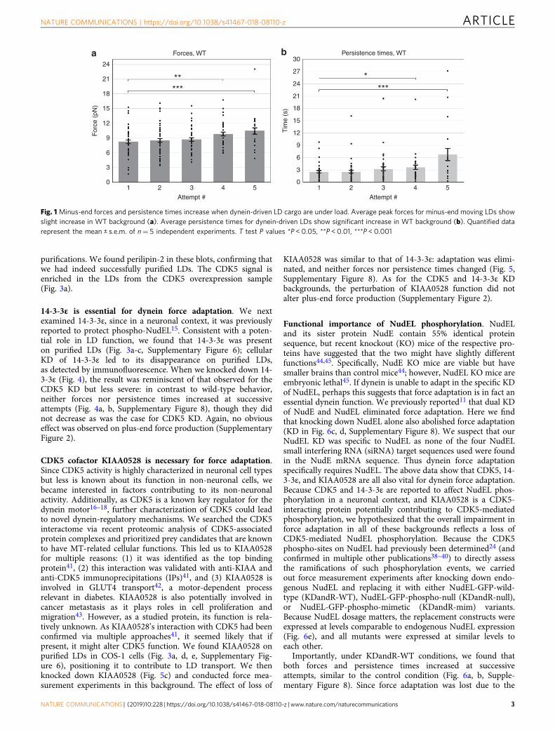
ResultsForce adaptation in wild-type COS-1 cells. We previouslyshowed that, under load, both forces and persistence times (thetime over which the force is maintained) in dynein-driven LDcargos increase at each attempt11; the increase is typically firstapparent in attempt 2 or 3 and easily statistically significant byattempt 4 or 5 (Fig. 1, Supplementary Figure 8). This trend occurswhen measurements are made both in unperturbed wild-typecells and in control cells treated with transfection reagent (Sup-plementary Figure 1).
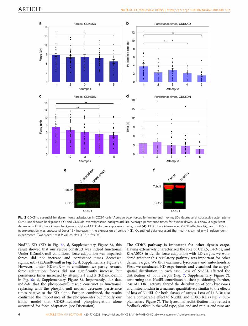
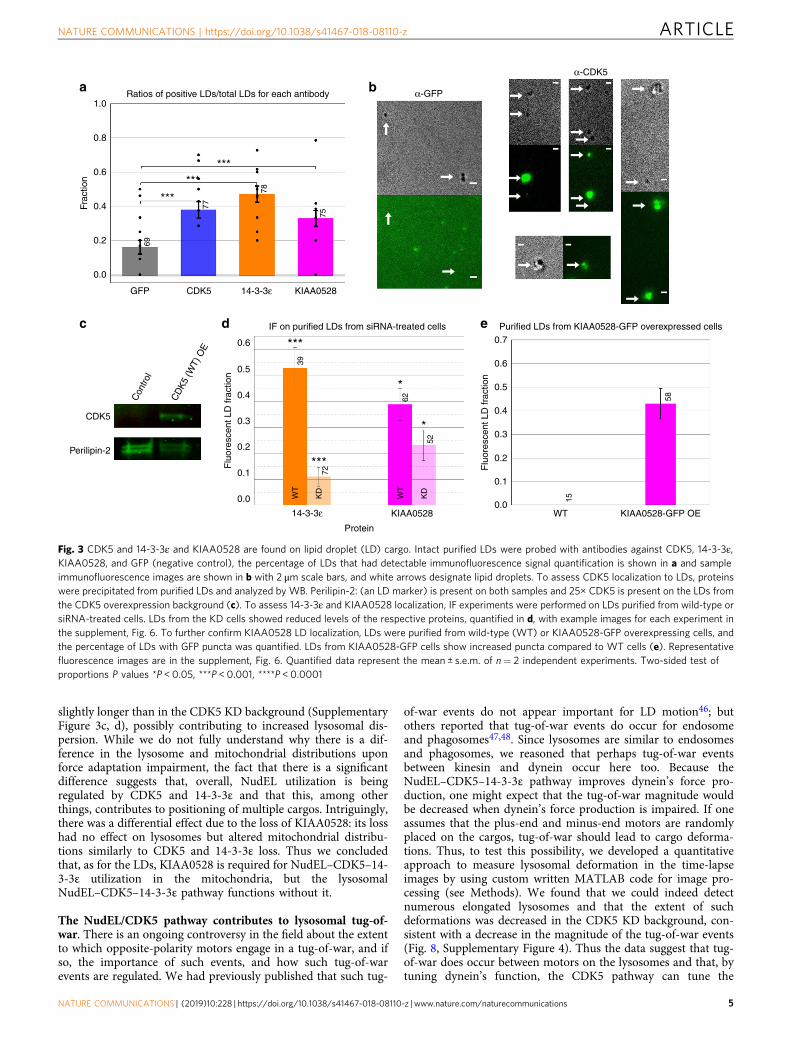
CDK5 is essential for dynein force adaptation. CDK5 phos-phorylation of NudEL has been shown by three separategroups24,38–40. Thus we explored the possibility that CDK5phosphorylation of NudEL is important for dynein force adap-tation. We knocked down endogenous CDK5, and under suchconditions, we found that dynein force production does notincrease at each attempt as for the wild type but instead forcesand persistence times decreased with subsequent attempts(Fig. 2a, b, Supplementary Figure 5, Supplementary Figure 8). Thesame phenotype was observed when we blocked CDK5 activity byoverexpressing a previously characterized dominant-negativeCDK5 construct (CDK5dn)16 (Fig. 2c, d, f, Supplementary Fig-ure 8). Neither CDK5 knockdown (KD) nor CDK5dn over-expression altered plus-end forces (Supplementary Figure 2).Thus inhibiting CDK5’s kinase activity coincides with alterationof dynein force adaptation in cells, where forces and forcedurations decrease rather than increase. After conductingimmunolocalization on purified LDs, we found that CDK5 wasindeed present on the LDs, supporting this CDK5 regulatoryfunction (Fig. 3a–c, Supplementary Figure 6). We confirmedCDK5’s presence on LDs by purifying LDs from control cells andcells overexpressing a wild-type CDK5 construct and conductinga western blot (WB) on the precipitated protein from these LD
ARTICLE NATURE COMMUNICATIONS | https://doi.org/10.1038/s41467-018-08110-z
2 NATURE COMMUNICATIONS | (2019) 10:228 | https://doi.org/10.1038/s41467-018-08110-z | www.nature.com/naturecommunications
purifications. We found perilipin-2 in these blots, confirming thatwe had indeed successfully purified LDs. The CDK5 signal isenriched in the LDs from the CDK5 overexpression sample(Fig. 3a).
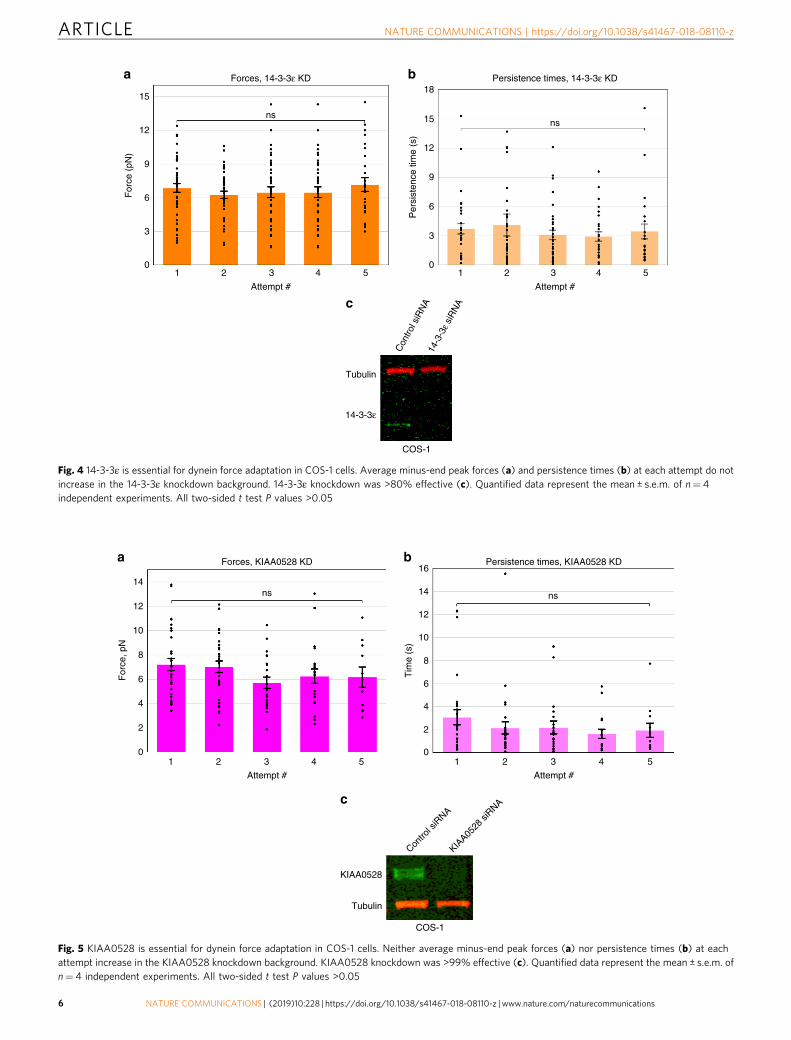
14-3-3ε is essential for dynein force adaptation. We nextexamined 14-3-3ε, since in a neuronal context, it was previouslyreported to protect phospho-NudEL15. Consistent with a poten-tial role in LD function, we found that 14-3-3ε was presenton purified LDs (Fig. 3a-c, Supplementary Figure 6); cellularKD of 14-3-3ε led to its disappearance on purified LDs,as detected by immunofluorescence. When we knocked down 14-3-3ε (Fig. 4), the result was reminiscent of that observed for theCDK5 KD but less severe: in contrast to wild-type behavior,neither forces nor persistence times increased at successiveattempts (Fig. 4a, b, Supplementary Figure 8), though they didnot decrease as was the case for CDK5 KD. Again, no obviouseffect was observed on plus-end force production (SupplementaryFigure 2).
CDK5 cofactor KIAA0528 is necessary for force adaptation.Since CDK5 activity is highly characterized in neuronal cell typesbut less is known about its function in non-neuronal cells, webecame interested in factors contributing to its non-neuronalactivity. Additionally, as CDK5 is a known key regulator for thedynein motor16–18, further characterization of CDK5 could leadto novel dynein-regulatory mechanisms. We searched the CDK5interactome via recent proteomic analysis of CDK5-associatedprotein complexes and prioritized prey candidates that are knownto have MT-related cellular functions. This led us to KIAA0528for multiple reasons: (1) it was identified as the top bindingprotein41, (2) this interaction was validated with anti-KIAA andanti-CDK5 immunoprecipitations (IPs)41, and (3) KIAA0528 isinvolved in GLUT4 transport42, a motor-dependent processrelevant in diabetes. KIAA0528 is also potentially involved incancer metastasis as it plays roles in cell proliferation andmigration43. However, as a studied protein, its function is rela-tively unknown. As KIAA0528’s interaction with CDK5 had beenconfirmed via multiple approaches41, it seemed likely that ifpresent, it might alter CDK5 function. We found KIAA0528 onpurified LDs in COS-1 cells (Fig. 3a, d, e, Supplementary Fig-ure 6), positioning it to contribute to LD transport. We thenknocked down KIAA0528 (Fig. 5c) and conducted force mea-surement experiments in this background. The effect of loss of
KIAA0528 was similar to that of 14-3-3ε: adaptation was elimi-nated, and neither forces nor persistence times changed (Fig. 5,Supplementary Figure 8). As for the CDK5 and 14-3-3ε KDbackgrounds, the perturbation of KIAA0528 function did notalter plus-end force production (Supplementary Figure 2).
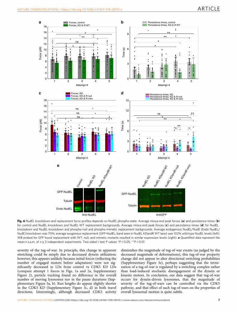
Functional importance of NudEL phosphorylation. NudELand its sister protein NudE contain 55% identical proteinsequence, but recent knockout (KO) mice of the respective pro-teins have suggested that the two might have slightly differentfunctions44,45. Specifically, NudE KO mice are viable but havesmaller brains than control mice44; however, NudEL KO mice areembryonic lethal45. If dynein is unable to adapt in the specific KDof NudEL, perhaps this suggests that force adaptation is in fact anessential dynein function. We previously reported11 that dual KDof NudE and NudEL eliminated force adaptation. Here we findthat knocking down NudEL alone also abolished force adaptation(KD in Fig. 6c, d, Supplementary Figure 8). We suspect that ourNudEL KD was specific to NudEL as none of the four NudELsmall interfering RNA (siRNA) target sequences used were foundin the NudE mRNA sequence. Thus dynein force adaptationspecifically requires NudEL. The above data show that CDK5, 14-3-3ε, and KIAA0528 are all also vital for dynein force adaptation.Because CDK5 and 14-3-3ε are reported to affect NudEL phos-phorylation in a neuronal context, and KIAA0528 is a CDK5-interacting protein potentially contributing to CDK5-mediatedphosphorylation, we hypothesized that the overall impairment inforce adaptation in all of these backgrounds reflects a loss ofCDK5-mediated NudEL phosphorylation. Because the CDK5phospho-sites on NudEL had previously been determined24 (andconfirmed in multiple other publications38–40) to directly assessthe ramifications of such phosphorylation events, we carriedout force measurement experiments after knocking down endo-genous NudEL and replacing it with either NudEL-GFP-wild-type (KDandR-WT), NudEL-GFP-phospho-null (KDandR-null),or NudEL-GFP-phospho-mimetic (KDandR-mim) variants.Because NudEL dosage matters, the replacement constructs wereexpressed at levels comparable to endogenous NudEL expression(Fig. 6e), and all mutants were expressed at similar levels toeach other.
Importantly, under KDandR-WT conditions, we found thatboth forces and persistence times increased at successiveattempts, similar to the control condition (Fig. 6a, b, Supple-mentary Figure 8). Since force adaptation was lost due to the
24
a bForces, WT Persistence times, WT
***** ***
*21
For
ce (
pN)
30
27
24
21
18
15
12
9
6
3
0
Tim
e (s
)
18
15
12
9
6
3
01 2 3 4 5
Attempt #
1 2 3 4 5
Attempt #
Fig. 1Minus-end forces and persistence times increase when dynein-driven LD cargo are under load. Average peak forces for minus-end moving LDs showslight increase in WT background (a). Average persistence times for dynein-driven LDs show significant increase in WT background (b). Quantified datarepresent the mean ± s.e.m. of n= 5 independent experiments. T test P values *P < 0.05, **P < 0.01, ***P < 0.001
NATURE COMMUNICATIONS | https://doi.org/10.1038/s41467-018-08110-z ARTICLE
NATURE COMMUNICATIONS | (2019) 10:228 | https://doi.org/10.1038/s41467-018-08110-z | www.nature.com/naturecommunications 3
NudEL KD (KD in Fig. 6c, d, Supplementary Figure 8), thisresult showed that our rescue construct was indeed functional.Under KDandR-null conditions, force adaptation was impaired:forces did not increase and persistence times decreasedsignificantly (KDandR-null in Fig. 6c, d, Supplementary Figure 8).However, under KDandR-mim conditions, we partly rescuedforce adaptation: forces did not significantly increase, butpersistence times increased by attempts 4 and 5 (KDandR-mimin Fig. 6c, d, Supplementary Figure 8). Importantly, our dataindicate that the phospho-null rescue construct is functional:replacing with the phospho-null mutant decreases persistencetimes relative to the KD alone. Further, combined, the resultsconfirmed the importance of the phospho-sites but modify ourinitial model that CDK5-mediated phosphorylation aloneaccounted for force adaptation (see Discussion).
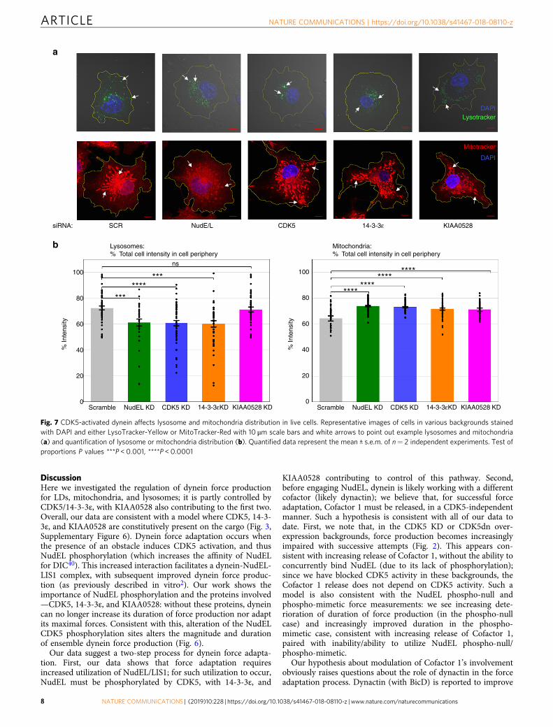
The CDK5 pathway is important for other dynein cargo.Having extensively characterized the role of CDK5, 14-3-3ε, andKIAA0528 in dynein force adaptation with LD cargos, we won-dered whether this regulatory pathway was important for otherdynein cargos. We thus examined lysosomes and mitochondria.First, we conducted KD experiments and visualized the cargos’spatial distribution in each case. Loss of NudEL affected thedistribution of both cargos (Fig. 7, Supplementary Figure 7),confirming that NudEL contributes to their positioning. Further,loss of CDK5 activity altered the distribution of both lysosomesand mitochondria in a manner quantitatively similar to the effectsof loss of NudEL for both classes of cargos. Loss of 14-3-3ε alsohad a comparable effect to NudEL and CDK5 KDs (Fig. 7, Sup-plementary Figure 7). The lysosomal redistribution may reflect afeedback effect: in the wild type, plus-end and minus-end runs are
18
12
10
8
Per
sist
ence
tim
e (s
)T
ime
(s)
For
ce (
pN)
For
ce (
pN)
6
4
2
0
12
14
10
8
6
4
2
0
15
12
9
6
3
0
18
15
12
9
6
3
0
1 2 3
Attempt #
Forces, CDK5KDa b
dc
Persistence times, CDK5KD
Forces, CDK5DN Persistence times, CDK5DN
**
**
****
*
*
*
4 5 1 2 3
Attempt #
4 5
1 2 3
Attempt #
f
COS-1
Tubulin
CDK5
Contro
l
CDK5DN
e
COS-1
Tubulin
Contro
l siR
NA
CDK5 siR
NA
CDK5
4 5 1 2 3
Attempt #
4 5
Fig. 2 CDK5 is essential for dynein force adaptation in COS-1 cells. Average peak forces for minus-end moving LDs decrease at successive attempts inCDK5 knockdown background (a) and CDK5dn overexpression background (c). Average persistence times for dynein-driven LDs show a significantdecrease in CDK5 knockdown background (b) and CDK5dn overexpression background (d). CDK5 knockdown was >90% effective (e), and CDK5dnoverexpression was successful (over 15× increase in the expression of control) (f). Quantified data represent the mean ± s.e.m. of n= 5 independentexperiments. Two-sided t test P values *P < 0.05, **P < 0.01
ARTICLE NATURE COMMUNICATIONS | https://doi.org/10.1038/s41467-018-08110-z
4 NATURE COMMUNICATIONS | (2019) 10:228 | https://doi.org/10.1038/s41467-018-08110-z | www.nature.com/naturecommunications
slightly longer than in the CDK5 KD background (SupplementaryFigure 3c, d), possibly contributing to increased lysosomal dis-persion. While we do not fully understand why there is a dif-ference in the lysosome and mitochondrial distributions uponforce adaptation impairment, the fact that there is a significantdifference suggests that, overall, NudEL utilization is beingregulated by CDK5 and 14-3-3ε and that this, among otherthings, contributes to positioning of multiple cargos. Intriguingly,there was a differential effect due to the loss of KIAA0528: its losshad no effect on lysosomes but altered mitochondrial distribu-tions similarly to CDK5 and 14-3-3ε loss. Thus we concludedthat, as for the LDs, KIAA0528 is required for NudEL–CDK5–14-3-3ε utilization in the mitochondria, but the lysosomalNudEL–CDK5–14-3-3ε pathway functions without it.
The NudEL/CDK5 pathway contributes to lysosomal tug-of-war. There is an ongoing controversy in the field about the extentto which opposite-polarity motors engage in a tug-of-war, and ifso, the importance of such events, and how such tug-of-warevents are regulated. We had previously published that such tug-
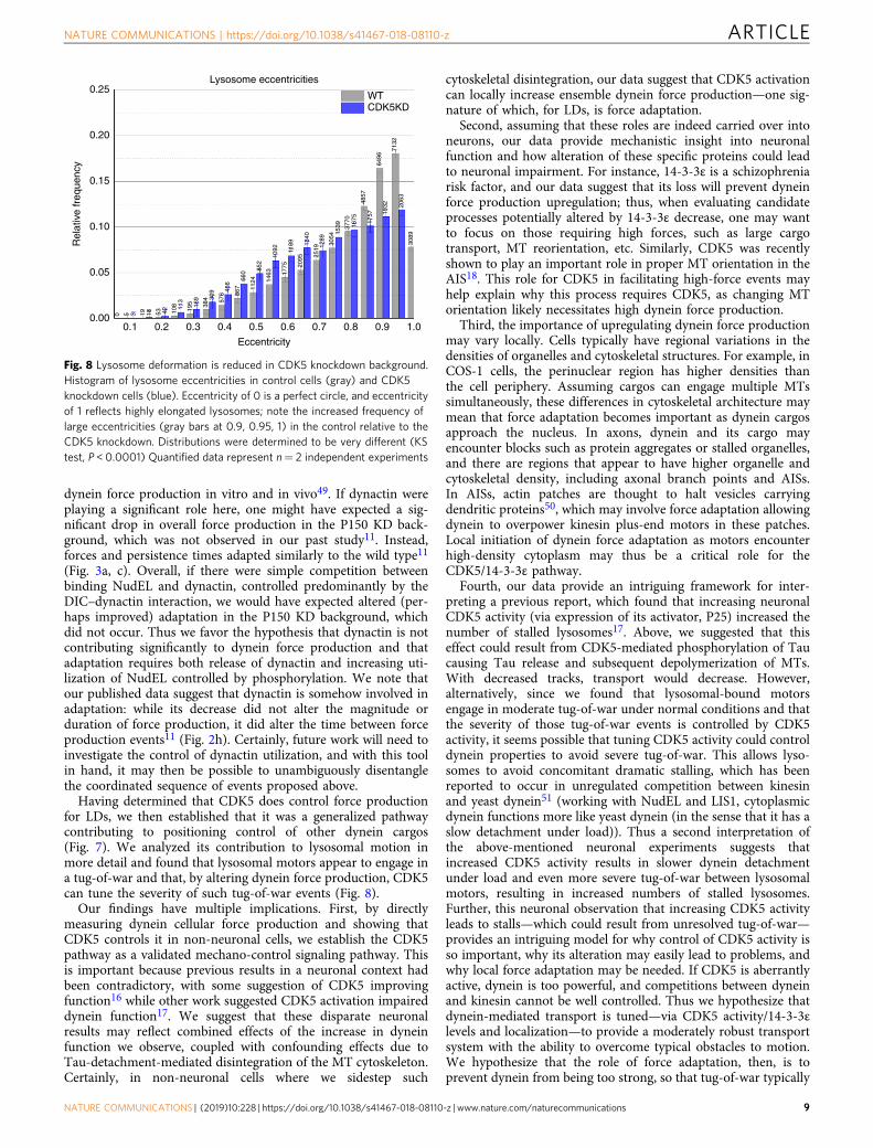
of-war events do not appear important for LD motion46; butothers reported that tug-of-war events do occur for endosomeand phagosomes47,48. Since lysosomes are similar to endosomesand phagosomes, we reasoned that perhaps tug-of-war eventsbetween kinesin and dynein occur here too. Because theNudEL–CDK5–14-3-3ε pathway improves dynein’s force pro-duction, one might expect that the tug-of-war magnitude wouldbe decreased when dynein’s force production is impaired. If oneassumes that the plus-end and minus-end motors are randomlyplaced on the cargos, tug-of-war should lead to cargo deforma-tions. Thus, to test this possibility, we developed a quantitativeapproach to measure lysosomal deformation in the time-lapseimages by using custom written MATLAB code for image pro-cessing (see Methods). We found that we could indeed detectnumerous elongated lysosomes and that the extent of suchdeformations was decreased in the CDK5 KD background, con-sistent with a decrease in the magnitude of the tug-of-war events(Fig. 8, Supplementary Figure 4). Thus the data suggest that tug-of-war does occur between motors on the lysosomes and that, bytuning dynein’s function, the CDK5 pathway can tune the
Ratios of positive LDs/total LDs for each antibodya
c d e
b
Con
trol
CD
K5 (W
T) O
E
CDK5
0.6 ***
***
*
*
0.5
0.4
Flu
ores
cent
LD
frac
tion
Flu
ores
cent
LD
frac
tion
0.3
0.2
0.1
0.0
0.6
0.7
0.5
0.4
0.3
0.2
0.1
0.0
Perilipin-2
39
72
WT
KD
WT
KD
62
52
15
58
14-3-3ε KIAA0528 WT KIAA0528-GFP OE
Protein
IF on purified LDs from siRNA-treated cells Purified LDs from KIAA0528-GFP overexpressed cells
α-GFP
α-CDK5
******
***
Frac
tion
1.0
0.8
0.6
0.4
0.2
0.0
GFP
69
77
78
75
CDK5 14-3-3ε KIAA0528
Fig. 3 CDK5 and 14-3-3ε and KIAA0528 are found on lipid droplet (LD) cargo. Intact purified LDs were probed with antibodies against CDK5, 14-3-3ε,KIAA0528, and GFP (negative control), the percentage of LDs that had detectable immunofluorescence signal quantification is shown in a and sampleimmunofluorescence images are shown in b with 2 μm scale bars, and white arrows designate lipid droplets. To assess CDK5 localization to LDs, proteinswere precipitated from purified LDs and analyzed by WB. Perilipin-2: (an LD marker) is present on both samples and 25× CDK5 is present on the LDs fromthe CDK5 overexpression background (c). To assess 14-3-3ε and KIAA0528 localization, IF experiments were performed on LDs purified from wild-type orsiRNA-treated cells. LDs from the KD cells showed reduced levels of the respective proteins, quantified in d, with example images for each experiment inthe supplement, Fig. 6. To further confirm KIAA0528 LD localization, LDs were purified from wild-type (WT) or KIAA0528-GFP overexpressing cells, andthe percentage of LDs with GFP puncta was quantified. LDs from KIAA0528-GFP cells show increased puncta compared to WT cells (e). Representativefluorescence images are in the supplement, Fig. 6. Quantified data represent the mean ± s.e.m. of n= 2 independent experiments. Two-sided test ofproportions P values *P < 0.05, ***P < 0.001, ****P < 0.0001
NATURE COMMUNICATIONS | https://doi.org/10.1038/s41467-018-08110-z ARTICLE
NATURE COMMUNICATIONS | (2019) 10:228 | https://doi.org/10.1038/s41467-018-08110-z | www.nature.com/naturecommunications 5
15
a b
c
12
9
For
ce (
pN)
6
3
0
15
18
12
9
Per
sist
ence
tim
e (s
)
6
3
01 2 3
Attempt #
4
Tubulin
COS-1
Con
trol s
iRN
A14
-3-3
ε siR
NA
14-3-3ε
5 1 2 3
Attempt #
Forces, 14-3-3ε KD
nsns
Persistence times, 14-3-3ε KD
4 5
Fig. 4 14-3-3ε is essential for dynein force adaptation in COS-1 cells. Average minus-end peak forces (a) and persistence times (b) at each attempt do notincrease in the 14-3-3ε knockdown background. 14-3-3ε knockdown was >80% effective (c). Quantified data represent the mean ± s.e.m. of n= 4independent experiments. All two-sided t test P values >0.05
14
a b
c
16
14
12
10
8
6
4
2
0
12
10
For
ce, p
N
Tim
e (s
)
8
6
4
2
01 2 3
Attempt # Attempt #
Forces, KIAA0528 KD
ns ns
Persistence times, KIAA0528 KD
4 5 1 2 3 4 5
KIAA0528
Tubulin
COS-1
Contro
l siR
NA
KIAA05
28 si
RNA
Fig. 5 KIAA0528 is essential for dynein force adaptation in COS-1 cells. Neither average minus-end peak forces (a) nor persistence times (b) at eachattempt increase in the KIAA0528 knockdown background. KIAA0528 knockdown was >99% effective (c). Quantified data represent the mean ± s.e.m. ofn= 4 independent experiments. All two-sided t test P values >0.05
ARTICLE NATURE COMMUNICATIONS | https://doi.org/10.1038/s41467-018-08110-z
6 NATURE COMMUNICATIONS | (2019) 10:228 | https://doi.org/10.1038/s41467-018-08110-z | www.nature.com/naturecommunications
severity of the tug-of-war. In principle, this change in apparentstretching could be simply due to decreased dynein utilization;however; this appears unlikely because initial forces (reflecting thenumber of engaged motors before adaptation) were not sig-nificantly decreased in LDs from control vs CDK5 KD LDs(compare attempt 1 forces in Figs. 1a and 2a, SupplementaryFigure 2), particle tracking found no difference in the overallnumber of moving lysosomes nor in the pause durations (Sup-plementary Figure 3a, b). Run lengths do appear slightly shorterin the CDK5 KD (Supplementary Figure 3c, d) in both traveldirections. Interestingly, although decreased CDK5 activity
diminishes the magnitude of tug-of-war events (as judged by thedecreased magnitude of deformations), this tug-of-war propertychange did not appear to alter directional switching probabilities(Supplementary Figure 3e), perhaps suggesting that the termi-nation of a tug-of-war is regulated by a switching complex ratherthan load-induced stochastic disengagement of the dynein orkinesin motors. In conclusion, our data suggest that tug-of-waroccurs for dynein-driven lysosomes, that the magnitude ofseverity of the tug-of-wars can be controlled via the CDK5pathway, and that effect of such tug-of-wars on the properties ofoverall lysosomal motion is quite subtle.
18
a b
c
e
d
***
****
*** *
*
****
***
Forces, control
Forces, KD & R nullForces, KD & R mim
Forces, KD & R WT
Forces, KDPersistence times, KD & R nullPersistence times, KD & R mim
Persistence times, KD
Persistence times, controlPersistence times, KD & R WT
16
14ns
ns
nsns
ns
12
10
9
6
3
0
For
ce (
pN)
Tim
e (s
)
8
6
4
2
0
18 15
12
9
6
3
0
16
14
12
10
For
ce (
pN)
Tim
e (s
)
8
6
4
2
0
1 2 3 4
Attempt #
5
1
Con
trol
Nud
EL K
D
Nud
EL K
Dan
dR W
T
Nud
EL K
Dan
dR W
TN
udEL
KD
andR
nul
lN
udEL
KD
andR
mim
Nud
EL K
D
2 3 4Attempt #
GFP-NudEL
Tubulin
GFP-NudEL
TubulinEndo NudEL
Anti-NudEL AntiGFP
5 1 2 3 4Attempt #
5
1 2 3 4
Attempt #
5
Fig. 6 NudEL knockdown and replacement force profiles depends on NudEL phospho-state. Average minus-end peak forces (a) and persistence times (b)for control and NudEL knockdown and NudEL-WT replacement backgrounds. Average minus-end peak forces (c) and persistence times (d) for NudELknockdown and NudEL knockdown and phospho-null and phospho-mimetic replacement backgrounds. Average endogenous NudEL/NudE (Endo NudEL/NudE) knockdown was 70%; average exogenous replacement (GFP-NudEL, band seen in NudEL KDandRWT lane) was 102% wild-type NudEL levels (left).WB probed for GFP found replacement with WT, null, and mimetic mutants resulted in similar expression levels (right). e Quantified data represent themean ± s.e.m. of n≥ 3 independent experiments. Two-sided t test P values *P < 0.05, **P < 0.01
NATURE COMMUNICATIONS | https://doi.org/10.1038/s41467-018-08110-z ARTICLE
NATURE COMMUNICATIONS | (2019) 10:228 | https://doi.org/10.1038/s41467-018-08110-z | www.nature.com/naturecommunications 7
DiscussionHere we investigated the regulation of dynein force productionfor LDs, mitochondria, and lysosomes; it is partly controlled byCDK5/14-3-3ε, with KIAA0528 also contributing to the first two.Overall, our data are consistent with a model where CDK5, 14-3-3ε, and KIAA0528 are constitutively present on the cargo (Fig. 3,Supplementary Figure 6). Dynein force adaptation occurs whenthe presence of an obstacle induces CDK5 activation, and thusNudEL phosphorylation (which increases the affinity of NudELfor DIC40). This increased interaction facilitates a dynein-NudEL-LIS1 complex, with subsequent improved dynein force produc-tion (as previously described in vitro2). Our work shows theimportance of NudEL phosphorylation and the proteins involved—CDK5, 14-3-3ε, and KIAA0528: without these proteins, dyneincan no longer increase its duration of force production nor adaptits maximal forces. Consistent with this, alteration of the NudELCDK5 phosphorylation sites alters the magnitude and durationof ensemble dynein force production (Fig. 6).
Our data suggest a two-step process for dynein force adapta-tion. First, our data shows that force adaptation requiresincreased utilization of NudEL/LIS1; for such utilization to occur,NudEL must be phosphorylated by CDK5, with 14-3-3ε, and
KIAA0528 contributing to control of this pathway. Second,before engaging NudEL, dynein is likely working with a differentcofactor (likely dynactin); we believe that, for successful forceadaptation, Cofactor 1 must be released, in a CDK5-independentmanner. Such a hypothesis is consistent with all of our data todate. First, we note that, in the CDK5 KD or CDK5dn over-expression backgrounds, force production becomes increasinglyimpaired with successive attempts (Fig. 2). This appears con-sistent with increasing release of Cofactor 1, without the ability toconcurrently bind NudEL (due to its lack of phosphorylation);since we have blocked CDK5 activity in these backgrounds, theCofactor 1 release does not depend on CDK5 activity. Such amodel is also consistent with the NudEL phospho-null andphospho-mimetic force measurements: we see increasing dete-rioration of duration of force production (in the phospho-nullcase) and increasingly improved duration in the phospho-mimetic case, consistent with increasing release of Cofactor 1,paired with inability/ability to utilize NudEL phospho-null/phospho-mimetic.
Our hypothesis about modulation of Cofactor 1’s involvementobviously raises questions about the role of dynactin in the forceadaptation process. Dynactin (with BicD) is reported to improve
100
siRNA:
a
b
SCR NudE/L CDK5 14-3-3ε KIAA0528
Mitotracker
DAPI
DAPILysotracker
ns
*******
********
*******
****
Lysosomes:% Total cell intensity in cell periphery
Mitochondria:% Total cell intensity in cell periphery
80
% In
tens
ity 60
40
20
0
100
80%
Inte
nsity 60
40
20
0Scramble NudEL KD CDK5 KD 14-3-3εKD KIAA0528 KD Scramble NudEL KD CDK5 KD 14-3-3εKD KIAA0528 KD
Fig. 7 CDK5-activated dynein affects lysosome and mitochondria distribution in live cells. Representative images of cells in various backgrounds stainedwith DAPI and either LysoTracker-Yellow or MitoTracker-Red with 10 μm scale bars and white arrows to point out example lysosomes and mitochondria(a) and quantification of lysosome or mitochondria distribution (b). Quantified data represent the mean ± s.e.m. of n= 2 independent experiments. Test ofproportions P values ***P < 0.001, ****P < 0.0001
ARTICLE NATURE COMMUNICATIONS | https://doi.org/10.1038/s41467-018-08110-z
8 NATURE COMMUNICATIONS | (2019) 10:228 | https://doi.org/10.1038/s41467-018-08110-z | www.nature.com/naturecommunications
dynein force production in vitro and in vivo49. If dynactin wereplaying a significant role here, one might have expected a sig-nificant drop in overall force production in the P150 KD back-ground, which was not observed in our past study11. Instead,forces and persistence times adapted similarly to the wild type11
(Fig. 3a, c). Overall, if there were simple competition betweenbinding NudEL and dynactin, controlled predominantly by theDIC–dynactin interaction, we would have expected altered (per-haps improved) adaptation in the P150 KD background, whichdid not occur. Thus we favor the hypothesis that dynactin is notcontributing significantly to dynein force production and thatadaptation requires both release of dynactin and increasing uti-lization of NudEL controlled by phosphorylation. We note thatour published data suggest that dynactin is somehow involved inadaptation: while its decrease did not alter the magnitude orduration of force production, it did alter the time between forceproduction events11 (Fig. 2h). Certainly, future work will need toinvestigate the control of dynactin utilization, and with this toolin hand, it may then be possible to unambiguously disentanglethe coordinated sequence of events proposed above.
Having determined that CDK5 does control force productionfor LDs, we then established that it was a generalized pathwaycontributing to positioning control of other dynein cargos(Fig. 7). We analyzed its contribution to lysosomal motion inmore detail and found that lysosomal motors appear to engage ina tug-of-war and that, by altering dynein force production, CDK5can tune the severity of such tug-of-war events (Fig. 8).
Our findings have multiple implications. First, by directlymeasuring dynein cellular force production and showing thatCDK5 controls it in non-neuronal cells, we establish the CDK5pathway as a validated mechano-control signaling pathway. Thisis important because previous results in a neuronal context hadbeen contradictory, with some suggestion of CDK5 improvingfunction16 while other work suggested CDK5 activation impaireddynein function17. We suggest that these disparate neuronalresults may reflect combined effects of the increase in dyneinfunction we observe, coupled with confounding effects due toTau-detachment-mediated disintegration of the MT cytoskeleton.Certainly, in non-neuronal cells where we sidestep such
cytoskeletal disintegration, our data suggest that CDK5 activationcan locally increase ensemble dynein force production—one sig-nature of which, for LDs, is force adaptation.
Second, assuming that these roles are indeed carried over intoneurons, our data provide mechanistic insight into neuronalfunction and how alteration of these specific proteins could leadto neuronal impairment. For instance, 14-3-3ε is a schizophreniarisk factor, and our data suggest that its loss will prevent dyneinforce production upregulation; thus, when evaluating candidateprocesses potentially altered by 14-3-3ε decrease, one may wantto focus on those requiring high forces, such as large cargotransport, MT reorientation, etc. Similarly, CDK5 was recentlyshown to play an important role in proper MT orientation in theAIS18. This role for CDK5 in facilitating high-force events mayhelp explain why this process requires CDK5, as changing MTorientation likely necessitates high dynein force production.
Third, the importance of upregulating dynein force productionmay vary locally. Cells typically have regional variations in thedensities of organelles and cytoskeletal structures. For example, inCOS-1 cells, the perinuclear region has higher densities thanthe cell periphery. Assuming cargos can engage multiple MTssimultaneously, these differences in cytoskeletal architecture maymean that force adaptation becomes important as dynein cargosapproach the nucleus. In axons, dynein and its cargo mayencounter blocks such as protein aggregates or stalled organelles,and there are regions that appear to have higher organelle andcytoskeletal density, including axonal branch points and AISs.In AISs, actin patches are thought to halt vesicles carryingdendritic proteins50, which may involve force adaptation allowingdynein to overpower kinesin plus-end motors in these patches.Local initiation of dynein force adaptation as motors encounterhigh-density cytoplasm may thus be a critical role for theCDK5/14-3-3ε pathway.
Fourth, our data provide an intriguing framework for inter-preting a previous report, which found that increasing neuronalCDK5 activity (via expression of its activator, P25) increased thenumber of stalled lysosomes17. Above, we suggested that thiseffect could result from CDK5-mediated phosphorylation of Taucausing Tau release and subsequent depolymerization of MTs.With decreased tracks, transport would decrease. However,alternatively, since we found that lysosomal-bound motorsengage in moderate tug-of-war under normal conditions and thatthe severity of those tug-of-war events is controlled by CDK5activity, it seems possible that tuning CDK5 activity could controldynein properties to avoid severe tug-of-war. This allows lyso-somes to avoid concomitant dramatic stalling, which has beenreported to occur in unregulated competition between kinesinand yeast dynein51 (working with NudEL and LIS1, cytoplasmicdynein functions more like yeast dynein (in the sense that it has aslow detachment under load)). Thus a second interpretation ofthe above-mentioned neuronal experiments suggests thatincreased CDK5 activity results in slower dynein detachmentunder load and even more severe tug-of-war between lysosomalmotors, resulting in increased numbers of stalled lysosomes.Further, this neuronal observation that increasing CDK5 activityleads to stalls—which could result from unresolved tug-of-war—provides an intriguing model for why control of CDK5 activity isso important, why its alteration may easily lead to problems, andwhy local force adaptation may be needed. If CDK5 is aberrantlyactive, dynein is too powerful, and competitions between dyneinand kinesin cannot be well controlled. Thus we hypothesize thatdynein-mediated transport is tuned—via CDK5 activity/14-3-3εlevels and localization—to provide a moderately robust transportsystem with the ability to overcome typical obstacles to motion.We hypothesize that the role of force adaptation, then, is toprevent dynein from being too strong, so that tug-of-war typically
0.25WTCDK5KD
0.20
0.15
Rel
ativ
e fr
eque
ncy
0.10
0.05
0.000.1
0 5 3 19 18 53 42 108 11
3
195 16
9
384 30
9
576
456
867
660
1124 14
63 1775
1092 11
89 1340
2519 12
89
3054
1539 37
7016
75 1757 19
32 2063
6496
7132
3089
4857
2095
852
0.2 0.3 0.4 0.5
Eccentricity
Lysosome eccentricities
0.6 0.7 0.8 0.9 1.0
Fig. 8 Lysosome deformation is reduced in CDK5 knockdown background.Histogram of lysosome eccentricities in control cells (gray) and CDK5knockdown cells (blue). Eccentricity of 0 is a perfect circle, and eccentricityof 1 reflects highly elongated lysosomes; note the increased frequency oflarge eccentricities (gray bars at 0.9, 0.95, 1) in the control relative to theCDK5 knockdown. Distributions were determined to be very different (KStest, P < 0.0001) Quantified data represent n= 2 independent experiments
NATURE COMMUNICATIONS | https://doi.org/10.1038/s41467-018-08110-z ARTICLE
NATURE COMMUNICATIONS | (2019) 10:228 | https://doi.org/10.1038/s41467-018-08110-z | www.nature.com/naturecommunications 9
resolves without aggravated traffic jams; it is only after repeatedstalls (indicating a significant barrier) that maximum adaptationengages, subsequently allowing for increased dynein performance.
Fifth, for bi-directionally moving cargos, there is typically tightcoupling between plus-end and minus-end motor activity:52,53
when one upregulates or downregulates one set of motors, there isusually feedback (though unknown mechanisms) resulting inconcomitant changes in the opposite direction. Interestingly,CDK5 modulation of dynein force production appears to pre-dominantly bypass such feedback: while the slight change inminus-end travel distances in the lysosomes is matched by plus-end changes (Supplementary Figure 3c, d), there is no suchmatching of effects on force production. Because the underlyingmechanism contributing to force feedback matching oppositedirections is unknown, we do not understand why such feedbackfails here. However, this decoupling may be functionally usefulallowing for selective directional control. In the wild-type forceadaptation, minus-end forces and durations increase with attemptnumber, but plus-end ones stay constant. In the CDK5 KD/dominant-negative overexpression backgrounds, minus-end for-ces and durations decrease, but plus-end forces and durationsremain unchanged (Supplementary Figure 2). Thus, at least forthe cargos/cells considered here, the CDK5 pathway appears toalmost entirely affect only minus-end motor activity and allowsfor selective directional force production control.
Finally, our findings may have implications for diabetes. LDbiology is linked to insulin resistance54, and their transport directlyaffects metabolism36, so alteration in their trafficking by changes inforce adaptation could potentially impact metabolic disease. Insulinsignaling can regulate dynein to influence lysosome motility inboth neurons and non-neuronal cells55. This involves inhibition ofglycogen synthase kinase (GSK)-3β, a kinase whose dysregulationis linked to both metabolic and neurological disorders56. GSK-3βphosphorylation of dynein reduces its interaction with NudEL55.CDK5 can inhibit GSK-3β, but to our knowledge the opposite hasnot yet been reported57. Nonetheless, it will be interesting todetermine whether GSK-3β modulates force adaptation, and if so,whether this occurs through altered NudEL phosphorylation byCDK5. Dynein is also required for internalization of adipocyteGLUT4 receptors in low insulin conditions58. Dynein forceadaptation could also modulate vesicle internalization. If that is thecase, alterations in force adaptation mechanisms could lead todefective glucose transport. Interestingly, KIAA0528 may beinvolved in GLUT4 insertion into the plasma membrane, althoughthis is not yet completely understood42,59.
In summary, we found that NudEL phosphorylation control bythe CDK5/14-3-3ε pathway directly regulates dynein force pro-duction in cells. It is required not only for the dynamic upregu-lation of dynein force underlying the previously discovered LDforce adaptation but also plays a general role for other cargossuch as lysosomes and mitochondria, contributing to control oftheir subcellular localization. Further, the differential effects ofKIAA0528 (affecting LDs and mitochondria but not lysosomes)suggest cargo-specificity could in part be controlled by cargo-specific CDK5-interacting regulatory proteins, like KIAA0528.Importantly, at least for lysosomes, the CDK5 pathway is able totune dynein’s force production capability and ultimately mod-ulate motor tug-of-war severity; this likely seems relevant forvarious cellular cargos. Future work will undoubtedly exploreramifications of control of tug-of-war severity, the role and reg-ulation of dynactin in this process, and how cargo-specific pro-teins like KIAA0528 mechanistically contribute to regulation.
MethodsCell culture and transfections. COS-1 cells (ATCC CRL-1650) were grown inDulbecco’s modified Eagle’s medium (Genesee) supplemented with 10% fetal
bovine serum and 1% Pen–Strep at 37 °C in 5% CO2. Gene expression KD forCDK5, 14-3-3ε, KIAA0528, NudE, and NudEL were completed by transienttransfection using commercially available siRNAs. siRNA for the control (sc-37007), CDK5 (sc-29263), 14-3-3ε (sc-29588), and KIAA0528 (sc-95830) siRNAswere obtained from Santa Cruz Biotechnology. NudE (SI00655858, SI4374356,SI4341386, and SI05147835) and NudEL (SI03246600, SI03246936, SI04264379,and SI04321191) siRNAs were obtained from Qiagen.
For the CDK5, 14-3-3ε, KIAA0528, NudE, and NudEL KDs, HiPerfect reagent(Qiagen) was used following the manufacturer’s instructions. CDK5 andKIAA0528 KDs were achieved by using a final concentration of 33 nM siRNA;14-3-3ε KD was achieved by using a final concentration of 66 nM siRNA; andNudE and NudEL KDs were achieved by using a final concentration of 5 nM ofeach siRNA. (Control transfections used the same final concentration of scrambledsiRNA as the target siRNA.)
For the CDK5dn overexpression and NudEL replacement, Lipofectamine-2000 (ThermoFisher Scientific) was used following the manufacturer’s instructions.Two micrograms of DNA was used for the overexpression and replacementexperiments.
Transient KDs were incubated for 48 h before force measurements, WB, IP, andtracking experiments were carried out. Transient overexpression or replacementexperiments were incubated for 24 h before force measurements, WB, and trackingexperiments were carried out. NudEL replacement expression was induced with20 nM doxycycline 18 h before force measurements, WB, fluorescence, andtracking experiments were carried out.
Expression vectors. The CMV-myc-CDK5dn, pEGFP-C1-NudEL(WT), andphospho-null mutant (1-5A) expression vectors were previously described16.
Mutagenesis and cloning. The NudEL-WT plasmid was mutagenized into aphospho-mimetic mutant using site-directed mutagenesis via Multi-Site-DirectedMutagenesis QuikChange Lightning (Agilent) in two rounds of mutagenesis. In thefirst round, F1–F3 primers were used; in the second round, P4–P5 primers wereused (Supplementary Table 1). CDK5 phospho-sites on NudEL 1-5 were replacedwith aspartic acid.
The three NudEL plasmids (WT, 1-5A, and 1-5D) were further mutagenized toachieve RNAi resistance. Two silent mutations were introduced in the middle ofeach of the four siRNA target sequences of the NudEL mRNA sequence (for a totalof eight mutations/construct). The NudEL plasmids were mutagenized using site-directed mutagenesis via Multi-Site-Directed Mutagenesis QuikChange Lightning(Agilent) in two rounds of mutagenesis. F1–F3 primers (Supplementary Table 2)were used in the first round; and then F4–F6 primers (Supplementary Table 2)were used in the second round.
The three RNAi-resistant NudEL constructs were then subcloned into theexpression pDONOR201 vector via Gateway Technology (Invitrogen)(Supplementary Table 3). These were then recombined into the gateway-compatible doxycycline-inducible destination vector and contained a C-terminal-S-FLAG-SBP (SFB) tag60.
Immunoblotting. Cells were washed with phosphate-buffered saline (PBS) andlysed using an ice-cold 1% NP-40 buffer containing a 1× protease inhibitor cocktail(Roche) and the supernatant was collected. Proteins from the lysate were denaturedusing an sodium dodecyl sulfate buffer and incubating at 70 °C for 10 min. Theproteins were then separated in a 4–12% Bis-tris gel (Life Technologies) andtransferred to a nitrocellulose membrane through a wet transfer method. Thenitrocellulose was blocked with 5% non-fat milk in Tris buffered saline (TBS:blocking buffer) for 45 min at room temperature (RT). Immunoblotting wascompleted with respective antibodies and blots were visualized with infrareddetection on the Odyssey (Licor). Primary antibodies were diluted in blockingbuffer with Tween20 at various concentrations (1:200–1:1000 v/v). Secondaryantibodies were diluted in blocking buffer with Tween20 at 1:10,000 v/v. Anti-bodies were purchased from: Cell Signaling Technologies 2506s (CDK5) and 9635s(14-3-3ε), Novus NB110-40878 (Perilipin-2), Bethyl A301-469A-M (KIAA0528),ABClonal A5776 (NudEL), Bioss bs-5522R (pSer231NudEL), Developmental Stu-dies Hybridoma Bank E7-s (Tubulin-β), Life Technologies (goat anti-mouse IgG680), and Li-cor (goat anti-rabbit IgG 800). Data shown here are representativeof 2–3 separate experiments.
Force measurements in cells. Force measurements on moving LDs in COS1 cellsis as described in refs. 11,61. LD positions in the laser trap were measured with highresolution using position sensitive detector (PSD) and cross-verified with analysisof differential interference contrast images using template matching or auto-correlation of LD intensity profile. Automated piezostage was used to position thetrap at the center of linearly moving LDs toward and away from the cell center.Detachments of LDs from MT within 200 nm from the center of the laser trap werecounted as escape attempts. Typically both failed and successful escape attemptscould be observed in the video as LDs that failed to escape would fall back to thetrap center with high velocity. However, only high-resolution PSD data (2 kHz)was used to quantify parameters of the escape attempts (position traces carried veryhigh slope due to rapid fall back to trap center) with better accuracy. During force
ARTICLE NATURE COMMUNICATIONS | https://doi.org/10.1038/s41467-018-08110-z
10 NATURE COMMUNICATIONS | (2019) 10:228 | https://doi.org/10.1038/s41467-018-08110-z | www.nature.com/naturecommunications
measurements, double trapping of LDs was quite common, and care was taken toanalyze only those LDs whose motion was uninterrupted by other organelles. Theideal region of the cell for measurements is halfway between periphery and nucleuswith an additional condition that there is a linear inward and outward flow oforganelle traffic to rule out the ambiguity in the direction of MTs. Note that successrate of trapping linearly moving LDs that last for 5 unperturbed attempts in the cellis very low and typically we scored a maximum of 6–8 clean LD tracks in 1 h.Please note that errors in escaped fractions in each attempt (f) were estimated with
ffiffiffiffiffiffiffiffiffiffiffiffiffiffiffiffiffiffiffiffiffiffiffiffi½f ð1� f Þ=n�p
for n droplets that made the escape attempt.
Lysosome distribution and particle tracking. After 48 h of incubation withtransfection reagents, COS-1 cells were stained with NucBlue Live ReadyProbesReagent (Life Technologies) and either 200 nM LysoTracker-Yellow or 200 nMMitoTracker-Red (Thermo Fisher Scientific) and incubated for 30 min beforevisualization. Live COS-1 cells were visualized at the University of California IrvineOptical Biology Core on the Zeiss LSM700 confocal laser scanning microscope. Atleast 30y cells were imaged. The cell perimeter, nuclear perimeter, and respectiveintensities were measured for each fluorescent image using ImageJ. The perinuclearregion was defined as 20% of the cytoplasm surrounding the nucleus. The cellperiphery was defined at the other 80% of the cytoplasm. The intensity of the cellperiphery was defined as the total cell intensity minus the intensity of the peri-nuclear region. The percentage of total cell intensity in the cell periphery wasdefined as: (the intensity of the cell periphery/total cell intensity) × 100 (previouslydescribed62).
LD immunofluorescence. LDs were purified from WT and siRNA-treated COS-1 cells using a sucrose gradient as previously described11. Probing for the presenceof proteins on the surface of LDs is carried out as described below. A samplechamber constructed with 0.17 × 40 × 22 mm3 clean coverslips, double-sidedadhesive tape, and microscope cover glass to hold about 20 μl of solution. Cov-erslips were cleaned with KOH solution followed by ddH2O and ethanol inultrasonic bath. About 25 μl of as-purified LDs in buffer were flown into thechamber and incubated for 10 min at RT. LD surface was blocked with 10% bovineserum albumin (BSA) in PBS for 20 min, followed by incubation with primaryantibodies to a final concentration of 2 μg/ml in PBS with 10% BSA and 0.1%Triton X. Unattached primary antibody (1:200 diluted) was washed by flushing thechamber with 30 μl of PBS with 10% BSA twice before adding secondary antibody.Primary antibodies used were: CDK5 (Cell Signaling 2506 S), KIAA0528 (BethylA301-468A), 14-3-3ε (Invitrogen 5H10L5), and green fluorescent protein (GFP;Cell Signaling 2912 S). Fluorescence imaging was carried out after 1-h incubationwith secondary antibody (0.02 mg/ml, Alexa Fluor 488-labeled anti-Rabbit) in theimaging buffer (80 mM PIPES, 1 mM MgCl2, 2 mM EGTA, 1 mg/ml casein, and1 mM dithiothreitol). An EMCCD camera (quantEM 512, Photometrics) and asingle-mode, 488 nm Ti-Sapphire excitation laser in semi-TIRF configuration wereused. Fluorescence signal integration time was fixed at 0.5 s.
Eccentricity and run distances of lysosomes. Cells were incubated with 200 nMof Lysotracker red DND-99 (Life Technologies) for 30 min in the medium used fortissue culture. A sample chamber similar to the one adopted for force measure-ments was used to image the cells. Images were acquired in semi-TIRF mode at 10fps using 568 nm excitation laser and the quantEM 512 camera. Spatial resolutionof the acquired images in the above set-up is 60 nm/pixel.
Eccentricity of lysosomes in each movie frame was determined using customMatlab code for image processing. One hundred consecutive images for each cell(18 cells in each condition from two experimental replicates) were used for thisanalysis, because the deformations could be dynamic, and we wanted to catch themif they occurred; the distributions shown thus reflect instantaneous multipledeterminations of each lysosome’s eccentricity. If a specific lysosome was visible inall 100 consecutive frames, and it were circular in the first 50 frames, and verystretched in the next 50, it would have yielded 50 eccentricity measurements thatwere 0, and 50 that were 1, all included in the histogram. Binary images weregenerated from the raw images, and the eccentricity and areas of individuallysosomes were measured (after appropriate filtering to reduce the background).Eccentricity values range from 0 to 1, with 0 corresponding to a perfect circle and 1representing highly deformed objects. Only lysosomes with areas in the range of8–200 square pixels were considered.
Particle tracking was carried out by custom softwares LVcorr and Marathon.LVcorr was used to generate long tracks (track length ~100 s and 90 tracks from 18cells in each condition) from the videos recorded above. Using tracks files fromLVcorr as input, the parsing software Marathon was used to extract the pauses andrun distances of lysosomes moving linearly in kinesin and dynein directions.
Immunoprecipitation. Cells were lysed and NudEL was immunoprecipitated usingthe Pierce Crosslink Magnetic IP/co-IP Kit (Thermo Scientific 88805) following themanufacturer’s instructions. Ten micrograms of NudEL antibody (abcam ab25959)was used for each IP. Elutions were then run on a WB probing for NudEL andpSer231NudEL.
Statistics. All graphs are mean and s.e.m. Statistical significance was determinedby Student’s t test, test of proportions, Kolmogorov–Smirnov test, and Wilcoxonsign-ranked t test. *P < 0.05, **P < 0.01, ***P < 0.001, ****P < 0.0001.
Code availability. LVcorr and Marathon are freely available on GrossLab website.Lysosomal eccentricity estimation code is available upon request by email to cor-responding author.
Data availabilityData supporting the findings of this manuscript are available from the corre-sponding author upon reasonable request.
Received: 8 February 2018 Accepted: 18 December 2018
References1. Bhabha, G., Johnson, G. T., Schroeder, C. M. & Vale, R. D. How dynein moves
along microtubules. Trends Biochem. Sci. 41, 94–105 (2016).2. McKenney, R. J., Vershinin, M., Kunwar, A., Vallee, R. B. & Gross, S. P. LIS1
and NudE induce a persistent dynein force-producing state. Cell 141, 304–314(2010).
3. Roberts, A. J., Kon, T., Knight, P. J., Sutoh, K. & Burgess, S. A. Functions andmechanics of dynein motor proteins. Nat. Rev. Mol. Cell Biol. 14, 713–726(2013).
4. Tripathy, S. K. et al. Autoregulatory mechanism for dynactin control ofprocessive and diffusive dynein transport. Nat. Cell Biol. 16, 1192–1201(2014).
5. Morgan, J. L., Song, Y. & Barbar, E. Structural dynamics and multiregioninteractions in dynein-dynactin recognition. J. Biol. Chem. 286, 39349–39359(2011).
6. McKenney, R. J., Weil, S. J., Scherer, J. & Vallee, R. B. Mutually exclusivecytoplasmic dynein regulation by NudE-Lis1 and dynactin. J. Biol. Chem. 286,39615–39622 (2011).
7. Nyarko, A., Song, Y. & Barbar, E. Intrinsic disorder in dynein intermediatechain modulates its interactions with NudE and dynactin. J. Biol. Chem. 287,24884–24893 (2012).
8. Barbar, E. Native disorder mediates binding of dynein to NudE and Dynactin.Biochem. Soc. Trans. 40, 1009–1013 (2012).
9. McKenney, R. J. et al. Activation of cytoplasmic dynein motility by dynactin-cargo adapter complexes. Science 345, 337–341 (2014).
10. Cardoso, C. et al. Refinement of a 400-kb critical region allows genotypicdifferentiation between isolated lissencephaly, Miller-Dieker syndrome, andother phenotypes secondary to deletions of 17p13. 3. Am. J. Hum. Genet. 72,918–930 (2003).
11. Reddy, B. J. N. et al. Load-induced enhancement of Dynein force productionby LIS1–NudE in vivo and in vitro. Nat. Commun. 7, 12259 (2016).
12. Tsai, J., Bremner, K. H. & Vallee, R. B. Dual subcellular roles for LIS1 anddynein in radial neuronal migration in live brain tissue. Nat. Neurosci. 10,970–979 (2007).
13. Gutierrez, P. A., Ackermann, B. E., Vershinin, M. & McKenney, R. J.Differential effects of the dynein-regulatory factor Lissencephaly-1 onprocessive dynein-dynactin motility. J. Biol. Chem. 292, 12245–12255 (2017).
14. Lu, M. & Prehoda, K. A NudE/14-3-3 pathway coordinates dynein and thekinesin khc73 to position the mitotic spindle. Dev. Cell 26, 369–380 (2013).
15. Wynshaw-Boris, A. Lissencephaly and LIS1: insights into the molecularmechanisms of neuronal migration and development. Clin. Genet. 72,296–304 (2007).
16. Pandey, JaiP. & Smith, D. S. A CDK5-dependent switch regulates Lis1/Ndel1/dynein-driven organelle transport in adult axons. J. Neurosci. 31, 17207–17219(2011).
17. Klinman, E. & Holzbaur, E. L. F. Stress-induced CDK5 activation disruptsaxonal transport via Lis1/Ndel1/Dynein. Cell Rep. 12, 462–473 (2015).
18. Klinman, E., Tokito, M. & Holzbaur, E. L. F. CDK5-dependent activationof dynein in the axon initial segment regulates polarized cargo transport inneurons. Traffic 18, 808–824 (2017).
19. Flaherty, D. B., Soria, J. P., Tomasiewicz, H. G. & Wood, J. G. Phosphorylationof human tau protein by and cdk5 are key participants. J. Neurosci. Res. 472,463–472 (2000).
20. Hamdane, M. et al. Mitotic-like tau phosphorylation by p25-Cdk5 kinasecomplex. J. Biol. Chem. 278, 34026–34034 (2003).
21. Noble, W. et al. Cdk5 is a key factor in tau aggregation and tangle formationin vivo. Neuron 38, 555–565 (2003).
22. Kimura, T., Ishiguro, K. & Hisanaga, S. Physiological and pathologicalphosphorylation of tau by. Front. Mol. Neurosci. 7, 1–10 (2014).
NATURE COMMUNICATIONS | https://doi.org/10.1038/s41467-018-08110-z ARTICLE
NATURE COMMUNICATIONS | (2019) 10:228 | https://doi.org/10.1038/s41467-018-08110-z | www.nature.com/naturecommunications 11
23. Castro-Alvarez, J. F., Uribe-arias, S. A. & Mejía-raigosa, D. Cyclin-dependentkinase 5, a node protein in diminished tauopathy: a systems biology approach.Front. Aging Neurosci. 6, 1–13 (2014).
24. Niethammer, M. et al. NUDEL is a novel Cdk5 substrate that associates withLIS1 and cytoplasmic dynein. Neuron 28, 697–711 (2000).
25. Arif, A. Extraneuronal activities and regulatory mechanisms of the atypicalcyclin-dependent kinase Cdk5. Biochem. Pharmacol. 84, 985–993 (2012).
26. Toyo-oka, K. et al. 14-3-3epsilon is important for neuronal migration bybinding to NUDEL: a molecular explanation for Miller-Dieker syndrome.Nat. Genet. 34, 274–285 (2003).
27. Middleton, Fa, Peng, L., Lewis, D. A., Levitt, P. & Mirnics, K. Alteredexpression of 14-3-3 genes in the prefrontal cortex of subjects withschizophrenia. Neuropsychopharmacology 30, 974–983 (2005).
28. Spalice, A. et al. Neuronal migration disorders: clinical, neuroradiologic andgenetics aspects. Acta Paediatr., Int. J. Paediatr. 98, 421–433 (2009).
29. Kawamoto, Y. et al. 14-3-3 proteins in Lewy bodies in Parkinson disease anddiffuse Lewy body disease brains. J. Neuropathol. Exp. Neurol. 61, 245–253(2002).
30. Zeng, C. P. et al. Increased identification of novel variants in type 2 diabetes,birth weight and their pleiotropic loci. J. Diabetes 9, 898–907 (2017).
31. Banks, A. S. et al. An Erk/Cdk5 axis controls the diabetogenic actions ofPPARγ. Nature 517, 391–395 (2015).
32. Zheng, Y. L. et al. Cdk5 inhibitory peptide (CIP) inhibits Cdk5/p25 activityinduced by high glucose in pancreatic beta cells and recovers insulin secretionfrom p25 damage. PLoS ONE 8, 3–10 (2013).
33. Ahmed, D. & Sharma, M. Cyclin-dependent kinase 5/p35/p39: a novel andimminent therapeutic target for diabetes mellitus. Int. J. Endocrinol. 2011,530274 (2011).
34. Lalioti, V. et al. The atypical kinase Cdk5 is activated by insulin, regulates theassociation between GLUT4 and E-Syt1, and modulates glucose transport in3T3-L1 adipocytes. Proc. Natl Acad. Sci. 106, 4249–4253 (2009).
35. Pozo, K. & Bibb, J. A. The emerging role of CDK5 in cancer. Trends Cancer 2,606–618 (2016).
36. Herms, A. et al. AMPK activation promotes lipid droplet dispersion ondetyrosinated microtubules to increase mitochondrial fatty acid oxidation.Nat. Commun. 6, 7176 (2015).
37. Billingsley, M. L. & Kincaid, R. L. Regulated phosphorylation anddephosphorylation of tau protein: effects on microtubule interaction,intracellular trafficking and neurodegeneration. Biochem. J. 591, 577–591(1997).
38. Sasaki, S. et al. A LIS1/NUDEL/cytoplasmic dynein heavy chain complexin the developing and adult nervous system. Neuron 28, 681–696 (2000).
39. Ko, J. et al. p35 and p39 are essential for cyclin-dependent kinase 5 functionduring neurodevelopment. J. Neurosci. 21, 6758–6771 (2001).
40. Hebbar, S. et al. Lis1 and Ndel1 influence the timing of nuclear envelopebreakdown in neural stem cells. J. Cell Biol. 182, 1063–1071 (2008).
41. Xu, S. et al. Proteomic analysis of the human cyclin-dependent kinase familyreveals a novel CDK5 complex involved in cell growth and migration. Mol.Cell. Proteomics 13, 2986–3000 (2014).
42. Xie, X. et al. C2 domain-containing phosphoprotein CDP138 regulatesGLUT4 insertion into the plasma membrane. Cell Metab. 14, 378–389 (2011).
43. Lu, Y. et al. CDP138 silencing inhibits TGF-β/Smad signaling to impairradioresistance and metastasis via GDF15 in lung cancer. Cell Death Dis. 8,e3036 (2017).
44. Stehman, S. A., Chen, Y., Mckenney, R. J. & Vallee, R. B. NudE and NudEL arerequired for mitotic progression and are involved in dynein recruitment tokinetochores. J. Cell Biol. 178, 583–594 (2007).
45. Sasaki, S. et al. Complete loss of Ndel1 results in neuronal migration defectsand early embryonic lethality. Mol. Cell. Biol. 25, 7812–7827 (2005).
46. Kunwar, A. et al. Mechanical stochastic tug-of-war models cannot explainbidirectional lipid-droplet transport. Proc. Natl Acad. Sci. 108, 18960–18965(2011).
47. Soppina, V., Rai, A. K., Ramaiya, A. J., Barak, P. & Mallik, R. Tug-of-warbetween dissimilar teams of microtubule motors regulates transport andfission of endosomes. Proc. Natl Acad. Sci. 106, 19381–19386 (2009).
48. Hendricks, A. G. et al. Motor coordination via a tug-of-war mechanism drivesbidirectional vesicle transport. Curr. Biol. 20, 697–702 (2010).
49. Belyy, V. et al. The mammalian dynein/dynactin complex is a strongopponent to kinesin in a tug-of-war competition. Nat. Cell Biol. 18, 1018–1024(2016).
50. Watanabe, K. et al. Networks of polarized actin filaments in the axon initialsegment provide a mechanism for sorting axonal and dendritic proteins.Cell Rep. 2, 1546–1553 (2012).
51. Derr, N. D. et al. Tug-of-war in motor protein ensembles revealed with aprogrammable DNA origami scaffold. Science 338, 662–666 (2012).
52. Gross, S. P. et al. Interactions and regulation of molecular motors in Xenopusmelanophores. J. Cell. Biol. 156, 855–865 (2002).
53. Shubeita, G. T. et al. Consequences of motor copy number on the intracellulartransport of kinesin-1-driven lipid droplets. Cell 135, 1098–1107 (2008).
54. Samuel, V. T. & Shulman, G. I. Mechanisms for insulin resistance: commonthreads and missing links. Cell 148, 852–871 (2012).
55. Gao, F. J. et al. GSK-3β phosphorylation of cytoplasmic dynein reduces Ndel1binding to intermediate chains and alters dynein motility. Traffic 16, 941–961(2015).
56. Saraswati, A. P., Ali Hussaini, S. M., Krishna, N. H., Babu, B. N. & Kamal, A.Glycogen synthase kinase-3 and its inhibitors: potential target for varioustherapeutic conditions. Eur. J. Med. Chem. 144, 843–858 (2017).
57. Engmann, O. Crosstalk between Cdk5 and GSK3β: implications forAlzheimer’s disease. Front. Mol. Neurosci. 2, 1–5 (2009).
58. Huang, J., Imamura, T. & Olefsky, J. M. Insulin can regulate GLUT4internalization by signaling to Rab5 and the motor protein dynein. Proc. NatlAcad. Sci. USA 98, 13084–13089 (2001).
59. Sadacca, L. A., Bruno, J., Wen, J., Xiong, W. & McGraw, T. E. Specializedsorting of GLUT4 and its recruitment to the cell surface are independentlyregulated by distinct Rabs. Mol. Biol. Cell 24, 2544–2557 (2013).
60. Wang, W. et al. PTPN14 is required for the density-dependent control ofYAP1. Genes Dev. 26, 1959–1971 (2012).
61. Jun, Y., Tripathy, S. K., Narayanareddy, B. R. J., Mattson-Hoss, M. K. & Gross,S. P. Calibration of optical tweezers for in vivo force measurements: how dodifferent approaches compare? Biophys. J. 107, 1474–1484 (2014).
62. Li, X. et al. A molecular mechanism to regulate lysosome motility for lysosomepositioning and tubulation. Nat. Cell Biol. 18, 404–417 (2016).
AcknowledgementsWe thank Adeela Syed at the UCI Optical Biology Core for her help and advice withthe lysosome and mitochondria distribution microscopy. Grant MacGregor graciouslyallowed use of Odyssey (Licor) for western blot visualization. D.E.C. was the recipientof the GAANN Fellowship (US Department of Education). Funding was provided byNIH R01 GM123068.
Author contributionsS.P.G. conceived the project; S.P.G., D.E.C., B.J.N.R., and W.W. designed the experi-ments; D.E.C., B.J.N.R., M.J.B., B.H., and H.H. performed experiments; D.E.C., B.J.N.R.,Z.M.A.-S., and S.J.S.C. analyzed data; B.J.N.R. developed software tools for video pro-cessing; S.P.G., D.E.C., B.J.N.R., and D.S.S. wrote the manuscript; and all authorsapproved the final version.
Additional informationSupplementary Information accompanies this paper at https://doi.org/10.1038/s41467-018-08110-z.
Competing interests: The authors declare no competing interests.
Reprints and permission information is available online at http://npg.nature.com/reprintsandpermissions/
Journal peer review information: Nature Communications thanks the anonymousreviewers for their contributions to the peer review of this work. Peer Reviewer Reportsare available.
Publisher’s note: Springer Nature remains neutral with regard to jurisdictional claims inpublished maps and institutional affiliations.
Open Access This article is licensed under a Creative CommonsAttribution 4.0 International License, which permits use, sharing,
adaptation, distribution and reproduction in any medium or format, as long as you giveappropriate credit to the original author(s) and the source, provide a link to the CreativeCommons license, and indicate if changes were made. The images or other third partymaterial in this article are included in the article’s Creative Commons license, unlessindicated otherwise in a credit line to the material. If material is not included in thearticle’s Creative Commons license and your intended use is not permitted by statutoryregulation or exceeds the permitted use, you will need to obtain permission directly fromthe copyright holder. To view a copy of this license, visit http://creativecommons.org/licenses/by/4.0/.
© The Author(s) 2019
ARTICLE NATURE COMMUNICATIONS | https://doi.org/10.1038/s41467-018-08110-z
12 NATURE COMMUNICATIONS | (2019) 10:228 | https://doi.org/10.1038/s41467-018-08110-z | www.nature.com/naturecommunications