Real-time in vivo Imaging of LPS-induced Local ...
9
Copyright © 2020 The Authors; exclusive licensee Bio-protocol LLC. 1 www.bio-protocol.org/e3724 Bio-protocol 10(16): e3724. DOI:10.21769/BioProtoc.3724 Real-time in vivo Imaging of LPS-induced Local Inflammation and Drug Deposition in NF-κB Reporter Mice Artur Schmidtchen 1, 2, 3 and Manoj Puthia 1, * 1 Division of Dermatology and Venereology, Department of Clinical Sciences, Lund University, SE-22184 Lund, Sweden; 2 Copenhagen Wound Healing Center, Bispebjerg Hospital, Department of Biomedical Sciences, University of Copenhagen, DK-2400, Denmark; 3 Dermatology, Skåne University Hospital, SE- 22185 Lund, Sweden *For correspondence: [email protected] [Abstract] Wound, biomaterial, and surgical infections are all characterized by a localized and excessive inflammation, motivating the development of in vivo methods focused on the analysis of local immune events. However, current inflammation models, such as the commonly used in vivo models of endotoxin-induced inflammation are based on systemic, usually intraperitoneal, administration of lipopolysaccharide (LPS), causing endotoxin shock. Here, we describe a model of LPS-induced local inflammation in NF-κB-RE-Luc reporter mice. LPS, alone or with added therapeutic substances, is delivered locally via a hydrogel which is deposited subcutaneously, providing a spatially defined environment, enabling in vivo bioimaging analyses of local NF-κB activation. Evaluation of drug efficacy can be analyzed longitudinally in the same mouse, and using fluorescently labeled drugs, local drug deposition can be simultaneously analyzed, and correlated to the site of inflammation. Finally, the protocol can also be used to study retention and systemic release of the drug from locally deposited gels and other biomaterials. Keywords: Inflammation, NF-κB, In vivo, Mouse model, Bioimaging, Therapy [Background] An excessive TLR response causes localized and sometimes disproportionate inflammation, as observed in different types of wound and biomaterial infections. These wound complications delay proper healing and increase the risk of severe infections and sepsis. Considering the latter, several experimental models of sepsis and endotoxin shock have been developed which study the development of systemic inflammation (Lewis et al., 2016). There is however a need for models that address localized inflammatory events from a mechanistic and therapeutic perspective. Activation of the transcription factor NF-κB is a key component of various inflammatory conditions, and hence, NF-κB is considered an important therapeutic target (Liu et al., 2017). Real-time, longitudinal in vivo imaging of NF-κB activation in NFκB-RE-Luc reporter mice is an important tool in studies on inflammatory disease and efficacy of drug treatments. NFκB-RE-Luc reporter mice carry a transgene containing NFκB- responsive elements from the CMVα promoter placed upstream of a basal SV40 promoter, and a modified firefly luciferase cDNA (Carlsen et al., 2002). This reporter element can be induced by LPS and TNF-α (Carlsen et al., 2002), and provides an excellent in vivo tool to monitor transcriptional responses of NF-κB. To achieve visualization of gene expression, luciferin, a substrate for luciferase, is Please cite this article as: Schmidtchen and Puthia, (2020). Real-time in vivo Imaging of LPS-induced Local Inflammation and Drug Deposition in NF-κB Reporter Mice,Bio-protocol 10 (16): e3724. DOI: 10.21769/BioProtoc.3724.
Transcript of Real-time in vivo Imaging of LPS-induced Local ...
Copyright © 2020 The Authors; exclusive licensee Bio-protocol LLC. 1
www.bio-protocol.org/e3724 Bio-protocol 10(16): e3724. DOI:10.21769/BioProtoc.3724
Real-time in vivo Imaging of LPS-induced Local Inflammation and Drug Deposition in NF-κB Reporter Mice
Artur Schmidtchen1, 2, 3 and Manoj Puthia1, *
1Division of Dermatology and Venereology, Department of Clinical Sciences, Lund University, SE-22184
Lund, Sweden; 2Copenhagen Wound Healing Center, Bispebjerg Hospital, Department of Biomedical
Sciences, University of Copenhagen, DK-2400, Denmark; 3Dermatology, Skåne University Hospital, SE-
22185 Lund, Sweden
*For correspondence: [email protected]
[Abstract] Wound, biomaterial, and surgical infections are all characterized by a localized and
excessive inflammation, motivating the development of in vivo methods focused on the analysis of local
immune events. However, current inflammation models, such as the commonly used in vivo models of
endotoxin-induced inflammation are based on systemic, usually intraperitoneal, administration of
lipopolysaccharide (LPS), causing endotoxin shock. Here, we describe a model of LPS-induced local
inflammation in NF-κB-RE-Luc reporter mice. LPS, alone or with added therapeutic substances, is
delivered locally via a hydrogel which is deposited subcutaneously, providing a spatially defined
environment, enabling in vivo bioimaging analyses of local NF-κB activation. Evaluation of drug efficacy
can be analyzed longitudinally in the same mouse, and using fluorescently labeled drugs, local drug
deposition can be simultaneously analyzed, and correlated to the site of inflammation. Finally, the
protocol can also be used to study retention and systemic release of the drug from locally deposited
gels and other biomaterials.
Keywords: Inflammation, NF-κB, In vivo, Mouse model, Bioimaging, Therapy
[Background] An excessive TLR response causes localized and sometimes disproportionate
inflammation, as observed in different types of wound and biomaterial infections. These wound
complications delay proper healing and increase the risk of severe infections and sepsis. Considering
the latter, several experimental models of sepsis and endotoxin shock have been developed which study
the development of systemic inflammation (Lewis et al., 2016). There is however a need for models that
address localized inflammatory events from a mechanistic and therapeutic perspective. Activation of the
transcription factor NF-κB is a key component of various inflammatory conditions, and hence, NF-κB is
considered an important therapeutic target (Liu et al., 2017). Real-time, longitudinal in vivo imaging of
NF-κB activation in NFκB-RE-Luc reporter mice is an important tool in studies on inflammatory disease
and efficacy of drug treatments. NFκB-RE-Luc reporter mice carry a transgene containing NFκB-
responsive elements from the CMVα promoter placed upstream of a basal SV40 promoter, and a
modified firefly luciferase cDNA (Carlsen et al., 2002). This reporter element can be induced by LPS and
TNF-α (Carlsen et al., 2002), and provides an excellent in vivo tool to monitor transcriptional responses
of NF-κB. To achieve visualization of gene expression, luciferin, a substrate for luciferase, is
Please cite this article as: Schmidtchen and Puthia, (2020). Real-time in vivo Imaging of LPS-induced Local Inflammation and Drug Deposition in NF-κB Reporter Mice,Bio-protocol 10 (16): e3724. DOI: 10.21769/BioProtoc.3724.

Copyright © 2020 The Authors; exclusive licensee Bio-protocol LLC. 2
www.bio-protocol.org/e3724 Bio-protocol 10(16): e3724. DOI:10.21769/BioProtoc.3724
administrated intraperitoneally to mice which in turn generate luminescent signals. Thereafter, in vivo
imaging using IVIS spectrum is used for acquisition and analysis of the recorded signals.
Here, we describe a model of LPS-induced local inflammation in NFκB-RE-Luc reporter mice. LPS,
mixed in a hydroxyethyl cellulose hydrogel, is injected subcutaneously on the back of mouse.
Subcutaneous deposition of hydrogel provides a defined and controlled environment for studies of local
NF-κB activation. In addition to LPS, therapeutic agents can also be included in the same hydrogel and
their efficacy can be analyzed longitudinally in the same mouse. Moreover, drug deposition and release
can also be imaged by using fluorescently labeled drugs. To evaluate local anti-inflammatory efficacy of
a peptide drug and for imaging its local distribution, this in vivo imaging protocol has successfully been
used by us (Puthia et al., 2020).
Materials and Reagents
1. 5 ml polypropylene tube
2. 50 ml tube
3. Syringes, 1 ml (Soft-Ject, Henke-Sass Wolf, catalog number: 5010-200V0)
4. Needle 23 G × 1’’ (BD, catalog number: 300800)
5. Syringe filter, 0.2 μm (Filtropur S 0.2, Sarstedt, catalog number: 83.1826.001)
6. Alcohol wipes (Cutisoft wipes, BSN Medical, catalog number: 204364)
7. BALB/c tg(NFκB-RE-Luc)-Xen reporter mice (Taconic Biosciences, catalog number: 10499) (8-
12 weeks old; male or female)
Note: BALB/c tg(NFκB-RE-Luc)-Xen reporter mice are used for inflammation imaging. If the
purpose is to image drug deposition only, other laboratory mouse strains can be used.
8. LPS from Escherichia coli O111:B4 (Sigma-Aldrich, catalog number: L3024)
9. TCP-25 (GKYGFYTHVFRLKKWIQKVIDQFGE)
As a drug, we used a thrombin-derived peptide TCP-25. For in vivo imaging, TCP-25 was mixed
with 2% (w/w) Cy5 labelled TCP-25 (Biopeptide, San Diego, CA, USA).
10. Hydroxyethyl cellulose (HEC, Natrosol 250 HX, MW 1,000,000; Ashland Industries Europe
GmbH, catalog number: 431292)
11. XenoLight D-luciferin, Potassium salt (PerkinElmer, catalog number: 122799)
12. DPBS, without calcium and magnesium (Thermo Fischer Scientific, catalog number: 14190250)
13. Endotoxin free water (Sigma, catalog number: TMS-011-A)
14. Isoflurane (Forane, Baxter, catalog number: CA2L9100)
15. 1.5% HEC hydrogel (see Recipes)
16. D-luciferin solution (see Recipes)
17. LPS solution (see Recipes)
Please cite this article as: Schmidtchen and Puthia, (2020). Real-time in vivo Imaging of LPS-induced Local Inflammation and Drug Deposition in NF-κB Reporter Mice,Bio-protocol 10 (16): e3724. DOI: 10.21769/BioProtoc.3724.

Copyright © 2020 The Authors; exclusive licensee Bio-protocol LLC. 3
www.bio-protocol.org/e3724 Bio-protocol 10(16): e3724. DOI:10.21769/BioProtoc.3724
Equipment
1. Magnetic stirrer
2. IVIS spectrum (PerkinElmer, model: catalog number: 124262)
3. Ultrasonic bath (Elma Schmidbauer GmbH, model: Elmasonic S30H)
4. Cordless hair clipper (Aesculap, catalog number: GT416)
5. Magnetic stirrer (Fisher Scientific, catalog number: 11715704)
Software
1. Living Image 4.5.5 Software (PerkinElmer)
2. Prism, version 8.3.0 (GraphPad Software, LLC.)
Procedure
A. Preparation of hydrogel-LPS mixture
1. To make hydrogel containing LPS, add 5 μg LPS (1 mg/ml solution) to 95 μl of 1.5% HEC gel.
Vortex the mixture vigorously for 5 min and centrifuge for 3 min (2,575 x g, room temperature)
to remove air bubbles. A range of 5-50 μg LPS can be used to obtain different grades of
inflammation.
Note: Gel LPS mixture should be prepared prior to shaving the mouse dorsum (i.e., before
Procedure C).
2. Slowly, take 200 μl of LPS gel mixture in a 1 ml syringe. Avoid taking in air bubbles. Attach a
23-gauge needle to the syringe and adjust the gel volume to 100 μl by slowly removing excess
gel from the syringe. Move to Procedure C.
Note: Hydrogel is quite thick, do not aspirate using needle as it will be difficult and will produce
air bubbles.
B. Preparation of hydrogel-Cy5-labeled drug mixture
1. In a 5 ml polypropylene tube, add the required amount of fluorescently labeled drug to the gel
and vortex vigorously for 5-10 min. Centrifuge for 3 min (2,575 x g, room temperature) to remove
air bubbles. In our study, for in vivo imaging, TCP-25 mixed with 2% (w/w) Cy5 labeled TCP-25
was added to the gel. We used a final concentration of 0.1% TCP-25 in the gel. To study
combined effects, drug/drugs can be added along with LPS in the same hydrogel.
Note: The hydrogel mixture should be prepared prior to shaving of mouse dorsum (i.e., before
Procedure C).
2. Slowly, take 200 μl of drug gel mixture in a 1 ml syringe. Avoid taking in air bubbles. Attach a
23-gauge needle to the syringe and adjust gel volume to 100 μl by slowly removing excess gel
from the syringe. Move to Procedure C.
Please cite this article as: Schmidtchen and Puthia, (2020). Real-time in vivo Imaging of LPS-induced Local Inflammation and Drug Deposition in NF-κB Reporter Mice,Bio-protocol 10 (16): e3724. DOI: 10.21769/BioProtoc.3724.

Copyright © 2020 The Authors; exclusive licensee Bio-protocol LLC. 4
www.bio-protocol.org/e3724 Bio-protocol 10(16): e3724. DOI:10.21769/BioProtoc.3724
Note: Hydrogel is quite thick, do not aspirate using needle as it will be difficult and will produce
air bubbles.
C. Preparation of mice for subcutaneous injection
1. After receiving BALB/c tg(NFκB-RE-Luc)-Xen reporter mice from supplier, keep them in
quarantine for at least one week (or depending on animal facility’s rules). A brief description of
mouse preparation for subcutaneous gel deposition and imaging is illustrated in Figure 1.
Figure 1. Preparation for subcutaneous gel deposition and IVIS imaging. A. Shaving of hair
from dorsum of mice with a cordless clipper. B. Subcutaneous deposition of hydrogel. C.
Intraperitoneal injection of the substrate D-luciferin. D. IVIS imaging.
2. Anesthetize mice with isoflurane-mixed oxygen (4% isoflurane for induction and 1.5-2% for
maintenance). This can be achieved in an IVIS induction chamber or in any other isoflurane-
oxygen delivery system. Shave hair from the dorsum (slightly below interscapular region) of
mice with a cordless clipper. Depilation can also be achieved with depilatory creams. If
depilatory creams are used, it should be performed at least 2 days prior to the subcutaneous
injection.
Note: Shaving or depilation is necessary as mouse hair can interfere with both luminescence
and fluorescence imaging.
3. Wipe and clean the dorsum with alcohol wipe. Move immediately to next step.
D. Subcutaneous deposition of hydrogel
1. Manually restrain the anesthetized mice using thumb and forefinger, lift and fold the skin from
interscapular area. Insert the needle under the skin and move forward at least 1 cm. Slowly,
Please cite this article as: Schmidtchen and Puthia, (2020). Real-time in vivo Imaging of LPS-induced Local Inflammation and Drug Deposition in NF-κB Reporter Mice,Bio-protocol 10 (16): e3724. DOI: 10.21769/BioProtoc.3724.

Copyright © 2020 The Authors; exclusive licensee Bio-protocol LLC. 5
www.bio-protocol.org/e3724 Bio-protocol 10(16): e3724. DOI:10.21769/BioProtoc.3724
inject 100 μl of LPS gel mixture, withdraw the needle and put your finger at the site of insertion
to prevent any leakage from the site of deposition.
Note: As mouse skin is quite loose, ensure that you inject gel in the midline otherwise gel maybe
placed to one side. For standard subcutaneous injection procedure please see Shimizu (2004).
2. Immediately after subcutaneous injection, transfer the mice to their respective cages.
E. IVIS imaging for NF-κB activity (luminescence imaging mode)
1. Fifteen min prior to IVIS imaging, administer D-luciferin solution intraperitoneally into the mice
(150 mg/kg body weight). Put the mice back to the cages.
Note: We image mice 3, 6 and 24 h post subcutaneous deposition of LPS gel.
2. Anesthetize mice in an induction chamber with 4% isoflurane-mixed oxygen. Complete
anesthesia takes approximately 3-5 min.
Note: Read IVIS spectrum user manual before using IVIS.
3. Once the mice are anesthetized, start the isoflurane supply (2%) for the IVIS imaging chamber.
Immediately, transfer mice to the imaging chamber.
Note: Turn on IVIS and start Living Image software at least 15 min prior to the imaging and
initialize it. Initialization process cools down the camera to -90 °C. Also ensure that stage heating
(37 °C) is working.
4. Place mice in prone position (backside up) on the stage. Position nose in nose cone for
anesthesia.
Note: Maximum five mice can be imaged at one time. If imaging more than one mouse at a time,
ensure that there is minimum lag time between D-luciferin injections. There are other good
sources for learning standard IVIS imaging in luminescence imaging mode (Cosette et al., 2016).
5. For imaging, set parameters in IVIS acquisition control panel. Select luminescent (imaging
mode), auto (exposure), and D (field of view for 5 mice), or C (field of view for 3 mice).
6. Start imaging by clicking ‘acquire’ in IVIS acquisition control panel.
7. In case of weak signal, auto exposure can be changed to desired time with a maximum of 5 min
of exposure. Field of view can be changed according to number of mice to be imaged at one
time or size of the area of interest.
F. IVIS imaging for drug deposition (fluorescence imaging mode)
Note: Perform fluorescence imaging of the drug immediately after the luminescence imaging. If
purpose of the experiment is only to visualize drug deposition in fluorescence imaging mode, there
is no need to inject D-luciferin substrate.
1. In IVIS acquisition control panel, set imaging mode to fluorescence, exposure time to auto,
excitation (650 nm) and emission (670 nm) for Cy5.
2. Start imaging by clicking ‘acquire’ in IVIS acquisition control panel.
Note: For fluorescent imaging, IVIS spectrum is capable of using wavelength range from 415-
850 nm. For in vivo and deep tissue imaging, it is important to note that wavelengths greater
Please cite this article as: Schmidtchen and Puthia, (2020). Real-time in vivo Imaging of LPS-induced Local Inflammation and Drug Deposition in NF-κB Reporter Mice,Bio-protocol 10 (16): e3724. DOI: 10.21769/BioProtoc.3724.
Copyright © 2020 The Authors; exclusive licensee Bio-protocol LLC. 6
www.bio-protocol.org/e3724 Bio-protocol 10(16): e3724. DOI:10.21769/BioProtoc.3724
than 600 nm are preferred. Animal tissue absorbs significant amounts of light below
wavelengths 600 nm. We have successfully used fluorescent labels such as Cy5, Alexa Fluor
660, Alexa Fluor 680, Alexa Fluor 750 and Cy7.
Data analysis
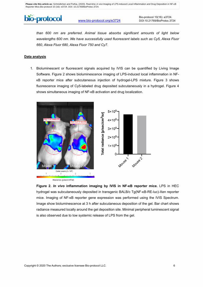
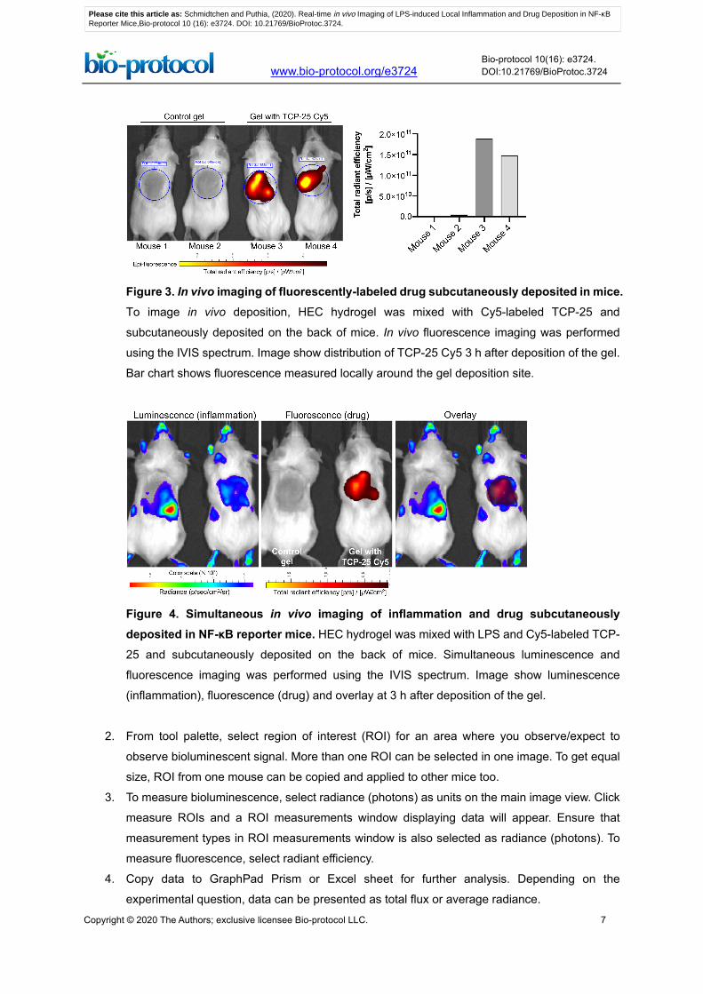
1. Bioluminescent or fluorescent signals acquired by IVIS can be quantified by Living Image
Software. Figure 2 shows bioluminescence imaging of LPS-induced local inflammation in NF-
κB reporter mice after subcutaneous injection of hydrogel-LPS mixture. Figure 3 shows
fluorescence imaging of Cy5-labeled drug deposited subcutaneously in a hydrogel. Figure 4
shows simultaneous imaging of NF-κB activation and drug localization.
Figure 2. In vivo inflammation imaging by IVIS in NF-κB reporter mice. LPS in HEC
hydrogel was subcutaneously deposited in transgenic BALB/c Tg(NF-κB-RE-luc)-Xen reporter
mice. Imaging of NF-κB reporter gene expression was performed using the IVIS Spectrum.
Image show bioluminescence at 3 h after subcutaneous deposition of the gel. Bar chart shows
radiance measured locally around the gel deposition site. Minimal peripheral luminescent signal
is also observed due to low systemic release of LPS from the gel.
Please cite this article as: Schmidtchen and Puthia, (2020). Real-time in vivo Imaging of LPS-induced Local Inflammation and Drug Deposition in NF-κB Reporter Mice,Bio-protocol 10 (16): e3724. DOI: 10.21769/BioProtoc.3724.
Copyright © 2020 The Authors; exclusive licensee Bio-protocol LLC. 7
www.bio-protocol.org/e3724 Bio-protocol 10(16): e3724. DOI:10.21769/BioProtoc.3724
Figure 3. In vivo imaging of fluorescently-labeled drug subcutaneously deposited in mice. To image in vivo deposition, HEC hydrogel was mixed with Cy5-labeled TCP-25 and
subcutaneously deposited on the back of mice. In vivo fluorescence imaging was performed
using the IVIS spectrum. Image show distribution of TCP-25 Cy5 3 h after deposition of the gel.
Bar chart shows fluorescence measured locally around the gel deposition site.
Figure 4. Simultaneous in vivo imaging of inflammation and drug subcutaneously deposited in NF-κB reporter mice. HEC hydrogel was mixed with LPS and Cy5-labeled TCP-
25 and subcutaneously deposited on the back of mice. Simultaneous luminescence and
fluorescence imaging was performed using the IVIS spectrum. Image show luminescence
(inflammation), fluorescence (drug) and overlay at 3 h after deposition of the gel.
2. From tool palette, select region of interest (ROI) for an area where you observe/expect to
observe bioluminescent signal. More than one ROI can be selected in one image. To get equal
size, ROI from one mouse can be copied and applied to other mice too.
3. To measure bioluminescence, select radiance (photons) as units on the main image view. Click
measure ROIs and a ROI measurements window displaying data will appear. Ensure that
measurement types in ROI measurements window is also selected as radiance (photons). To
measure fluorescence, select radiant efficiency.
4. Copy data to GraphPad Prism or Excel sheet for further analysis. Depending on the
experimental question, data can be presented as total flux or average radiance.
Please cite this article as: Schmidtchen and Puthia, (2020). Real-time in vivo Imaging of LPS-induced Local Inflammation and Drug Deposition in NF-κB Reporter Mice,Bio-protocol 10 (16): e3724. DOI: 10.21769/BioProtoc.3724.

Copyright © 2020 The Authors; exclusive licensee Bio-protocol LLC. 8
www.bio-protocol.org/e3724 Bio-protocol 10(16): e3724. DOI:10.21769/BioProtoc.3724
5. To analyze differences in the means between two groups, a Mann-Whitney test can be used.
To compare means between more than two groups, a Kruskal-Wallis test with post hoc (Dunn’s)
can be used.
Recipes
1. 1.5% hydrogel
a. Preheat 10 mM Tris buffer (pH 7.4) to 56 °C in a 50 ml tube. Do not make more than 10 ml
gel in a 50 ml tube as it otherwise will be difficult to stir
b. While stirring with a magnetic stirrer, slowly add the required amount of HEC (1.5% w/v)
c. Stir the mixture until a homogenous gel is formed. Remove air bubbles by centrifuging the
gel formulation for 3 min (3.5 × 1,000 rpm, room temperature). A well-mixed gel should not
have lumps
2. D-luciferin solution
a. Prepare 15 mg/ml D-luciferin solution in DPBS
b. Sterilize using a 0.2 μm syringe filter. Protect the solution from light
Note: Small aliquots of the prepared solution can be stored in sterile tubes at -20 °C. Once
the solution is thawed, use immediately and do not refreeze.
3. LPS solution
a. Prepare a 1 mg/ml LPS solution in endotoxin-free water
b. Sonicate for 1 min at highest setting in an ultrasonic bath (such as Elmasonic S30H)
Acknowledgments
This work was supported by grants from Alfred Österlunds Foundation, Edvard Welanders Stiftelse
and Finsenstiftelsen, The Knut and Alice Wallenberg Foundation, Thelma Zoégas Foundation, the
Royal Physiographic Society, the Swedish Strategic Research Foundation, Vinnova, the Swedish
Government Funds for Clinical Research (ALF), the Crafoord Foundation, the Novo Nordisk
Foundation (Novo Seeds NNF17OC0030158), and the Swedish Research Council (2012-1883,
2017-02341, 2018-05916). This protocol is derived from the original research paper (Puthia et al.,
2020)
Competing interests
A.S. is a shareholder and board member of in2cure AB, a company developing anti-inflammatory
peptides for therapeutic applications. M.P. has received consultancy fees from in2cure AB.
Please cite this article as: Schmidtchen and Puthia, (2020). Real-time in vivo Imaging of LPS-induced Local Inflammation and Drug Deposition in NF-κB Reporter Mice,Bio-protocol 10 (16): e3724. DOI: 10.21769/BioProtoc.3724.

Copyright © 2020 The Authors; exclusive licensee Bio-protocol LLC. 9
www.bio-protocol.org/e3724 Bio-protocol 10(16): e3724. DOI:10.21769/BioProtoc.3724
Ethics
All animal experiments are performed according to Swedish Animal Welfare Act SFS 1988:534 and
were approved by the Animal Ethics Committee of Malmö/Lund, Sweden (permit numbers M252-11,
M88-91/14, M5934-19, 8871-19).
References
1. Carlsen, H., Moskaug, J. O., Fromm, S. H. and Blomhoff, R. (2002). In vivo imaging of NF-kappa
B activity. J Immunol 168(3): 1441-1446.
2. Lewis, A. J., Seymour, C. W. and Rosengart, M. R. (2016). Current murine models of sepsis.
Surg Infect (Larchmt) 17(4): 385-393.
3. Liu, T., Zhang, L., Joo, D. and Sun, S. C. (2017). NF-κB signaling in inflammation. Signal
Transduct Target Ther 2.
4. Puthia, M., Butrym, M., Petrlova, J., Stromdahl, A. C., Andersson, M. A., Kjellstrom, S. and
Schmidtchen, A. (2020). A dual-action peptide-containing hydrogel targets wound infection and
inflammation. Sci Transl Med 12(524).
5. Shimizu, S. (2004). Routes of administration. In: The Laboratory Mouse. Elsevier ISBN 978-0-
12-336425-8. Pages 527-542.
6. Cosette, J., Ben Abdelwahed, R., Donnou-Triffault, S., Sautes-Fridman, C., Flaud, P. and Fisson,
S. (2016). Bioluminescence-Based Tumor Quantification Method for Monitoring Tumor
Progression and Treatment Effects in Mouse Lymphoma Models. J Vis Exp(113): e53609.
Please cite this article as: Schmidtchen and Puthia, (2020). Real-time in vivo Imaging of LPS-induced Local Inflammation and Drug Deposition in NF-κB Reporter Mice,Bio-protocol 10 (16): e3724. DOI: 10.21769/BioProtoc.3724.


![1 Mathematical Descriptions of Imaging Systems · SIMG-716 Linear Imaging Mathematics I 01 - Motivation 1 Mathematical Descriptions of Imaging Systems Input to Imaging System: f[x,y,z,λ,t]](https://static.fdocument.org/doc/165x107/60110d4541d0412d03031368/1-mathematical-descriptions-of-imaging-simg-716-linear-imaging-mathematics-i-01.jpg)
![Molecular imaging agents for detection of β-amyloid ... · The compound 18F-FDDNP (cf. Figure 2[2]) was the first PET probe sucessfully developed for in vivo molecular imaging of](https://static.fdocument.org/doc/165x107/601eb1a32c122f7f3152f1a3/molecular-imaging-agents-for-detection-of-amyloid-the-compound-18f-fddnp.jpg)