ΑΡΧΕΣ ΜΕΤΑΓΩΓΗΣ ΣΗΜΑΤΟΣ Τμήμα Ι Μονοπάτι σηματοδότησης EGF/EGFR-Ras-MAPK
From the Department of Medicine III, Grosshadern Hospital Ludwig-Maximilians-University, Munich
Chair: Prof. Dr. med. Wolfgang Hiddemann
Ras signaling enhances the activity of C/EBPαααα to induce granulocytic differentiation by
phosphorylation of serine 248
Thesis Submitted for the award of Ph.D degree in Human Biology
At the Faculty of Medicine Ludwig-Maximilians-University, Munich
Submitted by Sheo Mohan Singh
From
Gonda, India
2003

With permission from the Faculty of Medicine University of Munich
Supervisor/Examiner: Prof. Dr. W. Hiddemann Second Examiner: Prof. Dr. G.W. Bornkamm Co-Examiners: Prof. Dr. R. Lamerz Prof. Dr. Dr. h.c. W. Schramm
Prof. Dr. B. Emmerich
Co-Supervisor: Dr. Gerhard Behre
Dean: Prof. Dr. Dr. h.c. K. Peter
Date of Submission: 07 June, 2002 Date of Oral Exam: 22 July, 2003
TABLE OF CONTENTS PAGE NO.
1. INTRODUCTION 4
1.1 Role of C/EBPα in myelopoiesis 4
1.2 Disruption of C/EBPα activity and expression in acute myeloid leukemia 14
1.3 Ras signaling in leukemia and myeloid differentiation 17
1.4 Aim of this research 19
2. MATERIALS AND METHODS 20
2.1 Cell lines and cell culture 20
2.2 Reporter constructs and expression plasmids 21
2.3 Transient transfections 23
2.4 Electrophoretic mobility shift assay 24
2.5 In vivo labeling and phosphopeptide mapping 25
2.6 Western blot assay 27
3. RESULTS 29
3.1 Ras enhances the ability of C/EBPα to transactivate the GCSF receptor promoter 29
3.2 Ras enhances the ability of C/EBPα, but not of C/EBP β or C/EBPδ to transactivate a minimal TK promoter driven by C/EBP DNA binding sites 31
3.3 Ras enhances the transactivation function of a fusion protein containing a Gal4 DNA binding domain and a discrete region of the C/EBPα transactivation domain 34
3.4 Ras does not change DNA binding of C/EBPα 36
3.5 Ras activates C/EBPα via area 9 of the C/EBPα TAD 38
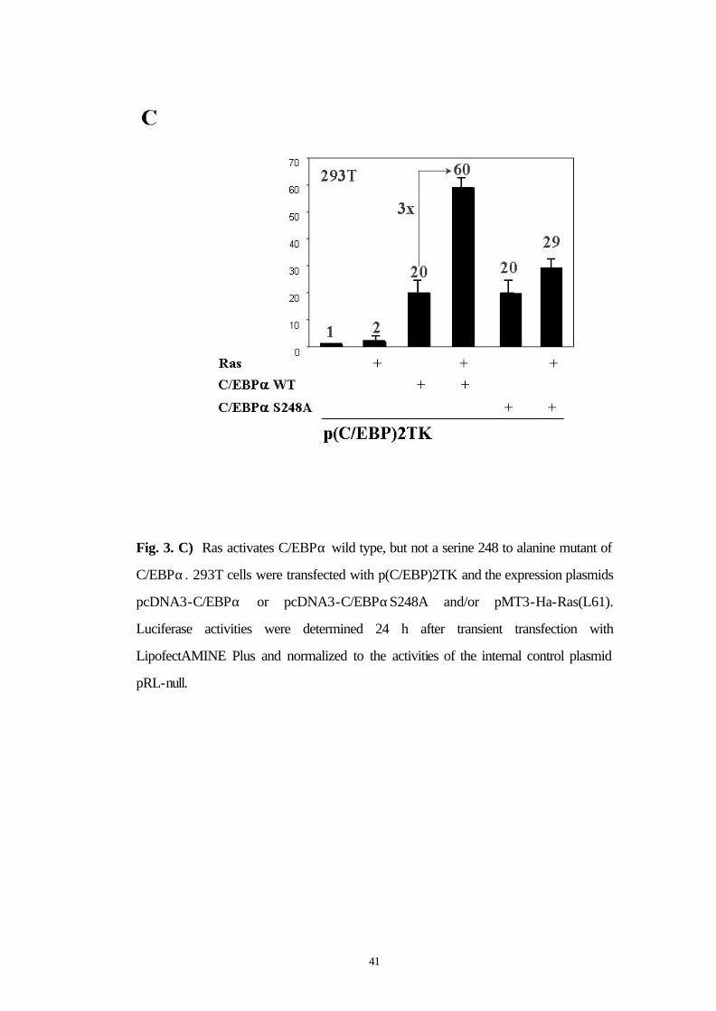
3.6 Ras activates C/EBPα wild type but not a S248A mutant 40
3.7 A PKC inhibitor blocks the activation of C/EBPα by Ras 42
3.8 Ras phosphorylates C/EBPα at S248 in vivo 45

2
3.9 Mutation of serine 248 to alanine obviates the ability of C/EBPα to induce differentiation 48
4. DISCUSSION 52
5. SUMMARY 59
6. ZUSAMMENFASSUNG 61
7. REFERENCES 63
8. ACKNOWLEDGEMENTS 73
9. CURRICULUM VITAE 74
10. PUBLICATIONS 77

3
To my wife Poonam, Who had the patience to live 7000 miles away from me for 3 long years…
4
1. INTRODUCTION
1.1 Role of C/EBPα in myelopoiesis
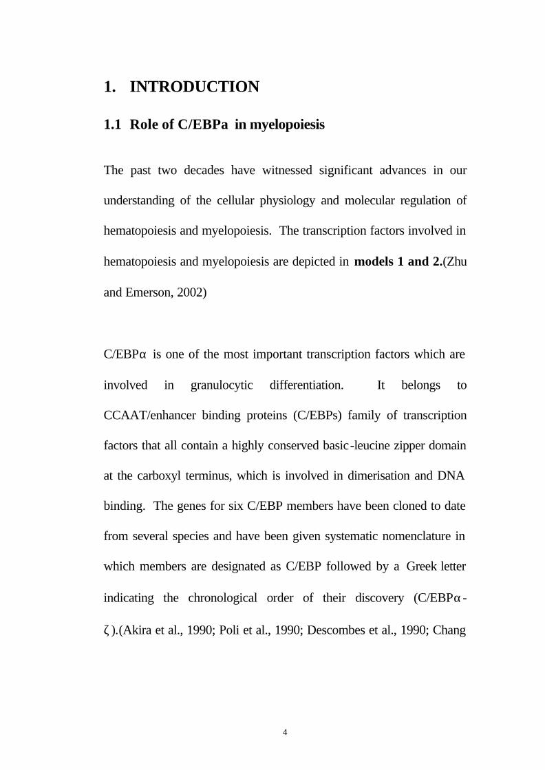
The past two decades have witnessed significant advances in our
understanding of the cellular physiology and molecular regulation of
hematopoiesis and myelopoiesis. The transcription factors involved in
hematopoiesis and myelopoiesis are depicted in models 1 and 2.(Zhu
and Emerson, 2002)
C/EBPα is one of the most important transcription factors which are
involved in granulocytic differentiation. It belongs to
CCAAT/enhancer binding proteins (C/EBPs) family of transcription
factors that all contain a highly conserved basic-leucine zipper domain
at the carboxyl terminus, which is involved in dimerisation and DNA
binding. The genes for six C/EBP members have been cloned to date
from several species and have been given systematic nomenclature in
which members are designated as C/EBP followed by a Greek letter
indicating the chronological order of their discovery (C/EBPα-
ζ).(Akira et al., 1990; Poli et al., 1990; Descombes et al., 1990; Chang
5
et al., 1990; Roman et al., 1990; Cao et al., 1991; Williams et al., 1991;
Ron and Habener, 1992; Ramji and Foka, 2002)
Model 1: Transcriptional regulation of early hematopoietic stem cell development. HSCs, as one progeny of hemangioblasts or hemogenic endothelium, are faced with the cell fate choice either to self-renew or to differentiate into committed common lymphoid or common myeloid hematopoietic precursors. The transcription factors involved in each development direction are depicted.
6
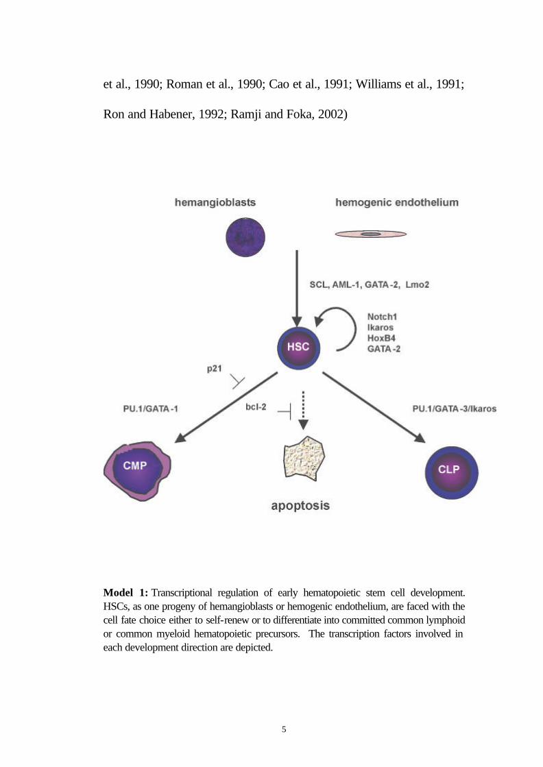
Model 2: Transcriptional regulation of common myeloid precursor (CMP) commitment. CMPs differentiate into either common precursors for granulocytic and monocytic lineages (GMPs) or common precursors for both erythroid and megakaryocytic lineages (EMPs). A separate, possible, pathway leading to eosinophils is depicted by dotted line. Dual expression of PU.1 and GATA-1 leads HSCs to CMPs, but then dominant expression of PU.1 is restricted to GMPs, while unopposed GATA-1 expression directs differentiation to EMPs.
The transcription factor C/EBPα plays a pivotal role during
differentiation in various cell types, including adipocytes and
hepatocytes, lung and ovary cells.(Cao et al., 1991; Lin and Lane,
1994; Scott et al., 1992; Flodby et al., 1996; Zhang et al., 1997;
Radomska et al., 1998; Umek et al., 1991) In hematopoiesis, C/EBPα
is expressed in myeloid cells.(Scott et al., 1992; Radomska et al.,
1998) It has been previously demonstrated that the expression of
7
C/EBPα correlates with the commitment of multipotential precursors
to the myeloid lineage, and is specifically upregulated during
neutrophilic differentiation.(Scott et al., 1992; Radomska et al., 1998)
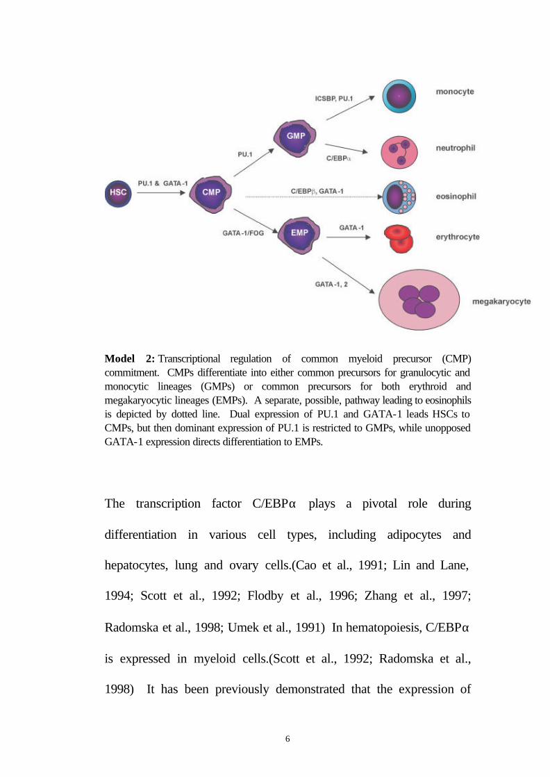
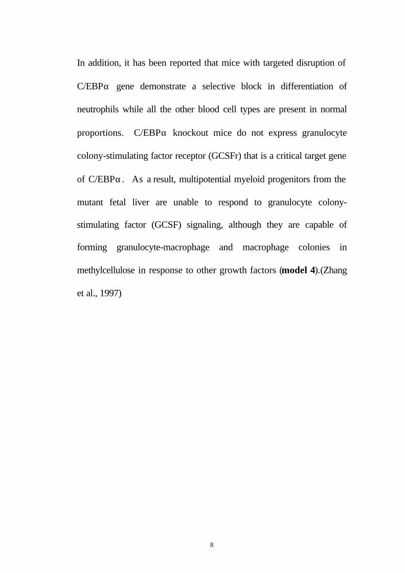
Furthermore, conditional expression of C/EBPα is sufficient to induce
neutrophilic differentiation (Radomska et al., 1998; Wang et al., 1999)
and can block the monocytic differentiation program in bipotential
myeloid precursors (model 3).(Radomska et al., 1998)
expression vector
C/EBPα
Zn
U937 cells
promoter C/EBPα
Conditional expression of C/EBPα is sufficient for induction of granulocytic differentiation of bipotential U937 cells
U937 with inducible C/EBPα- Zn + Zn
Radomska H et al., MCB 1998
Model 3: Overexpression of C/EBPα leads to granulocytic differentiation.
8
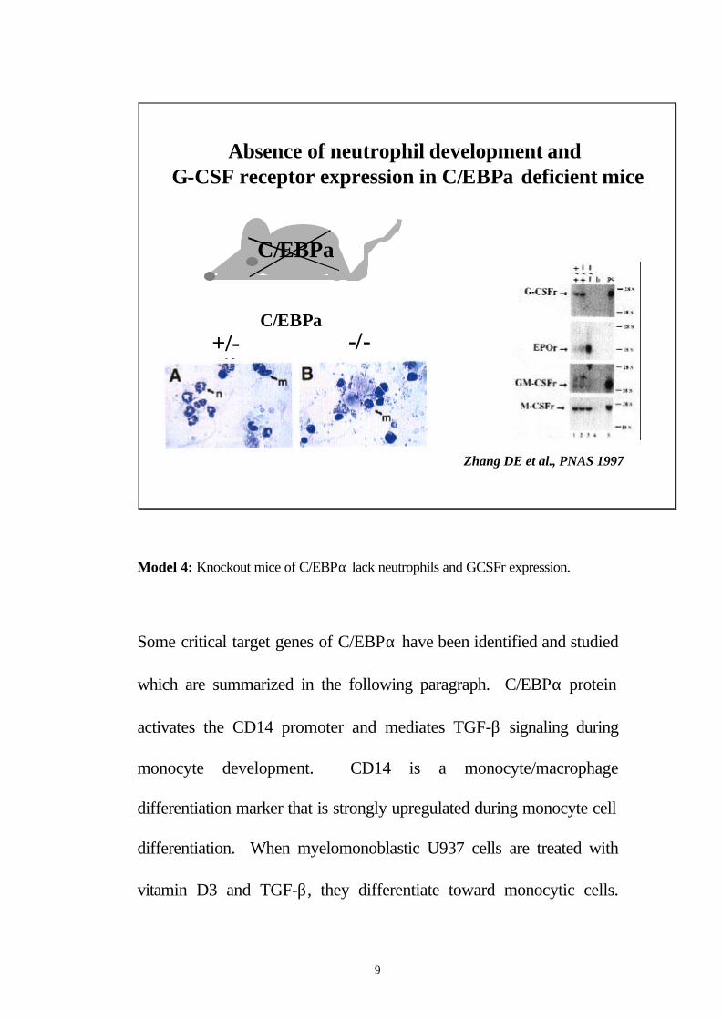
In addition, it has been reported that mice with targeted disruption of
C/EBPα gene demonstrate a selective block in differentiation of
neutrophils while all the other blood cell types are present in normal
proportions. C/EBPα knockout mice do not express granulocyte
colony-stimulating factor receptor (GCSFr) that is a critical target gene
of C/EBPα. As a result, multipotential myeloid progenitors from the
mutant fetal liver are unable to respond to granulocyte colony-
stimulating factor (GCSF) signaling, although they are capable of
forming granulocyte-macrophage and macrophage colonies in
methylcellulose in response to other growth factors (model 4).(Zhang
et al., 1997)
9
Absence of neutrophil development and G-CSF receptor expression in C/EBPα deficient mice
+/- -/-C/EBPα
Zhang DE et al., PNAS 1997
C/EBPα
Model 4: Knockout mice of C/EBPα lack neutrophils and GCSFr expression.
Some critical target genes of C/EBPα have been identified and studied
which are summarized in the following paragraph. C/EBPα protein
activates the CD14 promoter and mediates TGF-β signaling during
monocyte development. CD14 is a monocyte/macrophage
differentiation marker that is strongly upregulated during monocyte cell
differentiation. When myelomonoblastic U937 cells are treated with
vitamin D3 and TGF-β, they differentiate toward monocytic cells.
10
And there is a specific increase in the DNA binding and the expression
of C/EBPα and C/EBPβ during U937 monocytic cell
differentiation.(Zhang et al., 1997)
C/EBPα, -β, and –δ are also regulated during granulocyte lineage
specification. Ford et al. have shown that C/EBPα exists as multiple
phosphorylated forms in the nucleus of both multipotential and
granulocyte-committed hematopoietic progenitor cells. C/EBPβ is
unphosphorylated and cytoplasmically localized in multipotential cells
but exist as a phosphorylated nuclear enhancer-binding activity in
granulocyte-committed cells. GCSF-induced granulocytic
differentiation of multipotential progenitor cells results in activation of
C/EBPδ expression and functional recruitment of C/EBPδ and
C/EBPβ to the nucleus. These results suggest that the C-EBP family
members are critical regulators of myeloperoxidase gene expression
and are consistent with a model in which a temporal exchange of
C/EBP isoforms at the myeloperoxidase enhancer mediates the
transition from a primed state in multipotential cells to a
transcriptionally active configuration in promyelocytes.(Ford et al.,
1996)
11
Another target gene of C/EBPα in granulopoiesis is c-Myc. Johansen
et al. identified c-Myc as a C/EBPα negatively regulated gene. They
mapped an E2F binding site in the c-Myc promoter as the cis-acting
element critical for C/EBPα negative regulation. The identification of
c-Myc as a C/EBPα target gene is interesting, as it has been previously
shown that down-regulation of c-Myc can induce myeloid
differentiation. In this study they show that stable expression of c-
Myc from an exogenous promoter not responsive to C/EBPα-
mediated down-regulation forces myeloblasts to remain in an
undifferentiated state. Therefore, C/EBPα negative regulation of c-
Myc is critical for allowing early myeloid precursors to enter a
differentiation pathway. This is the first report to demonstrate that
C/EBPα directly affects the level of c-Myc expression and, thus, the
decision of myeloid blasts to enter into the granulocytic differentiation
pathway.(Johansen et al., 2001)
In 32D cl3 myeloblasts cell line the activation of C/EBPα-ER
construct by estradiol was sufficient to induce terminal granulocytic
differentiation and a G1 cell cycle arrest despite the continued presence
of IL-3. bcr-ablp210 prevented 32D cl3 cell differentiation, including
12
myeloperoxidase (MPO) RNA induction. Inhibition of cell growth by
C/EBPαWT-ER occurred even in 32D cl3 cells expressing bcr-ablp210
or in Ba/F3 B-lymphoid cells, without induction of differentiation. Cell
cycle arrest was associated with elevated p27Kip1 levels. PU.1 protein
and mRNA levels were increased within 4 hours of C/EBPαWT-ER
activation, in 32D cl3, 32D-bcr-ablp210, or Ba/F3 cells, and induction of
PU.1 mRNA occurred even in the presence of cycloheximide,
suggesting that induction of endogenous PU.1 RNA by C/EBPαWT-
ER results from direct transcriptional activation. However, activation
of PU.1-ER(T) in 32D cl3 cells induced MPO RNA but not cell cycle
arrest or terminal differentiation. Thus, in 32D cl3 myeloblasts,
C/EBPα acts independent of G-CSF signals, directly upstream of
PU.1, and upstream of p27Kip1 and additional factors to limit
proliferation and induce granulocytic differentiation.(Wang et al., 1999)
In a recent study it has been shown that C/EBPs are required for
granulopoiesis independent of their induction of the GCSFr.(Wang Qf
and Friedman, 2002)
Induction of granulocytic differentiation by C/EBPα seems to be by 2
pathways. In a cell line derived from the fetal liver of C/EBPα-
13
deficient animals it has been shown that conditional expression of
C/EBPα induces the C/EBP family members C/EBPβ and C/EBPε
and subsequently granulocytic differentiation. Similar results were
obtained when C/EBPα-/- cells were stimulated with the cytokines
interleukin-3 and granulocyte-macrophage colony-stimulating factor,
but not with all-trans retinoic acid, supporting a model of at least 2
pathways leading to the differentiation of myeloid progenitors to
granulocytes and implicating induction of other C/EBP family members
in granulopoiesis.(Zhang et al., 2002)
C/EBP family members are also involved in protein-protein interaction
and thereby regulate myeloid specific promoters. C/EBPα and
GCSFr signals cooperate to induce the myeloperoxidase and
neutrophil elastase genes(Wang et al., 2001), C/EBP and AML1
synergistically activate the macrophage colony-stimulating factor
receptor (MCSFr) promoter(Zhang et al., 1996), C/EBP, c-Myb, and
PU.1 cooperate to regulate the neutrophil elastase
promoter(Oelgeschlager et al., 1996), PU.1 and C/EBPα regulate the
granulocyte colony-stimulating factor receptor (GCSFr) promoter in
myeloid cells(Smith et al., 1996), and transcriptional coactivator ASC-
14
2 functionally interact with C/EBPα in granulocytic differentiation of
HL60 promyelocytic cells.(Hong et al., 2001)
1.2 Disruption of C/EBPα activity and expression in acute myeloid leukemia
We recently reported that dominant-negative mutations of C/EBPα are
found in patients with acute myeloid leukemia of subtypes M1 and M2.
Heterozygous mutations were found in CEBPA gene in ten patients
with acute myeloid leukemia (AML), five mutations in the amino
terminus truncated the full-length protein, but did not affect a 30-kD
protein initiated further downstream. The mutant proteins block wild-
type C/EBPα DNA binding and transactivation of granulocyte target
genes in a dominant-negative manner, and fails to induce granulocytic
differentiation. It is the first report of CEBPA gene mutations in
human neoplasia, and such mutations are likely to induce the
differentiation block found in AML.(Pabst et al., 2001a)
Furthermore, AML1-ETO associates with C/EBPα, inhibits C/EBPα
dependent transcription of myeloid cell specific rat defensin NP-3
promoter, and blocks granulocytic differentiation.(Westendorf et al.,
1998) We also demonstrated that the leukemic fusion protein AML1-
15
ETO, found in patients with acute myeloid leukemia with translocation
t(8;21), downregulates both C/EBPα expression and function in
primary AML patient samples.(Pabst et al., 2001b)
The hallmark of acute promyelocytic leukemia (APL) is the
translocation t(15;17) resulting in PML-RARα fusion protein and a
block in promyelocytic differentiation. In primary human APL cells
PML-RARα physically interacts with C/EBPα at diagnosis and
relapse, but not after All-Trans-Retinoic-Acid (ATRA)-induced
remission. Similar results are observed in primary leukemic cells from
transgenic mouse models of APL along with marked reduction of
C/EBPα DNA binding. Conditional expression of PML-RARα
abrogates C/EBPα DNA binding, transactivation, and differentiation of
myeloid cell lines in an ATRA-reversible manner. These studies
implicate disruption of C/EBPα DNA binding and function by physical
interaction with PML-RARα as an additional mechanism contributing
to the block in differentiation following expression of the fusion protein
in APL (Tracey Lodie and Dan Tenen, pers. communication).
16
These studies point to the crucial role of C/EBPα in both normal
myeloid differentiation and leukemogenesis. However, how the
transcriptional activity of C/EBPα is regulated both in normal
myelopoiesis as well as in leukemogenesis, is not fully understood.
In a recent review on transcriptional regulation of granulocyte and
monocyte development, Friedman, A.D. has set the perspective for the
future research in this field. Many questions remain unanswered
regarding the transcriptional regulation of granulocyte and monocyte
development. With respect to the cellular basis for initiating these
lineages: What are the relative contributions of granulocyte/monocyte
and B-cell/monocyte progenitors to mature blood elements; do some
granulocyte or monocyte progenitors develop directly from pluripotent
stem cells; how irreversible are commitment decisions? With respect
to gene regulation: Are there additional important transcriptional
regulators of myeloid genes remaining to be uncovered via detailed
investigation of promoters and distal enhancers; are there lineage-
restricted co-activators or co-repressors which participate in lineage
specification? With respect to key transcription factors, further
clarification of the regulatory network is needed: Which factor or
17
factors specify each lineage and at what levels of expression; can
family members act redundantly in this regard; what additional
cooperative mechanisms operate among transcriptional regulators;
what roles do cytokine receptor signaling and transcription factor
modifications play in each commitment decision and in each step of
lineage progression? The answers to these questions will provide
general lessons in developmental biology and insights into
leukemogenesis and will enable applications in clinical hematology,
oncology, and gene therapy.(Friedman, 2002)
1.3 Ras signaling in leukemia and myeloid differentiation
The Ras family of proteins are GTP-dependent molecular switches that
are essential for cell growth and differentiation.(Mccormick, 1995;
Gutkind, 1998) Ras exerts its effect on cell growth mainly via ETS
(Wasylyk et al., 1994) and AP-1 (Johnson et al., 1996) transcription
factors. For example, cells with a null mutation in the c-jun gene and
expressing oncogenic Ras lack many characteristics of Ras
transformation,(Johnson et al., 1993) and dominant negative mutants of
ETS-1, ETS-2, or PU.1 containing only the DNA binding domain
inhibit Ras activation of transcription and revert Ras-transformed
18
cells.(Wasylyk et al., 1994) In particular, Ras has been demonstrated
to play an important role in myeloid differentiation. Macrophage
differentiation and M-CSF-dependent survival are altered in transgenic
mice that express dominant suppressors of Ras signaling,(Jin et al.,
1995) while a number of hematopoietic cell lines undergo spontaneous
myeloid differentiation in response to expression of activated Ras.(Hibi
et al., 1993; Maher et al., 1996) In addition, M-CSF, granulocyte-
macrophage colony-stimulating factor, or interleukin-3-induced
monocytopoiesis of CD34+ cells is inhibited by N-Ras antisense
oligonucleotides.(Skorski et al., 1992)
19
1.4 Aim of this research
Since Ras signaling and the transcription factor C/EBPα both play
important roles in myeloid differentiation, we wanted to know how
C/EBPα is activated during granulocytic differentiation? We
hypothesized that Ras might be involved in the activation of C/EBPα.
Experiments were designed to test if Ras changes the DNA binding,
expression level or post-translational modification of C/EBPα? After
finding that Ras phosphorylates C/EBPα, we designed experiments to
map the site of phosphorylation in C/EBPα and to point mutate this
site to check the relevance of this phosphorylation. Then we wanted
to discover the biological relevance of C/EBPα phosphorylation in
myeloid cells by retroviral transduction of C/EBPα WT and
C/EBPαS248A mutant and look for its effect on granulocytic
differentiation.
Here we demonstrate data which suggest a model where Ras signaling
phosphorylates C/EBPα on serine 248 of the transactivation domain
resulting in an enhancement of the ability of C/EBPα to transactivate
the G-CSF receptor promoter, which contributes to the induction of
granulocytic differentiation.
20
2. MATERIALS AND METHODS
2.1 Cell lines and cell culture
Human kidney 293T cells and human kidney 293E1A cells (kindly
provided by John Blenis, Harvard Medical School, Boston, MA) were
maintained in Dulbecco's modified Eagle's medium supplemented with
10% fetal calf serum (HyClone). 32D cl3 cells were maintained in
phenol-red free Iscove’s Modified Dulbecco’s Medium with 10%
heat-inactivated fetal bovine serum (HI-FBS), 1 ng/ml IL-3 (R&D
Systems, Minneapolis, MN), and penicillin-streptomycin. ϕCRE cells
were maintained in Dulbecco’s Modified Eagle’s Medium with 10%
heat-inactivated calf serum and pen/strep. pBabePuro-
C/EBPα(S248A)-ER was introduced into ϕCRE cells using
lipofectamine (Gibco, BRL, Gaithersburg, MD), and a pool of
resistant cells was isolated using 2 µg/ml puromycin. 32D cl3 cells
were co-cultured in the presence of 4 µg/ml Polybrene with ϕCRE-
S248A cells which had been irradiated to 3000 cGy. After 48 hrs, the
32D cl3 cells were placed in 96 well dishes with 2 µg/ml puromycin,
and individual subclones were isolated. Estradiol was employed at 1
µM. Morphology was assessed by Wright’s-Giemsa staining of
21
cytospins. pBabePuro-C/EBPα(S248A)-ER was constructed by
ligating an MluI/NcoI segment containing the mutant serine in place of
the identical fragment in pBabePuro-C/EBPαWT-ER. Total cellular
extracts were subjected to Western blotting using antiserum
recognizing human ERα (Santa Cruz Biotechnology, San Diego, CA)
or murine actin (Sigma, St. Louis, MO).
2.2 Reporter constructs and expression plasmids
The human G-CSF receptor promoter ranging from bp -74 to +67 with
respect to the major transcription start site (Smith et al., 1996) was
subcloned in the firefly luciferase vector pXP2.(Nordeen, 1988) pTK
driven by 2 C/EBP sites (p(C/EBP)2TK) is a dimer of the C/EBP site
from the granulocyte colony-stimulating factor receptor promoter from
bp -57 to bp –37 with respect to the major transcription start
site,(Smith et al., 1996) subcloned into pTK81luc, a pXP2-based
luciferase construct with a TATA box only as a minimal
promoter.(Nordeen, 1988) As an internal control plasmid for co-
transfection assays, the pRL-null construct driving a Renilla luciferase
gene (Promega, Madison, WI) was used as described before.(Behre et
al., 1997; Behre et al., 1999; Zhang et al., 1999) The expression
22
plasmids pMSV-C/EBPα (rat), various deletion mutants of C/EBPα in
pMSV, pMSV-C/EBP β, pMSV-C/EBPδ, GAL4-C/EBPα area 1-9,
GAL4-C/EBPα area 1-3, and GAL4-C/EBPα area 4-9 and a minimal
promoter with GAL4 DNA binding sites were described
previously.(Friedman and McKnight, 1990; Oelgeschlager et al., 1996)
Human activated pMT3-Ha-Ras(L61) was kindly provided by Larry
Feig (Tufts University, Boston, MA).(Feig and Cooper, 1988; Feig,
1988)
To introduce a serine to alanine point mutation at serine 248 of rat
C/EBPα we performed PCR site-directed mutagenesis. pcDNA3-
C/EBPα was digested with NotI and the resulting small fragment of
C/EBPα coding sequence was used as template DNA for all PCR
reactions. Four primers were synthesized to amplify two internal
fragments from wild type C/EBPα: sense primer A (bp 693-716) and
antisense primer B (bp 763-740) were used to amplify fragment AB
(size 70 bp), sense primer C (bp 740-763) and antisense primer D (bp
860-837) were used to amplify fragment CD (size 120 bp). The point
mutation at bp 742 was introduced in primer B (from A to C) and
primer C (from T to G) which overlap with each other (bp 740-763),
23
so the resulting fragments will have the mutation incorporated during
the PCR amplification. The fragments AB and CD were purified from
agarose gels using a Gel Extraction kit (Qiagen). These 2 fragments
were joined by elongation reaction using primers A and D to create a
single fragment AD (size 167 bp) which would have the serine 248
(TCG) mutated to alanine (GCG). This fragment was then digested
with SfiI and XcmI and subcloned into SfiI/XcmI digested wild type
pcDNA3-C/EBPα. The point mutation was confirmed by DNA
sequencing and was used in subsequent experiments.
2.3 Transient transfections using LipofectAMINE Plus and reporter assays for firefly and Renilla luciferase
293T cells or 293E1A cells were transfected using LipofectAMINE
Plus (Life Technologies) as described by the manufacturer. Firefly
luciferase activities from the constructs pG-CSFR, pXP2, pGal4-DBD,
pTK and p(C/EBP)2TK and Renilla luciferase activity from the internal
control plasmid pRL-null were determined 24 h after the initiation of the
transfection protocols using the Dual-Luciferase Reporter Assay
System (Promega). Firefly luciferase activities were normalized to the
Renilla luciferase values of pRL-null. Results are given as means ±
24
S.E.M. of at least six independent experiments. The following DNA
concentrations of the reporter constructs and expression plasmids
were used for LipofectAMINE Plus transfections: 0.1 µg of pG-
CSFR, pXP2, pGal4-DBD, pTK and p(C/EBP)2TK; 0.01 µg of the
internal control plasmid pRL-null; 0.1 µg of the expression plasmids
for C/EBPα and C/EBPα mutants and for Ras(L61), and the same
concentrations of the empty expression vectors were used as controls,
respectively. In the transfections without cotransfection of Ras, the
empty vector pMT3 was included instead of pMT3-Ras. pRL-null
was chosen as internal control plasmid, because it was not
transactivated by Ras in 293T cells.(Behre et al., 1999; Behre et al.,
1997)
2.4 Electrophoretic mobility shift assay
Electrophoretic mobility shift assays were performed as described
previously.(Behre et al., 1999; Zhang et al., 1999) As a positive
control for C/EBPα binding, a double-stranded C/EBPα probe from
the human G-CSF receptor promoter (bp -57 to bp –37 with respect
to the major transcription start site, as described before in Fig. 9 of
reference (Smith et al., 1996) OligoA: AAG GTG TTG CAA TCC
25
CCA GC and OligoB: GCT GGG GAT TGC AAC ACC TT) was
labeled with Klenow polymerase and [32P]dCTP (NEN Life Science
Products) and incubated with 0.1 µg/µl of double-stranded poly(dI-
dC) (Sigma) with 1 µl of in vitro translated C/EBPα. In some
experiments, a 100-fold molar excess of the C/EBP probe was added
as specific unlabeled competitor. C/EBPα antibody (14AA) was used
for Gel Supershift (Cat # SC61 X, Santa Cruz Biotechnology).
2.5 In vivo labeling and phosphopeptide mapping
To detect changes in the phosphorylation pattern of C/EBPα upon
stimulation with activated Ras in vivo, 0.5 µg of pcDNA3-C/EBPα or
pcDNA3-C/EBPαS248A either with or without 0.25 µg of activated
Ras(L61), was transfected into 293T cells using LipofectAMINE Plus
(Life Technologies). 3 h after transfection, cells were starved in serum-
free Dulbecco's modified Eagle's medium. After 18 h, cells were
placed into serum-free and phosphate-free Dulbecco's modified Eagle's
medium (Life Technologies) for 30 min before they were metabolically
labeled with [32P]orthophosphate (2.5 mCi/ml). After 4 h, cells were
lysed with radioimmunoprecipitation assay buffer containing 1%
26
Nonidet P-40, 0.5% sodium deoxycholate, 0.1% SDS, 0.15 M NaCl, 5
mM EDTA, and 50 mM Tris (pH 8.0) and supplemented with
aprotinin, phenylmethylsulfonyl fluoride, pepstatin A, leupeptin,
antipain, and chymostatin as protease inhibitors (Sigma) and sodium
pyrophosphate, sodium fluoride, and sodium vanadate as phosphatase
inhibitors (Sigma). In parallel plates, 0.3 µg of the G-CSF receptor
promoter was co-transfected in 293T cells, and luciferase activities
were determined to ensure that Ras signaling enhances the
transactivation function of C/EBPα in the particular experiment used in
in vivo labeling and subsequent phosphopeptide mapping.
C/EBPα was isolated by immunoprecipitation from the lysates using a
rabbit polyclonal antibody against C/EBPα (Santa Cruz), washed four
times with radioimmunoprecipitation assay buffer, separated on 10%
SDS-polyacrylamide gels, and transferred to nitrocellulose (Bio-Rad)
for phosphopeptide mapping. After transfer, the C/EBPα protein
bands was excised. To determine the phosphorylated protein residues
of C/EBPα, C/EBPα protein bands were digested with 1-chloro-3-
tosylamido-7-amino-2-heptanone-treated chymotrypsin (Worthington)
and endoproteinase Glu-C (V8 protease) (Boehringer Mannheim) and
27
processed for phosphopeptide mapping as described
previously.(Boyle et al., 1991)
2.6 Western blot assay
24 h after the start of transfection, cells were lysed with
radioimmunoprecipitation assay buffer. Equal amounts of total protein
were separated on 10% SDS-polyacrylamide gels and transferred to
Immobilon-P membrane (Millipore). Membranes were incubated with
anti-C/EBPα antibody (Santa Cruz Biotechnology) or β-tubulin
antibody as an internal control (catalog no. 1111876; Boehringer
Mannheim) for 60 min and then with Protein A-horseradish peroxidase
conjugate (Amersham) for 45 min. Signals were detected with ECL
Western blotting detection reagents (Amersham). In parallel plates, the
G-CSF receptor promoter construct was co-transfected, and luciferase
activities were determined to ensure that Ras enhances the
transactivation function of C/EBPα in the particular experiment used
for Western blot analysis of C/EBPα expression and that the
transfection efficacy was the same (less than 10% difference between
plates) in the particular experiment. Differences in protein expression
were quantitated by ImageQuant software (Molecular Dynamics).

28
C/EBPα antibody (14AA, SC61) was used for Western Blot and
immunoprecipitation (Santa Cruz Biotechnology).
29
3. RESULTS
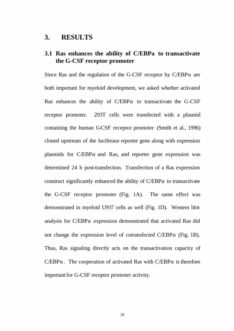
3.1 Ras enhances the ability of C/EBPα to transactivate the G-CSF receptor promoter
Since Ras and the regulation of the G-CSF receptor by C/EBPα are
both important for myeloid development, we asked whether activated
Ras enhances the ability of C/EBPα to transactivate the G-CSF
receptor promoter. 293T cells were transfected with a plasmid
containing the human G-CSF receptor promoter (Smith et al., 1996)
cloned upstream of the luciferase reporter gene along with expression
plasmids for C/EBPα and Ras, and reporter gene expression was
determined 24 h post-transfection. Transfection of a Ras expression
construct significantly enhanced the ability of C/EBPα to transactivate
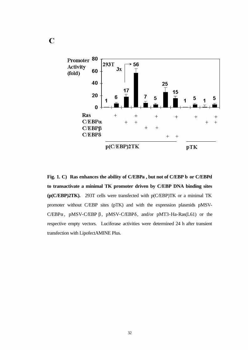
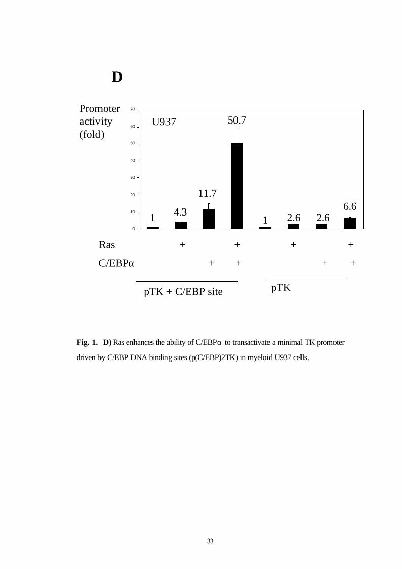
the G-CSF receptor promoter (Fig. 1A). The same effect was
demonstrated in myeloid U937 cells as well (Fig. 1D). Western blot
analysis for C/EBPα expression demonstrated that activated Ras did
not change the expression level of cotransfected C/EBPα (Fig. 1B).
Thus, Ras signaling directly acts on the transactivation capacity of
C/EBPα. The cooperation of activated Ras with C/EBPα is therefore
important for G-CSF receptor promoter activity.
30
Fig. 1. Ras signaling enhances the transcriptional activity of C/EBPα . A) Ras
enhances the ability of C/EBPα to transactivate the G-CSF receptor promoter. 293T
cells were transfected with the human G-CSF receptor promoter or the human G-CSF
receptor promoter with mutated C/EBP sites and with the expression plasmids pMSV-
C/EBPα (or empty vector pMSV) and pMT3-Ha-Ras(L61) (or empty vector pMT3).
Luciferase activities were determined 24 h after transient transfection with
LipofectAMINE Plus and normalized to the activities of the internal control plasmid
pRL-null. B) Western blot analysis for C/EBPα expression (and β-tubulin expression
as loading control) for the experiment shown in Fig. 1A.

31
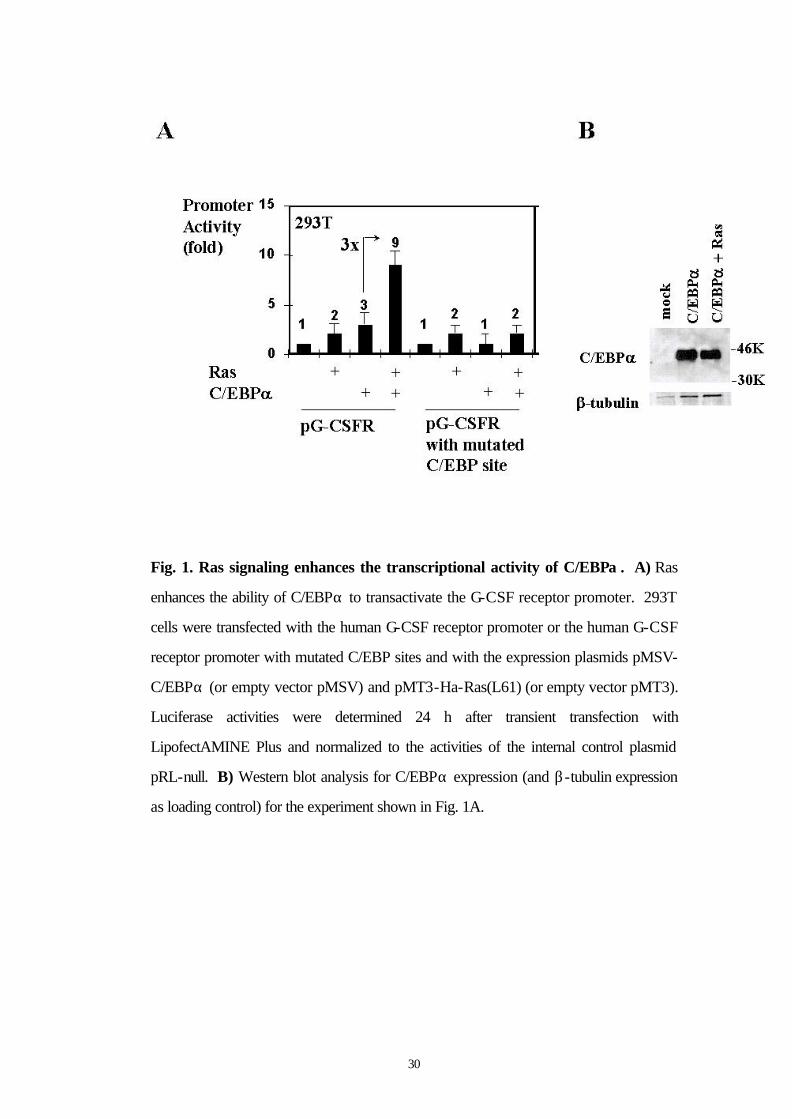
3.2 Ras enhances the ability of C/EBPα , but not of C/EBP β or C/EBPδ to transactivate a minimal TK promoter driven by C/EBP DNA binding sites
We next asked the following questions: (a) whether the binding of
C/EBPα to DNA was necessary for its activation by Ras and (b)
whether a C/EBPα binding site alone was sufficient for the Ras-
enhanced C/EBPα activation. We observed enhanced C/EBPα
transactivation mediated by activated Ras using a reporter construct
containing two C/EBPα binding sites cloned upstream of a minimal TK
promoter (p(C/EBP)TK) (Fig. 1C). In control experiments, no effect
of Ras on C/EBPα activity was observed when the minimal TK
promoter without C/EBPα binding sites was used (Fig. 1C). These
data indicate that C/EBPα binding to DNA is necessary for its
activation by Ras signaling and that C/EBPα binding sites are sufficient
to mediate this effect. Interestingly, the activation of C/EBPα by Ras
was specific for C/EBPα, because C/EBP β und C/EBPδ were not
activated functionally by Ras signaling in this assay (Fig. 1C).
32
Fig. 1. C) Ras enhances the ability of C/EBPα , but not of C/EBP β or C/EBPδ
to transactivate a minimal TK promoter driven by C/EBP DNA binding sites
(p(C/EBP)2TK). 293T cells were transfected with p(C/EBP)TK or a minimal TK
promoter without C/EBP sites (pTK) and with the expression plasmids pMSV-
C/EBPα, pMSV-C/EBP β , pMSV-C/EBPδ, and/or pMT3-Ha-Ras(L61) or the
respective empty vectors. Luciferase activities were determined 24 h after transient
transfection with LipofectAMINE Plus.
33
D
pTK + C/EBP site pTK
Ras + + + +
0
10
20
30
40
50
60
70
U937
C/EBPα + + + +
1 4.3
11.7
50.7
1 2.6 2.66.6
Promoter activity(fold)
Fig. 1. D) Ras enhances the ability of C/EBPα to transactivate a minimal TK promoter
driven by C/EBP DNA binding sites (p(C/EBP)2TK) in myeloid U937 cells.
34
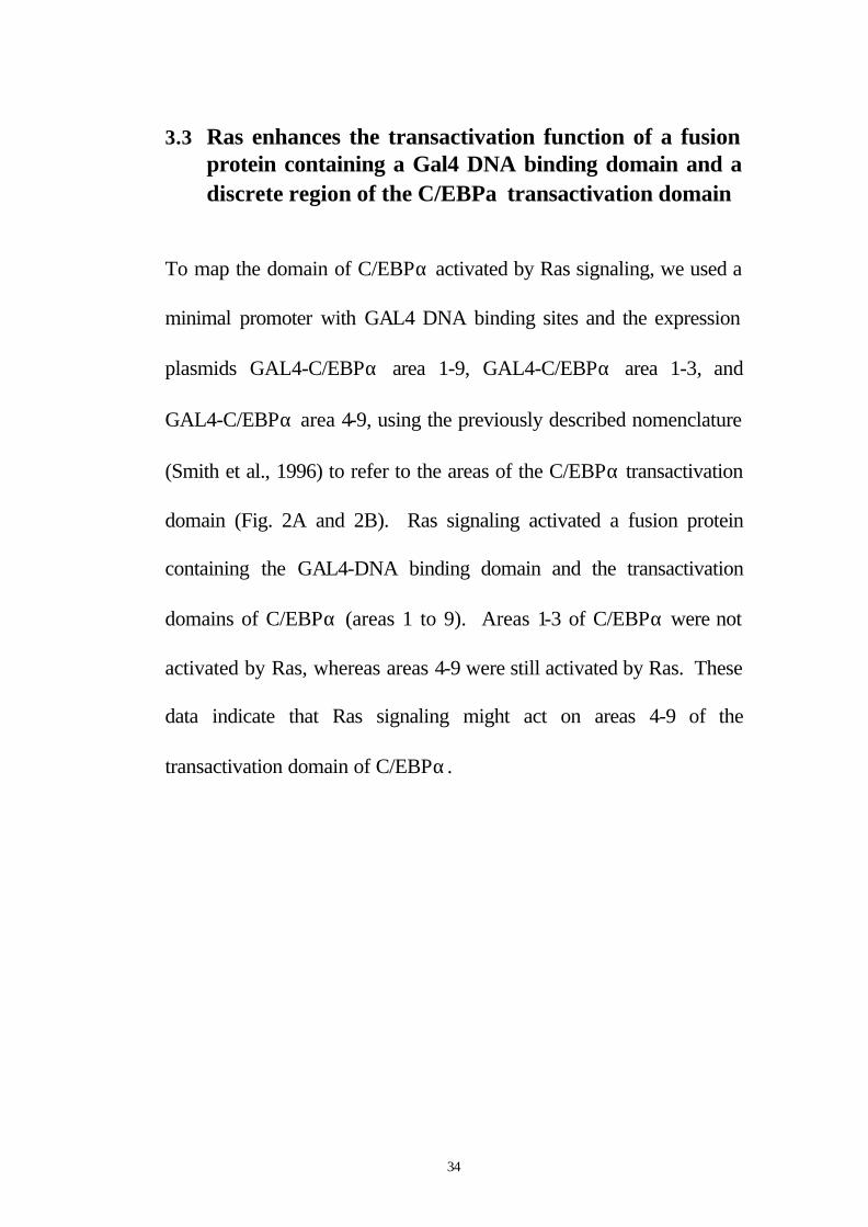
3.3 Ras enhances the transactivation function of a fusion protein containing a Gal4 DNA binding domain and a discrete region of the C/EBPα transactivation domain
To map the domain of C/EBPα activated by Ras signaling, we used a
minimal promoter with GAL4 DNA binding sites and the expression
plasmids GAL4-C/EBPα area 1-9, GAL4-C/EBPα area 1-3, and
GAL4-C/EBPα area 4-9, using the previously described nomenclature
(Smith et al., 1996) to refer to the areas of the C/EBPα transactivation
domain (Fig. 2A and 2B). Ras signaling activated a fusion protein
containing the GAL4-DNA binding domain and the transactivation
domains of C/EBPα (areas 1 to 9). Areas 1-3 of C/EBPα were not
activated by Ras, whereas areas 4-9 were still activated by Ras. These
data indicate that Ras signaling might act on areas 4-9 of the
transactivation domain of C/EBPα.
35
Fig. 2. Ras signaling activates C/EBPα via the C/EBPα transactivation domain. A) Ras enhances the transactivation function of a fusion protein containing a Gal4 DNA binding domain and areas 4-9 of the C/EBPα transactivation domain. 293E1A cells were transfected with a minimal promoter driven by GAL4 DNA binding sites and the expression plasmids GAL4-C/EBPα area 1-9, GAL4-C/EBPα area 1-3, GAL4-C/EBPα area 4-9, and/or pMT3-Ha-Ras(L61). B) Model of the construct GAL4-C/EBPα area 1-9 used in Fig. 2A. The transactivation domain of C/EBPα (area 1 to 9) is fused to the DNA binding domain of GAL4.
36
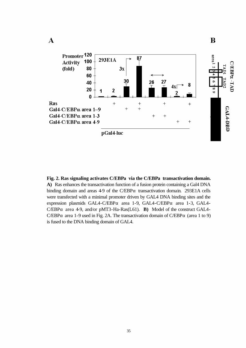
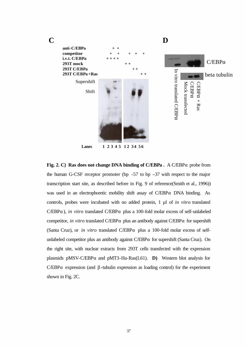
3.4 Ras does not change DNA binding of C/EBPα
To further elucidate the mechanism by which Ras augments the
transcriptional activity of C/EBPα, we performed experiments to
determine whether the activation of C/EBPα by Ras was partly due to
an increase of C/EBPα DNA binding. An electrophoretic mobility-
shift assay was performed using a bp -57 to bp -37 G-CSF receptor
promoter fragment containing its C/EBPα binding site.(Smith et al.,
1996) A positive control, in vitro translated C/EBPα, specifically
bound to this probe as expected (Fig. 2C). Nuclear extracts from
293T cells transfected with the expression plasmid of C/EBPα with or
without cotransfection of Ras bound to the oligo as well, and most
importantly, Ras signaling did not change the DNA binding of
C/EBPα. To rule out different C/EBPα protein contents of the
nuclear extracts used, we performed a Western blot analysis for
C/EBPα expression (Fig. 2D) which showed equal loading of
C/EBPα. These results indicate that binding of C/EBPα to the G-CSF
receptor promoter DNA is not enhanced by Ras signaling.
37
C
C/EBPα
beta tubulinIn vitro translated C
/EBPα
Mock
transfectedC
/EB
PαC
/EB
Pα+ R
asD
anti-C/EBPα + +competitor + + + + +i.v.t. C/EBPα + + + +293T mock + +293T C/EBPα + +293T C/EBPα+Ras + +
1 2 3 4 5 1 2 3 4 5 6Lanes
Shift
Supershift
Fig. 2. C) Ras does not change DNA binding of C/EBPα . A C/EBPα probe from
the human G-CSF receptor promoter (bp -57 to bp –37 with respect to the major
transcription start site, as described before in Fig. 9 of reference(Smith et al., 1996))
was used in an electrophoretic mobility shift assay of C/EBPα DNA binding. As
controls, probes were incubated with no added protein, 1 µl of in vitro translated
C/EBPα), in vitro translated C/EBPα plus a 100-fold molar excess of self-unlabeled
competitor, in vitro translated C/EBPα plus an antibody against C/EBPα for supershift
(Santa Cruz), or in vitro translated C/EBPα plus a 100-fold molar excess of self-
unlabeled competitor plus an antibody against C/EBPα for supershift (Santa Cruz). On
the right site, with nuclear extracts from 293T cells transfected with the expression
plasmids pMSV-C/EBPα and pMT3-Ha-Ras(L61). D) Western blot analysis for
C/EBPα expression (and β-tubulin expression as loading control) for the experiment
shown in Fig. 2C.
38
3.5 Ras activates C/EBPα via area 9 of the C/EBPα TAD
To further localize the site of C/EBPα activated by Ras signaling and
to fine map this site, we used various deletion mutants of C/EBPα
(Fig. 3A, 3B). C/EBPα∆4-9 was minimally activated and C/EBPα∆9
was not activated by Ras. All other mutants were activated at least 2-
fold by Ras. These studies demonstrate that Ras acts on area 9 of
C/EBPα.

39
Fig. 3. Ras signaling requires serine 248 of C/EBPα to activate C/EBPα . A)
Ras activates C/EBPα via area 9 of the C/EBPα transactivation domain. 293T cells
were transfected with p(C/EBP)2TK and the expression plasmids pMSV-C/EBPα or
various deletions mutants of C/EBPα and/or pMT3-Ha-Ras(L61). B) Model of
C/EBPα, with areas 1-9 containing the first and second transactivation domain (TAD1
and TAD2) and the DNA binding domain of C/EBPα with the basic region and leucine
zipper region.
40
3.6 Ras activates C/EBPα wild type but not a S248A mutant
Because Ras acts on area 9 of the transactivation domain of C/EBPα,
we focused on this area for generating C/EBPα point mutants. Area 9
of C/EBPα is depicted in detail in Figure 4B. There is only one
potential phosphorylation consensus site in area 9, at serine 248. We
generated a point mutant of C/EBPα at serine 248 by mutation to
alanine (C/EBPαS248A). C/EBPα wild type and C/EBPαS248A both
transactivated the G-CSF receptor promoter 20 fold. While Ras
signaling enhanced wild type C/EBPα transactivation capacity from 20
fold to 60 fold, Ras only enhanced the transactivation capacity of the
S248A mutant of C/EBPα from 20 to 29 fold (Fig. 3C). These results
suggest that Ras signaling might act on serine 248 of the C/EBPα
transactivation domain to enhance C/EBPα transcriptional activity.
41
Fig. 3. C) Ras activates C/EBPα wild type, but not a serine 248 to alanine mutant of
C/EBPα. 293T cells were transfected with p(C/EBP)2TK and the expression plasmids
pcDNA3-C/EBPα or pcDNA3-C/EBPαS248A and/or pMT3-Ha-Ras(L61).
Luciferase activities were determined 24 h after transient transfection with
LipofectAMINE Plus and normalized to the activities of the internal control plasmid
pRL-null.
42
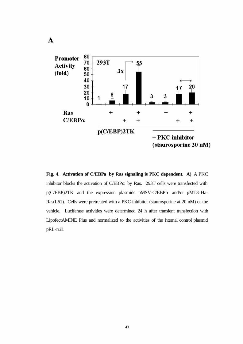
3.7 A PKC inhibitor blocks the activation of C/EBPα by Ras
Because serine 248 is a potential protein kinase C (PKC) consensus
site (Fig. 4B), we asked whether a PKC inhibitor (Staurosporine at 20
nM final concentration) could block the activation of C/EBPα by Ras
signaling. In fact, pretreatment of 293T cells with Staurosporine
abolished the activation of C/EBPα by Ras signaling (Fig. 4A). These
results support the hypothesis that the PKC pathway could be involved
in the Ras mediated activation of C/EBPα.
43
Fig. 4. Activation of C/EBPα by Ras signaling is PKC dependent. A) A PKC
inhibitor blocks the activation of C/EBPα by Ras. 293T cells were transfected with
p(C/EBP)2TK and the expression plasmids pMSV-C/EBPα and/or pMT3-Ha-
Ras(L61). Cells were pretreated with a PKC inhibitor (staurosporine at 20 nM) or the
vehicle. Luciferase activities were determined 24 h after transient transfection with
LipofectAMINE Plus and normalized to the activities of the internal control plasmid
pRL-null.
44
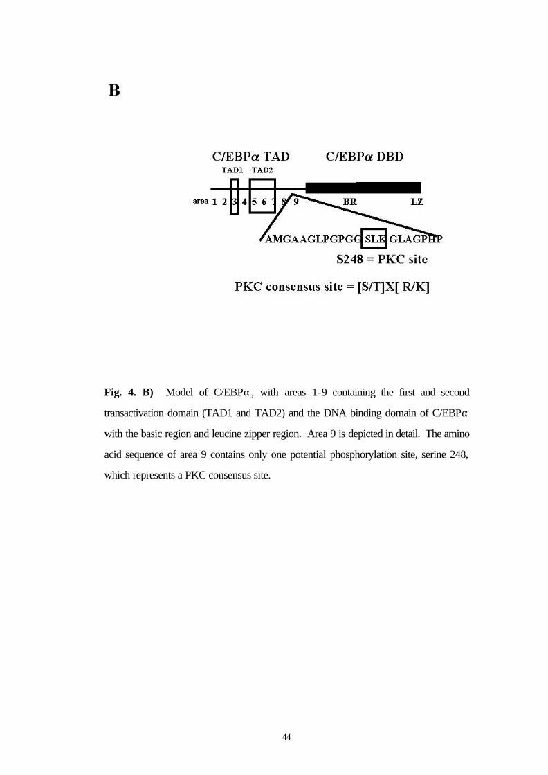
Fig. 4. B) Model of C/EBPα, with areas 1-9 containing the first and second
transactivation domain (TAD1 and TAD2) and the DNA binding domain of C/EBPα
with the basic region and leucine zipper region. Area 9 is depicted in detail. The amino
acid sequence of area 9 contains only one potential phosphorylation site, serine 248,
which represents a PKC consensus site.
45
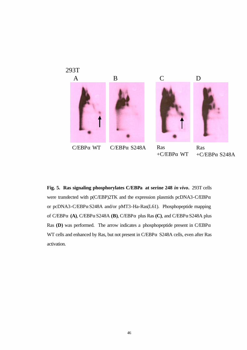
3.8 Ras phosphorylates C/EBPα at S248 in vivo
Because our data suggest that Ras activates C/EBPα via
phosphorylation on serine 248, we wanted to prove that Ras signaling,
in fact, phosphorylates C/EBPα on serine 248 in vivo. 293T cells
were transfected with p(C/EBP)2TK and with the expression plasmids
for C/EBPα or C/EBPαS248A and/or activated Ras.
Phosphopeptide mapping of cells transfected with C/EBPα,
C/EBPαS248A, C/EBPα plus Ras, and C/EBPαS248A plus Ras was
performed. Ras signaling (Fig. 5C) enhanced a phosphorylation spot
in the C/EBPα pattern (Fig. 5A) which was not visible in
C/EBPαS248A (Fig. 5B) or C/EBPαS248A plus cotransfected Ras
(Fig. 5D). Thus, we conclude that Ras signaling phosphorylates
C/EBPα on serine 248 in vivo.
46
293T
C/EBPα WT C/EBPα S248A Ras +C/EBPα WT
Ras +C/EBPα S248A
A B C D
Fig. 5. Ras signaling phosphorylates C/EBPα at serine 248 in vivo. 293T cells
were transfected with p(C/EBP)2TK and the expression plasmids pcDNA3-C/EBPα
or pcDNA3-C/EBPαS248A and/or pMT3-Ha-Ras(L61). Phosphopeptide mapping
of C/EBPα (A), C/EBPαS248A (B), C/EBPα plus Ras (C), and C/EBPαS248A plus
Ras (D) was performed. The arrow indicates a phosphopeptide present in C/EBPα
WT cells and enhanced by Ras, but not present in C/EBPα S248A cells, even after Ras
activation.
47
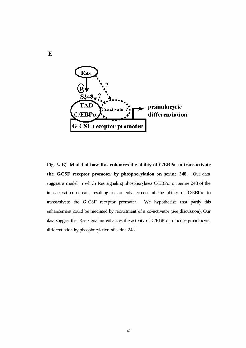
Fig. 5. E) Model of how Ras enhances the ability of C/EBPα to transactivate
the G-CSF receptor promoter by phosphorylation on serine 248. Our data
suggest a model in which Ras signaling phosphorylates C/EBPα on serine 248 of the
transactivation domain resulting in an enhancement of the ability of C/EBPα to
transactivate the G-CSF receptor promoter. We hypothesize that partly this
enhancement could be mediated by recruitment of a co-activator (see discussion). Our
data suggest that Ras signaling enhances the activity of C/EBPα to induce granulocytic
differentiation by phosphorylation of serine 248.
48
3.9 Mutation of serine 248 to alanine obviates the ability of C/EBPα to induce differentiation
To determine the biological importance of serine 248, we linked
C/EBPα(S248A) to the human Estradiol Receptor (ERα) ligand-
binding domain and isolated 32D cl3 cell lines expressing this protein.
Two subclones with protein expression as high as that we previously
achieved for C/EBPαWT-ER were obtained (Fig. 6A). In 32D-
C/EBPα(S248A)-ER-1 cells, the full-length species reproducibly
migrated faster than that found for clone 2 cells or for cells expressing
the wild-type protein. Smaller species, likely representing degradation
products, and a larger species (arrow), likely representing a dimer
formed via the leucine zipper, also ran faster than comparable bands in
the other lanes. Perhaps this difference in migration represents altered
phosphorylation at serine 248 or at another residue. It is not clear why
this faster migration was only seen with one subclone, but may
represent heterogeneity in the 32D cl3 cells themselves.
To assess induction of differentiation by C/EBPα(S248A)-ER, each of
the cell lines was placed in estradiol. Morphologically, after 3 days the
majority of 32D-C/EBPα(WT)-ER cells had differentiated to
49
neutrophils (Fig. 6C, right panels), whereas little morphologic change
was seen with 32D-C/EBPα(S248A)-ER-1 cells and clone 2 developed
primary granules without nuclear maturation (Fig 6C, left and middle
panels). To further assess differentiation, RNAs prepared daily from
cultures exposed to estradiol were subjected to Northern blotting for
MPO, LF, and b-actin (Fig. 6B). As reported previously,
C/EBPαWT-ER rapidly induced MPO and induced LF after 48 hrs.
In contrast, in 32D-C/EBPα(S248A)-ER-1 cells, neither MPO nor LF
were induced, and in clone 2 cells MPO was induced in a delayed
fashion and LF was not induced. Mutation of serine 248 to alanine did
not prevent the growth inhibitory properties of C/EBPαWT-ER in 32D
cl3 cells (data not shown).
50
actin
#1 #2 WTS248A
C/EBPα-ER
- 115 kd- 93 kd
- 48 kd
MPO
LF
actin
Days in E2: 0 1 2 3 0 1 2 3 0 1 2 3
C/EBPαWT-ERC/EBPαS248A-ER
clone#1 clone#2
Western blot for C/EBPαexpression
Northern blot for Myeloperoxidase and LactoferrinA B
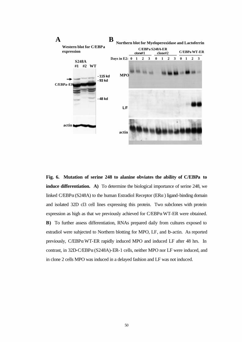
Fig. 6. Mutation of serine 248 to alanine obviates the ability of C/EBPα to
induce differentiation. A) To determine the biological importance of serine 248, we
linked C/EBPα(S248A) to the human Estradiol Receptor (ERα) ligand-binding domain
and isolated 32D cl3 cell lines expressing this protein. Two subclones with protein
expression as high as that we previously achieved for C/EBPαWT-ER were obtained.
B) To further assess differentiation, RNAs prepared daily from cultures exposed to
estradiol were subjected to Northern blotting for MPO, LF, and b-actin. As reported
previously, C/EBPαWT-ER rapidly induced MPO and induced LF after 48 hrs. In
contrast, in 32D-C/EBPα(S248A)-ER-1 cells, neither MPO nor LF were induced, and
in clone 2 cells MPO was induced in a delayed fashion and LF was not induced.
51
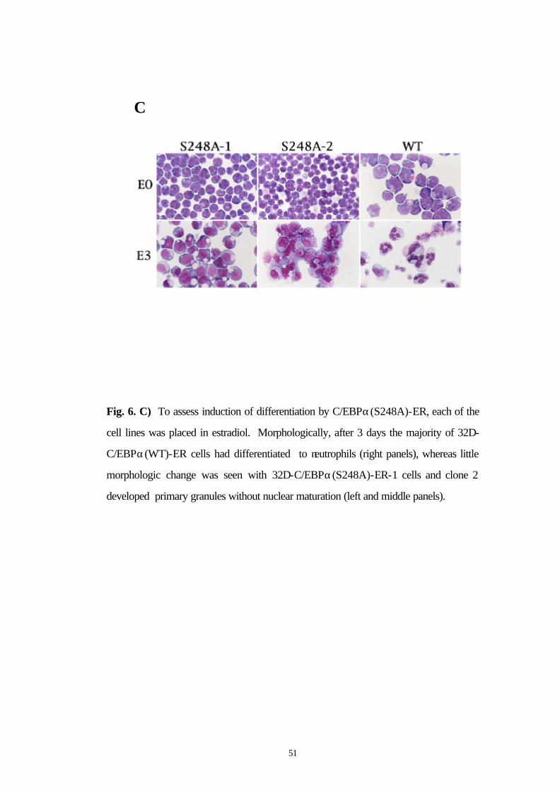
C
Fig. 6. C) To assess induction of differentiation by C/EBPα(S248A)-ER, each of the
cell lines was placed in estradiol. Morphologically, after 3 days the majority of 32D-
C/EBPα(WT)-ER cells had differentiated to neutrophils (right panels), whereas little
morphologic change was seen with 32D-C/EBPα(S248A)-ER-1 cells and clone 2
developed primary granules without nuclear maturation (left and middle panels).
52
4. DISCUSSION
As Ras signaling and the transcription factor C/EBPα play important
roles in myeloid differentiation, we hypothesized that Ras might be
involved in the activation of C/EBPα. Here we demonstrate that Ras
signaling phosphorylates C/EBPα on serine 248 of the transactivation
domain resulting in an enhancement of its transactivation capacity.
These data link the Ras pathway, which is involved in myelopoiesis
(Crespo and Leon, 2000) and acute myeloid leukemia(Schaich et al.,
2001; Zuber et al., 2000) to the differentiation inducing C/EBPα
pathway. Thus, activated Ras enhances the ability of C/EBPα to
transactivate the G-CSF receptor promoter and a minimal TK
promoter containing C/EBP DNA binding sites from 10 fold to 40 fold
(Fig. 1). The major role of phosphorylation sites in the activation and
deactivation of C/EBP family members has already been described by
other groups. Ford et al. suggested that C/EBPα exists in multiple
phosphorylated forms in the nucleus of both multipotential and
granulocytic-committed cells. (Ford et al., 1996) Furthermore, it has
been shown that C/EBPβ is expressed in both phosphorylated and
53
unphosphorylated forms in blasts from patients with acute myeloid
leukemia.(Iida et al., 2000) In terms of C/EBPβ, a regulation by the
Ras signal transduction pathway and Ras-induced phosphorylation of
C/EBPβ on threonine-235 is already known.(Nakajima et al., 1993)
Furthermore, the MAP kinases ERK1 and ERK2 have been reported to
mediate interferon-gamma responsiveness of C/EBPβ.(Ηυ ετ αλ.,
2001)
Regarding C/EBPα, Mahoney et al. reported that phosphorylation of
C/EBPα in vitro by protein kinase C attenuates its site-selective DNA
binding.(Mahoney et al., 1992) Subsequently, Pan et al. could identify
a specific increase in DNA binding and the expression of C/EBPα and
C/EBPβ during U937 monocytic cell differentiation, and related this to
the transforming growth factor β signaling pathway.(Pan et al., 1999)
Our data do not relate the effect of Ras signaling to the DNA binding
domain or DNA binding capacity of C/EBPα. Ras signaling acts on
the C/EBPα transactivation domain, because it enhances 4 fold the
transactivation function of a fusion protein containing a Gal4 DNA
binding domain and the C/EBPα transactivation domain (Fig. 2A), and
54
does not change the C/EBPα DNA binding capacity (Fig. 2C). In
fact, Ras acts on serine 248 of the C/EBPα transactivation domain,
because it does not enhance the 10-fold transactivation exhibited by a
C/EBPα mutant with a deletion of area 9 (Fig. 3A) or a serine 248 to
alanine point mutant in area 9 (Fig. 3C). Thus, we conclude that the
Ras pathway acts on the transactivation functions of C/EBPα, via
serine 248.
Ross et al. reported recently on other important phosphorylation sites
in the transactivation domain of C/EBPα.(Ross et al., 1999; Ross et
al., 2000) The insulin pathway activates GSK3 kinase which in turns
phosphorylates C/EBPα on threonines 222 and 226.(Ross et al., 1999)
This activation of C/EBPα is blocked by the Wnt signaling
pathway.(Ross et al., 2000) Wnt signaling maintains preadipocytes in
an undifferentiated state through inhibition of the adipogenic
transcription factors C/EBPα and peroxisome proliferator- activated
receptor gamma (PPARγ). When Wnt signaling in preadipocytes is
prevented by overexpression of axin or dominant-negative TCF4,
these cells differentiate into adipocytes.(Ross et al., 2000) Our data in
293T cells point to serine 248 as the major Ras-dependent

55
phosphorylation site of C/EBPα. Serine 248 of C/EBPα is a PKC
consensus site, and a PKC inhibitor blocks the activation of C/EBPα
by Ras (Fig. 4A). However, staurosporine is not completely specific
for PKC. Therefore, we have not excluded other kinases being
responsible for the activation of C/EBPα by Ras.
The serine to alanine mutant of C/EBPα did not completely eliminate
the activation of C/EBPα by Ras (Fig. 3C) whereas deletion of region
9 did (Fig. 3A). Perhaps a transcriptional co-activator binds optimally
to C/EBPα when serine 248 is phosphorylated (Fig. 5E), but can still
bind weakly to the S248A mutant. In this model the effect of Ras on
C/EBPα transactivation is at least partly indirect, via a co-activator.
This model is supported by the ability of Ras to induce basal activity
of p(C/EBP)TK; on the other hand our observation that Ras increases
phosphorylation of a C/EBPα peptide, but not of the same peptide
from C/EBPαS248A suggests a more direct effect on C/EBPα by Ras
(Fig. 5E). Because Ross et al. had reported that serine 230 was also
phosphorylated by insulin signaling in vivo, but not by GSK3,(Ross et
al., 1999; Ross et al., 2000) it is also possible that phosphorylation of
serine 230 might account for additional effects of Ras on C/EBPα.
56
However, we could not observe any loss of enhancement of
transactivation capacity of C/EBPα by Ras signaling using the serine
230 to alanine mutant of C/EBPα (data not shown, mutants kindly
provided by Dr. MacDougald, Ann Arbor). However, the very likely
additional Ras-dependent phosphorylation sites in C/EBPα besides
serine 248 (Fig. 5A-D) still need to be mapped and to be tested
functionally. Our current studies to identify interacting proteins and
interacting phosphoproteins of C/EBPα using proteomics techniques
(2D-gel electrophoresis of C/EBPα interacting proteins and
phosphoproteins and identification of those proteins by mass
spectrometry) might also contribute to answer the question whether the
effect of Ras signaling on serine 248 of C/EBPα requires a co-
activator binding to serine 248 or not.
Mutation of serine 248 to alanine had marked biologic consequences in
32D cl3 cells, obviating the ability of C/EBPα to induce differentiation
(Fig. 6). Whether this represents a defect in Ras-dependent
phosphorylation of this serine and consequent alteration of trans-
activating properties in myeloid cells or whether it represents a more
indirect effect in which a Ras-dependent pathway interacts with serine
57
248 or with another site in C/EBPα affected by mutation of serine 248
to alanine remains to be determined.
Further studies need to address the biological significance of serine
248 of C/EBPα by testing the serine to alanine 248 C/EBPα mutant in
biological assays of C/EBPα function such as in rescue assays in
C/EBPα knock-out mice. Of note, G-CSF, but not IL-3, signals
cooperate with C/EBPα to induce myeloid genes in Ba/F3 lymphoid
cells.(Wang et al., 2001) Because impairment of C/EBPα function can
contribute to the pathogenesis of acute myeloid leukemia,(Pabst et al.,
2001a; Pabst et al., 2001b) it needs to be addressed whether serine 248
of C/EBPα is mutated in patients with acute myeloid leukemia and/or
whether inactivating Ras mutations in acute myeloid leukemia might
lead to a loss of C/EBPα function.
In a nutshell, our data suggest so far that Ras signaling phosphorylates
C/EBPα on serine 248 of the transactivation domain resulting in an
enhancement of the ability of C/EBPα to transactivate the G-CSF
receptor promoter. Furthermore, our data suggest a model where Ras

58
signaling enhances the activity of C/EBPα to induce granulocytic
differentiation by phosphorylation of serine 248.
59
5. SUMMARY
The transcription factor C/EBPα regulates early steps of normal
granulocyte differentiation since mice with a disruption of the C/EBPα
gene do not express detectable levels of the G-CSF receptor and
produce no neutrophils. We have recently shown that C/EBPα
function is also impaired in acute myeloid leukemias. However, how
the transcriptional activity of C/EBPα is regulated both in myelopoiesis
and leukemogenesis, is not fully understood. The current study
demonstrates that activated Ras enhances the ability of C/EBPα to
transactivate the G-CSF receptor promoter and a minimal promoter
containing only C/EBP DNA binding sites. Ras signaling activates
C/EBPα via the transactivation domain, because it enhances the
transactivation function of a fusion protein containing a Gal4 DNA
binding domain and the C/EBPα transactivation domain, and does not
change C/EBPα DNA binding. Ras acts on serine 248 of the C/EBPα
transactivation domain, as it does not enhance the transactivation
function of a C/EBPα serine 248 to alanine point mutant. Interestingly,
serine 248 of C/EBPα is a PKC consensus site, and a PKC inhibitor
blocks the activation of C/EBPα by Ras. Ras signaling

60
phosphorylates C/EBPα on serine 248 in vivo. Finally, mutation of
serine 248 to alanine obviates the ability of C/EBPα to induce
granulocytic differentiation. These data suggest a model where Ras
signaling enhances the activity of C/EBPα to induce granulocytic
differentiation by phosphorylation of serine 248.
61
6. ZUSAMMENFASSUNG
Der Transkriptionsfaktor C/EBPα reguliert frühe Schritte in der
Differenzierung hämatopoetischer Vorläuferzellen zu Granulozyten. Bei
C/EBPα knockout Mäusen ist der G-CSF-Rezeptor nicht nachweisbar.
Diese Mäuse haben auch keine neutrophilen Granulozyten. Die Störung
der Funktion von C/EBPα bei Patienten mit akuten myeloischen
Leukämien ist bekannt. Es ist jedoch bislang weder die Regulation der
Effekte des Proteins C/EBPα auf die Myelopoese noch auf die
Leukämogenese zufriedenstellend beschrieben worden. Die vorliegende
Arbeit zeigt, daß aktiviertes Ras die Fähigkeit des
Transkriptionsfaktors C/EBPα, den Promoter des G-CSF Rezeptors
sowie einen Promoter, der nur die DNA bindenden Stellen enthält, zu
transaktivieren, verstärkt. Die Ras-Signaltransduktion ändert die
Bindung von C/EBPα an die DNA nicht und verstärkt die Fähigkeit der
Transaktivierung der beschriebenen Promotoren durch ein
Fusionsprotein aus einer Gal4 DNA bindenden Domäne und der
Transaktivierungsdomäne von C/EBPα. Also verstärkt Ras die
transkriptionelle Aktivität von C/EBPα in seiner
Transaktivierungsdomäne. In der vorliegenden Arbeit wurde die
62
Aminosäure, an der Ras C/EBPα aktiviert, bestimmt. Eine
Punktmutation, die die Aminosäure Serin 248 in C/EBPα zu Alanin
mutierte, führte zu einem Verlust der beschriebenen verstärkenden
Funktion des Protoonkogens Ras auf die transkriptionelle Aktivität von
C/EBPα. Die Aminosäure Serin 248 stellt eine PKC-Konsensus-Stelle
dar. Ein Inhibitor der Proteinkinase C konnte die durch Ras induzierte
Aktivierung von C/EBPα hemmen. Ras-Signaltransduktion führt in
vivo zu Phosphorylierung von C/EBPα. Ferner führte die Mutation
von Serin 248 zu Alanin zu einem Verlust der Fähigkeit C/EBPαs,
Differenzierung von Vorläuferzellen zu Granulozyten zu induzieren. Die
vorliegenden Daten unterstützen ein Modell, in dem Ras-
Signaltransduktion die Kapazität C/EBPαs, die Differenzierung zu der
granulozytischen Reihe zu induzieren, durch Phosphorylierung an Serin
248 verstärkt.
63
7. REFERENCES
Akira,S., Isshiki,H., Sugita,T., Tanabe,O., Kinoshita,S., Nishio,Y.,
Nakajima,T., Hirano,T., and Kishimoto,T. (1990)
A nuclear factor for IL-6 expression (NF-IL6) is a member of a C/EBP
family. EMBO J. 9 , 1897-1906.
Behre,G., Smith,L.T., and Tenen,D.G. (1997)
Use of a Renilla luciferase vector as a control for studies of Ras
transactivation. Blood 90, 158b.
Behre,G., Whitmarsh,A.J., Coghlan,M.P., Hoang,T., Carpenter,C.L.,
Zhang,D.-E., Davis,R.J., and Tenen,D.G. (1999)
c-Jun is a JNK independent coactivator of the PU.1 transcription factor.
Journal of Biological Chemistry 274, 4939-4946.
Boyle,W.J., van,d.G., and Hunter,T. (1991)
Phosphopeptide mapping and phosphoamino acid analysis by two-
dimensional separation on thin-layer cellulose plates. Methods Enzymol.
201:110-49, 110-149.
Cao,Z., Umek,R.M., and McKnight,S.L. (1991)
Regulated expression of three C/EBP isoforms during adipose conversion of
3T3-L1 cells. Genes Dev. 5, 1538-1552.
Chang,C.J., Chen,T.T., Lei,H.Y., Chen,D.S., and Lee,S.C. (1990)
Molecular cloning of a transcription factor, AGP/EBP, that belongs to
members of the C/EBP family. Mol. Cell Biol. 10, 6642-6653.
64
Crespo,P. and Leon,J. (2000)
Ras proteins in the control of the cell cycle and cell differentiation. Cell Mol.
Life Sci. 57 , 1613-1636.
Descombes,P., Chojkier,M., Lichtsteiner,S., Falvey,E., and Schibler,U.
(1990)
LAP, a novel member of the C/EBP gene family, encodes a liver-enriched
transcriptional activator protein. Genes Dev. 4, 1541-1551.
Feig,L.A. (1988)
Inhibition of NIH 3T3 cell proliferation by a mutant ras protein with
preferential affinity for GDP. Mol. Cell. Biol. 8, 3235-3243.
Feig,L.A. and Cooper,G.M. (1988)
Relationship among guanine nucleotide exchange, GTP hydrolysis, and
transforming potential of mutated ras proteins. Mol. Cell. Biol. 8, 2472-
2478.
Flodby,P., Barlow,C., Kylefjord,H., Ahrlund-Richter,L., and
Xanthopoulos,K.G. (1996)
Increased hepatic cell proliferation and lung abnormalities in mice deficient
in CCAAT/enhancer binding protein α. J Biol. Chem. 271, 24753-24760.
Ford,A.M., Bennett,C.A., Healy,L.E., Towatari,M., Greaves,M.F., and
Enver,T. (1996)
Regulation of the myeloperoxidase enhancer binding proteins PU.1, CEBPα,
β, and δ during granulocytic-lineage specification. Proc. Natl. Acad. Sci. USA
93, 10838-10843.
65
Friedman,A.D. (2002)
Transcriptional regulation of granulocyte and monocyte development.
Oncogene 21, 3377-3390.
Friedman,A.D. and McKnight,S.L. (1990)
Identification of two polypeptide segments of CCAAT/enhancer- binding
protein required for transcriptional activation of the serum albumin gene.
Genes Dev. 4, 1416-1426.
Gutkind,J.S. (1998)
The pathways connecting G protein-coupled receptors to the nucleus through
divergent mitogen-activated protein kinase cascades. J Biol. Chem. 273,
1839-1842.
Hibi,S., Lohler,J., Friel,J., Stocking,C., and Ostertag,W. (1993)
Induction of monocytic differentiation and tumorigenicity by v- Ha-ras in
differentiation arrested hematopoietic cells. Blood 81, 1841-1848.
Hong,S., Lee,M.Y., and Cheong,J. (2001)
Functional interaction of transcriptional coactivator ASC-2 and C/EBPalpha
in granulocyte differentiation of HL-60 promyelocytic cell. Biochem.
Biophys. Res. Commun. 282, 1257-1262.
Hu,J., Roy,S.K., Shapiro,P.S., Rodig,S.R., Reddy,S.P., Platanias,L.C.,
Schreiber,R.D., and Kalvakolanu,D.V. (2001)
ERK1 and ERK2 Activate CCAAAT/Enhancer-binding Protein-beta -
dependent Gene Transcription in Response to Interferon-gamma. J. Biol.
Chem. 276, 287-297.
66
Iida,H., Towatari,M., Iida,M., Tanimoto,M., Kodera,Y., Ford,A.M., and
Saito,H. (2000)
Protein expression and constitutive phosphorylation of hematopoietic
transcription factors PU.1 and C/EBP beta in acute myeloid leukemia blasts.
Int. J. Hematol. 71, 153-158.
Jin,D.I., Jameson,S.B., Reddy,M.A., Schenkman,D., and Ostrowski,M.C.
(1995)
Alterations in differentiation and behavior of monocytic phagocytes in
transgenic mice that express dominant suppressors of ras signaling. Mol.
Cell. Biol. 15, 693-703.
Johansen,L.M., Iwama,A., Lodie,T.A., Sasaki,K., Felsher,D.W.,
Golub,T.R., and Tenen,D.G. (2001)
c-Myc is a critical target for c/EBPalpha in granulopoiesis. Mol. Cell Biol.
21, 3789-3806.
Johnson,R., Spiegelman,B., Hanahan,D., and Wisdom,R. (1996)
Cellular transformation and malignancy induced by ras require c- jun. Mol
Cell Biol 16, 4504-4511.
Johnson,R.S., van Lingen,B., Papaioannou,V.E., and Spiegelman,B.M.
(1993)
A null mutation at the c-jun locus causes embryonic lethality and retarded cell
growth in culture. Genes Dev. 7, 1309-1317.
67
Lin,F.T. and Lane,M.D. (1994)
CCAAT/enhancer binding protein α is sufficient to initiate the 3T3-L1
adipocyte differentiation program. Proc. Natl. Acad. Sci. USA 91, 8757-
8761.
Maher,J., Baker,D., Dibb,N., and Roberts,I. (1996)
Mutant ras promotes haemopoietic cell proliferation or differentiation in a
cell-specific manner. Leukemia 10, 83-90.
Mahoney,C.W., Shuman,J., McKnight,S.L., Chen,H.C., and Huang,K.P.
(1992)
Phosphorylation of CCAAT-enhancer binding protein by protein kinase C
attenuates site-selective DNA binding. J Biol Chem. 267, 19396-19403.
Mccormick,F. (1995)
Ras-related proteins in signal transduction and growth control. Mol. Reprod.
Dev. 42, 500-506.
Nakajima,T., Kinoshita,S., Sasagawa,T., Sasaki,K., Naruto,M.,
Kishimoto,T., and Akira,S. (1993)
Phosphorylation at threonine-235 by a ras-dependent mitogen- activated
protein kinase cascade is essential for transcription factor NF-IL6. Proc.
Natl. Acad. Sci. USA 90, 2207-2211.
Nordeen,S.K. (1988)
Luciferase reporter gene vectors for analysis of promoters and enhancers.
Biotechniques 6, 454-458.
68
Oelgeschlager,M., Nuchprayoon,I., Luscher,B., and Friedman,A.D.
(1996)
C/EBP, c-Myb, and PU.1 cooperate to regulate the neutrophil elastase
promoter. Mol. Cell. Biol. 16, 4717-4725.
Pabst,T., Mueller,B.U., Harakawa,N., Schoch,C., Haferlach,T., Behre,G.,
Hiddemann,W., Zhang,D.E., and Tenen,D.G. (2001b)
AML1-ETO downregulates the granulocytic differentiation factor
C/EBPalpha in t(8;21) myeloid leukemia. Nat. Med. 7, 444-451.
Pabst,T., Mueller,B.U., Zhang,P., Radomska,H.S., Narravula,S.,
Schnittger,S., Behre,G., Hiddemann,W., and Tenen,D.G. (2001a).
Dominant-negative mutations of CEBPA, encoding CCAAT/enhancer binding
protein-alpha (C/EBPalpha), in acute myeloid leukemia. Nat. Genet. 27, 263-
270.
Pan,Z., Hetherington,C.J., and Zhang,D.E. (1999)
CCAAT/enhancer-binding protein activates the CD14 promoter and mediates
transforming growth factor beta signaling in monocyte development. J. Biol.
Chem. 274, 23242-23248.
Poli,V., Mancini,F.P., and Cortese,R. (1990)
IL-6DBP, a nuclear protein involved in interleukin-6 signal transduction,
defines a new family of leucine zipper proteins related to C/EBP. Cell 63,
643-653.
69
Radomska,H.S., Huettner,C.S., Zhang,P., Cheng,T., Scadden,D.T., and
Tenen,D.G. (1998)
CCAAT/Enhancer binding protein α is a regulatory switch sufficient for
induction of granulocytic development from bipotential myeloid progenitors.
Mol. Cell. Biol. 18, 4301-4309.
Ramji,D.P. and Foka,P. (2002)
CCAAT/Enhancer Binding Proteins: Structure, Function and Regulation.
Biochem. J. in press.
Roman,C., Platero,J.S., Shuman,J., and Calame,K. (1990)
Ig/EBP-1: a ubiquitously expressed immunoglobulin enhancer binding protein
that is similar to C/EBP and heterodimerizes with C/EBP. Genes Dev. 4,
1404-1415.
Ron,D. and Habener,J.F. (1992)
CHOP, a novel developmentally regulated nuclear protein that dimerizes with
transcription factors C/EBP and LAP and functions as a dominant-negative
inhibitor of gene transcription. Genes Dev. 6, 439-453.
Ross,S.E., Erickson,R.L., Hemati,N., and MacDougald,O.A. (1999)
Glycogen synthase kinase 3 is an insulin-regulated C/EBPalpha kinase. Mol.
Cell Biol. 19, 8433-8441.
Ross,S.E., Hemati,N., Longo,K.A., Bennett,C.N., Lucas,P.C.,
Erickson,R.L., and MacDougald,O.A. (2000)
Inhibition of adipogenesis by Wnt signaling. Science 289, 950-953.
70
Schaich,M., Ritter,M., Illmer,T., Lisske,P., Thiede,C., Schakel,U.,
Mohr,B., Ehninger,G., and Neubauer,A. (2001)
Mutations in ras proto-oncogenes are associated with lower mdr1 gene
expression in adult acute myeloid leukaemia. Br. J. Haematol. 112, 300-307.
Scott,L.M., Civin,C.I., Rorth,P., and Friedman,A.D. (1992)
A novel temporal expression pattern of three C/EBP family members in
differentiating myelomonocytic cells. Blood 80, 1725-1735.
Skorski,T., Szczylik,C., Ratajczak,M.Z., Malaguarnera,L., Gewirtz,A.M.,
and Calabretta,B. (1992)
Growth factor-dependent inhibition of normal hematopoiesis by N-ras
antisense oligodeoxynucleotides. J Exp. Med. 175, 743-750.
Smith,L.T., Hohaus,S., Gonzalez,D.A., Dziennis,S.E., and Tenen,D.G.
(1996)
PU.1 (Spi-1) and C/EBPα regulate the granulocyte colony-stimulating factor
receptor promoter in myeloid cells. Blood 88, 1234-1247.
Umek,R.M., Friedman,A.D., and McKnight,S.L. (1991)
CCAAT-enhancer binding protein: a component of a differentiation switch.
Science 251, 288-292.
Wang Qf,Q.F. and Friedman,A.D. (2002)
CCAAT/enhancer-binding proteins are required for granulopoiesis
independent of their induction of the granulocyte colony-stimulating factor
receptor. Blood 99, 2776-2785.
71
Wang,W., Wang,X., Ward,A.C., Touw,I.P., and Friedman,A.D. (2001)
C/EBPalpha and G-CSF receptor signals cooperate to induce the
myeloperoxidase and neutrophil elastase genes. Leukemia 15, 779-786.
Wang,X., Scott,E., Sawyers,C.L., and Friedman,A.D. (1999)
C/EBPalpha bypasses granulocyte colony-stimulating factor signals to rapidly
induce PU.1 gene expression, stimulate granulocytic differentiation, and limit
proliferation in 32D cl3 myeloblasts. Blood 94, 560-571.
Wasylyk,C., Maira,S.M., Sobieszczuk,P., and Wasylyk,B. (1994)
Reversion of Ras transformed cells by Ets transdominant mutants. Oncogene
9, 3665-3673.
Westendorf,J.J., Yamamoto,C.M., Lenny,N., Downing,J.R., Selsted,M.E.,
and Hiebert,S.W. (1998)
The t(8;21) fusion product, AML-1-ETO, associates with C/EBP-alpha,
inhibits C/EBP-alpha-dependent transcription, and blocks granulocytic
differentiation. Mol. Cell Biol. 18, 322-333.
Williams,S.C., Cantwell,C.A., and Johnson,P.F. (1991)
A family of C/EBP-related proteins capable of forming covalently linked
leucine zipper dimers in vitro. Genes Dev. 5, 1553-1567.
Zhang,D.-E., Hetherington,C.J., Meyers,S., Rhoades,K.L., Larson,C.J.,
Chen,H.M., Hiebert,S.W., and Tenen,D.G. (1996)
CCAAT enhancer binding protein (C/EBP) and AML1 (CBFα2)
synergistically activate the M-CSF receptor promoter. Mol. Cell. Biol. 16,
1231-1240.

72
Zhang,D.-E., Zhang,P., Wang,N.D., Hetherington,C.J., Darlington,G.J.,
and Tenen,D.G. (1997)
Absence of granulocyte colony-stimulating factor signaling and neutrophil
development in CCAAT enhancer binding protein α-deficient mice. Proc.
Natl. Acad. Sci. USA 94, 569-574.
Zhang,P., Behre,G., Pan J, Iwama,A., Wara-aswapati,N., Radomska,H.S.,
Auron,P.E., Tenen,D.G., and Sun,Z. (1999)
Negative cross-talk between hematopoietic regulators: GATA proteins
repress PU.1 function. Proc Natl Acad Sci U S A 96, 8705-8710.
Zhang,P., Nelson,E., Radomska,H.S., Iwasaki-Arai,J., Akashi,K.,
Friedman,A.D., and Tenen,D.G. (2002)
Induction of granulocytic differentiation by 2 pathways. Blood 99, 4406-
4412.
Zhu,J. and Emerson,S.G. (2002)
Hematopoietic cytokines, transcription factors and lineage commitment.
Oncogene 21, 3295-3313.
Zuber,J., Tchernitsa,O.I., Hinzmann,B., Schmitz,A.C., Grips,M.,
Hellriegel,M., Sers,C., Rosenthal,A., and Schafer,R. (2000)
A genome-wide survey of RAS transformation targets. Nat. Genet. 24, 144-
152.

73
8. ACKNOWLEDGEMENTS
I am very grateful to Prof. Wolfgang Hiddemann and Dr. Gerhard
Behre for providing the opportunity to work in their research lab at
Med III, Klinikum Großhadern, LMU, Munich.
I thank Prof. Alan Friedman and Dr. H. Liu of Johns Hopkins
Oncology Center, Baltimore, Maryland for doing the retroviral
transduction assays during the course of this work.
My colleagues (Arun, Abdul, Annika, Venkat, Rajani, Janki, Max,
Michi, Deepak, Alex, Nicolei, Matthias, Roman and Mulu to name a
few of them) have been very helpful in the lab and during scientific
discussions.
I must thank Julius, Sabrina, Manu and Brian for taking me out in
Munich beer gardens and pubs and for showing me that another world
also exists away from the lab.
And finally, this work was not possible without the support and
encouragement of my wife Poonam, who had the patience to live 7000
miles away from me for 3 years.
74
9. Curriculum Vitae
Name: Sheo Mohan Singh Date of Birth: 01 July 1971 Nationality: Indian Postal Address: Pfingstrosenstr. 64 App. 3/107 81377 Munich Germany Tel: 0049 89 7094 2964 (home), 0049 179 5249050 (mobile) Email: [email protected] Research Experience: Since June/2002 − Postdoc., Department of Medicine III, Grosshadern Hospital, University of Munich and GSF Hämatologikum, KKG Leukemia, Munich, Germany. Projects: 1. Target proteins of C/EBPα wild type and C/EBPα S248A mutant by proteomics. 2. Proteomics of AML1-ETO target proteins in t(8;21) myeloid leukemia. Feb/1999 – May/2002 Ph.D., Summa cum laude, Department of Medicine III, Grosshadern Hospital, University of Munich and GSF Hämatologikum, KKG Leukemia, Munich, Germany. Thesis title: Ras signaling enhances the activity of C/EBPα to induce granulocytic differentiation by phosphorylation of serine 248. March/1998 – Sept/1998 Senior Research Fellow, National Research Centre on Plant Biotechnology, IARI Campus, New Delhi, Worked on DBT project “Genetic engineering of Pigeonpea (Cajanus cajan L.) for insect resistance”. Aug/1997 – Feb/1998 Research Student, International Centre for Genetic Engineering and Biotechnology (ICGEB), New Delhi, Worked on “Molecular biology of Bacillus thuringiensis”.
75
Education: Oct/1994 – Oct/1995 Master of Science in Biotechnology, University of Kent at Canterbury, U.K. Thesis Title: Biochemical characterization of ectomycorrhizal fungi. Aug/1989 – July/1994 B.Sc. (Ag. & A.H.), First Class, Overall Grade Point 9.02 out of 10.00 C.S.A. University of Agriculture & Technology, Kanpur, India Membership in Professional Societies:
1. American Society of Hematology (ASH), USA 2. German Society of Hematology and Oncology (DGHO), Germany 3. Society for Biochemistry and Molecular Biology (GBM), Germany 4. The Biochemical Society, United Kingdom 5. Association of Microbiologists of India (Life member)
Talks: 1. Annual Meeting, American Society of Hematology (ASH), 6-10 December 2002,
Philadelphia, USA Title: Proteomics of AML1-ETO target proteins in t(8;21) myeloid leukemia
2. Annual meeting, German Society of Hematology and Oncology, 27-30 October
2002, Munich, Germany Title: Proteomics of AML1-ETO target proteins in t(8;21) myeloid leukemia
3. 2nd Harvard/Munich AML Workshop, March 10-13, 2000, Eibsee, Germany
Title: Ras signaling towards C/EBPa. 4. Annual meeting, German Society of Hematology and Oncology, 3 – 6 October
1999, Jena, Germany Title: Ras enhances the ability of C/EBPa to transactivate G-CSF receptor promoter by phosphorylating at S248 of C/EBPa transactivation domain.
List of publications:
1. Singh,S.M., Meisel,A., Kohlmann,A., Zhang,D.E., Haferlach,T., Tenen,D.G., Hiddemann,W., Behre,G. (2002). Proteomics of AML1-ETO target proteins in t(8;21) myeloid leukemia. Abstract 309 in Blood 100(11):84a, Talk at ASH 2002
76
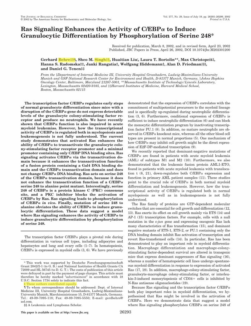
2. Singh,S.M.*, Behre,G.*, Liu,H., Bortolin,L.T., Christopeit,M., Radomska,H.S., Rangatia,J., Hiddemann,W., Friedman,A.D., Tenen,D.G. (2002). Ras signaling enhances the activity of C/EBPα to induce granulocytic differentiation by phosphorylation of serine 248. Journal of Biological Chemistry, 277:26293-26299, *Dual First Authors.
3. Behre,G., Reddy,V.A., Tenen,D.G., Hiddemann,W., Peer Zada, A.A., Singh,S.M. (2002). Proteomic analysis of transcription factor interactions in myeloid stem cell development and leukemia. Expert Opinion on Therapeutic Targets, 6:491-495
4. Peer Zada,A.A., Singh,S.M., Reddy,V.A., Meisel,A., Elsässer,A., Haferlach,T., Tenen,D.G., Hiddemann,W., Behre,G. (2003). CD44 ligation inhibits proliferation in acute myeloid leukemia cells by downregulating c-Jun expression and blocking cell cycle. Oncogene 22(15):2296-308
5. Vangala,R.K., Neumann,M.S., Rangatia,J., Singh,S.M., Tenen,D.G., Hiddemann,W., Behre,G. (2003). The myeloid master regulator PU.1 is inactivated by AML1-ETO in t(8;21) myeloid leukemia. Blood, 101:270-277
6. Rangatia,J., Vangala,R.K., Singh,S.M., Peer Zada,A.A., Elsässer,A., Kohlmann,A., Haferlach,T., Tenen,D.G., Hiddemann,W., Behre,G. (2003). Eleveated c-Jun expression in acute myeloid leukemias inhibits C/EBPα DNA binding via lucine zipper domain interaction. Oncogene 22(30):4760-4
7. Singh, D.M., Sonia, Sahoo, L., Singh, S.M., and Jaiwal, P.K. (1998). Biotechnological approaches for the improvement of Pigeonpea (Cajanus cajan (L.) Millsp.). In Recent Advances in Biotechnology (Ed. P.C. Trivedi), Jaipur, India
8. Singh, S.M. (1997). Degradation of polychlorinated biphenyls (PCBs) by isolates of ectomycorrhizal fungi Pisolithus arhizus. Indian Journal of Microbiology 37 (March 1997): 47-48
9. Singh, S.M. and Jefferies, P. (1997). Carbon and nitrogen nutrition in ectomycorrhizal fungi Pisolithus arhizus. Indian Journal of Microbiology 37 (June 1997): 69-72
10. Singh, S.M. and Mukerji, K.G. (1996). Shape of biotechnology to come. The Botanica 46: 13-17
Ras Signaling Enhances the Activity of C/EBP� to InduceGranulocytic Differentiation by Phosphorylation of Serine 248*
Received for publication, March 8, 2002, and in revised form, April 23, 2002Published, JBC Papers in Press, April 26, 2002, DOI 10.1074/jbc.M202301200
Gerhard Behre‡§¶, Sheo M. Singh‡§, Huaitian Liu�, Laura T. Bortolin**, Max Christopeit‡,Hanna S. Radomska‡‡, Janki Rangatia‡, Wolfgang Hiddemann‡, Alan D. Friedman�§§,and Daniel G. Tenen‡‡
From the ‡Department of Internal Medicine III, University Hospital Grosshadern, Ludwig-Maximilians-UniversityMunich and GSF-National Research Center for Environment and Health, D-81377 Munich, Germany, � Johns HopkinsOncology Center, Baltimore, Maryland 21287-5001, **Massachusetts Institute of Technology/Lincoln Laboratory,Lexington, Massachusetts 02420-9185, and ‡‡Harvard Institutes of Medicine, Harvard Medical School,Boston, Massachusetts 02115
The transcription factor C/EBP� regulates early stepsof normal granulocyte differentiation since mice with adisruption of the C/EBP� gene do not express detectablelevels of the granulocyte colony-stimulating factor re-ceptor and produce no neutrophils. We have recentlyshown that C/EBP� function is also impaired in acutemyeloid leukemias. However, how the transcriptionalactivity of C/EBP� is regulated both in myelopoiesis andleukemogenesis is not fully understood. The currentstudy demonstrates that activated Ras enhances theability of C/EBP� to transactivate the granulocyte colo-ny-stimulating factor receptor promoter and a minimalpromoter containing only C/EBP DNA binding sites. Rassignaling activates C/EBP� via the transactivation do-main because it enhances the transactivation functionof a fusion protein containing a Gal4 DNA binding do-main and the C/EBP� transactivation domain and doesnot change C/EBP� DNA binding. Ras acts on serine 248of the C/EBP� transactivation domain, because it doesnot enhance the transactivation function of a C/EBP�serine 248 to alanine point mutant. Interestingly, serine248 of C/EBP� is a protein kinase C (PKC) consensussite, and a PKC inhibitor blocks the activation ofC/EBP� by Ras. Ras signaling leads to phosphorylationof C/EBP� in vivo. Finally, mutation of serine 248 toalanine obviates the ability of C/EBP� to induce granu-locytic differentiation. These data suggest a modelwhere Ras signaling enhances the activity of C/EBP� toinduce granulocytic differentiation by phosphorylationof serine 248.
The transcription factor C/EBP� plays a pivotal role duringdifferentiation in various cell types, including adipocytes andhepatocytes and lung and ovary cells (1–7). In hematopoiesis,C/EBP� is expressed in myeloid cells (3, 6). We have previously
demonstrated that the expression of C/EBP� correlates with thecommitment of multipotential precursors to the myeloid lineageand is specifically up-regulated during neutrophilic differentia-tion (3, 6). Furthermore, conditional expression of C/EBP� issufficient to induce neutrophilic differentiation (6) and can blockthe monocytic differentiation program by inactivating transcrip-tion factor PU.1 (8). In addition, no mature neutrophils are ob-served in C/EBP� knockout mice, whereas all the other blood celltypes are present in normal proportions (5). One mechanism ofhow C/EBP� may inhibit cell growth might be the direct repres-sion of E2F-DP-mediated transcription (9).
We recently reported that dominant-negative mutations ofC/EBP� are found in patients with acute myeloid leukemia(AML) of subtypes M1 and M2 (10). Furthermore, we alsodemonstrated that the leukemic fusion protein AML1-ETO,found in patients with acute myeloid leukemia with transloca-tion t (8, 21), down-regulates both C/EBP� expression andfunction in primary AML patient samples (11). These studiespoint to the crucial role of C/EBP� in both normal myeloiddifferentiation and leukemogenesis. However, how the tran-scriptional activity of C/EBP� is regulated both in normalmyelopoiesis as well as in leukemogenesis is not fullyunderstood.
The Ras family of proteins are GTP-dependent molecularswitches that are essential for cell growth and differentiation (12,13). Ras exerts its effect on cell growth mainly via ETS (14) andAP-1 (15) transcription factors. For example, cells with a nullmutation in the c-jun gene and expressing oncogenic Ras lackmany characteristics of Ras transformation (15), and dominantnegative mutants of ETS-1, ETS-2, or PU.1 containing only theDNA binding domain inhibit Ras activation of transcription andrevert Ras-transformed cells (14). In particular, Ras has beendemonstrated to play an important role in myeloid differentia-tion. Macrophage differentiation and macrophage-colony-stimulating factor-dependent survival are altered in transgenicmice that express dominant suppressors of Ras signaling (16),whereas a number of hematopoietic cell lines undergo spontane-ous myeloid differentiation in response to expression of activatedRas (17, 18). In addition, macrophage-colony-stimulating factor,granulocyte-macrophage colony-stimulating factor, or interleu-kin-3-induced monocytopoiesis of CD34� cells is inhibited byN-Ras antisense oligonucleotides (19).
Because Ras signaling and the transcription factor C/EBP�both play important roles in myeloid differentiation, we hy-pothesized that Ras might be involved in the activation ofC/EBP�. Here we demonstrate data that suggest a modelwhere Ras signaling phosphorylates C/EBP� on serine 248 of
* This work was supported by Deutsche ForschungsgemeinschaftGrant 2042/2-1 (to G. B.) and National Institutes of Health Grants CA72009 and HL 56745 (to D. G. T.). The costs of publication of this articlewere defrayed in part by the payment of page charges. This article musttherefore be hereby marked “advertisement” in accordance with 18U.S.C. Section 1734 solely to indicate this fact.
§ These authors contributed equally.¶ To whom correspondence should be addressed: Dept. of Internal
Medicine III, University Hospital Grosshadern, Ludwig-Maximilians-University Munich, Marchioninistrasse 15, D-81377 Munich, Germany.Tel.: 49-89-7095-118; Fax: 49-89-7095-5550; E-mail: [email protected].
§§ A Leukemia and Lymphoma Scholar.
THE JOURNAL OF BIOLOGICAL CHEMISTRY Vol. 277, No. 29, Issue of July 19, pp. 26293–26299, 2002© 2002 by The American Society for Biochemistry and Molecular Biology, Inc. Printed in U.S.A.
This paper is available on line at http://www.jbc.org 26293
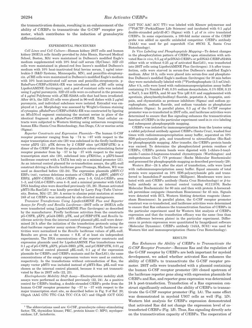
the transactivation domain, resulting in an enhancement of theability of C/EBP� to transactivate the G-CSF1 receptor pro-moter, which contributes to the induction of granulocyticdifferentiation.
EXPERIMENTAL PROCEDURES
Cell Lines and Cell Culture—Human kidney 293T cells and humankidney 293E1A cells (kindly provided by John Blenis, Harvard MedicalSchool, Boston, MA) were maintained in Dulbecco’s modified Eagle’smedium supplemented with 10% fetal calf serum (HyClone). 32D cl3cells were maintained in phenol-red free Iscove’s modified Dulbecco’smedium with 10% heat-inactivated fetal bovine serum, 1 ng/ml inter-leukin-3 (R&D Systems, Minneapolis, MN), and penicillin-streptomy-cin. �CRE cells were maintained in Dulbecco’s modified Eagle’s mediumwith 10% heat-inactivated calf serum and penicillin-streptomycin. p-BabePuro-C/EBP�(S248A)-ER was introduced into �CRE cells usingLipofectAMINE (Invitrogen), and a pool of resistant cells was isolatedusing 2 �g/ml puromycin. 32D cl3 cells were co-cultured in the presenceof 4 �g/ml Polybrene with �CRE-S248A cells that had been irradiated.After 48 h, the 32D cl3 cells were placed in 96-well dishes with 2 �g/mlpuromycin, and individual subclones were isolated. Estradiol was em-ployed at 1 �M. Morphology was assessed by Wright’s-Giemsa stainingof cytospins. pBabePuro-C/EBP�(S48A)-ER was constructed by ligatingan MluI/NcoI segment containing the mutant serine in place of theidentical fragment in pBabePuro-C/EBP�WT-ER. Total cellular ex-tracts were subjected to Western blotting using antiserum recognizinghuman ER� (Santa Cruz Biotechnology, San Diego, CA) or murine actin(Sigma).
Reporter Constructs and Expression Plasmids—The human G-CSFreceptor promoter ranging from bp �74 to �67 with respect to themajor transcription start site (20) was subcloned in the firefly luciferasevector pXP2 (21). pTK driven by 2 C/EBP sites (p(C/EBP)2TK) is adimer of the C/EBP site from the granulocyte colony-stimulating factorreceptor promoter from bp �57 to bp �37 with respect to the majortranscription start site (20), subcloned into pTK81luc, a pXP2-basedluciferase construct with a TATA box only as a minimal promoter (21).As an internal control plasmid for co-transfection assays, the pRL-nullconstruct driving a Renilla luciferase gene (Promega, Madison, WI) wasused as described before (22–24). The expression plasmids pMSV-C/EBP� (rat), various deletions mutants of C/EBP� in pMSV, pMSV-C/EBP�, pMSV-C/EBP�, GAL4-C/EBP� area 1–9, GAL4-C/EBP� area1–3, and GAL4-C/EBP� area 4–9, and a minimal promoter with GAL4DNA binding sites were described previously (25, 26). Human activatedpMT3-Ha-Ras(L61) was kindly provided by Larry Feig (Tufts Univer-sity, Boston, MA) (27, 28). A serine to alanine point mutation at serine248 of rat C/EBP� was introduced by site-directed mutagenesis.
Transient Transfections Using LipofectAMINE Plus and ReporterAssays for Firefly and Renilla Luciferase—293T cells or 293E1A cellswere transfected using LipofectAMINE Plus (Invitrogen) as describedby the manufacturer. Firefly luciferase activities from the constructspG-CSFR, pXP2, pGal4-DBD, pTK, and p(C/EBP)2TK and Renilla lu-ciferase activity from the internal control plasmid pRL-null were deter-mined 24 h after the initiation of the transfection protocols using thedual-luciferase reporter assay system (Promega). Firefly luciferase ac-tivities were normalized to the Renilla luciferase values of pRL-null.Results are given as the means � S.E. of at least six independentexperiments. The DNA concentrations of the reporter constructs andexpression plasmids used for LipofectAMINE Plus transfections were0.1 �g of pG-CSFR, pXP2, pGal4-DBD, pTK, and p(C/EBP)2TK, 0.01 �gof the internal control plasmid pRL-null, 0.1 �g of the expressionplasmids for C/EBP� and C/EBP� mutants and for Ras(L61); the sameconcentrations of the empty expression vectors were used as controls,respectively. In the transfections without cotransfection of Ras, theempty vector pMT3 was included instead of pMT3-Ras. pRL-null waschosen as the internal control plasmid, because it was not transacti-vated by Ras in 293T cells (22, 23).
Electrophoretic Mobility Shift Assay—Electrophoretic mobility shiftassays were performed as described previously (22, 24). As a positivecontrol for C/EBP� binding, a double-stranded C/EBP� probe from thehuman G-CSF receptor promoter (bp �57 to �37 with respect to themajor transcription start site, as described before in Fig. 9 of Ref. 20;OligoA (AAG GTG TTG CAA TCC CCA GC) and OligoB (GCT GGG
GAT TGC AAC ACC TT)) was labeled with Klenow polymerase and[32P]dCTP (PerkinElmer Life Sciences) and incubated with 0.1 �g/�ldouble-stranded poly(dI-dC) (Sigma) with 1 �l of in vitro translatedC/EBP�. In some experiments, a 100-fold molar excess of the C/EBPprobe was added as specific unlabeled competitor. C/EBP� antibody(14AA) was used for gel supershift (Cat #SC61 X, Santa CruzBiotechnology).
In Vivo Labeling and Phosphopeptide Mapping—To detect changesin the phosphorylation pattern of C/EBP� upon stimulation with acti-vated Ras in vivo, 0.5 �g of pcDNA3-C/EBP� or pcDNA3-C/EBP�S248Aeither with or without 0.25 �g of activated Ras(L61), was transfectedinto 293T cells using LipofectAMINE Plus (Invitrogen). 3 h after trans-fection, cells were starved in serum-free Dulbecco’s modified Eagle’smedium. After 18 h, cells were placed into serum-free and phosphate-free Dulbecco’s modified Eagle’s medium (Invitrogen) for 30 min beforethey were metabolically labeled with [32P]orthophosphate (2.5 mCi/ml).After 4 h, cells were lysed with radioimmunoprecipitation assay buffercontaining 1% Nonidet P-40, 0.5% sodium deoxycholate, 0.1% SDS, 0.15M NaCl, 5 mM EDTA, and 50 mM Tris (pH 8.0) and supplemented withaprotinin, phenylmethylsulfonyl fluoride, pepstatin A, leupeptin, anti-pain, and chymostatin as protease inhibitors (Sigma) and sodium py-rophosphate, sodium fluoride, and sodium vanadate as phosphataseinhibitors (Sigma). In parallel plates, 0.3 �g of the G-CSF receptorpromoter was co-transfected in 293T cells, and luciferase activities weredetermined to ensure that Ras signaling enhances the transactivationfunction of C/EBP� in the particular experiment used in in vivo labelingand subsequent phosphopeptide mapping.
C/EBP� was isolated by immunoprecipitation from the lysates usinga rabbit polyclonal antibody against C/EBP� (Santa Cruz), washed fourtimes with radioimmunoprecipitation assay buffer, separated on 10%SDS-polyacrylamide gels, and transferred to nitrocellulose (Bio-Rad)for phosphopeptide mapping. After transfer, the C/EBP� protein bandswas excised. To determine the phosphorylated protein residues ofC/EBP�, C/EBP� protein bands were digested with 1-chloro-3-tosyl-amido-7-amino-2-heptanone-treated chymotrypsin (Worthington) andendoproteinase Glu-C (V8 protease) (Roche Molecular Biochemicals)and processed for phosphopeptide mapping as described previously (29).
Western Blot—24 h after the start of transfection, cells were lysedwith radioimmunoprecipitation assay buffer. Equal amounts of totalprotein were separated on 10% SDS-polyacrylamide gels and trans-ferred to Immobilon-P membrane (Millipore). Membranes were incu-bated with anti-C/EBP� antibody (Santa Cruz Biotechnology) or �-tu-bulin antibody as an internal control (catalog #1111876; RocheMolecular Biochemicals) for 60 min and then with protein A-horserad-ish peroxidase conjugate (Amersham Biosciences) for 45 min. Signalswere detected with ECL Western blotting detection reagents (Amer-sham Biosciences). In parallel plates, the G-CSF receptor promoterconstruct was co-transfected, and luciferase activities were determinedto ensure that Ras enhances the transactivation function of C/EBP� inthe particular experiment used for Western blot analysis of C/EBP�expression and that the transfection efficacy was the same (less than10% difference between plates) in the particular experiment. Differ-ences in protein expression were quantitated by ImageQuant software(Molecular Dynamics). C/EBP� antibody (14AA, SC61) was used forWestern blot and immunoprecipitation (Santa Cruz Biotechnology).
RESULTS
Ras Enhances the Ability of C/EBP� to Transactivate theG-CSF Receptor Promoter—Because Ras and the regulation ofthe G-CSF receptor by C/EBP� are both important for myeloiddevelopment, we asked whether activated Ras enhances theability of C/EBP� to transactivate the G-CSF receptor pro-moter. 293T cells were transfected with a plasmid containingthe human G-CSF receptor promoter (20) cloned upstream ofthe luciferase reporter gene along with expression plasmids forC/EBP� and Ras, and reporter gene expression was determined24 h post-transfection. Transfection of a Ras expression con-struct significantly enhanced the ability of C/EBP� to transac-tivate the G-CSF receptor promoter (Fig. 1A). The same effectwas demonstrated in myeloid U937 cells as well (Fig. 1D).Western blot analysis for C/EBP� expression demonstratedthat activated Ras did not change the expression level of co-transfected C/EBP� (Fig. 1B). Thus, Ras signaling directly actson the transactivation capacity of C/EBP�. The cooperation of
1 The abbreviations used are: G-CSF, granulocyte colony-stimulatingfactor; TK, thymidine kinase; PKC, protein kinase C: MPO, myeloper-oxidase; LF, lactoferrin.
Ras Activates C/EBP�26294
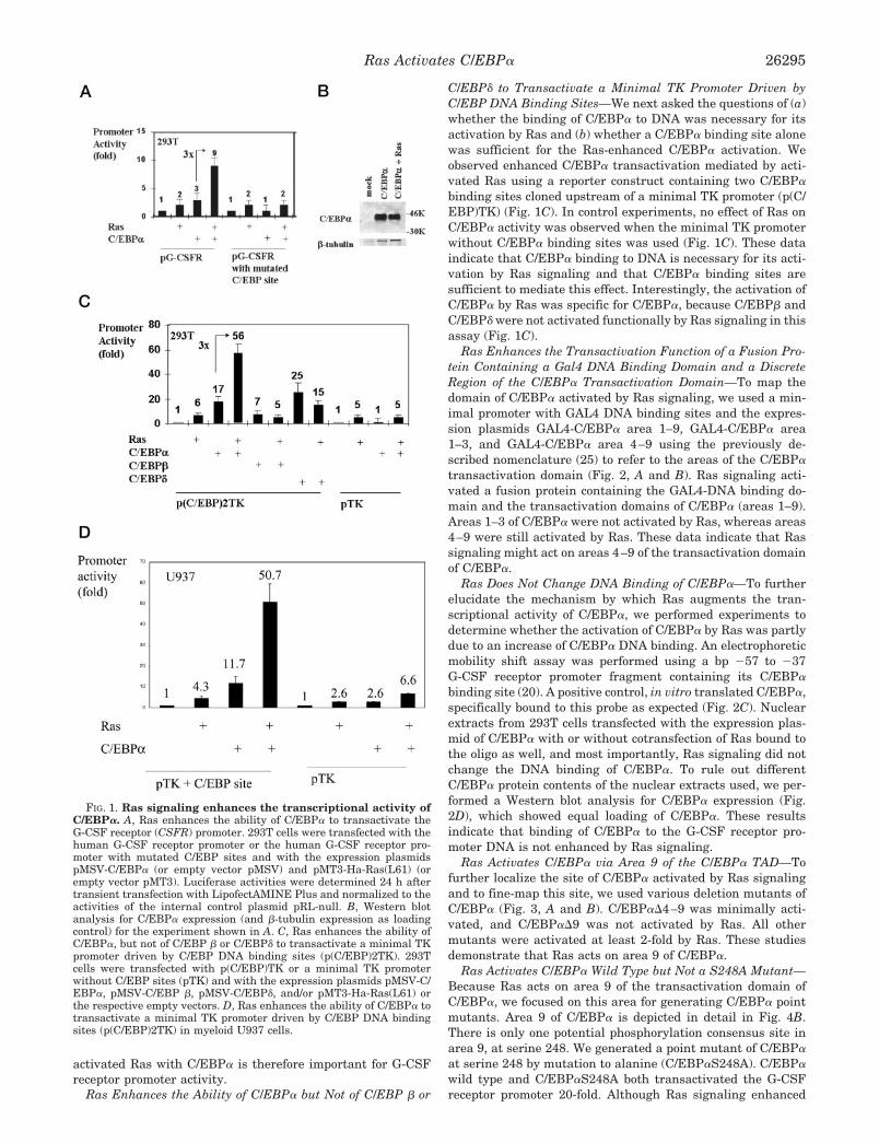
activated Ras with C/EBP� is therefore important for G-CSFreceptor promoter activity.
Ras Enhances the Ability of C/EBP� but Not of C/EBP � or
C/EBP� to Transactivate a Minimal TK Promoter Driven byC/EBP DNA Binding Sites—We next asked the questions of (a)whether the binding of C/EBP� to DNA was necessary for itsactivation by Ras and (b) whether a C/EBP� binding site alonewas sufficient for the Ras-enhanced C/EBP� activation. Weobserved enhanced C/EBP� transactivation mediated by acti-vated Ras using a reporter construct containing two C/EBP�
binding sites cloned upstream of a minimal TK promoter (p(C/EBP)TK) (Fig. 1C). In control experiments, no effect of Ras onC/EBP� activity was observed when the minimal TK promoterwithout C/EBP� binding sites was used (Fig. 1C). These dataindicate that C/EBP� binding to DNA is necessary for its acti-vation by Ras signaling and that C/EBP� binding sites aresufficient to mediate this effect. Interestingly, the activation ofC/EBP� by Ras was specific for C/EBP�, because C/EBP� andC/EBP� were not activated functionally by Ras signaling in thisassay (Fig. 1C).
Ras Enhances the Transactivation Function of a Fusion Pro-tein Containing a Gal4 DNA Binding Domain and a DiscreteRegion of the C/EBP� Transactivation Domain—To map thedomain of C/EBP� activated by Ras signaling, we used a min-imal promoter with GAL4 DNA binding sites and the expres-sion plasmids GAL4-C/EBP� area 1–9, GAL4-C/EBP� area1–3, and GAL4-C/EBP� area 4–9 using the previously de-scribed nomenclature (25) to refer to the areas of the C/EBP�
transactivation domain (Fig. 2, A and B). Ras signaling acti-vated a fusion protein containing the GAL4-DNA binding do-main and the transactivation domains of C/EBP� (areas 1–9).Areas 1–3 of C/EBP� were not activated by Ras, whereas areas4–9 were still activated by Ras. These data indicate that Rassignaling might act on areas 4–9 of the transactivation domainof C/EBP�.
Ras Does Not Change DNA Binding of C/EBP�—To furtherelucidate the mechanism by which Ras augments the tran-scriptional activity of C/EBP�, we performed experiments todetermine whether the activation of C/EBP� by Ras was partlydue to an increase of C/EBP� DNA binding. An electrophoreticmobility shift assay was performed using a bp �57 to �37G-CSF receptor promoter fragment containing its C/EBP�
binding site (20). A positive control, in vitro translated C/EBP�,specifically bound to this probe as expected (Fig. 2C). Nuclearextracts from 293T cells transfected with the expression plas-mid of C/EBP� with or without cotransfection of Ras bound tothe oligo as well, and most importantly, Ras signaling did notchange the DNA binding of C/EBP�. To rule out differentC/EBP� protein contents of the nuclear extracts used, we per-formed a Western blot analysis for C/EBP� expression (Fig.2D), which showed equal loading of C/EBP�. These resultsindicate that binding of C/EBP� to the G-CSF receptor pro-moter DNA is not enhanced by Ras signaling.
Ras Activates C/EBP� via Area 9 of the C/EBP� TAD—Tofurther localize the site of C/EBP� activated by Ras signalingand to fine-map this site, we used various deletion mutants ofC/EBP� (Fig. 3, A and B). C/EBP��4–9 was minimally acti-vated, and C/EBP��9 was not activated by Ras. All othermutants were activated at least 2-fold by Ras. These studiesdemonstrate that Ras acts on area 9 of C/EBP�.
Ras Activates C/EBP� Wild Type but Not a S248A Mutant—Because Ras acts on area 9 of the transactivation domain ofC/EBP�, we focused on this area for generating C/EBP� pointmutants. Area 9 of C/EBP� is depicted in detail in Fig. 4B.There is only one potential phosphorylation consensus site inarea 9, at serine 248. We generated a point mutant of C/EBP�
at serine 248 by mutation to alanine (C/EBP�S248A). C/EBP�
wild type and C/EBP�S248A both transactivated the G-CSFreceptor promoter 20-fold. Although Ras signaling enhanced
FIG. 1. Ras signaling enhances the transcriptional activity ofC/EBP�. A, Ras enhances the ability of C/EBP� to transactivate theG-CSF receptor (CSFR) promoter. 293T cells were transfected with thehuman G-CSF receptor promoter or the human G-CSF receptor pro-moter with mutated C/EBP sites and with the expression plasmidspMSV-C/EBP� (or empty vector pMSV) and pMT3-Ha-Ras(L61) (orempty vector pMT3). Luciferase activities were determined 24 h aftertransient transfection with LipofectAMINE Plus and normalized to theactivities of the internal control plasmid pRL-null. B, Western blotanalysis for C/EBP� expression (and �-tubulin expression as loadingcontrol) for the experiment shown in A. C, Ras enhances the ability ofC/EBP�, but not of C/EBP � or C/EBP� to transactivate a minimal TKpromoter driven by C/EBP DNA binding sites (p(C/EBP)2TK). 293Tcells were transfected with p(C/EBP)TK or a minimal TK promoterwithout C/EBP sites (pTK) and with the expression plasmids pMSV-C/EBP�, pMSV-C/EBP �, pMSV-C/EBP�, and/or pMT3-Ha-Ras(L61) orthe respective empty vectors. D, Ras enhances the ability of C/EBP� totransactivate a minimal TK promoter driven by C/EBP DNA bindingsites (p(C/EBP)2TK) in myeloid U937 cells.
Ras Activates C/EBP� 26295
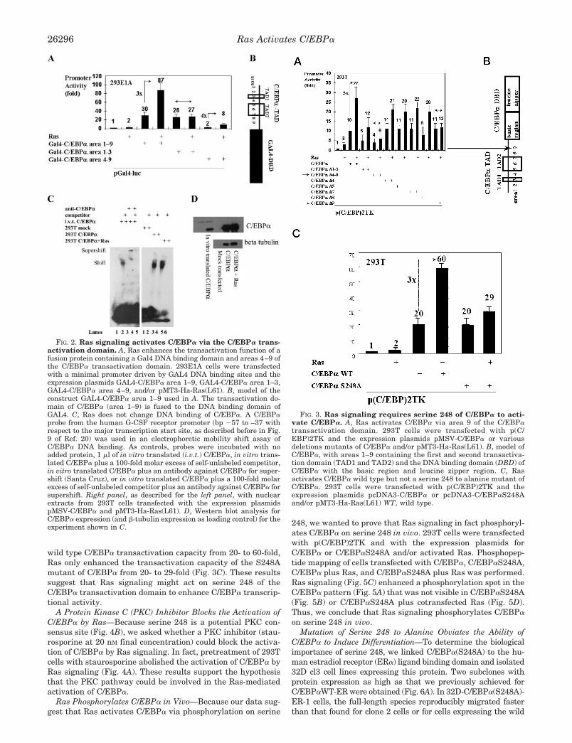
wild type C/EBP� transactivation capacity from 20- to 60-fold,Ras only enhanced the transactivation capacity of the S248Amutant of C/EBP� from 20- to 29-fold (Fig. 3C). These resultssuggest that Ras signaling might act on serine 248 of theC/EBP� transactivation domain to enhance C/EBP� transcrip-tional activity.
A Protein Kinase C (PKC) Inhibitor Blocks the Activation ofC/EBP� by Ras—Because serine 248 is a potential PKC con-sensus site (Fig. 4B), we asked whether a PKC inhibitor (stau-rosporine at 20 nM final concentration) could block the activa-tion of C/EBP� by Ras signaling. In fact, pretreatment of 293Tcells with staurosporine abolished the activation of C/EBP� byRas signaling (Fig. 4A). These results support the hypothesisthat the PKC pathway could be involved in the Ras-mediatedactivation of C/EBP�.
Ras Phosphorylates C/EBP� in Vivo—Because our data sug-gest that Ras activates C/EBP� via phosphorylation on serine
248, we wanted to prove that Ras signaling in fact phosphoryl-ates C/EBP� on serine 248 in vivo. 293T cells were transfectedwith p(C/EBP)2TK and with the expression plasmids forC/EBP� or C/EBP�S248A and/or activated Ras. Phosphopep-tide mapping of cells transfected with C/EBP�, C/EBP�S248A,C/EBP� plus Ras, and C/EBP�S248A plus Ras was performed.Ras signaling (Fig. 5C) enhanced a phosphorylation spot in theC/EBP� pattern (Fig. 5A) that was not visible in C/EBP�S248A(Fig. 5B) or C/EBP�S248A plus cotransfected Ras (Fig. 5D).Thus, we conclude that Ras signaling phosphorylates C/EBP�on serine 248 in vivo.
Mutation of Serine 248 to Alanine Obviates the Ability ofC/EBP� to Induce Differentiation—To determine the biologicalimportance of serine 248, we linked C/EBP�(S248A) to the hu-man estradiol receptor (ER�) ligand binding domain and isolated32D cl3 cell lines expressing this protein. Two subclones withprotein expression as high as that we previously achieved forC/EBP�WT-ER were obtained (Fig. 6A). In 32D-C/EBP�(S248A)-ER-1 cells, the full-length species reproducibly migrated fasterthan that found for clone 2 cells or for cells expressing the wild
FIG. 2. Ras signaling activates C/EBP� via the C/EBP� trans-activation domain. A, Ras enhances the transactivation function of afusion protein containing a Gal4 DNA binding domain and areas 4–9 ofthe C/EBP� transactivation domain. 293E1A cells were transfectedwith a minimal promoter driven by GAL4 DNA binding sites and theexpression plasmids GAL4-C/EBP� area 1–9, GAL4-C/EBP� area 1–3,GAL4-C/EBP� area 4–9, and/or pMT3-Ha-Ras(L61). B, model of theconstruct GAL4-C/EBP� area 1–9 used in A. The transactivation do-main of C/EBP� (area 1–9) is fused to the DNA binding domain ofGAL4. C, Ras does not change DNA binding of C/EBP�. A C/EBP�probe from the human G-CSF receptor promoter (bp �57 to –37 withrespect to the major transcription start site, as described before in Fig.9 of Ref. 20) was used in an electrophoretic mobility shift assay ofC/EBP� DNA binding. As controls, probes were incubated with noadded protein, 1 �l of in vitro translated (i.v.t.) C/EBP�, in vitro trans-lated C/EBP� plus a 100-fold molar excess of self-unlabeled competitor,in vitro translated C/EBP� plus an antibody against C/EBP� for super-shift (Santa Cruz), or in vitro translated C/EBP� plus a 100-fold molarexcess of self-unlabeled competitor plus an antibody against C/EBP� forsupershift. Right panel, as described for the left panel, with nuclearextracts from 293T cells transfected with the expression plasmidspMSV-C/EBP� and pMT3-Ha-Ras(L61). D, Western blot analysis forC/EBP� expression (and �-tubulin expression as loading control) for theexperiment shown in C.
FIG. 3. Ras signaling requires serine 248 of C/EBP� to acti-vate C/EBP�. A, Ras activates C/EBP� via area 9 of the C/EBP�transactivation domain. 293T cells were transfected with p(C/EBP)2TK and the expression plasmids pMSV-C/EBP� or variousdeletions mutants of C/EBP� and/or pMT3-Ha-Ras(L61). B, model ofC/EBP�, with areas 1–9 containing the first and second transactiva-tion domain (TAD1 and TAD2) and the DNA binding domain (DBD) ofC/EBP� with the basic region and leucine zipper region. C, Rasactivates C/EBP� wild type but not a serine 248 to alanine mutant ofC/EBP�. 293T cells were transfected with p(C/EBP)2TK and theexpression plasmids pcDNA3-C/EBP� or pcDNA3-C/EBP�S248Aand/or pMT3-Ha-Ras(L61) WT, wild type.
Ras Activates C/EBP�26296
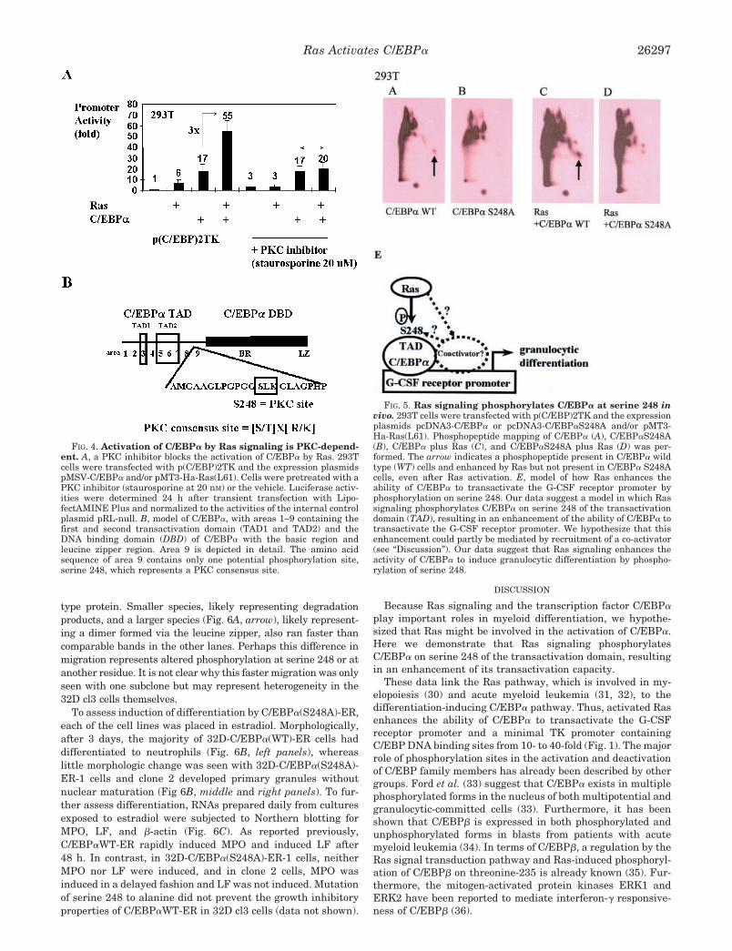
type protein. Smaller species, likely representing degradationproducts, and a larger species (Fig. 6A, arrow), likely represent-ing a dimer formed via the leucine zipper, also ran faster thancomparable bands in the other lanes. Perhaps this difference inmigration represents altered phosphorylation at serine 248 or atanother residue. It is not clear why this faster migration was onlyseen with one subclone but may represent heterogeneity in the32D cl3 cells themselves.
To assess induction of differentiation by C/EBP�(S248A)-ER,each of the cell lines was placed in estradiol. Morphologically,after 3 days, the majority of 32D-C/EBP�(WT)-ER cells haddifferentiated to neutrophils (Fig. 6B, left panels), whereaslittle morphologic change was seen with 32D-C/EBP�(S248A)-ER-1 cells and clone 2 developed primary granules withoutnuclear maturation (Fig 6B, middle and right panels). To fur-ther assess differentiation, RNAs prepared daily from culturesexposed to estradiol were subjected to Northern blotting forMPO, LF, and �-actin (Fig. 6C). As reported previously,C/EBP�WT-ER rapidly induced MPO and induced LF after48 h. In contrast, in 32D-C/EBP�(S248A)-ER-1 cells, neitherMPO nor LF were induced, and in clone 2 cells, MPO wasinduced in a delayed fashion and LF was not induced. Mutationof serine 248 to alanine did not prevent the growth inhibitoryproperties of C/EBP�WT-ER in 32D cl3 cells (data not shown).
DISCUSSION
Because Ras signaling and the transcription factor C/EBP�play important roles in myeloid differentiation, we hypothe-sized that Ras might be involved in the activation of C/EBP�.Here we demonstrate that Ras signaling phosphorylatesC/EBP� on serine 248 of the transactivation domain, resultingin an enhancement of its transactivation capacity.
These data link the Ras pathway, which is involved in my-elopoiesis (30) and acute myeloid leukemia (31, 32), to thedifferentiation-inducing C/EBP� pathway. Thus, activated Rasenhances the ability of C/EBP� to transactivate the G-CSFreceptor promoter and a minimal TK promoter containingC/EBP DNA binding sites from 10- to 40-fold (Fig. 1). The majorrole of phosphorylation sites in the activation and deactivationof C/EBP family members has already been described by othergroups. Ford et al. (33) suggest that C/EBP� exists in multiplephosphorylated forms in the nucleus of both multipotential andgranulocytic-committed cells (33). Furthermore, it has beenshown that C/EBP� is expressed in both phosphorylated andunphosphorylated forms in blasts from patients with acutemyeloid leukemia (34). In terms of C/EBP�, a regulation by theRas signal transduction pathway and Ras-induced phosphoryl-ation of C/EBP� on threonine-235 is already known (35). Fur-thermore, the mitogen-activated protein kinases ERK1 andERK2 have been reported to mediate interferon-� responsive-ness of C/EBP� (36).
FIG. 4. Activation of C/EBP� by Ras signaling is PKC-depend-ent. A, a PKC inhibitor blocks the activation of C/EBP� by Ras. 293Tcells were transfected with p(C/EBP)2TK and the expression plasmidspMSV-C/EBP� and/or pMT3-Ha-Ras(L61). Cells were pretreated with aPKC inhibitor (staurosporine at 20 nM) or the vehicle. Luciferase activ-ities were determined 24 h after transient transfection with Lipo-fectAMINE Plus and normalized to the activities of the internal controlplasmid pRL-null. B, model of C/EBP�, with areas 1–9 containing thefirst and second transactivation domain (TAD1 and TAD2) and theDNA binding domain (DBD) of C/EBP� with the basic region andleucine zipper region. Area 9 is depicted in detail. The amino acidsequence of area 9 contains only one potential phosphorylation site,serine 248, which represents a PKC consensus site.
FIG. 5. Ras signaling phosphorylates C/EBP� at serine 248 invivo. 293T cells were transfected with p(C/EBP)2TK and the expressionplasmids pcDNA3-C/EBP� or pcDNA3-C/EBP�S248A and/or pMT3-Ha-Ras(L61). Phosphopeptide mapping of C/EBP� (A), C/EBP�S248A(B), C/EBP� plus Ras (C), and C/EBP�S248A plus Ras (D) was per-formed. The arrow indicates a phosphopeptide present in C/EBP� wildtype (WT) cells and enhanced by Ras but not present in C/EBP� S248Acells, even after Ras activation. E, model of how Ras enhances theability of C/EBP� to transactivate the G-CSF receptor promoter byphosphorylation on serine 248. Our data suggest a model in which Rassignaling phosphorylates C/EBP� on serine 248 of the transactivationdomain (TAD), resulting in an enhancement of the ability of C/EBP� totransactivate the G-CSF receptor promoter. We hypothesize that thisenhancement could partly be mediated by recruitment of a co-activator(see “Discussion”). Our data suggest that Ras signaling enhances theactivity of C/EBP� to induce granulocytic differentiation by phospho-rylation of serine 248.
Ras Activates C/EBP� 26297

Regarding C/EBP�, Mahoney et al. (37) report that phospho-rylation of C/EBP� in vitro by protein kinase C attenuates itssite-selective DNA binding . Subsequently, Pan et al. (38) couldidentify a specific increase in DNA binding and the expressionof C/EBP� and C/EBP� during U937 monocytic cell differenti-ation and related this to the transforming growth factor �-sig-naling pathway. Our data do not relate the effect of Ras sig-naling to the DNA binding domain or DNA binding capacity ofC/EBP�. Ras signaling acts on the C/EBP� transactivationdomain, because it enhances 4-fold the transactivation functionof a fusion protein containing a Gal4 DNA binding domain andthe C/EBP� transactivation domain (Fig. 2A) and does notchange the C/EBP� DNA binding capacity (Fig. 2C). In fact,Ras acts on serine 248 of the C/EBP� transactivation domain,because it does not enhance the 10-fold transactivation exhib-ited by a C/EBP� mutant with a deletion of area 9 (Fig. 3A) ora serine 248 to alanine point mutant in area 9 (Fig. 3C). Thus,
we conclude that the Ras pathway acts on the transactivationfunctions of C/EBP� via serine 248.
Ross et al. (39, 40) report on other important phosphorylationsites in the transactivation domain of C/EBP�. Thus, GSK3kinase phosphorylates C/EBP� on threonines 222 and 226 (39).Furthermore, Wnt signaling maintains preadipocytes in anundifferentiated state and the adipogenic transcription factorsC/EBP� and peroxisome proliferator-activated receptor � arenot expressed. When Wnt signaling in preadipocytes is pre-vented by overexpression of axin or dominant-negative TCF4,these cells differentiate into adipocytes (40). Our data in 293Tcells point to serine 248 as the major Ras-dependent phospho-rylation site of C/EBP�. Serine 248 of C/EBP� is a PKC con-sensus site, and a PKC inhibitor blocks the activation ofC/EBP� by Ras (Fig. 4A). However, staurosporine is not com-pletely specific for PKC. Therefore, we have not excluded otherkinases as responsible for the activation of C/EBP� by Ras.
The serine to alanine mutant of C/EBP� did not completelyeliminate the activation of C/EBP� by Ras (Fig. 3C), whereasdeletion of region 9 did (Fig. 3A). Perhaps a transcriptionalco-activator binds optimally to C/EBP� when serine 248 isphosphorylated (Fig. 5E) but can still bind weakly to the S248Amutant. In this model the effect of Ras on C/EBP� transacti-vation is at least partly indirect via a co-activator. This modelis supported by the ability of Ras to induce basal activity ofp(C/EBP)TK; on the other hand, our observation that Ras in-creases phosphorylation of a C/EBP� peptide but not of thesame peptide from C/EBP�S248A suggests a more direct effecton C/EBP� by Ras (Fig. 5E). It is also possible that phospho-rylation of serine 230 (39) might account for additional effectsof Ras on C/EBP�. However, we could not observe any loss ofenhancement of transactivation capacity of C/EBP� by Rassignaling using the serine 230 to alanine mutant of C/EBP�
(data not shown; the mutants were kindly provided by Dr.MacDougald, Ann Arbor, MI). However, the very likely addi-tional Ras-dependent phosphorylation sites in C/EBP� besidesserine 248 (Fig. 5, A–D) still need to be mapped and to be testedfunctionally. Our current studies to identify interacting pro-teins of C/EBP� using proteomics techniques (two-dimensionalgel electrophoresis and mass spectrometry) might also contrib-ute to answering the question of whether the effect of Rassignaling on serine 248 of C/EBP� requires a co-activator bind-ing to serine 248 or not.
Mutation of serine 248 to alanine had marked biologic con-sequences in 32D cl3 cells, obviating the ability of C/EBP� toinduce differentiation (Fig. 6). Whether this represents a defectin Ras-dependent phosphorylation of this serine and conse-quent alteration of trans-activating properties in myeloid cellsor whether it represents a more indirect effect in which aRas-dependent pathway interacts with serine 248 or with an-other site in C/EBP� affected by mutation of serine 248 toalanine remains to be determined.
Further studies need to address the biological significance ofserine 248 of C/EBP� by testing the serine to alanine 248C/EBP� mutant in biological assays of C/EBP� function suchas in rescue assays in C/EBP� knock-out mice. Of note, G-CSF,but not interleukin-3, signals cooperate with C/EBP� to inducemyeloid genes in Ba/F3 lymphoid cells (41). Because impair-ment of C/EBP� function can contribute to the pathogenesis ofacute myeloid leukemia (10, 11), whether serine 248 of C/EBP�
is mutated in patients with acute myeloid leukemia and/orwhether inactivating Ras mutations in acute myeloid leukemiamight lead to a loss of C/EBP� function needs to be addressed.
In summary, our data suggest that Ras signaling phospho-rylates C/EBP� on serine 248 of the transactivation domain,resulting in an enhancement of the ability of C/EBP� to trans-
FIG. 6. Mutation of serine 248 to alanine obviates the ability ofC/EBP� to induce differentiation. To determine the biological im-portance of serine 248, we linked C/EBP�(S248A) to the human estra-diol receptor (ER�) ligand binding domain and isolated 32D cl3 celllines expressing this protein. Two subclones with protein expression ashigh as that we previously achieved for C/EBP�WT-ER were obtained(A). To assess induction of differentiation by C/EBP�(S248A)-ER, eachof the cell lines was placed in estradiol. Morphologically, after 3 daysthe majority of 32D-C/EBP�(WT)-ER cells had differentiated to neutro-phils (B, left panels), whereas little morphologic change was seen with32D-C/EBP�(S248A)-ER-1 cells, and clone 2 developed primary gran-ules without nuclear maturation (B, middle and right panels). To fur-ther assess differentiation, RNAs prepared daily from cultures exposedto estradiol were subjected to Northern blotting for MPO, LF, and�-actin (C). As reported previously, C/EBP�WT-ER rapidly inducedMPO and induced LF after 48 h. In contrast, in 32D-C/EBP�(S248A)-ER-1 cells, neither MPO nor LF were induced, and in clone 2 cells, MPOwas induced in a delayed fashion and LF was not induced. WT, wildtype.
Ras Activates C/EBP�26298
activate the G-CSF receptor promoter. Furthermore, our datasuggest a model where Ras signaling enhances the activity ofC/EBP� to induce granulocytic differentiation by phosphoryla-tion of serine 248.
REFERENCES
1. Cao, Z., Umek, R. M., and McKnight, S. L. (1991) Genes Dev. 5, 1538–15522. Lin, F. T., and Lane, M. D. (1994) Proc. Natl. Acad. Sci. U. S. A. 91, 8757–87613. Scott, L. M., Civin, C. I., Rorth, P., and Friedman, A. D. (1992) Blood 80,
1725–17354. Flodby, P., Barlow, C., Kylefjord, H., Ahrlund-Richter, L., and Xanthopoulos,
K. G. (1996) J. Biol. Chem. 271, 24753–247605. Zhang, D.-E., Zhang, P., Wang, N. D., Hetherington, C. J., Darlington, G. J.,
and Tenen, D. G. (1997) Proc. Natl. Acad. Sci. U. S. A. 94, 569–5746. Radomska, H. S., Huettner, C. S., Zhang, P., Cheng, T., Scadden, D. T., and
Tenen, D. G. (1998) Mol. Cell. Biol. 18, 4301–43097. Umek, R. M., Friedman, A. D., and McKnight, S. L. (1991) Science 251,
288–2928. Reddy, V. A., Iwama, A., Iotzova, G., Schulz, M., Elsasser, A., Vangala, R. K.,
Tenen, D. G., Hiddemann, W., and Behre, G. (2002) Blood, in press9. Slomiany, B. A., D’Arigo, K. L., Kelly, M. M., and Kurtz, D. T. (2000) Mol. Cell.
Biol. 20, 5986–599710. Pabst, T., Mueller, B. U., Zhang, P., Radomska, H. S., Narravula, S.,
Schnittger, S., Behre, G., Hiddemann, W., and Tenen, D. G. (2001) Nat.Genet. 27, 263–270
11. Pabst, T., Mueller, B. U., Harakawa, N., Schoch C, Haferlach, T., Behre, G.,Hiddemann, W., Zhang, D.-E., and Tenen, D. G. (2001) Nat. Med. 7,444–451
12. McCormick, F. (1995) Mol. Reprod. Dev. 42, 500–50613. Gutkind, J. S. (1998) J. Biol. Chem. 273, 1839–184214. Wasylyk, C., Maira, S. M., Sobieszczuk, P., and Wasylyk, B. (1994) Oncogene
9, 3665–367315. Johnson, R., Spiegelman, B., Hanahan, D., and Wisdom, R. (1996) Mol. Cell.
Biol. 16, 4504–451116. Jin, D. I., Jameson, S. B., Reddy, M. A., Schenkman, D., and Ostrowski, M. C.
(1995) Mol. Cell. Biol. 15, 693–70317. Hibi, S., Lohler, J., Friel, J., Stocking, C., and Ostertag, W. (1993) Blood 81,
1841–184818. Maher, J., Baker, D., Dibb, N., and Roberts, I. (1996) Leukemia (Baltimore) 10,
83–90
19. Skorski, T., Szczylik, C., Ratajczak, M. Z., Malaguarnera, L., Gewirtz, A. M.,and Calabretta, B. (1992) J. Exp. Med. 175, 743–750
20. Smith, L. T., Hohaus, S., Gonzalez, D. A., Dziennis, S. E., and Tenen, D. G.(1996) Blood 88, 1234–1247
21. Nordeen, S. K. (1988) Biotechniques 6, 454–45822. Behre, G., Whitmarsh, A. J., Coghlan, M. P., Hoang, T., Carpenter, C. L.,
Zhang, D.-E., Davis, R. J., and Tenen, D. G. (1999) J. Biol. Chem. 274,4939–4946
23. Behre, G., Smith, L. T., and Tenen, D. G. (1999) Biotechniques 26, 24–2624. Zhang, P., Behre, G., Pan J, Iwama, A., Wara-aswapati, N., Radomska, H. S.,
Auron, P. E., Tenen, D. G., and Sun, Z. (1999) Proc. Natl. Acad. Sci. U. S. A.96, 8705–8710
25. Friedman, A. D., and McKnight, S. L. (1990) Genes Dev. 4, 1416–142626. Oelgeschlager, M., Nuchprayoon, I., Luscher, B., and Friedman, A. D. (1996)
Mol. Cell. Biol. 16, 4717–472527. Feig, L. A., and Cooper, G. M. (1988) Mol. Cell. Biol. 8, 2472–247828. Feig, L. A. (1988) Mol. Cell. Biol. 8, 3235–324329. Boyle, W. J., Geer, P., and Hunter, T. (1991) Methods Enzymol. 201, 110–14930. Crespo, P., and Leon, J. (2000) Cell. Mol. Life Sci. 57, 1613–163631. Schaich, M., Ritter, M., Illmer, T., Lisske, P., Thiede, C., Schakel, U., Mohr, B.,
Ehninger, G., and Neubauer, A. (2001) Br. J. Haematol. 112, 300–30732. Zuber, J., Tchernitsa, O. I., Hinzmann, B., Schmitz, A. C., Grips, M.,
Hellriegel, M., Sers, C., Rosenthal, A., and Schafer, R. (2000) Nat. Genet. 24,144–152
33. Ford, A. M., Bennett, C. A., Healy, L. E., Towatari, M., Greaves, M. F., andEnver, T. (1996) Proc. Natl. Acad. Sci. U. S. A. 93, 10838–10843
34. Iida, H., Towatari, M., Iida, M., Tanimoto, M., Kodera, Y., Ford, A. M., andSaito, H. (2000) Int. J. Hematol. 71, 153–158
35. Nakajima, T., Kinoshita, S., Sasagawa, T., Sasaki, K., Naruto, M., Kishimoto,T., and Akira, S. (1993) Proc. Natl. Acad. Sci. U. S. A. 90, 2207–2211
36. Hu, J., Roy, S. K., Shapiro, P. S., Rodig, S. R., Reddy, S. P., Platanias, L. C.,Schreiber, R. D., and Kalvakolanu, D. V. (2001) J. Biol. Chem. 276, 287–297
37. Mahoney, C. W., Shuman, J., McKnight, S. L., Chen, H. C., and Huang, K. P.(1992) J. Biol. Chem. 267, 19396–19403
38. Pan, Z., Hetherington, C. J., and Zhang, D. E. (1999) J. Biol. Chem. 274,23242–23248
39. Ross, S. E., Erickson, R. L., Hemati, N., and MacDougald, O. A. (1999) Mol.Cell. Biol. 19, 8433–8441
40. Ross, S. E., Hemati, N., Longo, K. A., Bennett, C. N., Lucas, P. C., Erickson,R. L., and MacDougald, O. A. (2000) Science 289, 950–953
41. Wang, W., Wang, X., Ward AC, Touw, I. P., and Friedman, A. D. (2001)Leukemia (Baltimore) 15, 779–786
Ras Activates C/EBP� 26299
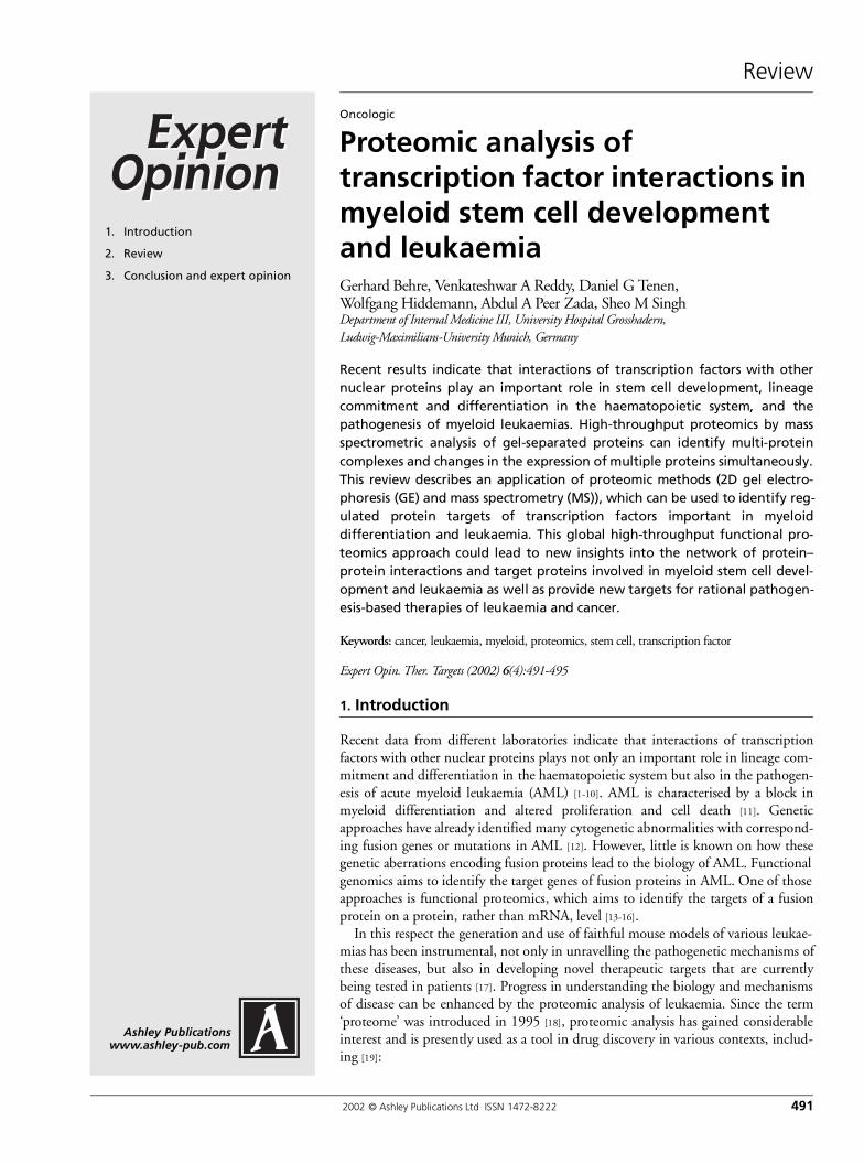
Review
2002 © Ashley Publications Ltd ISSN 1472-8222 491
Ashley Publicationswww.ashley-pub.com
1. Introduction
2. Review
3. Conclusion and expert opinion
Oncologic
Proteomic analysis of transcription factor interactions in myeloid stem cell development and leukaemiaGerhard Behre, Venkateshwar A Reddy, Daniel G Tenen, Wolfgang Hiddemann, Abdul A Peer Zada, Sheo M SinghDepartment of Internal Medicine III, University Hospital Grosshadern, Ludwig-Maximilians-University Munich, Germany
Recent results indicate that interactions of transcription factors with othernuclear proteins play an important role in stem cell development, lineagecommitment and differentiation in the haematopoietic system, and thepathogenesis of myeloid leukaemias. High-throughput proteomics by massspectrometric analysis of gel-separated proteins can identify multi-proteincomplexes and changes in the expression of multiple proteins simultaneously.This review describes an application of proteomic methods (2D gel electro-phoresis (GE) and mass spectrometry (MS)), which can be used to identify reg-ulated protein targets of transcription factors important in myeloiddifferentiation and leukaemia. This global high-throughput functional pro-teomics approach could lead to new insights into the network of protein–protein interactions and target proteins involved in myeloid stem cell devel-opment and leukaemia as well as provide new targets for rational pathogen-esis-based therapies of leukaemia and cancer.
Keywords: cancer, leukaemia, myeloid, proteomics, stem cell, transcription factor
Expert Opin. Ther. Targets (2002) 6(4):491-495
1. Introduction
Recent data from different laboratories indicate that interactions of transcriptionfactors with other nuclear proteins plays not only an important role in lineage com-mitment and differentiation in the haematopoietic system but also in the pathogen-esis of acute myeloid leukaemia (AML) [1-10]. AML is characterised by a block inmyeloid differentiation and altered proliferation and cell death [11]. Geneticapproaches have already identified many cytogenetic abnormalities with correspond-ing fusion genes or mutations in AML [12]. However, little is known on how thesegenetic aberrations encoding fusion proteins lead to the biology of AML. Functionalgenomics aims to identify the target genes of fusion proteins in AML. One of thoseapproaches is functional proteomics, which aims to identify the targets of a fusionprotein on a protein, rather than mRNA, level [13-16].
In this respect the generation and use of faithful mouse models of various leukae-mias has been instrumental, not only in unravelling the pathogenetic mechanisms ofthese diseases, but also in developing novel therapeutic targets that are currentlybeing tested in patients [17]. Progress in understanding the biology and mechanismsof disease can be enhanced by the proteomic analysis of leukaemia. Since the term‘proteome’ was introduced in 1995 [18], proteomic analysis has gained considerableinterest and is presently used as a tool in drug discovery in various contexts, includ-ing [19]:
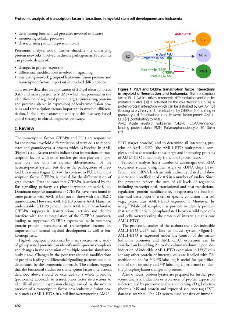
Proteomic analysis of transcription factor interactions in myeloid stem cell development and leukaemia
492 Expert Opin. Ther. Targets (2002) 6(4)
• determining biochemical processes involved in disease• monitoring cellular processes• characterising protein expression levels
Proteomic analysis would further elucidate the underlyingprotein networks involved in disease pathogenesis. Proteomicscan provide details of:
• changes in protein expression• differential modifications involved in signalling• interacting network groups of leukaemic fusion proteins and
transcription factors important in myeloid differentiation
This review describes an application of 2D gel electrophoresis(GE) and mass spectrometry (MS) which has potential in theidentification of regulated protein targets (interacting proteinsand proteins altered in expression) of leukaemic fusion pro-teins and transcription factors important in myeloid differen-tiation. It also demonstrates the utility of this discovery-basedglobal strategy in elucidating novel pathways.
2. Review
The transcription factors C/EBPα and PU.1 are responsiblefor the normal myeloid differentiation of stem cells to mono-cytes and granulocytes, a process which is blocked in AML(Figure 1) [1,2]. Recent results indicate that interactions of tran-scription factors with other nuclear proteins play an impor-tant role not only in normal differentiation of thehaematopoietic system, but also in the pathogenesis of mye-loid leukaemias (Figure 1) [3-10]. In contrast to PU.1, the tran-scription factor C/EBPα is crucial for the differentiation ofgranulocytes. Data indicates that C/EBPα is activated by theRas signalling pathway via phosphorylation on ser248 [10].Dominant negative mutations of C/EBPα have been found insome patients with AML [8] but not in those with the t(8;21)translocation. However, AML1-ETO positive AML blasts hadundetectable C/EBPα protein levels. AML1-ETO can bind toC/EBPα, suppress its transcriptional activity and therebyinterfere with the autoregulation of the C/EBPα promoter,leading to suppressed C/EBPα expression [9]. In summary,protein–protein interactions of transcription factors areimportant for normal myeloid development as well as leu-kaemogenesis.
High-throughput proteomics by mass spectrometric studyof gel separated proteins can identify multi-protein complexesand changes in the expression of multiple proteins simultane-ously [13-16]. Changes in the post-translational modificationsof proteins leading to differential signalling patterns could bedetermined by this proteomic approach. The authors suggestthat the functional studies on transcription factor interactionsdescribed above should be extended to a whole proteome(proteomic) approach to transcription factor interactions toidentify all protein expression changes caused by the overex-pression of a transcription factor or a leukaemic fusion pro-tein such as AML1-ETO, in a cell line overexpressing AML1-
ETO (target proteins) and to determine all interacting pro-teins of AML1-ETO (the AML1-ETO multiprotein com-plex), and to characterize those target and interacting proteinsof AML1-ETO functionally (functional proteomics).
Proteome analysis has a number of advantages over RNAexpression studies using filter arrays or cDNA chips [14-16].Protein and mRNA levels are only indirectly related and showa correlation coefficient of < 0.5 in a number of studies. Sincethe proteome reflects the sum of all levels of regulationincluding transcriptional, translational and post-translationalregulation (protein modification), it represents the best bio-chemical description of a cell or specific condition of a cell(e.g., plus/minus AML1-ETO expression). Moreover, byusing 32P-labelled samples, it is possible to identify proteinsthat are differentially phosphorylated between wild type cellsand cells overexpressing the protein of interest (in this caseAML1-ETO).
The proteomic studies of the authors use a Zn-inducibleAML1-ETO-U937 cell line as model system (Figure 2).AML1-ETO is expressed under the control of the metal-lothionin promoter and AML1-ETO expression can beswitched on by adding Zn to the culture medium. Upon Zn-induction of inducible AML1-ETO expression in U937 cells(or any other protein of interest), cells are labelled with 35S-methionine and/or 32P. 35S-labelling is useful for quantifica-tion of spot intensity and 32P-labelling is performed to iden-tify phosphorylation changes in proteins.
After 6 hours, protein lysates are prepared for further pro-teome analysis. Induction or repression of protein expressionis determined by proteome analysis combining 2D gel electro-phoresis, MS and protein and expressed sequence tag (EST)database searches. The 2D system used consists of immobi-
Figure 1. PU.1 and C/EBPαααα transcription factor interactionsin myeloid differentiation and leukaemia. The transcriptionfactor PU.1 (which drives monocytic differentiation and can bemutated in AML [3]) is activated by the co-activator c-Jun [4], aprotein-protein interaction which can be disturbed by GATA-1 [5](leading to erythrocytic differentiation), by C/EBPα [6] (resulting ingranulocytic differentiation) or the leukemic fusion protein AML1-ETO [7] (contributing to AML). AML: Acute myeloid leukaemia; C/EBPα: CCAAT/enhancerbinding protein alpha; PMN: Polymorphonucleocyte; SC: Stemcell.
AML1-ETO
Mono
PMN
c-Jun/ PU.1
C/EBPαααα
EryGATA1
1,2
5
Ras S248
mutated in AML
mutated in AMLSC

Behre, Reddy, Tenen, Hiddemann, Peer Zada, Singh
Expert Opin. Ther. Targets (2002) 6(4) 493
lised pH gradients first in 1D and then SDS-PAGE in 2D. Tocover a wide range of proteins and obtain a resolution suffi-ciently high enough to detect low abundance proteins, six dif-ferent gels in 2D are run of each sample: pH 3 – 10, 4 – 7 or6 – 11 for isoelectric focusing in 1D with 14% and 7% SDS-PAGE gels in 2D. Proteins are detected by silver staining andautoradiography. Proteins with an isoelectric point betweenpH 3 and pH 10 and ranging in size from 8 kDa to~ 100 kDa can be reliably analysed. To exclude gel to gel dif-ferences, each sample type has to be repeated at least 3 times.
Differential patterns of proteins obtained from 2D gels canbe analysed by Proteomweaver software, a new 2DGE imageanalysis software developed by Definiens AG based on cogni-tion network technology originally used in satellite imageclassification. This software detects spots in a fully automatedmanner with high precision and speed in a context-sensitiveway. Manual adjustments, such as background subtraction orparameter settings, are unnecessary – even normalisation isfully automated. Also spot matching can be performed with-out manual interference. The results of Proteomweaver can beinvestigated with advanced statistics tools. The most interest-ing spots in terms of expression levels change simultaneouslywith high statistical significance and can be displayed and/orsaved easily. Proteomweaver software seems to be faster, moreprecise and easier to use than to older software available onthe market.
After the determination of reproducible protein spotsupregulated or downregulated by AML1-ETO, these proteinsare identified by MS (MALDI-TOF, tandem MS (MS/MS))(Figure 2). For relatively abundant proteins, it is enough toscale up the sample load in the analytical gel system and stainproteins by coomassie blue to obtain enough material for fur-ther analysis. For less abundant proteins, narrow pH gradients(covering 1 – 2 pH units in 20 cm) are used in 1D allowing
higher loading of the gels. If the amount of the protein ofinterest is too low to be detected by coomassie blue, the cellscan be labelled metabolically with 35S-methionine to detectproteins by autoradiography and material from several gelsrun in parallel can be combined. Finally, low abundant pro-teins can be detected by MS/MS, rather than MALDI-TOFMS.
In-gel digestion of proteins with trypsin ensures that thereis almost no loss of protein and the resulting eluted peptidesare analysed directly by MALDI-TOF MS, which generatespeptide mass fingerprints. This enables known proteins inDNA or protein databases to be identified with the appropri-ate search programs (e.g., PeptIdent, PeptideSearch,MOWSE). From small amounts of starting material (< 0.1pMol), a dual capacity MALDI-TOF MS instrument canidentify a mass range of 50 – 10000 amu, effective flight path= 2.3 M, as well as > 500000 amu, effective flight path = 1.0M. The current protocol allows up to 30 proteins to be proc-essed within three working days. In the few cases where theprotein is not present in EST databases, de novo sequencing ofseveral peptides per protein has to be performed. For peptidesof low concentration not identified via the current annotateddatabases after MALDI-TOF analysis, a high sensitivity liquidchromatography ion-trap MS/MS system would be used.
Currently, there are some limitations to the very powerfulMALDI-TOF technology. Firstly, the database must containadequate stretches of protein sequence to enable comparisonwith the experimentally derived mass map derived from theunknown protein. This is not yet the case for the genomicdatabase due to incomplete coding information for all genes[13,20,21]. For mass maps not matched in databases, other MStechnologies, such as use of the MS/MS proposed, can beused to reanalyse and identify these unknown proteins presentin the fusion protein complex. The use of MS/MS allows
Figure 2. Proteomic analysis of transcription factor interactions: 2DGE and MALDI-TOF MS.AML: Acute myeloid leukaemia; MALDI-TOF: Matrix assisted laser desorption/ionisation-time-of-flight; MS: Mass spectrometry; PMF: Peptide mass fingerprint.
U937 AML1-ETO
U937 AML1 -ETO (Zn+2)
Database search with PMF
2D gel electrophoresis MALDI-TOF MS
Functional characterisation
%Intensity
Mass (m/z)
Proteomic analysis of transcription factor interactions in myeloid stem cell development and leukaemia
494 Expert Opin. Ther. Targets (2002) 6(4)
microsequencing by causing predictable fragmentations ofamino acid bands in peptides, allowing the determination ofindividual mass differences in the mass spectrum, which thencan be used to infer at which position an amino acid was lostfrom the peptide under analysis. The resulting MS/MS datacan be interpreted using programmes, such as SE-QUEST,which compare the experimental data obtained with predictedspectra from database sequences [22].
Due to the progress of the human genome project, ESTclones are already available for most of the proteins identifiedusing the method described. For those not yet available asESTs, the genes can be cloned with the aid of synthetic degen-erated deoxyoligonucleotides deduced from peptidesequences. For proteins with known sequences, clones can beretrieved from the laboratories that cloned the genes.
After target proteins of AML1-ETO (or of any other pro-tein of interest) have been identified in myeloid cells, whetherthe promoters of these genes have functional AML1B proteinbinding sites can be investigated. A search for AML1B con-sensus DNA binding sites in the respective promoters can beperformed using transfac software [101] and by performing gelshift assays with in vitro translated AML1B [1,2]. Luciferaseassays of promoter constructs with mutated AML1B sites(which are generated by polymerase chain reaction mutagene-sis) can be performed in myeloid U937 or nonmyeloid CV-1cells [1,2]. Finally, a gene whose expression is downregulated byAML1-ETO can be subcloned into the Zn-inducible vectorpPC18 to generate stable cell lines and to ask whether induci-ble overexpression of this protein can induce myeloid differ-entiation. This would indicate that the downregulation of thisprotein by AML1-ETO might have a biological significance.
To determine a multi-protein complex (e.g., for AML1-ETO), proteomic studies can be performed as outlined above
for target proteins. However, different starting material is usedfor 2DGE and subsequent MS. AML1-ETO is expressed as afusion protein with a cleavable affinity tag, glutathion-S-transferase (GST) to identify interacting proteins. In this case,AML1-ETO protein is immobilised onto agarose beads usinga GST tag. Nuclear cell extracts are incubated with the beadsand the beads are then washed extensively. Thrombin is usedto cleave between the GST and the AML1-ETO protein,resulting in the elution of all proteins that are specificallybound to AML1-ETO. The advantage of this method is thatproteins that are nonspecifically bound to the matrix or thetag itself are not eluted. The eluted proteins are resolved by1D or 2DGE and compared to GST alone. The bands orspots corresponding to proteins specifically bound to taggedproteins are excised and analysed by MS as described for tar-get proteins. Interacting proteins of AML1-ETO can be char-acterised for functional relevance by performing GST pull-down assays and luciferase assays of AML1-ETO responsivepromoters (such as the Bcl-2 promoter or a minimal thymi-dine kinase promoter driven by AML1B binding sites only).
3. Conclusion and expert opinion
Functional proteomics, the large-scale analysis of proteins andtheir interactions, will contribute greatly to the understandingof gene and protein function in the postgenomic era. The glo-bal high-throughput functional proteomics approachdescribed will likely lead to new insights into the network ofprotein–protein interactions and target proteins involved inmyelopoiesis and the development of leukaemia as well asprovide new targets for rational pathogenesis-based therapiesof leukaemia and cancer.
BibliographyPapers of special note have been highlighted as either of interest (•) or of considerable interest (••) to readers.
1. BEHRE G, ZHANG P, ZHANG D-E, TENEN DG: Analysis of the modulation of transcriptional activity in myelopoiesis and leukemogenesis. Methods (1999) 17:231-237.
2. BEHRE G, SMITH LT, TENEN DG: Use of a Renilla luciferase vector as a control for studies of Ras transactivation. Biotechniques (1999) 26:24-26.
3. MUELLER BU, PABST T, OSATO M et al.: Heterozygous PU.1 mutations are associated with acute myeloid leukaemia. Blood (2002) In press.
4. BEHRE G, WHITMARSH AJ, COGHLAN MP et al.: c-Jun is a JNK independent co-activator of the PU.1
transcription factor. J. Biol. Chem. (1999) 274:4939-4946.
5. ZHANG P, BEHRE G, PAN J et al.: Negative cross-talk between hematopoietic regulators: GATA proteins repress PU.1 function. Proc. Natl Acad. Sci. USA (1999) 96:8705-8710.
6. REDDY VA, IWAMA A, IOTZOVA G et al.: Granulocyte inducer C/EBPalpha inactivates the myeloid master regulator PU.1: possible role in lineage commitment decisions. Blood (2002) 100:483-490.
7. VANGALA R, NEUMANN SM, RANGATIA J et al.: The myeloid master regulator transcription factor PU.1 is inactivated by AML-ETO in t(8;21) myeloid leukemia. (2002) Submitted for publication.
8. PABST T, MUELLER BU, ZHANG P et al.: Dominant negative
mutations of CEBPA, encoding CCAAT/enhancer binding protein-alpha (C/EBPalpha), in acute myeloid leukaemia. Nat. Gen. (2001) 27:263-270.
9. PABST T, MUELLER BU, SCHOCH C et al.: AML1-ETO downregulates the granulocytic differentiation factor C/EBPalpha in t(8;21) myeloid leukaemia. Nat. Med. (2001) 7:444-451.
10. BEHRE G, SINGH SM, LIU H, BORTOLIN LT et al.: Ras signalling enhances the activity of c/ebpα to induce granulocytic differentiation by phosphorylation of serine 248. J. Biol. Chem. (2002) 277:26293-26299.
11. TENEN DG, HROMAS R, LICHT JD, ZHANG D-E: Transcription factors, normal myeloid development and leukaemia. Blood (1999) 90:489-519.
Behre, Reddy, Tenen, Hiddemann, Peer Zada, Singh
Expert Opin. Ther. Targets (2002) 6(4) 495
•• Most useful review on the role of transcription factors in myeloid development and leukaemia.
12. LOWENBERG B, DOWNING JR, BURNETT A: Acute myeloid leukaemia. N. Engl. J. Med. (1999) 341:1051-1062.
13. PANDEY A, MANN M: Proteomics to study genes and genomes. Nature (2000) 405:837-846.
• Most useful review and introduction into proteomics.
14. ABBOTT A: A postgenomic challenge: learning to read patterns of protein synthesis. Nature (1999) 402:713-720.
15. BANKS RE, DUNN MJ, HOCHSTRASSER DF et al.: Proteomics: new perspectives, new biomedical opportunities. Lancet (2000) 356:1749-1756.
16. ALAIYA AA, FRANZEN B, AUER G, LINDER S: Cancer proteomics: from identification of novel markers to creation of artifical learning models for tumour classification. Electrophoresis (2000) 21:1210-1217.
17. PANDOLFI PP: In vivo analysis of the molecular genetics of acute promyelocytic leukaemia. Oncogene (2001) 20:5726-5735.
18. WASINGER VC, CORDWELL SJ, CERPA-POLJAK A et al.: Progress with gene-product mapping of the mollicutes: Mycoplasma genitalium. Electrophoresis (1995) 16:1090-1094.
19. LARSEN MR, LARSEN PM, FEY SJ, ROEPSTORFF P: Characterization of differently processed forms of enolase 2 from Saccharomyces cerevisiae by two-dimensional gel electrophoresis and mass spectrometry. Electrophoresis (2001) 22:566-575.
20. YATES JR: Mass spectrometry. From genomics to proteomics. Trends Genet. (2000) 16:5-8.
21. MANN M, HENDRICKSON RC, PANDEY A: Analysis of proteins and proteomes by mass spectrometry. Ann. Rev. Biochem. (2001) 70:437-473.
22. YATES JR 3RD, MCCORMACK AL, LINK AJ, SCHIELTZ D, ENG J, HAYS L: Future prospects for the analysis of complex
biological systems using microcolumn liquid chromatography-electrospray tandem mass spectrometry. Analyst (1996) 121:65R-76R.
Website101. http://bioinformatic.weizmann.ac.il/
transfac
AffiliationGerhard Behre1,†, Venkateshwar A Reddy1, Daniel G Tenen2, Wolfgang Hiddemann1, Abdul A Peer Zada1, Sheo M Singh1
†Author for correspondence1Department of Internal Medicine III,University Hospital Grosshadern, Ludwig-Maximilians-University Munich and GSF-National Research Center for Environment and Health, Marchioninistr. 15, Munich, 81377, Germany2Harvard Institutes of Medicine, Harvard Medical School, Boston, MA 02115, USATel: +49 89 7095 118; Fax: +49 89 7095 5550; E.mail: [email protected]
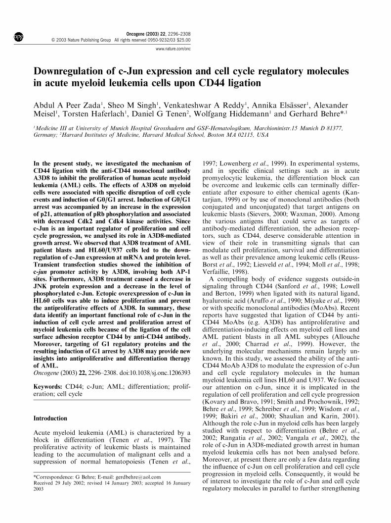
Downregulation of c-Jun expression and cell cycle regulatory molecules
in acute myeloid leukemia cells upon CD44 ligation
Abdul A Peer Zada1, Sheo M Singh1, Venkateshwar A Reddy1, Annika Elsasser1, AlexanderMeisel1, Torsten Haferlach1, Daniel G Tenen2, Wolfgang Hiddemann1 and Gerhard Behre*,1
1Medicine III at University of Munich Hospital Grosshadern and GSF-Hematologikum, Marchioninistr.15 Munich D 81377,Germany; 2Harvard Institutes of Medicine, Harvard Medical School, Boston MA 02115, USA
In the present study, we investigated the mechanism ofCD44 ligation with the anti-CD44 monoclonal antibodyA3D8 to inhibit the proliferation of human acute myeloidleukemia (AML) cells. The effects of A3D8 on myeloidcells were associated with specific disruption of cell cycleevents and induction of G0/G1 arrest. Induction of G0/G1arrest was accompanied by an increase in the expressionof p21, attenuation of pRb phosphorylation and associatedwith decreased Cdk2 and Cdk4 kinase activities. Sincec-Jun is an important regulator of proliferation and cellcycle progression, we analysed its role in A3D8-mediatedgrowth arrest. We observed that A3D8 treatment of AMLpatient blasts and HL60/U937 cells led to the down-regulation of c-Jun expression at mRNA and protein level.Transient transfection studies showed the inhibition ofc-jun promoter activity by A3D8, involving both AP-1sites. Furthermore, A3D8 treatment caused a decrease inJNK protein expression and a decrease in the level ofphosphorylated c-Jun. Ectopic overexpression of c-Jun inHL60 cells was able to induce proliferation and preventthe antiproliferative effects of A3D8. In summary, thesedata identify an important functional role of c-Jun in theinduction of cell cycle arrest and proliferation arrest ofmyeloid leukemia cells because of the ligation of the cellsurface adhesion receptor CD44 by anti-CD44 antibody.Moreover, targeting of G1 regulatory proteins and theresulting induction of G1 arrest by A3D8 may provide newinsights into antiproliferative and differentiation therapyof AML.Oncogene (2003) 22, 2296–2308. doi:10.1038/sj.onc.1206393
Keywords: CD44; c-Jun; AML; differentiation; prolif-eration; cell cycle
Introduction
Acute myeloid leukemia (AML) is characterized by ablock in differentiation (Tenen et al., 1997). Theproliferative activity of leukemic blasts is maintainedleading to the accumulation of malignant cells and asuppression of normal hematopoiesis (Tenen et al.,
1997; Lowenberg et al., 1999). In experimental systems,and in specific clinical settings such as in acutepromyelocytic leukemia, the differentiation block canbe overcome and leukemic cells can terminally differ-entiate after exposure to either chemical agents (Kan-tarjian, 1999) or by use of monoclonal antibodies (bothconjugated and unconjugated) that target antigens onleukemic blasts (Sievers, 2000; Waxman, 2000). Amongthe various antigens that could serve as targets ofantibody-mediated differentiation, the adhesion recep-tors, such as CD44, deserve considerable attention inview of their role in transmitting signals that canmodulate cell proliferation, survival and differentiationas well as their prevalence among leukemic cells (Reuss-Borst et al., 1992; Liesveld et al., 1994; Moll et al., 1998;Verfaillie, 1998).
A compelling body of evidence suggests outside-insignaling through CD44 (Sanford et al., 1998; Lowelland Berton, 1999) when ligated with its natural ligand,hyaluronic acid (Aruffo et al., 1990; Miyake et al., 1990)or with specific monoclonal antibodies (MoAbs). Recentreports have suggested that ligation of CD44 by anti-CD44 MoAbs (e.g. A3D8) has antiproliferative anddifferentiation-inducing effects on myeloid cell lines andAML patient blasts in all AML subtypes (Alloucheet al., 2000; Charrad et al., 1999). However, theunderlying molecular mechanisms remain largely un-known. In this study, we assessed the ability of the anti-CD44 MoAb A3D8 to modulate the expression of c-Junand cell cycle regulatory molecules in the humanmyeloid leukemia cell lines HL60 and U937. We focusedour attention on c-Jun, since it is implicated in theregulation of cell proliferation and cell cycle progression(Kovary and Bravo, 1991; Smith and Prochownik, 1992;Behre et al., 1999; Schreiber et al., 1999; Wisdom et al.,1999; Bakiri et al., 2000; Shaulian and Karin, 2001).Although the role c-Jun in myeloid cells has been largelystudied with respect to differentiation (Behre et al.,2002; Rangatia et al., 2002; Vangala et al., 2002), therole of c-Jun in A3D8-mediated growth arrest in humanmyeloid leukemia cells has not been analysed before.Moreover, at present there are only a few data regardingthe influence of c-Jun on cell proliferation and cell cycleprogression in myeloid cells. Consequently, it would beof interest to investigate the role of c-Jun and cell cycleregulatory molecules in parallel to further strengthening
Received 29 July 2002; revised 14 January 2003; accepted 16 January2003
*Correspondence: G Behre; E-mail: [email protected]
Oncogene (2003) 22, 2296–2308& 2003 Nature Publishing Group All rights reserved 0950-9232/03 $25.00
www.nature.com/onc
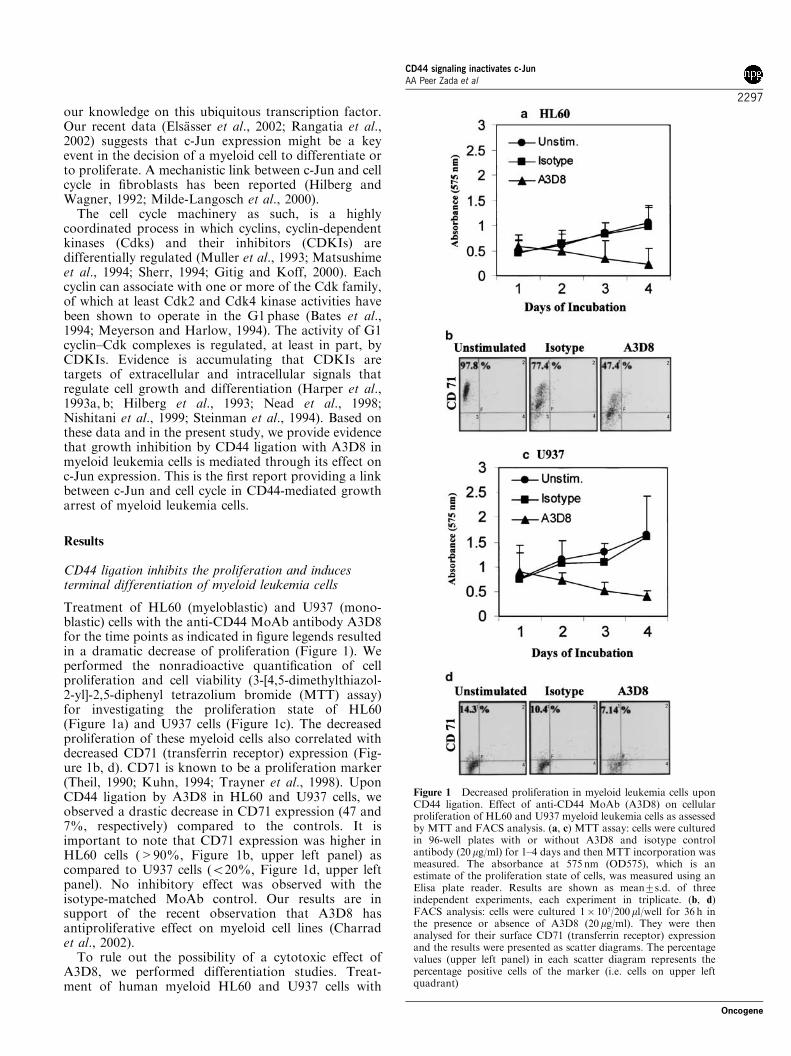
our knowledge on this ubiquitous transcription factor.Our recent data (Elsasser et al., 2002; Rangatia et al.,2002) suggests that c-Jun expression might be a keyevent in the decision of a myeloid cell to differentiate orto proliferate. A mechanistic link between c-Jun and cellcycle in fibroblasts has been reported (Hilberg andWagner, 1992; Milde-Langosch et al., 2000).
The cell cycle machinery as such, is a highlycoordinated process in which cyclins, cyclin-dependentkinases (Cdks) and their inhibitors (CDKIs) aredifferentially regulated (Muller et al., 1993; Matsushimeet al., 1994; Sherr, 1994; Gitig and Koff, 2000). Eachcyclin can associate with one or more of the Cdk family,of which at least Cdk2 and Cdk4 kinase activities havebeen shown to operate in the G1 phase (Bates et al.,1994; Meyerson and Harlow, 1994). The activity of G1cyclin–Cdk complexes is regulated, at least in part, byCDKIs. Evidence is accumulating that CDKIs aretargets of extracellular and intracellular signals thatregulate cell growth and differentiation (Harper et al.,1993a, b; Hilberg et al., 1993; Nead et al., 1998;Nishitani et al., 1999; Steinman et al., 1994). Based onthese data and in the present study, we provide evidencethat growth inhibition by CD44 ligation with A3D8 inmyeloid leukemia cells is mediated through its effect onc-Jun expression. This is the first report providing a linkbetween c-Jun and cell cycle in CD44-mediated growtharrest of myeloid leukemia cells.
Results
CD44 ligation inhibits the proliferation and inducesterminal differentiation of myeloid leukemia cells
Treatment of HL60 (myeloblastic) and U937 (mono-blastic) cells with the anti-CD44 MoAb antibody A3D8for the time points as indicated in figure legends resultedin a dramatic decrease of proliferation (Figure 1). Weperformed the nonradioactive quantification of cellproliferation and cell viability (3-[4,5-dimethylthiazol-2-yl]-2,5-diphenyl tetrazolium bromide (MTT) assay)for investigating the proliferation state of HL60(Figure 1a) and U937 cells (Figure 1c). The decreasedproliferation of these myeloid cells also correlated withdecreased CD71 (transferrin receptor) expression (Fig-ure 1b, d). CD71 is known to be a proliferation marker(Theil, 1990; Kuhn, 1994; Trayner et al., 1998). UponCD44 ligation by A3D8 in HL60 and U937 cells, weobserved a drastic decrease in CD71 expression (47 and7%, respectively) compared to the controls. It isimportant to note that CD71 expression was higher inHL60 cells (>90%, Figure 1b, upper left panel) ascompared to U937 cells (o20%, Figure 1d, upper leftpanel). No inhibitory effect was observed with theisotype-matched MoAb control. Our results are insupport of the recent observation that A3D8 hasantiproliferative effect on myeloid cell lines (Charradet al., 2002).
To rule out the possibility of a cytotoxic effect ofA3D8, we performed differentiation studies. Treat-ment of human myeloid HL60 and U937 cells with
Figure 1 Decreased proliferation in myeloid leukemia cells uponCD44 ligation. Effect of anti-CD44 MoAb (A3D8) on cellularproliferation of HL60 and U937 myeloid leukemia cells as assessedby MTT and FACS analysis. (a, c) MTT assay: cells were culturedin 96-well plates with or without A3D8 and isotype controlantibody (20 mg/ml) for 1–4 days and then MTT incorporation wasmeasured. The absorbance at 575 nm (OD575), which is anestimate of the proliferation state of cells, was measured using anElisa plate reader. Results are shown as mean7s.d. of threeindependent experiments, each experiment in triplicate. (b, d)FACS analysis: cells were cultured 1� 105/200ml/well for 36 h inthe presence or absence of A3D8 (20mg/ml). They were thenanalysed for their surface CD71 (transferrin receptor) expressionand the results were presented as scatter diagrams. The percentagevalues (upper left panel) in each scatter diagram represents thepercentage positive cells of the marker (i.e. cells on upper leftquadrant)
CD44 signaling inactivates c-JunAA Peer Zada et al
2297
Oncogene
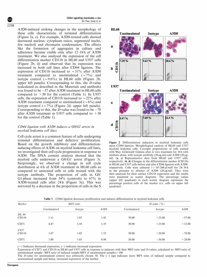
A3D8-induced striking changes in the morphology ofthese cells characteristic of terminal differentiation(Figure 2a, c). For example, A3D8-treated cells showeddecreased nucleus: cytoplasm ratios, segmented nuclei,few nucleoli and chromatin condensation. The effectslike the formation of aggregates in culture andadherence became visible only after 12–18 h of A3D8treatment. We also analysed the expression of the celldifferentiation marker CD11b in HL60 and U937 cells(Figure 2b, d) and observed that its expression wasincreased in both cell lines after CD44 ligation. Theexpression of CD11b increased to B31% after A3D8treatment compared to unstimulated (B7%) andisotype control (B9.6%) in HL60 cells (Figure 2b,upper left panels). Corresponding to this, the D-value(calculated as described in the Materials and methods)was found to be �57 after A3D8 treatment in HL60 cellscompared to +30 for the control (Table 1). In U937cells, the expression of CD11b increased to B22% afterA3D8 treatment compared to unstimulated (B6%) andisotype control (B7%) (Figure 2d, upper left panels).Corresponding to this, the D-value was found to be �78after A3D8 treatment in U937 cells compared to +30for the control (Table 1).
CD44 ligation with A3D8 induces a G0/G1 arrest inmyeloid leukemia cell lines
Cell cycle arrest is a common feature of cells undergoingterminal differentiation and defective proliferation.Based on the growth inhibitory and differentiation-inducing effects of A3D8 on myeloid leukemia cell lines,we investigated their cell cycle progression in response toA3D8. The DNA content analysis showed that themyeloid cells underwent a G0/G1 arrest (Figure 3).Surprisingly, we observed a change in cell cycledistribution at 6 h of A3D8 treatment in HL60 cells ascompared to untreated cells or cells treated with theisotype antibody. The proportion of cells in G0/G1 phase increased from 54% (controls) to 67% inA3D8-treated cells after 24 h (Figure 3a). This wasmirrored by a decrease in the proportion of cells in the S
Figure 2 Differentiation induction in myeloid leukemia cellsupon CD44 ligation. Morphological analysis of HL60 and U937myeloid leukemia cells: Cytospin preparations of cells stainedwith May–Grunwald–Giemsa after in vitro treatment for 36 h withmedium alone, with isotype antibody (20mg/ml) and A3D8 (20mg/ml). (a, c) Representative data from HL60 and U937 cells,respectively. (b, d) Changes in the differentiation marker (CD11b)in HL60 and U937 cells before and after CD44 ligation with A3D8,respectively. Cells were cultured 1� 105/200 ml/well for 24–36 hin the presence or absence of A3D8 (20 mg/ml). They werethen analysed for their surface CD11b expression and the resultswere presented as scatter diagrams. The percentage values(upper left quadrant) in each scatter diagram represents thepercentage positive cells of the marker (i.e. cells on upper leftquadrant)
Table 1 CD44 ligation decreases proliferation and induces differentiation in myeloid leukemia cells
Marker MFI ratio D-value (%)
Unstimulated Isotype A3D8 Unstimulated Isotype A3D8
HL 60CD11b 1.11 1.03 1.41 30.00 +23.00 �57.00
CD71 4.47 3.10 1.35 30.00 +25.00 +12.00
U937CD11b 1.07 1.02 1.32 30.00 +20.00 �78.00
CD71 1.00 1.03 0.98 30.00 �36.00 +24.00
(+) Indicates decreased expression, (�) indicates increased expression.Expression of CD71 and CD11b in HL60 and U937 cells in response to inducers with their MFI ratio and D-values, calculated as: MFI ratio ofunstimulated sample�MFI ratio of induced sample/MFI of unstimulated sample)� 100.The D-value for unstimulated control was arbitrarily chosen 30. The (�) sign indicates more MFI ratio of induced sample compared tounstimulated sample and hence, increased expression of the marker
CD44 signaling inactivates c-JunAA Peer Zada et al
2298
Oncogene
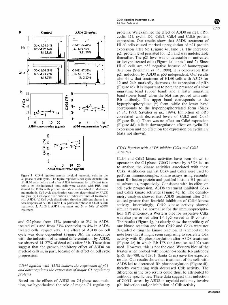
and G2 phase from 13% (controls) to 2% in A3D8-treated cells and from 23% (controls) to 4% in A3D8-treated cells, respectively. The effect of A3D8 on cellcycle was dose dependent (Figure 3b). In accordancewith the induction of terminal differentiation by A3D8,we observed 14–27% of dead cells after 36 h. These datasuggest that the growth inhibitory effect of A3D8 onmyeloid cells is, in part, because of its effect on cell cycleprogression.
CD44 ligation with A3D8 induces the expression of p21and downregulates the expression of major G1 regulatoryproteins
Based on the effects of A3D8 on G1 phase accumula-tion, we hypothesized the role of major G1 regulatory
proteins. We examined the effect of A3D8 on p21, pRb,cyclin D1, cyclin D2, Cdk2, Cdk4 and Cdk6 proteinexpression. Our results show that A3D8 treatment ofHL60 cells caused marked upregulation of p21 proteinexpression after 6 h (Figure 4a, lane 3). The increasedp21 protein level persisted for 12 h and was undetectablethereafter. The p21 level was undetectable in untreatedor isotype-treated cells (Figure 4a, lanes 1 and 2). SinceHL60 cells are p53 negative because of homozygousdeletions (Steinman et al., 1998), it is conceivable thatp21 induction by A3D8 is p53 independent. Our resultsalso show that treatment of HL60 cells with A3D8 for12 and 24 h markedly decreases the expression of pRb(Figure 4e). It is important to note the presence of a slowmigrating band (upper band) and a faster migratingband (lower band) when the blot was probed with anti-Rb antibody. The upper band corresponds to thehyperphosphorylated (*) form, while the lower bandcorresponds to the hypophosphorylated form (Slacket al., 1993; Savatier et al., 1994). Inhibition of pRbcorrelated with decreased levels of Cdk2 and Cdk4(Figure 4b, c). There was no effect on Cdk6 expression(Figure 4d), a little downregulation effect on cyclin D1expression and no effect on the expression on cyclin D2(data not shown).
CD44 ligation with A3D8 inhibits Cdk4 and Cdk2activities
Cdk4 and Cdk2 kinase activities have been shown tooperate in the G1 phase. G0/G1 arrest by A3D8 led usto analyse the kinase activities associated with theseCdks. Antibodies against Cdk4 and Cdk2 were used toperform immunocomplex kinase assays using recombi-nant Rb fusion protein and purified histone H1 proteinas substrates, respectively. Consistent with its effect oncell cycle progression, A3D8 treatment inhibited Cdk4and Cdk2 kinase activities (Figure 4g, h). The densito-metry analysis showed that A3D8 treatment after 24 hcaused greater than fourfold inhibition of Cdk4 kinaseactivity. Interestingly, Cdk2 kinase activity showedsimilar results. To normalize for the immunoprecipita-tion (IP) efficiency, a Western blot for respective Cdkswas also performed after IP. IgG served as IP control.The results (Figure 4g, h) clearly show the specificity ofour kinase reaction and that Cdk2 and Cdk4 were notdegraded during the kinase reaction. It is important tonote here that it might seem surprising to correlate Cdkactivity with Rb phosphorylation after A3D8 treatment(Figure 4e) in which Rb IF8 (anti-mouse, sc-102) wasused. However, this is not the case. Western blot of thelysates when probed with phospho-specific Rb antibody(pRb Ser-780, sc-12901, Santa Cruz) gave the expectedresults. Our results show that treatment of the cells withA3D8 led to decreased Rb phosphorylation (Figure 4f),thereby correlating with decreased Cdk activity. Thedifference in the two results could thus, be attributed toantibody specificities. These data suggest that inductionof G0/G1 arrest by A3D8 in myeloid cells may involvep21 induction and/or inhibition of Cdk activity.
Figure 3 CD44 ligation arrests myeloid leukemia cells in theG1phase of cell cycle. The figure represents cell cycle distributionof HL60 cells before and after A3D8 treatment for different timepoints. At the indicated time, cells were washed with PBS, andstained for DNA with propidium iodide as described in Materialsand methods. Cell cycle distribution was then determined by FACSanalysis. (a) Cell cycle distribution at indicated times of treatmentwith A3D8. (b) Cell cycle distribution showing different phases in adose response of A3D8. Lines: 1, A particular phase at 6 h of A3D8treatment. 2, At 24 h A3D8 treatment and 3, at 36 h of A3D8treatment
CD44 signaling inactivates c-JunAA Peer Zada et al
2299
Oncogene
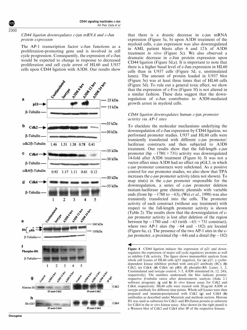
CD44 ligation downregulates c-jun mRNA and c-Junprotein expression
The AP-1 transcription factor c-Jun functions as aproliferation-promoting gene and is involved in cellcycle progression. Consequently, the expression of c-Junwould be expected to change in response to decreasedproliferation and cell cycle arrest of HL60 and U937cells upon CD44 ligation with A3D8. Our results show
that there is a drastic decrease in c-jun mRNAexpression (Figure 5a, b) upon A3D8 treatment of themyeloid cells, c-jun expression was also downregulatedin AML patient blasts after 6 and 12 h of A3D8treatment in vitro (Figure 5c). We also observed adramatic decrease in c-Jun protein expression uponCD44 ligation (Figure 5d,e). It is important to note thatthere is a higher basal level of c-Jun expression in HL60cells than in U937 cells (Figure 5d, e, unstimulatedlanes). The amount of protein loaded in U937 blot(Figure 5e) was at least three times that of HL60 cells(Figure 5d). To rule out a general toxic effect, we showthat the expression of c-Fos (Figure 5f) is not altered ina similar fashion. These data suggest that the down-regulation of c-Jun contributes to A3D8-mediatedgrowth arrest in myeloid cells.
CD44 ligation downregulates human c-jun promoteractivity via AP-1 sites
To elucidate the molecular mechanisms underlying thedownregulation of c-Jun expression by CD44 ligation, weperformed promoter studies. U937 and HL60 cells weretransiently transfected with different c-jun promoter/luciferase constructs and then subjected to A3D8treatment. Our results show that the full-length c-junpromoter (bp �1780/+731) activity was downregulated14-fold after A3D8 treatment (Figure 6). It was not avector effect since A3D8 had no effect on pGL3, in whichc-jun promoter constructs were subcloned. As a positivecontrol for our promoter studies, we also show that TPAincreases the c-jun promoter activity (data not shown). Tomap site(s) in the c-jun promoter responsible for thedownregulation, a series of c-jun promoter deletionmutant-luciferase gene chimeric plasmids with variableends (from bp �1780 to �63), (Wei et al., 1998) was alsotransiently transfected into the cells. The promoteractivity of each construct (without any treatment) withrespect to the full-length promoter activity is shown(Table 2). The results show that the downregulation of c-jun promoter activity is lost after deletion of the regionbetween bp �1780 and �63 (with �63/+731 construct),where two AP-1 sites (bp �64 and �182) are located(Figure 6a, c). The presence of the two AP-1 sites in the c-jun promoter, a proximal (bp �64) and a distal (bp �182)
Figure 4 CD44 ligation induces the expression of p21 and down-regulates the expression of major cell cycle regulatory proteins as wellas inhibits Cdk activity. The figure shows immunoblot analysis fromwhole cell lysates of HL60 cells (p53 negative), for (a) p21, a cyclin-dependent kinase inhibitor probed with anti-p21 antibody (SC); (b)Cdk2; (c) Cdk4; (d) Cdk6; (e) pRb; (f) phospho-Rb. Lanes: 1, 2,Unstimulated and isotype control, 3–5, A3D8 stimulated (6, 12, 24 h,respectively). The numbers underneath the blot indicate protein/respective b-tubulin ratios after densitometric analysis (Aida 2.1software program). (g and h) In vitro kinase assay for Cdk2 andCdk4, respectively: HL60 cells were treated with 20mg/ml A3D8 orisotype antibody for different time points. Whole cell lysates were thenprepared and immunoprecipitated with Cdk2 (g) and Cdk4 (h)antibodies as described under Materials and methods section. HistoneH1 was used as substrate for Cdk2- and Rb-fusion protein as substratefor Cdk4 in the in vitro kinase assay. Also shown (in the right panel) isa Western blot of Cdk2 and Cdk4 after IP of the respective kinases
CD44 signaling inactivates c-JunAA Peer Zada et al
2300
Oncogene
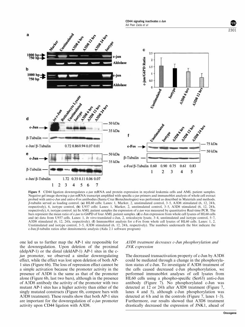
one led us to further map the AP-1 site responsible forthe downregulation. Upon deletion of the proximal(delpAP-1) or the distal (deldAP-1) AP-1 sites in the c-jun promoter, we observed a similar downregulatingeffect, while the effect was lost upon deletion of both AP-1 sites (Figure 6b). The loss of repression effect cannot bea simple activation because the promoter activity in thepresence of A3D8 is the same as that of the promoteralone (Figure 6b, last two bars), although in the presenceof A3D8 antibody the activity of the promoter with twomutant AP-1 sites has a higher activity than either of thesingly mutated constructs (Figure 6b, compare bars withA3D8 treatment). These results show that both AP-1 sitesare important for the downregulation of c-jun promoteractivity upon CD44 ligation with A3D8.
A3D8 treatment decreases c-Jun phosphorylation andJNK expression
The decreased transactivation property of c-Jun by A3D8could be mediated through a change in the phosphoryla-tion status of c-Jun. To investigate if A3D8 treatment ofthe cells caused decreased c-Jun phosphorylation, weperformed immunoblot analyses of cell lysates fromHL60 cells using a phospho-specific (Ser63) anti-c-Junantibody (Figure 7). No phosphorylated c-Jun wasdetected at 12 or 24h after A3D8 treatment (Figure 7,lanes 4 and 5), although c-Jun phosphorylation wasdetected at 6 h and in the controls (Figure 7, lanes 1–3).Furthermore, our results showed that A3D8 treatmentdrastically decreased the expression of JNK1, ahead of
Figure 5 CD44 ligation downregulates c-jun mRNA and protein expression in myeloid leukemia cells and AML patient samples.Negative gel image showing c-junmRNA transcript amplified with specific c-jun primers and immunoblot analysis of whole cell extractprobed with anti-c-Jun and anti-c-Fos antibodies (Santa Cruz Biotechnologies) was performed as described in Materials and methods.b-tubulin served as loading control. (a) HL60 cells: Lanes: 1, Marker, 2, unstimulated control, 3–5, A3D8 stimulated (6, 12, 24 h,respectively), 6, isotype control, (b) U937 cells: Lanes: 1, Marker, 2, unstimulated control, 3–5, A3D8 stimulated (6, 12, 24 h,respectively), 6, isotype control. (c) In AML patient samples the expression of c-jun was measured by quantitative Real-time PCR. Thebars represent the mean ratio of c-jun to G6PD of four AML patient samples. (d) c-Jun expression from whole cell lysates of HL60 cellsand (e) data from U937 cells. Lanes: 1, In vitro-translated c-Jun, 2, reticulocyte lysate, 3–4, unstimulated and isotype control, 5–7,A3D8 stimulated (6, 12, 24 h, respectively). (f) Immunoblot analysis for c-Fos from whole cell lysates of HL60 cells. Lanes: 1, 2,Unstimulated and isotype control, 3–5, A3D8 stimulated (6, 12, 24 h, respectively). The numbers underneath the blot indicate thec-Jun/b-tubulin ratios after densitometric analysis (Aida 2.1 software program)
CD44 signaling inactivates c-JunAA Peer Zada et al
2301
Oncogene
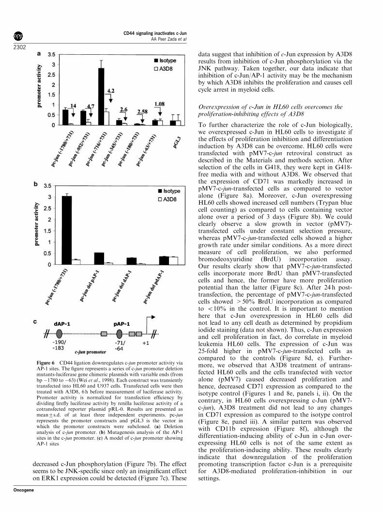
decreased c-Jun phosphorylation (Figure 7b). The effectseems to be JNK-specific since only an insignificant effecton ERK1 expression could be detected (Figure 7c). These
data suggest that inhibition of c-Jun expression by A3D8results from inhibition of c-Jun phosphorylation via theJNK pathway. Taken together, our data indicate thatinhibition of c-Jun/AP-1 activity may be the mechanismby which A3D8 inhibits the proliferation and causes cellcycle arrest in myeloid cells.
Overexpression of c-Jun in HL60 cells overcomes theproliferation-inhibiting effects of A3D8
To further characterize the role of c-Jun biologically,we overexpressed c-Jun in HL60 cells to investigate ifthe effects of proliferation inhibition and differentiationinduction by A3D8 can be overcome. HL60 cells weretransfected with pMV7-c-jun retroviral construct asdescribed in the Materials and methods section. Afterselection of the cells in G418, they were kept in G418-free media with and without A3D8. We observed thatthe expression of CD71 was markedly increased inpMV7-c-jun-transfected cells as compared to vectoralone (Figure 8a). Moreover, c-Jun overexpressingHL60 cells showed increased cell numbers (Trypan bluecell counting) as compared to cells containing vectoralone over a period of 3 days (Figure 8b). We couldclearly observe a slow growth in vector (pMV7)-transfected cells under constant selection pressure,whereas pMV7-c-jun-transfected cells showed a highergrowth rate under similar conditions. As a more directmeasure of cell proliferation, we also performedbromodeoxyuridine (BrdU) incorporation assay.Our results clearly show that pMV7-c-jun-transfectedcells incorporate more BrdU than pMV7-transfectedcells and hence, the former have more proliferationpotential than the latter (Figure 8c). After 24 h post-transfection, the percentage of pMV7-c-jun-transfectedcells showed >50% BrdU incorporation as comparedto o10% in the control. It is important to mentionhere that c-Jun overexpression in HL60 cells didnot lead to any cell death as determined by propidiumiodide staining (data not shown). Thus, c-Jun expressionand cell proliferation in fact, do correlate in myeloidleukemia HL60 cells. The expression of c-Jun was25-fold higher in pMV7-c-jun-transfected cells ascompared to the controls (Figure 8d, e). Further-more, we observed that A3D8 treatment of untrans-fected HL60 cells and the cells transfected with vectoralone (pMV7) caused decreased proliferation andhence, decreased CD71 expression as compared to theisotype control (Figures 1 and 8e, panels i, ii). On thecontrary, in HL60 cells overexpressing c-Jun (pMV7-c-jun), A3D8 treatment did not lead to any changesin CD71 expression as compared to the isotype control(Figure 8e, panel iii). A similar pattern was observedwith CD11b expression (Figure 8f), although thedifferentiation-inducing ability of c-Jun in c-Jun over-expressing HL60 cells is not of the same extent asthe proliferation-inducing ability. These results clearlyindicate that downregulation of the proliferationpromoting transcription factor c-Jun is a prerequisitefor A3D8-mediated proliferation-inhibition in oursettings.
Figure 6 CD44 ligation downregulates c-jun promoter activity viaAP-1 sites. The figure represents a series of c-jun promoter deletionmutants-luciferase gene chimeric plasmids with variable ends (frombp �1780 to �63) (Wei et al., 1998). Each construct was transientlytransfected into HL60 and U937 cells. Transfected cells were thentreated with A3D8, 6 h before measurement of luciferase activity.Promoter activity is normalized for transfection efficiency bydividing firefly luciferase activity by renilla luciferase activity of acotransfected reporter plasmid pRL-0. Results are presented asmean7s.d. of at least three independent experiments. pc-junrepresents the promoter constructs and pGL3 is the vector inwhich the promoter constructs were subcloned. (a) Deletionanalysis of c-jun promoter. (b) Mutagenesis analysis of the AP-1sites in the c-jun promoter. (c) A model of c-jun promoter showingAP-1 sites
CD44 signaling inactivates c-JunAA Peer Zada et al
2302
Oncogene
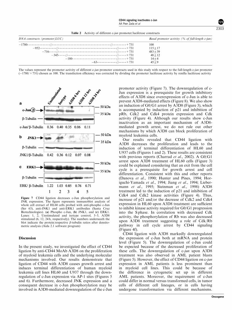
Discussion
In the present study, we investigated the effect of CD44ligation by anti-CD44 MoAb A3D8 on the proliferationof myeloid leukemia cells and the underlying molecularmechanisms involved. Our results demonstrate thatligation of CD44 with A3D8 causes growth arrest andinduces terminal differentiation of human myeloidleukemia cell lines HL60 and U937 through the down-regulation of c-Jun expression via AP-1 sites (Figures 5and 6). Furthermore, decreased JNK expression and aconsequent decrease in c-Jun phosphorylation may beinvolved in A3D8-mediated downregulation of the c-Jun
promoter activity (Figure 7). The downregulation of c-Jun expression is a prerequisite for growth inhibitoryeffects of A3D8 since overexpression of c-Jun is able toprevent A3D8-mediated effects (Figure 8). We also showan induction of G0/G1 arrest by A3D8 (Figure 3), whichis accompanied by induction of p21 and inhibition ofpRb, Cdk2 and Cdk4 protein expression and Cdkactivity (Figure 4). Although our results show c-Juninactivation as an important mechanism of A3D8-mediated growth arrest, we do not rule out othermechanisms by which A3D8 can block proliferation ofmyeloid leukemia cells.
Our results revealed that CD44 ligation withA3D8 decreases the proliferation and leads to theinduction of terminal differentiation of HL60 andU937 cells (Figures 1 and 2). These results are consistentwith previous reports (Charrad et al., 2002). A G0/G1arrest upon A3D8 treatment of HL60 cells (Figure 3)could be explained considering that an exit from the cellcycle is a prerequisite for growth arrest and celldifferentiation. Consistent with this and other reports(Danova et al., 1990; Hunter and Pines, 1994; Hor-iguchi-Yamada et al., 1994; Jiang et al., 1994; Lieber-mann et al., 1995; Steinman et al., 1998) A3D8treatment led to the induction of p21 and inhibition ofCdk4 and Cdk2 kinase activities (Figure 4). Thus,increase of p21 and/or the decrease of Cdk2 and Cdk4expression in HL60 upon A3D8 treatment are sufficientto inhibit kinase activity required for G0/G1 progressioninto the S phase. In correlation with decreased Cdkactivity, the phosphorylation of Rb was also decreasedupon A3D8 treatment suggesting a role of Cdk/Rbpathway in cell cycle arrest by CD44 signaling(Figure 4f).
CD44 ligation with A3D8 markedly downregulatedthe expression of c-Jun both at mRNA and proteinlevel (Figure 5). The downregulation of c-Jun couldbe expected because of the decreased proliferation ofthese cells. The downregulation of c-jun upon A3D8treatment was also observed in AML patient blasts(Figure 5). However, the effect of CD44 ligation on c-junexpression in AML patients is less prominent thanin myeloid cell lines. This could be because ofthe difference in cytogenetic set up in differentAML patients. Moreover, the requirement of c-Juncould differ in normal versus transformed cells, in tumorcells of different cell lineages, or in cells havingundergone transformation via different mechanisms.
Table 2 Activity of different c-jun promoter/luciferase constructs
DNA constructs (promoter/LUC) Basal promoter activity (% of full-length c-jun)
�1780- - - - - - - - - - - - - - -//- - - - - - - - - - - - - - - - - - - - - - - - - - - - - - - - - - - - - -//- - - - - - - - -+731 100�952- - - - - - - - - - -//- - - - - - - - - - - - - - - - - - - - - - - - - - - - - - - - - - - - - -//- - - - -+731 115717
�716- - - - -//- - - - - - - - - - - - - - - - - - - - - - - - - - - - - - - - - - - - - -//- - - - -+731 483739�345- - - - -//- - - - - - - - - - - - - - - - - - - - - - - - - - - - - - - -//- - - - -+731 48712
�180- - - - - - - - - - - - - - - - - - - - - - - - - - - - -//- - - - -+731 1474�63- - - - - - - - - - - - -//- - - - -+731 4579
The values represent the promoter activity of different c-jun promoter constructs used in this study with respect to the full-length c-jun promoter(�1780/+731) chosen as 100. The transfection efficiency was corrected by dividing the promoter luciferase activity by renilla luciferase activity
Figure 7 CD44 ligation decreases c-Jun phosphorylation andJNK expression. The figure represents immunoblot analysis ofwhole cell extract of HL60 cells probed with anti-phospho c-Jun(Ser 63), anti-JNK1 and anti-ERK1 antibodies (Santa CruzBiotechnologies). (a) Phospho c-Jun, (b) JNK1, and (c) ERK1.Lanes: 1, 2, Unstimulated and isotype control, 3–5, A3D8stimulated (6, 12, 24 h, respectively). The numbers underneath theblot indicate the protein/respective b-tubulin ratios after densito-metric analysis (Aida 2.1 software program)
CD44 signaling inactivates c-JunAA Peer Zada et al
2303
Oncogene
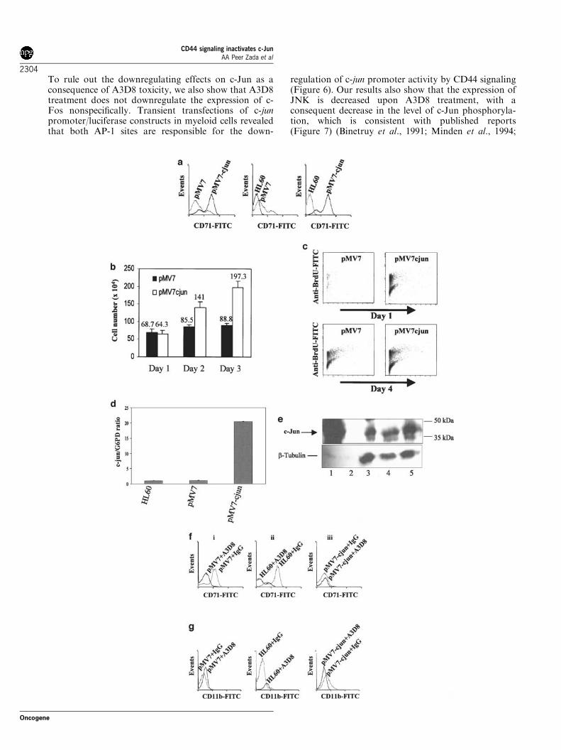
To rule out the downregulating effects on c-Jun as aconsequence of A3D8 toxicity, we also show that A3D8treatment does not downregulate the expression of c-Fos nonspecifically. Transient transfections of c-junpromoter/luciferase constructs in myeloid cells revealedthat both AP-1 sites are responsible for the down-
regulation of c-jun promoter activity by CD44 signaling(Figure 6). Our results also show that the expression ofJNK is decreased upon A3D8 treatment, with aconsequent decrease in the level of c-Jun phosphoryla-tion, which is consistent with published reports(Figure 7) (Binetruy et al., 1991; Minden et al., 1994;
CD44 signaling inactivates c-JunAA Peer Zada et al
2304
Oncogene
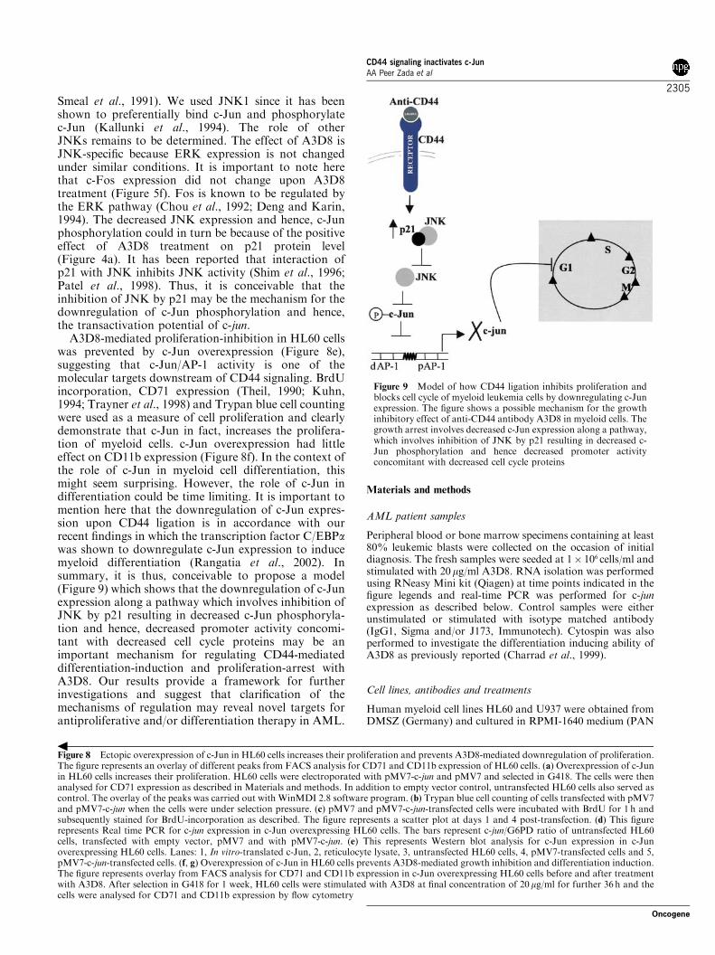
Smeal et al., 1991). We used JNK1 since it has beenshown to preferentially bind c-Jun and phosphorylatec-Jun (Kallunki et al., 1994). The role of otherJNKs remains to be determined. The effect of A3D8 isJNK-specific because ERK expression is not changedunder similar conditions. It is important to note herethat c-Fos expression did not change upon A3D8treatment (Figure 5f). Fos is known to be regulated bythe ERK pathway (Chou et al., 1992; Deng and Karin,1994). The decreased JNK expression and hence, c-Junphosphorylation could in turn be because of the positiveeffect of A3D8 treatment on p21 protein level(Figure 4a). It has been reported that interaction ofp21 with JNK inhibits JNK activity (Shim et al., 1996;Patel et al., 1998). Thus, it is conceivable that theinhibition of JNK by p21 may be the mechanism for thedownregulation of c-Jun phosphorylation and hence,the transactivation potential of c-jun.
A3D8-mediated proliferation-inhibition in HL60 cellswas prevented by c-Jun overexpression (Figure 8e),suggesting that c-Jun/AP-1 activity is one of themolecular targets downstream of CD44 signaling. BrdUincorporation, CD71 expression (Theil, 1990; Kuhn,1994; Trayner et al., 1998) and Trypan blue cell countingwere used as a measure of cell proliferation and clearlydemonstrate that c-Jun in fact, increases the prolifera-tion of myeloid cells. c-Jun overexpression had littleeffect on CD11b expression (Figure 8f). In the context ofthe role of c-Jun in myeloid cell differentiation, thismight seem surprising. However, the role of c-Jun indifferentiation could be time limiting. It is important tomention here that the downregulation of c-Jun expres-sion upon CD44 ligation is in accordance with ourrecent findings in which the transcription factor C/EBPawas shown to downregulate c-Jun expression to inducemyeloid differentiation (Rangatia et al., 2002). Insummary, it is thus, conceivable to propose a model(Figure 9) which shows that the downregulation of c-Junexpression along a pathway which involves inhibition ofJNK by p21 resulting in decreased c-Jun phosphoryla-tion and hence, decreased promoter activity concomi-tant with decreased cell cycle proteins may be animportant mechanism for regulating CD44-mediateddifferentiation-induction and proliferation-arrest withA3D8. Our results provide a framework for furtherinvestigations and suggest that clarification of themechanisms of regulation may reveal novel targets forantiproliferative and/or differentiation therapy in AML.
Materials and methods
AML patient samples
Peripheral blood or bone marrow specimens containing at least80% leukemic blasts were collected on the occasion of initialdiagnosis. The fresh samples were seeded at 1� 106 cells/ml andstimulated with 20mg/ml A3D8. RNA isolation was performedusing RNeasy Mini kit (Qiagen) at time points indicated in thefigure legends and real-time PCR was performed for c-junexpression as described below. Control samples were eitherunstimulated or stimulated with isotype matched antibody(IgG1, Sigma and/or J173, Immunotech). Cytospin was alsoperformed to investigate the differentiation inducing ability ofA3D8 as previously reported (Charrad et al., 1999).
Cell lines, antibodies and treatments
Human myeloid cell lines HL60 and U937 were obtained fromDMSZ (Germany) and cultured in RPMI-1640 medium (PAN
Figure 8 Ectopic overexpression of c-Jun in HL60 cells increases their proliferation and prevents A3D8-mediated downregulation of proliferation.The figure represents an overlay of different peaks from FACS analysis for CD71 and CD11b expression of HL60 cells. (a) Overexpression of c-Junin HL60 cells increases their proliferation. HL60 cells were electroporated with pMV7-c-jun and pMV7 and selected in G418. The cells were thenanalysed for CD71 expression as described in Materials and methods. In addition to empty vector control, untransfected HL60 cells also served ascontrol. The overlay of the peaks was carried out with WinMDI 2.8 software program. (b) Trypan blue cell counting of cells transfected with pMV7and pMV7-c-jun when the cells were under selection pressure. (c) pMV7 and pMV7-c-jun-transfected cells were incubated with BrdU for 1 h andsubsequently stained for BrdU-incorporation as described. The figure represents a scatter plot at days 1 and 4 post-transfection. (d) This figurerepresents Real time PCR for c-jun expression in c-Jun overexpressing HL60 cells. The bars represent c-jun/G6PD ratio of untransfected HL60cells, transfected with empty vector, pMV7 and with pMV7-c-jun. (e) This represents Western blot analysis for c-Jun expression in c-Junoverexpressing HL60 cells. Lanes: 1, In vitro-translated c-Jun, 2, reticulocyte lysate, 3, untransfected HL60 cells, 4, pMV7-transfected cells and 5,pMV7-c-jun-transfected cells. (f, g) Overexpression of c-Jun in HL60 cells prevents A3D8-mediated growth inhibition and differentiation induction.The figure represents overlay from FACS analysis for CD71 and CD11b expression in c-Jun overexpressing HL60 cells before and after treatmentwith A3D8. After selection in G418 for 1 week, HL60 cells were stimulated with A3D8 at final concentration of 20mg/ml for further 36 h and thecells were analysed for CD71 and CD11b expression by flow cytometry
Figure 9 Model of how CD44 ligation inhibits proliferation andblocks cell cycle of myeloid leukemia cells by downregulating c-Junexpression. The figure shows a possible mechanism for the growthinhibitory effect of anti-CD44 antibody A3D8 in myeloid cells. Thegrowth arrest involves decreased c-Jun expression along a pathway,which involves inhibition of JNK by p21 resulting in decreased c-Jun phosphorylation and hence decreased promoter activityconcomitant with decreased cell cycle proteins
CD44 signaling inactivates c-JunAA Peer Zada et al
2305
Oncogene
Biotech GMBH) supplemented with 2mm glutamine, 100 U/mlpenicillin and 100mg/ml streptomycin and 10% FBS (GIBCO-BRL, Eragny, France) at 371C with 5% CO2 enrichment. Thecells were suspended at 3� 105 cells/ml in 96-well tissue cultureplates (Nunc) with 200 ml/well. Monoclonal anti-CD44 anti-body, A3D8 (Sigma) was added at the final concentration of20 mg/ml. All the control samples were either untreated ortreated with 20mg/ml isotype-matched antibody (IgG1, Sigmaor J173, Immunotech). For direct immunofluorescence stain-ing, fluorescein isothiocyanate (FITC) MoAbs to CD11b(FITC, immunotech, IgG1) and CD71 (FITC, DAKO,IgG1) were used.
Proliferation assay
To assess the proliferation state of cells after varioustreatments, MTT proliferation assay kit (Boehringer Man-nheim, Germany) was used according to the manufacturer’sinstructions. Briefly, cells were incubated with or withoutA3D8 for 1–4 days in 96-well plates. A volume of 10 ml MTTlabeling reagent (5mg/ml) was added every day to each welland the plates were incubated at 371C for 4 h. The resultingformazon crystals were solubilized by adding 100ml ofsolubilization buffer (10% SDS in 0.01m HCl) per well andthe plates were incubated at 371C overnight. The absorbanceof the formazon measured at 575 nm was used to account forthe proliferation state of cells. Trypan blue cell exclusion wasalso used to assess the cell viability and the cell number.
Morphological analysis of cellular differentiation
After various cell treatments, HL60 and U937 cells were cyto-centrifuged on glass slides using a cytospin device (Shandon-Elliot, Paris, France). May–Grunwald–Giemsa-stained cytos-pin smears were then analysed under light microscopy. Theanalysis was performed on 200–300 cells.
Cell cycle analysis and flow cytometry
To investigate the surface expression of myeloid differentiationand proliferation markers, FACS analysis was performed.HL60 and U937 cells (3� 105 cells/ml) were stimulatedfor 36h with A3D8 (20mg/ml) and then stained with FITC orPE labeled antibodies at a concentration of 10mg/ml at41C for 30min and washed twice with FACS buffer (PBS, 3%FCS, 0.01% NaN3). Fluorescence was then analysed on CoulterEPICS XL/XL-MCL SystemII Software. Data were collectedafter 5000 cell analysis (per sample) and the results shown asscatter diagrams or expressed as D-value which is calculated as
Mean fluorescence intensity ðMFIÞ ratio
¼ Meanof sample stained with MoAb=
mean of isotype control MoAb
Then, the percentage of difference between MFI ratio ofsample incubated with or without A3D8 was calculated, givinga D-value (Pisani et al., 1997) for each sample. The D-value forunstimulated control was arbitrarily chosen and that ofstimulated sample expressed relative to it. Negative D-valuesindicate more MFI of stimulated sample than unstimulatedand hence, implies increased expression of the particularmarker. Similarly, positive D-values imply decreased expres-sion of the marker. For the cell cycle analysis, cells with orwithout A3D8 treatment were centrifuged at 1500 r.p.m. for3 min, washed with PBS and then the DNA was stained with100 mg/ml propidium iodide for 30min at 41C protected from
light. The cells were then analysed with the FACScan(Beckton-Dickinson) for different cell populations.
RNA isolation and semiquantitative RT–PCR
Total RNA was isolated from 7� 105 cells before and aftertreatment with A3D8 (20 mg/ml) and IgG1 (20 mg/ml) usingRNeasy Mini kit (Qiagen). RNA of 100 ng was used for first-strand cDNA synthesis in a 20 ml reaction with 10�RT buffer,dNTP (5mm), RNasin (1U/ml), oligo dT (1 mm) and thereaction was incubated at 371C for 90min. Equal amounts ofcDNA were taken for c-jun PCR amplification using a Qiagenkit. Aldolase was used as an internal control. Primers used forPCR amplification were for c-jun (Gene Bank Acc. no.J04111): forward primer 50-ACTGCAAAGA TGGAAAC-GAC-30 (bp 1264–1283) and reverse primer 50-AAAATGTTTGCAACTGCTGC-30 (bp 2235–2254); and for aldolase:forward primer 50-AGCTGTCTG ACATCG CTCACCG-30
and reverse primer 50-CACATA CTGG CAGCGCTTCAAG-30. The PCR cycling program consisted of 30 cyclesof 941C for 2min, 551C for 1min and 721C for 80 s using DNAthermal cycler (Perkin-Elmer). PCR products of B971 bp (c-jun) and B580 bp (Aldolase) were separated by 1.2% agarosegel electrophoresis and visualized by ethidium bromidestaining with UV irradiation.
Quantitative Real-time PCR in AML patient samples
Real-time PCR for c-jun and for the housekeeping geneglucose-6-phosphate dehydrogenase (G6PD) was performedusing the Light Cycler Technology (Roche Diagnostics,Mannheim, Germany). For amplification of G6PD, primerswere used according to Emig et al. (1999): G6PD-1 forwardprimer 50-CCGGATCGACCACTACCTGGGCAAG-30 andG6PD-2 reverse primer 50-GTTCCCCACGTACTGGC-CCAGGACCA-30. c-jun was amplified using the primerset according to Kiaris et al (1999). c-jun-1 forward primer50-GCATGAGGAACC GCATCGCTGCCTCCAAGT-30
and c-jun-2 reverse primer 50-GCGACCAAGTCCTTCCCACTCGTGCACACT-30. G6PD plasmid: pGdBBX,kindly provided by A Hochhaus, University of Mannheimwas serially diluted to 10 000, 1000 and 100 fg and used as astandard curve. The procedure for Real-time PCR wasessentially the same as described previously (Reddy et al., 2002).
Immunoblot analysis
Total cellular protein was extracted from HL60 and U937cells before and after A3D8 treatment and subjected toelectrophoresis on 10% SDS–PAGE gels. The Westernblotting procedure was performed and the blots detectedwith the ECL system as described previously (Behre et al.,1999). Anti-b-tubulin antibody (Boehringer, Mannheim)was used as internal loading control on the same blot afterstripping. Immunoblot analysis was performed for c-Jun(anti-rabbit, sc-45), c-Fos (anti-rabbit polyclonal, sc-52), JunB(anti-mouse monoclonal IgG1, sc-8051), Cdk2 (anti-rabbit,sc-163), Cdk4 (anti-rabbit, sc-260), Cdk6 (anti-rabbit, sc-177),cyclin D1 (anti-rabbit, sc-718), p21 (anti-mouse, sc-817), pRb(anti-mouse monoclonal, sc-102) and phospho-specificRb (anti-goat, pRb-Ser780, sc-12901). The other antibodiesused for the Immunoblot analysis were JNK1 (anti-rabbit,sc-474), ERK1 (anti-rabbit, sc-94 Santa Cruz), phospho-c-Jun (anti-mouse monoclonal, sc-822 Santa Cruz) andb-tubulin (anti-rabbit, sc-9104). All the blots were quantifiedby densitometry (Aida 2.1 software program) and theresults reported in relation to b-tubulin. Comparison of the
CD44 signaling inactivates c-JunAA Peer Zada et al
2306
Oncogene
differences in expression was done between isotype control andA3D8-treated samples. Unstimulated samples served as areference for the basal level of expression.
Immunocomplex kinase assay
After the described treatments, HL60 cells were washedwith cold PBS and RIPA lysates prepared at different timepoints. Lysates were collected by centrifugation for 30 minand protein concentrations were quantified by the Bradfordassay (Bio-Rad Laboratories, Germany). Protein of 200 mgwas incubated with 2 mg of anti-Cdk2 or anti-Cdk4 antibody at41C for 2 h with rotation. Protein A agarose beads (20 ml)were then added and the incubation continued for another 2 h.Immunocomplex beads were washed twice with PBS bufferand three times with kinase buffer (150 mm NaCl; 1 mm
EDTA; 50 mm Tris-HCl, pH 7.5; 10mm MgCl2; and 10 mm
DTT). Kinase activity was assayed by incubating the beadsat 371C for 30 min with 25 ml kinase buffer, 3mg histone H1(Upsdate, Germany; Cdk2) or Rb-fusion protein (Santa Cruz;Cdk4), 10mm ATP, and 4 mCi [g-32P]ATP (3000Ci/mmol).Samples were then boiled for 5 min in 2� sample buffer,electrophoresed through a 12% SDS–PAGE, dried, andphosphorylated histone H1 and Rb proteins were visualizedby autoradiography and quantified by Aida 2.1 softwareprogram.
Reporter constructs and transient transfections
The human full-length c-jun promoter plus 731 bp of down-stream sequences (�1780/+731) subcloned into the fireflyluciferase vector pGL3, and deletion mutants in the proximaland distal AP-1 sites of the same construct used in this studywere kindly provided by Wayne Vedeckis (Wei et al., 1998). Asan internal control plasmid for cotransfection assays, the pRL-0 construct driving the Renilla luciferase gene (Promega) wasused as described (Behre et al., 1999). Transient transfectionswere performed with these constructs by using Effectenetransfection reagent (Qiagen) in myeloid cells. Effectene allowstransfections in the absence of serum, which was important torule out any serum-induced fluctuations in c-jun promoteractivity. The cells were transiently transfected with 1mg c-junpromoter/luciferase constructs and pRL-0 plasmid per well ofthe six-well plates. At 18 h after transfection, A3D8 was addedto the wells to a final concentration of 20 mg/ml for additional6 h. Promoter activities were determined by measuring theluciferase activity with the Dual Luciferase Assay System(Promega). Firefly luciferase activities of different c-junpromoter constructs in pGL3 were normalized to the Renillaluciferase values of pRL-0 (Behre et al., 1999). Results are
given as mean7s.d. of at least three different independentexperiments.
Stable cell lines overexpressing c-Jun and BrdU incorporation
To generate cell lines overexpressing c-Jun, the retrovirus-derived cDNA expression vector was used for the study.This vector, designated pMV7-c-jun was kindly provided byDr Yaniv. The vectors are described elsewhere (Kirschmeieret al., 1988). pMV7-c-jun and the empty vector pMV7 (lackingthe c-Jun cDNA insert) were transfected into HL60 cells byelectroporation (300V/975 mF). After 24–48 h, the cells weretransferred into selective medium containing 1mg/ml G418and analysed for CD71 expression and BrdU incorporationby flow cytometry. Trypan blue cell counting and PI stainingwas also performed at different time points. In addition toCD71 expression and cell counting, BrdU, a thymidineanalogue which gets incorporated into cellular DNA wasalso used as a measure of cell proliferation in c-Jun over-expressing HL60 cells. The assay was performed by using insitu cell proliferation kit, FLUOS (Roche, Mannheim,Germany, cat. no. 1810740) according to the manufacturer’sinstructions. Briefly, after transfer into selection medium,HL60 cells (3� 105 cells/ml previously transfected withpMV7 and pMV7-c-jun) were incubated with 10 mm BrdU for1 h at 371C in 5% CO2. The cells were then fixed in 70%ethanol in 50mm glycine buffer, pH 2.0, washed in PBS,acid denatured (4m HCl) and incubated with anti-BrdU-FLUOS antibody solution for 45 min at 371C in 5% CO2.The cells were then analysed by flow cytometry and the datapresented as scatter diagrams. In a similar experiment, after 1week in the selection medium, the cells transfected with pMV7or pMV7-c-jun were then stimulated with A3D8 or IgG with afinal concentration of 20 mg/ml. At 36 h after treatment withA3D8, the cells were analysed for CD11b and CD71expression. The expression of c-Jun in c-Jun overexpressingHL60 cells was measured by Real-time PCR and Western blotanalysis.
Acknowledgements
We thank Dr Dirk Eick and Alloy Sheppers for valuablecomments and useful discussions, Christian Kurzreder,Doreen Westhaus and Vijay Rawat for help withflow cytometry experiments and Susanne Schnittger forproviding AML patient samples. This work was supportedby a DFG (German research foundation) grant given to GB(2042/2-1).
References
Allouche M, Charrad RS, Bettaieb A, Greenland C, GrignonC and Smadja-Joffe F. (2000). Blood, 96, 1187–1190.
Aruffo A, Stamenkovic I, Melnick M, Underhill CB and SeedB. (1990). Cell, 61, 1303–1313.
Bakiri L, Lallemand D, Bossy-Wetzel E, Yaniv M. (2000).EMBO J., 2, 2056–2068.
Bates S, Parry D, Bonetta L, Vousden K, Dickson C andPeters G. (1994). Oncogene, 9, 1633–1640.
Behre G, Singh SM, Liu H, Bortolin LT, Christopeit M,Radomska HS, Rangatia J, Hiddemann W, Friedman ADand Tenen DG. (2002). J. Biol. Chem., 277, 26293–26299.
Behre G, Smith LT and Tenen DG. (1999). Biotechniques, 26,24–26.
Behre G, Whitmarsh AJ, Coghlan MP, Hoang T, CarpenterCL, Zhang DE, Davis RJ and Tenen DG. (1999). J. Biol.Chem., 274, 4939–4946.
Behre G, Zhang P, Zhang DE and Tenen DG. (1999).Methods, 17, 231–237.
Binetruy B, Smeal T and Karin M. (1991). Nature, 351,
122–127.Charrad RS, Gadhoum Z, Qi J, Glachant A, Allouche M,
Jasmine C, Chomienne C and Smadja-Joffe F. (2002). Blood,1, 290–299.
Charrad RS, Li Y, Delpech B, Balitrand N, Clay D, Jasmin C,Chomienne C and Smadja-Joffe F. (1999). Nat. Med., 5,669–676.
CD44 signaling inactivates c-JunAA Peer Zada et al
2307
Oncogene
Chou SY, Baichwal V and Ferrell Jr JE. (1992). Mol. Biol.Cell., 3, 1117–1130.
Danova M, Giordano M, Mazzini G and Riccardi A. (1990).Leukemia Res., 14, 417–422.
Deng T and Karin M. (1994). Nature, 371, 171–175.Elsasser A, Franzen M, Reddy V and Behre G . (2002). ASH
[Abstract]. 100, 200.Emig M, Saussele S, Wittor H, Weisser A, Reiter A, Willer A,
Berger U, Hehlmann R, Cross NC and Hochhaus A. (1999).Leukemia, 13, 1825–1832.
Gitig DM and Koff A. (2000). Methods Mol. Biol., 142, 109–123.
Harper JW, Adami GR, Wei N, Keyomarsi K and Elledge SJ.(1993a). Cell, 75, 805–816.
Harper JW, Adami GR, Wei N, Keyomarsi K and Elledge SJ.(1993b). Cell, 19, 805–816.
Hilberg F, Aguzzi A, Howells N and Wagner. (1993). Nature,365, 179–181.
Hilberg F and Wagner EF. (1992). Oncogene, 7, 2371–2380.Horiguchi-Yamada J, Yamada H, Nakada S, Ochi K and
Nemoto T. (1994). Mol. Cell. Biochem., 132, 31–37.Hunter T and Pines J. (1994). Cell, 79, 573–582.Jiang H, Lin J, Su ZZ, Collart FR, Huberman E and Fisher
PB. (1994). Oncogene, 9, 3397–3406.Kallunki T, Su B and Tsigelny I. (1994). Genes Dev., 8, 2996–
3007.Kantarjian H. (1999). Semin. Hematol., 36, 16–25.Kiaris H, Schally AV, Sun B, Armatis P and Groot K. (1999).
Oncogene, 18, 7168–7173.Kirschmeier PT, Housey GM, Johnson MD, Perkins AS and
Weinstein IB. (1988). DNA, 7, 219–225.Kovary K and Bravo R. (1991). Mol. Cell. Biol., 11, 4466–
4472.Kuhn LC. (1994). Bailliere’s Clin. Haematol., 7, 763–785.Liesveld JL, Dipersio JF and Abboud CN. (1994). Leukemia
Lymphoma, 14, 19–28.Liebermann DA, Hoffman B and Steinman RA. (1995).
Oncogene, 11, 199–210.Lowell C-A and Berton G. (1999). J. Leukoc. Biol., 65, 313–320.Lowenberg B, Downing JR and Burnett A. (1999). N. Engl. J.
Med., 341, 1051–1062.Matsushime H, Quelle DE, Shurtleff SA, Shibuya M, Sherr CJ
and Kato JY. (1994). Mol. Cell. Biol., 14, 2066–2076.Meyerson M and Harlow E. (1994). Mol. Cell. Biol., 14, 2077–
2086.Milde-Langosch K, Bamberger AM, Methner C, Rieck G and
Loning T. (2000). Int. J. Cancer, 87, 468–472.Minden A, Lin A, Smeal T, Derijard B, Cobb M, Davis R and
Karin M. (1994). Mol. Cell. Biol., 14, 6683–6688.Miyake K, Underhill CB, Lesley J and Kincade PW. (1990). J.
Exp. Med., 172, 69–75.Moll J, Khaldoyanidi S, Sleeman JP, Achtnich M, Preuss I,
Ponta H and Herrlich P. (1998). J. Clin. Invest., 102, 1024–1034.
Muller R, Mumberg D and Lucibello FC. (1993). Biochim.Biophys. Acta, 1155, 151–179.
Nead MA, Baglia LA, Antinore MJ, Ludlow JW andMcCance DJ. (1998). EMBO J., 17, 2342–2352.
Nishitani J, Nishinaka T, Cheng CH, Rong W, YokoyamaKK and Chiu R. (1999). J. Biol. Chem., 274, 5454–5461.
Patel R, Bartosch B and Blank JL. (1998). J. Cell Sci., 111,2247–2255.
Pisani F, Del Poeta G, Aronica G, Venditti A, Caravita T andAmadori S. (1997). Ann. Hematol., 75, 145–147.
Rangatia JS, Vangala RK, Treiber N, Zhang P, RadomskaHS, Tenen DG, Hiddemann W and Behre G. (2002). Mol.Cell. Biol., 22, 8681–8785.
Reuss-Borst MA, Buhring HJ, Klein G and Muller CA.(1992). Ann. Hematol., 65, 169–174.
Reddy V, Iwama A, Iotzova G, Schulz M, Elsasser A, VangalaRK, Tenen DG, Hiddemann W and Behre G. (2002). Blood,100, 483–490.
Sanford J, Shattil, Hirokazu, Kashiwagi and Pampori N.(1998). Blood, 91, 2645–2657.
Savatier P, Huang S, Szekely L, Wiman KG and Samarut J.(1994). Oncogene, 9, 809–818.
Schreiber M, Kolbus A, Piu F, Szabowski A, Mohle-SteinleinU, Tian J, Karin M, Angel P and Wagner EF. (1999). GenesDev., 13, 607–619.
Shaulian E and Karin M. (2001). Oncogene, 20, 2390–2400.Sherr CJ. (1994). Stem Cells, 1, 47–55.Shim J, Lee H, Park J, Kim H and Choi EJ. (1996). Nature,381, 804–806.
Sievers EL. (2000). Curr. Opin. Oncol., 12, 30–35.Slack RS, Hamel PA, Bladon TS, Gill RM and McBurney
MW. (1993). Oncogene, 8, 1585–1591.Smeal T, Binetruy B, Mercola DA, Birrer M and Karin M.
(1991). Nature, 354, 494–496.Smith MJ and Prochownik EV. (1992). Blood, 79, 2107
2115.Steinman RA, Hoffman B, Iro A, Guillouf C, Liebermann DA
and El-Houseini ME. (1994). Oncogene, 9, 3389–3396.Steinman RA, Jianping H, Beatrice Y, Julie PG, Ball ED and
Nguyen A. (1998). Blood, 91, 4531–4542.Tenen DG, Hromas R, Licht JD and Zhang DE. (1997).
Blood, 90, 489–519.Theil EC. (1990). J. Biol. Chem., 265, 4771–4774.Trayner ID, Bustorff T, Etches AE, Mufti GJ, Foss Y and
Farzaneh F. (1998). Leukemia Res., 22, 537–547.Vangala RK, Heiss-Neumann MS, Rangatia JS, Singh SM,
Schoch C, Tenen DG, Hiddemann W and Behre G. (2002).Blood, 101, 270–277.
Verfaillie CM. (1998). Blood, 92, 2609–2612.Waxman S. (2000). Leukemia, 14, 491–496.Wei P, Inamdar N and Vedeckis WV. (1998). Mol. Endocrinol.,12, 1322–1333.
Wisdom R, Johnson RS and Moore C. (1999). EMBO J., 18,188–197.
CD44 signaling inactivates c-JunAA Peer Zada et al
2308
Oncogene
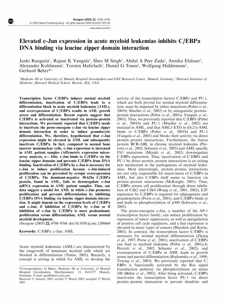
Elevated c-Jun expression in acute myeloid leukemias inhibits C/EBPaDNA binding via leucine zipper domain interaction
Janki Rangatia1, Rajani K Vangala1, Sheo M Singh1, Abdul A Peer Zada1, Annika Elsasser1,Alexander Kohlmann1, Torsten Haferlach1, Daniel G Tenen2, Wolfgang Hiddemann1,Gerhard Behre*,1
1Medicine III at University of Munich Hospital Grosshadern and GSF Research Center, Munich, Germany; 2Harvard Institutes ofMedicine, Harvard Medical School, Boston, MA, USA
Transcription factor C/EBPa induces normal myeloiddifferentiation, inactivation of C/EBPa leads to adifferentiation block in acute myeloid leukemias (AML),and overexpression of C/EBPa results in AML growtharrest and differentiation. Recent reports suggest thatC/EBPa is activated or inactivated via protein–proteininteractions. We previously reported that C/EBPa needsto inactivate the proto-oncogene c-Jun via leucine zipperdomain interaction in order to induce granulocyticdifferentiation. We, therefore, hypothesized that c-Junexpression might be elevated in AML and subsequentlyinactivate C/EBPa. In fact, compared to normal bonemarrow mononuclear cells, c-Jun expression is increasedin AML patient samples (Affymetrix expression micro-array analysis, n¼ 166). c-Jun binds to C/EBPa via theleucine zipper domains and prevents C/EBPa from DNAbinding. Inactivation of C/EBPa by c-Jun is necessary forc-Jun to induce proliferation because c-Jun-inducedproliferation can be prevented by ectopic overexpressionof C/EBPa. The dominant-negative 30-kDa C/EBPaprotein, found in AML, fails to downregulate c-JunmRNA expression in AML patient samples. Thus, ourdata suggest a model for AML in which c-Jun promotesproliferation and prevents differentiation by inhibitingC/EBPa DNA binding via leucine zipper domain interac-tion. It might depend on the expression levels of C/EBPaand c-Jun, if inhibition of C/EBPa by c-Jun or ifinhibition of c-Jun by C/EBPa is more predominant:proliferation versus differentiation; AML versus normalmyeloid development.Oncogene (2003) 22, 4760–4764. doi:10.1038/sj.onc.1206664
Keywords: C/EBPa; c-Jun; AML
Acute myeloid leukemias (AML) are characterized bythe outgrowth of immature myeloid cells which areblocked in differentiation (Tenen, 2003). Recently, aconcept is arising in which for AML to develop the
activity of the transcription factors C/EBPa and PU.1,which are both pivotal for normal myeloid differentia-tion, must be impaired by either mutations (Pabst et al.,2001b; Mueller et al., 2002) or by antagonistic protein–protein interactions (Pabst et al., 2001a; Vangala et al.,2003). Thus, we previously reported that C/EBPa (Pabstet al., 2001b) and PU.1 (Mueller et al., 2002) aremutated in AML, and that AML1-ETO in t(8;21)-AMLbinds to C/EBPa (Pabst et al., 2001b) and PU.1(Vangala et al., 2003) and blocks their activity via directprotein–protein interactions. Furthermore, the fusionprotein BCR-ABL in chronic myeloid leukemia (Per-rotti et al., 2002; Schuster et al., 2003) and AML-specificFlt3 mutations (Mizuki et al., 2002) downregulateC/EBPa expression. Thus, inactivation of C/EBPa andPU.1 by direct protein–protein interactions is an arisingnew mechanism in the pathogenesis of myeloid leuke-mias. Most interestingly, protein–protein interactionsare not only responsible for inactivation of C/EBPa inAML, but also C/EBPa itself seems to function viaprotein–protein interactions (McKnight, 2001). Thus,C/EBPa arrests cell proliferation through direct inhibi-tion of Cdk2 and Cdk4 (Wang et al., 2001, 2002), E2Frepression by C/EBPa is required for adipogenesis andgranulopoiesis (Porse et al., 2001), and C/EBPa binds toand leads to phosphorylation of p300 (Schwartz et al.,2003).The proto-oncogene c-Jun, a member of the AP-1
transcription factor family, can induce proliferation byrepression of tumor suppressors, as well as upregulationof positive cell cycle regulators, and c-Jun expression iselevated in many types of cancers (Shaulian and Karin,2002). In contrast, the transcription factor C/EBPa isnecessary for normal myeloid differentiation (Zhanget al., 1997; Porse et al., 2001), inactivation of C/EBPacan lead to myeloid leukemia (Pabst et al., 2001a,b;Perrotti et al., 2002; Schuster et al., 2002), andoverexpression of C/EBPa in AML leads to growtharrest and partial differentiation (Radomska et al., 1998;Truong et al., 2003). We previously reported that C/EBPa is functionally activated by the Ras signaltransduction pathway via phosphorylation on serine248 (Behre et al., 2002). After being activated, C/EBPainactivates the transcription factor PU.1 via directprotein–protein interaction to prevent dendritic and
Received 11 January 2003; revised 27 March 2003; accepted 27 March2003
*Correspondence: G Behre, Medicine III at University of MunichHospital Grosshadern, Marchioninistr. 15, D-81377 Munich,Germany; E-mail: [email protected]
Oncogene (2003) 22, 4760–4764& 2003 Nature Publishing Group All rights reserved 0950-9232/03 $25.00
www.nature.com/onc
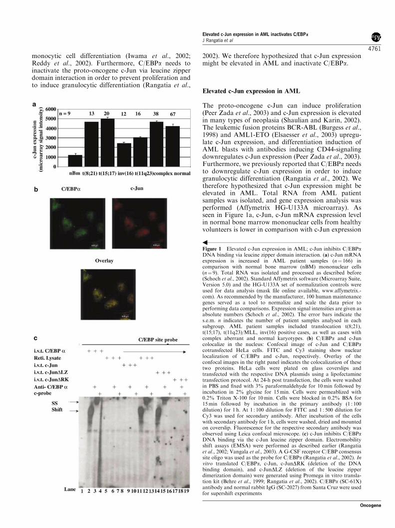
monocytic cell differentiation (Iwama et al., 2002;Reddy et al., 2002). Furthermore, C/EBPa needs toinactivate the proto-oncogene c-Jun via leucine zipperdomain interaction in order to prevent proliferation andto induce granulocytic differentiation (Rangatia et al.,
2002). We therefore hypothesized that c-Jun expressionmight be elevated in AML and inactivate C/EBPa.
Elevated c-Jun expression in AML
The proto-oncogene c-Jun can induce proliferation(Peer Zada et al., 2003) and c-Jun expression is elevatedin many types of neoplasia (Shaulian and Karin, 2002).The leukemic fusion proteins BCR-ABL (Burgess et al.,1998) and AML1-ETO (Elsaesser et al., 2003) upregu-late c-Jun expression, and differentiation induction ofAML blasts with antibodies inducing CD44-signalingdownregulates c-Jun expression (Peer Zada et al., 2003).Furthermore, we previously reported that C/EBPa needsto downregulate c-Jun expression in order to inducegranulocytic differentiation (Rangatia et al., 2002). Wetherefore hypothesized that c-Jun expression might beelevated in AML. Total RNA from AML patientsamples was isolated, and gene expression analysis wasperformed (Affymetrix HG-U133A microarray). Asseen in Figure 1a, c-Jun, c-Jun mRNA expression levelin normal bone marrow mononuclear cells from healthyvolunteers is lower in comparison with c-Jun expression
Figure 1 Elevated c-Jun expression in AML; c-Jun inhibits C/EBPaDNA binding via leucine zipper domain interaction. (a) c-Jun mRNAexpression is increased in AML patient samples (n¼ 166) incomparison with normal bone marrow (nBM) mononuclear cells(n¼ 9). Total RNA was isolated and processed as described before(Schoch et al., 2002). Standard Affymetrix software (Microarray Suite,Version 5.0) and the HG-U133A set of normalization controls wereused for data analysis (mask file online available, www.affymetrix.-com). As recommended by the manufacturer, 100 human maintenancegenes served as a tool to normalize and scale the data prior toperforming data comparisons. Expression signal intensities are given asabsolute numbers (Schoch et al., 2002). The error bars indicate thes.e.m. n indicates the number of patient samples analysed in eachsubgroup. AML patient samples included translocation t(8;21),t(15;17), t(11q23)/MLL, inv(16) positive cases, as well as cases withcomplex aberrant and normal karyotypes. (b) C/EBPa and c-Juncolocalize in the nucleus: Confocal image of c-Jun and C/EBPacotransfected HeLa cells. FITC and Cy3 staining show nuclearlocalization of C/EBPa and c-Jun, respectively. Overlay of theconfocal images in the right panel indicates the colocalization of thesetwo proteins. HeLa cells were plated on glass coverslips andtransfected with the respective DNA plasmids using a lipofectaminetransfection protocol. At 24-h post transfection, the cells were washedin PBS and fixed with 3% paraformaldehyde for 10min followed byincubation in 2% glycine for 15min. Cells were permeablized with0.2% Triton X-100 for 10min. Cells were blocked in 0.2% BSA for15min followed by incubation in the primary antibody (1 : 100dilution) for 1 h. At 1 : 100 dilution for FITC and 1 : 500 dilution forCy3 was used for secondary antibody. After incubation of the cellswith secondary antibody for 1 h, cells were washed, dried and mountedon coverslip. Fluorescence for the respective secondary antibody wasobserved using Leica confocal microscope. (c) c-Jun inhibits C/EBPaDNA binding via the c-Jun leucine zipper domain. Electromobilityshift assays (EMSA) were performed as described earlier (Rangatiaet al., 2002; Vangala et al., 2003). A G-CSF receptor C/EBP consensussite oligo was used as the probe for C/EBPa (Rangatia et al., 2002). Invitro translated C/EBPa, c-Jun, c-JunDRK (deletion of the DNAbinding domain), and c-JunDLZ (deletion of the leucine zipperdimerization domain) were generated using Promega in vitro transla-tion kit (Behre et al., 1999; Rangatia et al., 2002). C/EBPa (SC-61X)antibody and normal rabbit IgG (SC-2027) from Santa Cruz were usedfor supershift experiments
b
Elevated c-Jun expression in AML inactivates C/EBPa
J Rangatia et al
4761
Oncogene
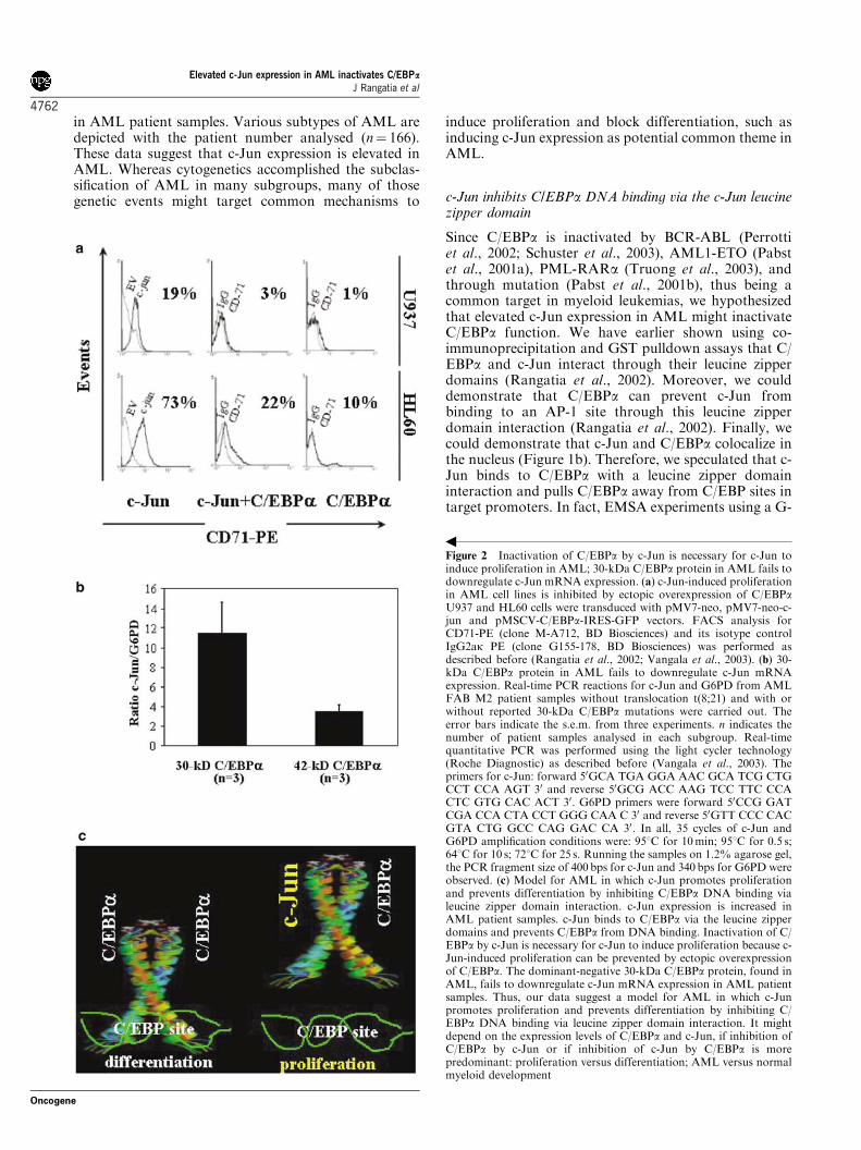
in AML patient samples. Various subtypes of AML aredepicted with the patient number analysed (n¼ 166).These data suggest that c-Jun expression is elevated inAML. Whereas cytogenetics accomplished the subclas-sification of AML in many subgroups, many of thosegenetic events might target common mechanisms to
induce proliferation and block differentiation, such asinducing c-Jun expression as potential common theme inAML.
c-Jun inhibits C/EBPa DNA binding via the c-Jun leucinezipper domain
Since C/EBPa is inactivated by BCR-ABL (Perrottiet al., 2002; Schuster et al., 2003), AML1-ETO (Pabstet al., 2001a), PML-RARa (Truong et al., 2003), andthrough mutation (Pabst et al., 2001b), thus being acommon target in myeloid leukemias, we hypothesizedthat elevated c-Jun expression in AML might inactivateC/EBPa function. We have earlier shown using co-immunoprecipitation and GST pulldown assays that C/EBPa and c-Jun interact through their leucine zipperdomains (Rangatia et al., 2002). Moreover, we coulddemonstrate that C/EBPa can prevent c-Jun frombinding to an AP-1 site through this leucine zipperdomain interaction (Rangatia et al., 2002). Finally, wecould demonstrate that c-Jun and C/EBPa colocalize inthe nucleus (Figure 1b). Therefore, we speculated that c-Jun binds to C/EBPa with a leucine zipper domaininteraction and pulls C/EBPa away from C/EBP sites intarget promoters. In fact, EMSA experiments using a G-
Figure 2 Inactivation of C/EBPa by c-Jun is necessary for c-Jun toinduce proliferation in AML; 30-kDa C/EBPa protein in AML fails todownregulate c-Jun mRNA expression. (a) c-Jun-induced proliferationin AML cell lines is inhibited by ectopic overexpression of C/EBPaU937 and HL60 cells were transduced with pMV7-neo, pMV7-neo-c-jun and pMSCV-C/EBPa-IRES-GFP vectors. FACS analysis forCD71-PE (clone M-A712, BD Biosciences) and its isotype controlIgG2ak PE (clone G155-178, BD Biosciences) was performed asdescribed before (Rangatia et al., 2002; Vangala et al., 2003). (b) 30-kDa C/EBPa protein in AML fails to downregulate c-Jun mRNAexpression. Real-time PCR reactions for c-Jun and G6PD from AMLFAB M2 patient samples without translocation t(8;21) and with orwithout reported 30-kDa C/EBPa mutations were carried out. Theerror bars indicate the s.e.m. from three experiments. n indicates thenumber of patient samples analysed in each subgroup. Real-timequantitative PCR was performed using the light cycler technology(Roche Diagnostic) as described before (Vangala et al., 2003). Theprimers for c-Jun: forward 50GCA TGA GGA AAC GCA TCG CTGCCT CCA AGT 30 and reverse 50GCG ACC AAG TCC TTC CCACTC GTG CAC ACT 30. G6PD primers were forward 50CCG GATCGA CCA CTA CCT GGG CAA C 30 and reverse 50GTT CCC CACGTA CTG GCC CAG GAC CA 30. In all, 35 cycles of c-Jun andG6PD amplification conditions were: 951C for 10min; 951C for 0.5 s;641C for 10 s; 721C for 25 s. Running the samples on 1.2% agarose gel,the PCR fragment size of 400 bps for c-Jun and 340bps for G6PD wereobserved. (c) Model for AML in which c-Jun promotes proliferationand prevents differentiation by inhibiting C/EBPa DNA binding vialeucine zipper domain interaction. c-Jun expression is increased inAML patient samples. c-Jun binds to C/EBPa via the leucine zipperdomains and prevents C/EBPa from DNA binding. Inactivation of C/EBPa by c-Jun is necessary for c-Jun to induce proliferation because c-Jun-induced proliferation can be prevented by ectopic overexpressionof C/EBPa. The dominant-negative 30-kDa C/EBPa protein, found inAML, fails to downregulate c-Jun mRNA expression in AML patientsamples. Thus, our data suggest a model for AML in which c-Junpromotes proliferation and prevents differentiation by inhibiting C/EBPa DNA binding via leucine zipper domain interaction. It mightdepend on the expression levels of C/EBPa and c-Jun, if inhibition ofC/EBPa by c-Jun or if inhibition of c-Jun by C/EBPa is morepredominant: proliferation versus differentiation; AML versus normalmyeloid development
b
Elevated c-Jun expression in AML inactivates C/EBPa
J Rangatia et al
4762
Oncogene
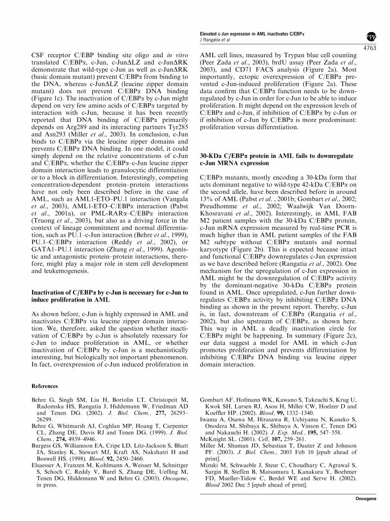
CSF receptor C/EBP binding site oligo and in vitrotranslated C/EBPa, c-Jun, c-JunDLZ and c-JunDRKdemonstrate that wild-type c-Jun as well as c-JunDRK(basic domain mutant) prevent C/EBPa from binding tothe DNA, whereas c-JunDLZ (leucine zipper domainmutant) does not prevent C/EBPa DNA binding(Figure 1c). The inactivation of C/EBPa by c-Jun mightdepend on very few amino acids of C/EBPa targeted byinteraction with c-Jun, because it has been recentlyreported that DNA binding of C/EBPa primarilydepends on Arg289 and its interacting partners Tyr285and Asn293 (Miller et al., 2003). In conclusion, c-Junbinds to C/EBPa via the leucine zipper domains andprevents C/EBPa DNA binding. In one model, it couldsimply depend on the relative concentrations of c-Junand C/EBPa, whether the C/EBPa–c-Jun leucine zipperdomain interaction leads to granulocytic differentiationor to a block in differentiation. Interestingly, competingconcentration-dependent protein–protein interactionshave not only been described before in the case ofAML, such as AML1-ETO–PU.1 interaction (Vangalaet al., 2003), AML1-ETO–C/EBPa interaction (Pabstet al., 2001a), or PML-RARa–C/EBPa interaction(Truong et al., 2003), but also as a driving force in thecontext of lineage commitment and normal differentia-tion, such as PU.1–c-Jun interaction (Behre et al., 1999),PU.1–C/EBPa interaction (Reddy et al., 2002), orGATA1–PU.1 interaction (Zhang et al., 1999). Agonis-tic and antagonistic protein–protein interactions, there-fore, might play a major role in stem cell developmentand leukemogenesis.
Inactivation of C/EBPa by c-Jun is necessary for c-Jun toinduce proliferation in AML
As shown before, c-Jun is highly expressed in AML andinactivates C/EBPa via leucine zipper domain interac-tion. We, therefore, asked the question whether inacti-vation of C/EBPa by c-Jun is absolutely recessary forc-Jun to induce proliferation in AML, or whetherinactivation of C/EBPa by c-Jun is a mechanisticallyinteresting, but biologically not important phenomenon.In fact, overexpression of c-Jun induced proliferation in
AML cell lines, measured by Trypan blue cell counting(Peer Zada et al., 2003), brdU assay (Peer Zada et al.,2003), and CD71 FACS analysis (Figure 2a). Mostimportantly, ectopic overexpression of C/EBPa pre-vented c-Jun-induced proliferation (Figure 2a). Thesedata confirm that C/EBPa function needs to be down-regulated by c-Jun in order for c-Jun to be able to induceproliferation. It might depend on the expression levels ofC/EBPa and c-Jun, if inhibition of C/EBPa by c-Jun orif inhibition of c-Jun by C/EBPa is more predominant:proliferation versus differentiation.
30-KDa C/EBPa protein in AML fails to downregulatec-Jun MRNA expression
C/EBPa mutants, mostly encoding a 30-kDa form thatacts dominant negative to wild-type 42-kDa C/EBPa onthe second allele, have been described before in around13% of AML (Pabst et al. , 2001b; Gombart et al., 2002;Preudhomme et al., 2002; Waalwijk Van Doorn-Khosravani et al., 2002). Interestingly, in AML FABM2 patient samples with the 30-kDa C/EBPa protein,c-Jun mRNA expression measured by real-time PCR ismuch higher than in AML patient samples of the FABM2 subtype without C/EBPa mutants and normalkaryotype (Figure 2b). This is expected because intactand functional C/EBPa downregulates c-Jun expressionas we have described before (Rangatia et al., 2002). Onemechanism for the upregulation of c-Jun expression inAML might be the downregulation of C/EBPa activityby the dominant-negative 30-kDa C/EBPa proteinfound in AML. Once upregulated, c-Jun further down-regulates C/EBPa activity by inhibiting C/EBPa DNAbinding as shown in the present report. Thereby, c-Junis, in fact, downstream of C/EBPa (Rangatia et al.,2002), but also upstream of C/EBPa, as shown here.This way in AML a deadly inactivation circle forC/EBPa might be happening. In summary (Figure 2c),our data suggest a model for AML in which c-Junpromotes proliferation and prevents differentiation byinhibiting C/EBPa DNA binding via leucine zipperdomain interaction.
References
Behre G, Singh SM, Liu H, Bortolin LT, Christopeit M,Radomska HS, Rangatia J, Hiddemann W, Friedman ADand Tenen DG. (2002). J. Biol. Chem., 277, 26293–26299.
Behre G, Whitmarsh AJ, Coghlan MP, Hoang T, CarpenterCL, Zhang DE, Davis RJ and Tenen DG. (1999). J. Biol.Chem., 274, 4939–4946.
Burgess GS, Williamson EA, Cripe LD, Litz-Jackson S, BhattJA, Stanley K, Stewart MJ, Kraft AS, Nakshatri H andBoswell HS. (1998). Blood, 92, 2450–2460.
Elsaesser A, Franzen M, Kohlmann A, Weisser M, SchnittgerS, Schoch C, Reddy V, Burel S, Zhang DE, Ueffing M,Tenen DG, Hiddemann W and Behre G. (2003). Oncogene,in press.
Gombart AF, Hofmann WK, Kawano S, Takeuchi S, Krug U,Kwok SH, Larsen RJ, Asou H, Miller CW, Hoelzer D andKoeffler HP. (2002). Blood, 99, 1332–1340.
Iwama A, Osawa M, Hirasawa R, Uchiyama N, Kaneko S,Onodera M, Shibuya K, Shibuya A, Vinson C, Tenen DGand Nakauchi H. (2002). J. Exp. Med., 195, 547–558.
McKnight SL. (2001). Cell, 107, 259–261.Miller M, Shuman JD, Sebastian T, Dauter Z and JohnsonPF. (2003). J. Biol. Chem., 2003 Feb 10 [epub ahead ofprint].
Mizuki M, Schwaeble J, Steur C, Choudhary C, Agrawal S,Sargin B, Steffen B, Matsumura I, Kanakura Y, BoehmerFD, Mueller-Tidow C, Berdel WE and Serve H. (2002).Blood 2002 Dec 5 [epub ahead of print].
Elevated c-Jun expression in AML inactivates C/EBPa
J Rangatia et al
4763
Oncogene
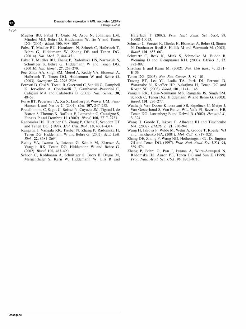
Mueller BU, Pabst T, Osato M, Asou N, Johansen LM,Minden MD, Behre G, Hiddemann W, Ito Y and TenenDG. (2002). Blood, 100, 998–1007.
Pabst T, Mueller BU, Harakawa N, Schoch C, Haferlach T,Behre G, Hiddemann W, Zhang DE and Tenen DG.(2001a). Nat. Med., 7, 444–451.
Pabst T, Mueller BU, Zhang P, Radomska HS, Narravula S,Schnittger S, Behre G, Hiddemann W and Tenen DG.(2001b). Nat. Genet., 27, 263–270.
Peer Zada AA, Singh SM, Meisel A, Reddy VA, Elsaesser A,Haferlach T, Tenen DG, Hiddemann W and Behre G.(2003). Oncogene, 22, 2296–2308.
Perrotti D, Cesi V, Trotta R, Guerzoni C, Santilli G, CampbellK, Iervolino A, Condorelli F, Gambacorti-Passerini C,Caligiuri MA and Calabretta B. (2002). Nat. Genet., 30,
48–58.Porse BT, Pedersen TA, Xu X, Lindberg B, Wewer UM, Friis-Hansen L and Nerlov C. (2001). Cell, 107, 247–258.
Preudhomme C, Sagot C, Boissel N, Cayuela JM, Tigaud I, deBotton S, Thomas X, Raffoux E, Lamandin C, Castaigne S,Fenaux P and Dombret H. (2002). Blood, 100, 2717–2723.
Radomska HS, Huettner CS, Zhang P, Cheng T, Scadden DTand Tenen DG. (1998). Mol. Cell. Biol., 18, 4301–4314.
Rangatia J, Vangala RK, Treiber N, Zhang P, Radomska H,Tenen DG, Hiddemann W and Behre G. (2002). Mol. Cell.Biol., 22, 8681–8694.
Reddy VA, Iwama A, Iotzova G, Schulz M, Elsasser A,Vangala RK, Tenen DG, Hiddemann W and Behre G.(2002). Blood, 100, 483–490.
Schoch C, Kohlmann A, Schnittger S, Brors B, Dugas M,Mergenthaler S, Kern W, Hiddemann W, Eils R and
Haferlach T. (2002). Proc. Natl. Acad. Sci. USA, 99,
10008–10013.Schuster C, Forster K, Dierks H, Elsaesser A, Behre G, SimonN, Danhauser-Riedl S, Hallek M and Warmuth M. (2003).Blood, 101, 655–663.
Schwartz C, Beck K, Mink S, Schmolke M, Budde B,Wenning D and Klempnauer KH. (2003). EMBO J., 22,
882–892.Shaulian E and Karin M. (2002). Nat. Cell Biol., 4, E131–E136.
Tenen DG. (2003). Nat. Rev. Cancer, 3, 89–101.Truong BT, Lee YJ, Lodie TA, Park DJ, Perrotti D,Watanabe N, Koeffler HP, Nakajima H, Tenen DG andKogan SC. (2003). Blood, 101, 1141–1148.
Vangala RK, Heiss-Neumann MS, Rangatia JS, Singh SM,Schoch C, Tenen DG, Hiddemann W and Behre G. (2003).Blood, 101, 270–277.
Waalwijk Van Doorn-Khosravani SB, Erpelinck C, Meijer J,Van Oosterhoud S, Van Putten WL, Valk PJ, Beverloo HB,Tenen DG, Lowenberg B and Delwel R. (2002). Hematol. J.,3, 324.
Wang H, Goode T, Iakova P, Albrecht JH and TimchenkoNA. (2002). EMBO J., 21, 930–941.
Wang H, Iakova P, Wilde M, Welm A, Goode T, Roesler WJand Timchenko NA. (2001). Mol. Cell, 8, 817–828.
Zhang DE, Zhang P, Wang ND, Hetherington CJ, DarlingtonGJ and Tenen DG. (1997). Proc. Natl. Acad. Sci. USA, 94,569–574.
Zhang P, Behre G, Pan J, Iwama A, Wara-Aswapati N,Radomska HS, Auron PE, Tenen DG and Sun Z. (1999).Proc. Natl. Acad. Sci. USA, 96, 8705–8710.
Elevated c-Jun expression in AML inactivates C/EBPa
J Rangatia et al
4764
Oncogene
NEOPLASIA
The myeloid master regulator transcription factor PU.1 is inactivated byAML1-ETO in t(8;21) myeloid leukemiaRajani K. Vangala, Marion S. Heiss-Neumann, Janki S. Rangatia, Sheo M. Singh, Claudia Schoch,Daniel G. Tenen, Wolfgang Hiddemann, and Gerhard Behre
The transcription factor PU.1 plays a piv-otal role in normal myeloid differentia-tion. PU.1�/� mice exhibit a complete blockin myeloid differentiation. HeterozygousPU.1 mutations were reported in somepatients with acute myeloid leukemia(AML), but not in AML with translocationt(8;21), which gives rise to the fusiongene AML1-ETO. Here we report a nega-tive functional impact of AML1-ETO onthe transcriptional activity of PU.1. AML1-ETO physically binds to PU.1 in t(8;21)�
Kasumi-1 cells. AML1-ETO binds to the
�3�4 region in the DNA-binding domain ofPU.1 and displaces the coactivator c-Junfrom PU.1, thus down-regulating the tran-scriptional activity of PU.1. This physicalinteraction of AML1-ETO and PU.1 did notabolish the DNA-binding capacity of PU.1.AML1-ETO down-regulates the transacti-vation capacity of PU.1 in myeloid U937cells, and the expression levels of PU.1target genes in AML French-American-British (FAB) subtype M2 patients witht(8;21) were lower than in patients withoutt(8;21). Conditional expression of AML1-
ETO causes proliferation in mouse bonemarrow cells and inhibits antiproliferativefunction of PU.1. Overexpression of PU.1,however, differentiates AML1-ETO–ex-pressing Kasumi-1 cells to the monocyticlineage. Thus, the function of PU.1 isdown-regulated by AML1-ETO in t(8;21)myeloid leukemia, whereas overexpres-sion of PU.1 restores normal differentia-tion. (Blood. 2003;101:000-000)
© 2003 by The American Society of Hematology
Introduction
The Ets family of transcription factors plays a key role in thegrowth, survival, differentiation, and activation of hematopoieticcells. This family of proteins is characterized by presence of an85-amino acid,1 winged helix-turn-helix DNA-binding domain.PU.1 is one of the most important Ets transcription factors.2 Itsexpression is limited to hematopoietic cells, including primitiveCD34� cells, macrophages, B lymphocytes, neutrophils, mastcells, and early erythroblasts.2,3 In vitro studies suggest that PU.1regulates the activity of a number of myeloid- and lymphoid-specific promoters and enhancers.4-10 PU.1 is a key transcriptionfactor for normal myeloid development as demonstrated by acomplete block of myeloid development in PU.1�/� mice.11,12 Fetalor newborn PU.1�/� mice have no detectable monocytes/macrophages or neutrophils.11,12 We have recently shown that PU.1is mutated in patients with acute myeloid leukemia (AML).13 Thesestudies all point to the crucial role of PU.1 in both normal myeloiddifferentiation and leukemogenesis.
AML1 is a member of the Runt-like transcription factors(Runx-1, -2, and -3) named after the Runt protein that regulatessegmentation during Drosophila embryogenesis.14-16 AML1 ap-pears to act as an “organizing” factor for many promoters andenhancers by interacting with various coactivators and DNA-binding transcription factors.17-22 The AML1 gene is one of the mostfrequently translocated or mutated genes in human cancer.23-25 Thet(8;21)(q22;q22) translocation fuses residues 1-177 of AML1(including the DNA-binding domain) to nearly all of ETO (also
known as CBF2T1).26 ETO is the human homolog of DrosophilaNERVY protein.27-29 The t(8;21) belongs to the most commonchromosomal abnormalities in AML, accounting for 10% of allAML cases and 40% of the AML French-American-British (FAB)M2 phenotype.30-33 AML1 activates transcription from enhancercore motifs (TGT/cGGY), which are present in a number of genesrelevant to myeloid development, including the macrophage colony-stimulating factor (M-CSF) receptor, granulocyte-macrophagecolony-stimulating factor (GM-CSF), myeloperoxidase, and neutro-phil elastase.34-39 Like AML1, AML1-ETO can act as a tran-scriptional activator,40-43 but it is also a transcriptional repressor inother contexts.44 Only one allele of AML1 is altered in leukemiacells expressing t(8;21), and AML1-ETO can efficiently repressAML1-dependent transcriptional activation. Therefore, AML1-ETO has been postulated to act as a dominant inhibitor of AML1function.34,37,44
Recently, we have shown that AML1-ETO blocks CCAATenhancer-binding protein (C/EBP�)–dependent activation of itsown promoter thus blocking normal granulocytic differentiation ofmyeloid cells.36 Furthermore, AML1-ETO was shown to repressAML1 and MEF-2–dependent gene activation.45 In our earlierstudies we demonstrated that c-Jun, a member of AP-1 transcrip-tion factor family, can interact with PU.1 at the �3�4 domain inPU.1 and coactivate the transcriptional activity of PU.1.46 Here weshow that AML1-ETO blocks the transcriptional activity of PU.1by displacing its coactivator c-Jun.
From the Department of Internal Medicine III, University Hospital Grosshadern,Ludwig-Maximilians-University Munich and GSF-National Research Center forEnvironment and Health, Munich, Germany; and Harvard Institutes ofMedicine, Harvard Medical School, Boston, MA.
Submitted April 30, 2002; accepted July 30, 2002. Prepublished online asBlood First Edition Paper, August 29, 2002; DOI 10.1182/blood-2002-04-1288.
Supported by a DFG (German research foundation) grant to G.B. (no. 2042/2-1).
Reprints: Gerhard Behre, Department of Internal Medicine III, UniversityHospital Grosshadern, Ludwig-Maximilians-University Munich, Marchioninistr15, D-81377 Munich, Germany; e-mail: [email protected].
The publication costs of this article were defrayed in part by page chargepayment. Therefore, and solely to indicate this fact, this article is herebymarked ‘‘advertisement’’ in accordance with 18 U.S.C. section 1734.
© 2003 by The American Society of Hematology
1BLOOD, 1 JANUARY 2003 � VOLUME 101, NUMBER 1
AQ: 1
AQ: 3
AQ: 15
AQ: 16
AQ: 3
AQ: 9
tapraid4/h8-bloo/h8-bloo/h80103/h83591d03a mazzam S�11 10/16/02 13:21 Art: 2002-04-1288
DOI: 10.1182/blood-2002-04-1288 Collection Codes: 00,00
Not for distribution: this preliminary material is embargoed until publication.
Materials and methods
Cell lines and cell culture
Human kidney 293T, mouse embryonal carcinoma F9, and ecotrophicPhoenix cells were maintained in Dulbecco modified Eagle medium(DMEM; Gibco, ) supplemented with 10% fetal bovine serum (FBS), 1%glutamine, and 1% Penstrep ( all from Gibco). Human monoblastic U937cells and t(8;21)� Kasumi-1 cells were cultured in RPMI 1640 mediumsupplemented with 10% FBS (both from Gibco).
Bone marrow cells were isolated from the femurs of Balb/C mice. Thefemurs were removed and stripped of the soft tissue and crushed to releasecells within marrow cavity. The red blood cells were lysed with a0.15-mol/L solution of ammonium chloride. The pelleted cells weresubjected to low-density mononuclear cell separation by incubating withdensity gradient (Histopaque 1083; Sigma, St Louis, MO) for 10 minutesand centrifuged at 600 rpm for 30 minutes, washed twice in phosphate-buffered saline (PBS), followed by culturing in Iscove modified Dulbeccomedium (IMDM; Stem Cell Technologies, ) supplemented with 10% FBS(Stem Cell Technologies), 50 ng/mL stem cell factor (R & D Systems,Minneapolis, MN), 50 ng/mL interleukin 6 (IL-6; R & D Systems), and 50ng/mL Flt-3 ligand (Flt-3L; R & D Systems).
Coimmunoprecipitation assay
Kasumi-1 cells (2 � 107) were lysed and 200 �g protein was used toperform immunoprecipitation as mentioned by Mao et al.45 The followingantibodies were used: rabbit IgG (Santa Cruz Biotechnologies, Santa Cruz,CA; catalog no. sc2027), goat IgG (Santa Cruz Biotechnologies; catalog no.2028), anti-AML1 antibody (Calbiochem, ; catalog no. PC284), anti-PU.1(Santa Cruz Biotechnologies; catalog no. sc352), and protein-A agarosebeads (Santa Cruz Biotechnologies; catalog no. sc2001).
Western blot
After plating in 100-mm plates, 293T cells were transfected using theLipofectAMINE Plus kit (Gibco) as per the manufacturer’s protocol. At 24hours after transfection, cells were harvested and lysed in RIPA lysis buffer,and immunoblot for PU.1 was performed with 100 �g protein as describedearlier.46-48 To generate protein lysates, 1 � 106 F9, Kasumi-1, or 293T cellswere lysed and nuclear extracts were prepared and immunoblot wasperformed with 100 �g protein for c-Jun (Santa Cruz Biotechnologies;catalog no. sc54). Mouse bone marrow cells transduced with PU.1,AML1-ETO, or respective empty vectors were similarly lysed (RIPA lysis)and 100 �g protein was used for immunoblot analysis for PU.1 andAML1-ETO (anti-ETO antibody, Santa Cruz Biotechnologies; catalog no.sc9737). Mouse monoclonal anti–�-tubulin purchased from Roche (; catalogno. 1111876) was used for immunoblot assay as internal control. ProteinA-peroxidase–conjugated for antirabbit (Amersham Pharmacia, ; catalogno. NA 9120), or antigoat peroxidase-conjugated immunoglobulins (Dako,; code no. p0449) were used as secondary antibodies.
Reporter constructs and expression plasmids
The human monocyte–specific M-CSF receptor promoter with or withoutAML1-binding site, p(PU.1)4TK, and p(mutPU.1)4TK (PU.1-binding sitesand mutated PU.1-binding sites subcloned into pTK61luciferase) weredescribed earlier.46 As an internal control plasmid for transient transfectionassay, we used the pRL-null construct driving a Renilla luciferase gene(Promega, Madison, WI).49 Other vectors used were pECE-PU.1-murine,pcDNA.1-PU.1, pGEX-2TK-PU.1 or �3�4, pS3H-c-Jun, and pSP6-c-Jun,as described previously.46,50 AML1B-pCMV5 and CBF�-pCMV5 weredescribed earlier.42 AML1-ETO-pcDNA3 was constructed by enzymaticdigestion of AML1-ETO-pCMV542 with XbaI and subcloning the resulting2258-bp fragment into the XbaI site of pcDNA3 plasmid (Invitrogen, ).
Transfection assays
Transient transfections in 293T or F9 cells were carried out with Lipo-fectAMINE transfection kit (Gibco) in 24-well plates as described ear-
lier.46,47,49 U937 cells were transiently transfected by electroporation inRPMI medium at 980 �F and 280 V. Firefly luciferase activities from theconstructs M-CSF receptor promoter luciferase, pXP2, p(PU.1)4TK,p(mutPU.1)4TK, and Renilla luciferase activity from internal controlplasmid pRL-null were determined 24 hours after transfection using DualLuciferase Reporter Assay System (Promega). Results are given asmeans � SEMs from at least 3 independent experiments.
Protein interaction assays
c-Jun and AML1-ETO were transcribed in vitro and translated in thepresence of [35S]-methionine (Amersham Pharmacia, ) using the T7/SP6-coupled reticulocyte system (Promega) in accordance with the manufac-turer’s instruction. Glutathione-S-transferase (GST) precipitation assayswere performed as described earlier.46,48
EMSA
�32P-adenosine triphosphate (ATP; Amersham Pharmacia)–labeled double-stranded oligonucleotides of PU.1 DNA-binding site51 and AML1-bindingsite52 for electrophoretic mobility shift assay (EMSA) were prepared. Theassay was performed with in vitro–translated proteins as mentionedearlier.11,47 For supershift experiments 3 �L of either anti-PU.1 or anti-ETOantibodies were added to the reaction mixture.
Retroviral transduction assay
Ecotrophic Phoenix cells (5 � 106) were plated in 10-cm plates andtransfected with 5 �g PINCO-GFP, PINCO-AML1-ETO-GFP, pGsam-PU.1-ires-NGFR, or pGsam-ires-NGFR vectors using LipofectAMINE transfec-tion kit (Gibco). At 24 hours after transfection, the transfection medium wasreplaced with IMDM (supplemented with 10% FBS, 50 ng/mL stem cellfactor, 50 ng/mL IL-6, and 50 ng/mL Flt-3L) for collection of the virusparticles. After the viral particle production, freshly isolated mouse bonemarrow cells were incubated with viral medium on fibronectin-coatedplates and centrifuged for 30 minutes at 1000g (this step was repeated every12 hours).53 At 60 hours after first transduction, NGFR� or enhanced greenfluorescence protein-positive (EGFP�) cells were isolated by fluorescence-activated cell sorting (FACS) analysis (Becton Dickinson, ). To detect theexpression of tNGFR on the cell surface, cells were stained with mouseantihuman NGFR (Chemicon, ; catalog no. MAB5246) followed byphycoerythrin (PE)–conjugated rabbit antimouse immunoglobulins (mouseIgG RPE; Dako; catalog no. R0439). Then, 1 � 104 transduced cells sortedfor NGFR positivity were plated in 1.2 mL mouse colony-forming medium(Stem Cell Technologies). After 3, 6, and 12 days of plating live cells werecounted by trypan blue staining.
Patient material and FACS analysis
Bone marrow cells from AML-M2 patients with or without t(8;21) wereobtained after informed consent was given by the patients. Mononuclearcells were isolated from the bone marrow by density gradient centrifugationwith Histopaque (Sigma). FACS analysis was performed with CD11b(Pharmingen, ; catalog no. 555388), CD14 (Pharmingen; catalog no.555397), and CD64 (Pharmingen; catalog no. 555527).
Transfection of Kasumi-1 cells and FACS analysis
Kasumi-1 cells were electroporated as mentioned above with pGsam-PU.1-ires-NGFR or pGsam-ires-NGFR vectors and sorted 24 hours aftertransfection for NGFR positivity by FACS (with anti-NGFR antibody fromChemicon, catalog no. MAB5246, and mouse IgG RPE from Dako, catalogno. R0439). Five days after sorting for NGFR expression, morphologicchanges were observed by Wright-Giemsa staining of cells. Then, 1 � 106
NGFR� Kasumi-1 cells were incubated with 10 �L recombinant PE-conjugated mouse monoclonal CD11b (Pharmingen; catalog no. 555388) orfluorescein isothiocyanate (FITC)–conjugated mouse monoclonal CD14(Pharmingen; catalog no. 555397) in 100 �L PBS for 60 minutes on ice,washed in PBS followed by analysis on a FACScan flow cytometer (BectonDickinson) using Cellquest software. The cells were also analyzed for the
2 VANGALA et al BLOOD, 1 JANUARY 2003 � VOLUME 101, NUMBER 1
AQ: 4AQ: 5
AQ: 6
AQ: 4
AQ: 6
AQ: 6AQ: 4
AQ: 4
AQ: 4
AQ: 4
AQ: 6
AQ: 4
AQ: 7
AQ: 4
AQ: 8AQ: 9AQ: 4AQ: 17AQ: 4
AQ: 10
AQ: 4
AQ: 29
tapraid4/h8-bloo/h8-bloo/h80103/h83591d03a mazzam S�11 10/16/02 13:21 Art: 2002-04-1288
Not for distribution: this preliminary material is embargoed until publication.
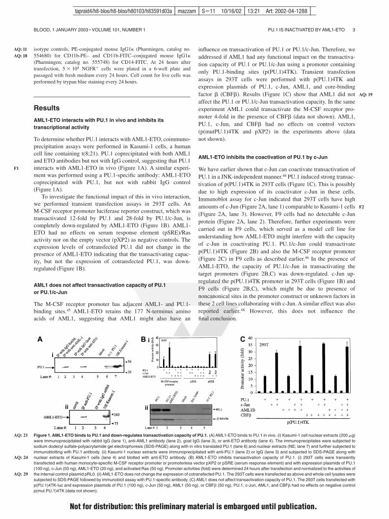
isotype controls, PE-conjugated mouse IgG1 (Pharmingen, catalog no.554680) for CD11b-PE– and CD11b-FITC–conjugated mouse IgG1(Pharmingen; catalog no. 555748) for CD14-FITC. At 24 hours aftertransfection, 5 � 104 NGFR� cells were plated in a 6-well plate andpassaged with fresh medium every 24 hours. Cell count for live cells wasperformed by trypan blue staining every 24 hours.
Results
AML1-ETO interacts with PU.1 in vivo and inhibits itstranscriptional activity
To determine whether PU.1 interacts with AML1-ETO, coimmuno-precipitation assays were performed in Kasumi-1 cells, a humancell line containing t(8;21). PU.1 coprecipitated with both AML1and ETO antibodies but not with IgG control, suggesting that PU.1interacts with AML1-ETO in vivo (Figure 1A). A similar experi-ment was performed using a PU.1-specific antibody: AML1-ETOcoprecipitated with PU.1, but not with rabbit IgG control(Figure 1A).
To investigate the functional impact of this in vivo interaction,we performed transient transfection assays in 293T cells. AnM-CSF receptor promoter luciferase reporter construct, which wastransactivated 12-fold by PU.1 and 28-fold by PU.1/c-Jun, iscompletely down-regulated by AML1-ETO (Figure 1B). AML1-ETO had no effects on serum response element (pSRE)/Rasactivity nor on the empty vector (pXP2) as negative controls. Theexpression levels of cotransfected PU.1 did not change in thepresence of AML1-ETO indicating that the transactivating capac-ity, but not the expression of cotransfected PU.1, was down-regulated (Figure 1B).
AML1 does not affect transactivation capacity of PU.1or PU.1/c-Jun
The M-CSF receptor promoter has adjacent AML1- and PU.1-binding sites.45 AML1-ETO retains the 177 N-terminus aminoacids of AML1, suggesting that AML1 might also have an
influence on transactivation of PU.1 or PU.1/c-Jun. Therefore, weaddressed if AML1 had any functional impact on the transactiva-tion capacity of PU.1 or PU.1/c-Jun using a promoter containingonly PU.1-binding sites (p(PU.1)4TK). Transient transfectionassays in 293T cells were performed with p(PU.1)4TK andexpression plasmids of PU.1, c-Jun, AML1, and core-bindingfactor � (CBF�). Results (Figure 1C) show that AML1 did notaffect the PU.1 or PU.1/c-Jun transactivation capacity. In the sameexperiment AML1 could transactivate the M-CSF receptor pro-moter 4-fold in the presence of CBF� (data not shown). AML1,PU.1, c-Jun, and CBF� had no effects on control vectors(p(mutPU.1)4TK and pXP2) in the experiments above (datanot shown).
AML1-ETO inhibits the coactivation of PU.1 by c-Jun
We have earlier shown that c-Jun can coactivate transactivation ofPU.1 in a JNK-independent manner.46 PU.1 induced strong transac-tivation of p(PU.1)4TK in 293T cells (Figure 1C). This is possiblydue to high expression of its coactivator c-Jun in these cells.Immunoblot assay for c-Jun indicated that 293T cells have highamounts of c-Jun (Figure 2A, lane 1) comparable to Kasumi-1 cells(Figure 2A, lane 3). However, F9 cells had no detectable c-Junprotein (Figure 2A, lane 2). Therefore, further experiments werecarried out in F9 cells, which served as a model cell line forunderstanding how AML1-ETO might interfere with the capacityof c-Jun in coactivating PU.1. PU.1/c-Jun could transactivatep(PU.1)4TK (Figure 2B) and also the M-CSF receptor promoter(Figure 2C) in F9 cells as described earlier.46 In the presence ofAML1-ETO, the capacity of PU.1/c-Jun in transactivating thetarget promoters (Figure 2B,C) was down-regulated. c-Jun up-regulated the p(PU.1)4TK promoter in 293T cells (Figure 1B) andF9 cells (Figure 2B,C), which might be due to presence ofnoncanonical sites in the promoter construct or unknown factors inthese 2 cell lines collaborating with c-Jun. A similar effect was alsoreported earlier.48 However, this does not influence thefinal conclusion.
Figure 1. AML1-ETO binds to PU.1 and down-regulates transactivation capacity of PU.1. (A) AML1-ETO binds to PU.1 in vivo. (i) Kasumi-1 cell nuclear extracts (200 �g)were immunoprecipitated with rabbit IgG (lane 1), anti-AML1 antibody (lane 2), goat IgG (lane 3), or anti-ETO antibody (lane 4). The immunoprecipitates were subjected tosodium dodecyl sulfate-polyacrylamide gel electrophoresis (SDS-PAGE) along with in vitro translated PU.1 (lane 6) and nuclear extracts (NE; lane 7) and further subjected toimmunoblotting with PU.1 antibody. (ii) Kasumi-1 nuclear extracts were immunoprecipitated with anti-PU.1 (lane 2) or IgG (lane 3) and subjected to SDS-PAGE along withnuclear extracts of Kasumi-1 cells (lane 4) and blotted with anti-ETO antibody. (B) AML1-ETO inhibits transactivation capacity of PU.1. (i) 293T cells were transientlytransfected with human monocyte-specific M-CSF receptor promoter or promoterless vector pXP2 or pSRE (serum response element) and with expression plasmids of PU.1(100 ng), c-Jun (50 ng), AML1-ETO (20 ng), and activated Ras (50 ng). Promoter activities (fold) were determined 24 hours after transfection and normalized to the activities ofthe internal control plasmid pRL0. (ii) AML1-ETO does not change the expression of cotransfected PU.1. The 293T cells were transfected as above and whole cell lysates weresubjected to SDS-PAGE followed by immunoblot assay with PU.1-specific antibody. (C) AML1 does not affect transactivation capacity of PU.1. The 293T cells transfected withp(PU.1)4TK-luc and expression plasmids of PU.1 (100 ng), c-Jun (50 ng), AML1 (50 ng), or CBF� (50 ng), PU.1, c-Jun, AML1, and CBF� had no effects on negative controlp(mut.PU.1)4TK (data not shown).
PU.1 IS INACTIVATED BY AML1-ETO 3BLOOD, 1 JANUARY 2003 � VOLUME 101, NUMBER 1
AQ: 11AQ: 18
F1
AQ: 21
AQ: 24
AQ: 29
AQ: 19
F2
tapraid4/h8-bloo/h8-bloo/h80103/h83591d03a mazzam S�11 10/16/02 13:21 Art: 2002-04-1288
Not for distribution: this preliminary material is embargoed until publication.
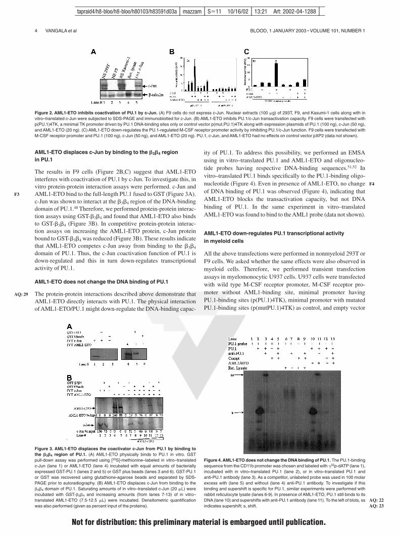
AML1-ETO displaces c-Jun by binding to the �3�4 regionin PU.1
The results in F9 cells (Figure 2B,C) suggest that AML1-ETOinterferes with coactivation of PU.1 by c-Jun. To investigate this, invitro protein-protein interaction assays were performed. c-Jun andAML1-ETO bind to the full-length PU.1 fused to GST (Figure 3A).c-Jun was shown to interact at the �3�4 region of the DNA-bindingdomain of PU.1.48 Therefore, we performed protein-protein interac-tion assays using GST-�3�4 and found that AML1-ETO also bindsto GST-�3�4 (Figure 3B). In competitive protein-protein interac-tion assays on increasing the AML1-ETO protein, c-Jun proteinbound to GST-�3�4 was reduced (Figure 3B). These results indicatethat AML1-ETO competes c-Jun away from binding to the �3�4
domain of PU.1. Thus, the c-Jun coactivation function of PU.1 isdown-regulated and this in turn down-regulates transcriptionalactivity of PU.1.
AML1-ETO does not change the DNA binding of PU.1
The protein-protein interactions described above demonstrate thatAML1-ETO directly interacts with PU.1. The physical interactionof AML1-ETO/PU.1 might down-regulate the DNA-binding capac-
ity of PU.1. To address this possibility, we performed an EMSAusing in vitro–translated PU.1 and AML1-ETO and oligonucleo-tide probes having respective DNA-binding sequences.51,52 Invitro–translated PU.1 binds specifically to the PU.1-binding oligo-nucleotide (Figure 4). Even in presence of AML1-ETO, no changeof DNA binding of PU.1 was observed (Figure 4), indicating thatAML1-ETO blocks the transactivation capacity, but not DNAbinding of PU.1. In the same experiment in vitro–translatedAML1-ETO was found to bind to the AML1 probe (data not shown).
AML1-ETO down-regulates PU.1 transcriptional activityin myeloid cells
All the above transfections were performed in nonmyeloid 293T orF9 cells. We asked whether the same effects were also observed inmyeloid cells. Therefore, we performed transient transfectionassays in myelomonocytic U937 cells. U937 cells were transfectedwith wild type M-CSF receptor promoter, M-CSF receptor pro-moter without AML1-binding site, minimal promoter havingPU.1-binding sites (p(PU.1)4TK), minimal promoter with mutatedPU.1-binding sites (p(mutPU.1)4TK) as control, and empty vector
Figure 2. AML1-ETO inhibits coactivation of PU.1 by c-Jun. (A) F9 cells do not express c-Jun. Nuclear extracts (100 �g) of 293T, F9, and Kasumi-1 cells along with invitro–translated c-Jun were subjected to SDS-PAGE and immunoblotted for c-Jun. (B) AML1-ETO inhibits PU.1/c-Jun transactivation capacity. F9 cells were transfected withp(PU.1)4TK, a minimal TK promoter driven by PU.1 DNA-binding sites only or control vector p(mut.PU.1)4TK along with expression plasmids of PU.1 (100 ng), c-Jun (50 ng),and AML1-ETO (20 ng). (C) AML1-ETO down-regulates the PU.1-regulated M-CSF receptor promoter activity by inhibiting PU.1/c-Jun function. F9 cells were transfected withM-CSF receptor promoter and PU.1 (100 ng), c-Jun (50 ng), and AML1-ETO (20 ng). PU.1, c-Jun, and AML1-ETO had no effects on control vector pXP2 (data not shown).
Figure 3. AML1-ETO displaces the coactivator c-Jun from PU.1 by binding tothe �3�4 region of PU.1. (A) AML1-ETO physically binds to PU.1 in vitro. GSTpull-down assay was performed using [35S]-methionine–labeled in vitro–translatedc-Jun (lane 1) or AML1-ETO (lane 4) incubated with equal amounts of bacteriallyexpressed GST-PU.1 (lanes 2 and 5) or GST plus beads (lanes 3 and 6). GST-PU.1or GST was recovered using glutathione-agarose beads and separated by SDS-PAGE prior to autoradiography. (B) AML1-ETO displaces c-Jun from binding to the�3�4 domain of PU.1. Saturating amounts of in vitro–translated c-Jun (20 �L) wereincubated with GST-�3�4 and increasing amounts (from lanes 7-13) of in vitro–translated AML1-ETO (7.5-12.5 �L) were incubated. Densitometric quantificationwas also performed (given as percent input of the proteins).
Figure 4. AML1-ETO does not change the DNA binding of PU.1. The PU.1-bindingsequence from the CD11b promoter was chosen and labeled with �32p-dATP (lane 1),incubated with in vitro–translated PU.1 (lane 2), or in vitro–translated PU.1 andanti-PU.1 antibody (lane 3). As a competitor, unlabeled probe was used in 100 molarexcess with (lane 5) and without (lane 4) anti-PU.1 antibody. To investigate if thisbinding and supershift is specific for PU.1, similar experiments were performed withrabbit reticulocyte lysate (lanes 6-9). In presence of AML1-ETO, PU.1 still binds to itsDNA (lane 10) and supershifts with anti-PU.1 antibody (lane 11). To the left of blots, ssindicates supershift; s, shift.
4 VANGALA et al BLOOD, 1 JANUARY 2003 � VOLUME 101, NUMBER 1
F3
AQ: 29
F4
AQ: 22AQ: 23
tapraid4/h8-bloo/h8-bloo/h80103/h83591d03a mazzam S�11 10/16/02 13:21 Art: 2002-04-1288
Not for distribution: this preliminary material is embargoed until publication.
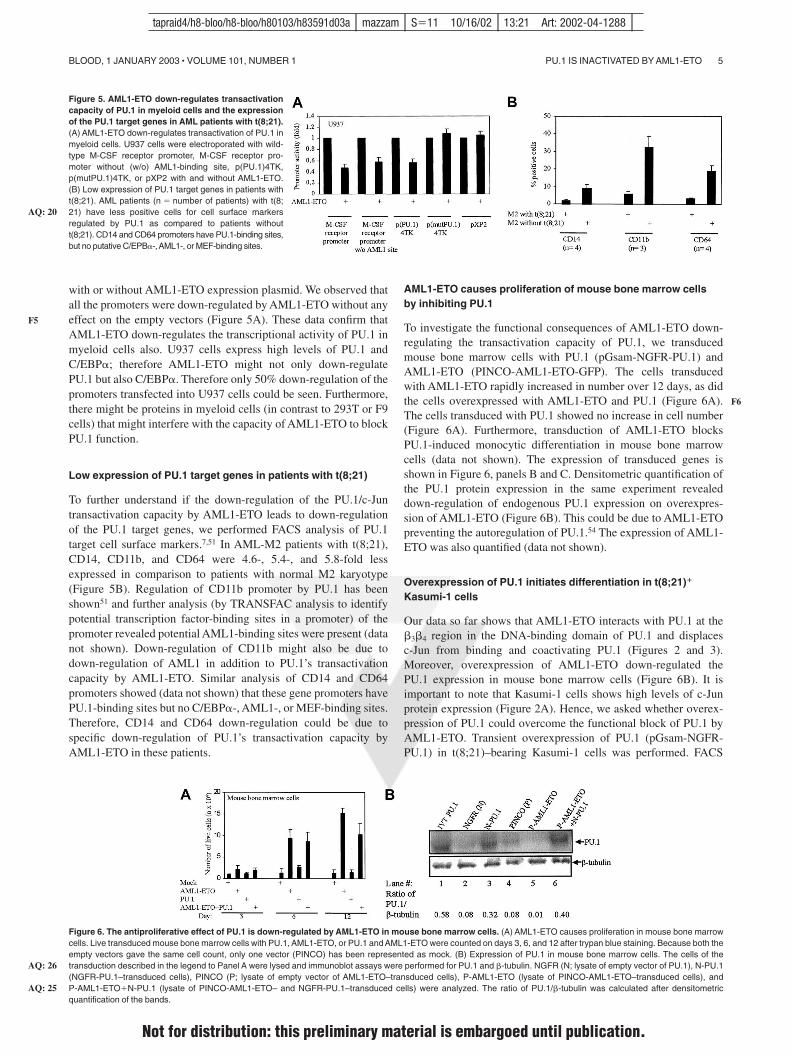
with or without AML1-ETO expression plasmid. We observed thatall the promoters were down-regulated by AML1-ETO without anyeffect on the empty vectors (Figure 5A). These data confirm thatAML1-ETO down-regulates the transcriptional activity of PU.1 inmyeloid cells also. U937 cells express high levels of PU.1 andC/EBP�; therefore AML1-ETO might not only down-regulatePU.1 but also C/EBP�. Therefore only 50% down-regulation of thepromoters transfected into U937 cells could be seen. Furthermore,there might be proteins in myeloid cells (in contrast to 293T or F9cells) that might interfere with the capacity of AML1-ETO to blockPU.1 function.
Low expression of PU.1 target genes in patients with t(8;21)
To further understand if the down-regulation of the PU.1/c-Juntransactivation capacity by AML1-ETO leads to down-regulationof the PU.1 target genes, we performed FACS analysis of PU.1target cell surface markers.7,51 In AML-M2 patients with t(8;21),CD14, CD11b, and CD64 were 4.6-, 5.4-, and 5.8-fold lessexpressed in comparison to patients with normal M2 karyotype(Figure 5B). Regulation of CD11b promoter by PU.1 has beenshown51 and further analysis (by TRANSFAC analysis to identifypotential transcription factor-binding sites in a promoter) of thepromoter revealed potential AML1-binding sites were present (datanot shown). Down-regulation of CD11b might also be due todown-regulation of AML1 in addition to PU.1’s transactivationcapacity by AML1-ETO. Similar analysis of CD14 and CD64promoters showed (data not shown) that these gene promoters havePU.1-binding sites but no C/EBP�-, AML1-, or MEF-binding sites.Therefore, CD14 and CD64 down-regulation could be due tospecific down-regulation of PU.1’s transactivation capacity byAML1-ETO in these patients.
AML1-ETO causes proliferation of mouse bone marrow cellsby inhibiting PU.1
To investigate the functional consequences of AML1-ETO down-regulating the transactivation capacity of PU.1, we transducedmouse bone marrow cells with PU.1 (pGsam-NGFR-PU.1) andAML1-ETO (PINCO-AML1-ETO-GFP). The cells transducedwith AML1-ETO rapidly increased in number over 12 days, as didthe cells overexpressed with AML1-ETO and PU.1 (Figure 6A).The cells transduced with PU.1 showed no increase in cell number(Figure 6A). Furthermore, transduction of AML1-ETO blocksPU.1-induced monocytic differentiation in mouse bone marrowcells (data not shown). The expression of transduced genes isshown in Figure 6, panels B and C. Densitometric quantification ofthe PU.1 protein expression in the same experiment revealeddown-regulation of endogenous PU.1 expression on overexpres-sion of AML1-ETO (Figure 6B). This could be due to AML1-ETOpreventing the autoregulation of PU.1.54 The expression of AML1-ETO was also quantified (data not shown).
Overexpression of PU.1 initiates differentiation in t(8;21)�
Kasumi-1 cells
Our data so far shows that AML1-ETO interacts with PU.1 at the�3�4 region in the DNA-binding domain of PU.1 and displacesc-Jun from binding and coactivating PU.1 (Figures 2 and 3).Moreover, overexpression of AML1-ETO down-regulated thePU.1 expression in mouse bone marrow cells (Figure 6B). It isimportant to note that Kasumi-1 cells shows high levels of c-Junprotein expression (Figure 2A). Hence, we asked whether overex-pression of PU.1 could overcome the functional block of PU.1 byAML1-ETO. Transient overexpression of PU.1 (pGsam-NGFR-PU.1) in t(8;21)–bearing Kasumi-1 cells was performed. FACS
Figure 5. AML1-ETO down-regulates transactivationcapacity of PU.1 in myeloid cells and the expressionof the PU.1 target genes in AML patients with t(8;21).(A) AML1-ETO down-regulates transactivation of PU.1 inmyeloid cells. U937 cells were electroporated with wild-type M-CSF receptor promoter, M-CSF receptor pro-moter without (w/o) AML1-binding site, p(PU.1)4TK,p(mutPU.1)4TK, or pXP2 with and without AML1-ETO.(B) Low expression of PU.1 target genes in patients witht(8;21). AML patients (n � number of patients) with t(8;21) have less positive cells for cell surface markersregulated by PU.1 as compared to patients withoutt(8;21). CD14 and CD64 promoters have PU.1-binding sites,but no putative C/EPB�-,AML1-, or MEF-binding sites.
Figure 6. The antiproliferative effect of PU.1 is down-regulated by AML1-ETO in mouse bone marrow cells. (A) AML1-ETO causes proliferation in mouse bone marrowcells. Live transduced mouse bone marrow cells with PU.1, AML1-ETO, or PU.1 and AML1-ETO were counted on days 3, 6, and 12 after trypan blue staining. Because both theempty vectors gave the same cell count, only one vector (PINCO) has been represented as mock. (B) Expression of PU.1 in mouse bone marrow cells. The cells of thetransduction described in the legend to Panel A were lysed and immunoblot assays were performed for PU.1 and �-tubulin. NGFR (N; lysate of empty vector of PU.1), N-PU.1(NGFR-PU.1–transduced cells), PINCO (P; lysate of empty vector of AML1-ETO–transduced cells), P-AML1-ETO (lysate of PINCO-AML1-ETO–transduced cells), andP-AML1-ETO�N-PU.1 (lysate of PINCO-AML1-ETO– and NGFR-PU.1–transduced cells) were analyzed. The ratio of PU.1/�-tubulin was calculated after densitometricquantification of the bands.
PU.1 IS INACTIVATED BY AML1-ETO 5BLOOD, 1 JANUARY 2003 � VOLUME 101, NUMBER 1
F5
AQ: 20
AQ: 26
AQ: 25
F6
tapraid4/h8-bloo/h8-bloo/h80103/h83591d03a mazzam S�11 10/16/02 13:21 Art: 2002-04-1288
Not for distribution: this preliminary material is embargoed until publication.
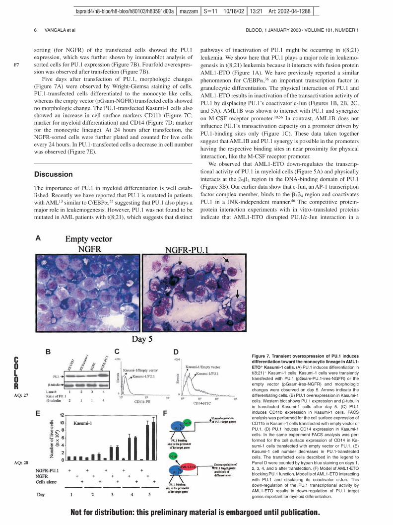
sorting (for NGFR) of the transfected cells showed the PU.1expression, which was further shown by immunoblot analysis ofsorted cells for PU.1 expression (Figure 7B). Fourfold overexpres-sion was observed after transfection (Figure 7B).
Five days after transfection of PU.1, morphologic changes(Figure 7A) were observed by Wright-Giemsa staining of cells.PU.1-transfected cells differentiated to the monocyte like cells,whereas the empty vector (pGsam-NGFR) transfected cells showedno morphologic change. The PU.1-transfected Kasumi-1 cells alsoshowed an increase in cell surface markers CD11b (Figure 7C;marker for myeloid differentiation) and CD14 (Figure 7D; markerfor the monocytic lineage). At 24 hours after transfection, theNGFR-sorted cells were further plated and counted for live cellsevery 24 hours. In PU.1-transfected cells a decrease in cell numberwas observed (Figure 7E).
Discussion
The importance of PU.1 in myeloid differentiation is well estab-lished. Recently we have reported that PU.1 is mutated in patientswith AML13 similar to C/EBP�,55 suggesting that PU.1 also plays amajor role in leukemogenesis. However, PU.1 was not found to bemutated in AML patients with t(8;21), which suggests that distinct
pathways of inactivation of PU.1 might be occurring in t(8;21)leukemia. We show here that PU.1 plays a major role in leukemo-genesis in t(8;21) leukemia because it interacts with fusion proteinAML1-ETO (Figure 1A). We have previously reported a similarphenomenon for C/EBP�,36 an important transcription factor ingranulocytic differentiation. The physical interaction of PU.1 andAML1-ETO results in inactivation of the transactivation activity ofPU.1 by displacing PU.1’s coactivator c-Jun (Figures 1B, 2B, 2C,and 5A). AML1B was shown to interact with PU.1 and synergizeon M-CSF receptor promoter.10,56 In contrast, AML1B does notinfluence PU.1’s transactivation capacity on a promoter driven byPU.1-binding sites only (Figure 1C). These data taken togethersuggest that AML1B and PU.1 synergy is possible in the promotershaving the respective binding sites in near proximity for physicalinteraction, like the M-CSF receptor promoter.
We observed that AML1-ETO down-regulates the transcrip-tional activity of PU.1 in myeloid cells (Figure 5A) and physicallyinteracts at the �3�4 region in the DNA-binding domain of PU.1(Figure 3B). Our earlier data show that c-Jun, an AP-1 transcriptionfactor complex member, binds to the �3�4 region and coactivatesPU.1 in a JNK-independent manner.46 The competitive protein-protein interaction experiments with in vitro–translated proteinsindicate that AML1-ETO disrupted PU.1/c-Jun interaction in a
Figure 7. Transient overexpression of PU.1 inducesdifferentiation toward the monocytic lineage in AML1-ETO� Kasumi-1 cells. (A) PU.1 induces differentiation int(8;21)� Kasumi-1 cells. Kasumi-1 cells were transientlytransfected with PU.1 (pGsam-PU.1-ires-NGFR) or theempty vector (pGsam-ires-NGFR) and morphologicchanges were observed on day 5. Arrows indicate thedifferentiating cells. (B) PU.1 overexpression in Kasumi-1cells. Western blot shows PU.1 expression and �-tubulinin transfected Kasumi-1 cells after day 5. (C) PU.1induces CD11b expression in Kasumi-1 cells. FACSanalysis was performed for the cell surface expression ofCD11b in Kasumi-1 cells transfected with empty vector orPU.1. (D) PU.1 induces CD14 expression in Kasumi-1cells. In the same experiment FACS analysis was per-formed for the cell surface expression of CD14 in Ka-sumi-1 cells transfected with empty vector or PU.1. (E)Kasumi-1 cell number decreases in PU.1-transfectedcells. The transfected cells described in the legend toPanel D were counted by trypan blue staining on days 1,2, 3, 4, and 5 after transfection. (F) Model of AML1-ETOblocking PU.1 function. Model is of AML1-ETO interactingwith PU.1 and displacing its coactivator c-Jun. Thisdown-regulation of the PU.1 transcriptional activity byAML1-ETO results in down-regulation of PU.1 targetgenes important for myeloid differentiation.
6 VANGALA et al BLOOD, 1 JANUARY 2003 � VOLUME 101, NUMBER 1
F7
AQ: 27
AQ: 28
COLOR
tapraid4/h8-bloo/h8-bloo/h80103/h83591d03a mazzam S�11 10/16/02 13:21 Art: 2002-04-1288
Not for distribution: this preliminary material is embargoed until publication.
competitive manner (Figure 3B), thus blocking c-Jun from coacti-vating PU.1. We have described a similar mechanism for GATA-150
and C/EBP�.48 The role of c-Jun in myeloid differentiation wasshown to be rather important, because it could enhance the extentof differentiation in U937 cells.57 In other studies AP-1 andC/EBP� were shown to cooperate in regulation of common targetgenes, including the human TSG-6,58 collagenase-1 gene,59 andtumor necrosis factor � (TNF-�).60 These data suggest that thecapacity of c-Jun to coactivate PU.1 is also a very importantmechanism, which is down-regulated by AML1-ETO.
We observed that physical interaction between AML1-ETO andPU.1 did not abolish the DNA- binding capacity of PU.1 (Figure4A), although AML1-ETO interacted with the PU.1 DNA-bindingdomain. Interestingly, in PU.1’s crystal structure,1 the �3�4 domaindoes not interact with DNA, but is exposed to the solvent. Thisstructural ability allows PU.1 to retain its DNA binding thoughbeing functionally repressed. We show here that the normalinteraction between coactivators and transcription factors arealtered in presence of AML1-ETO, which could be one of theimportant mechanisms for disrupted myelopoiesis in t(8;21)�
leukemia (Figure 7F).AML1B and AML1-ETO have been shown to transactivate the
M-CSF receptor,42 suggesting that interaction between AML1Band AML1-ETO could be important for leukemogenesis. Toinvestigate the importance of AML1-ETO/PU.1 interaction inleukemogenesis, transactivation, proliferation, and differentiationassays were performed in cells expressing wild-type AML1Bprotein. In the presence of AML1-ETO, the M-CSF receptorpromoter was down-regulated and similarly the AML1 site mutatedM-CSF receptor promoter and minimal promoter containing onlyPU.1-binding sites in U937 cells (Figure 5A). This could beexplained by a dual function of AML1-ETO in regulation of theM-CSF receptor expression. During normal myeloid differentia-tion, M-CSF receptor expression is required for G1-to-S phasetransition, which could be down-regulated by AML1-ETO throughthe functional interaction with PU.1, and then AML1-ETO cooper-ates with AML1B to up-regulate the M-CSF receptor expressionfor transformation and proliferation of abnormal progenitor cells.
In patients with t(8;21), expression of the cell surface markersCD11b, CD14, and CD64 was less in comparison to patientswithout t(8;21) (Figure 5B). CD14 and CD64 promoters haveputative PU.1 binding sites but not AML1-, C/EBP�-, or MEF-binding sites suggesting that down-regulation of the function ofPU.1 by AML1-ETO could possibly be an important step inprogression toward leukemia. CD11b, another marker for differen-tiation, was also less expressed in patients with t(8;21) in compari-son to patients without t(8;21) (Figure 5B). CD11b is regulated byPU.1 and its promoter contains putative binding sites of AML1. Inthis case AML1-ETO interaction and down-regulation of importantmyeloid transcription factors like PU.1 and AML1 could explainthe lower CD11b expression. The phenotype of PU.1�/� micesuggests that PU.1 is critical for myeloid differentiation anddevelopment.4,11,12 Interaction of PU.1 with AML1-ETO and
subsequent suppression of PU.1 target genes (Figure 5B) mightcontribute to the phenotypic changes seen in t(8;21). Furthermore,PU.1 is a self-regulatory protein54 and AML1-ETO overexpressionin mouse bone marrow cells down-regulated the endogenous PU.1expression (Figure 6B).
Because AML1�/� mice lack PU.1 expression,61 AML1-ETOcould down-regulate PU.1 expression through repressing AML1function. However, in a diseased condition or in presence ofAML1-ETO, like in Kasumi-1 cells, the expression of AML1 andPU.1 genes was still observed. Furthermore, AML1 does not have adown-regulatory effect on PU.1 (Figure 1C). Therefore, thepresence of AML1-ETO does not completely repress the expres-sion levels of these genes, but may block their functions byprotein-protein interactions. To analyze the functional impact ofAML1-ETO on PU.1 in the presence of wild-type AML1B protein,we performed experiments in cells expressing endogenous AML1Bprotein. Overexpression of PU.1 in mouse bone marrow cells leadsto a block in proliferation, but in presence of AML1-ETO thisfunction of PU.1 was abrogated (Figure 6A). Recently, it wasshown that AML1-ETO expression in human progenitor cells leadsto expansion of human hematopoietic stem cells.62 Therefore, theblock of differentiation and increase in abnormal proliferation ofhematopoietic stem cells could be due to down-regulation of theactivity of PU.1 in t(8;21) leukemia.
Overexpression of PU.1 in t(8;21)� Kasumi-1 cells differenti-ates them toward the monocytic lineage (Figure 7). Morphologi-cally, cells did not appear to be terminally differentiated eventhough the cell surface markers CD11b and CD14 were increasedin expression. It has been earlier shown that short-term activationof PU.1 in multipotent hematopoietic cells leads to immatureeosinophils.63 However, stable overexpression of PU.1 could leadto myeloid lineage in hematopoietic progenitor cells.63 Therefore,higher and stable expression of PU.1 in Kasumi-1 cells might beneeded to terminally differentiate toward the monocytic lineage.The cell number of PU.1-transfected Kasumi-1 cells decreasedover a course of time (Figure 7E) showing that PU.1 functions asan antiproliferative factor on overexpression in Kasumi-1 cells.However, this mechanism needs to be further elucidated. Ourdata suggest that the ectopic expression of PU.1 in Kasumi-1cells overcomes the functional block by AML1-ETO. PU.1 andC/EBP� are important factors for myeloid differentiation andAML1-ETO down-regulating these 2 factors could be an importantstep toward leukemia. This also suggests the possibility of usingthese 2 factors independently or in combination for therapy oft(8;21) myeloid leukemias.
Acknowledgments
We thank Dr Atsushi Iwama, University of Tsukuba, Japan forproviding pGsam-PU.1-ires-NGFR and PGsam-ires-NGFR retrovi-ral vectors.
References
1. Kodandapani R, Pio F, Ni CZ, et al. A new patternfor helix-turn-helix recognition revealed by PU.1ETS-domain-DNA complex. Nature. 1996;380:456-460.
2. Tenen DG, Hromas R, Licht, JD, Zhang DE. Tran-scription factors, normal myeloid development andleukemia. Blood. 1997;90:489-499.
3. Chen H, Zhang P, Voso MT, et al. Neutrophils and
monocytes express high levels of PU.1 (Spi-1) butnot Spi-B. Blood. 1995;85:2918-2928.
4. Anderson KL, Perkin H, Surth CD, et al. Tran-scription factor PU.1 is necessary for develop-ment of thymic and myeloid progenitor-deriveddendritic cells. J Immunol. 2000;164:1855-1861.
5. Behre G, Zhang P, Zhang DE, Tenen DG. Analysis of
the modulation of transcriptional activity in leukemo-genesis. Methods. 1999;17:231-237.
6. Hohaus S, Petrovick MS, Voso MT, et al. PU.1(Spi-1) and C/EBP alpha regulate expression ofthe granulocyte-macrophage colony-stimulatingfactor receptor alpha gene. Mol Cell Biol. 1995;15:5830-5845.
7. Olweus J, Thompson PA, Lund-Johansen F.
PU.1 IS INACTIVATED BY AML1-ETO 7BLOOD, 1 JANUARY 2003 � VOLUME 101, NUMBER 1
AQ: 12
tapraid4/h8-bloo/h8-bloo/h80103/h83591d03a mazzam S�11 10/16/02 13:21 Art: 2002-04-1288
Not for distribution: this preliminary material is embargoed until publication.
Granulocytic and monocytic differentiation of CD34hicells is associated with distinct changes in the ex-pression of the PU.1 regulated molecules, CD64 andmacrophage colony-stimulating factor receptor.Blood. 1996;88:3741-3754.
8. Smith LT, Hohaus S, Gonzalez DA, Dziennis SE,Tenen DG. PU.1 (Spi-1) and C/EBP alpha regulatethe granulocyte colony-stimulating factor receptorpromoter in myeloid cells. Blood. 1996;88:1234-1247.
9. Zhang DE, Hetherington CJ, Chen H, Tenen DG.The macrophage transcription factor PU.1 directstissue-specific expression of the macrophagecolony-stimulating factor receptor. Mol Cell Biol.1994;14:373-381.
10. Zhang DE, Hohaus S, Voso MT, et al. Function ofPU.1 (Spi-1), C/EBP, and AML1 in early myelopoi-esis regulation of multiple myeloid CSF receptor pro-moters. Curr Top Microbiol Immunol. 1996;211:137-147.
11. McKercher SR, Torbett BE, Anderson KL, et al. Tar-geted disruption of the PU.1 gene results in multiplehematopoietic abnormalities. EMBO J. 1996;15:5647-5658.
12. Scott EW, Simon MC, Anastasi J, Singh H. Require-ment of transcription factor PU.1 in the developmentof multiple hematopoietic lineages. Science. 1994;265:1573-1577.
13. Mueller BU, Pabst T, Osato M, et al. HeterozygousPU.1 mutations are associated with acute myeloidleukemia. Blood. In press.
14. Crute BE, Lewis AF, Wu Z, Bushweller JH, SpeckNA. Biochemical and biophysical properties of corebinding factors 2 (AML1) DNA binding domain. J BiolChem. 1996;271:26251-26260.
15. Daga A, Tighe JE, Calabi F. Leukemia/Drosophilahomology. Nature. 1992;356:484-489.
16. Golling G, Li L, Pepling M, Stebbins M, Gergen JP.Drosophila homologous of the proto-oncogene prod-uct PEBP2/CBF beta regulate the DNA binding prop-erties of the Runt. Mol Cell Biol. 1996;16:932-942.
17. Hernandez-Munian C, Krangel MS. c-Myb and corebinding factor PEBP2 display functional synergy butbind independently to adjacent sites in the T-cell re-ceptor enhancer. Mol Cell Biol. 1995;15:3090-3099.
18. Look AT. Oncogenic transcription factors in humanacute myeloid leukemias. Science. 1997;278:1059-1067.
19. Ness SA, Kowenz-Leutz E, Casini T, Graf T, Leutz A.Myb and MF-M: combinatorial activators of myeloidgenes in heterologous cell types. Genes Dev. 1993;7:749-759.
20. Ogawa E, Inuzuka M, Maruyama M, et al. Molecularcloning and characterization of PEBP2 beta, the het-erodimeric partner of novel Drosophila runt-relatedDNA binding protein PEBP2. Virology. 1993;194:314-331.
21. Sun W, O’Connell M, Speck NA. Characterization ofa protein that binds multiple sequences in mamma-lian type C retrovirus enhancers. J Virol. 1993;67:1986-1995.
22. Wang S, Wang Q, Crute BE, et al. Cloning and char-acterization of subunits of the T-cell receptor and mu-rine leukemia virus enhancer core-binding factor. MolCell Biol. 1993;13:3329-3332.
23. Nucifora G, Birn DJ, Erickson P, et al. Detection ofDNA rearrangements in the AML1 and ETO loci andan AML1-ETO fusion mRNA in patients with t(8;21)acute myeloid leukemia. Blood. 81;1993:883-888.
24. Osato M, Asou N, Abdalla E, et al. Biallelic and het-erozygous point mutations in the Runt domain of theAML1/PEBP2�B gene associated with myeloblasticleukemias. Blood. 1999;93:1817-1824.
25. Song WJ, Sullivan MG, Legare RD, et al. Haploinsuf-ficiency of CBFA2 causes familial thrombocytopeniawith propensity to develop acute myelogenous leu-kemia. Nat Genet. 1999;23:166-175.
26. Era T, Asou N, Kunisada T, et al. Identification of twotranscripts of AML1-ETO-fused gene in t(8;21) leuke-mic cells and expression of wild-type ETO gene in
hematopoietic cells. Genes Chromosomes Cancer.1995;13:25-33.
27. Erickson P Gao J, Chang KS, et al. Identification ofbreak point in t(8;21) acute myelogenous leukemiaand isolation of a fusion transcript, AML1-ETO, withsimilarity to Drosophila segmentation gene, runt.Blood. 1992;80:1825-1831.
28. Miyoshi H, Kozu T, Shimizu K, et al. The t(8;21)translocation in acute myeloid leukemia results inproduction of an AML1-MTG8 fusion transcript.EMBO J. 1993;12:2715-2721.
29. Okuda T, van Deursen J, Hiebert SW, Grosveld G,Downing JR. AML1, the target of multiple chromo-somal translocations in human leukemia, is essentialfor normal fetal liver hematopoiesis. Cell. 1996;84:321-330.
30. Bitter MA, Le Beau MM, Rowley JD, et al. Associa-tions between morphology, karyotype, and clinicalfeatures in myeloid leukemias. Hum Pathol. 1987;18:211-225.
31. Downing JR. The AML1-ETO chimeric transcriptionfactor in acute myeloid leukemia: biology and clinicalsignificance. Br J Haematol. 1999;106:296-308.
32. Lowenberg B, Downing JR, Burnett A. Acute myeloidleukemia. N Engl J Med. 1999;30:1051-1061.
33. Miyoshi H, Shimizu K, Kazu T, Maseki N, Kaneko Y,Ohki M. t(8;21) breakpoints on chromosome 21 inacute myeloid leukemia are clustered within a limitedregion of a single gene, AML1. Proc. Natl. Acad. Sci.U S A. 1991;88:10431-10434.
34. Frank R, Zhang J, Uchida H, Meyers S, Hiebert SW,Nimer SD. The AML1-ETO protein blocks transacti-vation of GM-CSF promoter by AML1B. Oncogene.1995;11:2667-2674.
35. Nuchprayoon L, Meyers S, Scott LM, Suzow J,Hiebert S, Friedman AD. PEBP2/CBF, the murinehomolog of human myeloid AML1 and PEBP2�/CBF� protooncoproteins, regulates the murine my-eloperoxidase and neutrophil elastase genes in im-mature myeloid cells. Mol Cell Biol. 1994;14:5558-5568.
36. Pabst T, Mueller BU, Schoch C, et al. AML1-ETOdownregulates the granulocytic differentiation factorC/EBPalpha in t(8;21) myeloid leukemia. Nat Med.2001;7:444-451.
37. Yergeau DA, Hetherington CJ, Wang Q, et al. Em-bryonic lethality and impairment of hematopoiesis inmice heterozygous for AML1-ETO fusion gene. NatGenet. 1997;15:303-306.
38. Zhang DE, Fujioka K, Hetherington CJ, et al. Identifi-cation of a region which directs the monocytic activityof the colony-stimulating factor 1 (macrophagecolony-stimulating factor) receptor promoter andbinds PEBP2/CBF (AML1). Mol Cell Biol. 1994;14:8085-8095.
39. Zhang DE, Hetherington CJ, Meyers S, et al. CCAATenhancer binding protein (C/EBP) and AML1 (CB-Falpha2) synergistically activates the macrophagecolony stimulating factor receptor promoter. Mol CellBiol. 1996;16:1231-1240.
40. Frank RC, Sun X, Berguido FJ, Jakubowaik A, NimerSD. The t(8;21) fusion protein, AML1-ETO trans-forms NIH3T3 cells and activates AP-1. Oncogene.1999;18:1701-1710.
41. Klampfer L, Zhang J, Zelenetz AO, Uchida H, NimerSD. The AML1-ETO fusion protein activates tran-scription of BCL-2. Proc Natl Acad Sci U S A. 1996;93:14059-14064.
42. Rhoades KL, Hetherington CJ, Rowley JD, et al.Synergistic up-regulation of the myeloid-specific pro-moter for macrophage colony-stimulating factor re-ceptor by AML1 and the t(8;21)fusion protein maycontribute to leukemogenesis. Proc Natl Acad SciU S A. 1996;93:11895-11900.
43. Rhoades KL, Heterington CJ, Harakawa N, et al.Analysis of the role of AML1-ETO in leukemogenesisusing inducible transgenic mouse model. Blood.2000;96:2108-2115.
44. Meyers S, Lenny N, Hiebert SW. The t(8;21) fusionprotein interferes with AML1B-dependent transcrip-tional activity. Mol Cell Biol. 1995;15:1974-1982.
45. Mao, S., Franks RC, Zhang J, Miyazaki Y, Nimer SD.Functional and physical interactions between AML1proteins and ETS protein: MEF; implications forpathogenesis of t(8;21) positive leukemias. Mol CellBiol. 1999;19:3635-3644.
46. Behre G, Whitmarsh AJ, Coghlan MP, et al. c-Jun isa JNK-independent coactivator of PU.1 transcriptionfactor. J Biol Chem. 1999;274:4939-4946.
47. Behre G, Singh SM, Liu H, et al. Ras signaling en-hances the activity of C/EBPalpha to induce granulo-cytic differentiation by phosphorylation of serine 248.J Biol Chem. 2002;277:26293-26299.
48. Reddy V, Iwama A, Iotzava G, et al. The granulocyticinducer C/EBPalpha inactivates the myeloid masterregulator PU.1: possible role in lineage commitmentdecisions. Blood. 2002;100:483-490.
49. Behre G, Smith LT, Tenen DG. Use of a promoterlessRenilla luciferase vectors as an internal control plas-mid for transient co-transfection assays of Ras-medi-ated transcription activation. Biotechniques. 1999;26:24-26.
50. Zhang.P., Behre G, Pan J, et al. Negative cross-talkbetween hematopoietic regulators: GATA proteinsrepress PU.1 function. Proc Natl Acad Sci U S A.1999;96:8705-8710.
51. Pahl HL, Scheibe RJ, Zhang DE, et al. The proto-oncogene PU.1 regulates expression of the myeloid-specific CD11b promoter. J Biol Chem. 1993;268:5014-5020.
52. Meyers S, Downing JR, Hiebert SW. Identification ofAML-1 and t(8;21) translocation protein (AML1-ETO)as sequence-specific DNA-binding proteins: the runthomology domain is required for DNA binding andprotein-protein interactions. Mol Cell Biol. 1993;13:6336-6345.
53. Grignani F, Kinsella T, Mencarelli A, Gergen JP. Highefficiency gene transfer and selection of human he-matopoietic progenitor cells with a hybrid EBV/retro-viral vector expressing the green fluorescence pro-tein. Cancer Res. 1998;58:14-19.
54. Chen H, Ray D, Zhang P, et al. PU.1 (Spi-1) auto-regulates its expression in myeloid cells. Oncogene.1995;11:1549-1560.
55. Pabst T, Mueller BU, Zhang P, et al. Dominant nega-tive mutations of C/EBPA, encoding CCAAT/en-hancer binding protein-alpha (C/EBPalpha), in acutemyeloid leukemia. Nat Genet. 2001;27:263-270.
56. Petrovick MS, Hiebert SW, Friedman AD, et al. Mul-tiple functional domains of AML1: PU.1 and C/EBPalpha synergize with different regions of AML1.Mol Cell Biol. 1998;18:3915-3925.
57. Szabo E, Francis J, Birrer MJ. Alterations in differen-tiation and apoptosis induced by bufalin in c-Junoverexpressing U937 cells. Int J Oncol. 1998;12:403-409.
58. Klampfer L, Lee TH, Hsu W, Vilcek J, Chen-Kiang S.NF-IL6 and AP-1 cooperatively modulate the activa-tion of TSG-6 gene by tumor necrosis factor alphaand interleukin-1. Mol Cell Biol. 1994;14:6561-6569.
59. Doyle GAR, Pierce RA, Parks WC. Transcriptionalinduction of collagenase-1 in differentiated mono-cyte-like U937 cells is regulated by AP-1 and up-stream C/EBPbeta site. J Biol Chem. 1997;272:11840-11849.
60. Zagariya A, Mungre S, Lovis R, et al. Tumor necrosisfactor alpha gene regulation: enhancement of C/EBPbeta-induced activation by c-Jun. Mol Cell Biol.1998;18:2815-2824.
61. Okada H, Watanabe T, Niki M, et al. AML1(�/�) em-bryos do not express certain hematopoiesis-relatedgene transcripts including those of the PU.1 gene.Oncogene. 1998;17:2287-2293.
62. Mulloy JC, Cammenga J, MacKenzie KL, et al. TheAML1-ETO fusion protein promotes the expansion ofhuman stem cells. Blood. 2002;99:15-23.
63. Nerlov C, Graf T. PU.1 induces myeloid lineage com-mitment in multipotent hematopoietic progenitors.Genes Dev. 1998;12:2403-2412.
8 VANGALA et al BLOOD, 1 JANUARY 2003 � VOLUME 101, NUMBER 1
AQ: 2
tapraid4/h8-bloo/h8-bloo/h80103/h83591d03a mazzam S�11 10/16/02 13:21 Art: 2002-04-1288
Not for distribution: this preliminary material is embargoed until publication.