r e Rese q Journal of Aquaculture Research & Development · Sturgeon biology is interesting because...
4
Open Access Volume 4 • Issue 4 • 1000180 J Aquac Res Development ISSN: 2155-9546 JARD, an open access journal Open Access Azarin et al., J Aquac Res Development 2013, 4:4 DOI: 10.4172/2155-9546.1000180 Open Access Keywords: Persian sturgeon; Acipenser persicus; 17α, 20β-Dihydroxyprogesterone; Oocyte maturation; Germinal vesicle breakdown Introduction Sturgeons are a very ancient fish group, existing since the Late Cretaceous with a wide distribution in the Northern Hemisphere. Sturgeon biology is interesting because of important conservation and economic issues involving these fishes. Sturgeons are the source of two high-value products: boneless and very tasteful meat and black caviar. Acipenser persicus is one of important species that live in the southern margin of the Caspian basin [1]. Sterlet (Acipenser ruthenus) is the species with relatively small size and rapid sexual maturation within the family Acipenseridae [2]. Unfortunately, nowadays these fishes have become an endangered species due to the damages of their natural spawning environments, overfishing for meat and caviar production and also water pollution [3,4]. Oocyte maturation (OM) in fish comprises the migration and breakdown of the germinal vesicle (GVBD) and is followed by egg release from the follicles. A two-stage concept of OM was suggested for teleost fish. e first stage consists of the acquisition of the follicle’s ability to produce the maturation inducing steroid (MIS) and the oocyte’s ability to respond to MIS. During the first stage, germinal vesicle migration (GVM) takes place. e second stage is comprised of the period of MIS production and resumption of oocyte meiosis, followed by GVBD [5]. In sturgeons, GV (germinal vesicle) requires a rather long period for complete migration during which oocyte sensitivity to the maturation- inducing hormone in vitro will change and also the position of the germinal vesicle in the oocyte has been used as a morphological character to select the proper female for induced spawning [6,7]. Several in vitro investigations have revealed that various steroids such as 17α, 20β-Dihydroxyprogesterone can stimulate oocyte maturation in teleosts [6]. e steroid 17α, 20β-DHP has been identified as the MIS in the majority of investigated orders, including Salmoniformes, Cypriniformes, Siluriformes, and Cyprinodontiformes. Sturgeon, which exhibit reproductive characteristics similar to amphibians in early development [8]. Studies related to fish ovarian follicle maturation have been carried out with three purposes. e first is connected to the search for phenomena involved from the end of the oocyte growth period to ovulation. e second group considered oocyte maturation as a bioassay to test the potency of different hormonal preparations. e third one was developed to determine the best physiological state of females for hormonal injection, especially for sturgeon [9]. Since the culture medium composition significantly affects the sensitivity of the sturgeon follicles to gonadotropins, the choice of culture medium for estimating the developmental stage and/or physiological state of sturgeon follicles according to their reaction to gonadotropins in vitro is of great importance [10]. Observation of germinal vesicle breakdown of the oocytes artificially induced by hormones is one of the main biological tools. As far as the choice of the incubation media is concerned, three different routes were followed for sturgeon. One was adopting the simplest media as illustrated by modified Ringer solution with sodiumbicarbonate, another was the utilization of commercial media namely Leibovitz medium, and the last was the development of a new medium based on fish plasma characteristics [11-14]. All bioassays with sturgeon follicles to date have been conducted *Corresponding author: Hajar Azarin, Department of Fisheries, Gorgan University of Agricultural Sciences and Natural Resources, Iran; Tel: + 911-347-0718; Fax: +171-442-4155; E-mail: [email protected] Received January 30, 2013; Accepted March 25, 2013; Published April 05, 2013 Citation: Azarin H, Imanpoor MR, Pourdehghani M (2013) Effect of 17α, 20β-Dihydroxyprogesterone on In vitro Oocyte Maturation in Persian Sturgeon (Acipenser persicus) and Sterlet (Acipenser ruthenus). J Aquac Res Development 4: 180 doi:10.4172/2155-9546.1000180 Copyright: © 2013 Azarin H, et al. This is an open-access article distributed under the terms of the Creative Commons Attribution License, which permits unrestricted use, distribution, and reproduction in any medium, provided the original author and source are credited. Abstract The in vitro effect of 17α, 20β-Dihydroxyprogesterone on the maturation of Persian sturgeon (Acipenser persicus) oocytes for 7, 10, 12, 24 and 30 hr and sterlet (Acipenser ruthenus) oocytes for 12, 18 and 24 hr of incubation was investigated. The oocytes were incubated in SIS, RM2, L-15 and PSACF in the presence or absence of 17α, 20β-Dihydroxyprogesterone at 1 μg/ml concentration. Results showed that in the Persian surgeon, incubation of oocytes after 7, 10, 12, 24 and 30 hr of in hormone free media had no effect on GVBD. The mean oocyte PI in the SIS, L-15, RM2 and PSACF media after 12 hr of incubation was 9.08 ± 4.65, 5.56 ± 3.40, 7.36 ± 2.95 and 5.86 ± 2.54 respectively. During the 24 and 30 hr constant exposure incubation, 17α, 20β-Dihydroxyprogesterone was more effective at inducing GVBD than 7, 10 and 12 of incubation. In the sterlet oocytes, GVBD did not occur after 24 hr of incubation and the mean oocyte PI in the L-15 and RM2 media after 12, 18 and 24 hr of incubation was 10.26 ± 3.63, 5.54 ± 3.59, 6.12 ± 4.86 and 8.6 ± 3.1, 5.98 ± 4.94, 5 ± 4.06 respectively. These results indicated a role for 17α, 20β-Dihydroxyprogesterone on oocyte maturation in the Persian sturgeon. Effect of 17α, 20β-Dihydroxyprogesterone on In vitro Oocyte Maturation in Persian Sturgeon (Acipenser persicus) and Sterlet (Acipenser ruthenus) Hajar Azarin 1 *, Mohammad Reza Imanpoor 2 and Mohammad Pourdehghani 3 1 Department of Fisheries, Gorgan University of Agricultural Sciences and Natural Resources, Iran 2 Gorgan University of Agricultural Sciences and Natural Resource, Iran 3 International Sturgeon Research Institute, Rasht, Iran Journal of Aquaculture Research & Development Research Article Jo u r n a l o f A q u a c u lt u r e R e s e a r c h & D e v e l o p m e n t ISSN: 2155-9546
Transcript of r e Rese q Journal of Aquaculture Research & Development · Sturgeon biology is interesting because...
Open Access
Volume 4 • Issue 4 • 1000180J Aquac Res DevelopmentISSN: 2155-9546 JARD, an open access journal
Open Access
Azarin et al., J Aquac Res Development 2013, 4:4 DOI: 10.4172/2155-9546.1000180
Open Access
Keywords: Persian sturgeon; Acipenser persicus; 17α,20β-Dihydroxyprogesterone; Oocyte maturation; Germinal vesicle breakdown
IntroductionSturgeons are a very ancient fish group, existing since the Late
Cretaceous with a wide distribution in the Northern Hemisphere. Sturgeon biology is interesting because of important conservation and economic issues involving these fishes. Sturgeons are the source of two high-value products: boneless and very tasteful meat and black caviar. Acipenser persicus is one of important species that live in the southern margin of the Caspian basin [1]. Sterlet (Acipenser ruthenus) is the species with relatively small size and rapid sexual maturation within the family Acipenseridae [2]. Unfortunately, nowadays these fishes have become an endangered species due to the damages of their natural spawning environments, overfishing for meat and caviar production and also water pollution [3,4].
Oocyte maturation (OM) in fish comprises the migration and breakdown of the germinal vesicle (GVBD) and is followed by egg release from the follicles. A two-stage concept of OM was suggested for teleost fish. The first stage consists of the acquisition of the follicle’s ability to produce the maturation inducing steroid (MIS) and the oocyte’s ability to respond to MIS. During the first stage, germinal vesicle migration (GVM) takes place. The second stage is comprised of the period of MIS production and resumption of oocyte meiosis, followed by GVBD [5].
In sturgeons, GV (germinal vesicle) requires a rather long period for complete migration during which oocyte sensitivity to the maturation-inducing hormone in vitro will change and also the position of the germinal vesicle in the oocyte has been used as a morphological character to select the proper female for induced spawning [6,7].
Several in vitro investigations have revealed that various steroids such as 17α, 20β-Dihydroxyprogesterone can stimulate oocyte maturation in teleosts [6]. The steroid 17α, 20β-DHP has been identified as the MIS in the majority of investigated orders, including Salmoniformes, Cypriniformes, Siluriformes, and Cyprinodontiformes. Sturgeon,
which exhibit reproductive characteristics similar to amphibians in early development [8].
Studies related to fish ovarian follicle maturation have been carried out with three purposes. The first is connected to the search for phenomena involved from the end of the oocyte growth period to ovulation. The second group considered oocyte maturation as a bioassay to test the potency of different hormonal preparations. The third one was developed to determine the best physiological state of females for hormonal injection, especially for sturgeon [9].
Since the culture medium composition significantly affects the sensitivity of the sturgeon follicles to gonadotropins, the choice of culture medium for estimating the developmental stage and/or physiological state of sturgeon follicles according to their reaction to gonadotropins in vitro is of great importance [10].
Observation of germinal vesicle breakdown of the oocytes artificially induced by hormones is one of the main biological tools. As far as the choice of the incubation media is concerned, three different routes were followed for sturgeon. One was adopting the simplest media as illustrated by modified Ringer solution with sodiumbicarbonate, another was the utilization of commercial media namely Leibovitz medium, and the last was the development of a new medium based on fish plasma characteristics [11-14].
All bioassays with sturgeon follicles to date have been conducted
*Corresponding author: Hajar Azarin, Department of Fisheries, Gorgan University of Agricultural Sciences and Natural Resources, Iran; Tel: + 911-347-0718; Fax: +171-442-4155; E-mail: [email protected]
Received January 30, 2013; Accepted March 25, 2013; Published April 05, 2013
Citation: Azarin H, Imanpoor MR, Pourdehghani M (2013) Effect of 17α, 20β-Dihydroxyprogesterone on In vitro Oocyte Maturation in Persian Sturgeon (Acipenser persicus) and Sterlet (Acipenser ruthenus). J Aquac Res Development 4: 180 doi:10.4172/2155-9546.1000180
Copyright: © 2013 Azarin H, et al. This is an open-access article distributed under the terms of the Creative Commons Attribution License, which permits unrestricted use, distribution, and reproduction in any medium, provided the original author and source are credited.
AbstractThe in vitro effect of 17α, 20β-Dihydroxyprogesterone on the maturation of Persian sturgeon (Acipenser persicus)
oocytes for 7, 10, 12, 24 and 30 hr and sterlet (Acipenser ruthenus) oocytes for 12, 18 and 24 hr of incubation was investigated. The oocytes were incubated in SIS, RM2, L-15 and PSACF in the presence or absence of 17α, 20β-Dihydroxyprogesterone at 1 μg/ml concentration. Results showed that in the Persian surgeon, incubation of oocytes after 7, 10, 12, 24 and 30 hr of in hormone free media had no effect on GVBD. The mean oocyte PI in the SIS, L-15, RM2 and PSACF media after 12 hr of incubation was 9.08 ± 4.65, 5.56 ± 3.40, 7.36 ± 2.95 and 5.86 ± 2.54 respectively. During the 24 and 30 hr constant exposure incubation, 17α, 20β-Dihydroxyprogesterone was more effective at inducing GVBD than 7, 10 and 12 of incubation. In the sterlet oocytes, GVBD did not occur after 24 hr of incubation and the mean oocyte PI in the L-15 and RM2 media after 12, 18 and 24 hr of incubation was 10.26 ± 3.63, 5.54 ± 3.59, 6.12 ± 4.86 and 8.6 ± 3.1, 5.98 ± 4.94, 5 ± 4.06 respectively. These results indicated a role for 17α, 20β-Dihydroxyprogesterone on oocyte maturation in the Persian sturgeon.
Effect of 17α, 20β-Dihydroxyprogesterone on In vitro Oocyte Maturation in Persian Sturgeon (Acipenser persicus) and Sterlet (Acipenser ruthenus)Hajar Azarin1*, Mohammad Reza Imanpoor2 and Mohammad Pourdehghani3
1Department of Fisheries, Gorgan University of Agricultural Sciences and Natural Resources, Iran2Gorgan University of Agricultural Sciences and Natural Resource, Iran 3International Sturgeon Research Institute, Rasht, Iran
Journal of AquacultureResearch & Development
Research Article
Jour
nal o
f Aqu
aculture Research &Developm
ent
ISSN: 2155-9546
Citation: Azarin H, Imanpoor MR, Pourdehghani M (2013) Effect of 17α, 20β-Dihydroxyprogesterone on In vitro Oocyte Maturation in Persian Sturgeon (Acipenser persicus) and Sterlet (Acipenser ruthenus). J Aquac Res Development 4: 180 doi:10.4172/2155-9546.1000180
Page 2 of 4
Volume 4 • Issue 4 • 1000180J Aquac Res DevelopmentISSN: 2155-9546 JARD, an open access journal
with long term exposure of follicles (6 to 40 hr) to various steroids [15]. It is expected that acute (short) exposure of ovarian follicles to the proposed MISs will better discriminate the putative MIS in sturgeon as the steroid will not be metabolized into a more active form during the incubation period as seen in Atlantic croaker (Micropogonias undulatus) and spotted seatrout (Cynoscion nebulosus) [16].
The present study was designed to determine which of the proposed MIS (17α, 20β-Dihydroxyprogesterone) is more effective at inducing GVBD in vitro in two sturgeon species: Persian sturgeon (Acipenser persicus) and sterlet (Acipenser ruthenus).
Materials and MethodsBroodstocks preparation
In this study, 16 female Persian sturgeon with the average weight of 21.5 ± 15.65 (kg) and average fork length of 1.01 ± 81.8 (m) were captured at six to eight week intervals beginning in March 2011 from south-east of Caspian Sea, during their upstream migration and then transported to Shahid Margani sturgeon fish farm (Golestan, Iran) by special car in tank with oxygenation. 10 female sterlet (mean weight ± SD: 5.75 ± 131.79 g, mean oocyte diameter ± SD: 2.33 ± 0.18 mm and oocyte polarization index ± SD: 8.5 ± 1.6) prepared from International Sturgeon Research Dr, Dadman (Guilan-Iran). A group of male breeder was captured and all broodstocks of both sexes were maintained in several separate circular tanks (8 m diameter, 1 m depth, 50 m3 volume). The females were survey for reproduction condition by oocyte diameter and degree of migration of germinal vesicle toward the animal pole.
Determination of Egg Polarization Index (PI)
For measuring GV position by polarization index (PI), a sample of 15-20 eggs for each female were boiled for 2 minutes and were cut along their animal-vegetal poles axis and observed under a dissection microscope with a micrometer eyepiece. The oocyte polarization index for GV position was calculated by the formula PI = a/A × 100, in which a: distance between GV and cell membrane, and A: diameter of oocyte along animal-vegetal axis. The females showed the polarization index less than 7%.
Preparation of incubation medium and test compound
The artificial media used to incubation the oocytes were commercial
media, namely Leibovitz medium (L-15), RM2, SIS and PSACF.
Hepes (4-2-hydroxyethyl-1-piperazineethanesulfonic acid) was added to the L-15 medium as a buffering agent at a concentration of 5 mM, penicillin at 70 mg L-1 and streptomycin sulfate at 100 mg L-1 and pH adjusted to 7.8 with 1 M NaOH.
The RM2 medium (Ringer solution modified for sturgeons) consisted of: 11/9 mM NaHCO3, 111 mM NaCl, 3/35 mM KCl, 2/14 mM CaCl2 (2H2O) and pH: 8/02.
The SIS medium (Siberian sturgeon medium based on plasma characteristics of Siberian sturgeon) consisted of: 128 mM NaCl, 2/7 mM KCl, 1/5 mM CaCl2(2H2O), 0/84 mM MgCl2(6H2O), 0/7 mM Na2SO4, 20 mM Hepes buffer and pH: 7/55.
The PSACF medium (Persian Sturgeon Artificial Coelomic Fluid) based on coelomic fluid characteristics of female Persian sturgeon (Acipenser persicus) and consisted of: 80 mM NaCl, 3/96 mM KCl, 0/78 mM MgSO4.7H20, 0/26 mM CaCl2, 2/42 mM glucose, 20 mM NaHCO3, 20 mM Hepes, 1g BSA (bovine serum albumin) and pH: 7/5.
All culture media were prepared with de-ionised water. The media were stored in a refrigerator at 4°C and were prepared fresh every week.
Progesterone (17α, 20β-Dihydroxyprogesterone (was dissolved in ethanol and added (0.2% in volume) to a final concentration of 1 μg/ml. The oocytes were removed with a metal probe through a small abdominal hole and put directly in plastic beakers containing 7.5 ml of the medium, which had been hormone-complemented shortly before sampling.
Capped plastic beakers were incubated under normal atmosphere at 18.5 ± 1°C for 7, 10, 12, 24 and 30 hr.
For each medium, one additional medium was used as control without hormone.
After incubation, the oocytes were fixed in formalin 10% solution. Then the oocytes boiled gently for about 1-3 minutes. After boiling, the oocytes are chilled by placing the beakers directly on ice for 15-30 minutes and then oocyte PI and GVBD in the in vitro maturation assay were determined at each sampling.
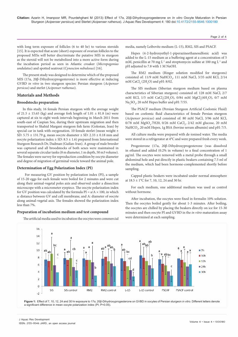
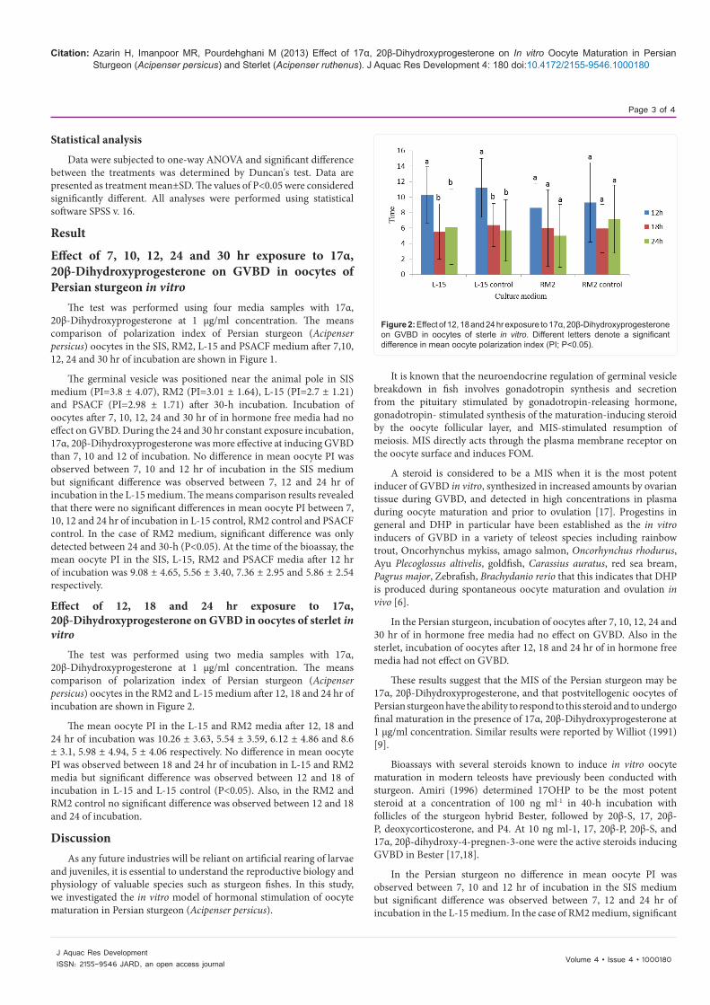
Figure 1: Effect of 7, 10, 12, 24 and 30 hr exposure to 17α, 20β-Dihydroxyprogesterone on GVBD in oocytes of Persian sturgeon in vitro. Different letters denote a significant difference in mean oocyte polarization index (PI; P<0.05).
Citation: Azarin H, Imanpoor MR, Pourdehghani M (2013) Effect of 17α, 20β-Dihydroxyprogesterone on In vitro Oocyte Maturation in Persian Sturgeon (Acipenser persicus) and Sterlet (Acipenser ruthenus). J Aquac Res Development 4: 180 doi:10.4172/2155-9546.1000180
Page 3 of 4
Volume 4 • Issue 4 • 1000180J Aquac Res DevelopmentISSN: 2155-9546 JARD, an open access journal
Statistical analysis
Data were subjected to one-way ANOVA and significant difference between the treatments was determined by Duncan's test. Data are presented as treatment mean±SD. The values of P<0.05 were considered significantly different. All analyses were performed using statistical software SPSS v. 16.
Result
Effect of 7, 10, 12, 24 and 30 hr exposure to 17α, 20β-Dihydroxyprogesterone on GVBD in oocytes of Persian sturgeon in vitro
The test was performed using four media samples with 17α, 20β-Dihydroxyprogesterone at 1 μg/ml concentration. The means comparison of polarization index of Persian sturgeon (Acipenser persicus) oocytes in the SIS, RM2, L-15 and PSACF medium after 7,10, 12, 24 and 30 hr of incubation are shown in Figure 1.
The germinal vesicle was positioned near the animal pole in SIS medium (PI=3.8 ± 4.07), RM2 (PI=3.01 ± 1.64), L-15 (PI=2.7 ± 1.21) and PSACF (PI=2.98 ± 1.71) after 30-h incubation. Incubation of oocytes after 7, 10, 12, 24 and 30 hr of in hormone free media had no effect on GVBD. During the 24 and 30 hr constant exposure incubation, 17α, 20β-Dihydroxyprogesterone was more effective at inducing GVBD than 7, 10 and 12 of incubation. No difference in mean oocyte PI was observed between 7, 10 and 12 hr of incubation in the SIS medium but significant difference was observed between 7, 12 and 24 hr of incubation in the L-15 medium. The means comparison results revealed that there were no significant differences in mean oocyte PI between 7, 10, 12 and 24 hr of incubation in L-15 control, RM2 control and PSACF control. In the case of RM2 medium, significant difference was only detected between 24 and 30-h (P<0.05). At the time of the bioassay, the mean oocyte PI in the SIS, L-15, RM2 and PSACF media after 12 hr of incubation was 9.08 ± 4.65, 5.56 ± 3.40, 7.36 ± 2.95 and 5.86 ± 2.54 respectively.
Effect of 12, 18 and 24 hr exposure to 17α, 20β-Dihydroxyprogesterone on GVBD in oocytes of sterlet in vitro
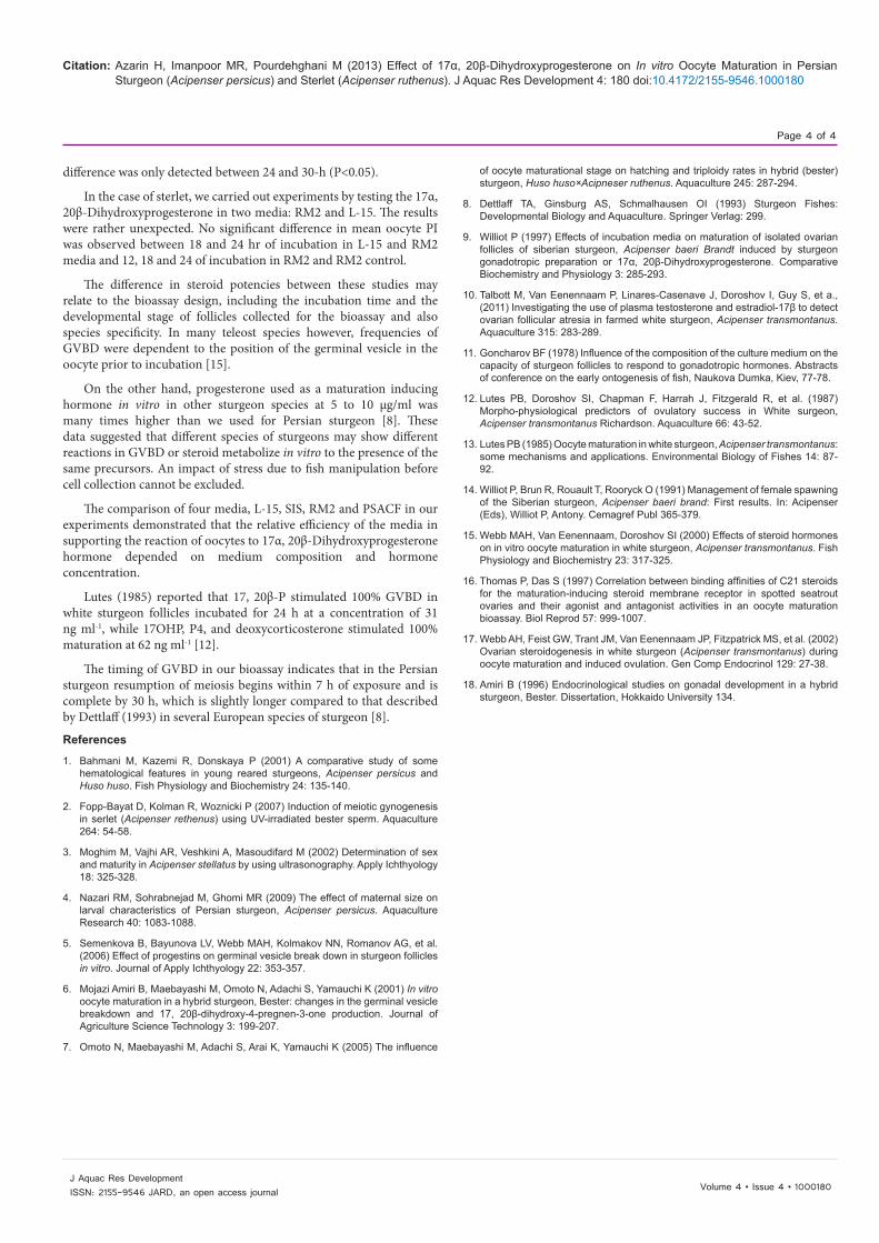
The test was performed using two media samples with 17α, 20β-Dihydroxyprogesterone at 1 μg/ml concentration. The means comparison of polarization index of Persian sturgeon (Acipenser persicus) oocytes in the RM2 and L-15 medium after 12, 18 and 24 hr of incubation are shown in Figure 2.
The mean oocyte PI in the L-15 and RM2 media after 12, 18 and 24 hr of incubation was 10.26 ± 3.63, 5.54 ± 3.59, 6.12 ± 4.86 and 8.6 ± 3.1, 5.98 ± 4.94, 5 ± 4.06 respectively. No difference in mean oocyte PI was observed between 18 and 24 hr of incubation in L-15 and RM2 media but significant difference was observed between 12 and 18 of incubation in L-15 and L-15 control (P<0.05). Also, in the RM2 and RM2 control no significant difference was observed between 12 and 18 and 24 of incubation.
DiscussionAs any future industries will be reliant on artificial rearing of larvae
and juveniles, it is essential to understand the reproductive biology and physiology of valuable species such as sturgeon fishes. In this study, we investigated the in vitro model of hormonal stimulation of oocyte maturation in Persian sturgeon (Acipenser persicus).
It is known that the neuroendocrine regulation of germinal vesicle breakdown in fish involves gonadotropin synthesis and secretion from the pituitary stimulated by gonadotropin-releasing hormone, gonadotropin- stimulated synthesis of the maturation-inducing steroid by the oocyte follicular layer, and MIS-stimulated resumption of meiosis. MIS directly acts through the plasma membrane receptor on the oocyte surface and induces FOM.
A steroid is considered to be a MIS when it is the most potent inducer of GVBD in vitro, synthesized in increased amounts by ovarian tissue during GVBD, and detected in high concentrations in plasma during oocyte maturation and prior to ovulation [17]. Progestins in general and DHP in particular have been established as the in vitro inducers of GVBD in a variety of teleost species including rainbow trout, Oncorhynchus mykiss, amago salmon, Oncorhynchus rhodurus, Ayu Plecoglossus altivelis, goldfish, Carassius auratus, red sea bream, Pagrus major, Zebrafish, Brachydanio rerio that this indicates that DHP is produced during spontaneous oocyte maturation and ovulation in vivo [6].
In the Persian sturgeon, incubation of oocytes after 7, 10, 12, 24 and 30 hr of in hormone free media had no effect on GVBD. Also in the sterlet, incubation of oocytes after 12, 18 and 24 hr of in hormone free media had not effect on GVBD.
These results suggest that the MIS of the Persian sturgeon may be 17α, 20β-Dihydroxyprogesterone, and that postvitellogenic oocytes of Persian sturgeon have the ability to respond to this steroid and to undergo final maturation in the presence of 17α, 20β-Dihydroxyprogesterone at 1 μg/ml concentration. Similar results were reported by Williot (1991) [9].
Bioassays with several steroids known to induce in vitro oocyte maturation in modern teleosts have previously been conducted with sturgeon. Amiri (1996) determined 17OHP to be the most potent steroid at a concentration of 100 ng ml-1 in 40-h incubation with follicles of the sturgeon hybrid Bester, followed by 20β-S, 17, 20β-P, deoxycorticosterone, and P4. At 10 ng ml-1, 17, 20β-P, 20β-S, and 17α, 20β-dihydroxy-4-pregnen-3-one were the active steroids inducing GVBD in Bester [17,18].
In the Persian sturgeon no difference in mean oocyte PI was observed between 7, 10 and 12 hr of incubation in the SIS medium but significant difference was observed between 7, 12 and 24 hr of incubation in the L-15 medium. In the case of RM2 medium, significant
Figure 2: Effect of 12, 18 and 24 hr exposure to 17α, 20β-Dihydroxyprogesterone on GVBD in oocytes of sterle in vitro. Different letters denote a significant difference in mean oocyte polarization index (PI; P<0.05).
Citation: Azarin H, Imanpoor MR, Pourdehghani M (2013) Effect of 17α, 20β-Dihydroxyprogesterone on In vitro Oocyte Maturation in Persian Sturgeon (Acipenser persicus) and Sterlet (Acipenser ruthenus). J Aquac Res Development 4: 180 doi:10.4172/2155-9546.1000180
Page 4 of 4
Volume 4 • Issue 4 • 1000180J Aquac Res DevelopmentISSN: 2155-9546 JARD, an open access journal
difference was only detected between 24 and 30-h (P<0.05).
In the case of sterlet, we carried out experiments by testing the 17α, 20β-Dihydroxyprogesterone in two media: RM2 and L-15. The results were rather unexpected. No significant difference in mean oocyte PI was observed between 18 and 24 hr of incubation in L-15 and RM2 media and 12, 18 and 24 of incubation in RM2 and RM2 control.
The difference in steroid potencies between these studies may relate to the bioassay design, including the incubation time and the developmental stage of follicles collected for the bioassay and also species specificity. In many teleost species however, frequencies of GVBD were dependent to the position of the germinal vesicle in the oocyte prior to incubation [15].
On the other hand, progesterone used as a maturation inducing hormone in vitro in other sturgeon species at 5 to 10 μg/ml was many times higher than we used for Persian sturgeon [8]. These data suggested that different species of sturgeons may show different reactions in GVBD or steroid metabolize in vitro to the presence of the same precursors. An impact of stress due to fish manipulation before cell collection cannot be excluded.
The comparison of four media, L-15, SIS, RM2 and PSACF in our experiments demonstrated that the relative efficiency of the media in supporting the reaction of oocytes to 17α, 20β-Dihydroxyprogesterone hormone depended on medium composition and hormone concentration.
Lutes (1985) reported that 17, 20β-P stimulated 100% GVBD in white sturgeon follicles incubated for 24 h at a concentration of 31 ng ml-1, while 17OHP, P4, and deoxycorticosterone stimulated 100% maturation at 62 ng ml-1 [12].
The timing of GVBD in our bioassay indicates that in the Persian sturgeon resumption of meiosis begins within 7 h of exposure and is complete by 30 h, which is slightly longer compared to that described by Dettlaff (1993) in several European species of sturgeon [8].
References
1. Bahmani M, Kazemi R, Donskaya P (2001) A comparative study of some hematological features in young reared sturgeons, Acipenser persicus and Huso huso. Fish Physiology and Biochemistry 24: 135-140.
2. Fopp-Bayat D, Kolman R, Woznicki P (2007) Induction of meiotic gynogenesis in serlet (Acipenser rethenus) using UV-irradiated bester sperm. Aquaculture 264: 54-58.
3. Moghim M, Vajhi AR, Veshkini A, Masoudifard M (2002) Determination of sex and maturity in Acipenser stellatus by using ultrasonography. Apply Ichthyology 18: 325-328.
4. Nazari RM, Sohrabnejad M, Ghomi MR (2009) The effect of maternal size on larval characteristics of Persian sturgeon, Acipenser persicus. Aquaculture Research 40: 1083-1088.
5. Semenkova B, Bayunova LV, Webb MAH, Kolmakov NN, Romanov AG, et al. (2006) Effect of progestins on germinal vesicle break down in sturgeon follicles in vitro. Journal of Apply Ichthyology 22: 353-357.
6. Mojazi Amiri B, Maebayashi M, Omoto N, Adachi S, Yamauchi K (2001) In vitro oocyte maturation in a hybrid sturgeon, Bester: changes in the germinal vesicle breakdown and 17, 20β-dihydroxy-4-pregnen-3-one production. Journal of Agriculture Science Technology 3: 199-207.
7. Omoto N, Maebayashi M, Adachi S, Arai K, Yamauchi K (2005) The influence
of oocyte maturational stage on hatching and triploidy rates in hybrid (bester) sturgeon, Huso huso×Acipneser ruthenus. Aquaculture 245: 287-294.
8. Dettlaff TA, Ginsburg AS, Schmalhausen OI (1993) Sturgeon Fishes: Developmental Biology and Aquaculture. Springer Verlag: 299.
9. Williot P (1997) Effects of incubation media on maturation of isolated ovarian follicles of siberian sturgeon, Acipenser baeri Brandt induced by sturgeon gonadotropic preparation or 17α, 20β-Dihydroxyprogesterone. Comparative Biochemistry and Physiology 3: 285-293.
10. Talbott M, Van Eenennaam P, Linares-Casenave J, Doroshov I, Guy S, et a., (2011) Investigating the use of plasma testosterone and estradiol-17β to detect ovarian follicular atresia in farmed white sturgeon, Acipenser transmontanus. Aquaculture 315: 283-289.
11. Goncharov BF (1978) Influence of the composition of the culture medium on the capacity of sturgeon follicles to respond to gonadotropic hormones. Abstracts of conference on the early ontogenesis of fish, Naukova Dumka, Kiev, 77-78.
12. Lutes PB, Doroshov SI, Chapman F, Harrah J, Fitzgerald R, et al. (1987) Morpho-physiological predictors of ovulatory success in White surgeon, Acipenser transmontanus Richardson. Aquaculture 66: 43-52.
13. Lutes PB (1985) Oocyte maturation in white sturgeon, Acipenser transmontanus: some mechanisms and applications. Environmental Biology of Fishes 14: 87-92.
14. Williot P, Brun R, Rouault T, Rooryck O (1991) Management of female spawning of the Siberian sturgeon, Acipenser baeri brand: First results. In: Acipenser (Eds), Williot P, Antony. Cemagref Publ 365-379.
15. Webb MAH, Van Eenennaam, Doroshov SI (2000) Effects of steroid hormones on in vitro oocyte maturation in white sturgeon, Acipenser transmontanus. Fish Physiology and Biochemistry 23: 317-325.
16. Thomas P, Das S (1997) Correlation between binding affinities of C21 steroids for the maturation-inducing steroid membrane receptor in spotted seatrout ovaries and their agonist and antagonist activities in an oocyte maturation bioassay. Biol Reprod 57: 999-1007.
17. Webb AH, Feist GW, Trant JM, Van Eenennaam JP, Fitzpatrick MS, et al. (2002) Ovarian steroidogenesis in white sturgeon (Acipenser transmontanus) during oocyte maturation and induced ovulation. Gen Comp Endocrinol 129: 27-38.
18. Amiri B (1996) Endocrinological studies on gonadal development in a hybrid sturgeon, Bester. Dissertation, Hokkaido University 134.

![r e Rese Journal of Aquaculture - OMICS International · are potent toxic, carcinogenic, ... Aflatoxin B1 (AFB1) ... damage in the liver of Nile tilapia [3]. Jantrorotai [13] mentioned](https://static.fdocument.org/doc/165x107/5b6cd5017f8b9a0b558c2067/r-e-rese-journal-of-aquaculture-omics-international-are-potent-toxic-carcinogenic.jpg)


![r e Rese Journal of Aquaculture · aquaculture enterprises these compounds have been tested and have ... differ which play a role in glucan-associated biological activity [24]. ...](https://static.fdocument.org/doc/165x107/5f0392087e708231d409b49c/r-e-rese-journal-of-aquaculture-aquaculture-enterprises-these-compounds-have-been.jpg)